the emerging role of copper-64 radiopharmaceuticals as
TRANSCRIPT

Review
s� K
EYNOTE
REV
IEW
Drug Discovery Today �Volume 23, Number 8 �August 2018 REVIEWS
Teaser Copper radioisotopes are emerging as potent tools for developing unprecedentedclinical approaches for cancer treatment by exploiting the intrinsic biological properties of
ionic copper and the richness of copper chemistry.
The emerging role of copper-64radiopharmaceuticals as cancertheranosticsAlessandra Boschi1, Petra Martini1,Emilija Janevik-Ivanovska2 and Adriano Duatti3
1Department of Morphology, Surgical and Experimental Medicine, University of Ferrara, 44121 Ferrara, Italy2 Faculty of Medical Sciences, University ‘Goce Del�cev’, Štip, Republic of Macedonia3Department of Chemical and Pharmaceutical Sciences, University of Ferrara, 44121 Ferrara, Italy
Copper radionuclides are rapidly emerging as potential diagnostic and
therapeutic tools in oncology, particularly 64Cu-radiopharmaceuticals for
targeting neuroendocrine, prostate, and hypoxic tumors. Unexpectedly,
experimental results are also revealing the impressive biological behavior
of simple [64Cu2+] ions. For example, it has been demonstrated that
administration of ionic [64Cu2+] in physiological solution allows the
selective targeting of a variety of malignancies. These remarkable
biological properties appear to be crucially linked to the natural role of
copper ions in cell proliferation. Here, we review the current status of64Cu-radiopharmaceuticals in molecular imaging and cancer therapy.
IntroductionMolecular imaging [1–6] is a fascinating concept that has deeply influenced modern diagnostic
imaging and therapy. However, its definition is rather vague and does not fully meet the strict
requirements of a rigorous scientific concept, resulting in an ongoing lengthy debate. In an
attempt to develop a definition that includes its most relevant characteristics, the Society of
Nuclear Medicine and Molecular Imaging (SNMMI) proposed the following statement:
‘Molecular imaging is the visualization, characterization, and measurement of biological pro-
cesses at the molecular and cellular levels in humans and other living systems’ [6].
Some ambitious interpretation entails that the meaning of the term ‘molecular’ should be
interpreted as the level of spatial resolution that can be attained by methods used for imaging
biomolecules in living systems. Only when the same atomic-scale resolution typical of structural
chemistry is achieved can the molecular attribute be applied. However, this result is still beyond
reach because there is no available imaging technology capable of truly detecting single
molecules in living tissues with atomic resolution.
Another interpretation suggests that molecular imaging corresponds to mapping the distribu-
tion and activity of molecules in living tissues. This description is linked to the concept of a
molecular imaging agent that is defined as a ‘probe’ used to visualize, characterize, and measure
Alessandra Boschi is
currently the head of the
Radiation Safety and Control
Section at the University of
Ferrara and a assistant
professor of radiochemistry.
Her research interests focus
mainly on the development of
novel chelating systems for
radiometals and the application of radionuclide imaging in
preclinical studies.
Petra Martini is currently a
postdoc at the Legnaro
National Laboratories of the
INFN. She is also a researcher
fellow at the University of
Ferrara. She has also worked
at TRIUMF Canada’s Particle
Accelerator Centre. Her main
research interests focus on
the production of novel radionuclides for medicine and the
development of automated methods for target processing,
separation, and purification of radionuclides from cyclotron-
irradiated targets.
Emilija Janevik-Ivanovska
is currently a professor in
pharmaceutical chemistry and
technology at the University of
Stip. She is also the director of
the University Institute for
Positron Emission
Tomography in Skopje. She is
also expert consultant to the
International Atomic Energy Agency and the European
Directorate for the Quality of Medicines. Her broad
research interests range from radiolabeling of peptides and
antibodies to the development of molecular imaging agents
labeled with positron-emitting radionuclides and
radionuclide therapy. She is also involved in various expert
committees for the analysis of regulatory aspects related to
radiopharmaceuticals.
Adriano Duatti is currently
a professor of general and
inorganic chemistry at the
University of Ferrara and a
research associate of the
Italian National Institute of
Nuclear Physics (INFN). He is
Head of the LARAMED
project at the INFN Legnaro
National Laboratories, which aims develop the production
of innovative and nonstandard radionuclides for medical
applications using a high-energy (70 MeV) and high-current
(800 microA) cyclotron. He is also a radiopharmaceutical
consultant to the Italian Ministry of Health. He has also
worked at the International Atomic Energy in Vienna,
Austria in the Radioisotope Products and Radiation
Technology Section. His main research interests focus on
the chemistry of metallic radiopharmaceuticals, molecular
imaging, and targeted radionuclide therapy.Corresponding author:
1359-6446/ã 2018 Elsevier Ltd. All rights reserved.https://doi.org/10.1016/j.drudis.2018.04.002 www.drugdiscoverytoday.com 1489

Reviews�K
EYNOTE
REV
IEW
REVIEWS Drug Discovery Today �Volume 23, Number 8 �August 2018
TABLE 1
Nuclear properties of copper radioisotopes
Radionuclide T1/2 Eb1 (MeV) (%) Eb� (MeV) (%) EC Eg (keV) (%)
67Cu 61.83 h – 0.576 (20) – 184.577 (48.7)0.4827 (22) 93.311 (16.1)0.3914 (57) 91.266 (7.0)0.1825 (1)
64Cu 12.7 h 0.65308 (17.4) 0.5787 (39) 43.6% 1345.77 (0.473)511 (34.79)
62Cu 9.74 min 2.927 (97.2) – 2.8% 511 (194.86)1173.02 (0.342)
61Cu 3.33 h 1.2164 (51) – 38.6% 656.008 (10.77)1.1489 (2.3) 511 (120.87)0.9334 (5.5) 373.05 (2.10)0.5604 (2.6) 282.956 (12.2)
67.412 (4.20)
60Cu 23.7 min 3.7719 (5) – 12.01% 3124.1 (4.8)2.9456 (15) 2158.90 (3.34)2.4784 (2.8) 1861.6 (4.8)1.9805 (49) 1791.6 (45.4)1.9105 (11.6) 1332.501 (88)1.8352 (4.59) 1035.2 (3.7)
826.4 (21.7)511 (185.19)467.3 (3.52)
biological processes in living systems. In principle, both endoge-
nous and exogenous molecules can be used as molecular imaging
agents. In this situation, molecular imaging exploits the properties
of a signal-generating agent to visualize the distribution of the
molecular probe by an appropriate imaging device. Thus, molecu-
lar imaging here is a methodology that makes use of molecular-
sized objects to collect fundamental biological information and,
accordingly, it cannot be viewed as ‘imaging of molecules’, but
rather as ‘imaging with molecules’.
Essentially, there are two approaches to accomplish this en-
deavor: (i) stimulate some endogenous biomolecule inside the cell
to emit a signal; or (ii) introduce into the cell a molecular probe
tagged with a moiety able to generate a signal. The first approach,
typically used with fluorescent proteins, usually does not allow a
signal to be channeled with sufficient intensity to be detectable for
diagnostic purposes. Conversely, the most obvious example of the
other type of approach is the radiolabeling of a molecule with a
g-emitting radionuclide.
Different classes of substance can be utilized as molecular
probes, including endogenous biomolecules, exogenous natural
products, and synthetic molecules. However, it is uncommon that
a radiolabeled probe perfectly matches the biological behavior of
the native substance from which it was derived. In this respect, the
radiotracers [11C-acetate], [15OH2], and [13NH3] represent sporadic
examples of perfect matching because the radiolabeled derivatives
are simply obtained by replacing one nonradioactive atom with
the corresponding radioactive counterpart without any alteration
of the primitive molecule [4]. Usually, the radiolabeling procedure
modifies some structural features of the precursor molecule, thus
giving rise to an authentic novel compound with its own biologi-
cal characteristics. This supports the notion that a radiolabeled
molecular probe, also called a radiopharmaceutical, is a unique
species with unique biodistribution properties and specific uptake
mechanisms.
Another category of radiopharmaceuticals that cannot be strict-
ly interpreted as radiolabeled molecules is currently emerging [7].
This comprises simple radioactive monoatomic ions derived from
bioelements having a well-recognized natural role in several bio-
logical processes. This type of radioactive tracer is not novel given
that radioactive iodine, in the form of a negatively charged ion,
was introduced into clinical use decades ago for the treatment of
thyroid diseases. This application sprang from the key role of
iodine in the metabolism of hormones by the thyroid. Yet, this
is a limited application restricted to a single organ. After the advent
of suitable production procedures based on a new generation of
advanced cyclotrons, several radioisotopes of elements with a
larger range of biological functions have recently become available
[8]. These include 64Cu, 67Cu [9], 63Zn [10], and 52Mn [11]. In
principle, these radiometals can be used to image their own
journey through a living organism after being administered as
simple metallic ions. This approach could be a genuine example of
molecular imaging given that the radiometal represents simulta-
neously both the imaging probe and the bioactive substrate.
Among the above-mentioned radioactive ions, [64Cu2+] is cur-
rently the most actively investigated and its potential for both
diagnosis and therapy is being unearthed. Here, we review the
status of research on the 64Cu. We show that [64Cu2+] can be
utilized not only to monitor its own natural biological behavior
1490 www.drugdiscoverytoday.com
in normal and diseased tissues, but also as radiolabeling precursor
for the preparation of novel metallic radiopharmaceuticals.
Copper radioisotopes for medicineThere are five important copper radioisotopes with nuclear char-
acteristics suitable for nuclear medicine applications (Table 1):64Cu, 67Cu, 62Cu, 61Cu and 60Cu [12].
64Cu decays mostly through the emission of b+ and b� particles
and Auger electrons [13]. In principle, the combination of these
radioactive emissions can be exploited for carrying out both
diagnosis and therapy using the same radiolabeled species, thus
providing an example of clinical approaches collectively referred
to as ‘theranostics’ [14]. This suggests that 64Cu is an almost ideal
example of a theranostic radionuclide.
Given that 64Cu cannot be conveniently produced by the decay
of isobars, it must be obtained by particle irradiation. Deuteron-
induced reactions are a competitive method, whereas neutron
bombardment in nuclear reactors typically yields the radionuclide
in low-specific activities. Usually, proton-induced reactions are the
most efficient. The maximum theoretical specific activity that can
be achieved is 1.43 � 1017 Bq g�1. To avoid the co-production of
radionuclidic impurities, proton irradiation always requires the
use of highly enriched 64Ni targets. Currently, high-yield produc-
tion of high-specific activity 64Cu is readily achievable using
biomedical cyclotrons through bombardment of enriched 64Ni
with low energy protons (10–18 MeV) [15–22]. A factor limiting
the cost-effectiveness of this method is the high commercial value
of enriched 64Ni (natural abundance of 64Ni is only 0.926%) and
the low proton currents (<100 mA) currently available with the
most common biomedical cyclotrons. Yet, the technology for
cyclotron-based production of 64Cu has advanced considerably
in recent years and fully automated systems for the target
Drug Discovery Today �Volume 23, Number 8 �August 2018 REVIEWS
Reductase
hCTR1
Efflux
ATPaseGolgi
ATOX
MT
CSS
COX17
SOD
SCO1
Drug Discovery Today
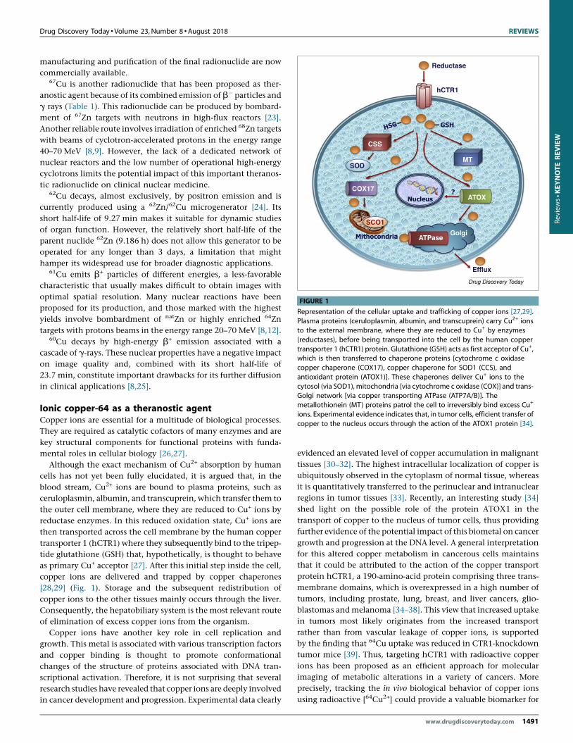
FIGURE 1
Representation of the cellular uptake and trafficking of copper ions [27,29].Plasma proteins (ceruloplasmin, albumin, and transcuprein) carry Cu2+ ionsto the external membrane, where they are reduced to Cu+ by enzymes(reductases), before being transported into the cell by the human coppertransporter 1 (hCTR1) protein. Glutathione (GSH) acts as first acceptor of Cu+,which is then transferred to chaperone proteins [cytochrome c oxidasecopper chaperone (COX17), copper chaperone for SOD1 (CCS), andantioxidant protein (ATOX1)]. These chaperones deliver Cu+ ions to thecytosol (via SOD1), mitochondria [via cytochrome c oxidase (COX)] and trans-Golgi network [via copper transporting ATPase (ATP7A/B)]. Themetallothionein (MT) proteins patrol the cell to irreversibly bind excess Cu+
ions. Experimental evidence indicates that, in tumor cells, efficient transfer ofcopper to the nucleus occurs through the action of the ATOX1 protein [34].
Review
s� K
EYNOTE
REV
IEW
manufacturing and purification of the final radionuclide are now
commercially available.67Cu is another radionuclide that has been proposed as ther-
anostic agent because of its combined emission of b� particles and
g rays (Table 1). This radionuclide can be produced by bombard-
ment of 67Zn targets with neutrons in high-flux reactors [23].
Another reliable route involves irradiation of enriched 68Zn targets
with beams of cyclotron-accelerated protons in the energy range
40–70 MeV [8,9]. However, the lack of a dedicated network of
nuclear reactors and the low number of operational high-energy
cyclotrons limits the potential impact of this important theranos-
tic radionuclide on clinical nuclear medicine.62Cu decays, almost exclusively, by positron emission and is
currently produced using a 62Zn/62Cu microgenerator [24]. Its
short half-life of 9.27 min makes it suitable for dynamic studies
of organ function. However, the relatively short half-life of the
parent nuclide 62Zn (9.186 h) does not allow this generator to be
operated for any longer than 3 days, a limitation that might
hamper its widespread use for broader diagnostic applications.61Cu emits b+ particles of different energies, a less-favorable
characteristic that usually makes difficult to obtain images with
optimal spatial resolution. Many nuclear reactions have been
proposed for its production, and those marked with the highest
yields involve bombardment of natZn or highly enriched 64Zn
targets with protons beams in the energy range 20–70 MeV [8,12].60Cu decays by high-energy b+ emission associated with a
cascade of g-rays. These nuclear properties have a negative impact
on image quality and, combined with its short half-life of
23.7 min, constitute important drawbacks for its further diffusion
in clinical applications [8,25].
Ionic copper-64 as a theranostic agentCopper ions are essential for a multitude of biological processes.
They are required as catalytic cofactors of many enzymes and are
key structural components for functional proteins with funda-
mental roles in cellular biology [26,27].
Although the exact mechanism of Cu2+ absorption by human
cells has not yet been fully elucidated, it is argued that, in the
blood stream, Cu2+ ions are bound to plasma proteins, such as
ceruloplasmin, albumin, and transcuprein, which transfer them to
the outer cell membrane, where they are reduced to Cu+ ions by
reductase enzymes. In this reduced oxidation state, Cu+ ions are
then transported across the cell membrane by the human copper
transporter 1 (hCTR1) where they subsequently bind to the tripep-
tide glutathione (GSH) that, hypothetically, is thought to behave
as primary Cu+ acceptor [27]. After this initial step inside the cell,
copper ions are delivered and trapped by copper chaperones
[28,29] (Fig. 1). Storage and the subsequent redistribution of
copper ions to the other tissues mainly occurs through the liver.
Consequently, the hepatobiliary system is the most relevant route
of elimination of excess copper ions from the organism.
Copper ions have another key role in cell replication and
growth. This metal is associated with various transcription factors
and copper binding is thought to promote conformational
changes of the structure of proteins associated with DNA tran-
scriptional activation. Therefore, it is not surprising that several
research studies have revealed that copper ions are deeply involved
in cancer development and progression. Experimental data clearly
evidenced an elevated level of copper accumulation in malignant
tissues [30–32]. The highest intracellular localization of copper is
ubiquitously observed in the cytoplasm of normal tissue, whereas
it is quantitatively transferred to the perinuclear and intranuclear
regions in tumor tissues [33]. Recently, an interesting study [34]
shed light on the possible role of the protein ATOX1 in the
transport of copper to the nucleus of tumor cells, thus providing
further evidence of the potential impact of this biometal on cancer
growth and progression at the DNA level. A general interpretation
for this altered copper metabolism in cancerous cells maintains
that it could be attributed to the action of the copper transport
protein hCTR1, a 190-amino-acid protein comprising three trans-
membrane domains, which is overexpressed in a high number of
tumors, including prostate, lung, breast, and liver cancers, glio-
blastomas and melanoma [34–38]. This view that increased uptake
in tumors most likely originates from the increased transport
rather than from vascular leakage of copper ions, is supported
by the finding that 64Cu uptake was reduced in CTR1-knockdown
tumor mice [39]. Thus, targeting hCTR1 with radioactive copper
ions has been proposed as an efficient approach for molecular
imaging of metabolic alterations in a variety of cancers. More
precisely, tracking the in vivo biological behavior of copper ions
using radioactive [64Cu2+] could provide a valuable biomarker for
www.drugdiscoverytoday.com 1491

REVIEWS Drug Discovery Today �Volume 23, Number 8 �August 2018
Reviews�K
EYNOTE
REV
IEW
disclosing fundamental molecular information in cancerous tis-
sues [35,40,41]. Notably, the associated b�-decay and Auger elec-
tron emissions could also be harnessed to trigger a therapeutic
effect on cancerous lesions, thus forming the basis for the use of64Cu as a genuine theranostic agent [42].
Results obtained from preclinical studies in animal models of a
variety of malignancies consistently supported the hypothesis that
[64Cu2+] ions could become a useful tool for molecular imaging
and therapy of various cancers [32,43–50]. These experiments
clearly confirmed that [64Cu2+] is selectively accumulated by vari-
ous cancerous tissues and that, unlike the radiotracer 2-deoxy-2-
[18F-fluoro-D-glucose] (18F-FDG), is not engaged in inflammatory
processes, thus suggesting that this radioactive probe behaves as a
highly specific tumor marker. In these studies, hCTR1 expression
was measured in a group of cell lines, including prostate, lung,
ovarian and breast cancers, melanoma, and glioblastoma and
compared with [64Cu2+] uptake. The experimental findings dem-
onstrated that the increased accumulation of [64Cu2+] was always
mediated by hCTR1 overexpression in those cancerous cells, thus
suggesting the potential theranostic use of [64Cu2+] for hCTR1-
expressing malignancies.
A few investigational clinical studies and preliminary experi-
mental data in humans convincingly supported preclinical obser-
vations. In these studies, [64Cu2+] was administered as simple
chloride salt dissolved in a sterile and pyrogen-free, buffered
solution. Specific activity was 3700 MBq/mg and volumic activity
925 MBq/ml. In normal subjects, the organ showing the highest
accumulation of radioactive [64Cu2+] was the liver, with lower
uptake also found in brain, kidneys, and pancreas.
A first investigation was carried out on a limited cohort of
patients to elucidate whether [64Cu2+] could be used for imaging
prostate cancer (PCa) in humans [51]. A group of patients treated
with adrenal deprivation therapy and another not receiving this
treatment, were selected for this study. Cancerous lesions were
sharply detected within 1 h after injection of [64Cu2+] (Fig. 2).
These observations confirmed that previous results collected in
animal models also hold in humans and that [64Cu2+] could be
utilized as a diagnostic agent for staging PCa. As expected, the
hepatobiliary system was the most relevant excretion route.
A first Phase II investigational clinical study was recently
reported [52] to investigate prospectively the biodistribution,
dosimetry, and lesion kinetics of [64Cu2+] in a significant number
of patients with PCa with biochemical relapse. This work was also
the first attempt to compare the diagnostic performances of PET/
CT, carried out with both [64Cu2+] and 18F-choline, with multi-
parametric MRI (mpMRI) in assessing PCa. Results collected in
these patients provided further evidence on the effectiveness of
[64Cu2+] in detecting local recurrence along with metastases in
bones and lymph nodes. Detection rate (DR) of [64Cu2+] was
significantly higher than observed for [18F-choline], particularly
in identifying local recurrence. In a patient-based analysis, [64Cu2
+] was found to detect more patients with PCa relapse compared
with [18F-choline] at prostate-specific antigen (PSA) levels
�4 ng mL�1, although for PSA values >4 ng mL�1, DR values
converged for the two diagnostic agents. In a lesion-based analysis,
a remarkably high DR for [64Cu2+] was also observed in patients
with PSA <1 ng ml �1, where PET/CT with [64Cu2+] outperformed
both 18F-Choline and mpMRI. The apparently superior perfor-
1492 www.drugdiscoverytoday.com
mance of 64Cu-PET/CT can be traced back to the favorable biodis-
tribution properties of [64Cu2+] ions. In particular, [64Cu2+] did not
significantly wash out from the kidneys and, thus, did not con-
centrate in the urinary tract. This enabled a careful assessment of
the pelvis and prostatic bed leading to the clear visualization of
small lesions not hidden by any significant bladder activity. Time–
activity curves showed that uptake of [64Cu2+] by PCa lesions was
rapid and peaked approximately 1 h after administration, whereas
its clearance was slow and dictated by the physical decay of the
radionuclide. Interestingly, the effective dose administered to
patients with [64Cu2+] was similar to that measured for other
common PET tracers (5.7 mSv).
Another preliminary exploratory clinical study was pursued to
assess the feasibility of PET/CT imaging of brain tumors with
[64Cu2+] in patients with glioblastoma multiforme. Comparison
with MRI, as reference imaging method, revealed sharp agreement
between the two diagnostic modalities. PET/CT imaging with
[64Cu2+] clearly detected brain cancerous tissues at 1 h post injec-
tion. Stable retention of radioactivity in brain lesions lasted for
more than 24 h. Again, these results confirmed preclinical obser-
vations and pointed to the potential of [64Cu2+] as a diagnostic
tracer for cerebral malignancies (Fig. 3). Remarkably, in the same
clinical study, a patient first found to have astrocytoma by MRI,
was subsequently imaged with [64Cu2+] and no uptake was ob-
served in the tumor region [53]. After removal of the lesion,
histological characterization disclosed its benign nature, a result
that sheds further light on the intrinsic affinity of 64Cu ions for
authentic malignant tissues.
Ongoing clinical studies are progressively revealing the high
specificity and uptake of [64Cu2+] for malignant tissues in the early
stages of cancer progression, thus providing strong evidence that
preclinical results can also be reproduced in humans. In particular,
current experimental efforts are devoted to scrutinizing whether
this tracer could be effective for the early diagnosis of cancerous
lesions in different organs. Apparently, this selectivity of [64Cu2+]
for different tumors appears always to be mediated by hCTR1
overexpression. A striking example was found in imaging liver
cancer with [64Cu2+]. Although it could be expected that visuali-
zation of lesions in the abdominal region would be hampered by
the natural, intense liver uptake of copper ions, the extremely high
affinity of [64Cu2+] for cancer cells in their initial stage of malig-
nant transformation, allowed early and efficient imaging of he-
patic metastases in a patient with hepatic cancer. This intense
accumulation is sustained by overexpression of hCTR1 copper
importers by hepatocytes in liver tissue [48,54,55].
Despite the constantly growing evidence for the high potential
of ionic 64Cu-PET to serve as a diagnostic tool for visualizing
pathological changes in copper metabolism in diseased tissues,
there are still open questions that need to be addressed. Primarily,
there are obvious limitations to the diagnostic utility of radio-
copper when regions of high physiological uptake, such as the
abdomen, are imaged. Furthermore, slight variations in radiation
dosimetry estimates have been reported in humans, owing to the
complex decay characteristics of 4Cu that make it difficult to
calculate the optimal and safest dose for patients [56]. These
critical issues need to be adequately investigated and clarified to
consolidate the clinical use of [64Cu2+] as novel cancer theranostic
agent.
Drug Discovery Today �Volume 23, Number 8 �August 2018 REVIEWS
Drug Discovery Today
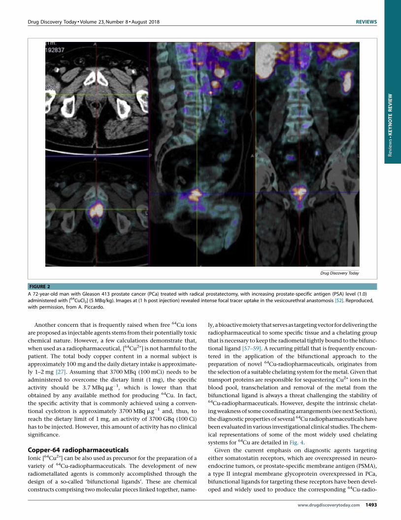
FIGURE 2
A 72-year-old man with Gleason 413 prostate cancer (PCa) treated with radical prostatectomy, with increasing prostate-specific antigen (PSA) level (1.0)administered with [64CuCl2] (5 MBq/kg). Images at (1 h post injection) revealed intense focal tracer uptake in the vesicourethral anastomosis [52]. Reproduced,with permission, from A. Piccardo.
Review
s� K
EYNOTE
REV
IEW
Another concern that is frequently raised when free 64Cu ions
are proposed as injectable agents stems from their potentially toxic
chemical nature. However, a few calculations demonstrate that,
when used as a radiopharmaceutical, [64Cu2+] is not harmful to the
patient. The total body copper content in a normal subject is
approximately 100 mg and the daily dietary intake is approximate-
ly 1–2 mg [27]. Assuming that 3700 MBq (100 mCi) needs to be
administered to overcome the dietary limit (1 mg), the specific
activity should be 3.7 MBq mg�1, which is lower than that
obtained by any available method for producing 64Cu. In fact,
the specific activity that is commonly achieved using a conven-
tional cyclotron is approximately 3700 MBq mg�1 and, thus, to
reach the dietary limit of 1 mg, an activity of 3700 GBq (100 Ci)
has to be injected. However, this amount of activity has no clinical
significance.
Copper-64 radiopharmaceuticalsIonic [64Cu2+] can be also used as precursor for the preparation of a
variety of 64Cu-radiopharmaceuticals. The development of new
radiometallated agents is commonly accomplished through the
design of a so-called ‘bifunctional ligands’. These are chemical
constructs comprising two molecular pieces linked together, name-
ly, a bioactive moiety that servesastargeting vector for deliveringthe
radiopharmaceutical to some specific tissue and a chelating group
that is necessary to keep the radiometal tightly bound to the bifunc-
tional ligand [57–59]. A recurring pitfall that is frequently encoun-
tered in the application of the bifunctional approach to the
preparation of novel 64Cu-radiopharmaceuticals, originates from
the selection of a suitable chelating system for the metal. Given that
transport proteins are responsible for sequestering Cu2+ ions in the
blood pool, transchelation and removal of the metal from the
bifunctional ligand is always a threat challenging the stability of64Cu-radiopharmaceuticals. However, despite the intrinsic chelat-
ing weakness of some coordinating arrangements (see next Section),
the diagnostic properties of several 64Cu radiopharmaceuticals have
been evaluated in various investigational clinical studies. The chem-
ical representations of some of the most widely used chelating
systems for 64Cu are detailed in Fig. 4.
Given the current emphasis on diagnostic agents targeting
either somatostatin receptors, which are overexpressed in neuro-
endocrine tumors, or prostate-specific membrane antigen (PSMA),
a type II integral membrane glycoprotein overexpressed in PCa,
bifunctional ligands for targeting these receptors have been devel-
oped and widely used to produce the corresponding 64Cu-radio-
www.drugdiscoverytoday.com 1493
REVIEWS Drug Discovery Today �Volume 23, Number 8 �August 2018
Drug Discovery Today
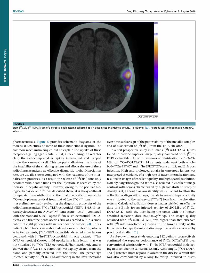
FIGURE 3
Brain [64Cu]Cu2+ PET/CT scan of a cerebral glioblastoma collected at 1 h post injection (injected activity, 13 MBq/kg) [53]. Reproduced, with permission, from C.Villano.
Reviews�K
EYNOTE
REV
IEW
pharmaceuticals. Figure 5 provides schematic diagrams of the
molecular structures of some of these bifunctional ligands. The
common mechanism singled out to explain the uptake of these
receptor-targeting agents entails that, after entering the receptor
cleft, the radiocompound is rapidly internalized and trapped
inside the cancerous cell. This property alleviates the issue of
the instability of the chelating system and allows the use of these
radiopharmaceuticals as effective diagnostic tools. Dissociation
rates are usually slower compared with the readiness of the inter-
nalization processes. As a result, the release of [64Cu2+] ions only
becomes visible some time after the injection, as revealed by the
increase in hepatic activity. However, owing to the peculiar bio-
logical behavior of Cu2+ ions described above, it is always difficult
to separate the contribution to the final diagnostic image of the64Cu-radiopharmaceutical from that of free [64Cu2+] ions.
A preliminary study evaluating the diagnostic properties of the
radiopharmaceutical [64Cu-TETA-octreotide] (TETA, 1,4,8,11-tet-
raazacyclotetradecane-N,N0,N00,N000-tetra-acetic acid) compared
with the standard SPECT agent [111In-DTPA-octreotide] (DTPA,
diethylene triamine penta-acetic acid) was carried out in a small
cohort of eight patients with neuroendocrine tumors [60]. In six
patients, both tracers were able to detect cancerous lesions, where-
as in two patients, [64Cu-TETA-octreotide] detected more lesions
compared with [111In-DTPA-octreotide]. In one patient, [111In-
DTPA-octreotide] showed mild uptake in a lung lesion that was
not visualized by [64Cu-TETA-octreotide]. Pharmacokinetic studies
showed that [64Cu-TETA-octreotide] was rapidly cleared from the
blood and partially excreted into the urine. The percentage
injected activity of [64Cu-TETA-octreotide] in the liver increased
1494 www.drugdiscoverytoday.com
over time, a clear sign of the poor stability of the metallic complex
and of dissociation of [64Cu2+] from the TETA chelator.
In a first prospective study in humans, [64Cu-DOTATATE] was
found to provide superior image quality compared with [111In-
DTPA-octreotide]. After intravenous administration of 193–232
MBq of [64Cu-DOTATATE], 14 patients underwent both whole-
body 64Cu-PET/CT and 111In-SPECT/CT scans at 1, 3, and 24 h post
injection. High and prolonged uptake in cancerous lesions was
interpreted as evidence of a high rate of tracer internalization and
resulted in images of excellent quality and high spatial resolution.
Notably, target:background ratios also resulted in excellent image
contrast with organs characterized by high somatostatin receptor
density. Yet, although in vivo stability was sufficient to allow the
collection of diagnostic images, the late increase in hepatic activity
was attributed to the leakage of [64Cu2+] ions from the chelating
system. Calculated radiation dose estimates yielded an effective
dose of 6.3 mSv for an injected activity of 200 MBq of [64Cu-
DOTATATE], with the liver being the organ with the highest
absorbed radiation dose (0.16 mGy/MBq). The image quality
obtained with [64Cu-DOTATATE] was higher than that observed
with [64Cu-TETA-octreotide], owing to the lower affinity of the
latter tracer for type 2 somatostatin receptors (sstr2), as revealed by
preclinical studies [61].
A subsequent larger study enrolling 112 patients prospectively
confirmed the superior performance of [64Cu-DOTATATE] over
conventional scintigraphy with [111In-DTPA-octreotide] in detect-
ing neuroendocrine cancerous lesions. Accordingly, [64Cu-DOTA-
TATE] detected more regions involved in the disease, a result that
was also corroborated by a long follow-up intended to assess

Drug Discovery Today �Volume 23, Number 8 �August 2018 REVIEWS
H H H H
HHHH
N N N
N N
N
N N
N N N
N N
N
N N
HO HO
HOHO
OH
OH
OH OH
OH
O OO
O
OOOO
OH
OOO
OO
N
N
NHO
HON
NNH
HN
OH
H
HH
H
N N
N N
N
N
NH2
NH2
CYCLEN CYCLAM
DOTA TETA
NOTA CB-TE2A
SAR(NH2)2
Drug Discovery Today
FIGURE 4
Schematic illustration of the structures of tetra-aza-macrocyclic andhexamine macrobicyclic ligands most frequently used for the preparation of64Cu-radiopharmaceuticals.
Review
s� K
EYNOTE
REV
IEW
whether foci were truly positive or false-positive. After a follow-up
of 42–60 months, no examples of false-positive results emerged
from 64Cu-PET images [62].
More interestingly, a head-to-head comparison between [64Cu-
DOTATATE] and [68Ga-DOTATOC], currently the most widely used
PET tracer for detecting neuroendocrine tumors, has been reported.
On a patient basis, no substantial differences were found between
the sensitivity of the two agents. However, [64Cu-DOTATATE] had a
remarkably higher detection rate of positive lesions, which was
tentatively attributed to the lower positron range of 64Cu compared
with68Ga, resulting in a better spatial resolution. Target:background
ratios in the various organs were not significantly different for the
two radiopharmaceuticals, thus yielding a similar image contrast.
However, a key advantage of [64Cu-DOTATATE] stemmed from the
longer half-life of the 64Cu radionuclide that allowed the patient to
be imaged between 1 h and 3 h after administration. This provided
an additional advantage over [68Ga-DOTATOC] by mitigating the
need for a tight routine workflow and increased the number of
patients who could be treated daily [63].
Preclinical and clinical studies are also underway with [64Cu-
SARTATE], which is an analog of [64Cu-DOTATATE] in which the
chelating system is replaced by a sarcophagine (SAR) moiety
(Fig. 5) [64]. In vitro and in vivo evaluation of [64Cu-SARTATE] in
animal models demonstrated its high selectivity for tumor cells
expressing sstr2. Comparison between the biodistribution data of
[64Cu-SARTATE] and [64Cu-DOTATATE] (DOTATATE, DOTA-Tyr3-
octreotate) showed that both radiopharmaceuticals exhibited ex-
cellent uptake in sstr2-positive tumors at 2 h post injection. How-
ever, tumor uptake of [64Cu-DOTATATE] significantly faded away
at 24 h, whereas activity of [64Cu-SARTATE] persisted, thus im-
proving image contrast at later time points. Accumulation of
[64Cu-SARTATE] in nontarget organs, such as liver and lungs,
was lower than that for [64Cu-DOTATATE]. Washout of [64Cu-
SARTATE] occurred mostly through the kidneys. The selectivity of
[64Cu-SARTATE] for sstr2 was confirmed by in vitro experiments
with A427-7 cells, demonstrating high cell-surface binding fol-
lowed by receptor-mediated endocytosis, as well as by biodistribu-
tion and imaging studies in A427-7 tumor-bearing mice. The high
tumor uptake and prolonged retention of [64Cu-SARTATE] could
reflect the robustness of the Cu2+ complex with the SAR ligand
preventing dissociation of the metal ion from the chelate. There-
fore, it was concluded that SAR-type ligands are able to form Cu2+
complexes with higher stability than DOTA-type ligands. This
superior coordinating ability could ensure their applicability for
use in routine imaging at later time points post injection in
accordance with the longer half-life of 64Cu [65].
Currently, much effort is devoted to the development of radio-
metal-based radiopharmaceuticals for targeting the integral mem-
brane glycoprotein PSMA [66]. Phosphoramidate-based and urea-
based molecules, two important categories of low-molecular-
weight PSMA inhibitors, have been synthesized to target PSMA
with high affinity and specificity. Ligands incorporating the pep-
tidomimetic motif Lys-urea-Glu have been widely investigated in
radiopharmaceutical development and the 64Cu-labeled ligand
DOTA-PSMA-617 (Fig. 5) was recently evaluated as a PET imaging
agent for PCa. Excellent, high-resolution PET images of the cancer-
ous lesions were detected with high lesion-to-background con-
trast. Again, the rapid internalization of the radiotracer into
subcellular organelles of PSMA-positive prostate tumor cells,
helped to overcome the problem of the in vivo instability of the64Cu-DOTA chelating system.
In a first in-human study, the diagnostic potential of [64Cu-
PSMA-617] was analyzed in a group of 29 patients with PCa. PET
images were acquired at 1 h and 2 h post injection, and metastatic
lesions were detected with high resolution and high lesion:back-
ground contrast. Patients with histologically proven local disease
were clearly identified by the tracer with high detection rates, also
in the presence of low circulating levels of PSA. Suspected lymph
node metastases, as detected by PET scans in two patients, were
subsequently confirmed by the corresponding MRI images. Sali-
vary glands, kidneys, and liver were among nontarget organs
showing the highest radiotracer uptake [67,68].
Another observational, prospective study was performed in a
selected number of patients with intermediate–high-risk PCa to
assess the diagnostic accuracy of [64Cu-PSMA-617] for primary
lymph node (LN) staging before laparoscopic radical prostatec-
tomy associated with extended pelvic LN dissection. The sensitiv-
ity, specificity, and positive (P) and negative (N) predictive values
for LN involvement were calculated based on final pathological
www.drugdiscoverytoday.com 1495

REVIEWS Drug Discovery Today �Volume 23, Number 8 �August 2018
H
HH
HN
NN
NN
N
HN O
O
OO
NH
HO
HO
N N
NNNH
OH
OO
OHO
HO
OO
O
O
HO
NH
NH
HNO
NH
X
HOO
OO
HO
OHN
HN
HN
HO
NH
NH
NH
NHSS
O
OO
O
X
OHH2N
= R'R =
X = R', DOTA-PSMA-617X = R, SARTATEX = R', DOTATATE
Drug Discovery Today
FIGURE 5
Schematic illustration of the structure of the bifunctional ligands DOTATATE, SARTATE, and DOTA-PSMA-617. For definitions, please see the main text.
Reviews�K
EYNOTE
REV
IEW
findings. Imaging-based LN staging showed that [64Cu-PSMA-617]
detected positive LN with a sensitivity of 87.5%, specificity of
100%, P of 100%, and N of 93.7%, thus suggesting a remarkable
diagnostic accuracy of [64Cu-PSMA-617] for primary LN staging
before radical prostatectomy. Again, it is difficult to determine
whether this excellent performance could be attributed only to the
high specificity of [64Cu-PSMA-617] for PCa cells without taking
into account the possible contribution of uptake by free [64Cu2+]
ions liberated as a result of the intrinsic instability of the Cu2
+-DOTA chelating arrangement [69].
A first-in-human clinical trial was carried out to study the agent
[64Cu-DOTA-AE105] in patients with breast, prostate, and bladder
cancer [70]. This new tracer features the high-affinity peptide antag-
onist AE105 targeting the urokinase-type plasminogen activator
receptor (uPAR), which is expressed in many types of cancer and
is predictive of metastatic invasion. The peptide AE105 was conju-
gated to the macrocyclic chelator DOTA and labeled with 64Cu
[71,72]. As expected, biodistribution data revealed high liver accu-
1496 www.drugdiscoverytoday.com
mulation, which was most likely caused by the in vivo instability of
the 64Cu-DOTA chelating system. In prostate as well as in breast and
bladder cancers, primary tumors and metastatic lymph nodes were
clearly identified with favorable tumor:background ratio, thus pro-
viding strong evidence for the target specific uptake of the [64Cu-
DOTA-AE105] consistent with uPAR expression also demonstrated
by immunohistochemistry assays carried out on isolated tissues.
An impressive example of controversial findings observed when
DOTA is used for the conjugation of 64Cu to biomolecules was
found in a clinical study aimed at determining whether HER2-
positive breast cancer (BCa) metastases could be visualized non-
invasively in the brain using the radiolabeled antibody [64Cu-
DOTA-trastuzumab] [73–75]. Metastatic brain lesions were effi-
ciently targeted by [64Cu-DOTA-trastuzumab], thus supporting
the conjecture that the radiolabeled antibody was able to penetrate
the blood–brain barrier (BBB). However, the large size and hydro-
philicity of therapeutic antibodies usually prevent crossing of the
BBB. In addition, tumor infiltration by monoclonal antibodies is

Drug Discovery Today �Volume 23, Number 8 �August 2018 REVIEWS
Review
s� K
EYNOTE
REV
IEW
normally hindered by high values of intratumoral interstitial
pressure. Although the authors speculated about a possible dis-
ruption of the BBB at the site of brain metastasis or the role of
immunoglobulin G in antibody transport, a simpler hypothesis
could take into account the observed high accumulation of 64Cu
ions in brain tumors. Indeed, the intrinsic instability of the 64Cu-
DOTA chelating arrangement could promote the partial release of
free 64Cu ions into the blood stream and subsequent targeting of
brain metastases by these ionic species.
A remarkable increase in stability was found when a different
chelating system was used for the design and preparation of the
tracer [64Cu-TP3805] [76]. Previous studies demonstrated that
vasoactive intestinal polypeptide receptor 1 (VPAC1) and pituitary
adenylate cyclase activating polypeptide (PACAP) are overex-
pressed at a high density on BCa cells, whereas on normal breast
tissue or benign masses, only a low number of receptors are
expressed. Therefore, a radiolabeled biomolecule was designed
by combining a PACAP analog with a C-terminal diaminodithiol
(N2S2) chelator to yield the corresponding bifunctional ligand
(TP3805). The resulting conjugate radiocompound was prepared
by reaction of TP3805 with 64Cu. Measurement of dissociation
constants supported the high in vivo stability of [64Cu-TP3805]
[77], which was, therefore, evaluated as a biomarker for the early
and accurate detection of BCa. In a preliminary investigation,
primary malignant tumors were visualized in 19 out of 20 patients
imaged with [64Cu-TP3805]. In addition, whole-body PET/CT
imaging revealed sentinel lymph nodes involvement in three
patients, thus suggesting a high affinity of the tracer for VPAC1
receptor-expressing tumors.
The remarkable improvement in stability observed in passing
from N4 tetramine ligands to N2S2 ligands is further magnified
when thiosemicarbazones, another class of N2S2 ligands, are used
as chelating agents for 64Cu. Bis(thiosemicarbazonato) copper(II)
complexes are a family of compounds with an important role in
the development copper-based radiopharmaceuticals (Fig. 6). This
class of radioactive agents has been extensively described in the
S
NN
NN
NH
HN
S
NN
N
N
N
SS
Cu
R3
R1R1
R4
R4 R2
Drug Discovery Today
FIGURE 6
Schematic illustration of the structures of bis-thiosemicarbazone ligands andof the corresponding Cu(II) complexes.
literature [78]. The bis(thiosemicarbazones) ligands can act as
tetradentate ligands for Cu2+, forming stable, neutral complexes
exhibiting high membrane permeability. The radioactive copper
complexes can be prepared in high radiochemical yield under mild
conditions. The biodistribution and specific targeting of these
small-molecule complexes can be manipulated by altering the
chemical substituents appended onto the bis-thiocarbazonato
moiety. Several 64Cu-complexes have been investigated as either
cardiac or hypoxia imaging agents and their stability in blood
firmly established. It was hypothesized that removal of the Cu2+
ion from this chelating system can occur only inside hypoxic cells
through a complex redox mechanism producing reduced Cu+
species. The compound [64Cu-ATSM] (where the ligand ATSM is
diacetyl-bis(N4-methylthiosemicarbazone) (Fig. 7) has been exten-
sively investigated as a marker for hypoxia [79–82]. It is a lipophil-
ic, low-molecular-weight complex characterized by high
membrane permeability and, therefore, rapid diffusion into cells.
It exhibits low redox potential, which was supposed to be a key
factor in favoring intracellular reduction of Cu2+ to Cu+. It was
posited that reduction is driven by mitochondria within hypoxic
cells with abnormally high electron concentrations, causing dis-
sociation of the reduced metallic ion from the ligand and its
subsequent cellular retention. Conversely, in normoxic cells,
[64Cu-ATSM] is kept in its oxidized form by a normal oxygen
pressure, thus allowing its outflow from the cell. Various preclini-
cal models disclosed some heterogeneity in hypoxia mapping by
[64Cu-ATSM], thus suggesting an underlying more complex phe-
nomenon. This led to a complicated interpretation of tumor
hypoxia. Experimental evidence suggested that [64Cu-ATSM]
could be more appropriately considered as a marker of over-
reduced intracellular state rather than a pure hypoxia agent. In
fact, the biochemical pathway leading to hypoxia usually switches
the intracellular redox potential to become progressively more
reductive, causing mitochondrial dysfunction because of excess of
electrons. This picture indicates that redox state and hypoxia are
both related and dependent phenomena, thus supporting the
interpretation that [64Cu-ATSM] is a generalized marker of an
over-reduced cell state and not specifically of hypoxia [80].
A chemical toolkit to assess the stability of copperradiopharmaceuticalsThe discovery of the remarkable biodistribution properties of the
radionuclide 64Cu, administered as simple cupric ions, also dis-
closed its possible therapeutic use for cancer treatment. However,
experiments carried out with [64CuCl2] have thus far demonstrat-
ed that this therapeutic potential can be fully exploited only when
the radionuclide is accumulated inside the cell nucleus in close
proximity to the genetic material. This condition is essential to
allow low-energy Auger electrons to deposit a cytotoxic dose in the
target tissue. Hence, it would be difficult to use, as therapeutic
agents, 64Cu radiopharmaceuticals that are unable to penetrate the
outer cellular membrane and reach the nucleus. For this purpose,67Cu displays nuclear properties that appear more attractive for
therapy of a larger class of tumors because of the emission of a
medium-energy b-particle.An additional crucial requirement, which holds for all diagnos-
tic and therapeutic radiopharmaceuticals, is that the radiolabeled
agent should always exhibit enough robustness in the biological
www.drugdiscoverytoday.com 1497
REVIEWS Drug Discovery Today �Volume 23, Number 8 �August 2018
Cu-CYCLAM
Cu-DOTA
Cu-TETA Cu-SAR(NH2)2
C
O
N
Cu
Drug Discovery Today
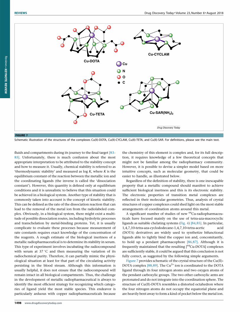
FIGURE 7
Schematic illustration of the structures of the complexes Cu(II)-DOTA, Cu(II)-CYCLAM, Cu(II)-TETA, and Cu(II)-SAR. For definitions, please see the main text.
Reviews�K
EYNOTE
REV
IEW
fluids and compartments during its journey to the final target [83–
85]. Unfortunately, there is much confusion about the most
appropriate interpretation to be attributed to the stability concept
and how to measure it. Usually, chemical stability is referred to as
‘thermodynamic stability’ and measured as log K, where K is the
equilibrium constant of the reaction between the metallic ion and
the coordinating ligands (the inverse is called the ‘dissociation
constant’). However, this quantity is defined only at equilibrium
conditions and it is unrealistic to believe that this situation could
be achieved in a biological system. Another type of stability that is
commonly taken into account is the concept of kinetic stability.
This can be defined as the rate of the dissociation reaction that can
lead to the removal of the metal ion from the radiolabeled com-
plex. Obviously, in a biological system, there might exist a multi-
tude of possible dissociation routes, including hydrolytic processes
and transchelation by metal-binding proteins. Yet, it is usually
complicate to evaluate these processes because measurement of
rate constants requires exact knowledge of the concentration of
the reagents. A rough estimate of the biological inertness of a
metallic radiopharmaceutical is to determine its stability in serum.
This type of experiment involves incubating the radiocompound
with serum at 37 �C and then measuring the variation of its
radiochemical purity. Therefore, it can partially mimic the physi-
ological situation at least for that part of the circulating activity
persisting in the blood stream. Although this information is
usually helpful, it does not ensure that the radiocompound will
remain intact in all biological compartments. Thus, the challenge
in the development of metallic radiopharmaceutical is always to
identify the most efficient strategy for recognizing which catego-
ries of ligand yield the most stable species. This endeavor is
particularly arduous with copper radiopharmaceuticals because
1498 www.drugdiscoverytoday.com
the chemistry of this element is complex and, for its full descrip-
tion, it requires knowledge of a few theoretical concepts that
might not be familiar among the radiopharmacy community.
However, it is possible to devise a simpler model based on more
intuitive concepts, such as molecular geometry, that could be
easier to handle, as illustrated below.
Regardless of the definition of stability, there is one inescapable
property that a metallic compound should manifest to achieve
sufficient biological inertness and this is its electronic stability.
The electronic properties of transition metal complexes are
reflected in their molecular geometries. Thus, analysis of crystal
structures of copper complexes could shed light on the most stable
arrangements of coordination atoms around this metal.
A significant number of studies of new 64Cu-radiopharmaceu-
ticals have focused mainly on the use of tetra-aza-macrocyclic
ligands as suitable chelating systems (Fig. 4) [84,85]. In particular,
1,4,7,10-tetra-aza-cyclododecane-1,4,7,10-tetra-acetic acid
(DOTA) derivatives are widely used to synthetize bifunctional
ligands able to tightly bind the copper ion and, concomitantly,
to hold up a pendant pharmacophore [86,87]. Although it is
frequently maintained that the resulting [64Cu-DOTA] complexes
are sufficiently stable, it could be argued that this conclusion is not
fully correct, as suggested by the following simple arguments.
Figure 7 provides schematic of the crystal structure of the Cu(II)-
DOTA complex [88,89]. The Cu2+ ion is coordinated to the DOTA
ligand through its four nitrogen atoms and two oxygen atoms of
the pendant carboxylic groups. The two other carboxylic arms are
protonated and do not integrate into the coordination sphere. The
structure of Cu(II)-DOTA resembles a distorted octahedron where
the four nitrogen atoms do not occupy the equatorial plane and
are heavily bent away to form a kind of pocket below the metal ion.

Drug Discovery Today �Volume 23, Number 8 �August 2018 REVIEWS
Review
s� K
EYNOTE
REV
IEW
Most importantly, two out of the four Cu–N bond distances are
elongated because of the so-called ‘Jahn–Teller’ effect. However,
when not constrained by inclusion into a multidentate ligand, a
set of four independent nitrogen atoms is usually arranged around
the Cu2+ ion in a square plane, as demonstrated by the structure of
the tetra-amine Cu(II) sulfate monohydrate salt [Cu(NH3)4(H2O)]
[SO4] [90]. In this complex, the nitrogen atoms of the four ammo-
nia molecules and the metal ion all lie in the square plan of a
square pyramid, where a weakly bound water molecule occupies
the apical position (Fig. 7). Therefore, it appears that the structural
features of the Cu(II)-DOTA complexes are in conflict with the
observed geometrical preferences of the Cu2+ ion when coordinat-
ed by four nitrogen atoms.
This structural tendency is not limited to unconstrained
amines, but is also observed in the complex [Cu(en)2] (en, ethy-
lenediamine) [91] and, most importantly, in [Cu(cyclam)][ClO4]2[92] and Cu(II)-TETA [93], where the four amine nitrogen atoms of
the ligands 1,4,8,11-tetrazazcyclotetradecane (CYCLAM) and
1,4,8,11-tetraazacyclotetradecane-1,4,8,11-tetraacetic acid (TETA)
are encircled in a ring. In these complexes, the four nitrogen atoms
achieve the preferred configuration (Fig. 7), because of their higher
flexibility, and it has been shown that they are more stable than Cu
(II)-DOTA in biological fluids [92,93].
A final example adding further evidence to the simple
model discussed here, is obtained by considering the ligands
CB-TE2A (1,4,8,11-tetra-azabicyclo[6.6.2]hexadecane-4,11-diace-
tic acid) [94,95] and SARNH2 [sarcophagine, 1,8-diamino-
3,6,10,13,16,19-hexaazabicyclo(6.6.6)eicosane] [96,97]. These
chelating systems belong to a class of cross-bridged tetra- and
hexamine macrobicyclic ligands (Fig. 4), and are pictured as
bearing a kind of molecular cleft where the metal ion could be
pocketed and, possibly, protected from transchelation. According
to crystal data reported for the complex Cu(II)-CB-TE2A, the four
nitrogen atoms do not achieve the most favorable square-planar
arrangement. Conversely, the crystal structure of Cu(II)-SARNH2
(Fig. 7) showed that four of the six nitrogen atoms attain almost
completely the most stable square-planar arrangement around the
metal. In agreement with the proposed interpretation, 64Cu-radio-
pharmaceuticals tethered to the CB-TE2A moiety were reported to
be unstable in biological fluids, whereas the sarcophagine motif
imparts high inertness to the corresponding complexes.
In summary, although some molecular architectures contribute
to protect the copper ion from being captured by endogenous
biomolecules, electronic factors do appear to have a primary role
in kinetic stabilization. This is neatly demonstrated by the sharp
increase in biological stability observed with ligands containing
the N2S2 chelating system [77,78]. Most importantly, when the
nitrogen and sulfur atoms are linked by p-electron delocalization,
as in bis-thiosemicarbazone ligands, the resulting coordination
geometry is perfectly square planar (Fig. 6) and achieves the high-
est stabilization [98].
Concluding remarksResearch on 64Cu is uncovering the tremendous impact on molec-
ular imaging that might stem from the use of radioisotopes of
elements that exhibit fundamental biological functions. When
this key characteristic is also complemented by nuclear properties
suitable for both diagnosis and therapy, it is reasonable to expect
that this category of radioisotopes could become essential for
theranostic applications. Previously, the only radioelement that
demonstrated these characteristics was iodine-131. Although io-
dine radioisotopes offered the very first example of this type of
radionuclide, the biological role of iodine is almost exclusively
confined to thyroid tissues. By contrast, the copper element is
essential for myriad cellular processes and, most importantly, is
involved in some fundamental steps of cellular replication that
also operate at the onset of a malignant transformation. Thus,
imaging the in vivo biological behavior of copper ions with the
corresponding, biologically equivalent, copper radioisotopes
could provide an unprecedented opportunity to achieve the pri-
mary objective of molecular imaging. This suggests that copper
radionuclides have enormous potential to promote the investiga-
tion of novel approaches for the detection and characterization of
cancer at the deepest molecular level.
References
1 Weissleder, R.M. and Pittet, M.J. (2008) Imaging in the era of molecular oncology.
Nature 452, 580–589
2 Mottaghy, F.M. (2009) Current and future aspects of molecular imaging. Methods
48, 81–82
3 Herschman, H.R. (2003) Molecular imaging: looking at problems, seeing solutions.
Science 302, 605–608
4 Ametamey, S.M. et al. (2008) Molecular imaging with PET. Chem. Rev. 108, 1501–
1516
5 Sharma, S. (2016) PET Radiopharmaceuticals for personalized medicine. Curr. Drug.
Targets 17, 1894–1907
6 Mankoff, D.A. (2007) A definition of molecular imaging. J. Nucl. Med. 48, 18N–21N
7 Duatti, A. (2015) Molecular imaging with endogenous and exogenous ligands:
the instance of antibodies, peptides, iodide and cupric ions. Nucl. Med. Biol. 42, 215–
218
8 Anon (2009) Cyclotron Produced Radionuclides: Physical Characteristics and Production
Methods. International Atomic Energy Agency
9 Medvedev, D.G. et al. (2012) Development of a large scale production of 67Cu from68Zn at the high energy proton accelerator: closing the 68Zn cycle. Appl. Radiat. Isot.
70, 423–429
10 Guerra Gomez, F.L. et al. (2012) Production and purification of the positron emitter
zinc-63. J. Label. Compd. Radiopharm. 55, 5–9
11 Fonslet, J. et al. (2017) Optimized procedures for manganese-52: production,
separation and radiolabeling. Appl. Radiat. Isot. 121, 38–43
12 Rowshanfarzad, P. et al. (2006) An overview of copper radionuclides and production
of 61Cu by proton irradiation of natZn at a medical cyclotron. Appl. Radiat. Isot. 64,
1563–1573
13 Anon (2016) Cyclotron Produced Radionuclides: Emerging Positron Emitters for Medical
Applications: 64Cu and 124I. International Atomic Energy Agency
14 Svenson, S. (2013) Theranostics: are we there yet? Mol. Pharm. 10, 848–856
15 McCarthy, D.W. et al. (1997) Efficient production of high specific activity 64Cu
using a biomedical cyclotron. Nucl. Med. Biol. 24, 35–43
16 Ohya, T. et al. (2016) Efficient preparation of high-quality 64Cu for routine use. Nucl.
Med. Biol. 43, 685–691
17 Thieme, S. et al. (2012) Module-assisted preparation of 64Cu with high specific
activity. Appl. Radiat. Isot. 70, 602–608
18 Xie, Q. et al. (2017) Establishing reliable Cu-64 production process: from target
plating to molecular specific tumor micro-PET imaging. Molecules 22, 1–10http://dx.
doi.org/10.3390/molecules22040641
19 Manrique-Arias, J.C. and Avila-Rodriguez, M.A. (2014) A simple and efficient
method of nickel electrodeposition for the cyclotron production of 64Cu. Appl.
Radiat. Isot. 89, 37–41
20 Ometakova, J. et al. (2012) Automated production of 64Cu prepared by 18 MeV
cyclotron. J. Radioanal. Nucl. Chem. 293, 217–222
www.drugdiscoverytoday.com 1499
REVIEWS Drug Discovery Today �Volume 23, Number 8 �August 2018
Reviews�K
EYNOTE
REV
IEW
21 Elomaa, V.V. et al. (2014) Automation of 64Cu production at Turku PET Centre. Appl.
Radiat. Isot. 89, 74–78
22 Spahn, I. et al. (2004) Enhanced production possibility of the therapeutic
radionuclides 64Cu, 67Cu and 89Sr via (n, p) reactions induced by fast spectral
neutrons. Radiochim. Acta 92, 183–186
23 Mastren, T. et al. (2014) Feasibility of isotope harvesting at a projectile
fragmentation facility: 67Cu. Sci. Rep. 4, 6706
24 Fukumura, T. et al. (2006) An improved 62Zn/62Cu generator based on a cation
exchanger and its fully remote-controlled preparation for clinical use. Nucl. Med.
Biol. 33, 821–827
25 McCarthy, D.W. et al. (1999) High purity production and potential applications of
copper-60 and copper-61. Nucl. Med. Biol. 26, 351–358
26 Rubino, J.T. and Franz, K.J. (2012) Coordination chemistry of copper proteins:
how nature handles a toxic cargo for essential function. J. Inorg. Biochem. 107, 129–
143
27 Kaplan, J.H. and Maryon, E.B. (2016) How mammalian cells acquire copper: an
essential but potentially toxic metal. Biophys. J. 110, 7–13
28 Inesi, G. (2017) Molecular features of copper binding proteins involved in copper
homeostasis. IUBMB Life 69, 211–217
29 Bharathi Devi, S.R. et al. (2016) Copper transporters and chaperones: their function
on angiogenesis and cellular signalling. J. Biosci. 41, 487–496
30 Brady, D.C. et al. (2014) Copper is required for oncogenic BRAF signalling and
tumorigenesis. Nature 509, 492–496
31 Ishida, S. et al. (2013) Bioavailable copper modulates oxidative phosphorylation and
growth of tumors. Proc. Natl. Acad. Sci. U. S. A. 110, 19507–19512
32 Denoyer, D. (2015) Targeting copper in cancer therapy: ‘Copper That Cancer’.
Metallomics 7, 1459–1476
33 Fuchs, A.G. and de Lustig, E.S. (1989) Localization of tissue copper in mouse
mammary tumors. Oncology 46, 183–187
34 Beaino, W. et al. (2014) Visualization of copper metabolism by 64CuCl2-PET. J. Biol.
Inorg. Chem. 19, 427–438
35 Peng, F. (2014) Positron emission tomography for measurement of copper fluxes in
live organisms. Ann. N. Y. Acad. Sci. 1314, 24–31
36 Hueting, R. (2014) Radiocopper for the imaging of copper metabolism. J. Label.
Comp. Radiopharm. 57, 231–238
37 Cai, H. et al. (2014) Reduced 64Cu uptake and tumor growth inhibition by
knockdown of human copper transporter 1 in xenograft mouse model of prostate
cancer. J. Nucl. Med. 55, 622–628
38 Kim, K.I. et al. (2014) Detection of increased 64Cu uptake by human copper
transporter 1 gene overexpression using PET with 64CuCl2 in human breast cancer
xenograft model. J. Nucl. Med. 55, 1692–1698
39 Jørgensen, J.T. et al. (2013) High tumor uptake of 64Cu: implications for molecular
imaging of tumor characteristics with copper-based PET tracers. Nucl. Med. Biol. 40,
345–350
40 Chakravarty, R. et al. (2016) 64Cu2+ ions as PET probe: an emerging paradigm in
molecular imaging of cancer. Mol. Pharm. 13, 3601–3612
41 Evangelista, L. et al. (2013) New issues for copper-64: from precursor to innovative
PET tracers in clinical oncology. Curr. Radiopharm. 6, 117–123
42 Qin, C. et al. (2014) Theranostics of malignant melanoma with 64CuCl2. J. Nucl. Med.
55, 812–817
43 Asabella, N.A. et al. (2014) The Copper Radioisotopes: A Systematic Review with
Special Interest to 64Cu. BioMed Res. Int. 2014, 1–9http://dx.doi.org/10.1155/2014/
786463
44 Ferrari, C. et al. (2015) Copper-64 dichloride as theranostic agent for glioblastoma
multiforme: a preclinical study. BioMed Res. Int. 2015, 1–6http://dx.doi.org/
10.1155/2015/129764
45 Safi, R. et al. (2014) Copper signaling axis as a target for prostate cancer therapeutics.
Cancer Res. 74, 5819–5831
46 Zhang, H. et al. (2011) Positron emission tomography of human hepatocellular
carcinoma xenografts in mice using copper (II)-64 chloride as a tracer with copper
(II)-64 chloride. Acad. Radiol. 18, 1561–1568
47 Peng, F. et al. (2006) PET of human prostate cancer xenografts in mice with
increased uptake of 64CuCl2. J. Nucl. Med. 47, 1649–1652
48 Peng, F. et al. (2005) Mouse extrahepatic hepatoma detected on microPET using
copper (II)-64 chloride uptake mediated by endogenous mouse copper transporter
1. Mol. Imaging Biol. 7, 325–329
49 Jiang, L. et al. (2017) Pilot study of 64Cu(I) for PET imaging of melanoma. Sci. Rep. 7,
2574
50 Catalogna, G. et al. (2017) The SGK kinase inhibitor SI113 sensitizes theranostic
effects of the 64CuCl2 in human glioblastoma multiforme cell lines. Cell Physiol.
Biochem. 43, 108–119
51 Capasso, E. et al. (2015) Role of 64CuCl2 PET/CT in staging of prostate cancer. Ann.
Nucl. Med. 29, 482–488
1500 www.drugdiscoverytoday.com
52 Piccardo, A. et al. (2018) 64CuCl2 PET/CT in prostate cancer relapse. J. Nucl. Med 59,
444–451
53 Panichelli, P. et al. (2016) Imaging of brain tumors with copper-64 chloride: early
experience and results. Cancer Biother. Radiopharm. 31, 159–167
54 Wachsmann, J. and Peng, F. (2016) Molecular imaging and therapy targeting
copper metabolism in hepatocellular carcinoma. World J. Gastroenterol. 22, 221–231
55 Kim, H. et al. (2009) Deletion of hepatic Ctr1 reveals its function in copper
acquisition and compensatory mechanisms for copper homeostasis. Am. J. Physiol.
Gastrointest. Liver Physiol. 296, G356–G364
56 Avila-Rodriguez, M.A. et al. (2017) Biodistribution and radiation dosimetry of64Cucopper dichloride: first-in-human study in healthy volunteers. EJNMMI Res. 7,
98
57 Price, E.W. and Orvig, C. (2014) Matching chelators to radiometals for
radiopharmaceuticals. Chem. Rev. 43, 260–290
58 Zeglis, B.M. and Lewis, J.S. (2011) A practical guide to the construction of
radiometallated bioconjugates for positron emission tomograph. Dalton Trans. 40,
6168–6195
59 Wadas, T.J. et al. (2010) Coordinating radiometals of copper, gallium, indium,
yttrium, and zirconium for PET and SPECT imaging of disease. Chem. Rev. 110,
2858–2902
60 Anderson, C.J. et al. (2001) 64Cu-TETA-octreotide as a PET imaging agent for
patients with neuroendocrine tumors. J. Nucl. Med. 42, 213–221
61 Pfeifer, A. et al. (2012) Clinical PET of neuroendocrine tumors using 64Cu-
DOTATATE: first-in-humans study. J. Nucl. Med. 53, 207–1215
62 Pfeifer, A. et al. (2015) 64Cu-DOTATATE PET for neuroendocrine tumors: a
prospective head-to-head comparison with 111In-DTPA-octreotide in 112 patients.
J. Nucl. Med. 56, 847–854
63 Johnbeck, C.B. et al. (2017) Head-to-head comparison of 64Cu-DOTATATE and68Ga-DOTATOC PET/CT: a prospective study of 59 patients with neuroendocrine
tumors. J. Nucl. Med. 58, 451–457
64 Cai, H. et al. (2010) An improved synthesis and biological evaluation of a new cage-
like bifunctional chelator, 4-((8-amino-3,6,10,13,16,19-hexaazabicyclo[6.6.6]
icosane-1-ylamino)methyl)benzoic acid, for 64Cu radiopharmaceuticals. Nucl. Med.
Biol. 37, 57–65
65 Paterson, B.M. et al. (2014) PET imaging of tumours with a 64Cu labeled
macrobicyclic cage amine ligand tethered to Tyr3-octreotate. Dalton Trans. 43,
1386–1396
66 Gourni, E. and Henriksen, G. (2017) Metal-based PSMA ligands. Molecules 22, 1–
34http://dx.doi.org/10.3390/molecules22040523
67 Grubmuller, B. et al. (2016) 64Cu-PSMA-617 PET/CT imaging of prostate
adenocarcinoma: first in-human studies. Cancer Biother. Radiopharm. 31,
277–286
68 Singh, A. et al. (2017) Imaging of prostate cancer using 64Cu-labeled prostate-
specific membrane antigen ligand. PET Clin. 12, 193–203
69 Cantiello, F. et al. (2017) Diagnostic accuracy of 64Copper prostate-specific
membrane antigen positron emission tomography/computed tomography for
primary lymph node staging of intermediate- to high-risk prostate cancer: our
preliminary experience. Urology 106, 139–145
70 Persson, M. et al. (2015) First-in-human uPAR PET: imaging of cancer aggressiveness.
Theranostics 5, 1303–1316
71 Kriegbaum, M.C. et al. (2011) Rational targeting of the urokinase receptor (uPAR):
Development of antagonists and non-invasive imaging probes. Curr. Drug Targets
12, 1711–1728
72 Persson, M. et al. (2014) Dosimetry of 64Cu-DOTA-AE105, a PET tracer for uPAR
imaging. Nucl. Med. Biol. 41, 290–295
73 Sasada, S. et al. (2017) Visualization of HER2-specific breast cancer intratumoral
heterogeneity using 64Cu-DOTA-trastuzumab PET. Eur. J. Nucl. Med. Mol. Imaging 44,
2146–2147
74 Kurihara, H. et al. (2015) 64Cu-DOTA-trastuzumab PET imaging and HER2
specificity of brain metastases in HER2-positive breast cancer patients. EJNMMI Res.
5, 8
75 Sasada, S. et al. (2017) 64Cu-DOTA-trastuzumab PET imaging for HER2-specific
primary lesions of breast cancer. Ann. Oncol. 8, 2028–2029
76 Thakur, M.L. et al. (2013) VPAC1 Receptors for imaging breast cancer: a feasibility
study. J. Nucl. Med. 54, 1019–1025
77 Zahng, K. et al. (2008) PET Imaging of VPAC1 expression in experimental and
spontaneous prostate cancer. J. Nucl. Med. 49, 112–121
78 Paterson, B.M. and Donnelly, P.S. (2011) Copper complexes of bis
(thiosemicarbazones): from chemotherapeutics to diagnostic and therapeutic
radiopharmaceuticals. Chem. Soc. Rev. 40, 3005–3018
79 Price, K.A. et al. (2011) Mechanisms controlling the cellular accumulation of copper
bis(thiosemicarbazonato) complexes. Inorg. Chem. 50, 9594–9605

Drug Discovery Today �Volume 23, Number 8 �August 2018 REVIEWS
Review
s� K
EYNOTE
REV
IEW
80 Colombie, M. et al. (2015) Focus on the controversial aspects of 64Cu-ATSM in
tumoral hypoxia mapping by PET imaging. Front. Med. 2, 58
81 Hueting, R. et al. (2014) A comparison of the behavior of 64Cu-acetate and 64Cu-
ATSM in vitro and in vivo. J. Nucl. Med. 55, 128–134
82 Vavere, A.L. and Lewis, J.S. (2007) Cu-ATSM: a radiopharmaceutical for the PET
imaging of hypoxia. Dalton Trans. 4893–4902
83 Maheshwari, V. et al. (2012) Measurement of the rate of copper(II) exchange for64Cu complexes of bifunctional chelators. Inorg. Chim. Acta 393, 318–323
84 Cai, Z. and Anderson, C.J. (2014) Chelators for copper radionuclides in positron
emission tomography radiopharmaceuticals. J. Label. Compd. Radiopharm. 57, 224–
230
85 Shokeen, M. and Wadas, T.J. (2011) The development of copper
radiopharmaceuticals for imaging and therapy. Med. Chem. 7, 413–429
86 Stasiuk, G.J. and Long, N.J. (2013) The ubiquitous DOTA and its derivatives: the
impact of 1,4,7,10-tetraazacyclododecane-1,4,7,10-tetraacetic acid on biomedical
imaging. Chem. Commun. 49, 2732–2746
87 Viola-Villegas, N. and Doyle, R.P. (2009) The coordination chemistry of 1,4,7,10-
tetraazacyclododecane-N,N0,N00,N000-tetraacetic acid (H4DOTA): structural overview
and analyses on structure–stability relationships. Coord. Chem. Rev. 253, 1906–1925
88 Kumar, K. et al. (1995) Synthesis, stability, and crystal structure studies of some Ca2
+, Cu2+, and Zn2+ complexes of macrocyclic polyamino carboxylates. Inorg. Chem.
34, 6472–6480
89 Riesen, A. et al. (1988) Structure of the barium salt of a Cu2+ complex with a tetraaza
macrocyclic tetraacetate. Acta Crystallogr. C Cryst. Struct. Commun. C44, 1740–1742
90 Morosin, B. (1969) The crystal structures of copper tetrammine complexes. A. Cu
(NH3)4SO2.H2O and Cu(NH3)4SeO4. Acta Cryst. B25, 19–30
91 Kovbasyuk, L.A. et al. (1997) Synthesis and structure of diaqua-bis
(ethylenediamine)copper(II) salts with anions of carbamic acids. Polyhedron 16,
1723–1729
92 Delgado, R. et al. (2007) Metal complexes of cyclen and cyclam derivatives useful for
medical applications: a discussion based on thermodynamic stability constants and
structural data. Dalton Trans. 2734–2745
93 Silversides, J.D. et al. (2007) Copper(II) cyclam-based complexes for
radiopharmaceutical applications: synthesis and structural analysis. Dalton Trans.
971–978
94 Wong, E.H. et al. (2000) Synthesis and characterization of cross-bridged cyclams and
pendant-armed derivatives and structural studies of their copper(II) complexes. J.
Am. Chem. Soc. 122, 10561–10572
95 Hubin, T.J. et al. (1998) Ultra rigid cross-bridged tetraazamacrocycles as ligands: the
challenge and the solution. Chem. Commun. 1675–1676
96 Bernhardt, P.V. et al. (1995) Copper(II) complexes of substituted macrobicyclic
hexaamines: combined trigonal and tetragonal distortions. Inorg. Chem. 34, 3589–
3599
97 Di Bartolo, N.M. et al. (2001) Synthesis of a new cage ligand, SarAr, and its
complexation with selected transition metal ions for potential use in radioimaging.
J. Chem. Soc. Dalton Trans. 2303–2309
98 Blower, P.J. (2003) Structural trends in copper(II) bis(thiosemicarbazone)
radiopharmaceuticals. Dalton Trans. 4416–4425
www.drugdiscoverytoday.com 1501