the endospore stain - dallas county community...
TRANSCRIPT

THE ENDOSPORE STAINEndospore production is a very important characteristic of some bacteria, allowing them toresist adverse environmental conditions such as desiccation, chemical exposure, extremeheat, etc. They were identified in the 1800s (John Tyndall developed a process fordestroying them with intermittent heat procedure), although the stain procedures to identifythem did not develop until the early twentieth century. Bacterial endospores are the mostresistant structures of all living organisms, and they can live in this dormant dehydrated statefor hundreds and hundreds of years (even some documented at thousands of years).Endospores are not for reproduction: One spore forms inside of the vegetative cell. Whenthe spore germinates, one vegetative cell will be produced. The stimulus for sporulation canvary—nutrient depletion, desiccation, chemicals, heat, etc.
As a spore forms inside of the vegetative cell, the spore wall chemically changes and thicken.This sporulation process changes the spore’s stainability, making it increasingly resistant tothe staining dyes, and so a gimmick—steaming---enhances the primary dye’s penetration.
The primary dye malachite green is a relatively weakly binding dye to the cell wall and sporewall. In fact, if washed well with water, the dye comes right out of the cell wall, however notfrom the spore wall once the dye is locked in. That is why there does not need to be adecolorizer in this stain: it is based on the binding of the malachite green and thepermeability of the spore vs. cell wall. . The steaming helps the malachite green to permeatethe low-permeability spore wall. A variety of chemicals comprise the spore wall (keratinprotein, calcium), but deeper in the wall is peptidoglycan. The keratin forming the outerportion of the endospore wall resists dye. The heating of the bacteria will make the sporewall more permeable to the malachite green, and it then attaches to the peptidoglycan. Oncein, the malachite green will not come out because the overlying spore wall becomes lesspermeable when the smear cools.
The identification of spores is also veryimportant for the clinical microbiologistwho is analyzing a patient's body fluid ortissue since there are not that manyspore-forming genera. In fact, there aretwo major pathogenic spore-forminggenera, Bacillus and Clostridium,together causing a number of lethaldiseases---botulism, gangrene, tetanus,and anthrax, to name a few. Somebacteria have to be put into unfavorablesituations (high cell density andstarvation are two key triggers) to go intosporulation; others will make sporeseasily without much provocation (Bacillussubtilus).
Fall 2011 – Jackie Reynolds, Richland College, BIOL 2420
2
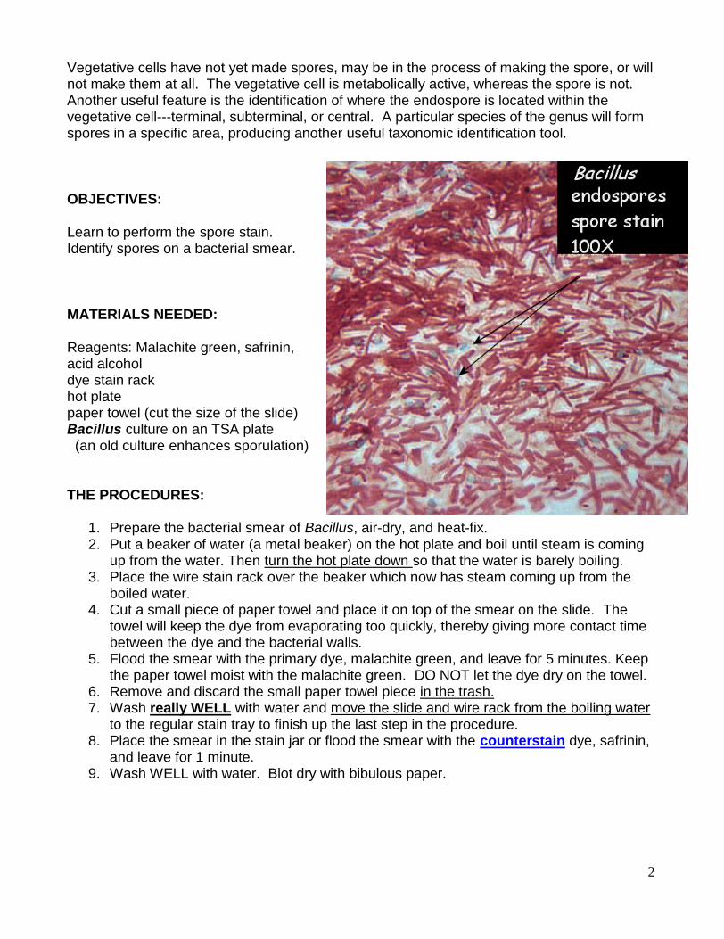
Vegetative cells have not yet made spores, may be in the process of making the spore, or willnot make them at all. The vegetative cell is metabolically active, whereas the spore is not.Another useful feature is the identification of where the endospore is located within thevegetative cell---terminal, subterminal, or central. A particular species of the genus will formspores in a specific area, producing another useful taxonomic identification tool.
OBJECTIVES:
Learn to perform the spore stain.Identify spores on a bacterial smear.
MATERIALS NEEDED:
Reagents: Malachite green, safrinin,acid alcoholdye stain rackhot platepaper towel (cut the size of the slide)Bacillus culture on an TSA plate
(an old culture enhances sporulation)
THE PROCEDURES:
1. Prepare the bacterial smear of Bacillus, air-dry, and heat-fix.2. Put a beaker of water (a metal beaker) on the hot plate and boil until steam is coming
up from the water. Then turn the hot plate down so that the water is barely boiling.3. Place the wire stain rack over the beaker which now has steam coming up from the
boiled water.4. Cut a small piece of paper towel and place it on top of the smear on the slide. The
towel will keep the dye from evaporating too quickly, thereby giving more contact timebetween the dye and the bacterial walls.
5. Flood the smear with the primary dye, malachite green, and leave for 5 minutes. Keepthe paper towel moist with the malachite green. DO NOT let the dye dry on the towel.
6. Remove and discard the small paper towel piece in the trash.7. Wash really WELL with water and move the slide and wire rack from the boiling water
to the regular stain tray to finish up the last step in the procedure.8. Place the smear in the stain jar or flood the smear with the counterstain dye, safrinin,
and leave for 1 minute.9. Wash WELL with water. Blot dry with bibulous paper.

3
LABORATORY REPORT SHEET
QUESTIONS:
1. Draw some spores along with some vegetative cells for comparison.
2. Can you find any spores still within the vegetative cell?
If so, notice where the spore is in the cell, if there is one---terminal, central,subterminal. Why might that information be helpful to a microbiologist?
3. What is the purpose of the steam in this stain?
4. Why does there not have to be a decolorizer in this stain?
5. If you mistakenly did this stain on an acid-fast Mycobacterium, what color do you thinkthe cells would come out to be?