the evaluation of potential human carcinogens: a histopathologist's point of view
TRANSCRIPT

ExpToxic Patho11996; 48: 169-174Gustav Fischer Verlag lena
Safety of Medicines Group, ZENECA Pharmaceuticals, Cheshire, UK
The evaluation of potential human carcinogens:A histopathologist's point of view
PETER GREAVES
With 4 figures and 4 tables
Address for correspondence: PETER GREAVES, MBChB, FRCPath, Safety of Medicines Group, ZENECA Pharmaceuticals, Alderley Park, Cheshire, UK.
Key words: Carcinogens, human, evaluation; Evaluation, carcinogens.
Summary
Over 100 marketed drugs induce neoplasia when administered at high doses to rats and mice for periods of up to twoyears. Despite their diverse chemical structures and biological activities, these compounds produce a relatively limited range of tumour types in rodents, most commonly in theliver. Tumours usually develop only after long periods oftime following high exposure to drug. The main exceptionsare DNA-reactive anticancer drugs such as alkylating agentswhich produce tumours rapidly in rodents in several organs.
In this laboratory, mouse carcinogenicity studies are performed using the C57BL/lOJ strain. This strain infrequentlydevelops hepatic tumours spontaneously but it is sensitiveto the effects of DNA-reactive carcinogens. Moreover, hepatic neoplasms regularly develop in male but not femaleC57BL/lOJ mice following long-term treatment with nongenotoxic drugs that produce hepatic enlargement associatedwith diverse hepatocellular effects. Studies in this strain withthe tumorigenic liver enlarger, phenobarbitone, have shownthat although such liver enlargement is characterised by abrief burst of hepatocyte replication, this is associated withpersistent regional modulation of hepatic growth stimulatory and inhibitory factors and their associated receptors.These findings indicate that there is a sustained alteration tothe internal hepatic environment characterised by regionalalterations to the balance of hepatocyte mitogens and inhibitors of replication and their respective receptors.
Thus, the development of hepatocellular tumours inC57BL/lOJ mice following two-year treatment with nongenotoxic drugs appears to be a regular response of an organ to an exaggerated and long-term disruption of its homeostasis. Agents that produce tumours in rodents in this wayseem likely to pose little or no risk to humans if administered under appropriate clinical circumstances at doses whichshow no significant disruption of organ homeostasis. However, drugs that produce this type of response need to be distinguished from those that induce unusual and rapid patternsof tumour development because these agents may have hightumorigenic potency of potential hazard to humans.
Introduction
FARBER (1988) has pointed out that the pathologist tendsto see cancer from a perspective that is different fromchemists and biochemists. Using liver cancer as a model,he has argued that the logical approach to the study of experimental cancer from a biochemical perspective is thestudy of the effects of all the different chemicals in various stages of carcinogenesis, initiation, promotion andprogression. The danger of this approach is becoming lostin minute detail that is uninterpretable without some overall orientation. By contrast, through the light microscopethe pathologist sees cancer development as a relativelyconsistent and reproducible biological sequence of eventscommon to a variety of different experimental models.
Over 100 DNA reactive and non-DNA reactive pharmaceuticals are reported to induce tumours in rodents.These drugs form a particularly useful subgroup of xenobiotics in the study of the carcinogenesis because of theknowledge of their pharmacology and exposure to humansunder carefully controlled circumstances (DAVIES andMONRO 1995). The liver represents the most common target organ for tumour induction in rodents by pharmaceutical agents.
The aim of this communication is to explore, from theperspective of the pathologist, the relationship betweennon-neoplastic alterations and tumours induced by noveldrugs in the rodent liver using data obtained in this laboratory in C57BL/lOJ mice, a strain which develops fewhepatocellular tumours spontaneously.
General principles
Based on the study of the pathological alterations produced in a wide range of animal tumour models developedover the last 50 years, GRASSO and colleagues (1991) have
ExpToxic Pathol48 (1996) 2-3 169

arguedthat a commonfactorwhichpredisposes to cancerdevelopment in rodents is sustained tissuedamage. Theyhave also suggested that a threshold exists below whichboth the earlynon-neoplastic changes and associated neoplasia do not occur. Moreover, they have noted that tumoursdo notdevelopat all if earlychanges regresseitherby cessation of treatmentor suppressed by use of an inhibitory agent.
A numberof authors have also suggested that there isa relationship betweenthe toxicityof xenobiotics in shortterm animal studiesas defined in acute toxicityor by themaximum tolerated dose (MTD) and carcinogenic potency(ZEISE et al. 1988; HASEMAN andSEILKOP 1992). Asmost non DNA-reactive pharmaceuticals are relativelynon-toxic in character, it is to be expectedthat their potential to producetumours will also generally be low.
Timeto tumour development
Although early changes in targetorgans are variable, thehistopathology of theend-stage tumours in a particular tissueis similar whatever the typeof inducing agent. Bycontrast,timeto tumour development canbe significantly different andcanbe a useful measure of tumorigenic potency.
Oneexampleof timedifferences to developtumours isin the solid state model. Implantation of smooth plasticdiscs into the subcutaneous tissue of mice produces tumours in a shorter time than implantation of rough discsof the material and same size. BRAND and colleagues(1976) showed that by about 16 months after implanta-
Table 1.Hepatocellular lesions inuntreated C57BL/1OJ micefrom control groups in two year carcinogenicity studiesconducted at Alderley Parkbetween 1986 and 1990.
Hepatic lesions Males Femalesn=500 n=500
Foci 4 (0.8 %) 2 (0.5 %)Adenomas 5 (1.0 %) 1 (0.2%)Carcinomas 15(3.0%) 2(0.1%)
tionof smooth implants 660square mm,over50 %of micehad developed sarcomas whereas it took over 24 monthsfor rough implants of the same size to produce a similarresponse. It is believedthat this differenceis linkedto thenatureofthe inflammatory responseinducedby these implants, rough implants inciting a moreintense foreign bodyreaction with prolonged cellularity and delayed fibroticconversion than smoothimplants (BRAND et al. 1976).
An examplefromthis laboratory where time to tumourdevelopment was a key element in the drug developmentdecision making process is tamoxifen, an anti-oestrogenwidely used in the treatment of advanced breast cancer.Tamoxifen showedrelatively low toxicity in animalsandno mutagenic or clastogenic activity in a battery of shorttermtests (TUCKER et al. 1984). In a conventional carcinogenicity studyconducted at threedose levelsin the Alderley Park Wistar rat, hepatocellular carcinomas started toappear after only about 6 months treatment and by 12months over50 % of rats in the highdosegrouphad developed hepatocellular carcinomas (GREAVES et al. 1993).The rapidity of development of malignant tumours in amannersuggestive of DNA-reactive drugs was pivotal inthe decision ofZENECA Pharmaceuticals not to progressthe use of tamoxifen into the treatmentof non-lifethreateningconditions. Subsequent investigations have shownthat DNA-adducts form in vivo in the rat liver and thesemay result inDNA damage (HAN andLIEHR 1992; SARGENTet al. 1994).
Hepatic tumours in the C57BLIlOJ mouse
ZENECA Pharmaceuticals at Alderley Parkhave traditionally used the C57BLlI0J APfSD strain of mouse forcarcinogenicity studies, largely because like the C57BLl6mouse but in contrast to theB6C3Fl (C57BLl6 x C3H/He)mouse, it develops few spontaneous hepatocellular tumoursspontaneously (table 1). The C57BL mouse, derived in1921, wasdividedinto the 6 and 10sublinesin the colonyat the Jackson Laboratory. The C57BLl1O and C57BLl6strains are closely related. Among 32 polymorphic locifor which both strains have been typed, they only differat two (Lv and H-9). In 1977,C57BLl1OJ mice were im-
Table 2. Overall tumour profile in the C57BLlI0J mouse and tumours induced by 2-acetylamino-fluorene (2-AAF) administered for periods of up to 16months mixed in diet.
Tumour type Males Females
Controls 0.03 % 0.06% Controls 0.03 % 0.06%2-AAF 2-AAF 2-AAF 2-AAF
Number examined 65 35 28 61 35 28Hepatocellular 1 3 2 0 1 13Gallbladder 0 0 2 0 2 2Harderian gland 4 3 10 3 3 1Bladder 0 17 2 0 0 7
170 Exp Toxic Pathol48 (1996)2-3

Table 3. Hepatic changes induced by a variety of novel drugs of different therapeuticclassesadministered to C57BLllOJ micefor two years in conventional carcinogenicitybioassays.
Study Relative liver Pathology Increase in neoplasmsnumber weight in high in highdose group
dose group%
140 Hypertrophy and Male liverperoxisome proliferation
2 132 Hypertrophy andperoxisome proliferation
3 100
4 100
5 100
6 128 Hypertrophy and MFOinduction
7 100
8 118 Hypertrophy and MFO Male thyroidinduction
9 169 Hypertrophy and MFO Maleliverinduction
10 167 Hypertrophy but no Male liversignificant MFO induction
ported from the Jackson Laboratory to Alderley Park andthe colony set up by Caesareansection and fosteringontoCharlesRiverCD-l mice. The C57BLlIOJ mousehas beenshown to be sensitive to genotoxic carcinogens, notably2-acetylaminofluorine (2-AAF, table 2). The tumours induced in bladder and liver were broadly similar to that reported in other strains treatedwith 2-AAF, althoughstraindifferences are recorded (WOOD 1969).
It is also of note that studies in this laboratory of spontaneousandinducedhepatocellular tumoursin the C57BLIIOJ strain unlike B6C3Fl mice showed few mutations ofB-ras , K-ras or N-ras genes (LORD et al. 1992).As a consequence, ras genes do not represent markers for in vivogenotoxic activity in C57BLllOJ mice.
Characteristics of responses of C57BL/IOJmice to liver enlargers
Resultsof ten unselected conventional regulatory oncogenicitystudiesconductedperformed over the last 10yearswith pharmaceutical agents of different therapeutic classes in this strain at Alderley Park suggest a relationshipbetweendrug-induced hepatic enlargement and the developmentof hepatocellular tumours. Once liverenlargementbecame a significant response to treatment, hepatocellular tumours developed at theend of twoyearstudies in males butnot in females (table3). A statistically significant response
was limited to groups given doses of drug that producedliver enlargement characterised by relative liver weightsat least 140% of controls. This did not seem to be directlyrelated to the degree of enzyme induction or peroxisomalproliferation. Indeed,one agent was devoidof any significant enzymeor peroxisomal potential,yet still inducedtumours in groups of animals with marked liver weight increases. In one case, treatment induced mixed functionoxidases sufficiently to inducealter thyroidhormone metabolism with consequent thyroid adenoma formation aftertwoyears withoutincreasing the incidence of hepatictumours.
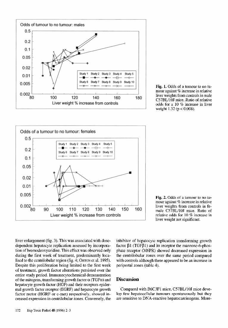
This association can also be expressing by plotting theodds of a tumouror the probability of a tumour to the probability of no tumour against relative liver weight (fig. 1and 2). When analysed using the logarithm of the odds(correlation coefficients, linear regression and analysisof covariance), there was evidence for an association between liver weight and the development of tumours formales but not females. The ratio of relative odds for every10 % increase in relative liver weight was 1.32for males(p < 0.008) and 0.94 for females (NS).
Growth factors associated with of hepaticenlargement in the C57BL/IOJ mouse
Administrationof phenobarbitone to C57BLl1OJ micefor up to 13 weeks produced a dose and time dependent
Exp Toxic Pathol 48 (1996) 2-3 171
Odds of tumour to no tumour: males0.5 r----------------------,
Fig. 1. Odds of a tumour to no tumour against % increase in relativeliver weights from controls in maleC57BL/IOJ mice. Ratio of relativeodds for a 10 % increase in liverweight 1.32 (p < 0.008).
Study 1 Study 2 Study 3 Study 4 Study 5
-+- -e-- ---+-Study 6 Study 7 Study 8 Study 9 Study 10
~ -- ----~ --+--
100 120 140 160 180Liver weight % increase from controls
0.2
0.1
0.05
0.02
0.01
0.005
0.002 '--~------'-_~_ ___'__~_....L...__~____J'__~_-'
80
Odds of a tumour to no tumour: females0.5 .------------------- - - -----,
0.2
0.1
0.05 -
0.02
0.01
0.005 -
Study 1 Study 2 Study 3 Study 4 Study 5
- -e-- ---+Study 6 Study 7 Study 8 Study 9 Study 10
~--- ~ -+--
90 100 110 120 130 140 150 160Liver weight % increase from controls
Fig. 2. Odds of a tumour to no tumour against % increase in relativeliver weights from controls in female C57BL/l OJ mice. Ratio ofrelative odds for 10 % increase inliver weight not significant.
liver enlargement (fig. 3). This was associated with dosedependent hepatocyte replication assessed by incorporation of bromodeoxyuridine. This effect was observed onlyduring the first week of treatment, predominantly localised to the centrilobular region (fig. 4; ORTON et al. 1995).Despite this proliferation being limited to the first weekof treatment, growth factor alterations persisted over theentire study period. Immunocytochemical demonstrationof the mitogens, transforming growth factor a (TGFa) andhepatocyte growth factor (HGF) and their receptors epidermal growth factor receptor (EGRF) and hepatocyte growthfactor rector (HGRF or c-met) respectively, showed increased expression in centrilobular zones. Conversely, the
inhibitor of hepatocyte replication transforming growthfactor BI (TGFBl) and its receptor the mannose-6-phosphate receptor (M6PR) showed decreased expression inthe centrilobular zones over the same period comparedwith controls although there appeared to be an increase inperiportal zones (table 4).
Discussion
Compared with B6C3FI mice, C57BL/lOJ mice develop few hepatocellular tumours spontaneously but theyare sensitive to DNA-reactive hepatocarcinogens. More-
172 Exp Toxic Pathol 48 (1996) 2-3

250 r--------------------, ,------,
Fig. 3. Liver weights in C57BL/1OJmice as percentage of controls following administration of phenobarbitone. Mice were given approximately 70, 140, 210, 280 and 350 mg/kg/day mixed in diet and groups oftreated and controls were killed at 1,4 and 13 weeks. Results are the meanof 12 (week 1) or 6 mice (weeks 4and 13). Liver: body weight ratiohigher in treated mice at all dosesand time points than controls (alldifference statistically significant atp < 0.05, Students t-test followinganalysis of covariance).
*' 200 .E.21~ 150
!.~ 100iiiQ)
a: 50 '
o70 140 210
mglkg280 350
. 1week0 4 weeks0 13 weeks
40 .--- - - - - - - - - - - - - - - - - ---, r---- - - -,
Fig. 4. Time and dose dependencyof hepatocyte replication after phenobarbitone. Results are of mean of6 mice. Replication was increased atweek I only (statistically significantby analysis of covariance after takingthe empirical logit transformation).5-bromo-2-deoxyuridine (BrdU) wasgiven seven days prior to necropsyvia subcutaneous implanted osmoticminipump.
5''E 30 .!!!..
*'~ 20 ..SCl
~Q)
~ 10...J
o o
P<O.O lP<O.Ol
70 140 210mglkg
P<O.OOl
P<O.OOl
280 350
. 1 week0 4 weeks0 13 weeks
Table 4. Intensity and distribution of immunostaining in hepatocytes for EGRF, TGFa, M6PR andTGFp I following administration of approximately 350 mg/kg/day of phenobarbitone for periodsof 1 week, 4 weeks and 13 weeks. Avidin-biotin method. EGFR: anti-human mouse monoclonal;TGFa: Oncogene Science, Cambridge; M6PR: rabbit polyclonal raised against M6PR from Caro-lyn Scott; TGFPI: rabbit polyclonal from Randy Jirtle, Duke University. Note that bile duct epi-thelial cells stained intensely for EGRF, TGFa, HGRF and HGF in both controls and treated mice.
Antigen Zone Control Week I Week 4 Week 13
EGRF Centrilobular + +++ +++ ++Periportal + + + +
TGFa Centrilobular + ++ ++ ++Periportal + 0 0 0
M6PR Centrilobular + 0 0 0Periportal + ++ ++ ++
TGFpl Centrilobular ++ 0 0 0Periportal ++ +++ +++ +++
HGFR Centrilobular + +++ +++ +++(c-met) Periportal ++ + + +
HGF Centrilobular + +++ +++ +++Periportal ++ + + +
Immunostaining: 0 negative, + minimal, ++ moderate, +++ marked.
Exp Toxic Pathol 48 (1996) 2-3 173

over, when non-DNA reactive drugs that produce significant enlargement of the liver (above 140 % of controls)are administered to C57BLllOJ mice for two years, malebut not female mice consistently develop statistically significant increases in the numbers of hepatocellular tumours. Studies with the liver enlarger, phenobarbitonesuggest that this increased liver weight is associated withsustained alterations in growth factors and their receptors. Although the actual mitotic activity may not persist,our findings suggest that there is a sustained alteration tothe internal environment which is characterised by regional alterations to the balance of hepatocyte mitogens andinhibitors of replication and their receptors.
The growth factors TGFa, TGF~ and HGF and theirreceptors play pivotal roles in cell-cell interactions, although their precise role and how their effects are co-ordinated with other ligand-receptor systems remains unclear. Nevertheless, M6PR and TGF~ have been shownto be important factors in the development of hepatocellular cancer (JIRTLE 1994). Recent evidence showing mutations in the M6PR gene in hepatocellular cancer suggests that this gene functions as a tumour suppressor gene(DE SOUZA et al. 1995).JIRTLE and colleagues (1994) haveshownthat chronicexposure of rats to phenobarbitone altersTGF~ and reduces the expression of its receptor M6PR innormal but not initiated liver cells. This suggests that hepatic tumours represent the result of natural selection ofcells that are resistant to the growth-inhibitory state produced by changes in growth factors and growth factor expression as a result of administration of phenobarbitone.
These data at the molecular level support the biochemical evidence that neoplastic cells are the result of adaptation to the pressures of natural selection. Based on biochemical studies, FARBER and RUBIN (1991)elaborated theconcept of "cellular adaptation" in which cells may begenetically programmed to adapt in different ways to excessiveor 'pathological' stimuli. Thus,cancerdevelopmentcan be seen as a genetically programmed process that represents a form of physiological adaption of survival value for the host. Thus, it can be argued that the internalenvironmental pressures are sufficient in the male but notfemale C57BLlIOJ mouse liver to favour the developmentof an aberrantadaptiveresponse, i.e. nodulesand neoplasia.
Seen from this perspective, hepatic development at theend of long-term rodent studies with non-DNA reactivedrugs at high doses appears to be a natural consequenceof significant disruption of organ homeostasis with littledirect relevance for humans when these drugs are employed at therapeutic doses.
Nevertheless, it is important for the pathologist tomake the distinction between drugs that produce tumoursonly in small numbers after long periods of significantdisruption of organ homeostasis from those such as tamoxifen which show a rapid and unusual pattern of induction of numerous malignant tumours. Rapid developmentof tumours is characteristic of more toxic DNA-reactiveanticancer drugs which may represent greater potentialrisk to humans.
174 Exp Toxic Pathol48 (1996) 2-3
Acknowledgements: To the Safety of MedicinesMolecular Toxicology Team: SUE DOUGHTY, PETER LORD, TERRYORTON and for statistical analysis: BRIAN MIDDLETON.
References
BRAND KG, JOHNSON KH, BUOEN LC: Foreign body tumorigenesis. CRC Crit Rev Toxicol1976; 4: 353-394.
DAVIES TS, MONRO A: Marketed human pharmaceuticalsreportedto be tumorigenic in rodents. J Am CoIlToxicol1995; 14: 90-107.
FARBER E: Cancerdevelopment as viewedby a pathologist:A differentperspective. Mod Patho11988; 1: 2-3.
FARBER E, RUBIN H: Cellular adaptation in the origin anddevelopment of cancer. CancerRes 1991; 51: 2751-2761.
DE SOUZA AT, HANKINS GR, WASHINGTON MK,et al.: M6P/IGF2Rgene is mutatedin human hepatocellular carcinomas with loss of heterozygosity. Nature Genetics 1995;11: 447-449.
GRASSO P: Persistentorgan damage and cancer productionin rats and mice. Arch Toxicol1987; suppl. 11: 75-83.
GREAVES P, GOONETILLEKE R, NUNN G, et al.: Two-yearcarcinogenicity studyof tamoxifen in AlderleyPark Wistar-derived rats. CancerRes 1993;53: 3919-3924.
HAN Y, LIEHR JG: Induction of covalentDNAadductsin rodents by tamoxifen. CancerRes 1992;52: 1360-1363.
HASEMAN JK, SEILKOP SK: An examinationof the association between maximum-tolerated dose and carcinogenicity in 326 long-termstudies in rats and mice. Fund ApplToxico11992; 19: 207-213.
JIRTLE RL: Liver tumor promotion: A processof natural selection. In: COCKBURN A, SMITH L (eds.): Nongeotoxiccarcinogenesis. Tenth Workshop of the Ernst ScheringResearchFoundation, Springer 1994;pp. 157-172.
JIRTLE RL, HANKINS GR, REISENBICHLER H, et al.: Regulation of mannose 6-phosphate/insulin-like growth factorII receptors and transforming growth factor beta duringlivertumorpromotion withphenobarbital. Carcinogenesis1994;15: 1473-1478.
LORD PG, HARDAKER KJ, LOUGHLIN JM, et al.: Point mutation analysisof ras genes in spontaneous and chemicallyC57BL/IOJ mouse liver tumours. Carcinogenesis 1992;13: 1383-1387.
ORTON TC, DOUGHTY SE, KLAINOWSKI AE, et al.: Phenobarbitone induced mouse hepatocyte proliferation is associatedwith increasedEGR- and TGFa expressionand reducedTGF~1 and M6PRexpression. Carcinogenesis in press.
SARGENT LM, DRAGAN YP, BAHNUB N, et al.: Tamoxifeninduces hepatic aneuploidy and mitotic spindle disruption after a single in vivo administration to female Sprague-Dawley rats. Cancer Res 54: 3357-3360.
TUCKER MJ, ADAM HK, PATTERSON JS: Tamoxifen. In:LAURENCE DR, MCCLEAN AE, WETHERALL M (eds.):Safety Testing of New Drugs, Laboratory Predictions andClinicalPerformance. Academic Press London 1984pp.125-161.
WOOD M: Factors influencing the induction of tumours ofthe urinarybladderand liverby 2-acetylaminofluorene inthe mouse.Eur J Cancer 1969; 5: 41-47.
ZEISE L, CROUCH EAC, WILSON R: A possible relationshipbetweentoxicityand carcinogenicity. J Am CoIlToxicol1986; 5: 137-151.