the evolution of brain activation during temporal processinghitchco4/rao.pdftemporal relationship...
TRANSCRIPT
nature neuroscience • volume 4 no 3 • march 2001 317
articles
Humans are remarkably proficient at perceiving the passage oftime and producing precisely timed behaviors, many of whichdepend upon explicit prospective temporal judgments. For theseevents, multiple processes seem to determine our subjective per-ception of current time for intervals lasting several hundreds ofmilliseconds to several seconds. Most theories of prospective tim-ing embody similar components1, including an internal time-keeper, attention and memory2,3. A clock metaphor is used todescribe the timekeeper mechanism, which represents subjectivetime through the accumulation or readout of pulses, possibly gen-erated by oscillators. Our perception of time, however, is inti-mately related to the level of attention given to the passage of time.When attention is diverted, a systematic shortening of subjectiveduration occurs, implying that pulses from the timekeeper maybe lost4. Attention may also mediate the flexible starting and stop-ping of pulses from the timekeeper, which enables anticipation ofpredictable events5. Hence, a representation of subjective timeemerges from the interplay between timekeeping and attentionmechanisms. This representation is then passed on to workingmemory, a short-term repository where interval representationsare maintained and manipulated in accord with current goals (forexample, comparing two intervals of time)6. Working memoryfunctions can therefore alter stored representations of time as well.The combination of these different component processes givesrise to the subjective perception of time, although the relative con-tribution of each might differ depending on the interval durationor the cognitive demands of timing events7.
The neural systems that support different component process-es of time perception are a matter of debate. The basal gangliaand lateral cerebellum have been logical candidates for hypo-thetical timekeeping operations, as damage to these brain regions
commonly disrupts behaviors that depend upon precise timing,such as rhythmic movements in Parkinson’s disease8 and regu-lation of agonist–antagonist muscle activity (for example, dys-metria) in cerebellar damage9. Although these movementabnormalities could be attributed to disruption of more gener-alized motor execution functions, the basal ganglia and cerebel-lum do seem to mediate time perception. Studies of Parkinson’sdisease patients10,11 and pharmacological investigations in ani-mals12,13 have argued that timekeeping operations are regulatedthrough dopamine neurotransmission in the striatum. Humanlesion studies indicate that the lateral cerebellar hemisphere andits primary output, the dentate nucleus14–18, are also involved intimekeeping mechanisms. Nonetheless, it has been difficult toisolate timekeeping and attention operations from working-memory and response implementation processes1. Timingdeficits after basal ganglia or cerebellar damage could also be dueto abnormalities in interconnecting cortical systems commonlyassociated with some or all of these processes19,20. Fewer studieshave examined the involvement of the cerebral cortex in timeperception. Focal lesion investigations in animals and humanshave shown that the frontal and parietal lobes are also essentialfor accurate time perception, perhaps due to their purportedattention and working memory functions14,21,22. Others haveposited a role for the supplementary motor area23, but this hasbeen difficult to assess because focal lesions are uncommon inthis region.
Functional imaging techniques can be used to dissect the con-tribution of each component of multiple neural systems,although studies of timing using these methods have producedconflicting or ambiguous results to date7. Most research24–27 hasfocused on motor timing, making it difficult to separate activa-
The evolution of brain activationduring temporal processing
Stephen M. Rao1,4, Andrew R. Mayer1 and Deborah L. Harrington1,2,3,4
1 Department of Neurology, Medical College of Wisconsin, Milwaukee, Wisconsin 53226, USA
2 Department of Veterans Affairs, Albuquerque, New Mexico 87108, USA
3 Department of Neurology, University of New Mexico, Albuquerque, New Mexico 87113, USA
4 These authors contributed equally to this work
Correspondence should be addressed to D.L.H. ([email protected])
Timing is crucial to many aspects of human performance. To better understand its neuralunderpinnings, we used event-related fMRI to examine the time course of activation associated withdifferent components of a time perception task. We distinguished systems associated with encodingtime intervals from those related to comparing intervals and implementing a response. Activation inthe basal ganglia occurred early, and was uniquely associated with encoding time intervals, whereascerebellar activation unfolded late, suggesting an involvement in processes other than explicittiming. Early cortical activation associated with encoding of time intervals was observed in the rightinferior parietal cortex and bilateral premotor cortex, implicating these systems in attention andtemporary maintenance of intervals. Late activation in the right dorsolateral prefrontal cortexemerged during comparison of time intervals. Our results illustrate a dynamic network of cortical-subcortical activation associated with different components of temporal information processing.
©20
01 N
atu
re P
ub
lish
ing
Gro
up
h
ttp
://n
euro
sci.n
atu
re.c
om
© 2001 Nature Publishing Group http://neurosci.nature.com

318 nature neuroscience • volume 4 no 3 • march 2001
tion in systems traditionally associated with motor control, suchas the basal ganglia and cerebellum, from those supporting time-keeping or other cognitive processes. Two PET studies28,29 havespecifically examined time perception. Unfortunately, the timescale of PET scanning is limited to blocked-trial designs that can-not disentangle processing associated with encoding an intervalfrom processing associated with decision making and imple-menting a response. We reasoned that fundamental insights intothis issue could be gained by studying the time course of brainactivation patterns associated with different components of atime perception task. The present study exploited the finer tem-poral resolution of event-related functional magnetic resonanceimaging (fMRI) to isolate patterns of brain activation that cor-related with encoding time intervals from those associated withcomparing two time intervals and implementing a response. Tim-ing theory suggests that activation in systems integrally involvedin encoding or formulating a representation of time (pacemakerand attention operations) should develop at the onset of a to-be-timed event2,3, followed by activation in systems concerned withmanipulating information in working memory (comparing inter-vals) and implementing a response.
We obtained fMRI scans of seventeen subjects as they per-formed three different tasks, the order of which was counterbal-anced across subjects. In the time (T) discrimination condition,two tones (50 ms) separated by 1200 ms (standard tone-pair) werepresented, followed by a 1-s delay and then a comparison tone-pair (Fig. 1a). Subjects indicated whether the comparison tone-pair was longer or shorter than the standard. To better separateneural systems specific to timing, subjects also performed a pitch(P) discrimination condition in which the auditory events weresimilar except that subjects indicated whether the fourth tone washigher or lower in pitch than the first three tones (Fig. 1b). Neur-al systems involved with processing time and pitch informationwere identified by contrasting imaging runs in each discrimina-tion condition with a sensorimotor control (C) condition in whichsubjects responded after the presentation of two isochronous tonepairs of identical pitch (Fig. 1c). The T and P conditions were then
contrasted to specify systems unique to time discriminations.These subtractions were conducted at each of four scanning inter-vals after trial onset (2.5, 5.0, 7.5 and 10.0 s). In all conditions, thetypical motor response occurred approximately 4.5 s after trialonset (Fig. 2). Allowing 5 s for the hemodynamic response to peak,we proposed that the 2.5- and 5.0-s intervals after trial onsetshould reveal brain activation patterns specific to encoding timeintervals. In contrast, the 10.0-s scanning interval should includeactivations associated with contrasting the standard and com-parison intervals and implementing the response. Overlap betweenthese processes should be particularly evident during the 7.5-sscan, due to encoding of the comparison interval. The resultsreported here show early sustained activation of the basal gangliaand right inferior parietal cortex, implicating these systems in for-mulating representations of time. Though activation in the cere-bellum was more robust during time than pitch discriminations,activation was located in the vermis and unfolded late, suggest-ing a more general involvement in cognitive or sensorimotor func-tions. The evolution of activation in the bilateral premotor andright DLPF cortex differed from each other, consistent with pre-vious work implicating these systems in different aspects of work-ing memory.
articles
Fig. 1. Trial events in the time perception (a), pitch perception (b), andcontrol (c) conditions. In the time perception condition, subjects indi-cated whether the comparison interval (defined by tones 3 and 4) waslonger or shorter than the standard interval (defined by tones 1 and 2).In the pitch perception condition, subjects indicated whether the com-parison tone (tone 4) was higher or lower in pitch than the standardtones (tones 1, 2 and 3). In the control condition, subjects pressed akey after the presentation of the four tones.
Fig. 2. Temporal relationship among the trial events, acquisition ofimages and hypothetical hemodynamic response functions to differenttask components. Seven scans were acquired during each 17.5-s trial(a 2.5-s interval between the seventh image and the first image of thenext trial is not illustrated on the timeline). The first scan wasacquired at the onset of the first tone (T1). The fourth tone (T4) waspresented an average of 3.4 s after trial onset. The typical key pressresponse occurred 4.5 s after trial onset. The two hypothetical timecourse functions illustrate early versus late MR signal responses to dif-ferent trial events. An early response corresponding with the encod-ing of temporal information (red plot) would have a maximal signalchange at 2.5 and 5.0 s after trial onset. In contrast, a late responsedue to decision making and response preparation processes (blueplot) would be observed primarily at 7.5 and 10.0 s after trial onset.
a
b
c
©20
01 N
atu
re P
ub
lish
ing
Gro
up
h
ttp
://n
euro
sci.n
atu
re.c
om
© 2001 Nature Publishing Group http://neurosci.nature.com

nature neuroscience • volume 4 no 3 • march 2001 319
RESULTSBehavioral data collected during scanningshowed that response times and accuracy corre-lated with the difficulty of time and pitch dis-criminations. Reaction time was typically longer(Fig. 3a, F5,76 = 4.2, p < 0.01; Fig. 3c, F6,87 = 4.0,p < 0.01) and accuracy poorer (Fig. 3b, F4,57 = 8.1, p < 0.001; Fig. 3d, F7,112 = 2.7, p < 0.025) when the comparison stimuli werecloser in time or in pitch to the standard stimu-lus. There were no significant differences betweenthe two discrimination conditions in overallaccuracy (T, 83 ± 3%; P, 78 ± 3%) or reactiontime (T, 1111 ± 76 ms; P, 1076 ± 54 ms). Reac-tion times for the C condition (707 ± 39 ms)were significantly faster (F1,16 = 48.9, p < 0.0001)than those for the time and pitch conditions.
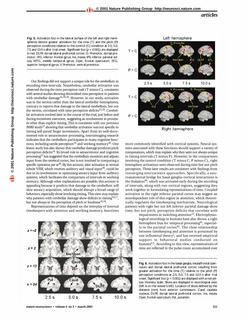
During the early imaging epochs (2.5 and 5.0 s), which emphasize encoding of temporalinformation, subcortical activations specific tothe T condition (Table 1) were observed withinthe right putamen, head of the caudate nucleusbilaterally, and right centromedian and ven-troanterior thalamic nuclei (Fig. 4a). Early acti-vation specific to the T condition was alsoobserved in various cortical regions (Fig. 5):right intraparietal sulcus (BA 40), bilateral dor-sal and left ventral premotor areas (BA 6), andbilateral lateral temporalcortex (BA 21/22). Activa-tion specific to the T condi-tion was sustained duringthe 7.5- and/or 10.0-s imag-ing epochs in most of theseregions. In the P condition,areas of activation duringthe early imaging epochsoverlapped with those in theT condition. In both the T and P conditions (Table 2),activity unfolded early with-in the medial wall (preSMAand SMA proper, BA 6, andanterior cingulate, BA 32;Fig. 4c) and the anteriorinsula/frontal operculum(Fig. 4a), but was sustainedduring later epochs as well.
During the later imagingepochs (7.5 and 10.0 s),which included decision andresponse selection compo-nents of the tasks, activationspecific to the T condition(Table 1) was observed inthe posterior vermis (tuber)of lobule VIIB of the cere-bellum (Fig. 4b) and theright dorsolateral prefrontal(DLPF) cortex (BA 46/10/9;Fig. 5). All other activationfoci were observed in the lefthemisphere in both the
articles
Fig. 3. Mean (± standard error of mean) reaction time and percent correct for the time per-ception (a, b) and the pitch perception (c, d) conditions. Data are depicted as a function of thecomparison interval or comparison pitch.
a
c d
b
Table 1. Stereotaxic brain atlas coordinates49 for Time > Control subtraction.
Location (Brodmann Area) Hemisphere 2.5 5.0 7.5 10.0
Basal GangliaMedial caudate (head) R 12, 7, 3 12, 6, 4
L –12, 7, 5 –9, 7, 2 –8, 4, 8Lateral caudate (body) R 15, 6, 19Putamen R 22, 8, –1 23, 6, 8
26, 6, –2L –20, –1, 5
CerebellumVermis (tuber, lobule VIIB) B –3, –70, –30 2, –70, –29
ThalamusCentromedian nucleus R 4, –21, 0 4, –21, 0Ventroanterior nucleus R 4, –11, 0 5, –10, 0
FrontalDorsal premotor (6) R 23, –7, 48 23, –3, 52 46, 1, 49
L –45, –7, 47Ventral premotor (6) R 46, 8, 24
L –54, –13, 26 –51, –15, 27Dorsolateral (46/10/9) R 34, 23, 25 31, 46, 22
41, 29, 22Parietal
Intraparietal sulcus,Angular gyrus (40) R 38, –40, 41 36, –43, 40 37, –47, 38 30, –56, 35Superior parietal lobule,Precuneus (7) R 10, –68, 44
TemporalSuperior temporal (22) R 51, –39, 6Middle temporal (21) L –46, –56, 4
R, right; L, left; B, bilateral. The activations reported in this table were not observed in the Pitch > Control subtraction.
©20
01 N
atu
re P
ub
lish
ing
Gro
up
h
ttp
://n
euro
sci.n
atu
re.c
om
© 2001 Nature Publishing Group http://neurosci.nature.com

320 nature neuroscience • volume 4 no 3 • march 2001
T and P conditions (Table 2), and included the inferior frontalgyrus (Broca’s area, BA 44/45), intraparietal sulcus (BA 40), supe-rior parietal lobule/precuneus (BA 7) and DLPF cortex.
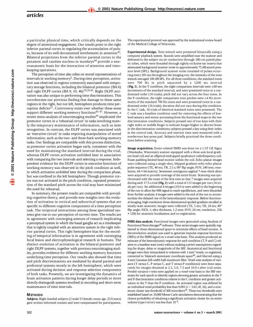
The results from the T minus P subtraction were similar tothe results for the T minus C subtraction (Fig. 6). During the
earlier imaging epochs (2.5 and 5.0 s), subcortical activationsunique to the T condition were in the right hemisphere andincluded the putamen (x, y, z = 24, 7, –2), caudate (15, 6, 13) andinsula/frontal operculum (29, 16, 2). The later region, however,was also activated during the 7.5-s epoch in the pitch condition(Table 2, Fig. 4a). During the later imaging epochs (7.5 s), the right DLPF cortex (21, 21, 30) was also unique to theT condition (Fig. 6).
DISCUSSIONThe present findings provide compelling evidence for the involve-ment of the basal ganglia in formulating representations of time.Activation in the right putamen and caudate were uniquely asso-ciated with encoding time intervals. These results corroboratestudies in Parkinson’s disease showing that dopaminergic treat-ment improves motor timing30,31 and time perception32. Phar-macological challenges in animals also suggest that dopaminergicantagonists and agonists respectively slow down and speed uptiming operations12,13. Contrary to one proposal33, these andother studies10,11,27 show that the basal ganglia are involved intiming a wide range of intervals, from hundreds of milliseconds(300 ms) to tens of seconds (20 s). Collectively, these resultsimplicate striatal dopaminergic neurotransmission in hypothet-ical internal timekeeping mechanisms.
articles
Fig. 4. Activation foci in the basal ganglia (a), cerebellum (b), and pre-supplementary motor area/anterior cingulate (c) resulting from subtrac-tion of the control (C) condition from the time (T) and the pitch (P)perception conditions at 2.5, 5.0, 7.5 and 10.0 s after trial onset.Significant foci (p < 0.001) are displayed with a red-yellow intensity scaledenoting greater activation for the T or P conditions. Slices are displayedin neurological view (left is on the viewer’s left). Location of slices definedby the distance (mm) from anterior commissure: x, right (+)/left (–); y,anterior (+)/posterior (–); z, superior (+)/inferior (–). Caud, caudatenucleus; Cing, anterior cingulate area; Ins, insula; Oper, frontal opercu-lum; Put, putamen; Thal, thalamus; SMA, supplementary motor area.
Table 2. Stereotaxic brain atlas coordinates49 for regions commonly activated in subtractions of Time and Pitchperception conditions relative to Control condition.
Time > Control Pitch > Control
Location (Brodmann Area) Hemisphere 2.5 5.0 7.5 10.0 2.5 5.0 7.5 10.0
FrontalInsula/operculum (47) R 31, 17, 3 35, 16, 3 34, 17, 4 34, 17, 0
L –35, 11, 5 –34, 15, 2 –36, 12, 4 –34, 18, 1 –36, 17, 0
PreSMA (6),
Anterior cingulate (32) L –4, –1, 56 –4, 6, 49 –7, 10, 45 –5, 12, 43 –6, 7, 48 –4, 8, 49
Inferior frontal gyrus (44/45) R 37, 1, 32 37, 4, 28
L –46, 4, 21 –47, 5, 18 –45, 4, 22 –44, 7, 26
Dorsolateral (46/10/9) L –39, 42, 12 –36, 46, 13 –36, 40, 8
–42, 26, 28 –40, 14, 29
ParietalIntraparietal sulcus,
Angular gyrus (40) L –31, –49, 37 –29, –52, 33
–36, –53, 44 –32, –47, 38–30, –55, 36
Superior parietal lobule,
Precuneus (7) L –21, –66, 49 –28, –49, 43 –13, –72, 50 –43, –57, 50
–21, –63, 51 –25, –65, 50
R, right; L, left; B, bilateral
a
b
c
©20
01 N
atu
re P
ub
lish
ing
Gro
up
h
ttp
://n
euro
sci.n
atu
re.c
om
© 2001 Nature Publishing Group http://neurosci.nature.com
nature neuroscience • volume 4 no 3 • march 2001 321
Our findings did not support a unique role for the cerebellum inencoding time intervals. Nonetheless, cerebellar activation wasobserved during the time perception task (T minus C), consistentwith several studies showing diminished time perception in patientswith cerebellar damage16,18,34. However, in our study, activationwas in the vermis rather than the lateral cerebellar hemispheres,contrary to reports that damage to the lateral cerebellum, but notthe vermis, correlated with time perception deficits15,18. Cerebel-lar activation evolved later in the course of the trial, just before andduring movement execution, suggesting an involvement in process-es other than explicit timing. This is consistent with our previousfMRI study27 showing that cerebellar activation was not specific totiming self-paced finger movements. Apart from its well-docu-mented role in sensorimotor processing, neuroimaging researchindicates that the cerebellum participates in many cognitive func-tions, including tactile perception35 and working memory36. Onelesion study has also shown that cerebellar damage produces pitchperception deficits14. Its broad role in sensorimotor and cognitiveprocessing37 has suggested that the cerebellum monitors and adjustsinput from the cerebral cortex, but is not involved in computing aspecific operation per se38. By this account, later activation in vermallobule VIIB, which receives auditory and visual input39, could bedue to its involvement in optimizing sensory input from auditorysystems, which facilitates the comparison of intervals in workingmemory. Although other explanations are possible, this account isappealing because it predicts that damage to the cerebellum willslow sensory acquisition, which should disrupt a broad range ofbehaviors, especially those involving timing. This view may explainwhy patients with cerebellar damage show deficits in timing16,17,but not always in the perception of pitch or loudness16,18.
Representations of time depend on the interplay of internaltimekeepers with attention and working memory, functions
more commonly identified with cortical systems. Neural sys-tems associated with these functions should support a variety ofcomputations, which may explain why they were not always uniqueto timing intervals (T minus P). However, in the comparisonsinvolving the control condition (T minus C, P minus C), righthemisphere activations were observed during time but not pitchperception. These later results are consistent with findings fromconverging neuroscience approaches. Specifically, a neu-roanatomical bridge for basal ganglia–cortical interactions isthe thalamus40, which was activated early during the encodingof intervals, along with two cortical regions, suggesting theywork together in formulating representations of time. Coupledactivation in the right inferior parietal cortex may suggest aninterdependent role of this region in attention, which theoret-ically regulates the timekeeping mechanism. Neurologicalpatients with right but not left inferior parietal damage showtime, but not pitch, perception deficits that correlate with
impairments in switching attention21. Electrophysio-logical recordings in humans have also shown a righthemisphere bias for temporal processing41, especial-ly in the parietal cortex42. The close relationshipbetween timekeeping and attention is presumed byone influential theory2, and has received empiricalsupport in behavioral studies conducted onhumans4,5. According to this view, representations oftime are reflected in the pulse count accumulated over
articles
Fig. 5. Activation foci in the lateral surface of the left and right hemi-spheres denote greater activation for the time (T) and the pitch (P)perception conditions relative to the control (C) condition at 2.5, 5.0,7.5 and 10.0 s after trial onset. Significant foci (p < 0.001) are displayedin red. DLPF, dorsal lateral prefrontal cortex; D. Premotor, dorsal pre-motor; IFG, inferior frontal gyrus; Ins, insula; IPS, inferior parietal sul-cus; MTG, middle temporal gyrus; Oper, frontal operculum; STG,superior temporal gyrus; V. Premotor, ventral premotor.
Fig. 6. Activation foci in the basal ganglia, insula/frontal oper-culum and dorsal lateral prefrontal cortex resulting fromgreater activation for the time (T) relative to the pitch (P)perception conditions at 2.5, 5.0, 7.5 and 10.0 s after trialonset. Significant foci (p < 0.001) are displayed with a red-yel-low intensity scale. Slices are displayed in neurological view(left is on the viewer’s left). Location of slices defined by thedistance (mm) from anterior commissure. Caud, caudatenucleus; DLPF, dorsal lateral prefrontal cortex; Ins, insula;Oper, frontal operculum; Put, putamen.
©20
01 N
atu
re P
ub
lish
ing
Gro
up
h
ttp
://n
euro
sci.n
atu
re.c
om
© 2001 Nature Publishing Group http://neurosci.nature.com
322 nature neuroscience • volume 4 no 3 • march 2001
a particular physical time, which critically depends on thedegree of attentional engagement. Our results point to the rightinferior parietal cortex in regulating the accumulation of puls-es, because of its well-documented involvement in attention43.Bilateral projections from the inferior parietal cortex to theputamen and caudate nucleus in monkeys44 provide a neu-roanatomic basis for the interaction of attention and time-keeping operations.
The perception of time also relies on stored representations ofintervals in working memory2. During time perception, activa-tion was observed in regions commonly associated with tempo-rary storage functions, including the bilateral premotor (BA 6)and right DLPF cortex (BA 9, 10, 46)19,20,45. Right DLPF acti-vation was also unique to performing time discriminations. Thiscorroborates our previous finding that damage to these sameregions in the right, but not left, hemisphere produces time per-ception deficits21. Controversy exists over whether these areassupport different working memory functions45–47. However, arecent meta-analysis of neuroimaging studies20 implicated thepremotor cortex in a ‘rehearsal circuit’ in tasks involving main-ly the temporary maintenance of information, such as itemrecognition. In contrast, the DLPF cortex was associated withan ‘executive circuit’ in tasks requiring manipulation of storedinformation, such as the two- and three-back working-memorytasks. Our findings are compatible with this process distinction,as premotor cortex activation began early, consistent with theneed for maintaining the standard interval during the trial,whereas DLPF cortex activation unfolded later in associationwith comparing the two intervals and selecting a response. Inde-pendent evidence for the DLPF cortex in executive functions ofworking memory was observed in the pitch condition as well,in which activation unfolded later during the comparison phase,but was confined to the left hemisphere. Though premotor cor-tex was not activated in the pitch condition, repeated presenta-tion of the standard pitch across the trial may have minimizedthe need for rehearsal.
In summary, the present results are compatible with prevail-ing cognitive theory, and provide new insights into the evolu-tion of activation in cortical and subcortical systems that arespecific to different cognitive components of a time perceptiontask. The reciprocal interactions among these specialized sys-tems give rise to our perception of current time. The results arein agreement with converging avenues of research implicatinga perceptual system in which the basal ganglia act as a timekeeperthat is tightly coupled with an attention system in the right infe-rior parietal cortex. This right hemisphere bias for the encod-ing of temporal information is in agreement with convergingfocal lesion and electrophysiological research in humans. Thedistinct evolution of activation in the bilateral premotor andright DLPF systems, together with previous neuroimaging stud-ies, provides evidence for different working memory functionsunderlying time perception. Our results also showed that timeand pitch discriminations are mediated by shared parietal andprefrontal systems mostly in the left hemisphere, which wereactivated during decision and response selection componentsof both tasks. Presently, we are investigating the dynamics ofbrain activation patterns during longer delay periods to moredirectly distinguish systems involved in encoding and short-termmaintenance of time intervals.
METHODSSubjects. Right-handed subjects (2 male/15 female; mean age, 23.9 years)gave written informed consent and were compensated for participation.
The experimental protocol was approved by the institutional review boardof the Medical College of Wisconsin.
Experimental design. Tone stimuli were presented binaurally using acomputer playback system. Sounds were amplified near the scanner anddelivered to the subject via air conduction through 180-cm paired plas-tic tubes, which were threaded through tightly occlusive ear inserts thatattenuated background scanner noise to approximately 75 dB sound pres-sure level (SPL). Background scanner noise consisted of pulses occur-ring every 205 ms throughout the imaging run; the intensity of the tonestimuli averaged 100 dB SPL. For all three conditions, the standard toneswere 700 Hz in pitch separated by a 1200 ms interval (Fig. 1). In the T condition, the eight comparison intervals were ±60-msincrements of the standard interval, and were presented twice in a ran-domized order (16 trials); pitch did not vary across the four tones. Inthe P condition, the eight comparison tone pitches were ±4 Hz incre-ments of the standard 700 Hz tones and were presented twice in a ran-domized order (16 trials); duration did not vary during this condition.In the C task, 16 trials of identical standard tones were presented. The C task was a baseline condition used for removing the effects of low-level sensory and motor processing from the functional maps in the twodiscrimination conditions. Subjects pressed one of two keys with theirright index or middle finger to indicate longer/higher or shorter/lowerin the discrimination conditions; subjects pressed a key using their indexin the control task. Accuracy and reaction time were measured with anonferrous key-press pad. Subjects briefly practiced the three condi-tions before scanning.
Image acquisition. Event-related fMRI was done on a 1.5T GE Signa(Waukesha, Wisconsin) scanner equipped with a three-axis local gradi-ent head coil and an elliptical endcapped quadrature radiofrequency coil.Foam padding limited head motion within the coil. Echo-planar imageswere collected using a single-shot, blipped gradient-echo echo-planarpulse sequence (TE, 40 ms; TR, 2.5 s; 90° flip angle; FOV, 240 mm; reso-lution, 64 × 64 matrix). Seventeen contiguous sagittal 7-mm-thick sliceswere acquired to provide coverage of the entire brain. Scanning was syn-chronized with the onset of the first tone so that 7 images were acquiredduring each 17.5-s trial (Fig. 2) with a total of 112 images per run (16 tri-als per run). An additional 4 images (10.0 s) were added to the beginningof the run to allow the MR signal to reach equilibrium, and were discardedfrom further analysis; 4 images were added to the end of the run to accom-modate the delayed rise of the hemodynamic response. Before function-al imaging, high-resolution three-dimensional spoiled gradient-recalled atsteady-state anatomic images were collected (TE, 5 ms; TR, 24 ms; 40°flip angle; NEX, 1; slice thickness, 1.2 mm; FOV, 24 cm; resolution, 256× 128) for anatomic localization and co-registration.
fMRI data analysis. Functional images were generated using Analysis ofFunctional NeuroImages48 software. Time series images were spatially reg-istered in three-dimensional space to minimize effects of head motion. Adeconvolution analysis was used to generate impulse response functions(IRFs) of the fMRI signal on a voxel-wise basis. This analysis produced anestimate of the hemodynamic response for each condition (T, P and C) rel-ative to a baseline state (rest) without making a priori assumptions regard-ing the shape, delay or magnitude of the IRF. Anatomical and functionalimages were then interpolated to volumes with 1 mm3 voxels, co-registered,converted to Talairach stereotaxic coordinate space49, and blurred using a 4 mm Gaussian full-width half-maximum filter. Voxel-wise analyses of vari-ance (T versus C, P versus C, and T versus P conditions) were done sepa-rately for images obtained at 2.5, 5.0, 7.5 and 10.0 s after trial onset.Pooled-variance t-tests were applied on a voxel-wise basis to the IRF esti-mates for each epoch to identify regions showing greater activation in the Tand P discrimination conditions relative to the C condition and greater acti-vation in the T than the P condition. An activated region was defined byan individual voxel probability less than 0.001 (t > 3.61; df, 16), and a min-imum cluster size threshold of 300 microliters50. These two thresholds wereestablished based on 10,000 Monte Carlo simulations demonstrating that thechance probability of obtaining a significant activation cluster for an entirevolume (type I error) was less than 10–6.
articles©
2001
Nat
ure
Pu
blis
hin
g G
rou
p
htt
p:/
/neu
rosc
i.nat
ure
.co
m© 2001 Nature Publishing Group http://neurosci.nature.com
nature neuroscience • volume 4 no 3 • march 2001 323
ACKNOWLEDGEMENTSThis study was funded in part by grants from the Department of Veterans Affairs
and National Foundation for Functional Brain Imaging (D.L.H.), the National
Institutes of Health (P01 MH51358, R01 MH57836, M01 RR00058) and W.M.
Keck Foundation (S.M.R.). We thank R. Cox, E. DeYoe, S. Durgerian, R. Lee,
G. Mallory, L. Mead, J. Neitz and B. Ward for their assistance.
RECEIVED 6 SEPTEMBER; ACCEPTED 21 DECEMBER 2000
1. Wearden, J. H. “Beyond the fields we know...”: Exploring and developingscalar timing theory. Behavioural Processes 45, 3–21 (1999).
2. Gibbon, J., Church, R. M. & Meck, W. H. Scalar timing in memory. Ann. NYAcad. Sci. 423, 52–77 (1984).
3. Matell, M. S. & Meck, W. H. Neuropsychological mechanisms of intervaltiming behavior. Bioessays 22, 94–103 (2000).
4. Brown, S. W. Time perception and attention: the effects of prospective versusretrospective paradigms and task demands on perceived duration. Percept.Psychophys. 38, 115–124 (1985).
5. Zakay, D. & Block, R. A. in Time, Internal Clocks, and Movement (eds. Pastor,M. A. & Artieda, J.) 143–164 (Elsevier Science BV, Amsterdam, 1996).
6. Baddeley, A. Working Memory (Clarendon/Oxford Univ. Press, Oxford,1986).
7. Harrington, D. L. & Haaland, K. Y. Neural underpinnings of temporalprocessing: A review of focal lesion, pharmacological, and functional imagingresearch. Rev. Neurosci. 10, 91–116 (1999).
8. Narabayashi, H. & Nakamura, R. in Clinical Neurophysiology of Parkinsonism.(eds. Delwaide, P. J. & Agnoli, A.) 45–57 (Elsevier, New York, 1985).
9. Manto, M. Pathophysiology of cerebellar dysmetria: the imbalance betweenthe agonist and the antagonist electromyographic activities. Eur. Neurol. 36,333–337 (1996).
10. Artieda, J., Pastor, M. A., Lacruz, F. & Obeso, J. A. Temporal discrimination isabnormal in Parkinson’s disease. Brain 115, 199–210 (1992).
11. Harrington, D. L., Haaland, K. Y. & Hermanowicz, N. Temporal processing inthe basal ganglia. Neuropsycholologia 12, 3–12 (1998).
12. Maricq, A. V. & Church, R. M. The differential effects of haloperidol andmethamphetamine on time estimation in the rat. Psychopharmacology 79,10–15 (1983).
13. Meck, W. H. Affinity for the dopamine D2 receptor predicts neurolepticpotency in decreasing the speed of an internal clock. Pharmacol. Biochem.Behav. 25, 1185–1189 (1986).
14. Casini, L. & Ivry, R. Effects of divided attention on temporal processing inpatients with lesions of the cerebellum or frontal lobe. Neuropsychologia 13,10–21 (1999).
15. Ivry, R. B., Keele, S. W. & Diener, H. C. Dissociation of the lateral and medialcerebellum in movement timing and movement execution. Exp. Brain Res.73, 167–180 (1988).
16. Ivry, R. B. & Keele, S. W. Timing functions of the cerebellum. J. Cogn.Neurosci. 1, 136–152 (1989).
17. Malapani, C., Dubois, B., Rancurel, G. & Gibbon, J. Cerebellar dysfunctionsof temporal processing in the seconds range in humans. Neuroreport 9,3907–3912 (1998).
18. Mangels, J. A., Ivry, R. & Shimizu, N. Dissociable contributions of theprefrontal and neocerebellar cortex to time perception. Cogn. Brain Res. 7,15–39 (1998).
19. Goldman-Rakic, P. S. Regional and cellular fractionation of workingmemory. Proc. Natl. Acad. Sci. USA 93, 13473–13480 (1996).
20. Smith, E. & Jonides, J. Storage and executive processes in the frontal lobes.Science 283, 1657–1661 (1999).
21. Harrington, D. L., Haaland, K. Y. & Knight, R. T. Cortical networksunderlying mechanisms of time perception. J. Neurosci. 18, 1085–1095(1998).
22. Meck, W. H., Church, R. M., Wenk, G. L. & Olton, D. S. Nucleus basalismagnocellularis and medial septal area lesions differentially impair temporalmemory. J. Neurosci. 7, 3505–3511 (1987).
23. Halsband, U., Ito, N., Tanji, J. & Freund, H. J. The role of premotor cortex and
the supplementary motor area in the temporal control of movement in man.Brain 116, 243–266 (1993).
24. Larsson, J., Gulyas, B. & Roland, P. E. Cortical representation of self-pacedfinger movement. Neuroreport 7, 463–468 (1996).
25. Lejeune, H. et al. The basic pattern of activation in motor and sensorytemporal tasks: positron emission tomography data. Neurosci. Lett. 235,21–24 (1997).
26. Penhune, V. B., Zatorre, R. J. & Evans, A. Cerebellar contributions to motortiming: a PET study of auditory and visual rhythm reproduction. J. Cogn.Neurosci. 10, 752–765 (1998).
27. Rao, S. M. et al. Distributed neural systems underlying the timing ofmovements. J. Neurosci. 17, 5528–5535 (1997).
28. Jueptner, I. H. et al. Localization of a cerebellar timing process using PET.Neurology 45, 1540–1545 (1995).
29. Maquet, P. et al. Brain activation induced by estimation of duration: a PETstudy. Neuroimage 3, 119–126 (1996).
30. O’Boyle, D. J., Freeman, J. S. & Cody, F. W. J. The accuracy and precision oftiming of self-paced, repetitive movements in subjects with Parkinson’sdisease. Brain 119, 51–70 (1996).
31. Pastor, M. A., Artieda, J., Jahanshahi, M. & Obeso, J. A. Performance ofrepetitive wrist movements in Parkinson’s disease. Brain 115, 875–891(1992).
32. Malapani, C. et al. Coupled temporal memories in Parkinson’s disease: adopamine-related dysfunction. J. Cogn. Neurosci. 10, 316–331 (1998).
33. Ivry, R. The representation of temporal information in perception and motorcontrol. Curr. Opin. Neurobiol. 6, 851–857 (1996).
34. Casini, L. & Macar, F. Effects of attention manipulation on judgments ofduration and of intensity in the visual modality. Mem. Cognit. 25, 812–818(1997).
35. Gao, J. H. et al. Cerebellum implicated in sensory acquisition anddiscrimination rather than motor control. Science 272, 545–547 (1996).
36. Desmond, J. E., Gabrieli, J. D. E., Wagner, A. D., Ginier, B. L. & Glover, G. H.Lobular patterns of cerebellar activation in verbal working-memory andfinger-tapping tasks as revealed by functional MRI. J. Neurosci. 17,9675–9685 (1997).
37. Schmahmann, J. D. The Cerebellum and Cognition (Academic, New York, 1997).38. Bower, J. M. Is the cerebellum sensory for motor’s sake, or motor for
sensory’s sake: the view from the whiskers of a rat? Prog. Brain Res. 114,463–496 (1997).
39. Brodal, P. The ponto-cerebellar projection in the rhesus monkey: anexperimental study with retrograde axonal transport of horseradishperoxidase. Neuroscience 4, 193–208 (1979).
40. Alexander, G. E., DeLong, M. R. & Strick, P. L. in Annual Review ofNeuroscience (ed. Cowan, W. M.) 357–381 (Society for Neuroscience,Washington, DC, 1986).
41. Brunia, C. & Damen, E. Distribution of slow brain potentials related tomotor preparation and stimulus anticipation in a time estimation task.Electroencephalogr. Clin. Neurophysiol. 69, 234–243 (1988).
42. Mohl, W. & Pfurtscheller, G. The role of the right parietal region in amovement time estimation task. Neuroreport 2, 309–312 (1991).
43. Posner, M. I., Walker, J. A., Friedrich, F. J. & Rafal, R. D. Effects of parietalinjury on covert orienting of attention. J. Neurosci. 4, 1863–1874 (1984).
44. Cavada, C. & Goldman-Rakic, P. S. Topographic segregation ofcorticostriatal projections from posterior parietal subdivisions in themacaque monkey. Neuroscience 42, 683–696 (1991).
45. Cohen, J. et al. Temporal dynamics of brain activation during a workingmemory task. Nature 386, 604–607 (1997).
46. Awh, E., Smith, E. E. & Jonides, J. Human rehearsal processes and the frontallobes: PET evidence. Ann. NY Acad. Sci. 769, 97–117 (1995).
47. Prabhakaran, V., Narayanan, K., Zhao, Z. & Gabrieli, J. D. E. Integration ofdiverse information in working memory within the frontal lobe. Nat.Neurosci. 3, 85–90 (2000).
48. Cox, R. W. AFNI: Software for analysis and visualization of functionalmagnetic resonance neuroimages. Comput. Biomed. Res. 29, 162–173 (1996).
49. Talairach, J. & Tournoux, P. Co-Planar Stereotaxic Atlas of the Human Brain(Thieme, New York, 1988).
50. Forman, S. D. et al. Improved assessment of significant activation infunctional magnetic resonance imaging (fMRI): use of a cluster-sizethreshold. Magn. Reson. Med. 33, 636–647 (1995)
articles©
2001
Nat
ure
Pu
blis
hin
g G
rou
p
htt
p:/
/neu
rosc
i.nat
ure
.co
m© 2001 Nature Publishing Group http://neurosci.nature.com