the exceptional case of a large extra-uterine, so-called
TRANSCRIPT
319
Introduction
Uterine leiomyomas, also called fibroids, are common benign adenomas, which sometimes cause metrorrhagia. The growth of fibroids is hormone- dependant, but it is possible that they can shrink after the menopause. However, they may also gain volume during oestrogen replacement therapy. Whilst the overwhelming majority of fibroids are situated in the uterus, extra-uterine, parasitic leiomyomas do occasionally occur (Mahmoud et al., 2015). Several publications report a possible association between leiomyomas and thyroid diseases, namely hypothyroidism (Ott et al., 2014), thyroid adenomas (Kim et al., 2010) and fibroadenomas of the breast (Spinos et al., 2007). An association between leiomyomas and autoimmune Hashimoto’s thyroiditis has not been described before. Rare cases of leiomyoma of the thyroid gland (Biankin and Cachia, 1999) or adrenal gland (Goldman and Brodey, 1994, Huei et al., 2017) have been reported.
To the best of our knowledge, no cases have been published combining a large extra-uterine leiomyoma with Hashimoto’s thyroiditis in a post-
menopausal patient receiving adequate hormone replacement therapy with thyroid hormone (Levothyroxine) and oestrogen (Oestrogel).
Case description
The patient was a 65-year-old obese woman with a BMI of 34.8 kg/m2. She was known to suffer from Hashimoto’s thyroiditis, with elevated anti-thyroglobulin antibodies (232 IU/mL), which has been adequately treated for over 10 years. Treatment with 100 μg of levothyroxine resulted in normal hormone concentrations in the blood: TSH: 0.5.mud/L, free T4: 20.2pmol/L, free T3: 4.9 pmol/L.There were no other biological markers of autoimmune disease. Due to insulin resistance combined with hyperinsulinemia, the patient was successfully treated with the combination of Momordica charantia extract and alpha-lipoic acid (Comhaire, 2015), with a normal postprandial C-peptide concentration of 1.67 nmol/L. She had no cardiac, pulmonary, or systemic diseases.
After the birth of her third daughter, she was sterilized by tubal ligature. Due to disturbing menopausal complaints, the patient applied
The exceptional case of a large extra-uterine, so-called parasitic leiomyoma in a post-menopausal woman with Hashimoto’s thyroiditis
W. Decleer1,2, A. DebeuckelAere3, F. comhAire2
1Fertility Centre, AZ Jan Palfijn Ghent, Watersportlaan 5, 9000 Ghent, Belgium; 2Fertility Clinic, Weststraat 16/18, 9880 Aalter, Belgium; 3Master student Medical Science, KU Leuven, 3000 Leuven, Belgium.
Correspondence at: Dr. Wim Decleer: [email protected]
Abstract
A large tumour mass was detected in a 65-year-old patient during a routine gynaecological examination. This patient had been treated for over 10 years with levothyroxine for Hashimoto’s thyroiditis and was also given transdermal oestrogen replacement therapy. Before the operation, detailed imaging by CT scan and MRI was performed. A tumour weighing 1.056 grams and measuring 23x12x7 cm was successfully removed through laparotomy. Histopathology revealed the diagnosis of an extra-uterine, so-called parasitic leiomyoma. Post-surgery recovery was uneventful, but Tibolone treatment was indicated due to disturbing menopausal complaints.
Key words: Parasitic leiomyoma, Hashimoto’s thyroiditis, extra-uterine, post-menopausal.
Facts Views Vis Obgyn, 2020, 12 (4): 319-323 Case report
320 Facts Views Vis Obgyn
transdermal oestradiol (0.75 mg/day of Oestrogel, Besins, Brussels, Belgium).
In July 2019, the patient was examined by her usual gynaecologist who detected a large intra-abdominal mass, detached from the uterus.
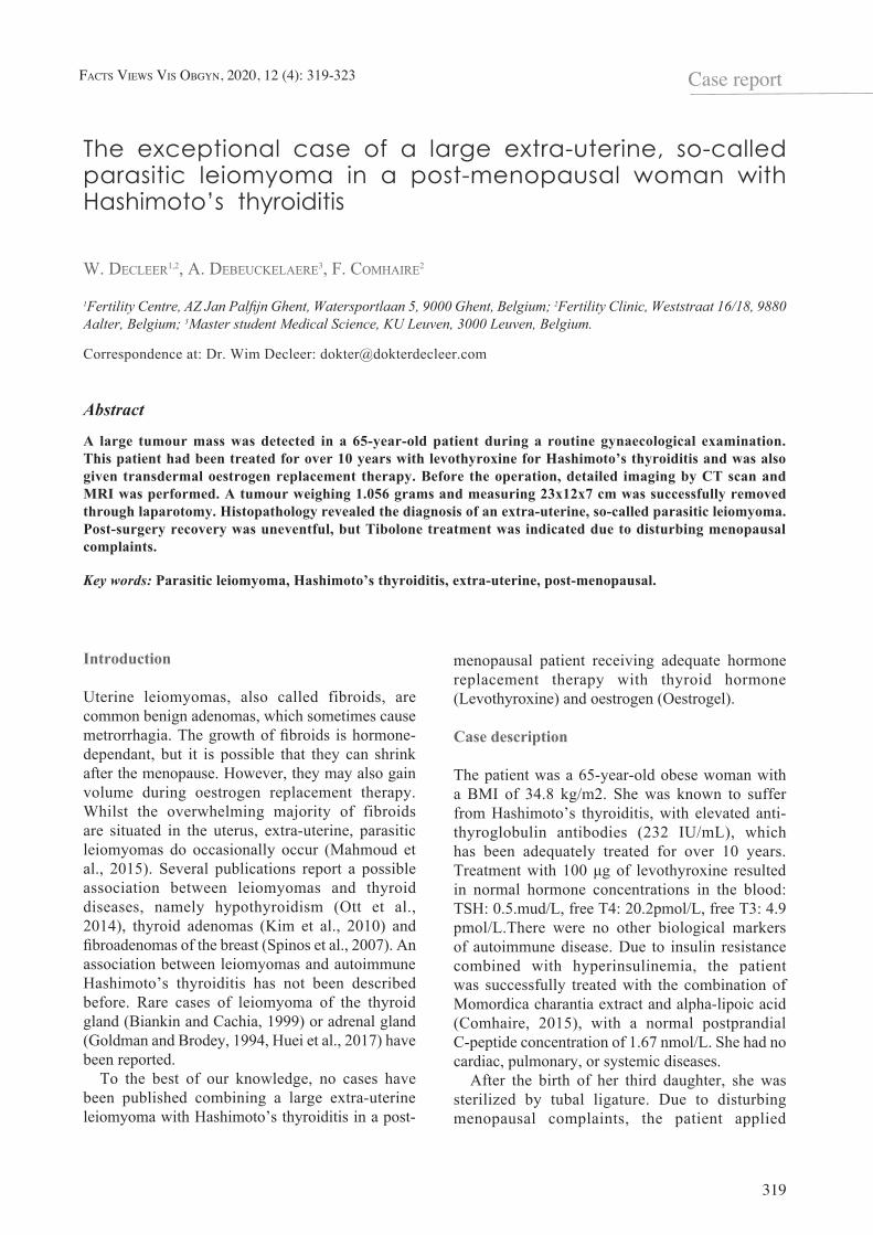
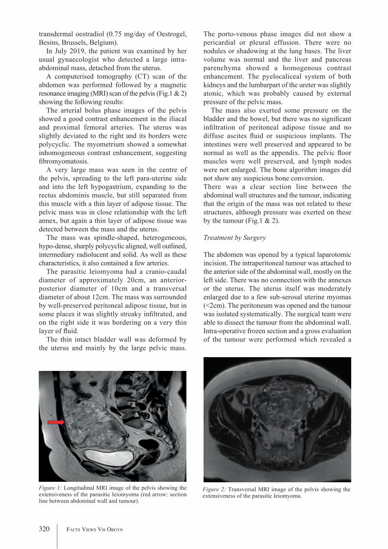
A computerised tomography (CT) scan of the abdomen was performed followed by a magnetic resonance imaging (MRI) scan of the pelvis (Fig.1 & 2) showing the following results:
The arterial bolus phase images of the pelvis showed a good contrast enhancement in the iliacal and proximal femoral arteries. The uterus was slightly deviated to the right and its borders were polycyclic. The myometrium showed a somewhat inhomogeneous contrast enhancement, suggesting fi bromyomatosis.
A very large mass was seen in the centre of the pelvis, spreading to the left para-uterine side and into the left hypogastrium, expanding to the rectus abdominis muscle, but still separated from this muscle with a thin layer of adipose tissue. The pelvic mass was in close relationship with the left annex, but again a thin layer of adipose tissue was detected between the mass and the uterus.
The mass was spindle-shaped, heterogeneous, hypo-dense, sharply polycyclic aligned, well outlined, intermediary radiolucent and solid. As well as these characteristics, it also contained a few arteries.
The parasitic leiomyoma had a cranio-caudal diameter of approximately 20cm, an anterior-posterior diameter of 10cm and a transversal diameter of about 12cm. The mass was surrounded by well-preserved peritoneal adipose tissue, but in some places it was slightly streaky infi ltrated, and on the right side it was bordering on a very thin layer of fl uid.
The thin intact bladder wall was deformed by the uterus and mainly by the large pelvic mass.
Figure 1: Longitudinal MRI image of the pelvis showing the extensiveness of the parasitic leiomyoma (red arrow: section line between abdominal wall and tumour).
Figure 2: Transversal MRI image of the pelvis showing the extensiveness of the parasitic leiomyoma.
The porto-venous phase images did not show a pericardial or pleural effusion. There were no nodules or shadowing at the lung bases. The liver volume was normal and the liver and pancreas parenchyma showed a homogenous contrast enhancement. The pyelocaliceal system of both kidneys and the lumbarpart of the ureter was slightly atonic, which was probably caused by external pressure of the pelvic mass.
The mass also exerted some pressure on the bladder and the bowel, but there was no signifi cant infi ltration of peritoneal adipose tissue and no diffuse ascites fl uid or suspicious implants. The intestines were well preserved and appeared to be normal as well as the appendix. The pelvic fl oor muscles were well preserved, and lymph nodes were not enlarged. The bone algorithm images did not show any suspicious bone conversion.There was a clear section line between the abdominal wall structures and the tumour, indicating that the origin of the mass was not related to these structures, although pressure was exerted on these by the tumour (Fig.1 & 2).
Treatment by Surgery
The abdomen was opened by a typical laparotomic incision. The intraperitoneal tumour was attached to the anterior side of the abdominal wall, mostly on the left side. There was no connection with the annexes or the uterus. The uterus itself was moderately enlarged due to a few sub-serosal uterine myomas (<2cm). The peritoneum was opened and the tumour was isolated systematically. The surgical team were able to dissect the tumour from the abdominal wall. Intra-operative frozen section and a gross evaluation of the tumour were performed which revealed a

THE EXCEPTIONAL CASE OF A LARGE EXTRA-UTERINE – DECLEER et Al. 321
parasitic leiomyomas are a rare variant of a pedunculated (uterine) sub-serosal leiomyoma occurring outside the uterus and completely separated from it (Shaukat et al., 2019; Sarmalkar et al., 2016; Grover and Bhalla, 2015). Extra-uterine leiomyoma was first described by Kelly and Cullen in 1909. As per FIGO classification system, so-called parasitic fibroids have been categorized as type 8 leiomyomas with no myometrial involvement and uterine attachment (Berek, 2013).
This mass usually grows intra-peritoneal, more specifically in the pelvis. However, some parasitic myomata have been described in the upper abdomen, on the omentum, in the vagina, sub- or supra-fascial near the port site, sigmoid colon, cervical, retroperitoneal (Cucinella et al., 2011; Epstein et al., 2009; Nezhat and Kho, 2010; Lete et al., 2016),in the thyroid gland (Biankin and Cachia, 1999) and in the adrenal gland (Goldman and Brodey, 1994; Huei et al., 2017).
Parasitic leiomyomas can be either asymptomatic (Salih et al., 2017) or cause complaints such as chronic abdominal pain, urinary dysfunction, abdominal feeling of pressure, palpable mass and rarely acute abdominal pain caused by necrosis or torsion (Cucinella et al., 2011). The diagnosis is often incidentally made upon radiologic examination or at laparoscopic surgery for another reason (Grover and Bhalla, 2015). The diagnosis can pose clinical and diagnostic challenges because these leiomyomas can mimic malignancy due to their unusual location, growth pattern and volume.
The most accurate diagnostic method is a pelvic MRI allowing a detailed description of the tumour and the anatomical relationship with surrounding structures. In addition to these features, MRI may also detect alternative and/or coexistent pelvic or abdominal pathology.
The classic appearance of a typical fibroid on MRI is a well-circumscribed mass with homogeneous T2 hypo-intensity and T1 iso-intensity relative to the myometrium (Bolan and Caserta, 2016). However, only histopathological analysis can confirm the diagnosis of a benign parasitic leiomyoma.
An interesting and rather unique feature of the present case is that the leiomyoma occurred in a patient with Hashimoto’s thyroiditis, but without hypothy roidism. There were no other characteristics of autoimmune disease, but the patient presented with insulin resistance and hyperinsu linemia related to obesity. Insulin is a growth factor and hyperinsulinemiais associated with tumour development (Tsujimoto et al., 2017).Thus, hyperinsulinemia may have contributed to the rapid expansion of this large, though benign tumour mass.
benign stromal tumour of approximately 23 x 12 x 7cm. The left rectus abdominis muscle was slightly damaged during the process of tumour removal, which was immediately restored with Vicryl 2 in separate U-sutures. The peritoneum fascia, subcutis and cutis were closed, and intra-abdominal tubular drainage was placed with the installation of a subfascial Penrose drain.
Post-surgery
The post-operative course was uneventful, and no complications occurred. Because of this favourable course, the patient was able to leave the hospital after four days. Four weeks later, she was seen at an outpatient consultation (FC) in good health with slight local thickening of the abdominal wall at the left side. No residual tumour residue was detected.The patient complained of disturbing vasomotor hot flushes, cognitive and emotional instability, and sleep disturbance. It was decided to temporarily restart transdermal oestrogen supplementation, which was immediately replaced by long-term Tibolone intake (Gregoriou et al., 1997,Gregoriou et al., 2001).
Histopathology
Macroscopically, the mass was described as a large nodular tumour of 1056 grams and 23 x 12 x 7 cm. On cut section, it appeared as a solid homogenous light-brown, tan mass. Paraffin sections showed a moderately to poorly cellular tumour, composed of spindle cells, predominantly arranged in bundles. The nuclei were spindle-shaped with blunt ends. Mitoses were few, and no abnormal mitotic figures were seen. The cells had an eosinophilic cytoplasm and were separated by a wide and oedematous interstitium.The sections showed dispersed mast cells andplasmocytes. At the periphery, a pseudocapsule of connective tissue was noticed. Arterial and venous vessels in the capsule were found at the site of the adhesion. Immunohistochemical investigation was performed and resulted in: (1) Smooth muscle actine: positive; (2) Desmin: positive; (3) S100 protein: negative; (4) Ki-67 proliferation marker: <5% positive cell nuclei. In conclusion, the mass was described as a giant leiomyoma (max. diameter 23 cm) with benign degenerative changes and no signs of malignancy.
Discussion
Whereas uterine leiomyomas are common gynaecological tumours, extra-uterine so-called
322 Facts Views Vis Obgyn
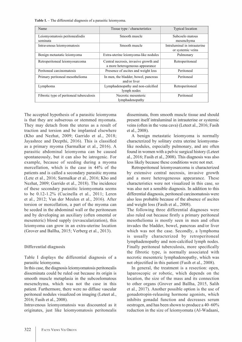
Name Tissue type / characteristics Typical location
Leiomyomatosis peritonealisdisseminata
Smooth muscle Subcoelo matous mesenchyma
Intravenous leiomyomatosis Smooth muscle Intraluminal in intrauterine or systemic veins
Benign metastatic leiomyoma Extra-uterine leiomyoma-like nodules Pulmonary
Retroperitoneal leiomyosarcoma Central necrosis, invasive growth and a more heterogeneous appearance
Retroperitoneal
Peritoneal carcinomatosis Presence of ascites and weight loss Peritoneal
Primary peritoneal mesothelioma In men, the bladder, bowel, pancreas and/or liver
Peritoneal
Lymphoma Lymphadenopathy and non-calcified lymph nodes
Retroperitoneal
Fibrotic type of peritoneal tuberculosis Necrotic mesenteric lymphadenopathy
Peritoneal
disseminata, from smooth muscle tissue and should present itself intraluminal in intrauterine or systemic veins (often in the vena cava) (Leteet al., 2016; Fasih et al., 2008).
A benign metastatic leiomyoma is normally characterized by solitary extra uterine leiomyoma-like nodules, especially pulmonary, and are often found in women with a pelvic surgical history (Leteet al., 2016; Fasih et al., 2008). This diagnosis was also less likely because these conditions were not met.
Retroperitoneal leiomyosarcoma is characterized by extensive central necrosis, invasive growth and a more heterogeneous appearance. These characteristics were not visualized in this case, so was also not a sensible diagnosis. In addition to this differential diagnosis, peritoneal carcinomatosis were also less probable because of the absence of ascites and weight loss (Fasih et al., 2008).The following three differential diagnoses were also ruled out because firstly a primary peritoneal mesothelioma is mostly seen in men and often invades the bladder, bowel, pancreas and/or liver which was not the case. Secondly, a lymphoma is usually characterized by retroperitoneal lymphadenopathy and non-calcified lymph nodes. Finally peritoneal tuberculosis, more specifically the fibrotic type, is normally associated with necrotic mesenteric lymphadenopathy, which was not objectified in this patient (Fasih et al., 2008).
In general, the treatment is a resection: open, laparoscopic or robotic, which depends on the location, the size of the mass and its connection to other organs (Grover and Ballha, 2015, Salih et al., 2017). Another possible option is the use of gonadotropin-releasing hormone agonists, which inhibits gonadal function and decreases serum oestrogen, and has been shown to produce a 40–60% reduction in the size of leiomyomata (Al-Wadaani,
The accepted hypothesis of a parasitic leiomyoma is that they are subserous or stemmed myomata. They may detach from the uterus as a result of traction and torsion and be implanted elsewhere (Kho and Nezhat, 2009; Garrido et al., 2018; Jayashree and Deepthi, 2016). This is classified as a primary myoma (Sarmalkar et al., 2016). A parasitic abdominal leiomyoma can be caused spontaneously, but it can also be iatrogenic. For example, because of seeding during a myoma morcellation, which is the case in 44% of the patients and is called a secondary parasitic myoma (Lete et al., 2016; Sarmalkar et al., 2016; Kho and Nezhat, 2009; Garrido et al., 2018). The incidence of these secondary parasitic leiomyomata seems to be 0.12-1.2% (Cucinella et al., 2011; Leren et al., 2012; Van der Meulen et al., 2016). After torsion or morcellation, a part of the myoma can be seeded in the abdominal wall or the peritoneum and by developing an auxiliary (often omental or mesenteric) blood supply (revascularization), this leiomyoma can grow in an extra-uterine location (Grover and Ballha, 2015; Verberg et al., 2013).
Differential diagnosis
Table I displays the differential diagnosis of a parasitic leiomyoma. In this case, the diagnosis leiomyomatosis peritonealis disseminata could be ruled out because its origin is smooth muscle metaplasia in the subcoelomatous mesenchyma, which was not the case in this patient. Furthermore, there were no diffuse vascular peritoneal nodules visualized on imaging (Leteet al., 2016; Fasih et al., 2008).Intravenous leiomyomatosis was discounted as it originates, just like leiomyomatosis peritonealis
Table I. – The differential diagnosis of a parasitic leiomyoma.

THE EXCEPTIONAL CASE OF A LARGE EXTRA-UTERINE – DECLEER et Al. 323
Huei TJ, Lip HT, Rahman MS et al. Large adrenal leiomyoma presented as adrenal incidentaloma in an AIDS patient: a rare entity. Med J Malaysia. 2017;72:65-7.
Jayashree A K, Deepthi R. Primary parasitic leiomyoma: a case report. J Clin Biomed Sci. 2016;6:36-8.
Kelly HA, Cullen TS. Myomata of the uterus. WB Saunders, Philadelphia. 1909.
Kho KA, Nezhat C. Parasitic myomas. Obstet Gynecol. 2009;114:611-5.
Kim MH, Park YR, Lim DJ et al.The relationship between thyroid nodules and uterine fibroids. Endocr J. 2010;57:615-21.
Leren V, Langebrekke A, Qvigstad E. Parasitic leiomyomas after laparoscopic surgery with morcellation. Acta Obstet Gynecol Scand. 2012;91:1233-6.
Lete I, González J, Ugarte L et al. Parasitic leiomyomas: a systematic review. Eur J Obstet Gynecol Reprod Biol. 2016;203:250-9.
Mahmoud MS, Desai K, Nezhat FR. Leiomyomas beyond the uterus; benign metastasizing leiomyomatosis with paraaortic metastasizing endometriosis and intravenous leiomyomatosis: a case series and review of the literature. Arch Gynecol Obstet. 2015;291:223–30.
Nezhat C, Kho K. Iatrogenic myomas: new class of myomas? J Minim Invasive Gynecol. 2010;17:544-50.
Ott J, Kurz C, Braun R et al.Overt hypothyroidism is associated with the presence of uterine leiomyoma: a retrospective analysis. Eur J Obstet Gynecol Reprod Biol. 2014;177:19-22.
Salih AM, Kakamad FH,Dahat AH et al. Parasitic leiomyoma: A case report with literature review. Int J Surg Case Rep. 2017;41:33-5.
Sarmalkar M, Nayak A, Singh N et al. A rare case of primary parasitic leiomyoma mimicking as ovarian mass: a clinical dilemma. Int J Reprod Contracept Obstet Gynecol. 2016;5:545-8.
Shaukat I, Yassin S, Paudel A et al. Unusual presentation of parasitic leiomyoma; a tale of twists and turns. J Community Hosp Intern Med Perspect. 2019;9:168-70.
Spinos N, Terzis G, Crysanthopoulou A et al. Increased frequency of thyroid nodules and breast fibroadenomas in women with uterine fibroids. Thyroid. 2007;17:1257-9.
Tsujimoto T, Kajio H, Sugiyama T. Association between hyperinsulinemia and increased risk of cancer in nonobese and obese people: a population-based observational study. Int J Cancer. 2017;141:102-11.
Van der Meulen JF, Pijnenborg JM, Boomsma CM et al.Parasitic myoma after laparoscopic morcellation: a systematic review of the literature. BJOG. 2016;123:69-75.
Verberg MF, Boomsma CM, Pijnenborg JM. Eenparasitairmyoom: onverwachte bevinding na laparoscopisch ehysterectomie. Ned Tijdschr Geneeskd. 2013;157:A6683.
2012). Other reported treatment modalities include: careful observation, myomectomy, hysterectomy with bilateral oophorectomy and medical treatments such as progestin and aromatase inhibitors (Mahmoud et al., 2015).
In the case reported, there was no need for hysterectomy nor ovariectomy since the tumour was completely isolated from the female genital organs.
References
Al-Wadaani HA. Anterior abdominal wall leiomyoma arising de novo in a perimenopausal woman. Oman Med J. 2012;27:323-5.
Berek JS. Uterine fibroids. In: Berek JS, eds. Berek& Novak’s Gynecology. 15th ed. Philadelphia, PA: Lipincott Williams & Wilkins. 2013;444-5.
Biankin SA, Cachia AR. Leiomyoma of the thyroid gland. Pathology. 1999;31:64-6.
Bolan C, Caserta MP. MR imaging of atypical fibroids. Abdom Radiol (NY). 2016;41:2332-49.
Comhaire F. Treating hyperinsulinemia with Momordica charantia. J Metab Syndr. 2015;4:3
Cucinella G, Granese R, Calagna G et al. Parasitic myomas after laparoscopic surgery: an emerging complication in the use of morcellator? Description of four cases. Fertil Steril. 2011;96:90-6.
Epstein JH, Nejat EJ, Tsai T. Parasitic myomas after laparoscopic myomectomy: case report. Fertil Steril. 2009;91:932.e13-4.
Fasih N, Prasad Shanbhogue AK, Macdonald DB et al.Leiomyomas beyond the uterus: unusual locations, rare manifestations. Radiographics. 2008;28:1931-48.
Garrido Oyarzún MF, Saco A, Castelo-Branco C. Anterior abdominal wall parasitic leiomyoma: case report. Gynecol Endocrinol. 2018;34:103-6.
Goldman RL, Brodey PA. Symptomatic leiomyoma of the adrenal. Clin Imaging. 1994;18:277-8.
Gregoriou O, Konidaris S, Botsis D et al. Long term effects of Tibolone on postmenopausal women with uterine myomas. Maturitas. 2001;40:95-9.
Gregoriou O, Vitoratos N, Papadias C et al. Effect of Tibolone on postmenopausal women with myomas. Maturitas. 1997;27:187-91.
Grover A, Bhalla S. Parasitic leiomyoma: A rare complication following laparoscopic myomectomy with review of literature. Curr Med Res Pract.2015;5:278-81.