the expression of the athb-8homeobox gene is restricted to...
TRANSCRIPT

4171Development 121, 4171-4182 (1995)Printed in Great Britain © The Company of Biologists Limited 1995DEV0036
The expression of the Athb-8 homeobox gene is restricted to provascular
cells in Arabidopsis thaliana
Simona Baima1, Fabio Nobili1, Giovanna Sessa2, Sabrina Lucchetti1, Ida Ruberti2 and Giorgio Morelli1,*1Unità di Nutrizione Sperimentale, Istituto Nazionale della Nutrizione, Via Ardeatina 546, 00178 Rome, Italy2Centro di Studio per gli Acidi Nucleici, c/o Dipartimento di Genetica e Biologia Molecolare, Università di Roma La Sapienza, P. le Aldo Moro 5 00185 Rome, Italy
*Author for correspondence
We have characterized an Arabidopsis homeobox genecoding for a putative DNA binding protein that representsan early marker for vascular development. The full-lengthcDNA encodes a protein of 833 amino acids that we havedesignated Athb-8; it contains the conserved DNA bindingdomain that characterizes the HD-Zip family of transcrip-tion factors. RNA analysis showed that the Athb-8 gene isexpressed during the vegetative and the reproductivephases of plant growth. A higher steady-state level of theAthb-8 mRNA was found in flowering stem and root. In situmRNA analysis of Arabidopsis plants demonstrated thatAthb-8 expression is restricted to the procambial cells of
embryo and developing organs. Moreover, Athb-8-GUSexpression was found in single parenchyma cells which aredifferentiating into tracheary elements in wounded tobaccotransgenic plants. Finally, we showed that the auxin,indole-3-acetic acid, which is involved in vascular develop-ment and differentiation, modulates the expression of thegene. Taken together, these results suggest that Athb-8might be a regulator of vascular development in Arabidop-sis thaliana.
Key words: Arabidopsis, auxin, HD-Zip motif, procambium,vascular development, homeobox gene
SUMMARY
INTRODUCTION
In flowering plants, the body organization is generated by twodistinct processes. During embryogenesis, the polar axis of theplant is established, domains that set up the organization of theplant body are defined, and the primary tissue and organsystems are delineated. The primary body organization of themature embryo may be described as a superimposition of twopatterns, one axial and one radial. The apical-basal pattern isarranged along the main body axis of polarity. Shoot meristem,cotyledon(s), hypocotyl, radicle (embryonic root) and rootmeristem are morphologically distinct elements of the axialpattern. The radial pattern, which is perpendicular to the axis,involves the three major tissues of the primary plant body: theouter epidermis, the inner mass of ground tissue and thecentrally located vascular strands (Johri, 1984; Mayer et al.,1991). During post-embryonic development, the major portionof the morphogenetic programs and patterning occurs progres-sively from the shoot and root meristems. Leaf primordia andthe procambium, as well as the cortical ground tissue, arisefrom the shoot meristem (Steeves and Sussex, 1989). The rootmeristem is composed of few mitotically quiescent central cellssurrounded by morphologically distinct initials. Further differ-entiation of these cells gives rise to cell files which form theepidermis and lateral root-cap, the cortex and endodermis, thestele and columella (Benfey and Schiefelbein, 1994; Doerner,1993; Esau, 1977).
Although genetic approaches are giving significant insightsinto the processes involved in the formation of embryonicpattern, apical meristems and organs (reviewed by Doerner,1993; Ma, 1994; Medford, 1992; Meyerowitz, 1994; Schiefel-bein, 1994; Weigel, 1993), not much is known about develop-mental regulation in higher plants.
Many animal homeodomain (HD) proteins are believed toplay a critical role in diverse developmental processes,including the control of pattern formation in insect and ver-tebrate embryos, and the specification of cell fates in manytissues (reviewed by Affolter et al., 1990). In maize, gain-of-function mutations of the KNOTTED1 homeobox (HB) generesult in an alteration of leaf morphology (Hake, 1992). In Ara-bidopsis, mutations in the HB gene GLABRA2 result inabnormal trichome expansion (Rerie et al., 1994). Therefore itis tempting to speculate that HD proteins in plants will beinvolved in differentiation and/or developmental control asthey are in animals.
In an attempt to identify putative developmental regulatorygenes in Arabidopsis, the isolation and the characterization ofhomeobox genes was recently undertaken (Ruberti et al.,1991). The Arabidopsis Athb-1 and -2 homeodomains exhibita strong homology with the helix-3 region and the highlyconserved residues of most of the HD sequences. The Ara-bidopsis homeobox genes also contain a second element thatpotentially codes for a leucine zipper motif (Zip), locatedimmediately 3′ to the homeobox; therefore these gene products

4172 S. Baima and others
have been designated as HD-Zip proteins (Ruberti et al., 1991).Other members of the Arabidopsis HD-Zip class of proteinshave been characterized (Carabelli et al., 1993; Mattsson et al.,1992; Schena and Davis, 1992), and the isolation of newmembers of this class of proteins has been recently reported(Schena and Davis, 1994; Sessa et al., 1994; Soderman et al.,1994). On the basis of sequence homology, we tentativelygrouped the HD-Zip proteins characterized so far into fourfamilies designated HD-ZIP I-IV (Carabelli et al., 1993; Sessaet al., 1994).
We have found that HD-Zip proteins interact with DNArecognition elements in a fundamentally different fashion fromthe classic homeodomain proteins, thereby constituting adistinct class of regulatory proteins (Sessa et al., 1993). Theapparent uniqueness of HD-Zip proteins to higher plants andthe observation that light quality strongly affects the expressionof the Athb-2 and -4 genes suggest that these factors mightcontrol developmental pathways that are peculiar to plants(Carabelli et al., 1993).
Here, we present the characterization of the Athb-8 gene,which encodes a novel type of HD-Zip protein. In situ mRNAanalysis of Arabidopsis plants demonstrated that the Athb-8gene is transcribed in procambia of embryo and developingorgans, and during the regeneration of vascular strands. Thepeculiar spatial and temporal expression found suggests thatthe Athb-8 gene product might play a role in the regulation ofvascular development in Arabidopsis thaliana.
MATERIALS AND METHODS
Plant materialA. thaliana Columbia ecotype plants were grown as previouslydescribed by Carabelli et al. (1993). Roots were obtained from plantsgrown for 4 weeks in liquid medium. Young seedlings were obtainedfrom vernalized seeds germinated on solid medium and grown for 5days in the light (GS). Soil grown plants were taken to analyze laterstages. Whole plants were collected at 2 and 3 weeks and the wholeaerial portion at 4 weeks. Green siliques were harvested from 5 to 6week old plants.
A. thaliana Wassilevskija (WS) ecotype plants grown as describedabove were used for Agrobacterium mediated transformation.Selection of transgenic seeds was done on 0.5× Murashige and Skoogsalt mixture + Gamborg’s B5 vitamins in the presence of 50 µg/mlkanamycin.
Tobacco plants (Nicotiana tabacum var. SR1) were germinated onsolid 1× Murashige and Skoog salt mixture, 3% sucrose with theaddition of 50 µg/ml of kanamycin in the case of transgenic lines andthen transferred to soil. All the tobacco plants were grown in a 16hour light:8 hour dark cycle at 26°C in a growth chamber.
Screening of cDNA and genomic libraries A λEMBL-3 library containing partially digested MboI fragments ofArabidopsis Columbia DNA (Clontech) was screened using the HB-2 degenerate oligonucleotide (5′-TGGTTYCARAAYMGNMG-3′)that corresponds to a conserved six amino acid sequence from thehelix-3 region of Athb-1 and -2 (Sessa et al., 1994). Filters were pre-hybridized, hybridized and washed as previously described (Rubertiet al., 1991) except that the washing temperature was 47°C. An A.thaliana Columbia cDNA library (Clontech) was screened using a32P-labelled genomic fragment spanning the homeodomain codingregion. Three independent, overlapping Athb-8 hybridizing λ cDNAclones (λ21, λ71, λ73) were subcloned into pBluescript KSII (Strat-agene) using standard techniques.
PCR cloning of the 5′ transcribed region of the Athb-8geneTwo oligonucleotides, 73un20 and 73un21, were designed from thegenomic sequence and used as upstream primers in combination withthe rev73B downstream primer. Synthesis of cDNA from 250 ng ofleaf poly(A)+ RNA with random hexamers was performed using theGeneAmp RNA PCR kit (Perkin Elmer-Cetus Corp.). No amplifica-tion product was obtained using the 73un21 primer. The 932 bp PCRproduct obtained with the 73un20 and rev73B primers was restrictedwith HindIII and the resulting 663 bp fragment was subcloned (p5′73HindIII) into pBluescript KSII (Stratagene). The 5′ Athb-8 fragmentwas obtained by amplification of a genomic subclone with 73un20and 73rev12 primers. PCR amplifications were performed accordingto standard protocols (Innis and Gelfand, 1990).
The sequences of the oligonucleotide primers were as follows:
73un20: 5′-AGCACACCCACCCATAAAC-3′ (from nucleotides 88 to 107 in Fig. 2A)
73un21: 5′-ACAGCCCGACACACATGTC-3′ (from 1 to 19)rev73B: 5′-GCGGTTCCAGTCGCC-3′ (from 1736 to 1722)73rev12: 5′-CCCTCTTTCTCTCTCAG-3′ (from 334 to 318).
DNA sequencing Sequencing of the double stranded DNA was carried out withSequenase 2 (USB) according to manufacturer’s instructions.Oligonucleotides were synthesized on a Beckman DNA-SM synthe-sizer and purified according to manufacturer’s instructions. Thesequence of the four cDNAs was determined on both strands.
DNA and RNA gel blot analyses Genomic DNA isolation, DNA restriction analysis, gel electrophore-sis and Southern blot onto Hybond-N membrane (Amersham) weredone as previously described (Carabelli et al., 1993). A fragment cor-responding to the 3′ end of the Athb-8 cDNA (a 272 bp EcoRI DNAfragment of the 3′ Athb-8 clone, Fig. 1A) was used as probe. 32Prandom primed labelling of the probe and hybridization conditionswere performed according to standard protocols.
RNA was isolated and analysed as previously described (Carabelliet al., 1993). Poly(A)+ RNA was isolated from total RNA withDynabeads Oligo(dT)25 (Dynal AS) according to manufacturer’sinstructions. For northern blot analysis 10 µg of total RNA or 2.5 µgof poly(A)+ RNA from A. thaliana were denatured, electrophoresedon 1.2% agarose-1.9% formaldehyde gels and transferred ontoHybond-C extra membranes (Amersham). The amount of Athb-8 tran-scripts was quantitated by scanning the X-ray films with a laser den-sitometer (Ultroscan XL, LKB) and normalized to the amount of totalRNA by transcript quantification of the nuclear gene encoding the βsubunit of the mitochondrial ATPase, a gene which is known to beconstitutively expressed (Kuhlemeier et al., 1987).
In situ hybridizationLinearized plasmid templates were used to generate 35S-UTP-labelledantisense and sense RNA probes by runoff transcription. Either thep71∆HindIII clone (a 190 bp cDNA fragment subcloned into pBlue-script KSII) or the 272 bp 3′ Athb-8 clone (Fig. 1A) were used toprovide templates for the T3 and T7 polymerases. Probes were usedat a final concentration of 1-1.5×10⁸ dpm/ml. Fixation of tissue,preparation of sections, hybridization and washes were carried out asdescribed by Drews et al. (1991) with minor modifications (hybridiz-ation temperatures were 46°C and 50°C for the p71∆HindIII probesand p3′ Athb-8 probes, respectively). Hybridizations with the tran-scribed sense RNA were performed as a negative control. Slides wereexposed for 10-30 days.
Construction of promoter-GUS fusion and planttransformationA 1.7 kb DNA fragment of Athb-8 genomic sequence upstream of the

4173Arabidopsis Athb-8 expression in procambial cells
putative initiation codon was amplified by PCR and cloned in framewith the β-glucuronidase (GUS) coding sequence in the pBI101.1binary vector (Clontech). The pAthb-8-GUS construct was checkedby sequencing and introduced into the LBA4404 Agrobacteriumtumefaciens strain by standard methods. The LBA/pAthb-8-GUSstrain and a corresponding control were used for Agrobacteriummediated transformation of Arabidopsis and tobacco according to themethods of Bechtold et al. (1993) and Horsch et al. (1985), respec-tively.
The resultant transgenic plants were screened for the level of GUSactivity by standard fluorimetric assay (Jefferson et al, 1987) and for3:1 segregation on plates containing kanamycin. Three lines of bothArabidopsis and tobacco transgenic plants were selected and used forfurther characterization.
Histochemical localization of GUS activityHistochemical staining for GUS activity was performed using 5-bromo-4-chloro-3-indolyl β-D-glucuronide (X-Gluc) as chromogenicsubstrate (Jefferson et al.,1987). Freshly cut plant tissue was incubatedin GUS staining solution containing 50 mM sodium phosphate pH7.0, 0.1% Triton X-100, K3/K4 FeCN 0.5 mM, and 1 mM X-Gluc at37°C for 1-24 hours depending on staining intensity. Photosynthetictissues were cleared in methanol: acetic acid (3:1) and then transferredto 70% lactic acid. Photographs were taken under a Wild MZ8 stereo-microscope (Leica).
Wounding Razor blade wounds were made in the middle of internodes that wereabout 10 cm behind the shoot tip. A cut penetrating approximately75% of the diameter of the stem was made horizontally. The incisionsevered the cortex and vascular tissue and extended into the pith.About 3 mm under this cut, a second horizontal cut was made fromthe opposite side. The region between the cuts has been called‘bridge’. Parafilm was placed in each wound to prevent possiblecontact between the two sides of the cut and to ensure that materialcould be transported in the stem only across the bridge. Afterwounding, the plants lost water and were splinted to maintain rigidity.In about 1 week the wounded plants started to recover, as indicatedby regain of turgidity.
B
A
Hormone treatmentFor auxin induction studies, plants were treated essentially asdescribed by Gee et al. (1991). Leaves from 4 week old plants werecut in pieces, about 1 cm2, and washed shaking for 4 hours in 10 mMpotassium phosphate buffer (pH 6), 2% sucrose to remove endogen-ous auxins. Samples were then incubated for 1 hour in fresh bufferalone or with increasing concentrations of indole-3-acetic acid (IAA)(from 10−9 to 10−5 M), frozen in liquid N2 and processed for RNAextraction.
Computer analysis Comparative sequence analysis were performed with the PILEUPprogram from the Genetics Computer Group (Devereux, 1991).
RESULTS
Identification of a novel member of the plant HD-Zipprotein familyTo identify other genes coding for HD proteins, we tookadvantage of the strategy previously used to isolate Athb-1 and-2 (Ruberti et al., 1991). For the new screening we designedHB-2 (see Materials and methods), a degenerate oligonu-cleotide shorter than HB-1. HB-2 corresponds to a conservedsix amino acid (aa) sequence from the helix-3 region of Athb-1 and -2. Several clones were isolated in this screening andbased on cross-hybridization studies they were shown tocontain different genomic fragments (Sessa et al., 1994). Aclone, designated Athb-8, was further characterized. Genomicsequences flanking the homeobox region were cloned andsequenced by standard techniques.
To determine the entire coding sequence, the HB-2 hybridiz-ing genomic fragment was used to screen an ArabidopsiscDNA library. Three clones were obtained and sequenced. Nodifferences in the DNA sequence were observed between theoverlapping regions of different cDNA inserts. A single longopen reading frame, starting within the homeobox region, was
Fig. 1. Organization of the Athb-8 gene. (A) A sketch of theexon-intron organization of the Athb-8 gene is shown at thetop. Filled and open boxes represent coding and non codingexons, respectively. The dotted and hatched boxes indicatethe homeodomain (HD) and the leucine-zipper (Zip),respectively. Thin lines represent introns. The wavy linerepresents the Athb-8 mRNA. cDNA clones and DNA probesare indicated by thick lines. E, EcoRI; B, BamHI; H, HindIII(other HindIII sites are present but not shown). (B) Southernblot of A. thaliana genomic DNA (1.5 µg) digested withEcoRI (E) or BamHI (B) and probed with the 3′ Athb-8fragment. Numbers indicate the size, in bp, of DNAmolecular mass markers.

4174 S. Baima and others
identified in this sequence. The same primers used for sequenc-ing the cDNA clones were utilized for DNA sequence analysisof the corresponding genomic clone. To determine whichgenomic sequences located 5′ to the Athb-8 cDNA are tran-scribed, synthesis of cDNA from poly(A)+ RNA and PCRamplification were performed. Random hexanucleotide
A
(Carabelli et al., 1993; Mattsson et al., 1992; Ruberti et al., 1991; Soderpositions of the introns in Athb-1, -2 and -8 are indicated with white arro
primers were used to prime first strand cDNA synthesis fromA. thaliana leaf poly(A)+ RNA. An attempt to obtain a PCRproduct using the forward primer 73un21 with the reverseprimer rev73B was unsuccessful. However, a 932 bp productwas obtained with the 73un20 and rev73B primers. DNAsequence analysis of this subcloned fragment (p5′73HindIII)
Fig. 2. DNA and amino acid sequences ofAthb-8. (A) The genomic DNA sequence isshown. Dotted lines represent 5′ and 3′untranslated regions as deduced from cDNAsequences. Dashed lines represent introns. Thededuced amino acid sequence is shown belowthe sequence of each exon. Two ATGs presentin the 5′ untranslated region are underlined.(B) Amino acid comparison of the Athb-8 HD-Zip domain with other Athb HD-Zip domains
man et al., 1994). Single letter code designates amino acid residues. Thewheads. EMBL Accession No. Z50851 ATHB8GN.
4175Arabidopsis Athb-8 expression in procambial cells
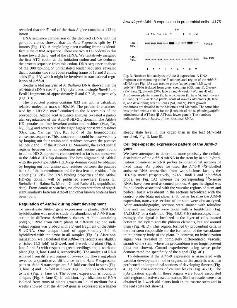
Fig. 3. Northern blot analysis of Athb-8 expression. A DNAfragment corresponding to the 5′ untranslated region of the Athb-8cDNA (see Fig. 1A) was used to probe (upper panel) 2.5 µg ofpoly(A)+ RNA isolated from green seedlings (GS, lane 1), 2-week(2W, lane 2), 3-week (3W, lane 3) and 4-week (4W, lane 4) oldArabidopsis plants, stems (S, lane 5), leaves (L, lane 6), and flowers(F, lane 7) of 5-week old plants, roots of 4-week old plants (R, lane8) and developing green siliques (Sil, lane 9). Plant growthconditions are detailed in the Materials and Methods. The same blotwas probed with a cDNA for the β subunit of the N. plumbaginifoliamitochondrial ATPase (β-ATPase, lower panel). The numbersindicate the size, in bases, of the ribosomal RNAs.
revealed that the 5′ end of the Athb-8 gene contains a 413 bpintron.
DNA sequence comparison of the deduced cDNA with thegenomic clones showed that the Athb-8 gene is split by 17introns (Fig. 1A). A single long open reading frame is identi-fied in the cDNA sequence. There are two ATG codons in thisframe toward the 5′ end of the cDNA. We tentatively assignedthe first ATG codon as the initiation codon and we deducedthe protein sequence from this codon. DNA sequence analysisof the 308 bp-long 5′ untranslated leader sequence revealedthat it contains two short open reading frame of 13 and 3 aminoacids (Fig. 2A) which might be involved in translational regu-lation of Athb-8.
Southern blot analysis of A. thaliana DNA showed that thep3′Athb-8 cDNA (see Fig. 1A) hybridizes to single BamHI andEcoRI fragments of approximately 5 and 0.7 kb, respectively(Fig. 1B).
The predicted protein contains 833 aas with a calculatedrelative molecular mass of 92x103. The protein is character-ized by a HD-Zip motif confined to the N terminus of thepolypeptide. Amino acid sequence analysis revealed a partic-ular organization of the Athb-8 HD-Zip domain. The Athb-8HD contains the four invariant amino acid residues (W48, F49,N51, R53) and seven out of the eight highly conserved residues(Q12, L16, Y20, I40, V47, R55, K57) of the homeodomainconsensus sequence. This conservation could be observed onlyby looping out four amino acid residues between the putativehelices 2 and 3 of the Athb-8 HD. Moreover, the exact spatialregister between the homeodomain and leucine zipper foundin all the HD-Zip proteins characterized so far is not conservedin the Athb-8 HD-Zip domain. The best alignment of Athb-8with the prototype Athb-1 HD-Zip domain could be obtainedby looping out four amino acid residues between the putativehelix 3 of the homeodomain and the first leucine residue of thezipper (Fig. 2B). The DNA binding properties of the Athb-8HD-Zip domain will be published elsewhere (Sessa G.,Steindler C., Baima S., Morelli G. and Ruberti I., unpublisheddata). From database searches, no obvious stretches of signif-icant similarity between Athb-8 and other known proteins havebeen found.
Regulation of Athb-8 during plant developmentTo investigate Athb-8 gene expression in plants, RNA blothybridization was used to study the abundance of Athb-8 tran-scripts in different Arabidopsis tissues. A blot containingpoly(A)+ RNA from entire plants of different age and indi-vidual organs was probed with a 5′ end fragment of the Athb-8 cDNA. One unique band of approximately 3.4 kbhybridized with the probe in all samples (Fig. 3). After nor-malization, we calculated that Athb-8 transcripts are slightlyenriched (1.3 fold) in 2-week and 3-week old plant (Fig. 3,lane 2 and 3) with respect to green seedlings and 4-week oldplant (Fig. 3, lane 1 and 4, respectively). The analysis of RNAisolated from different organs of 5-week old flowering plantsrevealed a quantitative difference in the Athb-8 expressionpattern. Athb-8 transcripts are 5.8-fold enriched in stem (Fig.3, lane 5) and 1.5-fold in flower (Fig. 3, lane 7) with respectto leaf (Fig. 3, lane 6). The lowest expression is found insiliques (Fig. 3, lane 9). In addition, hybridization of RNAisolated from roots of plants grown on liquid medium for 4weeks showed that the Athb-8 gene is expressed at a higher
steady state level in this organ than in the leaf (4.7-foldenriched, Fig. 3, lane 8).
Cell type-specific expression pattern of the Athb-8geneWe have attempted to determine more precisely the cellulardistribution of the Athb-8 mRNA in the stem by in situ hybrid-ization of anti-sense RNA probes to longitudinal sections offixed tissue. As probes we used the Athb-8 35S-labelledantisense RNA, transcribed from two subclones lacking theHD-Zip motif (respectively, p71∆ HindIII and p3′Athb-8probe, see Fig. 1A) whereas the corresponding 35S-senseRNAs have been used as control probes. A specific signal wasfound clearly associated with the vascular regions of stem andpedicel, but it was absent in the sections hybridized with thecontrol probe (data not shown). To better localize the Athb-8expression, transverse sections of the stem were also analyzed.After autoradiography, sections were stained with toluidineblue and micrographs were taken with a bright-field (Fig.4A,D,E,G) or a dark-field (Fig. 4B,C,F,H) microscope. Inter-estingly, the signal is localized to the layer of cells locatedbetween the xylem and the phloem elements, but not aroundthem (Fig. 4B,D). This region, formed by procambial cells, isthe meristem responsible for the formation of the vasculaturein the primary body of the plant. In contrast, no hybridizationsignal was revealed in completely differentiated vascularstrands of the stem, where the procambium is no longer present(data not shown). Control experiments using sense probedemonstrated the specificity of the signal (Fig. 4C).
To determine if the Athb-8 expression is associated withvascular development in other organs, in situ analysis was alsoperformed on longitudinal sections of developing flowers (Fig.4E,F) and cross-sections of cauline leaves (Fig. 4G,H). Thehybridization signals in these organs were found associatedwith regions of active vascularization. Similar results were alsoobtained in 2-week old plants both in the rosette stem and inthe leaf (data not shown).

4176 S. Baima and others
Fig. 4. Distribution of Athb-8mRNA in flowering stem,flower and cauline leaf. Stemsfrom flowering plants werecut below the inflorescence,fixed and paraffin embedded.Sections were hybridized with35S-labelled RNAs transcribedfrom the p71∆HindIII clone(Fig. 1A) in the antisense(A,B,D,E,F,G,H) or in thesense (C) orientation. Highlyrefrangible, purple structuresare xylematic elements.Similar results were alsoobtained with the p3′Athb-8probe. (A,B,C) Transversesections of an elongatingstem. Bar, 50 µm. (D) Detailed view of Bshowing a developingvascular bundle where theprocambium is clearlyvisualized. Bar, 10 µm. (E,F) Longitudinal sectionthrough a developing floralbud. Arrows indicate regionsof active vascularization inreceptacle, ovary and antherfilament. Bar, 50 µm. (G,H) Cross-section of acauline leaf. A lateral vein inthis section is in the medialposition. The hybridizationsignal between xylem andphloem is shown by an arrow.Bar, 20 µm. (A,D,E,G)Bright-field micrographs.(B,C,F,H) Dark-fieldmicrographs.
Several genes have been shown to be expressed in regionsof the meristems, as well as in procambium. To test whetherAthb-8 expression is procambium specific, as opposed tomeristem specific, we analyzed longitudinal sections of
vegetative and floral apices. No hybridization signal wasdetected in these two meristems. Instead, it was possible todetect a low hybridization signal on a side of the vegetativemeristem where the initiation of the procambium in associ-

4177Arabidopsis Athb-8 expression in procambial cells
Fig. 5. Athb-8 gene expressionin root and embryo.(A) Longitudinal sectionthrough the root meristem andthe elongation zone.(B,C) Transverse section of theelongation zone.(D) Longitudinal section of alinear cotyledon stage (torpedo)embryo. (A,B,D) Bright-fieldmicrographs. (C) Dark-fieldmicrograph. Bar, 10 µm.
ation with the young leaf primordium occurs (data not shown).
A similar analysis was also performed on longitudinal andtransverse sections of root apices. In situ hybridizationrevealed Athb-8 mRNA localization in a region correspond-ing to the meristem and elongation zone (Fig. 5A). Thespecific signal in the elongation zone of the root is clearlyassociated with the central cell file corresponding to the pro-cambium (Fig. 5B, C). During the analysis we occasionallyobserved lateral root primordia. Lateral root formation beginsby periclinal and anticlinal divisions in pericycle cells of theprimary root (Dolan et al., 1993). Upon formation of a dome-shaped structure (primordium) by pericycle derivatives, anew root meristem is derived from a subset of these cells. Asignificant level of Athb-8 expression is visible (just abovethe periphery of the vascular cylinder) in the center of the pri-mordium (data not shown).
Finally, we have performed in situ hybridization experi-ments to determine if the Athb-8 gene is transcribed duringembryogenesis. Analysis of longitudinal sections of siliquesrevealed a low hybridization signal in the central region of theembryo at the linear cotyledon stage (Fig. 5D). This group of
narrow elongated cells is the procambium, which extend intothe cotyledons in this stage embryo.
To characterize further the cell-specific expression of theAthb-8 gene, we analyzed the expression of the Athb-8-GUSchimeric gene in Arabidopsis. For this purpose, we fused the1.7-kb DNA fragment lying directly upstream from the firstATG of the Athb-8 coding region in frame with the GUSreporter gene (Jefferson et al. 1987). The entire leadersequence of the Athb-8 mRNA, which contains two short openreading frames and an intron, was included in the construct. Insuch a construct, GUS should undergo the same transcriptionaland translational regulation as Athb-8. Transgenic Arabidopsisseedlings harboring the chimeric Athb-8-GUS gene wereanalyzed by histochemical staining. In cotyledons GUSstaining is visible along vascular strands (Fig. 6A), which areeasily identified by the autofluorescence of tracheid cells (Fig.6B). The GUS staining is also visible in regions of cotyledonsand leaflets where the vasculature will be formed (indicated byarrows, Fig. 6A). In the primary root, a specific GUS stainingis found in the meristem and in the elongation zone, and it isassociated with central files of cells corresponding to the pro-cambium and the columella (Fig. 6C).
Fig. 6. Athb-8-GUS geneexpression in transgenicArabidopsis plants.Histochemical localization ofGUS activity in (A,B)cotyledons and (C) primaryroot. The arrows in A and Bindicate regions where maturevascular elements have not yetbeen formed. (A) Bright-fieldmicrograph. (B) Dark-fieldmicrograph. The scale barrepresents 100 µm in A and Band 10 µm in C.

4178 S. Baima and others
Athb-8 is expressed during the early stages ofrevascularizationOne of the most widely used approaches to study determinantsof vascular differentiation is the analysis of revascularizationafter severing vascular bundles. We therefore decided to inves-tigate Athb-8 expression under these particular experimentalconditions.
As a convenient model system to address this question, wehave chosen transgenic tobacco plants harboring the Athb-8-GUS chimeric gene. In fact, tobacco is easy to manipulate inwounding experiments, and Athb-8 expression can be easilymonitored by histochemical detection of GUS activity.
To ascertain if the Athb-8 promoter sequence chosen wasalso active in transgenic tobacco plants, we analyzed GUSexpression by histochemical staining (Fig. 7). GUS staining isvisible in the vascular system of seedlings (Fig. 7A), in theelongation zone of the primary root (Fig. 7B) and in lateral rootprimordia (Fig. 7C). Moreover, GUS activity is detectable inembryos starting at the heart stage (Fig. 7D). Therefore theexpression pattern of the chimeric gene in tobacco is coinci-dent with that of the Athb-8 gene in Arabidopsis.
To determine whether the Athb-8 promoter is active duringrevascularization, we analyzed young internodes wounded bya double cut. The two partially overlapping transverse cuts(about 3 mm apart) severed all the vascular bundles but main-tained a narrow bridge of pith cells connecting the apical andbasal portion of the stem. After wounding the apical portion
Fig. 7. Athb-8-GUS gene expression in transgenic tobacco plants. Histoc(C) lateral root, (D) heart stage embryo. (B,C) Dark-field micrographs. T
withered and stopped growing but it regained a healthy appear-ance in approximately 7 days, suggesting that functional con-nections had been re-established. We followed the expressionof Athb-8-GUS gene in wounded stems by histochemicalstaining at different time points after cutting. Unwoundedstems do not show any specific staining (Fig. 8A,B). Con-versely, stems stained 1 hour after the wound appeareddiffusely blue coloured indicating that the Athb-8 promoter iswound inducible (Fig. 8C). A rapid induction of Athb-8 uponwounding was also observed in leaves in both tobacco and Ara-bidopsis transgenic plants (data not shown). Eight hours afterwounding GUS activity was lower and the pattern of bluestaining was changed. GUS activity was mainly restricted tothe portion of the stem above the cut region (Fig. 8D). Thestaining of a straight and narrow portion of the bridge showsthat the expression is maintained preferentially on the side ofthe nearest upper leaf trace (Fig. 8D). Sometimes we found thatthe GUS activity is retained in the basal portion of the stemaround the uncut vascular bundles (Fig. 8D). However, instems in which the upper cut severed all the vascular bundlesof the nearest leaf no staining was visible in the bridge (datanot shown). About 7 days after wounding the expression ofGUS activity in recovered plants was mainly present in thebridge and in a narrow region immediately above the marginof the upper cut (Fig. 8E).
To understand better the localization of Athb-8-GUSexpression we analyzed in more detail the wounded region of
hemical localization of GUS activity in (A) seedling, (B) primary root,he scale bar represents 1 mm in A and 100 µm in B,C and D.

4179Arabidopsis Athb-8 expression in procambial cells
recovered stems. The analysis of transverse sections of thestem showed that new vessels with typical lignified secondarywalls had been formed in the pith from parenchyma cells whichhad undergone cell division and redifferentiation (Fig. 8F,G).Many small, intensely blue stained cells resembling a woundcambium are often present near the new rows of trachearyelements (Fig. 8G). Although new vessel elements differenti-ate preferentially in close proximity to the old vascular system,we focused our attention on more clearly distinguishablevascular elements that are formed in the pith as isolated cells,
small clusters or strands. Sections of stems at earlier phases ofrecovery showed that many single or grouped parenchymacells positively stain for GUS activity (Fig. 8H). These cellsare most likely vascular cell precursors at very early stages ofdifferentiation because we occasionally observed cells whichhad both a pale blue colour and the typical pattern of secondarywall deposition (Fig. 8I).
From these experiments we can therefore conclude thatAthb-8 gene is expressed at early stages of vascular cells deter-mination during revascularization.
Fig. 8. Athb-8 -GUS geneexpression during woundrecovery. Localization of GUSactivity in control (A,B) andwounded stems 1 hour (C), 8hours (D) and 1 week (E-I) aftercutting. (A,C,D,E) Outsideview. All the stems are orientedwith the apical end upward. Anarrow indicates the position ofthe leaf trace in D. (B) Hand-cuttransverse section of the controlstem. (F-I) Hand-cut transversesections through the bridgeregion. A partially differentiatedtracheary element showing apale blue colour is indicated byan arrowhead in I. P,parenchyma; TE, trachearyelements; V, vascular ring; WC,wound cambium. The scale barrepresents 500 µm in A, C, D, Eand 100 µm in F-I.

4180 S. Baima and others
Fig. 9. The expression of the Athb-8 gene is regulated by auxin.Hormone-depleted leaf samples were incubated for 1 hour in thepresence of 10−5-10−9 M IAA (lanes 2-6) or in mock buffer (lane 1).Northern blot conditions were as described in Fig. 3 except that 10µg of total RNA were loaded in each lane.
Auxin induces Athb-8 mRNA accumulationIt is very well known that auxins are hormones that regulatevarious aspects of plant growth and development, such as cellelongation, cell division, cell differentiation and morphogen-esis (review by Estelle, 1992). The formation of the vascularapparatus involves several of these aspects and there is con-vincing evidence that an auxin flux is responsible for theorderly pattern of vascular differentiation from leaves to root(Sachs, 1969; 1981). Moreover, the stimulus for vascular red-ifferentiation after wounding is thought to be the auxin releasedfrom the severed bundle itself (Steeves and Sussex, 1989).Therefore, we analyzed the effect of auxin on the expressionof the Athb-8 gene. In a preliminary experiment leaf tissueswere incubated in the presence of 50 µM indol-3-acetic acid(IAA) for 30 minutes and 1 hour and the correspondent RNAswere analyzed by northern blot (see Materials and Methods).The result obtained revealed a higher steady state level of Athb-8 transcripts in the auxin-treated tissues with respect to thecontrol samples, with a higher accumulation after 1 hour (datanot shown).
To study dose-dependent accumulation of Athb-8 mRNA,leaf tissues were incubated for 1 hour with 10−5-10−9 M IAAand the correspondent RNAs were analyzed. A 2.6- and a 4-fold increase in the steady state level of Athb-8 mRNA occursin leaves treated with 10−6 and 10−5 M IAA, respectively (Fig.9). Similar results were also obtained with the synthetic auxinsnaphthalenelacetic acid and 2,4-dichlorophenoxyacetic acid,while no induction was observed with 2-(p-chlorophenoxy)-isobutyric acid, an auxin analog without biological activity(data not shown).
DISCUSSION
In this study, we describe the characterization of an Ara-bidopsis gene, Athb-8, encoding a homeodomain-leucinezipper protein of 833 amino acids which belongs to the HD-ZIP III family previously described (Sessa et al., 1994).
Homeobox genes in plants, by analogy to the functionalroles of the products of animal HB genes, are thought to codefor transcriptional regulators which mediate important devel-opmental processes. This hypothesis has been supported by the
analysis of the maize KNOTTED-1 (KN1) gene, the firstisolated and well characterized plant HB gene (Hake, 1992).
So far, no mutationally defined loci can be correlated withgenes encoding HD-Zip factors although a less conservedleucine-zipper domain is present in Athb-10 (Sessa et al., 1994)which corresponds to GLABRA2, a homeodomain proteininvolved in trichome development (Rerie et al., 1994). Inter-estingly, outside the HD-Zip region we found that Athb-8shares some homologies with GLABRA2. The completealignment of Athb-8 with GLABRA2 will be publishedelsewhere (Sessa G., Steindler C., Baima S., Morelli G. andRuberti I., unpublished data). In addition, the expressionanalysis of Athb-1 (Aoyama et al., 1993), Athb-2/HAT4 andAthb-4 (Carabelli et al., 1993; Schena et al., 1993) in Ara-bidopsis and in transgenic plants suggested that HD-Zipproteins may control important aspects of plant development.
Here, we show that Athb-8 expression is associated with thedevelopment of the vascular system. Vascular developmentinvolves first the formation from the apical meristems ofspecific cells (procambium) which are committed at some pointto differentiate into xylem and phloem. Subsequently, itinvolves the cytodifferentiation of these procambial cells intomore specialized vascular cells (Aloni, 1987; Esau, 1977;Shininger, 1979). In the young, terminal regions of the stemand in differentiating leaves the procambium consists ofdiscrete cellular strands composed of elongated cells. In theroots, the procambium is a core of tissue from which thevascular cylinder originates. So far, procambial cells have beendescribed only cytologically and there are no specific bio-chemical characteristics attributable to this cell type.Moreover, it is not possible to define the stage at which thecells are committed to xylem or phloem differentiation unlessthey have clearly differentiated. Various efforts have beenmade to identify specific markers of vascular development.Recently, many cDNA clones for genes expressed preferen-tially in cells that redifferentiate into tracheary elements havebeen isolated from Zinnia elegans (Demura and Fukuda, 1994).A genetic approach has been taken by different groups toisolate developmental mutants. Several of them have mutationsthat interfere with the normal pattern of vascular development(Bowman et al., 1994), but only the Arabidopsis wooden legmutant shows a specific defect in the vascular tissue (Schereset al., 1995).
We have examined the expression of Athb-8 in several plantorgans. RNA gel analysis and in situ hybridization studies ofAthb-8 expression in Arabidopsis led to the conclusion that thegene is mainly restricted to the procambium. These data havebeen confirmed also by histochemical localization of Athb-8-GUS expression in Arabidopsis and tobacco transgenic plants.By comparing the expression of the gene in the vegetativemeristems of the plant with that in the floral meristems and inthe embryos, we can draw the conclusion that the Athb-8expression is not linked to the mitotic activity per se, but it israther restricted to the procambial cells. Moreover, in all thetissues analyzed, the expression of Athb-8 in the vascularsystem is confined to procambial cells and it is never found interminally differentiated vascular cells.
Histological studies have revealed that progenitors of pro-cambium can generally be discerned during the transition fromthe globular to the heart stage of embryogenesis (review byWest and Harada, 1993). We were able to detect a low but sig-

4181Arabidopsis Athb-8 expression in procambial cells
nificant level of Athb-8 expression in the torpedo stage of theArabidopsis embryo. This means that Athb-8 is alwaysexpressed in provascular cells even when they are not differ-entiating into more mature vascular cells. Therefore, Athb-8might be considered a specific molecular marker of earlyvascular development. Consistent with this hypothesis is theobservation that the Athb-8-GUS chimeric gene is inducedearly during re-vascularization of wounded stems. When avascular bundle is severed, nearby parenchyma cells resumecell division and redifferentiate into both xylem and phloem(Steeves and Sussex, 1989). Therefore, differentiation oftracheary elements after wounding has been widely used as anexperimental model system to study vascular developmentwithout the superimposition of spatial and temporal factorssuch as in the apical meristem.
We cut internodes of Athb-8-GUS transgenic tobacco plantsso that only a narrow transverse bridge connected the apicaland basal portion of the stem. In this way, new trachearyelements were differentiated in the middle of the pith to forman horizontal vascular connection between the old, interruptedvascular strands. We were able to detect GUS staining in singleparenchyma cells of the bridge which were not distinguishablefrom the neighbour cells for any other morphological aspect.As we occasionally observed a lighter staining in singletracheary cells at early stages of differentiation, we concludedthat the single stained parenchyma cells in the pith are theprecursor of these vascular elements.
It is well known that auxin is the major signal involved inthe control of several aspects of plant vascular differentiation(Aloni, 1987). One of the main peculiarities of auxin is that,of all the known plant hormones, it is the only one that exhibitspolar transport (Kaldewey, 1984). At early stages of plantdevelopment, an auxin polar transport system may be involvedin the establishment of bilateral symmetry in embryos (Liu etal., 1993). In postembryonic development, young growingleaves or developing buds induce the formation of vasculartissue below them. In a series of elegant experimentsperformed in pea seedlings, Sachs provided evidence support-ing the hypothesis that an auxin flux determines the orderlypattern of vascular differentiation from leaves to root (Sachs,1969, 1981). In the ‘canalization hypothesis’ he proposed thatthe diffusion of auxin from an auxin source induces theformation of a polar auxin transport system along a narrow fileof procambial cells. The polar transport of auxin finally resultsin the formation of vascular strands (Sachs, 1981, 1991). Thishypothesis has been widely supported by experimental data onvascular redifferentiation in wounded stems (Gersani, 1985;Sachs, 1981; Sussex et al., 1972). In our experimental system,detection of GUS activity in stems at different times afterwounding revealed interesting features of the regulation ofAthb-8 expression. Athb-8 is strongly and rapidly (1 hour)induced at wound sites. Few hours later, the gene expressionbecome regulated in a polar fashion, being more expressed inthe apical than in the basal portion of the cut region. Moreover,the position of the leaf trace nearest the upper cut stronglyinfluenced the time of activation of Athb-8 expression in thebridge region. All these observations implicated auxin in theregulation of Athb-8 expression. In fact, it has been shown thatseveral auxin-regulated genes are also wound inducible (An etal., 1990; Eberner et al., 1993). It has been suggested thatflavonoids released after wounding may act as natural
inhibitors of auxin transport (Jacobs and Rubery, 1988),resulting in locally high auxin concentration. This fact mayexplain the dual regulation of the auxin-regulated genes andtherefore the wound inducible expression of the Athb-8-GUSgene. Moreover, the local increase of auxin descending alongthe leaf traces and coming out from severed vascular bundles(Gersani, 1985; Sussex et al., 1972) can well explain the polarand asymmetrical GUS staining of wounded Athb-8-GUSplants. A more direct confirmation that auxin might have a rolein regulating Athb-8 expression in wounded plants comes fromthe results obtained incubating Arabidopsis tissues in thepresence of IAA. In fact, a four-fold accumulation of the Athb-8 mRNA has been obtained in wounded leaves 1 hour after thebeginning of hormone treatment (Fig. 9). Similar results werealso obtained in cut flowering stem (S. B., unpublished data).
In conclusion, the expression pattern and the auxin regula-tion of the gene encoding Athb-8 suggest a role for this factorin provascular specification. A more detailed study aimed atthe functional assessment of Athb-8 in transgenic plants as wellas the analysis of mutants with specifically altered vasculardevelopment will help to define the role of Athb-8 in the deter-mination and differentiation of provascular cells and the regu-latory cascades in which this gene participates.
We wish to thank E. Meyerowitz for making available a detailedin situ hybridization protocol; M. Altamura and A. Spena for helpfulsuggestions. We would also like to thank Giuseppe Crocchioni,Roberto Gargamelli, Francesco Vignolini and Kerstin Langenkemperfor skilled technical assistance. S. B. was supported by a fellowship‘V.V. Landi’ of the Accademia Nazionale dei Lincei and was recipientof an EMBO Short-term fellowship. This work was supported, in part,by Piano Nazionale ‘Sviluppo di Tecnologie Avanzate Applicate allePiante’, Ministero Agricoltura e Foreste and by National ResearchCouncil of Italy, Special Project RAISA, Sub-project N.2, PaperN.2171.
REFERENCES
Affolter, M., Schier, A. and Gehring, W. J. (1990). Homeodomain proteinsand the regulation of gene expression. Curr. Opin. Cell Biol. 2, 485-495.
Aloni, R. (1987). Differentiation of vascular tissues. Annu. Rev. Plant Phys. 38,179-204.
An, G., Costa, M. A. and Ha, S. B. (1990). Nopaline synthase promoter iswound inducible and auxin inducible. Plant Cell 2, 225-233.
Aoyama, T., Dong, C.-H., Carabelli, M., Sessa, G., Ruberti, I., Morelli, G.and Chua, N.-H. (1993). Developmental regulation of gene expression inplant cells. In XV International Botanical Congress Abstract, p. 197.
Bechtold, N., Ellis, J., E. and Pellettier, G. (1993). In planta Agrobacteriummediated gene transfer by infiltration of adult Arabidopsis thaliana plants. C.R. Acad. Sci. Paris 316, 1194-1199.
Benfey, P. N. and Schiefelbein, J. W. (1994). Getting to the root of plantdevelopment: the genetics of Arabidopsis root formation. Trends Genet. 10,84-88.
Bowman, J. (1994). Arabidopsis. An Atlas of Morphology and Development.New York: Springer-Verlag.
Carabelli, M., Sessa, G., Baima, S., Morelli, G. and Ruberti, I. (1993). TheArabidopsis Athb-2 and -4 genes are strongly induced by far-red-rich light.Plant J. 4, 469-479.
Demura, T. and Fukuda, H. (1994). Novel vascular cell-specific genes whoseexpression is regulated temporally and spatially during vascular systemdevelopment. Plant Cell 6, 967-981.
Devereux, J. (1991). Program Manual, Sequence Analysis Software Package,Version 7. Madison: Genetics Computer Group.
Doerner, P. (1993). Patterning the Arabidopsis root. Curr. Biol. 3, 867-869.Dolan, L., Janmaat, K., Willemsen, V., Linstead, P., Poethig, S., Roberts,
K. and Scheres, B. (1993). Cellular organisation of the Arabidopsis thalianaroot. Development 119, 71-84.

4182 S. Baima and others
Drews, G. N., Bowman, J. L. and Meyerowitz, E. M. (1991). Negativeregulation of the Arabidopsis homeotic gene AGAMOUS by the APETALA2product. Cell 65, 991-1002.
Eberner, W., Fowler, T. J., Suzuki, H., Shaver, J. and Tierney, M. L.(1993). Expression of DcPRP1 is linked to carrot storage root formation andis induced by wounding and auxin treatment. Plant Physiol. 101, 259-265.
Esau, K. (1977). Anatomy of Seed Plants. New York: John Wiley & Sons.Estelle, M. (1992). The plant hormone auxin: insight in sight. BioEssays 14,
439-443.Gee, M. A., Hagen, G. and Guilfoyle, T. (1991). Tissue-specific and organ-
specific expression of soybean auxin-responsive transcripts GH3 andSAURS. Plant Cell 3, 419-430.
Gersani, M. (1985). Appearance of new transport capacity in wounded plants.J. Exp. Bot. 36, 1809-1816.
Hake, S. (1992). Unraveling the knots in plant development. Trends Genet. 8,109-114.
Horsch, R. B., Fry, J. E., Hoffmann, N. L., Eichholtz, D., Rogers, S.G. andFraley, R. T. (1985). A simple and general method for transferring genesinto plants. Science 227, 1229-1231.
Innis, M. I. and Gelfand, D. H. (1990). Optimization of PCRs. In PCRProtocols: A Guide to Methods and Applications (eds M. I. Innis, D. H.Gelfand, J. J. Sninsky and T. J. White), pp. 3-12. San Diego: Academic Press.
Jacobs, M. and Rubery, P. H. (1988). Naturally occurring auxin transportregulators. Science 241, 346-349.
Jefferson, R. A., Kavanagh, T. A. and Bevan M. W. (1987). GUS fusions: β-glucuronidase as a sensitive and versatile gene fusion marker in higherplants. EMBO J. 6, 3901-3907.
Johri, B. M. (1984). Embryology of Angiosperms. Berlin: Springer.Kaldewey, H. (1984). Transport and other modes of movement of hormones
(mainly auxins). In Hormonal Regulation of Development II (ed.T. K. Scott),pp. 80-148. Heidelberg: Springer-Verlag.
Kuhlemeier, C., Green, P. J. and Chua, N.-H. (1987). Regulation of geneexpression in higher plants. Ann. Rev. Plant Physiol. Plant Mol. Biol. 38,221-257.
Liu, C.-M., Xu, Z.-H. and Chua, N.-H. (1993). Auxin polar transport isessential for the establishment of bilateral symmetry during early plantembryogenesis. Plant Cell 5, 621-630.
Ma, H. (1994). The unfolding drama of flower development: recent resultsfrom genetic and molecular analyses. Genes Dev. 8, 745-756.
Mattsson, J., Soderman, E., Svenson, M., Borkird, C. and Engstrom, P.(1992). A new homeobox-leucine zipper gene from Arabidopsis thaliana.Plant Mol. Biol. 18, 1019-1022.
Mayer, U., Torres Ruiz, R. A., Berleth, T., Miséra, S. and Jurgens, G.(1991). Mutations affecting body organization in the Arabidopsis embryo.Nature, 353, 402-407.
Medford, J. I. (1992). Vegetative apical meristems. Plant Cell 4, 1029-1039.Meyerowitz, E. M. (1994). Pattern formation in plant development: four
vignettes. Curr. Opin. Gen. Dev. 4, 602-608.Rerie, W. G., Feldmann, K. A. and Marks, M. D. (1994). The GLABRA2
gene encodes a homeodomain protein required for normal trichomedevelopment in Arabidopsis. Genes Dev. 8, 1388-1399.
Ruberti, I., Sessa, G., Lucchetti, S. and Morelli, G. (1991). A novel class ofplant proteins containing a homeodomain with a closely linked leucine zippermotif. EMBO J. 10, 1787-1791.
Sachs, T. (1969). Polarity and the induction of organized vascular tissues. Ann.Bot. 33, 263-275.
Sachs, T. (1981). The control of the patterned differentiation of vasculartissues. Adv. Bot. Res. 9, 151-262.
Sachs, T. (1991). Cell polarity and tissue patterning in plants. DevelopmentSupplement 1, 83-93.
Scheres, B., Di Laurenzio, L., Willemsen, V., Hauser, M-T., Janmaat, K.,Weisbeek, P. and Benfey, P. N. (1995). Mutations affecting the radialorganisation of the Arabidopsis root display specific defects throughout theembryonic axis. Development 121, 53-62.
Schena, M. and Davis, R. W. (1992). HD-Zip proteins: Members of anArabidopsis homeodomain protein superfamily. Proc. Natl. Acad. Sci. USA89, 3894-3898.
Schena, M., Lloyd, A. M. and Davis, R. W. (1993). The HAT4 gene ofArabidopsis encodes a putative developmental regulator. Genes Dev. 7, 367-379.
Schena, M. and Davis, R. W. (1994). Structure of homeobox-leucine zippergenes suggests a model for the evolution of gene families. Proc. Natl. Acad.Sci. USA 91, 8393-8397.
Schiefelbein, J. W. (1994). Cell fate and cell morphogenesis in higher plants.Curr. Opin. Gen. Dev. 4, 647-651.
Sessa, G., Morelli, G. and Ruberti, I. (1993). The Athb-1 and -2 HD-Zipdomains homodimerize forming complexes of different DNA bindingspecifities. EMBO J. 12, 3507-3517.
Sessa, G., Carabelli, M., Ruberti, I., Lucchetti, S., Baima, S. and Morelli,G. (1994). Identification of distinct families of HD-ZIP proteins inArabidopsis thaliana. In Molecular-Genetic Analysis of Plant Developmentand Metabolism (eds. P. Puigdomenech and G. Coruzzi), pp. 411-426.Berlin: Springer.
Shininger, T. (1979) The control of vascular development. Annu. Rev. PlantPhys. 30, 313-337.
Soderman, E., Mattsson, J., Svenson, M., Borkird, C. and Engstrom, P.(1994). Expression patterns of novel genes encoding homeodomain leucine-zipper proteins in Arabidopsis thaliana. Plant Mol. Biol. 26, 145-154.
Steeves,T. A. and Sussex, I. M. (1989). Patterns in Plant Development, 2nded. Cambridge: Cambridge University Press.
Sussex, I. M., Clutter, M. E. and Goldsmith, H. M. (1972). Wound recoveryby pith cell redifferentiation: structural changes. Amer. J. Bot. 59, 797-804.
Weigel, D. (1993). Patterning the Arabidopsis embryo. Curr. Biol. 3, 443-445.West, M. A. L. and Harada, J. J. (1993). Embryogenesis in higher plants: an
overview. Plant Cell 5, 1361-1369.
(Accepted 14 August 1995)