the factor v-activating enzyme (rvv-v) from russell's viper venom
TRANSCRIPT

THE JOURNAL OF BIOLOGICAL CHEMISTRY 0 1988 by The American Society for Biochemistry and Molecular Biology, Inc.
Vol. 263, No. 33, Issue of November 25, PP. 17471-17481,1988 Printed in U.S.A.
The Factor V-activating Enzyme (RVV-V) from Russell’s Viper Venom IDENTIFICATION OF ISOPROTEINS RVV-Va, -Vg, AND -Vy AND THEIR COMPLETE AMINO ACID SEQUENCES*
(Received for publication, June 16, 1988)
Fuminori TokunagaS, Katsuaki NagasawaQ, Shigehiko Tarnurag, Toshiyuki MiyataQ, Sadaaki IwanagaQll, and Walter KisielII From the $Department of Biology, Faculty of Science, Kyushu University 33 and the $Department of Molecular Biology, Graduate School of Medical Science, Kyushu University, Fukuokn 812, Japan and the 11 Department of Pathlogy, Blood Systems Research Foundation Laboratory, University of New Mexico, School of Medicine, Albuquerque, New Mexico 87131
The complete amino acid sequences of two isopro- teins of the factor V-activating enzyme (RVV-V) iso- lated from Vipera rwselli (Russell’s viper) venom were determined by sequencing S-pyridylethylated deriva- tives of the proteins and their peptide fragments gen- erated by either chemical (cyanogen bromide and 242- nitrophenylsulfenyl)-3-methyl-3-bromoindolenine) or enzymatic (trypsin, a-chymotrypsin, and lysyl endo- peptidase) cleavages. Both enzymes, designated RVV- Va and RVV-Vy, consist of 236 amino acid residues and have a N-linked oligosaccharide chain at AsnZzs. The six amino acid substitutions between RVV-Va and -Vy are: Thrzz(a)-Alaz”(y), Glyze(a)-Alazs(y), Gln”’(a)- Glu’”(y), Ile’e2(a)-Met’92(y), Gln’gS(a)-His’eS(y), and Asnzz4(a)-SerZz4(y). The molecular weights were cal- culated as 26,182 for RVV-Va and 26,167 for RVV- Vy. The sequences of the RVV-V isoproteins exhibited 62% identity with that of batroxobin, a thrombin-like enzyme present in Bothrops atrox venom, and 33% identity with that of human thrombin B chain. The most interesting difference between the structures of RVV-V and other trypsin-type serine proteases is that the conservative Ser214-Trp216-Gly2’a sequence (chy- motrypsinogen numbering), considered as the site of antiparallel B-sheet formation between the protein sub- strate and most serine proteases, has been replaced by the corresponding sequence Ala-Gly-Gly.
~ ~~ ~ ~ _ _ _ _ _ _____ ~~ ~ ~ ~ ~
Vipera russelli (Russell’s viper) venom contains two pro- teases, designated RVV-V’ and RVV-X, which induce the coagulation of mammalian plasma. RVV-X activates factors X and IX by limited proteolysis in a calcium-dependent manner (1-3), whereas RVV-V activates factor V in a calcium- independent manner (4-7). RVV-V has been purified to ho- mogeneity by Kisiel (8) and Kisiel and Canfield (9) and
* This work was supported by a grant-in-aid from the Ministry of Education, Science, and Culture of Japan (to S. I.) and by a grant from Blood Systems, Inc. (to W. K.). The costs of publication of this article were defrayed in part by the payment of page charges. This article must therefore be hereby marked “advertisement” in accord- ance with 18 U.S.C. Section 1734 solely to indicate this fact.
llTo whom correspondence should be addressed Dept. of Biology, Faculty of Science, Kyushu University 33,6-10-1 Hakozaki, Higashi- ku, Fukuoka 812, Japan.
The abbreviations used are: RVV-V, the factor V-activating en- zyme from Russell’s viper venom; Pe-, S-pyridylethylated; HPLC, high performance liquid chromatography; SDS-PAGE, sodium do- decyl sulfate-polyacrylamide gel electrophoresis; BNPS-skatole, 2-(2- nitrophenylsulfenyl)-3-methyl-3-bromoindolenine; TFA, trifluoroa- cetic acid.
characterized to be a glycoprotein containing 6% carbohydrate with an apparent molecular weight of 29,000. Esmon and Jackson (6) have reported that RVV-V exhibits arginine esterase activity and is sensitive to the serine protease inhib- itor diisopropyl fluorophosphate. Moreover, the purified prep- aration of RVV-V activates factor V to the same extent as a- thrombin. Unlike a-thrombin or thrombocytin, a factor V- activating enzyme from Bothrops atrox venom (9), RVV-V shows no apparent effects on factor VI11 (5), factor XI11 (9), fibrinogen (6), or prothrombin (6). Whereas the activation of factor V with a-thrombin results in the cleavage of three peptide linkages and the release of the large connecting frag- ment, the RVV-V-mediated activation of factor V occurs as a result of a single cleavage in factor V, generating the heavy and light chains of factor V, (10). Furthermore, it has recently been shown that RVV-V cleaves the Arg’545-Ser1546 linkage in the human factor V molecule (11, 12). This peptide bond is one of the three thrombin cleavage sites.
Since RVV-V is a specific activator of factor V, we deter- mined the entire amino acid sequence of RVV-V in order to extend our information concerning the substrate specificity relationships of serine proteases. In the course of this study, we resolved three isoproteins of RVV-V, designated RVV-Va, -V@, and -Vy, and determined the complete sequences of the two major isoenzymes, RVV-Va and -Vy.
MATERIALS AND METHODS AND RESULTS~
NH2-terminul Sequence and Cyanogen Bromide Cleavage- The NH2-terminal sequence of Pe-RVV-V allowed identifi- cation of the first 20 residues (Table M2). Subsequently, Pe- RVV-V (20 nmol) was treated with CNBr, and the resulting fragments were separated by gel filtration using a TSK G2000SW column and reverse-phase HPLC, as shown in Figs. M1 and M2. According to previous reports (8, 9), RVV-V contains 3 Met residues; and thus, four CNBr fragments (CN1-CN4) were expected. However, the amino acid compo- sitions and the NHz-terminal sequences of the isolated CNBr fragments, as shown in Tables M1 and M2, indicated that the RVV-V preparation contains at least two molecules, one of which contains 3 Met residues, whereas the other has an additional Met residue near the COOH-terminal end of the molecule. The evidence was as follows. 1) The NH2-terminal sequences of CN4t and CN4a are identical, and that of CN4b
Portions of this paper (including “Materials and Methods,” part of “Results,” Figs. Ml-Ml5, and Tables I and M1-M21) are presented in miniprint at the end of this paper. Miniprint is easily read with the aid of a standard magnifying glass. Full size photocopies are included in the microfilm edition of the Journal that is available from Waverly Press.
17471
17472 Complete Amino Acid Sequence of RVV- V
is homologous to the COOH-terminal sequence of other serine proteases; 2) the sum of the total amino acid residues of CN4a and CN4b is in good agreement with that of CN4t; and 3) CN4a, but not CN4t, contains a homoserine residue, and both CN4t and CN4b have an oligosaccharide unit.
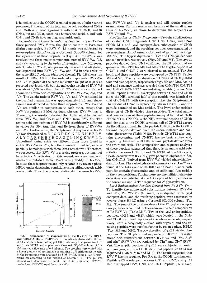
Separation and Characterization of Isoproteins of R VV- V- Since purified RVV-V was thought to contain at least two distinct molecules, Pe-RVV-V (13 nmol) was subjected to reverse-phase HPLC using a Cosmosil 5C4-300 column for the separation of these isoproteins (Fig. lA). Pe-RVV-V was resolved into three major components, named RVV-Va, -Vp, and -Vy, according to the order of retention time. Moreover, intact native RVV-V, not previously reduced and alkylated, was separated into RVV-Vy, -Va, and -Vp in this order by the same HPLC column (data not shown). Fig. 1B shows the result of SDS-PAGE of the isolated components. RVV-Va and -Vy migrated at the same molecular weight of 29,000 as reported previously (8), but the molecular weight of RVV-Vp was about 1,500 less than that of RVV-Va and -Vy. Table I shows the amino acid compositions of Pe-RVV-Va, -Vp, and -Vy. The weight ratio of RVV-Va, -Vp, and -Vy contained in the purified preparation was approximately 2:1:6; and gluco- samine was detected in these three isoproteins. RVV-Va and -Vy are similar in composition to each other, except that RVV-Va contains 3 Met residues, whereas RVV-Vy has 4. Therefore, the results indicated that CN4t must be derived from RVV-Va, and CN4a and CN4b from RVV-Vy. The amino acid composition of RVV-Vp is significantly different in values for Gly, Arg, Thr, and Ile from those of RVV-Va and -Vy. Furthermore, the NH2-terminal sequence of RVV- VP was determined as: V-I-G-G-D-E-C-N-I-N-E-H-P-F-L-V-
Underlined amino acids are different from those found in either RVV-Va or -Vy, but the amino-terminal sequence is partially homologous with them (data not shown). Therefore, we suspected that RVV-Vp is a serine protease, but may not be a factor V activator. In this regard, we were unable to assess the putative factor V-activating ability in RVV-Vp because these isoproteins are only separable by reverse-phase HPLC under denaturing conditions using trifluoroacetic acid/ acetonitrile. Thus, the precise relationship between RVV-Vp
A-L-Y-T-A-R-S-K-R-F-H-C-A-G-T-L-I-N-Q-E-W-V-L-T. - _ "_
A
.l. 0.48 t pe-"-v'l I
0
- a - 3 0 k D a
-20.1 kDs
-14.4kDa
FIG. 1. Separation of isoproteins of Pe-RVV-V by HPLC and SDS-PAGE. A, Pe-RVV-V (13 nmol) was dissolved in 0.8 ml of 10 mM phosphate buffer, pH 6.0, containing 6 M guanidine HC1 and 1 mM EDTA and applied to a Cosmosil 5C4-300 column (4.6 X 150 mm) at a flow rate of 0.5 ml/min. The proteins were eluted with a linear gradient of acetonitrile containing 0.1% trifluoroacetic acid. B, the isoproteins were analyzed by SDS-PAGE using a 12.5% sepa- rating gel according to the method of Laemmli (17). The gel was stained with Coomassie Brilliant Blue R-250. Left lane, RVV-Va; center lane, RVV-Vp; right lane, RVV-Vy.
and RVV-Va and -Vy is unclear and will require further examination. For this reason and because of the small quan- tities of RVV-Vp, we chose to determine the sequences of RVV-Va and -Vy.
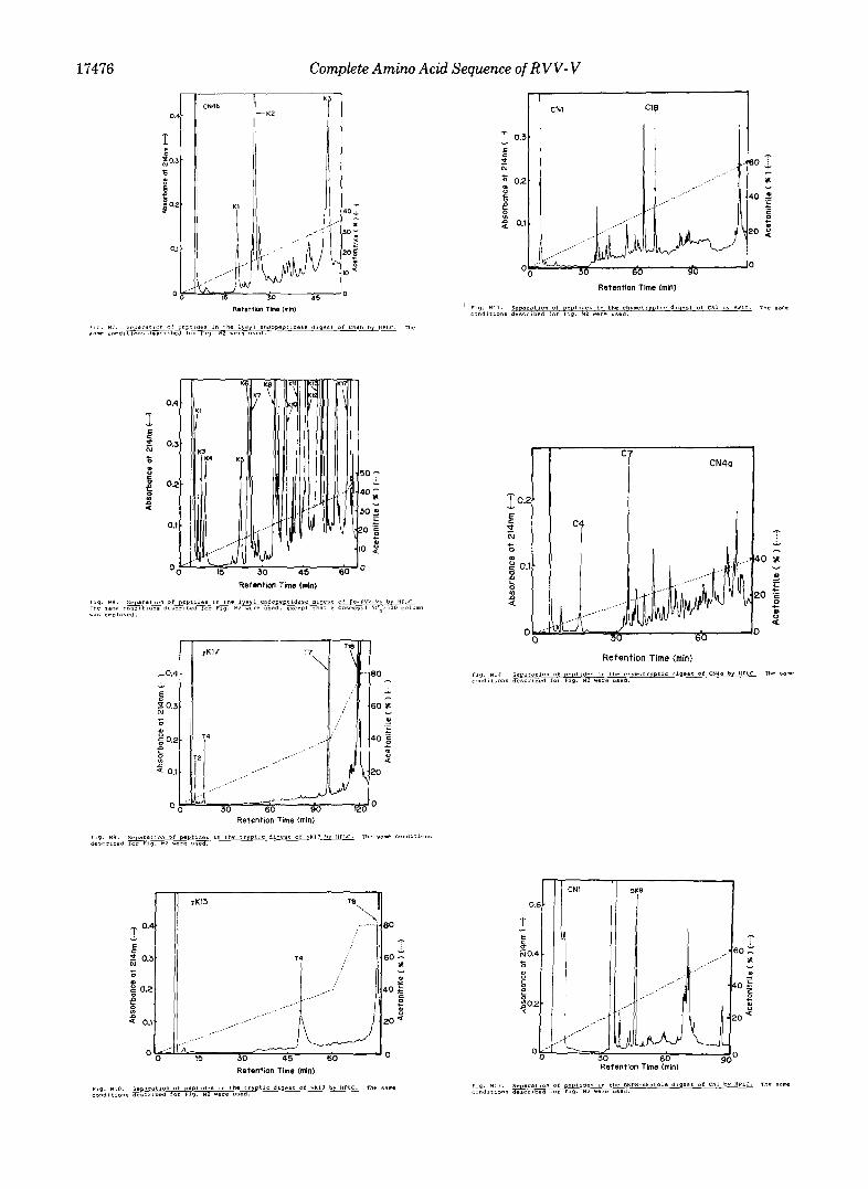
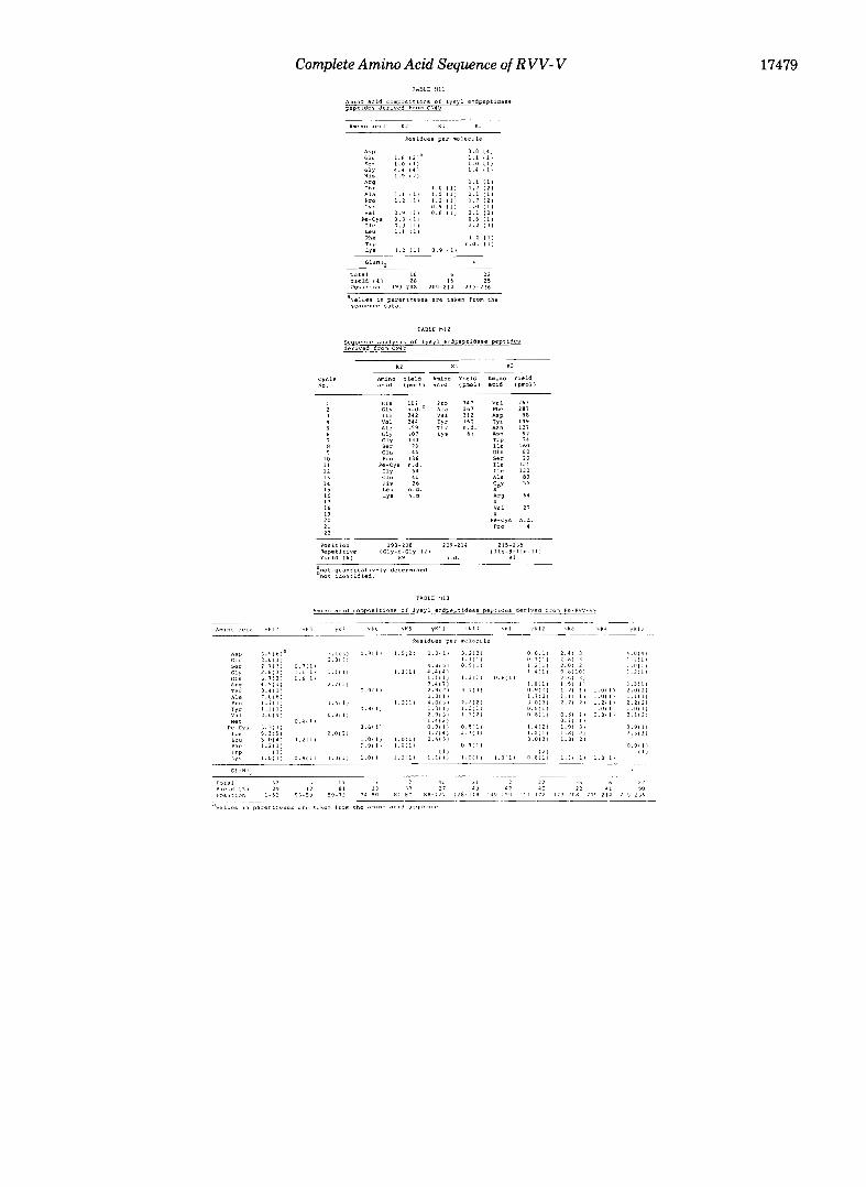
Subdigestion of CNBr Fragments-Trypsin subdigestions of isolated CNBr fragments CN2, CN3, CN4a, and CN4t (Table Ml), and lysyl endopeptidase subdigestion of CN4b were performed, and the resulting peptides were separated by reverse-phase HPLC using a Cosmosil 5Cls-P column (Figs. M3-M7). The tryptic digestion of CN2 and CN3 yielded four and six peptides, respectively (Figs. M3 and M4). The tryptic peptides derived from CN2 confirmed the NH2-terminal se- quence of CN2 (Tables M3 and M4). The tryptic peptides of CN3T11 and CN3T12 were cleaved at an unusual Tyr-Ser bond, and these peptides were overlapped by CN3T13 (Tables M5 and M6). The trypsin digestion of CN4a and CN4t yielded four and five peptides, respectively (Figs. M5 and M6). Amino acid and sequence analyses revealed that CN4aT14-CN4T13 and CN4aT19-CN4tT15 are indistinguishable (Tables M7- M10). Peptide CN4tT12 overlapped between CN4a and CN4b since the NH2-terminal half of this peptide was identical to that of CN4aT6, with the exception that the NH2-terminal His residue of CN4b is replaced by Gln in CN4tT12 and the peptide contained no Met residue. The lysyl endopeptidase digestion of CN4b yielded three peptides. The total amino acid compositions of these peptides are equal to that of CN4b (Table M11). CN4bK2 is the NHz-terminal peptide of CN4b and identical to the COOH-terminal half of CN4tT12 except for the NH2-terminal His. The peptide CN4bK3 is the COOH- terminal peptide derived from the entire molecule and con- tains glucosamine (Table M12). Peptide CN4tT18 also con- tains glucosamine, and CN4tT6 has no basic amino acid, suggesting that it is the COOH-terminal peptide derived from the entire molecule. The composition and sequence analyses of these peptides suggested that there is an amino acid sub- stitution between CN4bK3 and CN4tT18. At the 10th cycle, CN4b (derived from RVV-Vy) gave phenylthiohydantoin-Ser, but CN4tT18 (derived from RVV-Va) yielded phenylthiohy- dantoin-Asn. The carbohydrate attachment site at was found at the 15th cycle of CN4bK3 and CN4tT18 since both peptides contain glucosamine and an additional Asx residue in their compositions. Furthermore, no phenylthiohydantoin- derivative was detected at the 15th cycle of both peptides in the consensus Asn-X-Thr sequence for N-glycosylation.
Lysyl Endopeptidase Peptides Derived from Pe-RVV-Vy- To identify the amino acid substitutions between RVV-Va and -Vy, Pe-RVV-Vy (30 nmol) was digested with lysyl endopeptidase, and the resulting peptides were separated by reverse-phase HPLC using a Cosmosil 5C4-300 column (Fig. M8). The sum of the total residues of the 12 lysyl endopepti- dase peptides accounted for the entire amino acid composition of Pe-RVV-Vy (Table M13). Two of the lysyl endopeptidase peptides, yK17 and yK13, which were located in the NHz- and COOH-terminal peptides of the whole molecule, respec- tively, were subsequently cleaved with trypsin; and the re- sulting peptides were purified further by reverse-phase HPLC (Figs. M9 and M10). Tryptic digestion of yK17 yielded four peptides. The NH2-terminal sequence of yK17T18 revealed amino acid substitutions between RVV-Va and -Vy. Ala22 and Ala2' (RVV-Vy) are replaced by Thr2' and Gly9 (RVV- Va). The tryptic peptides of yK13 were subjected to amino acid analyses, and the COOH-terminal peptide yK13T4 was sequenced (Tables M15 and M16). The result suggested that RVV-V has the sequence Pro-Pro at the COOH-terminal end. Peptide yK3 overlapped between CN1 and CN2, and y K l l also overlapped CN2, CN3, and CN4. Peptide yK12 over-
Complete Amino Acid Sequence of R VV- V 17473
lapped between CN4aT19 and CN4aT9. The positions of peptides yK6, yK5, yK4, and yK13T8 were assigned from their amino acid compositions. Amino acid and sequence analyses of peptide yK8 revealed further that three additional amino acid substitutions are found between RVV-Va and -Vy. The sequence Gln'91-Ile192-Gln'93 of CN4tT12 derived from RVV-Va is replaced by G l ~ ' ~ ' - M e t ' ~ ~ - H i s ' ~ ~ of yK8 derived from RVV-Vy (Table M14).
DISCUSSION
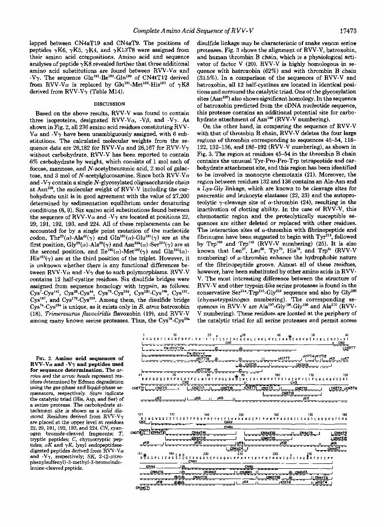
Based on the above results, RVV-V was found to contain three isoproteins, designated RVV-Va, -Vat and -Vy. As shown in Fig. 2, all 236 amino acid residues constituting RVV- Va and -Vy have been unambiguously assigned, with 6 sub- stitutions. The calculated molecular weights from the se- quence data are 26,182 for RVV-VCY and 26,167 for RVV-Vy without carbohydrate. RVV-V has been reported to contain 6% carbohydrate by weight, which consists of 1 mol each of fucose, mannose, and N-acetylneuramic acid, 2 mol of galac- tose, and 3 mol of N-acetylglucosamine. Since both RVV-Va and -Vy contain a single N-glycosylated oligosaccharide chain at AsnZz9, the molecular weight of RVV-V including the car- bohydrate unit is in good agreement with the value of 27,200 determined by sedimentation equilibrium under denaturing conditions (8,9). Six amino acid substitutions found between the sequence of RVV-Va and -Vy are located at positions 22, 29, 191, 192, 193, and 224. All of these replacements can be accounted for by a single point mutation of the nucleotide codon, Thr22(a)-Ala22(y) and Gln'g'(a)-Glu'g'(y) are at the first position, G13g(a)-Ala"(y) and A ~ n ~ ~ " ( a ) - S e r ~ ~ " ( y ) are at the second position, and Ile'92(a)-Met'92(y) and Gln'93(a)- Hislg3(y) are at the third position of the triplet. However, it is unknown whether there is any functional differences be- tween RVV-Va and -Vy due to such polymorphisms. RVV-V contains 12 half-cystine residues. Six disulfide bridges were assigned from sequence homology with trypsin, as follows:
CyP7, and C y ~ ' ~ ~ - C y s ~ ~ ~ . Among them, the disulfide bridge C y ~ ~ ' - C y s ~ ~ ~ is unique, as it exists only in B. atrox batroxobin (18), Trimeresurus flavoviridis flavoxobin (19), and RVV-V among many known serine proteases. Thus, the C y ~ ~ ~ - C y s ~ ~ "
Cys7-Cys'"', CysZ8-Cys44, Cys76-CysZ3", Cys'20-Cys1~, cys'52-
FIG. 2. Amino acid sequences of RVV-Vu and -Vy and peptides used for sequence determination. The ar- rows and the arrow heads represent res- idues determined by Edman degradation using the gas-phase and liquid-phase se- quenators, respectively. Stars indicate the catalytic triad (His, Asp, and Ser) of a serine protease. The carbohydrate at- tachment site is shown as a solid dia- mond. Residues derived from RVV-VT are placed at the upper level at residues 22,29,191,192,193, and 224. CN, cyan- ogen bromide-cleaved fragments: T, tryptic peptides; C, chymotryptic pep- tides; aK and -yK, lysyl endopeptidase- digested peptides derived from RVV-Va and -Vr, respectively; SK, 2-(2-nitro- phenylsuifenyl)-3-methyl-3-bromoindo- lenine-cleaved peptide.
1 i n
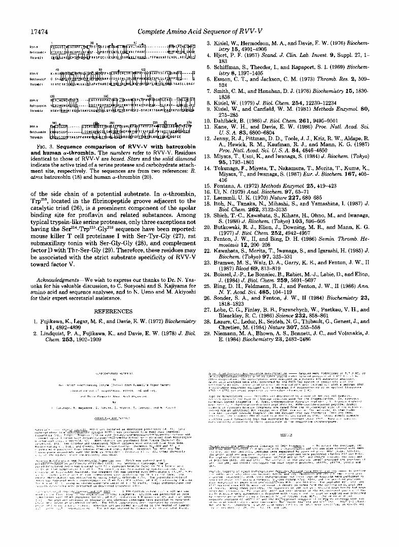
disulfide linkage may be characteristic of snake venom serine proteases. Fig. 3 shows the alignment of RVV-V, batroxobin, and human thrombin B chain, which is a physiological acti- vator of factor V (20). RVV-V is highly homologous in se- quence with batroxobin (62%) and with thrombin B chain (33.5%). In a comparison of the sequences of RVV-V and batroxobin, all 12 half-cystines are located in identical posi- tions and surround the catalytic triad. One of the glycosylation sites (AsnZz9) also shows significant homology. In the sequence of batroxobin predicted from the cDNA nucleotide sequence, this protease contains an additional potential site for carbo- hydrate attachment of Am'@ (RVV-V numbering).
On the other hand, in comparing the sequence of RVV-V with that of thrombin B chain, RVV-V deletes the four large regions of thrombin corresponding to sequences 45-54, 118- 122,132-136, and 186-192 (RVV-V numbering), as shown in Fig. 3. The region at residues 45-54 in the thrombin B chain contains the unusual Tyr-Pro-Pro-Trp tetrapeptide and car- bohydrate attachment site, and this region has been identified to be involved in monocyte chemotaxis (21). Moreover, the region between residues 132 and 136 contains an Ala-Asn and a Lys-Gly linkage, which are known to be cleavage sites for pancreatic and leukocyte elastases (22, 23) and the autopro- teolytic y-cleavage site of a-thrombin (24), resulting in the inactivation of clotting ability. In the case of RVV-V, this chemotactic region and the proteolytically susceptible se- quences are either deleted or replaced with other residues. The interaction sites of a-thrombin with fibrinopeptide and fibrinogen have been suggested to begin with Tyr212, followed by Trp'09 and Trp'" (RVV-V numbering) (25). It is also known that Leuz7, LeuSS, Tyr7', His76, and Trp8' (RVV-V numbering) of a-thrombin enhance the hydrophobic nature of the fibrinopeptide groove. Almost all of these residues, however, have been substituted by other amino acids in RVV- V. The most interesting difference between the structure of RVV-V and other trypsin-like serine proteases is found in the conservative Ser214-Trp216-Gly216 sequence and also by Glyz6 (chymotrypsinogen numbering). The corresponding se- quences in RVV-V are Ala'97-Gly'98-Gly'99 and Alaz1' (RVV- V numbering). These residues are located at the periphery of the catalytic triad for all serine proteases and permit access
V Y G C D E C N I ' ~ E H P f L V A L I T S T S S T I H C G G A L I N R E W V L T A A ~ C O ~ R N I R I K L G ~ H S ~ N I A A" 40 50 60
1 I Pa-RW-Vm """"""""""_ CNl I f CN2 "_"
0 0 """"_ """"""_ - _" ""_ g N 2 T 7
1"" "" L""""" ps%5v-v-
I rK17T1.9 @ 0 II yK17T7 lrKfiLY2 rK3 l e
R n E n E a l R v P R c K Y F c L N T K F p N c L D Y B I W L I R L R R ~ v ~ " ~ ~ ~ ~ A P v ~ L P s ~ s ~ c v c s ~ c
"""""""" -L "" """"_"""""""" """"""- II I @ CNlClB CNlSKB I """_ """" _"" - "_"
I n I mKl7TlB' 0 """"""""_""""" """ 61 70 80 90 100 110 120
CN2 """"""""""""""- LC """* CN3
cN2TuL"c_N_- 2Tll 1 L """ CN """"" ZT17 11 CN2T19 GN3fi l CN3Tll I I CN3Tl2 J I CNST5 llCN3TB """_ """""" _"" -
!. _--- 2?'3?? -____-_ J """"""_ rK7 I I rK6 II 1K5 II rM1 '" "" """ "" """"""" "
17474 Complete Amino Acid Sequence of R VV- V
RVV-Y
Batrmobln Thronbln
RVV-V
BstrmObln
ThrOnbln
RVV-V
B a t r m b l n Thrmblo
RVV-V
B l t l O x O D l n
ThrOaOln
FIG.
~ ~ ~ ~ ~ I I H ~ ~ ~ - - - - - - - - - - D ~ ~ ~ V I G G D E D l N E H P F L A F Y l - - RYFCC T L I N Y V L T A A H - - - - - - - - - - N R F R l H L C H
I E D A E C Y Y R K S P Q E L L C S D R T A A H L L l P P Y D K N F T E N D L L V R l
E I A A S L L P A G l
and human &thrombin.-The numbers refer to RVV-V. Residues identical to those of RVV-V are boxed. Stars and the solid diamond indicate the active triad of a serine protease and carbohydrate attach- ment site, respectively. The sequences are from two references: B. atrox batroxobin (18) and human a-thrombin (20).
of the side chain of a potential substrate. In a-thrombin, Trp216, located in the fibrinopeptide groove adjacent to the catalytic triad (26), is a prominent component of the apolar binding site for proflavin and related substances. Among typical trypsin-like serine proteases, only three exceptions not having the Se314-Trp215-Gly216 sequence have been reported mouse killer T cell proteinase I with Ser-Tyr-Gly (27), rat submaxillary tonin with Ser-Gly-Gly (28), and complement factor D with Thr-Ser-Gly (29). Therefore, these residues may be associated with the strict substrate specificity of RVV-V toward factor V.
Acknowledgments-We wish to express our thanks to Dr. N. Yas- uoka for his valuable discussion, to C. Sueyoshi and S. Kajiyama for amino acid and sequence analyses, and to N. Ueno and M. Akiyoshi for their expert secretarial assistance.
REFERENCES
1. Fujikawa, K., Legaz, M. E., and Davie, E. W . (1972) Biochemistry
2. Lindquist, P. A., Fujikawa, K., and Davie, E. W . (1978) J. Bwl. 11,4892-4899
Chem. 253.1902-1909
3.
4.
5.
6.
7.
8. 9.
10. 11.
12.
13.
14.
15. 16. 17. 18.
19.
20.
21.
22.
23.
24.
25.
26.
27.
28.
29.
Kisiel, W . , Hermodson, M. A., and Davie, E. W . (1976) Biochem-
Hjort, P. F. (1957) Scand. J. Clin. Lab. Znuest. 9, Suppl. 27, 1-
Schiffman, S., Theoder, I., and Rapaport, S. I. (1969) Biochem-
Esmon, C. T., and Jackson, C. M. (1973) Thromb. Res. 2, 509-
Smith, C. M., and Hanahan, D. J. (1976) Biochemistry 15,1830-
Kisiel, W . (1979) J. Bwl. Chem. 254,12230-12234 Kisiel, W., and Canfield, W . M. (1981) Methods Enzymol. 80,
Dahlback, B. (1986) J. Biol. Chem. 261,9495-9501 Kane, W . H., and Davie, E. W . (1986) Proc. Nutl. Acad. Sci.
Jenny, R. J., Pittman, D. D., Toole, J. J., Kriz, R. W . , Aldape, R. A., Hewick, R. M., Kaufman, R. J., and Mann, K. G. (1987) Proc. Natl. Acad. Sci. U. S. A. 84, 4846-4850
Miyata, T., Usui, K., and Iwanaga, S. (1984) J. Biochern. (Tokyo)
Tokunaga, F., Miyata, T., Nakamura, T., Morita, T., Kuma, K., Miyata, T., and Iwanaga, S. (1987) Eur. J. Bwchem. 167,405- 416
istry 15,4901-4906
183
istry 8, 1397-1405
524
1838
215-285
U. S. A. 83,6800-6804
95, 1793-1801
Fontana, A. (1972) Methods Enzymol. 25, 419-423 Ui, N. (1979) Anal. Bimhem. 97, 65-71 Laemmli, U. K. (1970) Nature 227,680-685 Itoh, N., Tanaka, N., Mihashi, S., and Yamashina, I. (1987) J.
Shieh, T.-C., Kawabata, S., Kihara, H., Ohno, M., and Iwanaga,
Butkowski, R. J., Elion, J., Downing, M. R., and Mann, K. G.
Fenton, J. W . , 11, and Bing, D. H. (1986) Semin. Thromb. He-
Kawabata, S., Morita, T., Iwanaga, S., and Igarashi, H. (1985) J.
Bramwe, M. S., Walz, D. A., Garry, K. E., and Fenton, J. W . , I1
Boissel, J.-P., Le Bonniec, B., Rabiet, M.-J., Labie, D., and Elion,
Bing, D. H., Feldmann, R. J., and Fenton, J. W . , I1 (1986) Ann.
Sonder, S. A., and Fenton, J . W., I1 (1984) Biochemistry 23,
Lobe, C. G., Finlay, B. B., Paranchych, W . , Paetkau, V. H., and
Lazure, C., Leduc, R., Seidah, N. G., Thibault, G., Genest, J., and
Niemann, M. A., Bhown, A. S., Bennett, J. C., and Volanakis, J.
Biol. Chem. 262,3132-3135
S. (1988) J. Biochem. (Tokyo) 103, 596-605
(1977) J. Biol. Chem. 252,4942-4957
mostasis 12,200-208
Bwchem. (Tokyo) 9 7 , 325-331
(1987) Blood 69,813-819
J. (1984) J. Biol. Chem. 259,5691-5697
N. Y. A d . Sci. 485,104-119
1818-1823
Bleackley, R. C. (1986) Science 232,858-861
Chretien, M. (1984) Nature 307, 555-558
E. (1984) Biochemistry 23,2482-2486

Complete Amino Acid Sequence of RVV- V 17475
0.4
CN3
Retention Time (mh)
17476 Complete Amino Acid Sequence of R VV- V
0.4
I 5 03
E
*
n
0 2
0.1
0
CNl "'7
Retention Time (rnin)
30 45 60
CN4a

Complete Amino Acid Sequence of R VV- V 17477
r
0.3.
02 -
: 0.1 . P f 0 ,
f 0.3.
0
L
In 9
02 *
0.1.
0 .
L P I 2 , 4 . 5 1 5 1
H I S 5r r
Le" I i *
0 5 " - 9
u.5 0 . 4 L O
P"e 0. L
1 5 2 6 1 7

17478 Complete Amino Acid Sequence of RVV- V TABLE M 3
l .2 , l l
].ai*, O . Y I L ,
1 . 0 1 1 1
1 2 1 2 . 2 1 2 1
Complete Amino Acid Sequence of R VV- V 17479

17480 Complete Amino Acid Sequence of RVV- V
Complete Amino Acid Sequence of R VV- V 17481
ClC"". . .