the fetal ultrasound assessment of fetal skeletal...
TRANSCRIPT
The FetalSkeletal System
Carol B. Benson, MD
Ultrasound Assessmentof Fetal Skeletal System
Extremities
Spine
Calvarium
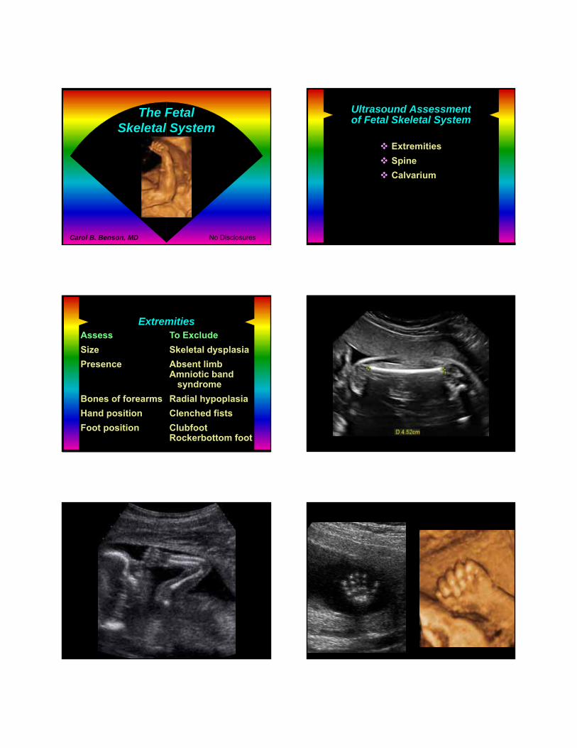
ExtremitiesAssess To Exclude
Size Skeletal dysplasia
Presence Absent limbAmniotic band
syndrome
Bones of forearms Radial hypoplasia
Hand position Clenched fists
Foot position ClubfootRockerbottom foot
Calderon Femur length
Michel arm 3 bones
Caraballo nl hand
No Disclosures

Foot
Femur Length
Normal for gestational age= Mean ± 2 SD
Femur length falls 2 - 4 SD below meanMost are growth restrictedWithout skeletal dysplasia
Femur length falls > 4 SD below meanUsually a skeletal dysplasiaBones appear abnormal by US
Steimle Femur length Long Bones are Too Short
Skeletal dysplasia
Dysotosis
Malformation
Deformationamniotic band syndromerestrictive uterine environment
Lethal Skeletal Dysplasias
Neonate cannot surviveusually due to respiratory failure
Ultrasound diagnosis typicallymade in 2nd trimester
Thanatophoric dysplasia Osteogenesis imperfecta Type 2 Achondrogenesis Congenital hypophosphatasia Short rib – polydactyly syndrome
Nonlethal Skeletal Dysplasias
Infants typically surviveUltrasound diagnosis typically
not made in 2nd trimestersometimes made in 3rd trimester
Heterozygous achondroplasia Osteogenesis imperfecta Types 1&4 Asphyxiating thoracic dystrophy
Ultrasound assessment
Degree of shortening of long bonestypically > 4 SD below mean for GA
Distribution of involved bonesextremities, spine, calvarium, ribs
Bony abnormalities
↓ mineralization, fractures, bowing
Polydactyly
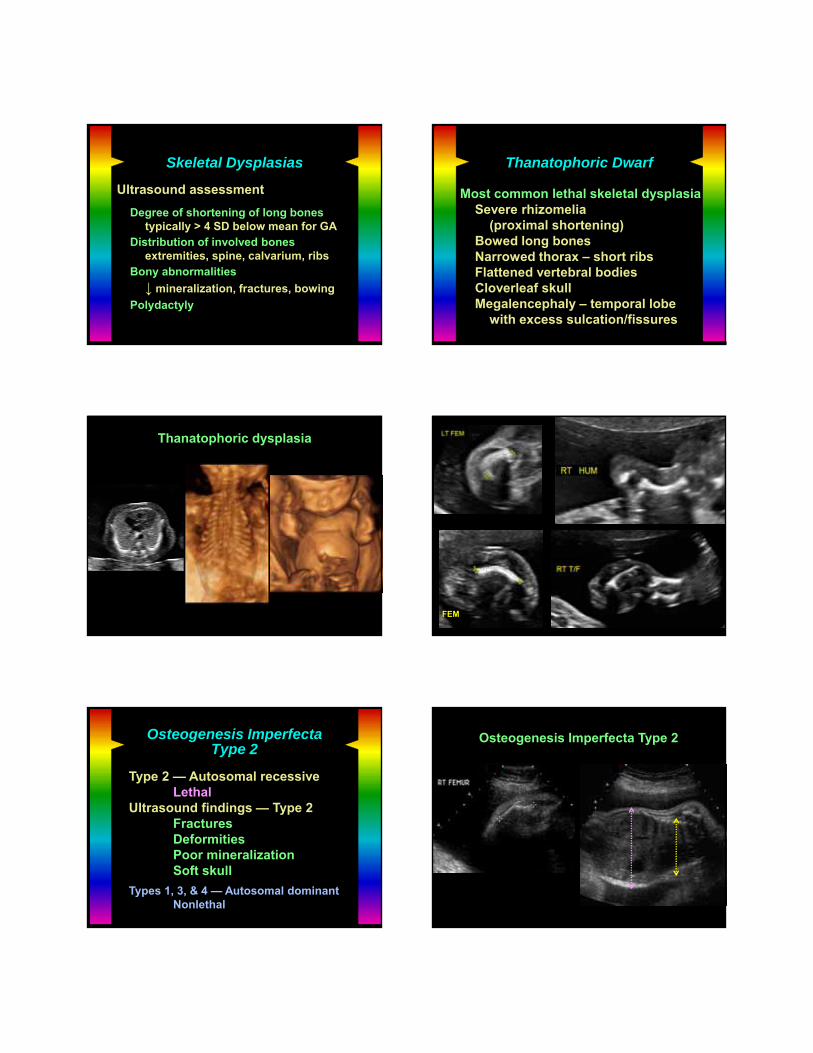
Skeletal Dysplasias Thanatophoric Dwarf
Most common lethal skeletal dysplasiaSevere rhizomelia
(proximal shortening)Bowed long bonesNarrowed thorax – short ribsFlattened vertebral bodiesCloverleaf skullMegalencephaly – temporal lobe
with excess sulcation/fissures
Kristian 19wthanatophoric
Thanatophoric dysplasia
Kristian 19wthanatophoric
FEM
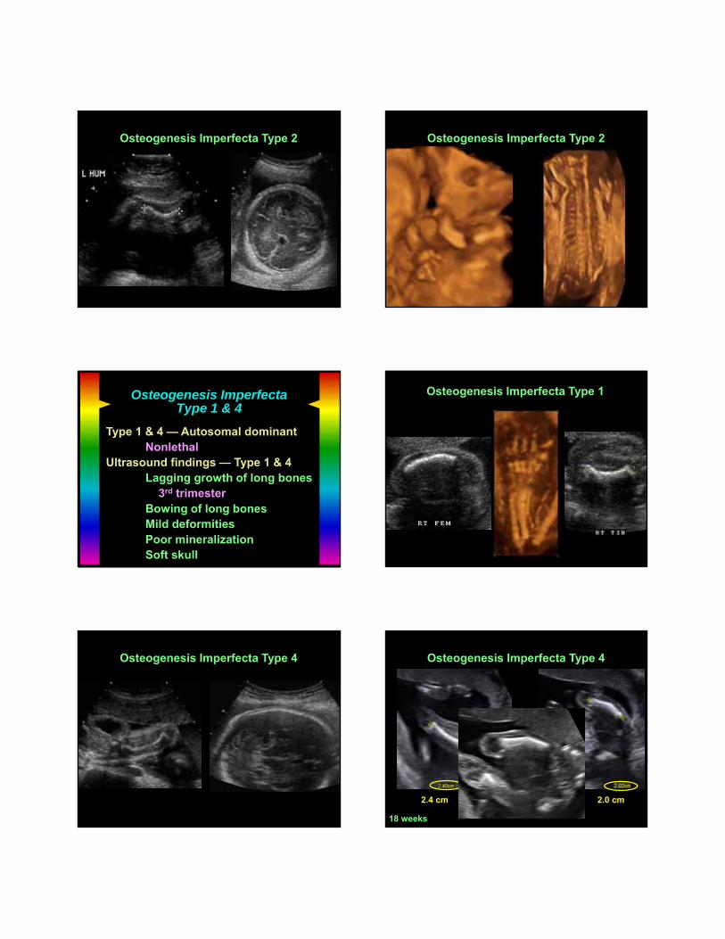
Osteogenesis ImperfectaType 2
Type 2 — Autosomal recessiveLethal
Ultrasound findings — Type 2FracturesDeformitiesPoor mineralizationSoft skull
Types 1, 3, & 4 — Autosomal dominantNonlethal
Lopez OI
Osteogenesis Imperfecta Type 2
Lopez OI
Osteogenesis Imperfecta Type 2
Astacio OI 3D
Osteogenesis Imperfecta Type 2
Osteogenesis ImperfectaType 1 & 4
Type 1 & 4 — Autosomal dominantNonlethal
Ultrasound findings — Type 1 & 4Lagging growth of long bones
3rd trimesterBowing of long bonesMild deformitiesPoor mineralizationSoft skull
McBride OI type 4
Osteogenesis Imperfecta Type 1
Al-Owfi OI type 4
Osteogenesis Imperfecta Type 4
Jones fem fxOI 4
Osteogenesis Imperfecta Type 4
2.4 cm 2.0 cm
18 weeks

Arthrogryposis
Multiple joint contracturesEtiologies
Limitation to movementOligohydramniosMultiple gestationBicornuate uterus
Abnormal nerve function Abnormal musculature Defective connective tissue
ArthrogryposisMultiplex Congenita
Ultrasound findingsContracturesFetal growth restrictionPolyhydramniosHydrops
Beausoleil arthrogryposis
Arthrogryposis
Eiresarthrogryposis
McGuiganarthrogryposis
Banerjee Larsen synarthrogryposis
Arthrogryposisfrom
Larsen syndrome(rare genetic syndrome)

Abnormal Hands & Forearms
Inherently abnormalMalformationDysplasia
Normal tissues effected by externalfactors
Deformationuterine constraintsamniotic band syndrome
Disruption (e.g., teratogen)
Radial Ray AnomaliesAssociated with
SyndromesCornelia de LangeFanconi anemiaHolt-OramRadial aplasia-ThrombocytopeniaPoland syndromeNager acrofacial dysostosisVACTERL
Trisomies 13 & 18
Henebry VACTERLabnl thumb
Radial Ray DefectAbnormal Thumb
VACTERL
Martins hypoplradius VACTERL
Hypoplastic radiusAbnormal thumb
VACTERL
Landry absent radius tri 18
Absent radiusLimb Reduction Defects
Terminal transverse deletions(e.g., absent hands)
Isolatedsporadic, unilateralamniotic band syndromevascular accident
SyndromesOrofacial (e.g., Poland)Amniotic bands
Penney absenttoes ABS Osgood absent hand
Osgood absent hand 3D bones
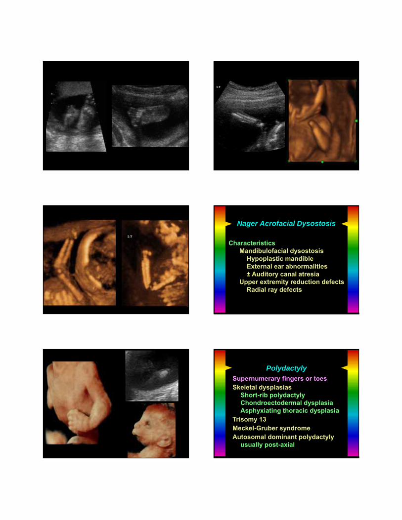
Nager Acrofacial Dysostosis
CharacteristicsMandibulofacial dysostosis
Hypoplastic mandibleExternal ear abnormalities± Auditory canal atresia
Upper extremity reduction defectsRadial ray defects
Alshamsi Nager’s
PolydactylySupernumerary fingers or toesSkeletal dysplasias
Short-rib polydactylyChondroectodermal dysplasiaAsphyxiating thoracic dysplasia
Trisomy 13Meckel-Gruber syndromeAutosomal dominant polydactyly
usually post-axial
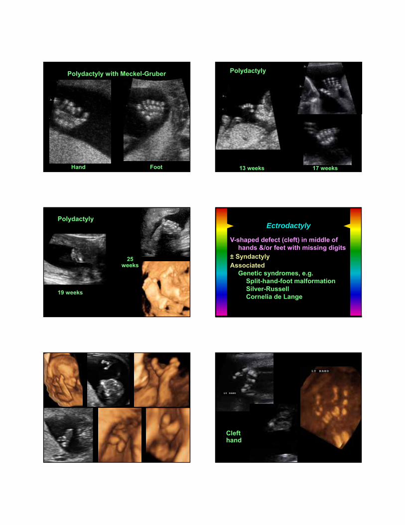
MacGowan polydactyly withMeckel Gruber
Polydactyly with Meckel-Gruber
Hand Foot
IsabellePolydactyly
13 weeks 17 weeks
Polydactyly
TorresPolydactyly
Polydactyly
19 weeks
25weeks
Ectrodactyly
V-shaped defect (cleft) in middle ofhands &/or feet with missing digits
± Syndactyly Associated
Genetic syndromes, e.g.Split-hand-foot malformationSilver-RussellCornelia de Lange
Dowd ectrodacthands & feet
Appleton absent finger
Ligibel cleft handmissing finger
Clefthand
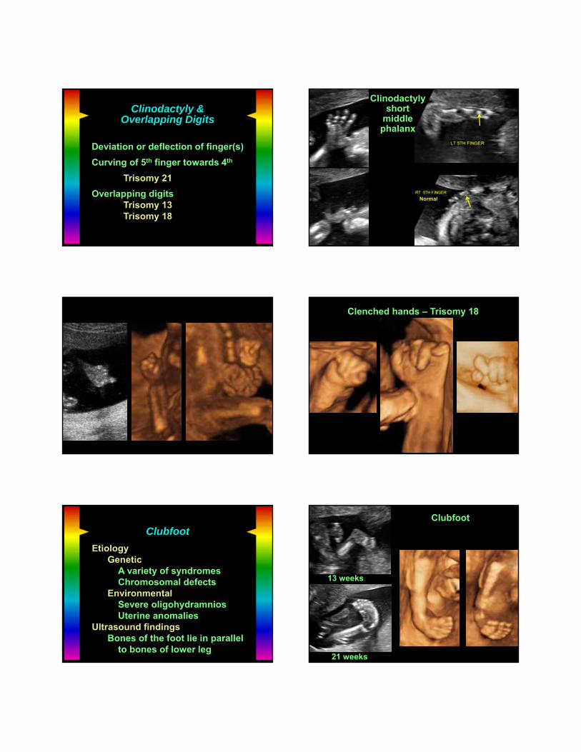
Clinodactyly &Overlapping Digits
Deviation or deflection of finger(s)
Curving of 5th finger towards 4th
Trisomy 21
Overlapping digitsTrisomy 13Trisomy 18
Subenko clinodactyly
Clinodactylyshort
middlephalanx
Normal
Sin abnl hands
Dixonclinodactyly
Clenched hands – Trisomy 18
Clubfoot
EtiologyGenetic
A variety of syndromesChromosomal defects
EnvironmentalSevere oligohydramniosUterine anomalies
Ultrasound findingsBones of the foot lie in parallel
to bones of lower leg
Lashley clubfeet
Clubfoot
21 weeks
13 weeks

Rockerbottom Foot
EtiologyTrisomy 18Skeletal dysplasia
Ultrasound findingsRounded bottom of foot
Woodlandrockerbottom feet
Rockerbottom feet – Trisomy 18
Atlas BRB rockerbottom feet
Rockerbottom feetAmniotic Band Syndrome
Early rupture of amnion
Fibrous bands entrap or adhere to fetus
Limb amputations or deformities
EncephalocelesFacial clefts
Ventral wall defectsEctopia cordis
Ultrasound Findings
Fetal deformitiesSkeletalCraniofacialVentral wall
Adherent bands
DDx: Amniotic “sheets”
Amniotic Band Syndrome
Dailey ABS anomalous hand
Amniotic BandSyndrome
Juris ABS missing hand
Amniotic BandSyndrome
13 weeks
21 weeks
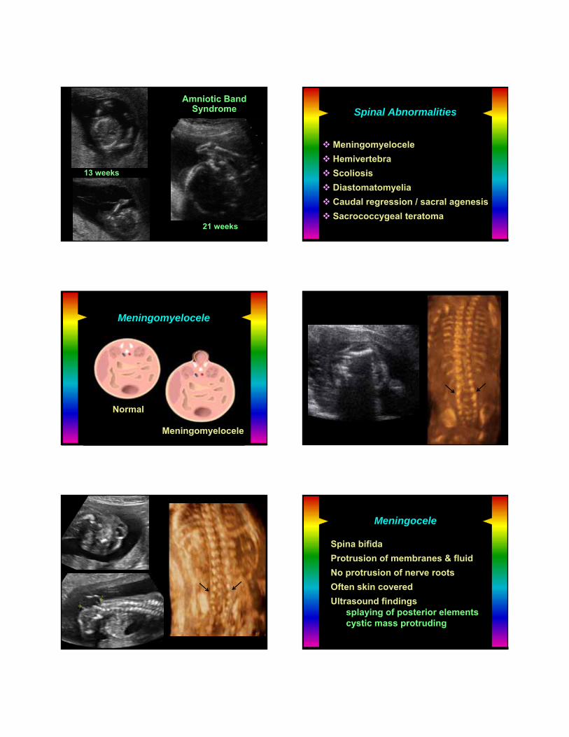
Spinal Abnormalities
Meningomyelocele
Hemivertebra
Scoliosis
Diastomatomyelia
Caudal regression / sacral agenesis
Sacrococcygeal teratoma
Meningomyelocele
Normal
Meningomyelocele
GuilmetMeningomyelocele
Bardales tri 18Meningomyelocele
Meningocele
Spina bifida
Protrusion of membranes & fluid
No protrusion of nerve roots
Often skin covered
Ultrasound findingssplaying of posterior elementscystic mass protruding
Cabral 19w 3DMeningocele
19 weeks
Reif 20w Meningocele
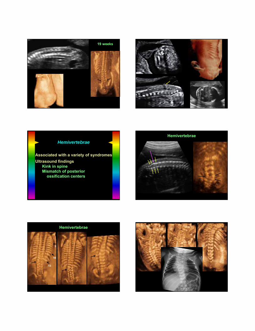
Hemivertebrae
Associated with a variety of syndromes
Ultrasound findingsKink in spineMismatch of posterior
ossification centers
Johnson hemivertebrae
Hemivertebrae
Hazen Hemivert
Hemivertebrae
1
5
10
13
Rossman hemivert
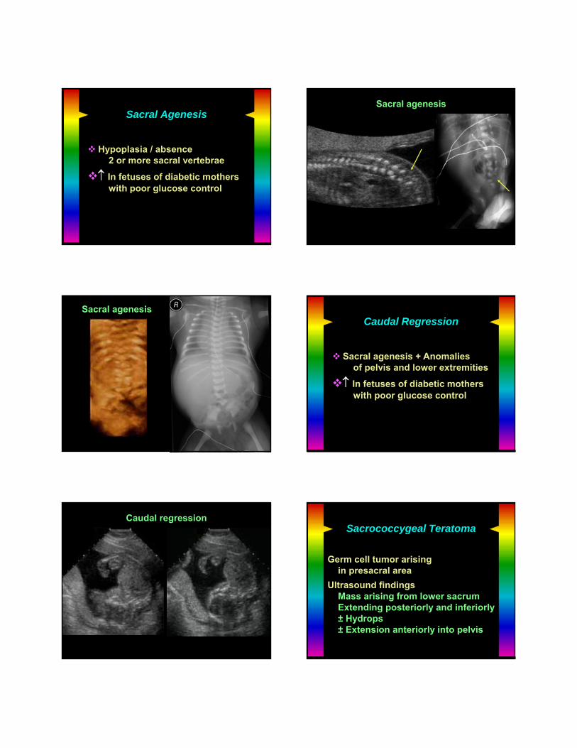
Sacral Agenesis
Hypoplasia / absence2 or more sacral vertebrae
In fetuses of diabetic motherswith poor glucose control
MacDougall sacral agen
Sacral agenesis
Bensonsacral agenesis
Sacral agenesisCaudal Regression
Sacral agenesis + Anomalies of pelvis and lower extremities
In fetuses of diabetic motherswith poor glucose control
Kelshan caudal regression
Caudal regressionSacrococcygeal Teratoma
Germ cell tumor arisingin presacral area
Ultrasound findingsMass arising from lower sacrumExtending posteriorly and inferiorly± Hydrops± Extension anteriorly into pelvis
Bonica SCT
Sacrococcygeal TeratomaSacral erosion
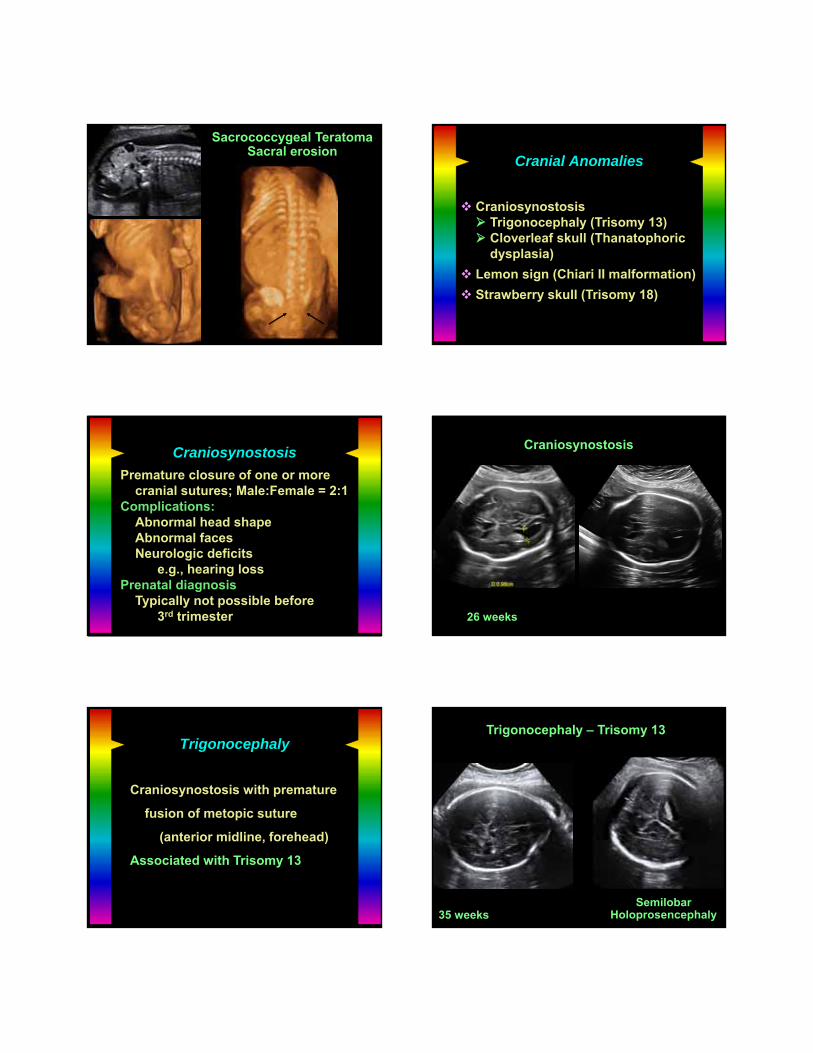
Cranial Anomalies
Craniosynostosis Trigonocephaly (Trisomy 13) Cloverleaf skull (Thanatophoric
dysplasia)
Lemon sign (Chiari II malformation)
Strawberry skull (Trisomy 18)
Craniosynostosis
Premature closure of one or morecranial sutures; Male:Female = 2:1
Complications:Abnormal head shapeAbnormal facesNeurologic deficits
e.g., hearing lossPrenatal diagnosis
Typically not possible before 3rd trimester
Harrigan craniosyn
Craniosynostosis
26 weeks
Trigonocephaly
Craniosynostosis with premature
fusion of metopic suture
(anterior midline, forehead)
Associated with Trisomy 13
Britt tri 13trigonoceph
Trigonocephaly – Trisomy 13
35 weeksSemilobar
Holoprosencephaly

Cloverleaf Skull
Craniosynostosis causing
trilobed shape
prominent forehead
Associated with
Thanatophoric dysplasia
Cole thanat skull
Cloverleaf skull – Thanatophoric dysplasia
37 weeks
Lemon-Shaped Cranium
Associated with Chiari II malformationMeningomyelocele
Noguerialemon sign
Lemon Sign withMeningomyelocele
Strawberry-Shaped Cranium
Associated with Trisomy 18 Schindlerstrawb sk
Strawberry-Shaped CraniumTrisomy 18 – 15 weeks