the g tonucleicacid - blossombio.comblossombio.com/pdf/products/bro_naldcat.pdf · tonucleicacid...
TRANSCRIPT

���
� � �� �� � � ���� � ���
� � � ��� � �� � � � � �� � �
The Guide
to Nucleic Acid
Labelling
and Detection
The Guide
to Nucleic Acid
Labelling
and Detection

Contents
Introduction page 2
Choosing a non-radioactive labelling system page 3
AlkPhos Direct Labelling and Detection Systems page 6
Gene Images Random-Prime Labelling and Detection Systems page 10
Gene Images 3’-Oligolabelling and Detection System page 12
ECL Direct Labelling and Detection System page 14
Choosing a radioactive labelling system page 16
Rediprime II DNA Labelling System page 18
5’-End Labelling Kit page 20
3’-End Labelling Kit page 21
Ready-To-Go PCR Beads page 22
RNA Labelling Kit page 24
Rapid-hyb Buffer page 25
Redivue Nucleotides page 26
Hybond Membranes for Nucleic Acid Blotting page 28
Autoradiography Films page 30
Amersham Biosciences Scanner page 32
Accessory products page 34
11..
The choice is influenced by many factors, not all of whichare directly related to the application. For exampleradioactive labelling has the advantage of high sensitivityand extremely robust protocols, however safety and long-term probe storage are serious considerations.
The aim of this guide is to give an overall introduction to the different radioactive and non-radioactivemethodologies available, highlighting their advantages anddrawbacks. Individual products are then presented in moredetail allowing you to choose the most appropriate systemfor your application.
Introduction
22..
For many years the use of radiolabels was the method ofchoice for nucleic acid labelling and detection. Morerecently, however, considerable research has lead to thedevelopment of a comprehensive range of quality systemsusing both radioactive and non-radioactive technologies.This extensive range now provides you with an excellentchoice of systems for use with DNA, RNA andoligonucleotides in a variety of applications.
Significant developments in the area of non-radioactivelabelling and detection include the use of light-baseddetection systems and direct labelling. The introduction ofthese techniques means that non-radioactive methodsnow present a viable alternative to the use of traditionalradioactive techniques for many applications.
This breadth of choice can in itself cause problems.Choosing the most appropriate system for a particularapplication can be a challenge for even the mostexperienced researchers.
Table 1 Factors affecting the choice of labelling and detection system
Factor Considerations
Blotting system Southern, Northern, dot/slot, library screen.Amount of target nucleic acid present
Label Radioactive or non-radioactiveDirect or indirect
Probe DNA, RNA, oligonucleotideSize and purity of probe
Speed and convenience Multiple exposuresTime constraints
Probe storage Stability
Results Autoradiography filmImage analysis with scanners
Membrane Charged or uncharged nylonNitrocellulose
Labelling
Non-radioactive labelling methods are separated into twodifferent approaches. Early non-radioactive systems utilizedindirect labelling techniques, but these have now beenlargely superseded by the introduction of superior directlabelling methods, an example of which is AlkPhos Direct™.
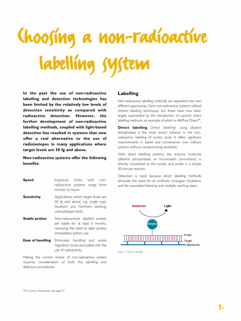
Direct labelling Direct labelling* using alkalinephosphatase is the most recent advance in the non-radioactive labelling of nucleic acids. It offers significantimprovements in speed and convenience over indirectsystems without compromising sensitivity.
With direct labelling systems, the enzyme molecule(alkaline phosphatase or horseradish peroxidase) is directly crosslinked to the nucleic acid probe in a simple 30-minute reaction.
Detection is rapid because direct labelling methodseliminate the need for an antibody conjugate incubationand the associated blocking and multiple washing steps.
Choosing a non-radioactive
labelling system
33..
In the past the use of non-radioactivelabelling and detection technologies hasbeen limited by the relatively low levels ofdetection sensitivity as compared withradioactive detection. However, the further development of non-radioactivelabelling methods, coupled with light-baseddetection has resulted in systems that nowoffer a real alternative to the use ofradioisotopes in many applications wheretarget levels are 50 fg and above.
Non-radioactive systems offer the followingbenefits:
Speed Exposure times with non-radioactive systems range fromminutes to hours.
Sensitivity Applications where target levels are50 fg and above, e.g. single copySouthern and Northern blotting,colony/plaque blots.
Stable probes Non-radioactively labelled probesare stable for at least 6 months,removing the need to label probesimmediately before use.
Ease of handling Eliminates handling and wasteregulatory issues associated with theuse of radioactivity.
Making the correct choice of non-radioactive systemrequires consideration of both the labelling and detection procedures.
Enzyme
Substrate Light
Probe
TargetMembrane
Figure 1 Direct labelling
*For licence information see page 37
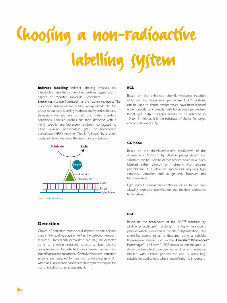
Indirect labelling Indirect labelling involves theintroduction into the probe of nucleotides tagged with ahapten or ‘reporter’ molecule. Amersham Biosciences kits use fluorescein as the hapten molecule. Thenucleotide analogues are readily incorporated into theprobe by standard labelling methods and hybridization andstringency washing are carried out under standardconditions. Labelled probes are then detected with ahighly specific anti-fluorescein antibody conjugated toeither alkaline phosphatase (AP) or horseradishperoxidase (HRP) enzyme. This is followed by enzymecatalysed detection, using the appropriate substrate.
44..
Figure 2 Indirect labelling
Detection
Choice of detection method will depend on the enzymeused in the labelling stage as well as the detection mediumrequired. Horseradish peroxidase can only be detectedusing a chemiluminescent substrate, but alkalinephosphatase can be detected using chemiluminescent andchemifluorescent substrates. Chemiluminescent detectionsystems are designed for use with autoradiography filmwhereas fluorescence based detection systems require theuse of suitable scanning equipment.
ProbeFluorescein
Antibody
TargetMembrane
Enzyme
Substrate Light
ECL
Based on the enhanced chemiluminescent reaction of luminol with horseradish peroxidase, ECL™ substrate can be used to detect probes which have been labelledeither directly or indirectly with horseradish peroxidase.Rapid light output enables results to be achieved in 10 to 15 minutes. It is the substrate of choice for targetamounts above 500 fg.
CDP-Star
Based on the chemiluminescent breakdown of thedioxetane CDP-Star™ by alkaline phosphatase*, thissubstrate can be used to detect probes which have beenlabelled either directly or indirectly with alkalinephosphatase. It is ideal for applications requiring highsensitivity detection such as genomic Southern andNorthern blots.
Light output is rapid and continues for up to five daysallowing exposure optimization and multiple exposures to be taken.
ECF
Based on the breakdown of the ECF™ substrate byalkaline phosphatase*, resulting in a highly fluorescentproduct which is localized at the site of hybridization. Thischemifluorescent signal is detected using a suitablefluorescence scanner such as the Amersham Biosciences ™
FluorImager™ or Storm™. ECF detection can be used todetect probes which have been either directly or indirectlylabelled with alkaline phosphatase and is particularlysuitable for applications where quantification is important.
Choosing a non-radioactive
labelling system

55..
*For licence information see page 37.
Labelling and Sensitivity Time from Duration of Strip before Quantification Recommended application
detection system hybridization light output re-probing
to detection
AlkPhos Direct 0.06 pg 1 hour 5 days yes no Single copy Southern
and Northern
ECL Direct 0.5 pg 1 hour 1-2 hours no no High target applications
e.g. colony/plaques
Gene Images 0.1 pg 3 hours 5 days yes no High sensitivity Northerns
Random-Prime
Gene Images 3’- end 0.1 pg 3 hours 5 days yes no Oligo screening with
labelling with CDP-Star stringency control
Gene Images 3’- end 120 pg 3 hours 1-2 days yes yes Quantification
labelling with ECF
ECL Random-Prime 0.5 pg 3 hours 1-2 hours yes no Medium target Southern
with DNA probes
ECL 3’-end labelling 0.2 pg 3 hours 1-2 hours yes no Medium to high target
Southern with oligo probes
ECF Random-Prime 0.25 pg 3 hours 1-2 days yes yes Quantification
Table 2 Guide to the properties of non-radioactive labelling and detection systems

Protocol Summary
Probe labelling
Denatured or single-stranded DNA or RNA probe ismixed with the labelling buffer and alkaline phosphatase.Formaldehyde is then added to covalently crosslink theenzyme to the probe. There is no need to purify theprobe before hybridization.
Hybridization and stringency
The probe is hybridized to the blot using the speciallyformulated AlkPhos Direct hybridization buffer, whichstabilizes the activity of the enzyme. After a 15 minute pre-hybridization step, hybridization with probe is performedovernight at the required temperature. For higher targetamounts, a 2 to 4 hour hybridization may be sufficient.
AlkPhos Direct Labelling
and Detection
AlkPhos Direct™ combines the convenienceof direct enzyme labelling (no blocking orantibody stages) with those of alkalinephosphatase detection (long light outputand high sensitivity).
Labelling is complete within 30 minutes in asingle tube protocol and the resultingprobe is ready for use in hybridizationswithout further purification.
Due to the thermostable nature of theenzyme, hybridization stringency can becontrolled by adjusting temperature as wellas salt concentration.
The system can be used with either chemiluminescent or fluorescent detection reagents. The most sensitiveresult is achieved with CDP-Star, achemiluminescent substrate with a fastlight output. Alternatively, the use of ECFsubstrate generates a fluorescent signalsuitable for use with fluorescence scanningdevices such as Amersham BiosciencesFluorImager or Storm instruments.
66..
Probe
AP
Probe
Target DNAMembrane
AP
Figure 3 Probe labelling
Figure 4 Hybridization

77..
Systems
Detection
Chemiluminescence with CDP-Star:
Hybridized blots are detected with CDP-Star reagent.Following a two-minute incubation, blots are exposed to Hyperfilm™ MP for 1 to 2 hours or a light capturescanning device. Sensitivities as low as 60fg can beachieved using CDP-Star.
or
Chemifluorescence with ECF:
Hybridized blots are detected with ECF substrate. Non-fluorescent substrate is catalysed by alkalinephosphatase to produce a fluorescent signal whichaccumulates over time; low sensitivity applications yieldresults after 1 hour, high sensitivity applications usuallyrequire overnight incubation.
Recommended Applications
AlkPhos Direct is a fast and easy-to-use system suitablefor the majority of routine blotting applications usingeither DNA, RNA or oligonucleotide probes.
The versatility of the system, combined with the fact thatboth DNA and RNA probes are labelled equallyefficiently, makes it the ideal choice for a busy laboratorycarrying out a variety of different blotting andhybridization techniques.
For Southern and Northern blotting applicationsAlkPhos Direct, combined with CDP-Star detection,offers a highly sensitive system capable of detectingdown to 60 fg of target DNA. Thus making it the idealchoice for the non-radioactive detection of single-copygenomic Southern blots.
Re-probing can be difficult with non-radioactive systemsdue to the need to remove both probe and antibodylayers in an indirect system. The simplicity of the AlkPhosDirect system means that there are less components tobe removed during the stripping procedure. Proberemoval is therefore more effective with less damage tothe membrane.
For labelling oligonucleotides, an optimized protocol hasbeen developed which is capable of labelling probesdown to 17 basepairs in length. Request TechTip number174 for additional information.
Quantification of dot and slot blots is possible by usingECF detection in conjunction with AlkPhos Direct.Analysing results on a fluorescence scanner such asAmersham Biosciences Storm or FluorImager allowsaccurate quantification and analysis of results.
Figure 5 Detection
Probe
Target DNAMembrane
CDP-Staror ECF substrate Signal
AP
Figure 5 Detection

Stability Probes are stable for up to six months if storedat –20°C in 50% glycerol.
Efficiency Nucleic acid probes are labelled efficiently and uniformly with enzyme.
Accuracy The crosslinking labelling reaction does notresult in any net synthesis of probe and the amount ofprobe present before and after labelling does not change.Therefore probe concentrations during hybridization canbe determined more accurately.
Speed Time savings of 3 to 4 hours over conventionalindirect methods are achievable as a result of the rapid and simple labelling reaction, and the absence ofantibody incubations.
Consistency Quality control on each batch ensuresconsistent performance and provides confidence in probelabelling and concentration.
Stringency control Due to the thermostable nature ofthe enzyme, hybridization stringency can be controlled byelevating temperatures up to 75°C as well as bydecreasing salt concentrations.
Versatility Labelling of DNA, RNA and oligonucleotideprobes with detection by chemiluminescence orchemifluorescence offers a high degree of versatility.
AlkPhos Direct Labelling
and Detection
88..
Figure 7
Northern blot (Hybond-N+) ofhuman skeletal muscle (totalloadings of 0.5, 0.25, 0.125,0.063 µg). Hybridized withGAPDH probe.(A) First detection(B) CDP-Star re-applied
after stripping(C) CDP-Star re-applied
after re-probingC
A
B
Figure 6
Southern blot
Cosmid DNA digested with Not I and EcoR I, probed with a 1.1kb probelabelled with AlkPhos Direct and a competitor’s hapten-based system
(A) AlkPhos Direct
(B) Competitor’s hapten-based system
Courtesy of Janet Bartels, Yale University
BA

99..
Systems
Ordering Information
AlkPhos Direct Labelling and Detection 25 reactions RPN 3690System with CDP-Star
AlkPhos Direct Labelling and Detection 50 reactions RPN 3691System with CDP-Star
AlkPhos Direct Labelling and Detection System with ECF 25 reactions RPN 3692
AlkPhos Direct Labelling Module 25 reactions RPN 3680
AlkPhos Direct Labelling Module 50 reactions RPN 3681
AlkPhos Direct Hybridization Buffer For 5000 cm2 membrane RPN 3688
CDP-Star Detection Reagent For 2500cm2 membrane RPN 3682
CDP-Star Detection Reagent For 5000cm2 membrane RPN 3683
ECF Substrate For 2500cm2 membrane RPN 3685
Related products
ECL Direct Nucleic Acid Labelling and Detection System RPN3000
Hybond-N+ Membranes see page 28
Hyperfilm MP see page 30
Figure 8
Dot blots loaded with 5, 2, 1, 0.5, 0.1 and 0.01ng of a 1.5kb N-rasinsert on Hybond-N+ nylon membrane. The 7th dot was a control of5ng carrier DNA. Results were obtained with CDP-Star detection reagent.
A CBFigure 9
Control of stringency by wash temperature. Human genomicSouthern blots (2, 1 and 0.5µg loadings) hybridized with BCL2 at 5ng/ml. Washed at:
(A) 50°C (B) 55°C (C) 60°C
0.2% blocking reagent included in the primary wash buffer.

1100..
Gene Images Random-Prime
Labelling and Detection
Gene Images™ Random-Prime is designed to
generate fluorescein labelled DNA probes
using a random prime labelling reaction.
The reaction requires only 50 ng of template
DNA as the strand displacement properties
of the enzyme, coupled with non-limiting
concentrations of nucleotide, result in net
synthesis of probe. The use of fluorescein allows
the efficiency of the reaction to be checked
before hybridization by a fluorescence assay.
Protocol Summary
Probe labelling
Fluorescein dUTP is incorporated into DNA in a randomprime labelling reaction using Klenow DNA polymerase.Nonamer primers are used to ensure efficient hybridizationto probe. Probes can be used without purification.
Optional rapid labelling assay
The efficiency of probe labelling can be checked by spotting a small aliquot of labelled probe onto Hybond™-N+membrane, washing to remove unincorporated fluoresceinand visualizing on a UV transilluminator or fluorescent scanner.
Hybridization
For optimum performance use the recommendedhybridization buffer.
Sensitive detection is achieved by the use of a monoclonalantibody which is highly specific for alkaline phosphatase.50 fg of target can be routinely detected in a humangenomic Southern blot.
Detection
Chemiluminescence with CDP-Star:Hybridized blots are detected with CDP-Star reagent.Following a 2 minute incubation, blots are exposed toHyperfilm MP for 1 to 2 hours. Sensitivities as low as 50 fgcan be detected.
or
Chemifluorescence with ECF:Hybridized blots are detected with ECF substrate. Non-fluorescent substrate is catalysed by alkalinephosphatase to produce a fluorescent signal whichaccumulates over time; low sensitivity applications yieldresults after 1 hour, high sensitivity applications usuallyrequire overnight incubation.
Recommended Applications
Probes labelled with the Gene Images Random-PrimeLabelling and Detection System can be used for mostmembrane applications especially in situations where thereis insufficient probe for labelling by direct methods.
Gene Images Random-Prime Labelling and DetectionSystems may be used with either the detection reagent CDP-Star for high sensitivity applications, or with ECFsubstrate for quantification on fluorescence scanninginstrumentation.
Gene Images Random-Prime is especially recommendedfor high sensitivity Northern applications.
ProbeFluorescein
Antibody
TargetMembrane
Enzyme
Substrate Light
Figure 11 Indirect labelling
ProbeFl Fl Fl Fl Fl Fl Fl
Figure 10 Probe labelling

1111..
Systems
Ordering Information
Gene Images Random-Prime Labelling and Detection System 30 reactions RPN 3500
Gene Images Random-Prime Labelling and Detection System 60 reactions RPN 3501
Gene Images Random-Prime Labelling Module 30 reactions RPN 3540
Gene Images Random-Prime Labelling Module 60 reactions RPN 3541
CDP-Star Detection Module For 2500cm2 membrane RPN 3510
CDP-Star Detection Module For 5000cm2 membrane RPN 3511
ECF Dectection Module For 2500cm2 membrane RPN 5750
Related products
AlkPhos Direct Labelling and Detection System RPN3690
Hybond-N+ membranes see page 28
Confidence The use of fluorescein-dUTP allows a simpleand rapid (30 minute) probe labelling assay to beperformed by monitoring the fluorescence of labelledprobes on a UV transilluminator. This enables theefficiency of the labelling reaction to be checked beforeproceeding with the hybridization.
Stability Probes are stable for at least 9 months underrecommended conditions minimizing the requirement forcontinual labelling of frequently used probes.
A B
(iii)
(ii)
(i)
Figure 13
Rapid labelling assay
(a) Serial dilutions of fluorescein nucleotide1/25, 1/50, 1/100, 1/250, 1/500,1/1000. Membrane not washed
(b) Aliquots of labelling reactions:
(i) labelling reaction inhibited withEDTA at time 0
(ii) labelled lambda DNA digestedwith Hind III
(iii) labelled N-ras clone
Membrane washed in 2xSSC at 60˚C for 15 minutes. Visualized on a UVtransilluminator
Figure 12
5, 2, 1 and 0.5 µg of human liver mRNA were blottedonto Hybond-N+, fixed using the Amersham crosslinkerand hybridized with a fluorescein-labelled DNA proge,specific for p53, at 65°C. CDP-Star detection, 1 hourexposure on Hyperfilm-MP.

Hybridization
For optimum performance use the recommendedhybridization buffer.
Detection
Chemiluminescence with CDP-Star:
Hybridized blots are detected with CDP-Star reagent.Following a two minute incubation, blots are exposed toautoradiography film for 1 to 2 hours or detected using alight collection device.
or
Chemifluorescence with ECF:
Hybridized blots are detected with ECF substrate. Non-fluorescent substrate is catalysed by alkalinephosphatase to produce a fluorescent signal whichaccumulates over time; low sensitivity applications yieldresults after 1 hour; high sensitivity applications usuallyrequire overnight incubation.
Gene Images 3’-Oligolabelling
and Detection
Gene Images™ 3’-Oligolabelling system isbased on the labelling of standard unmodifiedoligonucleotide sequences with fluorescein-dUTP in a reaction catalysed by terminaldeoxynucleotidyl transferase. The optimizedprotocol results in a tail of 6-8 nucleotides,which provides good sensitivity with lowbackground, and does not interfere with thespecificity of hybridization. The use offluorescein as the label allows the efficiencyof the labelling reaction to be checked usinga fluorescence assay before hybridization.
Protocol Summary
Probe labelling
Standard oligonucleotides are tailed with fluorescein-dUTPusing the enzyme terminal deoxynucleotidyl transferase.There is no need to purify the probe before hybridization.
Optional rapid labelling assay
Labelling efficiency can be checked by spotting a smallaliquot of labelled probe onto Hybond-N+ membrane,washing to remove unincorporated fluorescein andvisualizing on a transilluminator or fluorescence scanner.
1122..
Figure 14 Probe labelling
LightCDP-Star
AP
Probe
Target DNA
Membrane
FlFlFlFl
Figure 15 Gene Images 3’-Oligolabelling with CDP-Star detection

1133..
System
Figure 16
Dot blots of M13 mp8 (500 - 0.625 attomoles).3’-end labelled M13 17mer oligonucleotide probe.5 ng/ml, 2 hour hybridization and detected usingCDP-Star. 1 hour exposure to Hyperfilm MP.
Recommended Applications
The 3’-Oligolabelling system can be used to label probesfor a wide variety of applications. The indirect labellingmethod and high sensitivity makes it especially suited tooligo screening applications requiring a high degree ofstringency control.
Ease of use Simple protocols, and ready-to-use reagentsensures first-time success.
Confidence The use of fluorescein-dUTP allows a simpleand rapid (30 minute) probe labelling assay to beperformed by monitoring the fluorescence of labelledprobes on a UV transilluminator. This enables the efficiencyof the labelling reaction to be checked before proceedingwith the hybridization. Use with any oligonucleotide, specialsynthesis is not required.
Stability Labelled probes are stable for at least 9 months,under recommended conditions allowing batches of probeto be prepared and stored until needed.
Ordering Information
Gene Images 3’-Oligolabelling To label 1000 pmol oligo RPN5776and CDP-Star Detection System
Gene Images 3’-Oligolabelling Module To label 1000 pmol oligo RPN5770
CDP-Star Detection Module For 2500 cm2 membrane RPN3510
CDP-Star Detection Module For 5000 cm2 membrane RPN3511
ECF Detection Module For 2500 cm2 membrane RPN5750
Related products
AlkPhos Direct Labelling and Detection System with CDP-Star RPN3690

Hybridization
The labelled probe is hybridized to the blot using thespecially formulated ECL Gold Buffer, which stabilizes the activity of the enzyme. Following a 15 minute pre-hybridization, hybridization with probe is performedovernight. For colony or plaque screens a 2 to 4 hourhybridization is sufficient.
Detection
The hybridized blot is soaked in the detection reagent.Where the probe is bound there is peroxidase-catalysedoxidation of luminol and subsequent enhancedchemiluminescence. The resulting light signal is detectedon autoradiography film.
ECL Direct Labelling
and Detection System
The ECL Direct™ nucleic acid labelling anddetection system is based on the directlabelling of DNA or RNA probes with theenzyme horseradish peroxidase (HRP) in a simple chemical reaction. Labelling takesonly 10 minutes in a single tube reaction,and the resulting probe can be used directlyin hybridization experiments withoutfurther purification. Detection of hybridizedprobe is achieved by generation of light viaHRP catalysed oxidation of luminol with theuse of an enhancer maximizing light output.
Protocol Summary
Probe labelling
The labelling solution is added to the unlabelled,denatured single-stranded DNA or RNA probe. Thepositively charged HRP complex is bound electrostaticallyto the negatively charged DNA. Glutaraldehyde is addedto covalently cross-link the HRP to the probe. There is noneed to purify the probe before hybridization. Labelledprobes can be stores in 50% glycerol for several monthsat -15°C to -30°C.
1144..
ProbeHRP
+
+- -
- --+
HRP+
++
HRP+
++
Figure 17 Probe labelling
HRP HRP HRP HRP HRP
Probe
Target DNAMembrane
Figure 18
Probe
HRP
Hyperfilm ECL
Light
Substrate +Enhancer
Figure 19 Detection

Recommended Applications
ECL Direct™ is a fast and easy system for high to mediumtarget amount applications such as colony/plaque screens,dot blots, genome mapping, and PCR* product analysis. Itis particularly useful for applications which require a blot tobe re-probed several times and screening applicationsrequiring rapid throughput and results.
Ease of use Simple protocol coupled with high initialsignal output makes it the most rapid non-radioactivesystem available.
Speed Less than 4 hours from probe labelling to detection inhigh target amount applications with no lengthy antibody steps.
Ease of re-probing HRP is inactivated by thechemiluminescent reaction therefore no need to stripblots before reprobing. This saves time and avoids possiblemembrane damage.
Consistency Consistent results combining strong signalswith very low backgrounds on nylon membranes.
Convenience Convenient, ready-to-use ECL DirectGold hybridization buffer formulated with an exclusive rateenhancer*.
Efficiency DNA or RNA probes are labelled equallyefficiently.
Stability Probes can be stored for at least 3 months in 50% glycerol, avoiding the need for frequent labelling.
1155..
Figure 20
Random YAC DNAdigested with PvuIIand hybridized withAluI human DNArepetitive probe(exposure time of 30 minutes), using ECL Direct labelling.
Ordering Information
ECL Direct Labelling and Detection System To label 5 µg nucleic acid RPN 3000
ECL Direct Labelling and Detection System To label 10 µg nucleic acid RPN 3001
ECL Direct Detection Reagents For 2000cm2 membrane RPN 3004For 4000cm2 membrane RPN 2105
ECL Direct Gold Buffer For 4000cm2 membrane RPN 3006
ECL Direct Labelling System To label 5 ng nucleic acid RPN 3005
Related products
AlkPhos Direct Labelling and Detection System RPN3690
A B
Figure 21
Comparison of 1st and 6th re-probing of Southern blotted human genomic DNA bound to Hybond-N+. Minimalreduction in signal intensity is observed.
A) 6th re-probing
B) 1st probing
*For licence information see page 37

Choice of radiolabel
The selection of radiolabel for a particular applicationdepends on the level of sensitivity and resolution required.For most membrane hybridization techniques sensitivity ismost important, so a high-emission energy label willprovide sensitive results with shorter exposure times butresolution will be compromised.
In applications where resolution is most important, such asmicrosatellite analysis and sequencing, a lower energy-emitting isotope will give improved resolution, but longerexposure times will be required.
The three most widely used isotopes in the life sciencefield are:
32P For high-sensitivity membrane applications 32P isthe label of choice due to its high β-energy and theelevated specific activity of 32P-labelled probes. Labellednucleotides with a specific activity of >110 TBq/mmol, >3000 Ci/mMol are available for these applications.
33P With the advantage of a lower emission energy,33P offers increased resolution as compared to 32P. It may be used for membrane hybridizations, but its lower energy emission means that longer exposure timesare required. It is ideally suited to sequencing and in situhybridization applications.
35S This label can be used for hybridizations but isnot recommended because of its low emission energy.However, it provides high resolution and is suited tosequencing and in situ hybridization applications.
Choice of labelling method
Random prime The most widely used method for the uniform labelling of DNA to provide high label density probes.
Ready-To-Go™ DNA Labelling Beads, Rediprime™ II,Megaprime™ and Multiprime™ are all based on the randomprime technique which uses random sequence hexamersor nonamers to prime DNA synthesis on a denaturedDNA template at numerous sites along its length.
Choosing a radioactive
labelling system
Labelling with 32P-labelled nucleotidesremains the most sensitive and robusttechnology available. Radiolabelled probesoffer the following benefits:
Sensitivity 32P-labelled probes offer the highest level ofsensitivity, ideal for detection where there is a very lowabundance of target.
Prolonged emission Radioactively labelled blotscontinue to emit signal for several weeks enabling multiple exposures and long exposure times to be carried out as required.
Reliability Radioactive labelling systems are veryforgiving. Precise optimization of hybridization conditions isless critical, and hence less time is used determining idealhybridization and stringency conditions.
Flexibility Incorporation of radiolabelled nucleotidesinto a probe can be controlled by the labelling conditions. Therefore probes can be produced with differing specific activities and incorporation levels can be measured accurately, allowing exact probeconcentrations to be determined.
1166..
Figure 22 Ready-To-Go pre-dispensed reagent beads

1177..
Labelling Technology Nucleotide Amount of Labelling Probe specific Recommendedsystem template time activity (dpm/µg) application
Rediprime II Random-Prime dCTP only 25 ng 10 minutes 2 x 10⁹ membrane hybridization
Ready-To-Go Random-Prime dCTP only 10 ng - 1 µg 5 minutes 2 x 10⁹ membrane hybridizationDNA labelling beads
Megaprime Random-Prime any dNTP 25 ng 10 minutes 2 x 10⁹ membrane hybridization
Nick translation Nick translation any dNTP 1 µg 2-3 hours 2 x 10⁹ Production of largeamounts of probe
RNA labelling SP6/T7 RNA UTP 1 µg 1-2 hours 2 x 10⁹ in situpolymerase hybridization
5’-end labelling T4 Polynucleotide dATP 10 pmol ends 1 hour 5 x 10⁶ membrane hybridizationkinase in situ hybridization
3’-end labelling Terminal deoxynucleo- any dNTP 10 pmol ends 30-60 5 x 10⁶ membrane hybridizationtidyl transferase minutes in situ hybridization
The reaction uses the Klenow fragment of DNApolymerase I and leads to an efficient use of labellednucleotides and net synthesis of probe. Therefore, verysmall amounts of input DNA are required, enabling highspecific activity DNA probes to be produced withrelatively small quantities of added label.
Nick translation This method provides uniformlylabelled DNA probes by the introduction of random nicksinto the template by the action of DNase I and subsequentfilling in by DNA polymerase I. The existing nucleotidesequence of the DNA probe is renewed without net synthesis occurring. Nick translation can be used for the production of large amounts of high-specific activity probes.
End labelling This is the method of choice for labellingoligonucleotide probes. Either the 3’ or 5’ end can belabelled by the use of different techniques. End-labelledoligonucleotide probes may be used for membranehybridizations. Probes labelled with 35S and 33P are suitablefor use with in situ hybridization.
RNA labelling This technique produces radiolabelledRNA probes from inserts cloned into appropriate vectors.SP6 and T7 RNA polymerase may be used to produceasymmetric probes from the same or differentvectors. Asymmetric probes are used in insitu hybridization applications to provideboth positive results and negative controls.
Figure 23 Redivial™ showing removal of the splash guard which secures the radioactive solution at thebottom of the vial during ambient shipment. The splashguard is easily removed and disposed of as active waste.
Table 3 Guide to the properties of radioactive labelling systems

1188..
Rediprime II
DNA Labelling System
Amersham Biosciences ' premiumradioactive labelling system consists ofindividually dispensed reaction mixes whichare dried in the presence of a stabilizer anda dye to make labelling of probes easier. Thesystem can be stored at 4°C or at roomtemperature ready for use.
A C DB
Rediprime II reaction mixes have been formulatedusing an improved exonuclease free Klenow to giveprobes with specific activities of 1.9 x 10⁹ dpm/mg orgreater after 10 minutes incubation at 37°C with themajority of DNA substrates. When used withRedivue™ [32P]dCTP, Rediprime II reactions can beset up and completed to produce a DNA probeready for hybridization in less than 15 minutes.
Protocol Summary
Figure 24
Schematic diagram of the Rediprime II protocol
(A) Add denatured template to a final volume of 45µl
(B) Flick tube and spin briefly
(C) Add 5ml Redivue [32P]dCTP, pipette up and down and spin briefly
(D) Incubate for 10 minutes at 37°C
1199..
Recommended Applications
Labelling of DNA from a variety of sources to a highspecific activity for use in Southern and Northern blothybridizations. The system is designed for use withRedivue [32P]dCTP with a specif ic activity of110TBq/mmol, 3000 Ci/mmol.
Speed Quick and convenient protocol requires theaddition of template and labelled nucleotide only.
Stability Ambient temperature stable, therefore can be stored at room temperature.
Flexibility DNA can be labelled in the presence of Low Melting Point agarose or restriction enzyme buffers.
Efficiency Each labelling mix can label up to 25 ng of DNA to a specific activity of >10 9 dpm/µg.
Ordering Information
Rediprime II DNA Labelling System 30 pre-mixed labelling reactions RPN1633For use with radiolabelled dCTP
Rediprime II DNA Labelling System 60 pre-mixed labelling reactions RPN1634For use with radiolabelled dCTP
Related products
Redivue [α- 32P]dCTP, 3000Ci/mmol AA0005
Megaprime DNA Labelling System RPN1604
Rapid-hyb Buffer RPN1635
Hybond Nylon Membranes see page 28
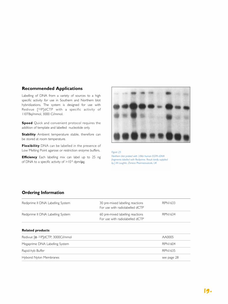
Figure 25
Northern blot probed with 3.8kb human EGFR cDNAfragments labelled with Rediprime. Result kindly suppliedby J M Loughlin, Zeneca Pharmaceuticals, UK

OH 3'
P* 5'
5' *P
3' HO
OH 3'
P 5'
5' P
3' HO
OH 3'
OH 5'
5' HO
3' HO
Alkaline phosphataseT4 PNK ADP,
[γ-32P]ATP
T4 PNK [γ-32P]ATP
5’-End Labelling Kit
The 5’-End Labelling Kit exploits an optimized exchange buffer and T4 polynucleotide kinase to labeloligonucleotides with or without a 5’-phosphate group. Labelling is completewithin 30 minutes.
Protocol Summary
2200..
Recommended Applications
Oligonucleotides labelled in this way are suitable for use in high target amount applications. For example as probes or primers to track PCR products, or in gel-shift and fragment analysis assays.
Flexibility Very small fragments of both DNA and RNAcan be labelled.
Reliability Location of the labelled group is known.
Ordering Information
5’-End Labelling Kit 20 reactions RPN1509
Related products
Redivue [γ-32P]dATP AA0018
3’-End Labelling Kit N4020
Figure 26
The 5’ -End Labelling reaction using T4 polynucleotide kinase

3’-End Labelling Kit
Terminal deoxynucleotidyl transferase addsdeoxyribonucleotides onto the 3’-ends of DNA fragments. It can be used inconjunction with 32P-, 33P- 35S- or 3H-labelled nucleotides to label DNA for avariety of applications.
Protocol Summary
Recommended ApplicationsOne of the major applications for the 3’-End Labelling Kit is in the production of 32P end-labelledoligonucleotide probes for screening applicationsinvolving colony, plaque or PCR clones. 35S or 33P end-labelled probes are used for in situ hybridization applications.
Flexibilty Template independent and all types of 3’-Endscan be labelled.
Reliability Location of the labelled group is known.
2211..
Ordering Information
3’-End Labelling Kit 20 reactions N4020
Related products
Standard formulation radiolabelled nucleotides RPN1509
5’-End Labelling Kit RPN1509
OH 3'
P 5'
5' P
3' HO
OH *3'
P 5'
5' P
3' *HO
OH *3'
P 5'
5' P
3' *HO
Terminal deoxynucleotidyltransferase, [α-32P]ddATPTerminal deoxynucleotidyl
transferase, [α-32P]dNTPs
Figure 27
The 3’ -End Labelling reaction using terminaldeoxynucleotidyl transferase
Protocol Summary
Each pre-dispensed Ready-To-Go PCR Bead contains TaqDNA polymerase, nucleotides and buffer, all optimizedfor PCR.
Ready-To-Go PCR Beads are designed for 25 µl reactions.Simply add DNA template and primers and begintemperature cycling.
The beads are provided in either 0.5 ml or 0.2 ml tubesthat are compatible with most thermocyclers. The 0.2 mltubes come assembled in a convenient 96-well (8 x 12)plate format that allows individual strips of tubes to beeasily removed. This flexibility allows the use of either theentire 96-well plate, strips of 8 or individual 0.2 ml tubes.
Recommended Applications
Ready-To-Go PCR Beads can be used with a variety oftemplates including genomic DNA, viral DNA, plasmidDNA and cDNA. They provide an excellent source ofprobe template for both radioactive and non-radioactivelabelling techniques. In addition low levels ofradioactivity may be directly incorporated for use inapplications such as single stranded conformationalpolymorphism (SSCP) assays.
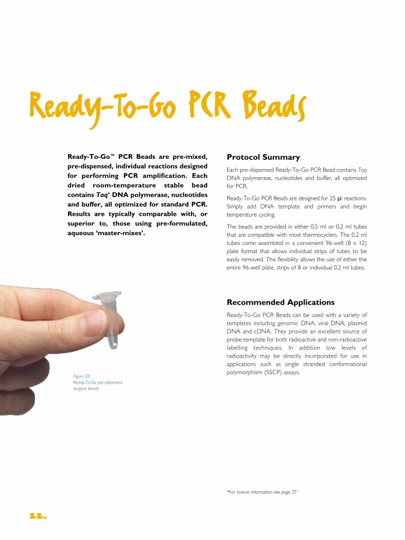
Ready-To-Go PCR Beads
Ready-To-Go™ PCR Beads are pre-mixed,pre-dispensed, individual reactions designedfor performing PCR amplification. Eachdried room-temperature stable beadcontains Taq* DNA polymerase, nucleotidesand buffer, all optimized for standard PCR.Results are typically comparable with, orsuperior to, those using pre-formulated,aqueous ‘master-mixes’.
2222..
*For licence information see page 37
Figure 28 Ready-To-Go pre-dispensed reagent beads

Convienience PCR components are provided in pre-formulated, single dose beads, reducing risk of pipettingerrors and introducing contaminants. Pre-dispensed,individually packed PCR reactions, ensures a freshreaction that dissolves quickly in a PCR compatible tube.
Consistency Application testing ensures each batch ofbeads delivers consistent results.
Compatability Each reaction is provided in a thin-walled0.5 ml or 0.2ml tube, which fit directly into mostthermocyclers.
2233..
Figure 30
PCR results comparingperformance of Ready-To-GoPCR Beads with pre-formulated,aqueous ‘master mixes’. Both thePCR Beads and the ‘master-mixes’ contained all componentsfor PCR except template andprimer. 100 ng of humangenomic template and primersspecific for human aromatase,a single-copy gene, were addedto each reaction to a final volume of 25 µl.Reactions were subjected to 35 cycles of : 95°C for 1 minute,55°C for 1 minute; 72°C for 2minutes. An equal volume ofeach reaction was loaded ontoan agarose gel. M = 100 base-pair ladder. Datacourtesy of Denise Garvin.
Ordering Information
Ready-To-Go PCR Beads (0.5 ml tubes) 100 reactions 27-9555-01
Ready-To-Go-PCR Beads (0.2 ml tubes/plate) 96 reactions 27-9553-01
Ready-To-Go-PCR Beads (0.2 ml tubes/plate) 5 x 96 reactions 27-9553-02
Related products
Ready-To-Go DNA Labelling Beads 27-9240-01
A BFigure 29
Comparison of Ready-To-Go Beads with conventional PCR mixtures for the amplification of genes form interferon-γ activated human aorticendothelial cells. Panel (A) amplification of TGF-α . Panel (B) amplification of β-actin. M = 100 Base Pair Ladder, beads = Ready -To-Go PCR Beads; mix =conventional PCR mixture; + = control reaction supplied with PCR beads.
RNA Labelling Kit
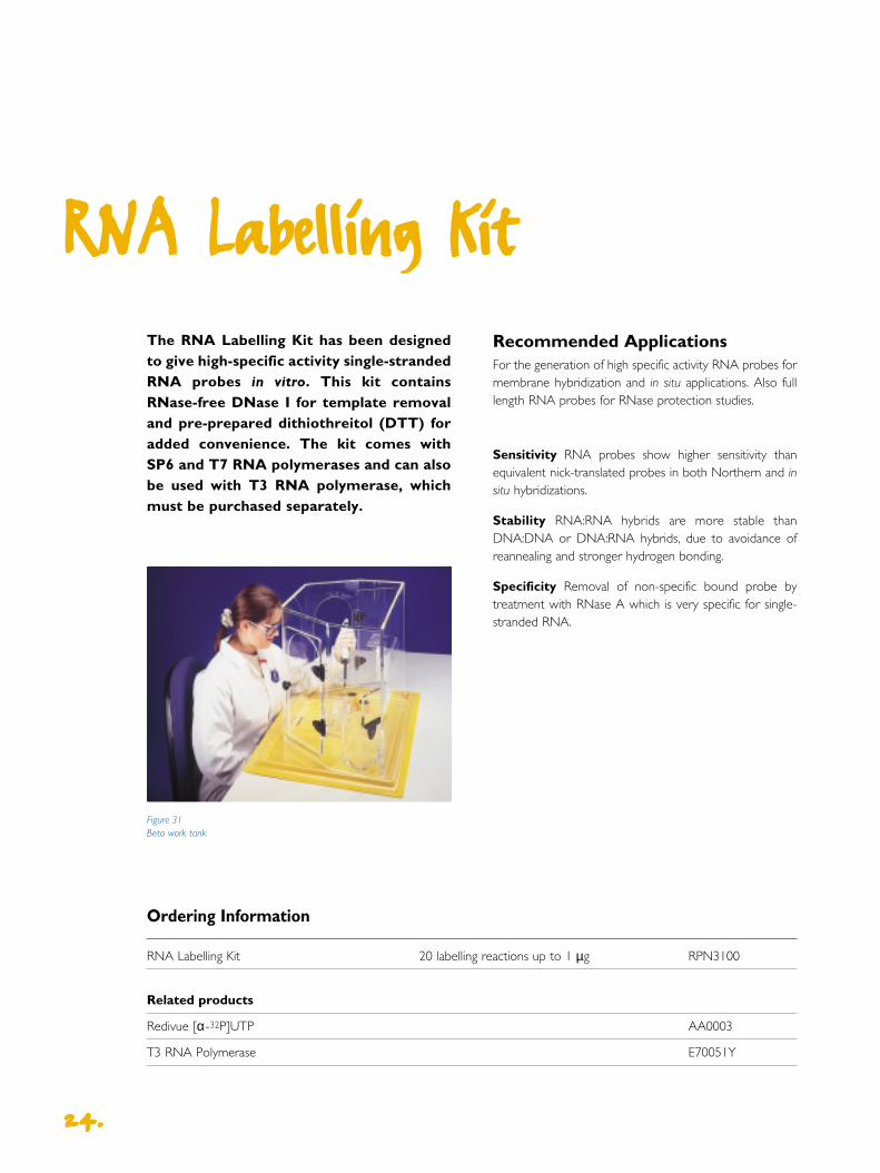
The RNA Labelling Kit has been designedto give high-specific activity single-strandedRNA probes in vitro. This kit containsRNase-free DNase I for template removaland pre-prepared dithiothreitol (DTT) foradded convenience. The kit comes withSP6 and T7 RNA polymerases and can alsobe used with T3 RNA polymerase, whichmust be purchased separately.
Recommended ApplicationsFor the generation of high specific activity RNA probes formembrane hybridization and in situ applications. Also fulllength RNA probes for RNase protection studies.
Sensitivity RNA probes show higher sensitivity thanequivalent nick-translated probes in both Northern and insitu hybridizations.
Stability RNA:RNA hybrids are more stable thanDNA:DNA or DNA:RNA hybrids, due to avoidance ofreannealing and stronger hydrogen bonding.
Specificity Removal of non-specific bound probe bytreatment with RNase A which is very specific for single-stranded RNA.
Ordering Information
RNA Labelling Kit 20 labelling reactions up to 1 µg RPN3100
Related products
Redivue [α-32P]UTP AA0003
T3 RNA Polymerase E70051Y
2244..
Figure 31Beta work tank

2255..
Rapid-hyb™ Buffer is a rate enhancinghybridization buffer for rapid hybridizationof radiolabelled nucleic acid probes to membrane-bound targets. In someNorthern blotting experiments Rapid-hybBuffer contributed to a five foldimprovement in sensitivity.
Recommended Applications
Rapid-hyb Buffer is optimized for use in a wide range ofapplications, including Southern, Northern, dot/slot blotsand colony/plaque lifts.
Speed Single-copy gene detection is possible after only a 2 hour hybridization with 32P-labelled probes.
High signal to noise ratio Inclusion of chemical blockingagents ensures low backgrounds.
Stability Stores at room temperature – ready to usewithout addition of carrier DNA.
Rapid-hyb Buffer
Compatibility Compatible with DNA, RNA andoligonucleotide probes.
Versatility A wide range of hybridization temperatures(42°C – 70°C) can be used for optimal results.
Ordering Information
Rapid-hyb Buffer 125 ml RPN1635
Rapid-hyb Buffer 500 ml RPN1636
Related products
Rediprime II DNA Labelling System RPN1633
5’-End Labelling Kit RPN1509
3’-End Labelling Kit N4020
2255..
Figure 32
Northern blot analysis using:
(A) Standard hybridization buffer (B) Rapid-hyb buffer
Northern blots of HeLa cell total RNA (0.5 µg loadings). Linearized pHSP70probe labelled with (α-32P)dCTP using megaprime labelling system.Hybridizations were at 65°C for 1 hour using a probe concentration of 2 ng/ml. Exposure to Hyperfilm MP overnight.
A B

Compatibility Exhaustive testing of each Redivueproduct ensures equivalent performance to standardformat radiolabelled nucleotides.
Consistency Elimination of repeated freeze thaw cyclesensuring consistent performance.
Redivue Nucleotides
Redivue™ 32P, 33P and 35S nucleotidescontain a novel red dye and stabilizersolution, providing advantages in bothhandling and storage. Redivue is mucheasier to use being clearly visible in yourpipette tip and in reaction mixes down to 1 µl in 50 µl. Redivue nucleotides can beused straight from the refrigerator in liquidform which saves time and avoids freezethaw cycles.
Recommended Applications
Redivue nucleotides can be substituted directly forstandard formulation products in the majority of molecularbiology applications.
Ease of use Addition of an intense red dye improvesvisibility and handling characteristics.
Stability Stabilized format allows storage at 4°C in aconvenient liquid form.
2266..
Figure 35
Detection of B-myb mRNA by Northern hybridization using Redivue or standard [α-32P]dCTP
Results supplied by Dr E Lam, Ludwig Institute for Cancer Research, London
Figure 33
Use straight from thefridge. Dispensing issimpler with easy to use Redivue nucleotides
Figure 34
Redivue nucleotides can make experiments quicker to set up

2277..
Ordering Information
Redivue Nucleotides
[α-32P]dATP ~220 TBq/mmol, ~6000 Ci/mmol AA0074
[α-32P]dATP ~110 TBq/mmol, ~3000 Ci/mmol AA0004
[α-32P]dATP ~30 TBq/mmol, ~800 Ci/mmol AA0084
[α-32P]dATP ~15 TBq/mmol, ~400 Ci/mmol AA0064
[α-32P]dCTP ~220 TBq/mmol, ~6000 Ci/mmol AA0075
[α-32P]dCTP ~110 TBq/mmol, ~3000 Ci/mmol AA0005
[α-32P]dCTP ~30 TBq/mmol, ~800 Ci/mmol AA0085
[α-32P]dCTP ~15 TBq/mmol, ~400 Ci/mmol AA0065
[α-32P]dGTP ~110 TBq/mmol, ~3000 Ci/mmol AA0006
[α-32P]dGTP ~30 TBq/mmol, ~800 Ci/mmol AA0086
[α-32P]dGTP ~15 TBq/mmol, ~400 Ci/mmol AA0066
[α-32P]dTTP ~110 TBq/mmol, ~3000 Ci/mmol AA0007
[α-32P]dTTP ~30 TBq/mmol, ~800 Ci/mmol AA0087
[α-32P]dTTP ~15 TBq/mmol, ~400 Ci/mmol AA0067
[α-32P]dUTP ~110 TBq/mmol, ~3000 Ci/mmol AA0003
[γ-32P]dATP > 185 TBq/mmol, >5000 Ci/mmol AA0018
[γ-32P]dATP ~110 TBq/mmol, ~3000 Ci/mmol AA0068
[γ-33P]dATP ≥ 92.5 TBq/mmol, ≥ 2500 Ci/mmol AH9968
[α-33P]dATP ≥ 92.5 TBq/mmol, ≥ 2500 Ci/mmol AH9904
[α-33P]dCTP ≥ 92.5 TBq/mmol, ≥ 2500 Ci/mmol AH9905
[α-33P]dUTP ≥ 92.5 TBq/mmol, ≥ 2500 Ci/mmol AH9903
[35S]dATPαS > 37 TBq/mmol, >1000 Ci/mmol AG1000
[35S]dATPαS ~22 TBq/mmol, ~600 Ci/mmol AG1001
[35S]dATPαS ~15 TBq/mmol, ~400 Ci/mmol AG1002
[35S]dCTPαS > 37 TBq/mmol, >1000 Ci/mmol AG0135
[35S]dCTPαS ~22 TBq/mmol, ~600 Ci/mmol AG0035
[35S]dCTPαS ~15 TBq/mmol, ~400 Ci/mmol AG0025
Related products
Rediprime II DNA Labelling System RPN1633
Ready-To-Go DNA Labelling Beads (dCTP) 27-9240-01
5’-End Labelling Kit RPN1509
3’-End Labelling Kit RPN4020
RNA Labelling Kit RPN3100
Ready-To-Go PCR Beads 27-9555-01

Hybond Membranes for
Nucleic Ac id Blotting
Hybond™ membranes are available tosupport all major nucleic acid blottingapplications.
Rely on Amersham Biosciences' technical experience in all types of blottingfor clear guidance on the choice ofmembrane best suited for your application.
Individual technical advice is available on allaspects of blotting and hybridization viae:mail at
Choosing the correct membrane
The following table is designed to help in the choice of the best possible membrane for each application
2288..
Application Method Recommended membrane
Southern blotting Radioactive detection Hybond-XL
Non-radioactive detection Hybond-N+
Alkali blotting Hybond-XL / Hybond-N+
Low volume hybridizations Hybond-NX
Rapid-hyb Buffer Hybond-XL
Fingerprinting Hybond-Nfp
Northern blotting Radioactive detection Hybond-XL
Non-radioactive detection Hybond-N+
Low volume hybridizations Hybond-NX
Rapid-hyb Buffer Hybond-XL
Dot and slot blots Radioactive detection Hybond-XL
Non-radioactive detection Hybond-N+
Colony and plaque blots Radioactive detection Hybond-XL
Non-radioactive detection Hybond-N+
Table 4 Choosing the correct membrane
Hybond-XL Hybond-XL is a charged nylonmembrane designed exclusively for radioactive nucleic acid transfer applications to achieve an improved signal-to-noise ratio. It has a greater concentration and amore even distribution of positive charge than othercharged nylon membranes. Hybond-XL producesexcellent results with a wide variety of applications andtarget/probe combinations. This membrane retains all theadvantages of other nylon membranes such as high nucleicacid binding capacity and high tensile strength.
Hybond-N+ Hybond-N+ is a positively chargednylon membrane which yields excellent sensitivity in bothalkali blotting and conventional Southerns. Nucleic acidsamples may be fixed by simple alkali treatment or alkaliblotting rather than UV exposure or baking, though UVfixation is recommended for maximum reproducibility.
Hybond-N Hybond-N is a neutral nylonmembrane capable of high sensitivity in DNA and RNAblotting. It is a strong supported membrane that isinherently hydrophilic and requires no pre-wetting. It iseasy to quickly crosslink nucleic acids to nylon using UVlight. This makes Hybond-N ideal for any standardradioactive Southern or Northern blotting procedure,except for customers using low hybridization volumeswhere Hybond-NX is recommended.
Hybond-NX Hybond-NX is a neutral nylonmembrane with all the properties of Hybond-N but hasbeen specially developed for use with high throughputapplications where low hybridization buffer volumes areused. Hybond-NX gives cleaner background thanHybond-N when low buffer volumes are used.
Hybond-Nfp Hybond-Nfp is a neutral nylonmembrane with all of the properties of Hybond-N, it waslaunched in response to the needs of researchersperforming DNA fingerprinting. These customerspreferred to validate a sample of a particular batch in theirapplication before ordering the rest of the batch.Validation in this demanding technique can take up to 2months, therefore to remove this inconvenience we offerHybond-Nfp which is pre-validated by independent thirdparty DNA fingerprinting experts.
2299..
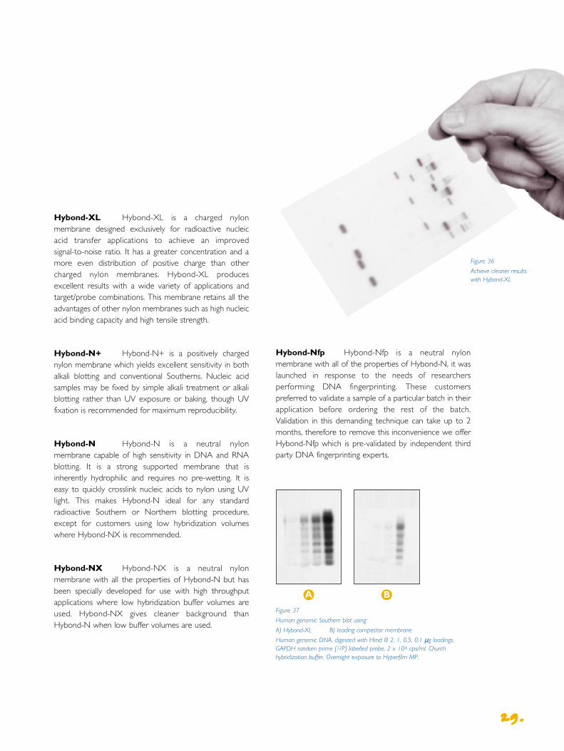
Figure 37
Human genomic Southern blot using:
A) Hybond-XL B) leading competitor membrane
Human genomic DNA, digested with Hind III 2, 1, 0.5, 0.1 µg loadings.GAPDH random prime [32P] labelled probe, 2 x 10⁶ cps/ml. Churchhybridization buffer. Overnight exposure to Hyperfilm MP.
Figure 36
Achieve cleaner resultswith Hybond-XL
A B

Use the following table to help choose of the best possible film and exposure conditions for your application.
Autoradiography Films
Amersham Biosciences ' expertisein the labelling and detection of biological molecules supports a range of autoradiography products designedspecifically for life science applications.
This range includes the market leadingKodak™ X-Omat™ AR film and the high speedBioMax™ MR and BioMax MS films whichutilize Kodak T-grain emulsion technology.
3300..
Application Result required Label Method Film
Southern/Northern blots High speed and sensitivity 32P Pre-flash with Hyperfilm MPColony/plaque blots Accurate quantitation screens at -70°C
Maximum speed and sensitivity 32P Screens at -70°C BioMax MS
Optimum resolution 33P/35S Direct autoradiography Hyperfilm βmaxBioMax MR
Non-radioactive ECL Pre-flash and direct detection Hyperfilm ECL
Slot blots High speed and sensitivityaccurate quantitation 32P Pre-flash with screens at -70°C Hyperfilm MP
Maximum speed and sensitivity 32P Screens at -70°C BioMax MS
non-radioactive ECL Pre-flash and direct detection Hyperfilm ECL
Dideoxy sequencing High speed 32P Direct Hyperfilm MPautoradiography BioMax MR
Optimum resolution and maximum Directspeed for difficult sequences 35S autoradiography Biomax MR
Good resolution and speed Directfor routine work 35S autoradiography Hyperfilm MP
Cycle sequencing Optimum resolution and maximum Directspeed for difficult sequences 33P autoradiography BioMax MR
Good resolution and speed Directfor routine work 33P autoradiography Hyperfilm MP
In situ hybridization Macroscale in situ optimization Directof parameters 32P autoradiography Hyperfilm MP
BioMax MS This film and screen combinationoffers exceptional sensitivity in 32P and 125I applications. The high performance film of choice forradioactive blotting.
X-Omat AR A general-purpose film for use with allcommonly used isotopes. Coated with emulsion on both sides for both direct exposure and use withintensifying screens.
Related autoradiographyaccessories
Hypercassette™ autoradiography cassettesHyperprocessor™ automatic film processorHyperscreen™ intensifying screensHypertorch™
Kodak BioMax TranScreen intensifying screensSafelightsSensitize™ preflash unitTrackertape™
Ordering information
For further information please request ‘The CompleteAutoradiography Product Guide’.
Hyperfilm MP An excellent multipurpose film. The clear base gives high contrast and good resolutionwhile its double-sided construction and compatibility with intensifying screens means that it performs well inblotting applications.
Hyperfilm β-max A specialized film with highsensitivity, high resolution and a good linear range.
Hyperfilm ECL The recommended film forchemiluminescent applications. The clear base gives highcontrast and good resolution while its sensitivity profilematches the emission spectrum of ECL and otherchemiluminescent systems.
BioMax MR An excellent film for the directautoradiography of 35S, 14C and 33P. This clear, single-sidedfilm gives high resolution and contrast and the special T-grain technology makes it twice as fast as X-Omat ARwith weak emitters.
3311..
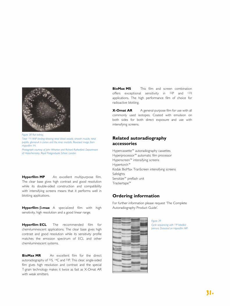
Figure 39
Cycle sequencing with 33P labelledprimers. Detected on Hyperfilm MP.
Figure 38 Rat kidney.
Total 125I ANP binding showing renal blood vessels, smooth muscle, renalpapilla, glomeruli in cortex and the inner medulla. Reversed image fromHyperfilm 3H.
Photograph courtesy of John Wharton and Richard Rutherford. Departmentof Histochemistry, Royal Postgraduate School, London.

Storm
The Amersham Biosciences ™ Storm™ imaging system for gel,blot and macroarray analysis combines proven storagephosphor imaging technology for radioisotope detectionwith fluorescence and chemifluorescence imaging for non-radioactive detection. Researchers can make the move tonon-radioactive gel and blot analysis without sacrificingconventional radioisotope techniques. Storm scans storagephosphor screens and fluorescent and chemifluorescentgels and blots in a large format 35 x 43cm scanning area orsmaller. ImageQuant™ software for Windows NT™ orMacintosh™ is included. ECF detection reagents provideoptimized chemifluorescent detection of Southern andNorthern blots on the Storm system.
Place your storage phosphor screen, gel or blot on the sample area, and point and click to start your scan.The Storm system uses patented Variable Mode Imaging (VMI) architecture to activate the correct opticalcomponents for storage phosphor autoradiography, directfluorescence or chemifluorescence.
Amersham Biosciences Scanners
3322..
Figure 40
Storm imaging system in use
Figure 41
Large samples fit on theStorm systems 35 x 43cmscan area. Storm offers thehigh resolution needed forDNA base identification.
3333..
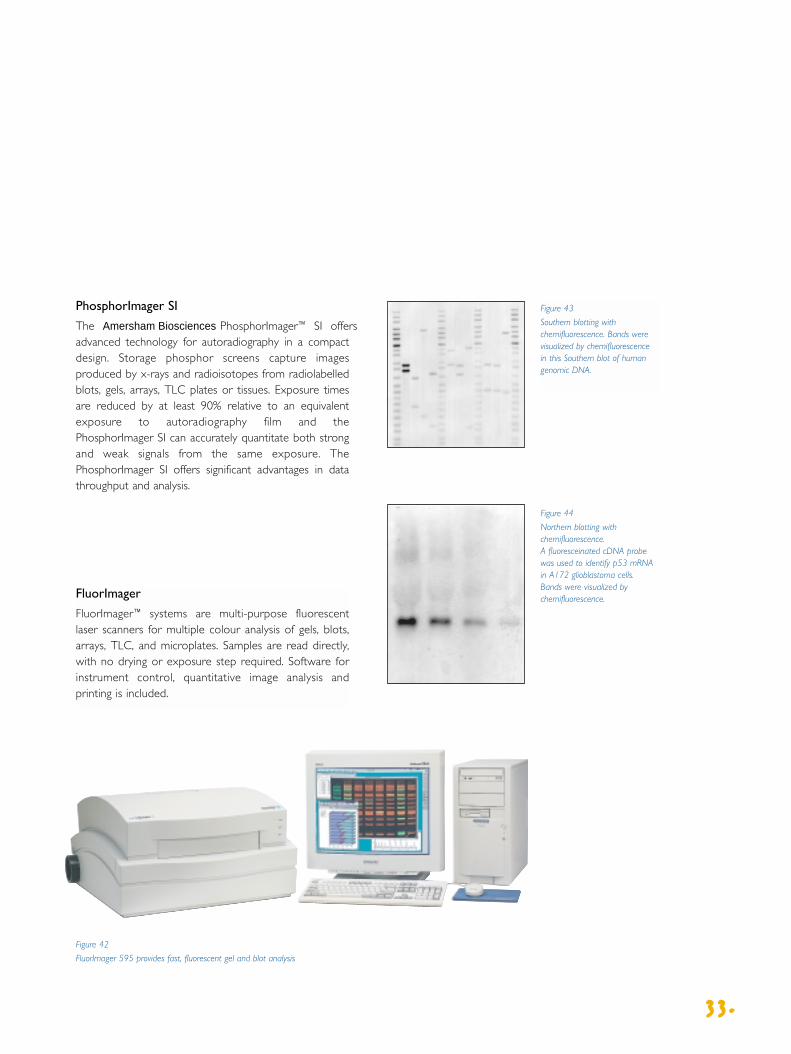
FluorImager
FluorImager™ systems are multi-purpose fluorescentlaser scanners for multiple colour analysis of gels, blots,arrays, TLC, and microplates. Samples are read directly,with no drying or exposure step required. Software forinstrument control, quantitative image analysis andprinting is included.
Figure 43
Southern blotting withchemifluorescence. Bands werevisualized by chemifluorescence in this Southern blot of humangenomic DNA.
Figure 44
Northern blotting withchemifluorescence. A fluoresceinated cDNA probe was used to identify p53 mRNAin A172 glioblastoma cells. Bands were visualized bychemifluorescence.
Figure 42
FluorImager 595 provides fast, fluorescent gel and blot analysis
PhosphorImager SI
The Amersham Biosciences PhosphorImager™ SI offersadvanced technology for autoradiography in a compactdesign. Storage phosphor screens capture imagesproduced by x-rays and radioisotopes from radiolabelledblots, gels, arrays, TLC plates or tissues. Exposure timesare reduced by at least 90% relative to an equivalentexposure to autoradiography film and thePhosphorImager SI can accurately quantitate both strongand weak signals from the same exposure. ThePhosphorImager SI offers significant advantages in datathroughput and analysis.

Vacuum Blotting Units
Vacuum blotting is a quick and reproduciblemethod for transferring nucleic acid tomembranes. The specially designed vacuumpump draws the DNA/RNA through theagarose gel and onto the membrane,therefore reducing transfer times to lessthan an hour.
Speed Rapidly transfer DNA or RNA from agarose gelsup to 20 x 30 cm in size.
Efficiency Efficient and reproducible transfers improveblotting results.
Convenience Minimize gel handling by performing allpre-transfer steps in the unit.
Accuracy Use with VacuGene™ XL blotting pumpensures precise vacuum control.
Accessory products
UV Crosslinker
The Amersham Biosciences™ UV Crosslinker reduces the time required to fix nucleic acidson membranes from hours to less than a minute.
UV-fixed samples give consistently betterhybridization signals than baked samples,especially after repeated re-probings.Nucleic acids can be UV-crosslinked tonylon, nitrocellulose and supported nitro-cellulose membranes.
Reliability Short-wave UV light crosslinks DNA or RNAsecurely to membranes for repeated re-probings.
Accuracy Microprocessor controlled for accurate andreproducible UV dosage.
Convenience Keypad selection of pre-programmed oruser-set energy or time exposures.
Flexibility Spacious chamber accepts membranes up to 25 x 30cm, but compact footprint requires only
35 x 40cm of bench space.
3344..
Figure 45
The Amersham Biosciences UVCrosslinker
Ordering Information
Ultraviolet Crosslinker 115 V 60 Hz 80-6222-31Includes five 8 W short-wave UV lamps
Ultraviolet Crosslinker 230 V 50 Hz 80-6222-50Includes five 8 W short-wave UV lamps
8 W, 254 nm replacement UV lamp 80-6223-64
Ordering Information
VacuGene XL Vacuum Blotting System 13-2223-01
VacuGene XL Vacuum Blotting Unit 80-1266-24
VacuGene XL Vacuum Blotting Pump 110 V 80-1265-14
VacuGene XL Vacuum Blotting Pump 220 V 80-1265-15
3355..
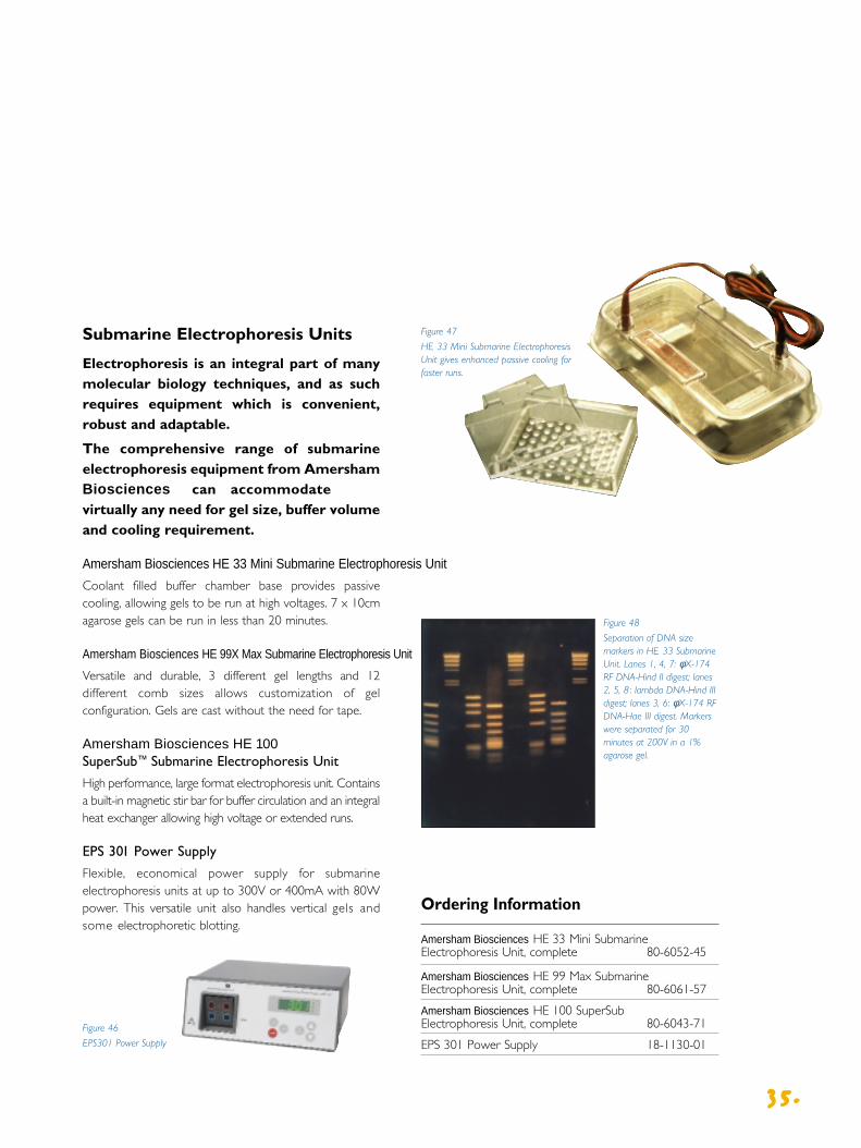
Figure 47
HE 33 Mini Submarine ElectrophoresisUnit gives enhanced passive cooling forfaster runs.
Ordering Information
Amersham Biosciences HE 33 Mini SubmarineElectrophoresis Unit, complete 80-6052-45
Amersham Biosciences HE 99 Max Submarine Electrophoresis Unit, complete 80-6061-57
Amersham Biosciences HE 100 SuperSub Electrophoresis Unit, complete 80-6043-71
EPS 301 Power Supply 18-1130-01
Figure 48
Separation of DNA sizemarkers in HE 33 SubmarineUnit. Lanes 1, 4, 7: φX-174RF DNA-Hind II digest; lanes2, 5, 8 : lambda DNA-Hind IIIdigest; lanes 3, 6: φX-174 RFDNA-Hae III digest. Markerswere separated for 30minutes at 200V in a 1%agarose gel.
Submarine Electrophoresis Units
Electrophoresis is an integral part of manymolecular biology techniques, and as suchrequires equipment which is convenient,robust and adaptable.
The comprehensive range of submarineelectrophoresis equipment from AmershamBiosciences can accommodatevirtually any need for gel size, buffer volumeand cooling requirement.
Amersham Biosciences HE 33 Mini Submarine Electrophoresis UnitCoolant filled buffer chamber base provides passivecooling, allowing gels to be run at high voltages. 7 x 10cmagarose gels can be run in less than 20 minutes.
Amersham Biosciences HE 99X Max Submarine Electrophoresis UnitVersatile and durable, 3 different gel lengths and 12different comb sizes allows customization of gelconfiguration. Gels are cast without the need for tape.
Amersham Biosciences HE 100 SuperSub™ Submarine Electrophoresis Unit
High performance, large format electrophoresis unit. Containsa built-in magnetic stir bar for buffer circulation and an integralheat exchanger allowing high voltage or extended runs.
EPS 301 Power Supply
Flexible, economical power supply for submarineelectrophoresis units at up to 300V or 400mA with 80Wpower. This versatile unit also handles vertical gels andsome electrophoretic blotting.
Figure 46
EPS301 Power Supply

Ordering Information
Ethidium Bromide Solution, 10 mg/ml 17-1328-01
Agarose NA, 100 g 17-0554-02
Agarose Prep, 50 g 80-1103-07
3366..
Ordering Information
Hybridization Oven/Shaker 220/240 V 50 Hz RPN2510
Hybridization Oven/Shaker 110/115 V 60 Hz RPN2511
Hybridization Oven/Shaker 100 V 50/60 Hz RPN2512
Electrophoresis Chemicals
In gel electrophoresis the quality of thechemicals used for gel casting and gelstaining is critical to obtain good results.
Ethidium Bromide Widely used for detecting DNAand RNA, ethidium bromide binds to double-strandedregions to form UV fluorescent complexes. As little as 50 ng of DNA can be detected with concentrations of 0.5 – 1.0 µg/ml of stain.
Agarose Agarose with low electroendosmosis is recommended for most DNA and RNA applications. To ensure maximum enzyme efficiency, a highly purifiedAgarose NA can be used. For preparative work, AgarosePrep with low gelling temperature should be used.
Hybridization Oven/Shaker
The Hybridization Oven/Shaker rigidlycontrols the hybridization temperature andfrequency of agitation, thus ensuring thatstringency and washing conditions aremaintained. Developed for use with bothradioactive and non-radioactive labellingsystems. A choice of hybridization modes is available by use of a variable speedrotisserie or platform shaker.
Economy Reduced hybridization buffer volumesminimizes probe usage.
Safety Acrylic/polycarbonate door minimizes exposure to beta radiation.
Versatility Interchangeable rotisserie and platform shaker.
Figure 49
Hybridization Oven Shaker
Accessory products

Trademarks
AlkPhos Direct, ECF, ECL, ECL Direct, FluorImager, Gene Images, Amersham Biosciences,Hybond, Hypercassette, Hyperfilm, Hyperprocessor, Hyperscreen, Hypertorch,ImageQuant, Megaprime, Amersham Biosciences, Multiprime, PhosphorImager,Rapid-hyb, Ready-To-Go Rediprime, Redivial, Redivue, Sensitize, Storm,SuperSub, TrackerTape, TransVac, Vacublot, VacuGene are trademarks ofAmersham Biosciences Limited or its subsidiaries.
Amersham and Amersham Biosciences are trademarks of Amersham plc
CDP-Star is a trademark of Tropix Inc
Kodak, BioMax,TranScreen, and X-Omat are trademarks of Eastman Kodak Co.
Windows NT is a trademark of Microsoft Corp.
Macintosh is a trademark of Apple Computers.
Licence information
CDP-Star is protected by Tropix Inc. under one or more of US patent numbers5326882 and 4931569
ECF substrate is manufactured for Amersham Biosciences Limited by JBL Scientific Inc, and is sold under licence from JBL Scientific Inc.
ECL Direct/AlkPhos Direct labelling reagents are covered by European patentnumber 120376B1 and sold under licence from European Molecular BiologyLaboratory. ECL Direct (RPN3000/3001/3005) is covered by European patentnumbers 373017 and 513040 and foreign equivalents and is sold under licencefrom Instituit Pasteur.
Gene Images hybridization buffer is covered by US patent number 5512436 andforeign equivalents.
PCR: Taq DNA polymerase is sold under licensing arrangements with RocheMolecular Systems, F Hoffmann-La Roche Ltd and the Perkin Elmer Corporation.Purchase of these products is accompanied by a limited licence to use it in thepolymerase Chain Reaction (PCR) process for research in conjunction with athermal cycler whose use in the automated performance of the PCR process iscovered by the up-front licence fee, either by payment to Perkin-Elmer or aspurchased, i.e. an authorized thermal cycler.
The Polymerase Chain Reaction (PCR) is covered by patents owned by RocheMolecular Systems and F Hoffmann-La Roche Ltd. A licence to use the PCRprocess for certain research and development activities accompanies thepurchase of certain reagents from licensed suppliers such as AmershamBiosciences Limited and affiliates when used in conjunction with anauthorized thermal cycler.
© Amersham Biosciences UK Limited 1998 - All rights reserved
All goods and services are sold subject to the terms and conditions of sale of thecompany within the Amersham Biosciences group which supplies them. A copy of these terms and conditions is available on request.
Amersham Biosciences UK Limited Amersham Place LittleChalfont Buckinghamshire England HP7 9NA
Amersham Biosciences AB S-751 82 Uppsala Sweden
Amersham Biosciences Inc 800 Centennial Avenue PO Box 1327Piscataway NJ 08855 USA
3377..

S1295/98/05
For more information about
Nucleic Acid Labelling and Detection
visit our website at:
www.amershambiosciences.com
Asia PacificAmersham Biosciences Asia Pacific LtdTel: +852 2811 8693Fax: +852 2811 5051
AustralasiaAmersham Biosciences Australia Pty LtdTel: +61 2 9894 5188Fax: +61 2 9899 7511
AustriaMEDPROTel: +43 1479 15 690Fax: +43 1479 83 5217
BelgiumAmersham Biosciences BeneluxTel: 0800 73888Fax: +32 3272 1637
CanadaAmersham Biosciences, Inc.Tel: 1 800 463 5800Fax: 1 800 567 1008
Central EuropeAmersham Biosciences Export GmbHTel: +43 1 982 3826Fax: +43 1 985 8327
DenmarkAmersham Biosciences Tel: +33 4516 24000Fax: +33 4516 2424
FinlandAmersham Biosciences Europe GmbHTel: 358 (9) 512 3940Fax: 358 (9) 512 1710
Russian FederationZAO Amersham Biosciences Tel: +7 (950) 232 0250 / 956 1137Fax: +7 (950) 232 6377
FranceAmersham Biosciences Tel: +33 169 18 28 00Fax: +33 169 18 28 28
GermanyAmersham Biosciences GmbHTel: +49 0761 4903 0Fax: +49 0761 4903 247
ItalyAmersham Italia SrlTel: +39 02 27322 1Fax: +39 02 27302 212
JapanAmersham Biosciences KKTel: 81 3 5992 2828Fax: 81 3 5992 5186
Latin AmericaAmersham Biosciences do Brasil LtdaTel: +55 11 3667 5700Fax: +55 11 3667 58 99
Middle East and AfricaAmersham Biosciences Export GmbHTel: +30 (1) 96 00 687Fax: +30 (1) 96 00 693
NetherlandsAmersham Biosciences BeneluxTel: 0165 580 410Fax: 0165 580 401
NorwayAmersham Biosciences Tel: 63 89 23 10Fax: 63 89 23 15
South East AsiaAmersham Biosciences ABTel: 60 3 724 2080Fax: 60 3 724 2090
South East EuropeAmersham Biosciences GmbHTel: +43 (1) 982 3826Fax: +43 (1) 985 8327
SpainAmersham Biosciences Europe GmbHTel: 93 594 49 82Fax: 93 594 49 55
SwedenAmersham Biosciences ABTel: 018 164000Fax: 018 712777
SwitzerlandAmersham Biosciences Europe GmbHTel: 01 802 81 50Fax: 01 802 81 51
UKAmersham Biosciences UK LimitedTel: 0800 515313Fax: 0800 616927
USAAmersham Biosciences IncTel: +1 800 526 3593Fax: +1 800 329 3593