the gray zone around dental implants: keys to esthetic...
TRANSCRIPT
2THE AMERICAN JOURNAL OF ESTHETIC DENTISTRYTHE AMERICAN JOURNAL OF ESTHETIC DENTISTRY
The Gray Zone
Around Dental Implants:
Keys to Esthetic Success
Iñaki Gamborena, DMD, MSD, FID
Clinical Assistant Professor, Department of Preventive and Restorative
Sciences, University of Pennsylvania School of Dental Medicine, Philadelphia,
Pennsylvania, USA; Private Practice, San Sebastian, Spain.
Markus B. Blatz, DMD, PhD
Professor and Chairman, Department of Preventive and Restorative
Sciences, University of Pennsylvania School of Dental Medicine, Philadelphia,
Pennsylvania, USA.
Correspondence to:
Dr Iñaki Gamborena
Resurecccion mª de Azkue, 6 20018
San Sebastian, Spain.
Email: [email protected]

VOLUME 1 • NUMBER 1 • FALL 2011VOLUME 1 • NUMBER 1 • FALL 2011
Single-implant restorations in the anterior maxilla have become a routine treat-
ment option. While customized tooth-colored prosthetic components show great-
ly improved clinical outcomes, esthetic success relies not only on the restorative
result, but also on the condition of the soft tissues. A common esthetic shortcom-
ing is the grayish appearance of the peri-implant soft tissues, which are difficult
to manipulate around dental implants. The parameters and clinical guidelines that
should be used to influence esthetic success and avoid the gray zone around
implant restorations can be categorized into five key factors: (1) optimal three-
dimensional implant placement for functional and esthetic long-term implant
success; (2) maximized soft tissue thickness to conceal the implant-restorative
interface; (3) proper abutment selection to improve biocompatibility, tissue stabil-
ity, color, translucency, and fluorescence; (4) careful crown restoration to imitate
the natural teeth; and (5) awareness of the lip line, which may greatly influence the
final outcome. Mimicking the inherent optical properties, especially fluorescence,
of natural teeth with implant components and crown materials is fundamental for
ideal restorative and soft tissue esthetics. (Am J Esthet Dent 2011;1:xxx–xxx.)
3

GAMBORENA ANd BLAtz
4THE AMERICAN JOURNAL OF ESTHETIC DENTISTRYTHE AMERICAN JOURNAL OF ESTHETIC DENTISTRY
The esthetic success of a dental
restoration is judged by its inte-
gration with the surrounding dentition
in respect to position, angulation, di-
mensions, proportions, shape, surface
morphology, and shade.1–3 Other cru-
cial esthetic parameters that are often
overlooked include the morphology,
texture, and ultimately the color of the
surrounding gingiva.3,4 The soft tissue
is the natural frame of the teeth and
any dental restoration and is, therefore,
a fundamental parameter for esthetic
success.1,3,5 This aspect is often ne-
glected because successful soft tissue
outcomes—including handling, manip-
ulation, and healing—are very demand-
ing, time intensive, and unpredictable.5
Magne et al6 described a prevalence
of grayish soft tissue discolorations
around tooth-supported full-coverage
porcelain-fused-to-metal and even all-
ceramic restorations. Interestingly, oth-
er perioral facial parameters such as
position of the upper lip and height of
the smile line7–9 also seem to influence
the degree of gingiva discoloration.
The authors note that “this problem is
particularly evident in the presence of
the upper lip, which can generate an
‘umbrella effect’ characterized by gray
marginal gingivae and dark interdental
papillae.”6
This umbrella effect is magnified with
dental implant restorations in the an-
terior maxilla because the supporting
hard and soft tissues are often com-
promised even before restorative treat-
ment and are influenced by the color
and design of the implant, its prosthetic
components, and the definitive resto-
ration.10–17 Therefore, ideal periodontal
and restorative esthetic success with
maxillary anterior implant-supported
restorations presents a great challenge
for the entire dental team and depends
on a variety of parameters.10–14
The parameters and clinical guide-
lines that should be used to influence
esthetic success and avoid the gray
zone around implant restorations can
be categorized into five key factors: (1) optimal three-dimensional (3D) implant
placement for functional and esthetic
long-term implant success; (2) maxi-
mized soft tissue thickness to conceal
the implant-prosthetic component inter-
face; (3) proper abutment selection to
improve biocompatibility, tissue stabil-
ity, and color to provide a perfect blend
with surrounding tissues and teeth; (4) careful crown restoration to imitate the
natural teeth; and (5) awareness of the
lip line, which may greatly influence the
final outcome.
3D IMPLANT PLACEMENT
The fundamental factor for long-term
functional and esthetic success as well
as soft tissue color and stability is opti-
mal 3D implant placement.18 A simple
but essential guideline is to position the
implant as close as possible to where
the natural tooth was or ideally would
be.10 If a line is drawn at the center of
the implant along its long axis and ex-
tending through the tooth restoration,
it should run through the center of the
incisal edge of the prospective tooth
(Fig 1). The greater the 3D mismatch
between the crown and implant body,
the poorer and less stable the final

GAMBORENA ANd BLAtz
5VOLUME 1 • NUMBER 1 • FALL 2011
outcome will be. The incisal edge is
also the target for the angulation of
the implant. An implant that is angu-
lated too far to the buccal aspect will
result in greater tissue recession under
functional load. Conversely, a palatally
placed implant leads to a more ex-
treme emergence profile, resulting in
increased bone resorption and thinning
of the tissues. Both situations will lead
to an intensified grayish appearance of
the soft tissues at the gingival margin.
The third dimension is determined
by the depth of the implant in respect
to the marginal bone and soft tissue.
An implant placed at the proper depth
allows for the development of an ideal
emergence profile and a soft tissue col-
lar void of a gray zone. It is impossible
to create a proper emergence profile
when the implant is placed too shal-
low, while an implant placed too deep
is difficult to manage clinically and in-
creases the possibility of peri-implant
infection, inflammation, and bone loss.
A surgical guide fabricated from the
diagnostic wax-up/setup is an indis-
pensible tool to ensure proper 3D im-
plant placement. The anticipated incisal
edge position of the final tooth restora-
tion determines the position, angulation,
and depth of the implant in all three di-
mensions, which directly influence the
position, height, and thickness of the
surrounding hard and soft tissues.10,12
SOFT TISSUE THICkNESS
Even in cases where ideal implant
placement was achieved, the esthetic
outcome may become compromised
over time due to resorption of the mar-
ginal bone and soft tissues.5,19
Fig 1 (right) Maxillary anterior implants should
be positioned and angulated so that a virtual line
through the center of the implant along its long
axis would run through the center of the incisal
edge of the prospective crown.

GAMBORENA ANd BLAtz
6THE AMERICAN JOURNAL OF ESTHETIC DENTISTRY
Case 1 (Figs 2 to 6) illustrates a situa-
tion where a single implant was placed
immediately after extraction of the
maxillary right central incisor without
any hard or soft tissue augmentation. A
modified metal abutment was fabricat-
ed, and the definitive restoration was
inserted (Figs 2 and 3). A follow-up
photograph taken several years post-
operatively reveals a grayish appear-
ance of the soft tissue surrounding the
implant restoration (Fig 4). This discol-
oration becomes increasingly evident
11 years after completion as a result of
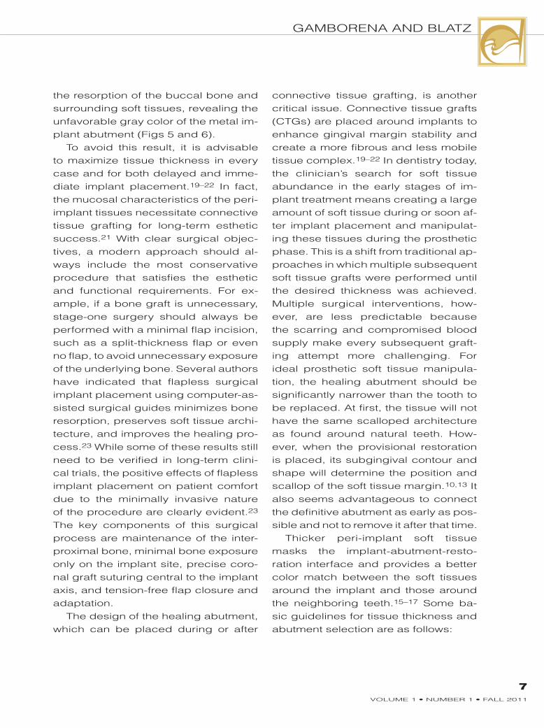
Figs 2a and 2b A modified metal abutment was used after im-
mediate implant placement at the maxillary right central incisor site
without bone or soft tissue augmentation.
Fig 3 Postoperative situation
showing the implant-supported
crown.
Fig 4 (above left) Follow-up view after several years reveals a
grayish appearance of the soft tissues.
Fig 5 (above right) Follow-up view after 11 years showing soft tis-
sue discoloration due to the metal abutment.
Fig 6 (left) Periapical radiograph after 11 years reveals loss of
buccal bone.
Case 1
GAMBORENA ANd BLAtz
7VOLUME 1 • NUMBER 1 • FALL 2011
the resorption of the buccal bone and
surrounding soft tissues, revealing the
unfavorable gray color of the metal im-
plant abutment (Figs 5 and 6).
To avoid this result, it is advisable
to maximize tissue thickness in every
case and for both delayed and imme-
diate implant placement.19–22 In fact,
the mucosal characteristics of the peri-
implant tissues necessitate connective
tissue grafting for long-term esthetic
success.21 With clear surgical objec-
tives, a modern approach should al-
ways include the most conservative
procedure that satisfies the esthetic
and functional requirements. For ex-
ample, if a bone graft is unnecessary,
stage-one surgery should always be
performed with a minimal flap incision,
such as a split-thickness flap or even
no flap, to avoid unnecessary exposure
of the underlying bone. Several authors
have indicated that flapless surgical
implant placement using computer-as-
sisted surgical guides minimizes bone
resorption, preserves soft tissue archi-
tecture, and improves the healing pro-
cess.23 While some of these results still
need to be verified in long-term clini-
cal trials, the positive effects of flapless
implant placement on patient comfort
due to the minimally invasive nature
of the procedure are clearly evident.23
The key components of this surgical
process are maintenance of the inter-
proximal bone, minimal bone exposure
only on the implant site, precise coro-
nal graft suturing central to the implant
axis, and tension-free flap closure and
adaptation.
The design of the healing abutment,
which can be placed during or after
connective tissue grafting, is another
critical issue. Connective tissue grafts
(CTGs) are placed around implants to
enhance gingival margin stability and
create a more fibrous and less mobile
tissue complex.19–22 In dentistry today,
the clinician’s search for soft tissue
abundance in the early stages of im-
plant treatment means creating a large
amount of soft tissue during or soon af-
ter implant placement and manipulat-
ing these tissues during the prosthetic
phase. This is a shift from traditional ap-
proaches in which multiple subsequent
soft tissue grafts were performed until
the desired thickness was achieved.
Multiple surgical interventions, how-
ever, are less predictable because
the scarring and compromised blood
supply make every subsequent graft-
ing attempt more challenging. For
ideal prosthetic soft tissue manipula-
tion, the healing abutment should be
significantly narrower than the tooth to
be replaced. At first, the tissue will not
have the same scalloped architecture
as found around natural teeth. How-
ever, when the provisional restoration
is placed, its subgingival contour and
shape will determine the position and
scallop of the soft tissue margin.10,13 It
also seems advantageous to connect
the definitive abutment as early as pos-
sible and not to remove it after that time.
Thicker peri-implant soft tissue
masks the implant-abutment-resto-
ration interface and provides a better
color match between the soft tissues
around the implant and those around
the neighboring teeth.15–17 Some ba-
sic guidelines for tissue thickness and
abutment selection are as follows:

GAMBORENA ANd BLAtz
8THE AMERICAN JOURNAL OF ESTHETIC DENTISTRY
� A soft tissue thickness greater than
3 mm allows for the use of titanium
or zirconia abutments without nega-
tive esthetic implications.
� A thin soft tissue of less than 2 to
3 mm requires either a CTG or
zirconia abutment.
� A dentin-colored abutment is
always preferred.
In Case 2 (Figs 7 to 13), a colored in-
stead of a white zirconia abutment was
placed due to the presence of less than
1 mm of labial soft tissue. This approach,
along with the adequate soft tissue sup-
port and contour, provided a satisfying
outcome.
ABUtMENt sELEctiON
In an evaluation of the soft tissue around
single-tooth implant crowns, Fürhauser
et al24 showed that the color of the peri-
implant soft tissue matched that of the
reference tooth in no more than one-
third of cases. Another study found that
all-ceramic implant abutment and crown
Case 2
Fig 7 Thin peri-implant soft
tissue of only 1 mm was
evident on the buccal aspect.
Fig 8 A custom-colored
zirconia abutment (Procera, No-
bel Biocare) was fabricated to
optimize the esthetic outcome.
Fig 9 Colored zirconia abut-
ment and alumina crown
(Procera Crown Alumina, Nobel
Biocare).
Fig 10 Intraoral occlusal view
showing the soft tissue support.
Fig 11 Postoperative buc-
cal view. The tooth-colored
abutment and all-ceramic
crown blend favorably with the
adjacent teeth and surrounding
soft tissue despite the compro-
mised soft tissue thickness.
Fig 12 Postoperative peri-
apical radiograph.
Fig 13 Occlusal view of the
definitive implant-supported
restoration.
GAMBORENA ANd BLAtz
9VOLUME 1 • NUMBER 1 • FALL 2011
materials provide a better soft tissue
color match with neighboring teeth than
do conventional metal-alloy compo-
nents.16 zirconia has been shown to be
the preferred implant abutment material
due to its high strength13,25,26 and ex-
cellent biocompatibility.27–29 The short-
comings of zirconia include its higher
cost and unfavorable optical properties
in regard to color and fluorescence.30
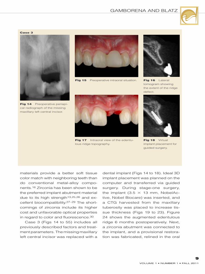
Case 3 (Figs 14 to 55) includes all
previously described factors and treat-
ment parameters. The missing maxillary
left central incisor was replaced with a
Case 3
Fig 14 Preoperative periapi-
cal radiograph of the missing
maxillary left central incisor.
Fig 15 Preoperative intraoral situation.
Fig 17 Intraoral view of the edentu-
lous ridge topography.
Fig 18 Virtual
implant placement for
guided surgery.
Fig 16 Lateral
tomogram showing
the extent of the ridge
defect.
dental implant (Figs 14 to 18). Ideal 3D
implant placement was planned on the
computer and transferred via guided
surgery. During stage-one surgery,
the implant (3.5 × 13 mm, NobelAc-
tive, Nobel Biocare) was inserted, and
a CTG harvested from the maxillary
tuberosity was placed to increase tis-
sue thickness (Figs 19 to 23). Figure
24 shows the augmented edentulous
ridge 6 months postoperatively. Next,
a zirconia abutment was connected to
the implant, and a provisional restora-
tion was fabricated, relined in the oral
9VOLUME 1 • NUMBER 1 • FALL 2011
GAMBORENA ANd BLAtz
10THE AMERICAN JOURNAL OF ESTHETIC DENTISTRY
cavity, and cemented (Figs 25 to 29).
The different lighting conditions (natu-
ral and ultraviolet [UV] light) shown in
Figs 30 to 33 reveal the optical short-
comings of these materials, especially
the lack of natural fluorescence. Figure
Fig 19 Implant placement (3.5 × 13 mm,
NobelActive, Nobel Biocare).
Fig 20 A subepithelial CTG was harvested
from the maxillary tuberosity to augment the
deficient ridge.
Fig 21 After placement of the CTG, the flaps
were adapted without tension and sutured with thin
suture material to limit trauma.
Fig 22 Labial view of the adapted flap after
suturing.
Fig 23 Intraoral situation 1 week postopera-
tively.
Fig 24 Postoperative situation after 6 months
reveals improved ridge morphology.
Case 3 Continued
GAMBORENA ANd BLAtz
11VOLUME 1 • NUMBER 1 • FALL 2011
34 shows the detailed optical charac-
teristics of natural enamel and dentin
under different light sources.
Fluorescence is a crucial property
for natural esthetics.30–32 Colorants
and fluorescent modifiers that can be
applied to zirconia abutments even af-
ter milling and finishing have recently
been developed.30 The abutment or
framework is dipped into a fluores-
cent coloring liquid before sintering
to infiltrate the zirconia (Colour Liquid
Case 3 Continued
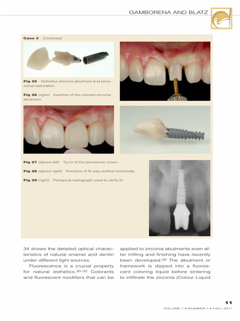
Fig 25 Definitive zirconia abutment and provi-
sional restoration.
Fig 26 (right) Insertion of the colored zirconia
abutment.
Fig 27 (above left) Try-in of the provisional crown.
Fig 28 (above right) Precision of fit was verified extraorally.
Fig 29 (right) Periapical radiograph used to verify fit.

GAMBORENA ANd BLAtz
12THE AMERICAN JOURNAL OF ESTHETIC DENTISTRY
Fluoreszenz, zirkonzahn). the abut-
ment is blow dried after the dipping
process to remove the excess and then
placed under a drying lamp to prevent
damage to the heating elements of the
sinter furnace.
In addition to the regular zirconia,
a more translucent zirconia (Prettau
zirconia “translucent,” zirkonzahn)
along with 16 coloring liquids (zirkon-
zahn) are available. Figures 35 to 37
illustrate the infiltration process and its
Fig 32 Optical properties of the zirconia abut-
ment under natural light.
Fig 33 Optical properties of the zirconia abut-
ment under UV light reveals a lack of fluores-
cence.
Fig 34 Color charac-
teristics of natural enamel
and dentin: (a) The three
basic color zones; (b)
areas of brightness/value;
(c) enamel characteristics
under a polarizing filter;
(d) color characteristics
of dentin; (e) degrees
of dentin fluorescence
under UV light.
Fig 31 Optical properties of the provisional
crown under UV light. Note the lack of fluores-
cence.
Fig 30 Optical properties of the provisional
crown under natural light.
Case 3 Continued

GAMBORENA ANd BLAtz
13VOLUME 1 • NUMBER 1 • FALL 2011
effect on the optical appearance under
different light sources. Three different
abutments were fabricated: translu-
cent zirconia with and without fluores-
cence and conventional zirconia with
fluorescence. Figures 38 to 40 show the
patient’s favorable soft tissue thick-
ness and the clinical try-in of the three
different abutments under regular and
UV light. Interestingly, the translucent
abutment provided the best match in
natural light but the worst under UV
Fig 35 Three different abutments were fab-
ricated with conventional zirconia, a more
translucent zirconia (Prettau zirconia “translu-
cent”), and fluorescent colorants (Colour Liquid
Fluoreszenz): colored translucent zirconia with
fluorescence (transl + fluoresc) and without fluo-
rescence (translucent), and conventional zirconia
with fluorescence (zr + fluoresc). Natural light
reveals the chroma characteristics.
Fig 36 Fabrication of a fluorescent abutment:
(a) Provisional composite abutment; (b) duplicat-
ed zirconia abutment before the sinter process;
(c) dipping of the zirconia abutment into fluo-
rescent colorants before sintering; (d) definitive
abutment after sintering.
Fig 37 The three different abutments under UV
light. Conventional colored zirconia and fluoresc-
ing liquid (zr + fluoresc) reveal the most favora-
ble effect.
Case 3 Continued

GAMBORENA ANd BLAtz
14THE AMERICAN JOURNAL OF ESTHETIC DENTISTRY
light. The most favorable fluorescent ef-
fect was achieved with colored conven-
tional zirconia and fluorescing liquid.
In summary, the selection of zirconia
implant abutments should be based on
the following factors:
� 3D implant position: The screw-
access opening in the abutment
should not compromise mechani-
cal strength, and the circumferential
thickness should be at least 0.8 mm.
� Soft tissue thickness: A minimum of
3 mm is ideal.
� Interocclusal space: Sufficient abut-
ment height is required for ideal
strength and resistance.
� Implant abutment color: The order
of priority should be fluorescence/
value, translucency, and shade
(chroma and hue).
� Color of the intended crown restora-
tion (alumina versus zirconia).
For optimal stability and fit of the
coping, the preparation margin of the
implant abutment is generally a circum-
ferential chamfer or rounded shoulder.
On the labial aspect, the margin is typi-
cally placed deeper than on the palatal
aspect, but should not extend more than
1 mm subgingivally to avoid difficulties
during cement removal. The abutment
should support approximately 90% of
the total surrounding soft tissue con-
tour, with the crown supporting no more
than 10%.30
Fig 38 Intraoral try-in of the three abutments
under natural light.
Fig 39 Ideal soft tissue thickness (> 3 mm).
Fig 40 Intraoral try-in of the three abutments
under UV light.
Case 3 Continued

GAMBORENA ANd BLAtz
15VOLUME 1 • NUMBER 1 • FALL 2011
The provisional restoration generally
remains in place for 4 to 6 weeks until
the position of the tissue is stable. A
final impression of the abutment should
then be made to transfer this informa-
tion to the laboratory for fabrication of
the definitive restoration.
CROWN RESTORATION
The definitive crown material is se-
lected based on its core structure to
enhance the optical characteristics of
the intended restoration. The coping
is chosen by its ability either to mask
underlying structures or to complement
the underlying abutment color. zirconia
is increasingly used as a coping ma-
terial due to its versatility in respect to
strength, thickness, color, and translu-
cency, but especially due to its inherent
brightness and options for fluorescence
through infiltration.13–17,30 It seems only
logical that when a fluorescent abut-
ment is used, the material selected for
the definitive crown should also offer a
certain degree of fluorescence to match
the adjacent natural dentition.30–32 It is
important to evaluate the optical prop-
erties of the coping in relation to the
remaining natural dentition under differ-
ent light sources. UV light reveals the
dramatic effects of fluorescence, which
provides the vitality and brightness ex-
hibited by natural teeth.
Fluorescence is an inherent property
of natural teeth31,32 but is rarely found
in “esthetic” dental materials.33–40 In
natural teeth, the root and coronal den-
tin show the highest degree of fluores-
cence, especially in the gingival third,
while enamel has low fluorescent prop-
erties.30–32 Ceramic coping materials
such as alumina37 and zirconia39 do
not provide natural fluorescence and,
therefore, are treated with fluorescent
modifiers and/or veneered with fluores-
cent dentin stains, liners, and shoulder
porcelains.30,37,39 As in natural teeth,
the fluorescent effect is most prominent
in the gingival third of the restoration.
Therefore, natural fluorescence does
not only influence the optical effects of
the restoration itself, but also greatly in-
fluences the color and appearance of
the surrounding soft tissues.30
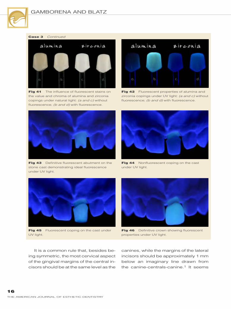
Figures 41 to 55 show the selection
of the definitive coping material and the
final outcome of Case 3. Figures 41 and
42 reveal the influence of fluorescent
stains on the value and chroma of alu-
mina and zirconia copings under natu-
ral and UV light. The impact of using a
fluorescent (Fig 43) versus a nonfluo-
rescent coping (Fig 44) is quite obvious
on the stone cast (Figs 45 and 46) and
even more so in the oral cavity (Figs 47
to 55). The definitive implant-supported
crown shows optical and fluorescent
properties that ideally match the exist-
ing natural dentition under various light
sources.
LIP LINE
A high lip line or “smile line” that reveals
all anterior teeth and large amounts of
gingival tissue7,8 is a great challenge
for the dental team since it is impossible
to hide the implant-restorative interface.
A high smile line may be due to vertical
maxillary excess or a hypermobile lip.
GAMBORENA ANd BLAtz
16THE AMERICAN JOURNAL OF ESTHETIC DENTISTRY
It is a common rule that, besides be-
ing symmetric, the most cervical aspect
of the gingival margins of the central in-
cisors should be at the same level as the
canines, while the margins of the lateral
incisors should be approximately 1 mm
below an imaginary line drawn from
the canine-centrals-canine.1 It seems
Fig 43 Definitive fluorescent abutment on the
stone cast demonstrating ideal fluorescence
under UV light.
Fig 44 Nonfluorescent coping on the cast
under UV light.
Fig 45 Fluorescent coping on the cast under
UV light.
Fig 46 Definitive crown showing fluorescent
properties under UV light.
Fig 41 The influence of fluorescent stains on
the value and chroma of alumina and zirconia
copings under natural light: (a and c) without
fluorescence; (b and d) with fluorescence.
Fig 42 Fluorescent properties of alumina and
zirconia copings under UV light: (a and c) without
fluorescence; (b and d) with fluorescence.
Case 3 Continued

GAMBORENA ANd BLAtz
17VOLUME 1 • NUMBER 1 • FALL 2011
advisable for central incisor implant
restorations to initially place the gingi-
val margin slightly more incisally. This
slight “overcompensation” will prove
extremely helpful to counterbalance
tissue recession typically seen over
time. The CTG now becomes an essen-
tial aspect for functional and esthetic
Case 3 Continued
Fig 48 Intraoral try-in under UV light demon-
strates ideal blending of the fluorescent properties
of the definitive crown with the adjacent teeth.
Fig 49 Definitive implant restoration.
Fig 51 (right) The definitive abutment and res-
toration provide the same degree of fluorescence
as a natural tooth.
Fig 47 Intraoral try-in of definitive crown under
natural light shows an excellent blend with the
shade of the adjacent teeth.
Fig 50 Postoperative occlusal view showing
the soft tissue support and contour.
GAMBORENA ANd BLAtz
18THE AMERICAN JOURNAL OF ESTHETIC DENTISTRY
integration of the implant-supported
restoration, especially in the presence
of a high lip line.
Occasionally, unfavorable changes
of the gingival margin levels may occur
at the teeth adjacent to the implant res-
toration. These are based on the dis-
tance between the free gingival margin
and the supporting bone and may re-
quire more extensive tissue grafting
to control gingival levels at both the
natural and implant-supported teeth.
The added connective tissue causes
the fibrotic mucosa around the implant
to migrate more coronally. In rare cas-
es, the additional soft tissue becomes
so abundant that a gingivectomy fol-
lowed by a fibrotomy becomes neces-
sary to establish ideal crown lengths
and gingival margin contours.
The unfavorable umbrella effect is
most prevalent in patients with a high
Fig 52 Postoperative view under natural light
showing the color match of the implant-support-
ed crown with the natural dentition.
Fig 53 Postoperative view under UV light
showing the ideal blend of fluorescent properties
between the restoration and natural dentition.
Fig 54 Postoperative intraoral situation. No gray zone is evident. Fig 55 One-year postopera-
tive peri apical radiograph of
the implant at the left central
incisor site.
Case 3 Continued

GAMBORENA ANd BLAtz
19VOLUME 1 • NUMBER 1 • FALL 2011
smile line.6 The gray zone may become
visible at the implant restoration site
even when all of the key factors are im-
plemented perfectly. Differences in soft
tissue thickness and volume may cause
these color dissimilarities, which are
then amplified by the shear presence of
the upper lip, causing a shadow on the
soft tissue and the light to be reflected
and transferred in a different manner.
Case 4 (Figs 56 to 61) demonstrates
a situation in which the maxillary left
central incisor was replaced with an im-
plant-supported crown in a patient with
Fig 56 Intraoral view of the definitive zirconia
abutment.
Fig 57 A CTG was placed earlier to ensure
ideal soft tissue thickness.
Case 4
Fig 58 Intraoral situation after 1 year reveals
differences in peri-implant soft tissue color and
morphology.
Fig 59 Preoperative view showing the patient’s
high lip line.
Fig 60 Postoperative situation. An unfavorable
gray zone is visible during an average smile.
Fig 61 A high smile reveals the gray zone
through the umbrella effect.

GAMBORENA ANd BLAtz
20THE AMERICAN JOURNAL OF ESTHETIC DENTISTRY
a high smile line. A CTG was placed to
enhance the soft tissue contours. While
all of the key aspects were implement-
ed successfully, the slight differences
in soft tissue volume created an unfa-
vorable grayish effect (Figs 60 and 61).
In contrast to the previous case,
Case 5 (Figs 62 to 67) exemplifies
successful implementation of these
key factors in a patient with a high lip
line for long-term esthetic and func-
tional success.
Fig 62 Intraoral view of the crown prepara-
tion of the right central incisor and the definitive
colored zirconia implant abutment at the left
central incisor.
Fig 63 UV light reveals the natural fluores-
cence of the modified zirconia implant abutment.
Fig 64 Preoperative intraoral view of the failing
central incisor crowns.
Fig 65 Postoperative intraoral situation show-
ing the color and soft tissue match.
Fig 66 Initial situation. Note the high lip line. Fig 67 Successful implementation of the five
key factors prevented the appearance of a gray
zone despite the high smile line.
Case 5

GAMBORENA ANd BLAtz
21VOLUME 1 • NUMBER 1 • FALL 2011
CONCLUSIONS
Five key factors were identified to avoid
the gray zone around maxillary ante-
rior implant restorations: 3D implant
placement, soft tissue thickness, abut-
ment selection, crown restoration, and
lip line. Mimicking the inherent optical
properties, especially fluorescence, of
natural teeth with ideal prosthetic im-
plant components and crown materials
is fundamental for ultimate restorative
and soft tissue esthetics.
ACkNOWLEDGMENTS
The authors would like to thank Iñigo Casares for the beautiful porcelain work featured in this article Fernando zozaya for the detailed fabrication on the zirconia abut-ments.
REFERENCES
1. Chiche G, Pinault A. Esthetics of Anterior Fixed Prosthodon-tics. Chicago: Quintessence, 1994.
2. Matthews TG. The anatomy of a smile. J Prosthet Dent 1978; 39:128–134.
3. Fradeani M. Esthetic Analysis: A Systematic Approach to Prosthetic Treatment. Chicago: Quintessence, 2005.
4. Bitter RN. the periodontal factor in esthetic smile design—Altering gingival display. Gen Dent 2007;55:616–622.
5. Blatz MB, Hürzeler MB, strub JR. Reconstruction of the lost interproximal papilla—Presen-tation of some surgical and non-surgical procedures. Int J Periodontics Restorative Dent 1999;19:395–406.
6. Magne P, Magne M, Belser U. The esthetic width in fixed prosthodontics. J Prosthodont 1999;8:106–118.
7. Vig RG, Brundo Gc. the kinetics of anterior tooth display. J Prosthet Dent 1978;39:502–504.
8. Passia N, Blatz MB, strub JR. Is the smile line a valid parameter for esthetic evalua-tion? A review of the literature. Eur J Esthet Dent (in press).
9. Fradeani M. Evaluation of dentolabial parameters as part of a comprehensive esthetic analysis. Eur J Esthet Dent 2006;1:62–69.
10. Gamborena i, Blatz MB. Current clinical and technical protocols for single-tooth immediate implant procedures. Quintessence Dent Technol 2008;31:49–60.
11. Holst s, Blatz MB, Hegenbarth E, Wichmann M, Eitner S. Prosthodontic considerations for predictable single-implant esthetics in the anterior maxilla. J Oral Maxillofac Surg 2005;63(suppl 2):89–96.
12. kois JC, kan JY. Predictable peri-implant gingival aesthet-ics: Surgical and prosthodontic rationales. Pract Proced Aesthet Dent 2001;13:691–698.
13. Blatz MB, Bergler M, Holst s, Block M. zirconia abutments for single-tooth implants—Rationale and clinical guide-lines. J Oral Maxillofac Surg 2009;67(suppl):74–81.
14. Yildirim M, Edelhoff D, Hanish O, Spiekermann H. Ceramic abutments—A new era in achieving optimal esthetics in implant dentistry. Int J Peri-odontics Restorative Dent 2000;20:81–91.
15. Jung RE, Sailer I, Hämmerle CH, Attin T, Schmidlin P. In vitro color changes of soft tissues caused by restorative materi-als. Int J Periodontics Restora-tive Dent 2007;27:251–257.
16. Jung RE, Holderegger C, Sailer I, khraisat A, Suter A, Hämmerle CH. The effect of all-ceramic and porcelain-fused-to-metal restorations on marginal peri-implant soft tissue color: A randomized controlled clinical trial. Int J Periodontics Restorative Dent 2008;28:357–365.
17. van Brakel R, Noordmans HJ, Frenken J, de Roode R, de Wit GC, Cune MS. The effect of zirconia and titanium implant abutments on light reflection of the supporting soft tissues [epub ahead of print 20 Jan 2011]. Clin Oral Implants Res.
18. Garber DA. The esthetic dental implant: Letting restoration be the guide. J Am Dent Assoc 1995;126:319–325.
19. Grunder U. Crestal ridge width changes when placing implants at the time of tooth extraction with and without soft tissue augmentation after a healing period of 6 months: Report of 24 consecutive cases. Int J Periodontics Restorative Dent 2011;31:9–17.
GAMBORENA ANd BLAtz
22THE AMERICAN JOURNAL OF ESTHETIC DENTISTRY
22THE AMERICAN JOURNAL OF ESTHETIC DENTISTRY
20. Linkevicius T, Apse P, Grybauskas S, Puisys A. The influence of soft tissue thickness on crestal bone changes around implants: A 1-year prospective controlled clinical trial. Int J Oral Maxillo-fac Implants 2009;24:712–719.
21. Speroni S, Cicciu M, Maridati P, Grossi GB, Maiorana c. Clinical investigation of mucosal thickness stability after soft tissue grafting around implants: A 3-year retrospec-tive study. Indian J Dent Res 2010;21:474–479.
22. Wiesner G, Esposito M, Worthington H, Schlee M. Connective tissue grafts for thickening peri-implant tissues at implant placement. One-year results from an explana-tory split-mouth randomised controlled clinical trial. Eur J Oral Implantol 2010;3:27–35.
23. Esposito M, Grusovin MG, Maghaireh H, Coulthard P, Worthington HV. Interventions for replacing missing teeth: Management of soft tissues for dental implants. Cochrane Database Syst Rev 2007;(3):CD006697.
24. Fürhauser R, Florescu D, Benesch t, Haas R, Mailath G, Watzek G. Evaluation of soft tissue around single-tooth implant crowns: The pink esthetic score. Clin Oral Implants Res 2005;16:639–644.
25. Att W, kurun S, Gerds T, Strub JR. Fracture resistance of single-tooth implant-supported all-ceramic restorations after exposure to the artificial mouth. J Oral Rehabil 2006; 33:380–386.
26. Nothdurft FP, Merker S, Pospiech PR. Fracture behaviour of implant-implant- and implant-tooth-supported all-ceramic fixed dental prostheses utilising zirconium dioxide implant abutments. Clin Oral Investig 2011;15: 89–97.
27. Scarano A, Piattelli M, Caputi S, Favero GA, Piattelli A. Bacterial adhesion on com-mercially pure titanium and zirconium oxide disks: An in vivo human study. J Periodon-tol 2004;75:292–296.
28. Rimondini L, Cerroni L, carrassi A, torricelli P. Bacte-rial colonization of zirconia ceramic surfaces: An in vitro and in vivo study. Int J Oral Maxillofac Implants 2002;17: 793–798.
29. Degidi M, Artese L, Scarano A, Perrotti V, Gehrke P, Piattelli A. Inflammatory infiltrate, microvessel density, nitric oxide synthase expression, vascular endothelial growth factor expression, and proliferative activity in peri-implant soft tissues around titanium and zirconium oxide healing caps. J Periodontol 2006;77:73–80.
30. Gamborena i, Blatz MB. Fluoresence—Mimicking nature for ultimate esthetics in implant dentistry. Quintessence Dent Technol 2011;34:7–23.
31. Benedict Hc. A note on the fluo-rescence of teeth in ultra-violet rays. Science 1928;67:442.
32. Araki T, Miyazaki E, kawata T, Miyata k. Measurements of fluorescence heterogeneity in human teeth using polarization microfluorometry. Appl Spectrosc 1990;44:627–631.
33. Lee Yk, Lu H, Powers JM. Fluorescence of layered resin composites. J Esthet Restor Dent 2005;17:93–100.
34. Tani k, Watari F, Uo M, Morita M. Discrimination between composite resin and teeth using fluorescence properties. Dent Mater J 2003;22:569–580.
35. Sant’Anna Aguiar Dos Reis R, Casemiro LA, Carlino GV, et al. Evaluation of fluorescence of dental composites using contrast ratios to adjacent tooth structure: A pilot study. J Esthet Restor Dent 2007;19:199–206.
36. Monsénégo G, Burdairon G, clerjaud B. Fluorescence of dental porcelain. J Prosthet Dent 1993;69:106–113.
37. Komine F, Blatz MB, Yamamoto S, Matsumura H. A modified layering technique to enhance fluorescence in glass-infiltrated aluminum oxide ceramic restorations: Case report. Quintessence Int 2008;39: 11–16.
38. Marchack BW, Futatsuki Y, Marchack cB, White sN. Customization of milled zirconia copings for all-ceramic crowns: A clinical report. J Prosthet Dent 2008;99:169–173.
39. Nik Mohd Polo kinin NM, Wan Mohd Arif Wi, zainal Arifm A. Study on the effect of Y2O3 addition to the fluorescent property of dental porcelain. Med J Malaysia 2004;59 (suppl B):23–24.
40. Ferreira zandoná AG, kleinrichert T, Analoui M, schemehorn BR, Eckert GJ, Stookey Gk. Effect of two fluorescent dyes on color of restorative materials. Am J Dent 1997;10:203–207.