the gypsy moth fungus - · pdf filethe gypsy moth fungus linda butler professor of entomology...
TRANSCRIPT
The Gypsy Moth Fungus
Linda Butler Professor of Entomology Division of Plant and Soil Sciences West Virginia University 1998In recent years, dramatic collapses of gypsy moth populations in the eastern states are due, in large part, to the fungus Entomophaga maimaiga. A native of Japan, this fungus is a natural enemy of gypsy moth larvae.
Questions have arisen as to the origin of the fungus in North America. The "gypsy fungus" was collected in Japan and deliberately released near Boston between 1910 and 1911, but subsequent studies did not recover it. Spring of 1989 was particularly rainy in the Northeast when gypsy moth populations appeared to be increasing. When larval population collapses were noticed, examination of larval cadavers revealed spores of an entomophthoralean fungus soon shown to be E. maimaiga. Dr. Ann Hajek (Cornell University) has evaluated many possibilities as to origin of the fungus identified in 1989. She and her co-workers concluded the most likely scenarios are that the weak fungus introduced in 1910 evolved into a virulent strain or that the fungus was accidentally introduced recently into the Northeast (Hajek et al. 1995).
How does the fungus spread? The fungus can be inadvertently or deliberately spread by humans moving soil or gypsy moth larval cadavers containing the resting spores, but the conidiaspores (produced on the outside of the caterpillar body) are small and airborne enabling rapid natural spread. "Such rapid spread was completely unexpected and has never before been documented for an entomopathogenic fungus" (Hajek et al. 1996).
How much impact does the fungus have on defoliating populations of gypsy moth? We have only to look at gypsy moth defoliation figures for the northeastern and mid-Atlantic states to see the effect of the fungus. Between 1995 and 1996, many states experienced a decrease in defoliation: Delaware, 99% decrease; Maryland, 88%; Michigan, 96%; Pennsylvania, 94%; Virginia, 100%; and West Virginia, 31%. In many of West Virginia's leading-edge counties, the fungus is catching up to the gypsy moth. More than 66,000 acres were sprayed for gypsy moth in West Virginia in 1996, but egg mass surveys show that little more than 10,000 acres will qualify for treatment in 1997 (Jan D. Hacker, WVDA, personal communication).
Are we sure the collapse is due to fungus rather than the gypsy moth virus? Yes. The differences can be confirmed easily by an experienced person examining the larval cadavers for fungal conidia and viral polyhedra. Larvae killed by both virus and fungus are particularly evident on lower tree trunks. Viral-killed larvae often hang in an inverted V, and the bodies are moist and ooze dark fluid.

Typical appearance of gypsy moth larvaekilled by Entomophaga maimaiga.
Fungal-killed cadavers typically are oriented vertically on the tree trunk with the head down and the abdominal prolegs spread out. The cadavers tend to dry and, soon after death, white, powdery spores may be noted on the caterpillar hairs (Hajek and Snyder 1992; Reardon and Hajek 1993).
Before 1989, gypsy moth larval collapses occurred periodically, but typically only in the aftermath of population outbreaks. The fungus appears to be equally virulent in low or high gypsy moth populations.
How host specific is Entomophaga maimaga? The fungus affects only larvae of Lepidoptera. In laboratory bioassays, 78 species of Lepidoptera representing 17 families were dipped in fungal conidial suspensions, representing a "worst-case" scenario. Cadavers of 36% of the species produced spores, although infection levels were less than 50% for most of those species. Highest infection levels were seen in native members of the gypsy moth family, Lymantriidae (Hajek et al. 1995). In a related field study, 1,511 nontarget caterpillars from 52 species belonging to 7 families were collected from sites (primarily Virginia) with high gypsy moth fungal infections. Only two specimens, 1 of 318 forest tent caterpillars and 1 of 96 Catocala ilia (underwing) larvae, were infected (Hajek et al. 1996).
Will the fungus continue to suppress gypsy moth populations? No one can really predict what will happen in future years. Although fungi, in general, do better in wet years, E. maimaiga has spread well and suppressed larvae in some dry seasons. Typically, natural enemies never eradicate their hosts, so we will always have gypsy moth as a part of our forest landscape. Populations at or behind leading edges, however, may not be as damaging as they have been in the past.
ReferencesHajek, A.E., R.A. Humber, and J.S. Elkinton. 1995. The mysterious origin of Entomophaga
maimaiga in North America. Am. Entomol. 41:31-42.
Hajek, A.E., L. Butler, and M.M. Wheeler. 1995. Laboratory bioassays testing the host rangeof the gypsy moth fungal pathogen Entomophaga maimaiga. Biol. Cont. 5:530-544.
Hajek, A.E., L. Butler, S.R.A. Walsh, J.C. Silver, F.P. Hain, F.L. Hastings, T.M. Odell,and D.R.Smitley. 1996. Host range of the gypsy moth (Lepidoptera: Lymantriidae) laboratory, Environ. Entomol. [Forum] 25:709-721.

Hajek, A.E., J.S. Elkinton, and J.J. Witcosky. 1996. Introduction and spread of the fungalpathogen Entomophaga maimaiga along the leading edge of gypsy moth spread. Environ.Entomol. 25:1235-1247.
Hajek, A.E. and A.L. Snyder. 1992. Natural Enemies: Tools for Integrated Pest Management.Field identification of the gypsy moth fungus, Entomophaga maimaiga. USDA, Forest Service NE Area, NA-PR-02-92.
Reardon, R. and A.E. Hajek. 1993. Entomophaga maimaiga in North America: A review.USDA, Forest Service, AIPM Technology Transfer. USDA, Forest Service NA-TP-15- 93.
Ann E. HajekDepartment of Entomology
Cornell UniversityIthaca, NY 14853
(prepared from the videotaped presentation*)
The use of fungi to control gypsy moth is a much younger field of study than many of the systems we've talked about so far in this conference. I have been working with fungal pathogens, primarily of gypsy moths, for the past 10 years.
Gypsy moths are very destructive, and they impact not only forests but also urban and suburban areas. In 1981, a peak year for gypsy moth populations in northeastern North America, gypsy moth larvae caused 13 million acres of defoliation.
The purple sections of the above map indicate areas of infestation where gypsy moth larvae are found. New England has an established population of gypsy moth, as does Michigan and Wisconsin. The orange shows areas of spread, which usually experience heavy damage when the gypsy moth first enters the area. However, gypsy moths are also caught in pheromone traps across the country (yellow and orange areas), indicating that they are continually found at very low levels in scattered locations across the United States.
Gypsy moth is a native of temperate Asia and Europe. It was accidentally introduced into the Boston area in 1868 or 1869. Over the past 125+ years, gypsy moth has been of concern to many people in

North America. The generally accepted hypothesis about natural regulation of gypsy moth populations has been that at low densities, the most important mortality factors are vertebrate predators, at medium densities parasitoids are most important (usually Diptera or Hymenoptera), and at high densities, the gypsy moth nuclear polyhedrosis virus (NPV), a baculovirus, is the most frequent agent regulating populations. NPV causes big crashes in the gypsy moth population (=epizootics) and people often tended to wait for these epizootics to control high populations.
This general theory about gypsy moth population regulation has been challenged. In 1989, gypsy moths in the northeastern U.S. were seen dying of what appeared to be a viral epizootic. What made this remarkable was that the populations were not at high density but were just beginning to increase and were not yet dense enough for an NPV epizootic. When cadavers were dissected, fungal spores were found inside. No such fungus was previously known as an important gypsy moth pathogen in North America. In fact, only in Japan was any fungal pathogen known to be important in gypsy moth control.
To determine what fungus was attacking the gypsy moths, we did isozyme studies and RFLPs. We knew the fungus was in the Entomophthorales and we compared strains of the fungus found on gypsy moth cadavers from Connecticut, New Hampshire, and Massachusetts with Entomophaga species infecting forest Lepidoptera in North America as well as with the Japanese species that infects gypsy moth, E. maimaiga. These molecular analyses both showed a perfect match between northeastern isolates and E. maimaiga, the fungal species known only from the Japanese gypsy moth.
This fungus was known to have been introduced from Japan in 1910 and 1911, but no one had seen it since its introduction until 1989. It is quite a mystery whether the fungus was established at the time of its original introduction, and if so, why it wasn't detected during the years between 1911 and 1989?
From E. maimaiga epizootics occurring in North American gypsy moth populations in 1989 and 1990, it became clear that this fungus was capable of becoming an important mortality factor. The question arose regarding potential use of this fungus for biological control but what would we release? There are several life stages of species in the Entomophthorales:
• The conidia are relatively short-lived. These spores are actively discharged from cadavers and can immediately germinate and cause infection.
• Hyphal bodies occur within infected insects and mycelium grows on the outside of cadavers. However, these are vegetative growth stages and are not environmentally resistant.
• The resting spores are relatively long-lived. If you dissect infected late instars, you find resting spores inside that are about 30 micrometers in diameter.
Based on what we knew about the life stages of this fungus, the resting spores appeared to be the best stage of the fungus to work with.
In releasing this fungus, we established these objectives:
1. To establish E. maimaiga in new areas. As gypsy moth spreads into new areas, the fungus also spreads, but we believe there is the opportunity to establish E. maimaiga faster through manipulation.
2. To augment the fungus in areas where it was not well established so that it could reach higher densities sooner. To increase fungal populations in areas where the fungus is established and thereby promote the occurrence of epizootics.
We initially obtained E. maimaiga resting spores for fungal releases by collecting soil that contained resting spores. Infected gypsy moth larvae die hanging onto tree trunks and cadavers

subsequently fall to the bases of trees. We've documented high titers of resting spores at the bases of these trees. Concerns regarding this strategy for obtaining resting spores are that
3. the soil containing the resting spores needs to come from areas without deleterious microbes also inhabiting the soil, and
4. the soil must be processed to confirm the presence of resting spores and to quantify them.
We could also collect and distribute the cadavers, but this strategy also has a specific problem: the gypsy moth has only one generation per year, and the larvae are generally present from April through July (varying by year and latitude). Cadavers do not hang on trees very long and, once they fall to the ground, they are not recognizable for long. Therefore, we would need to know in July the extent to which one would want to be distributing resting spores the following May. Unfortunately, gypsy moth populations are difficult to predict and one does not always know in July, before all adults have finished laying eggs for the next generation, the extent to which gypsy moth populations would need to be controlled the following spring.
At present we cannot produce resting spores in vitro - we can only produce them in insects. There is a constitutive dormancy and we've been studying this dormancy to discover in what ways we might be able manipulate this fungus while still releasing resting spores that have satisfied the dormancy requirements.
In 1991 after 2 years of observing these epizootics of E. maimaiga, the Forest Service provided funds to investigate establishment of E. maimaiga after resting spore release. This was a large study including almost 50 release plots in 4 states over 2 years.
The gray area on the above map shows where the fungus was found in 1989-1990 surveys. The open dots show our 1991 releases of resting spores and the black dots show 1992 releases. We made the releases along the leading area of gypsy moth spread.
The lines on the graph above show gypsy moth egg mass density in control plots (blue lines) and release plots (yellow lines). The concentration of egg masses at the beginning of the study is shown on the left. We tried to control for egg mass density when choosing our plots.
During 1991, there were 34 fungal release plots in 4 different states. After the release there was a drought, and fungal activity is, of course, decreased during dry conditions. Nevertheless, we did achieve establishment of E. maimaiga that same year in the majority of sites.
The second release year, 1992, was a more normal year with regards to precipitation. The fungus showed up in almost all of the plots where we had released it in 1991 but it also spread into many control plots. During 1992, gypsy moth populations throughout the area declined due to fungal infection. There were also population decreases in some control plots where the fungus had moved in, but we documented increases in other control plots.
In follow-up checks in 1994, the fungus had persisted well in the seven 1991 or 1992 release plots studied. Gypsy moths were still present in these plots also, but there was a high degree of fungal infection in the host population, providing long-term fungal control.
Number of E. maimaiga Release Sites
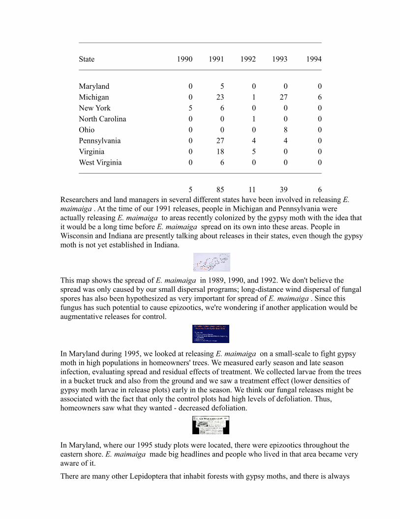
State 1990 1991 1992 1993 1994
Maryland 0 5 0 0 0Michigan 0 23 1 27 6New York 5 6 0 0 0North Carolina 0 0 1 0 0Ohio 0 0 0 8 0Pennsylvania 0 27 4 4 0Virginia 0 18 5 0 0West Virginia 0 6 0 0 0
5 85 11 39 6Researchers and land managers in several different states have been involved in releasing E. maimaiga . At the time of our 1991 releases, people in Michigan and Pennsylvania were actually releasing E. maimaiga to areas recently colonized by the gypsy moth with the idea that it would be a long time before E. maimaiga spread on its own into these areas. People in Wisconsin and Indiana are presently talking about releases in their states, even though the gypsy moth is not yet established in Indiana.
This map shows the spread of E. maimaiga in 1989, 1990, and 1992. We don't believe the spread was only caused by our small dispersal programs; long-distance wind dispersal of fungal spores has also been hypothesized as very important for spread of E. maimaiga . Since this fungus has such potential to cause epizootics, we're wondering if another application would be augmentative releases for control.
In Maryland during 1995, we looked at releasing E. maimaiga on a small-scale to fight gypsy moth in high populations in homeowners' trees. We measured early season and late season infection, evaluating spread and residual effects of treatment. We collected larvae from the trees in a bucket truck and also from the ground and we saw a treatment effect (lower densities of gypsy moth larvae in release plots) early in the season. We think our fungal releases might be associated with the fact that only the control plots had high levels of defoliation. Thus, homeowners saw what they wanted - decreased defoliation.
In Maryland, where our 1995 study plots were located, there were epizootics throughout the eastern shore. E. maimaiga made big headlines and people who lived in that area became very aware of it.
There are many other Lepidoptera that inhabit forests with gypsy moths, and there is always

concern about impact of gypsy moth control tactics on non-target hosts. We conducted lab bioassays challenging field-collected insects from West Virginia forests with E. maimaiga . In all we evaluated 78 different species of Lepidoptera. In the lab, given optimal conditions and doses, we found a smattering of infection across different super-families. For the most part, the levels of infection were pretty low when you consider that we optimized infection conditions.
In the Sphingidae, we found high levels of infection in Manduca sexta , the tobacco hornworm, and we're interested in looking at that further. There was no infection in other sphingid species tested. In the Noctuoidea, the only high levels of infection that we found was in the Lymantriidae, the family that includes gypsy moth. Based on these results, it seemed that E. maimaiga is quite specific to the family that includes gypsy moth, although it might cause low levels of infection in a number of other species.
In the field, the only cadavers we found on trees were gypsy moths. We went to locations where there was an active epizootic of E. maimaiga occurring in gypsy moth populations and we collected more than 1500 insects of 53 different species. Of those 1500 individuals, we found 1 individual of lasiocampid and 1 individual from the Noctuidae that were infected.
We would like to continue our studies by investigating further the lack of agreement between the lab infection rates and the field infection rates. Also, we're interested in working further on the epizootiology and potential use of resting spores for control as well as the potential for in vitro and in vivo production of these spores.
Based on our results to date, E. maimaiga isn't the silver bullet that will end gypsy moth outbreaks in North America, but it is a very important natural enemy regulating gypsy moth populations and is capable of creating epizootics in both low density and high density populations. We think that it will have the long-term effect of decreasing high level gypsy moth populations and perhaps will decrease outbreak frequency.
©All material is protected by Section 107 of the 1976 copyright law.Copyright is held by Cornell University.Use of this material for educational purposes is encouraged. Please notify the editor of such use and cite the author, date, site name, Cornell University, and the URL.
Return to Proceedings Index
Last modified December 22, 1998http://www.nysaes.cornell.edu/ent/bcconf/talks/hajek.html
G. C. Hartmann1 and S. S. Wasti1
(1) Department of Biology, Rhode Island College, 02908 Providence, R.I., USAAbstract Larvae of the gypsy mothPorthetria dispar (L.) were infected with sporulating cultures of the fungus,Entomophthora virulenta HALL & DUNN using the spore shower technique. The infectivity of the parasite in the host and its histopathology was observed. Penetration of the integument was recorded at 24 hours post-inoculation and conidiophore production occurred at 72 hours after infection. Experiments on mammalian toxicity against laboratory mice showed no significant differences between control and treated mice in body weight, blood cell counts, food and liquid consumption and in the post-mortem examination.

Forestry Dept Ohio State:
THE GYPSY MOTH FUNGUSThere is good news to report in the fight against the gypsy moth. A new weapon has emerged. A fungus, Entomophaga maimaiga (Em), has emerged as a tool that can be used against this voracious feeder. This highly virulent and host-specific fungal pathogen of gypsy moth larvae, is known as one of the most important causes of mortality in Japanese gypsy moth populations. The fungus was probably imported from Japan to areas near Boston, Massachusetts around 1910. This attempt to establish the fungus seemed to fail since extensive surveys did not reveal the pathogen. Em was not observed in North America until June, 1989 when dead caterpillars found clinging to trees in the northeastern U.S. revealed its presence. Ohio first documented the fungus in Trumbull county in 1993.
“If these moisture conditions are present and the temperature is between 14 and 26 degrees Celsius, infection can occur. You might say “when it rains it spores!”
The life cycle of Em closely parallels that of the gypsy moth. The fungus over winters in the soil in the form of dormant resting spores. As springtime temperatures and moisture levels reach proper levels (usually about 2 weeks prior to gypsy moth egg hatch), the resting spores germinate and begin forcibly releasing fragile, short lived conidia (active, infectious spores). Caterpillars are infected by coming in contact with soil borne resting spores or the germinating conidia. An enzyme helps the fungus penetrate the larva’s body. Disease develops in the caterpillar, resulting in death within 7-10 days. After death, fungal hyphae form in the caterpillar’s body, producing conidia (outside the larval body) and/or resting spores (inside the larval body). Conidia produced at this time can infect other caterpillars. The process can be repeated as long as weather conditions are favorable, and usually ceases about 2 weeks after gypsy moth pupation (mid-July). Resting spores from dead larvae are eventually leached back to the soil. Entomophaga killed caterpillars typically hang from tree trunks from their prolegs in a head down position. They also have a “rubbery” texture and appear dry. Some dead larvae will fall from tree trunks in 9-10 days, while some will remain attached throughout the autumn and winter.
Adequate moisture is key to the biology and pathogenicity of Em. This water dependence can be seen in two of the fungus’ important processes. In the spring, resting spores germinate best 1-2 days after precipitation because high humidity (approaching 100%) is required for conidial development and discharge. Also, conidia production from dead larvae usually occurs on days when there is rainfall because free water is needed for conidial germination. If these moisture conditions are present and the temperature is between 14 and 26 degrees Celsius, infection can occur. You might say “when it rains it spores!”
This relationship of Em to moisture levels is exhibited in the recent history of the fungus. In 1989 near record rainfall occurred throughout the northeastern U.S. during May and June. This was when the fungus was first discovered killing gypsy moth caterpillars in North America. Near record precipitation also fell in Ohio during the Spring of 1996, and an amazingly rapid spread of Em coincided. Since 1993, the Ohio Department of Natural Resources (ODNR) and ODA carefully monitored a few local populations of Em (many were experimental introductions of the fungus). After the summer of 1996, it was clear that Em had spread into most counties harboring a significant gypsy moth population. Research indicates the typical spread would be closer to 1 kilometer per year.
“The relationship between Em and gypsy moth appears to be very exclusive.”
E. maimaiga shows promise as a gypsy moth management tool for many reasons. Host-specificity is a prime example. Other species of Entomophaga native to the northeastern U.S. can not successfully infect gypsy moth caterpillars. Similarly, Em does not often infect other species of Lepidoptera

(butterflies and moths). The relationship between Em and gypsy moth appears to be very exclusive. The list of possible non-target organisms for E. maimaiga is very short. First, Em is only likely to affect Lepidopteran larvae which are active during roughly the same time period as gypsy moth larvae. Research has shown that only a few species of heavily contoured or densely hairy caterpillars can be infected with Em, even using extreme laboratory methods. It is very rare to find a naturally infected caterpillar (other than gypsy moth) in the field. This type of host specific attribute is a big plus for any potential management tool. The fungus also works at low, as well as high, gypsy moth population levels. This sets it apart from natural controls such as the nucleopolyhedrosis virus (NPV), a viral disease of gypsy moth which kills caterpillars under stress from high population densities and diminishing food supplies. Research also shows indications of a strong synergistic relationship between Em and NPV. Improving the effectiveness of other natural controls is another positive attribute for Em as a gypsy moth control tool. The toughness of the resting spores also increases the effectiveness of Em as a management tactic. Resting spores can survive 2-3 years without the gypsy moth host.
Some are touting E. maimaiga as the ‘cure-all’ for forest and tree health concerns caused by the gypsy moth. This will probably not be the case. This fungus is one of the most promising gypsy moth management tools to surface since our struggle with the insect began, however, other management techniques will still be needed in many instances. Em will probably not affect all gypsy moth populations the same way in a given year. In some areas gypsy moth populations will totally collapse, some areas will show a population reduction, while others show little impact on the gypsy moth population. Many areas at the leading edge of an infestation do not even harbor the fungus. It usually takes 2-4 years for the fungus to establish itself naturally in a gypsy moth population. The initial outbreak will have already occurred and the most severe tree mortality often results from these first defoliation events. In addition, defoliation of trees will still be evident, even in areas where caterpillars are infected with Em. Often, infected caterpillars will not die until the 4th or 5th instar, by which time they will have already caused significant defoliation. Although gypsy moth larval mortality rates due to Em are often 75-100%, some caterpillars are not infected and continue to feed and eventually reproduce. Also, forested ecosystems offer plenty of shade and leaf litter to help maintain the high moisture levels required by Em. The survival and effectiveness of the fungus in ore open fields or in grassy suburbia is unknown.
OHIO RESEARCH PROJECTSince 1993, ODNR and ODA have established 18 experimental E. maimaiga plots in cooperation with California University of Pennsylvania (CUP). The research plots were largely located on public lands (for instance, state forests), to insure future access to the plots and to maintain some management consistency (no pesticide treatment and retention of test trees). The purpose of this ongoing research project is threefold: to gather information on our ability to artificially introduce Em into a low level population of gypsy moth; the fungus’ ability to control the number of gypsy moths in an area; and, the manner of fungal spread through a given area.
“It seems crucial to introduce the fungus when the gypsy moth populations are low, so that it can build its levels and provide control prior to the first (and most damaging) outbreak.”
Even during the drought of 1991 and in very low density gypsy moth populations, the fungus was established at all but one test site. During dry years the fungus seems to establish, but does not kill as many caterpillars as in a moist year. Success was also seen in terms of gypsy moth population control. Em was introduced into one site with egg mass counts of 800 per acre. This level of infestation would normally result in noticeable defoliation. This population of gypsy moth was reduced to a level where caterpillars were difficult to find and no noticeable defoliation occurred. It is important to remember, however, that this test plot was monitored during the near record moisture levels in the Spring of 1996. Em killed gypsy moth larvae could be found in almost every infested county during this same time

period. It would be misleading to assume that the mortality rate would be this high during low to normal precipitation periods. The plots should be monitored for a few more years to develop a clearer picture of the relationship dynamics between the gypsy moth and the fungus. The wet spring of 1996 clouded the data regarding fungal spread. Prior to 1996, the data showed a spread of slightly more than 1 kilometer per year, which is typical of other research findings. After the wet period, however, the fungus appeared to spread more rapidly and over longer distances. Now that it is found in so many places, it is difficult to actually trace the spread from the point of introduction.
The project is showing evidence that Em can successfully be introduced into low density gypsy moth populations. Once established, E maimaiga can reduce the number of gypsy moth larvae and limit population expansion. It seems crucial to introduce the fungus when gypsy moth populations are low, so that it can build its levels and provide control prior to the first (and most damaging) outbreak. It appears promising that Em can help us manage gypsy moth by keeping populations below damaging levels, effectively limiting defoliation.
Authors: Ann E. Hajek; Micheal M. Wheeler; Callie C. Eastburn; Leah S. Bauer DOI: 10.1080/09583150120076184 Publication Frequency: 10 issues per year
Published in: Biocontrol Science and Technology, Volume 11, Issue 5 October 2001 , pages 637 - 647 Subjects: Crop Science; Entomology; Pest Management; Plant Ecology; Plant Pathology; Number of References: 22 Formats available: PDF (English) Article Requests: Order Reprints : Request Permissions
• In order to give pricing details we need to know your country. Please register and/or sign in to identify your country.
• Sign In
• Online Sample

Abstract The fungal pathogen, Entomophaga maimaiga causes epizootics in populations of the important North American forest defoliator gypsy moth ( Lymantria dispar ). Increasing use of this fungus for biological control is dependent on our ability to produce and manipulate the long-lived overwintering resting spores (azygospores). E. maimaiga resting spores undergo obligate dormancy before germination so we investigated conditions required for survival during dormancy as well as the dynamics of subsequent germination. After formation in the field during summer, resting spores were stored under various moisture levels, temperatures, and with and without soil in the laboratory and field. The following spring, for samples maintained in the field, germination was greatest among resting spores stored in plastic bags containing either moistened paper towels or sterile soil. Resting spores did not require light during storage to subsequently germinate. In the laboratory, only resting spores maintained with either sterile or unsterilized soil at 4°C (but not at 20 or -20°C) germinated the following spring, but at a much lower percentage than most field treatments. To further investigate the effects of relative humidity (RH) during storage, field-collected resting spores were placed at a range of humidities at 4°C. After 9.5 months, resting spore germination was highest at 58% RH and no resting spores stored at 88 or 100% RH germinated. To evaluate the dynamics of infections initiated by resting spores after storage, gypsy moth larvae were exposed to soil containing resting spores that had been collected in the field and stored at 4°C for varying lengths of time. No differences in infection occurred among larvae exposed to fall-collected soil samples stored at 4oC over the winter, versus soil samples collected from the same location the following spring. Springcollected resting spores stored at 4°C did not go into secondary dormancy. At the time that cold storage of soil containing resting spores began in spring, infection among exposed larvae was initiated within a few days after bringing the soil to 15°C. This same pattern was also found for spring-collected resting spore-bearing soil that was assayed after cold storage for 2-7 months. However, after 31-32 months in cold storage, infections started 14-18 days after soil was brought to 15°C, indicating a delay in resting spore activity after prolonged cold storage.

Authors: Ann E. Hajek; Micheal M. Wheeler; Callie C. Eastburn; Leah S. Bauer DOI: 10.1080/09583150120076184 Publication Frequency: 10 issues per year Published in: Biocontrol Science and Technology, Volume 11, Issue 5 October 2001 , pages 637 - 647 Subjects: Crop Science; Entomology; Pest Management; Plant Ecology; Plant Pathology; Number of References: 22 Formats available: PDF (English) Article Requests: Order Reprints : Request Permissions
• In order to give pricing details we need to know your country. Please register and/or sign in to identify your country.
• Sign In • Online Sample
Abstract The fungal pathogen, Entomophaga maimaiga causes epizootics in populations of the important North American forest defoliator gypsy moth ( Lymantria dispar ). Increasing use of this fungus for biological control is dependent on our ability to produce and manipulate the long-lived overwintering resting spores (azygospores). E. maimaiga resting spores undergo obligate dormancy before germination so we investigated conditions required for survival during dormancy as well as the dynamics of subsequent germination. After formation in the field during summer, resting spores were stored under various moisture levels, temperatures, and with and without soil in the laboratory and field. The following spring, for samples maintained in the field, germination was greatest among resting spores stored in plastic bags containing either moistened paper towels or sterile soil. Resting spores did not require light during storage to subsequently germinate. In the laboratory, only resting spores maintained with either sterile or unsterilized soil at 4°C (but not at 20 or -20°C) germinated the following spring, but at a much lower percentage than most field treatments. To further investigate the effects of relative humidity (RH) during storage, field-collected resting spores were placed at a range of humidities at 4°C. After 9.5 months, resting spore germination was highest at 58% RH and no resting spores stored at 88 or 100% RH germinated. To evaluate the dynamics of infections initiated by resting spores after storage, gypsy moth larvae were exposed to soil containing resting spores that had been collected in the field and stored at 4°C for varying lengths of time. No differences in infection occurred among larvae exposed to fall-collected soil samples stored at 4oC over the winter, versus soil samples collected from the same location the following spring. Springcollected resting spores stored at 4°C did not go into secondary dormancy. At the time that cold storage of soil containing resting spores began in spring, infection among exposed larvae was initiated within a few days after bringing the soil to 15°C. This same pattern was also found for spring-collected resting spore-bearing soil that was assayed after cold storage for 2-7 months. However, after 31-32 months in cold storage, infections started 14-18 days after soil was brought to 15°C, indicating a delay in resting spore activity after prolonged cold storage.
Authors: Ann E. Hajek; Micheal M. Wheeler; Callie C. Eastburn; Leah S. Bauer

DOI: 10.1080/09583150120076184 Publication Frequency: 10 issues per year Published in: Biocontrol Science and Technology, Volume 11, Issue 5 October 2001 , pages 637 - 647 Subjects: Crop Science; Entomology; Pest Management; Plant Ecology; Plant Pathology; Number of References: 22 Formats available: PDF (English) Article Requests: Order Reprints : Request Permissions
• In order to give pricing details we need to know your country. Please register and/or sign in to identify your country.
• Sign In • Online Sample
Abstract The fungal pathogen, Entomophaga maimaiga causes epizootics in populations of the important North American forest defoliator gypsy moth ( Lymantria dispar ). Increasing use of this fungus for biological control is dependent on our ability to produce and manipulate the long-lived overwintering resting spores (azygospores). E. maimaiga resting spores undergo obligate dormancy before germination so we investigated conditions required for survival during dormancy as well as the dynamics of subsequent germination. After formation in the field during summer, resting spores were stored under various moisture levels, temperatures, and with and without soil in the laboratory and field. The following spring, for samples maintained in the field, germination was greatest among resting spores stored in plastic bags containing either moistened paper towels or sterile soil. Resting spores did not require light during storage to subsequently germinate. In the laboratory, only resting spores maintained with either sterile or unsterilized soil at 4°C (but not at 20 or -20°C) germinated the following spring, but at a much lower percentage than most field treatments. To further investigate the effects of relative humidity (RH) during storage, field-collected resting spores were placed at a range of humidities at 4°C. After 9.5 months, resting spore germination was highest at 58% RH and no resting spores stored at 88 or 100% RH germinated. To evaluate the dynamics of infections initiated by resting spores after storage, gypsy moth larvae were exposed to soil containing resting spores that had been collected in the field and stored at 4°C for varying lengths of time. No differences in infection occurred among larvae exposed to fall-collected soil samples stored at 4oC over the winter, versus soil samples collected from the same location the following spring. Springcollected resting spores stored at 4°C did not go into secondary dormancy. At the time that cold storage of soil containing resting spores began in spring, infection among exposed larvae was initiated within a few days after bringing the soil to 15°C. This same pattern was also found for spring-collected resting spore-bearing soil that was assayed after cold storage for 2-7 months. However, after 31-32 months in cold storage, infections started 14-18 days after soil was brought to 15°C, indicating a delay in resting spore activity after prolonged cold storage.