the hip as a cable truss -...
TRANSCRIPT

The Hip as a Cable TrussMartin Moeser and Lothar Meinel, Germany, 18 April, 2010
Nature is a master of lightweight construction (some animals can fly).
Prevailing opinion: The bones of vertebrates are subject to bending.
But: Bending is characteristic of heavyweight construction. Authors` opinion: The bones are stressed purely on their axes, and only under compression. The tension is submitted by ligaments and muscles.
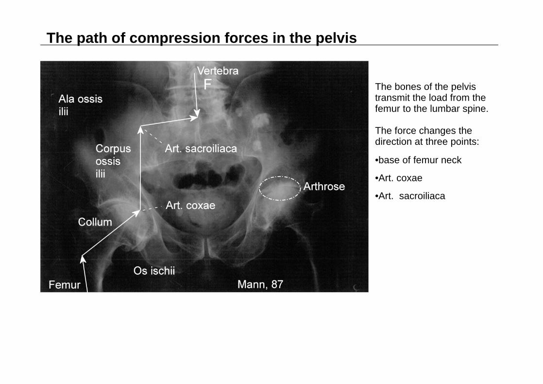
The path of compression forces in the pelvis
The bones of the pelvis transmit the load from the femur to the lumbar spine.
The force changes the direction at three points:
•base of femur neck
•Art. coxae
•Art. sacroiliaca
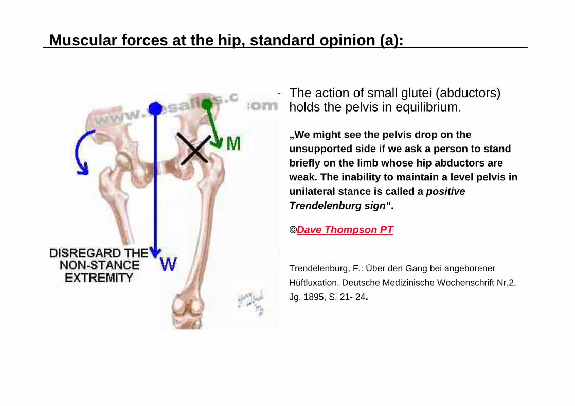
Muscular forces at the hip, standard opinion (a):
The action of small glutei (abductors) holds the pelvis in equilibrium.
„We might see the pelvis drop on the unsupported side if we ask a person to stand briefly on the limb whose hip abductors are weak. The inability to maintain a level pelvis in unilateral stance is called a positive Trendelenburg sign“.
©Dave Thompson PT
Trendelenburg, F.: Über den Gang bei angeborener Hüftluxation. Deutsche Medizinische Wochenschrift Nr.2, Jg. 1895, S. 21- 24.

Muscular forces at the hip, standard opinion (b): bending bones
Mechanic consideration of the hip by Friedrich Pauwels (1885-1980):
The leverage of abductors (M) is rather small, thus a strong resultant force (R) is formed (Pauwels, F.: Biomechanics of the locomotor apparatus. Springer, Berlin 1980).
The resultant is obliquely oriented to the femur neck producing bending in the femur neck, the femur itself and the tibia. The surfaces of the joints of hip and knee are loaded unequally.
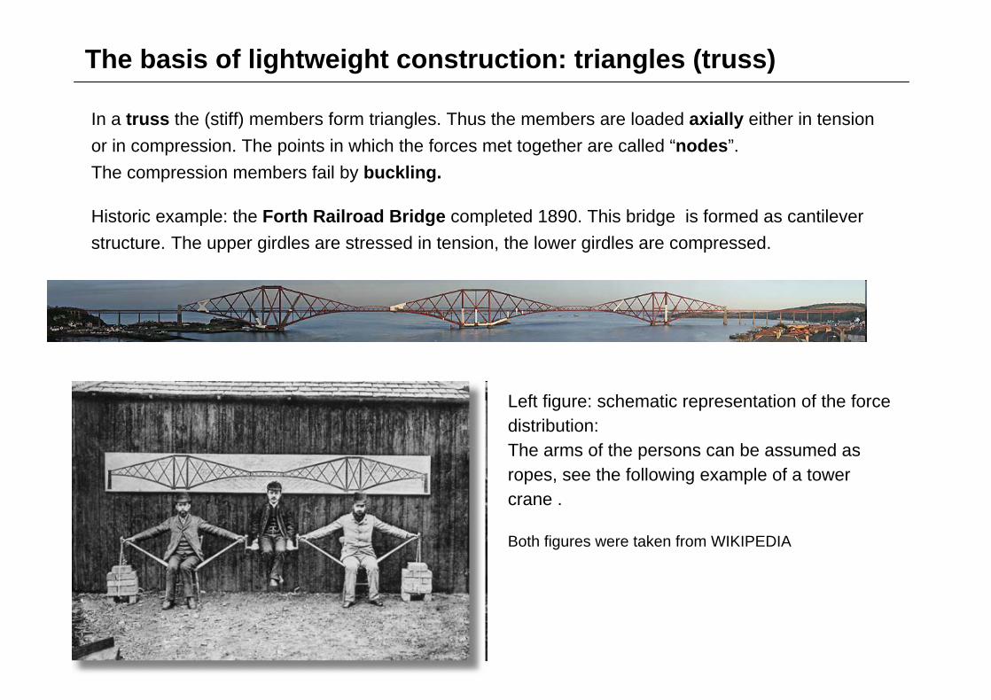
The basis of lightweight construction: triangles (truss)
In a truss the (stiff) members form triangles. Thus the members are loaded axially either in tension or in compression. The points in which the forces met together are called “nodes”. The compression members fail by buckling.
Historic example: the Forth Railroad Bridge completed 1890. This bridge is formed as cantilever structure. The upper girdles are stressed in tension, the lower girdles are compressed.
Left figure: schematic representation of the force distribution:The arms of the persons can be assumed as ropes, see the following example of a tower crane .
Both figures were taken from WIKIPEDIA

Tower crane with moveable jib The tension elements can be replaced by ropes forming the mentioned cable truss. An example is given by the tower crane constructed as base rotator with “needle” jib (moveable jibs). This kind of crane is self erecting. The counterweight rests on the crane foot opposite to the load jib. The left crane is a pure type, but the in right crane the tower is a little slanted (right figure: University of Waterloo).

Essential ligaments und muscles for our model (a)
Lig. sacrotuberale
Lig. sacrospinale
Mm. rotatores (M. quadratus femoris, Mm. obturatori)
The rotatores provide a horizontal tension from the base of the femur neck to the ischium.
The ischium is protected by the stretch of lig. sacrotuberale.
Source of the figure:
Bertolini, R; Leutert, G.: Atlas der Anatomie des Menschen.
Bd 1. VEB Georg Thieme, Leipzig 1978

Essential ligaments und muscles for our model (b)
Tractus iliotibialis
The iliotibial tract is attached as well as to the crista iliaca and the gluteus maximus. The tract is specific for human beings. This role was first described by J. H. Maissiat (Maissiat`s band).
Massiat, J. H.: Etudes de physique animale. Paris: Bethune et Plon 1843
Source of the figure: Bertolini, R; Leutert, G. :Atlas…

Tower crane with moveable jib as basis for unilateral stance
A special type of tower crane (with moveable jib) serves as basis of our consideration. The crane is a balancing one:
Femur and tibia are arranged on a line and form the „tower“. As weight jib, the femoral neck is prolonged to the spinal column and forms the pelvis.
The greater trochanter serves as counter jib. Here pelvis and trochanter are dealt as wheel discs. There diameters are in the ratio of 4:1.
The main jib is stretched by a rope formed by the extended iliotibial tract. Via the greater trochanter, the iliotibial tract (ext.) pulls down from the pelvis to the foot.
The load F is given by the full body weight. Due to the given leverage, the force of the tractus is 4 F und that of the femur-tibia about 5 F.

Introduction of the hip joint (a)
The hip joint divides the weight jib into the real femoral neck (collum ) and the corpus ossis ilii, thus a stabilisation is needed. This is accomplished by the ischium (os isschii) and the wing of the ilium (ala ossis ilii). In turn, they must also be braced:
The rotatores lead from the base of the femoral neck to the ischium (lower chord). The sacrotuberous ligament stretches from the ischium to the sacrum.
As „upper chord“, the iliolumbar ligament stretches to the iliac crest. The pelvis as a whole is stretched back by the iliotibial tract.Thus an osseous cross formed.
The „real“ femur (stronger inclined than the femur-tibia combination) is prolonged to the load line (reduced system).
e

Introduction of the hip joint (b)
The region of pelvis is magnified. Thus the values of forces can be read easily.

Introduction of the iliosacral joint and the knee
The sacrum is relatively broad and shows practically rib stumps (pars lateralis). It is divided from the ilium by the iliosacral joint forming the second joint in the weight jib of our tower crane.
The iliosacral joint is held from above by ligaments stretching from the ilium to the sacrum (ll. sacroiliaca).
Knee:Normally there is a slight bend of some 6o between tibia and femur called physiological genu valgum. This bend is due to a splitting of the iliotibial tract. One tract branch inserts at the lateral side of the tibia but the other branch aims at the middle of the knee. This medial branch is called here „tractus genualis“.
At tibia, the force of the lateral branch of the tract is transmitted to the foot by lateral muscles (mm. peronei).
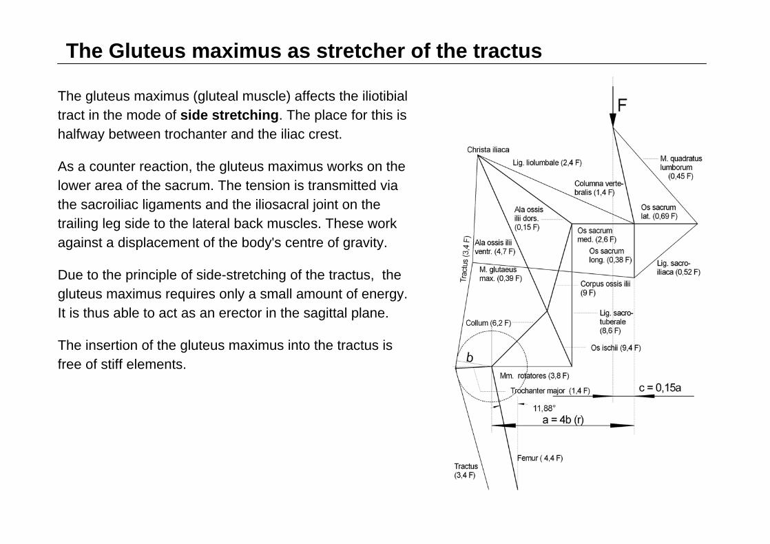
The Gluteus maximus as stretcher of the tractus
The gluteus maximus (gluteal muscle) affects the iliotibial tract in the mode of side stretching. The place for this is halfway between trochanter and the iliac crest.
As a counter reaction, the gluteus maximus works on the lower area of the sacrum. The tension is transmitted via the sacroiliac ligaments and the iliosacral joint on the trailing leg side to the lateral back muscles. These work against a displacement of the body's centre of gravity.
Due to the principle of side-stretching of the tractus, the gluteus maximus requires only a small amount of energy. It is thus able to act as an erector in the sagittal plane.
The insertion of the gluteus maximus into the tractus is free of stiff elements.

Balance of forces at the base of collum (clinical differences)At the hip the rotators are the essential muscles. Thus, the neck attachment becomes the critical node. The respective cycle of forces is shown in the figure: The femoral force pushes to the left and upwards on the node, while under the pressure of the tractus, the trochanter acts almost horizontally to the right. Being the strongest force, the femoral neck shifts to the left and downwards The cycle is closed by the pull of the rotators to the right (case a).
Case b: The rotators produce only half of the required force. The vector for the femoral neck force becomes relatively steep.Case c: We double the tension of the rotators. The vector for the neck force becomes relatively flat and considerably longer. The response of the bone depends on whether it is still growing or not. In children, the femoral neck will grow according to the predetermined direction of force…

The hip in the transversal plane, the two-legged stance
We start with the frontal plane, the two-legged stance.
In opposite to the one-legged stance, the iliotibial tract is absent.
Via the sacrum, the both halves of the pelvis are hold together by the sacrospinous ligament.From above, the sacrum is hold by sacroiliac ligaments
Now the body weight is called G.

The pelvis forms an osseous rhombus which is stretched from the outer side.dorsal part: iliumventral part: pubic boneouter stretching: dorsal by the rotatores, ventral by the pubofemoral ligament (here prolonged to symphis pubica).
Introduction of load occurs via femoral neck with a horizontal force component = 0,50 G.
This force widens the rhomb in the transversal axis. An outer force (F) is needed as compensation delivered by the spinal column and the inguinal ligament (horizontal components).
The hip as a cable truss in the transversal plane
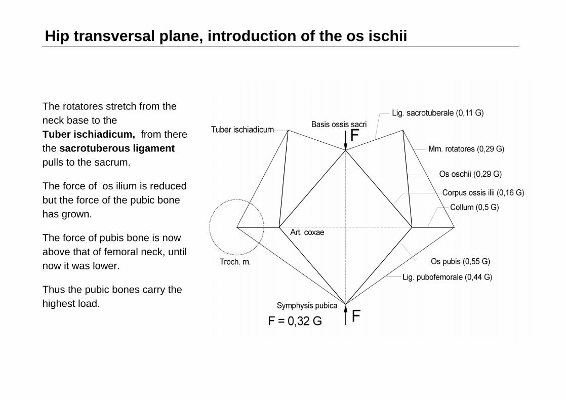
Hip transversal plane, introduction of the os ischii
The rotatores stretch from the neck base to the Tuber ischiadicum, from there the sacrotuberous ligament pulls to the sacrum.
The force of os ilium is reduced but the force of the pubic bone has grown.
The force of pubis bone is now above that of femoral neck, until now it was lower.
Thus the pubic bones carry the highest load.

Hip transversal plane, introduction of the iliosacral joint
The dorsal part of the pelvis ring is now arranged like a trapezium.
The sacrum forms the final part of the ring.
In the centre of the sacrum, at the basis ossis sacri, the lumbar spine attaches.
Stretching from the neck base to the ischium occurs by the rotatores, and further by the sacrotuberous ligament.

Hip transversal plane, antetorsion of collum
The femoral neck gets a torsion in the ventral direction.
The load of the ventral part of the pelvis ring with pubic bone and und pubofemoral ligament is further increased.
Due to symmetry of the pelvis ring it is sufficient to sketch only one half.
In the one-legged stance the forces grow tenfold.
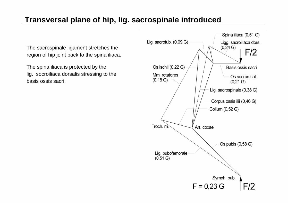
Transversal plane of hip, lig. sacrospinale introduced
The sacrospinale ligament stretches the region of hip joint back to the spina iliaca.
The spina iliaca is protected by the lig. socroiliaca dorsalis stressing to the basis ossis sacri.

Anatomical variations in antetorsion
Here again, the rotators are the essential muscles and the neck attachment is the critical node. The respective cycle of forces is shown in the figure: The femoral force pushes on the node to the left right and upwards. The cycle is closed by the pull of the rotators to the left/downward und the lig. pubofemorale to the left/upward (case a).
Case b: The rotators produce only half of the required force. The vector for the neck force becomes relatively steep (antetorsion increases).

Summary
The cable truss is based on a continuous path of stiff members. It fills the weight gap between tensegrity structures and the normal truss. The cable truss is preferred for the construction of cranes.
A special type of tower crane is suited to explain the function of long bones of leg (and arm) and the attached regions (hip and shoulder).
The leading opinion that the bones of the leg are bent relies on an obsolete understanding of the kind of the muscles involved.