the hong kong w ë medical diary - fmshk. · pdf filedr. albert mp lee digital dentistry...
TRANSCRIPT

ISSN 1812 - 1691
THE HONG KONG
MEDICAL DIARY
OFFICIAL PUBLICATION FOR THE FEDERATION OF MEDICAL SOCIETIES OF HONG KONG
www.fmshk.org VOL.15 NO.3 MARCH 2010
Dentistry

Contents
1
VOL.15 NO.3 MARCH 2010
I usually spend the Easter Holidays with my family in Japan. The Country extends over a long latitude so there are always cherry blossoms somewhere. I took this shot at Daiseki-ji ( ) in Shizuoka-ken ( ) last year. Quintessential Japanese spring: cherry blossoms, a Japanese style roof, and Mt. Fuji with a blue sky as the backdrop.
Nikon D80 with 18-55mm lens at 55mm, Auto at F/11, CPL, tripod.
The Cover Shot
Contents
Dental Bulletin
Editorial
� 2
Dr. Albert MP LEE
� MCHK CME Programme Self-assessment Questions
� 4
Dr. Gregory TAYLOR
� 12Intra-oral Autogenous Bone Grafting for Dental Implant Site Preparation
Dr. SF LEUNG
� 16Cone Beam Computed Tomography in Endodontics
Dr. Chun-hung CHU
� 21Management of Dentine Hypersensitivity
6
� Motor Racing
Life Style
Dr. Edgar SHIH
Dr. Chun-yan TSE
25
� Advance Directives: Their Role in Clinical Practice and Their Difficulties
Advance Directives Consultation Paper
27
Prof. EC HUI
� Introducing the Use of Advance Care Planning and Advance Directives in Hong Kong?
29
Mr. David KAN
� Advance Directives - the Legal Issues 32
Dr. SF LEUNG BDS(HK), MSc(Lond.), FRACDSSpecialist in Endodontics, Part-time Lecturer, Faculty of Dentistry, The University of Hong Kong
Digital Dentistry
Dr. Hannah Daile CHUAProf. Lim K CHEUNG
The Roles of Dental Professionals in the Management of Obstructive Sleep Apnoea
� 8
Dr. Mike Y Y LEUNG Prof. Lim K CHEUNG
New Approach in Extraction of Impacted Wisdom Teeth
35Medical Diary of March
Calendar of Events
37� Meetings
VOL.11 NO.5 MAY 2006 Editorial
2
VOL.15 NO.3 MARCH 2010
Computer technology, scanners and software have been developed in recent years for application in various aspects of dentistry. Nowadays many dental procedures can be assisted by these new technologies to facilitate dental clinicians in diagnosis, treatment planning, patient education and even delivery of treatment procedures. One of the remarkable applications is developed in orthodontics for the treatment of malocclusion. The procedures involve simply taking dental impressions of both the upper and lower arches. A computer scanner is then used to scan the impressions and a 3-D digital image of the dental arches is created by the computer software. According to instructions by the clinician, a 3-D virtual course of treatment is generated in the computer and custom-made aligners are made in a series of minor tooth movements until the set of teeth is moved to an ideal position.
The latest cutting edge technology using the conoscopic system or CAD/CAM system also involves in fabrication of dental prostheses like crowns & bridges and dental veneers. By combining high-precision scanning technology, intuitive design software and industrial manufacturing machines, excellent high quality metal-free dental prostheses can be produced to meet the demand of esthetic and strength for masticatory function. The working processes can be achieved within a few minutes after scanning the master dental models or impressions that minimise the complicated laboratory procedures in the conventional way.
In conjunction with cone-beam CT scans and software, high-quality 3-D images of dento-maxillofacial structures can be reproduced for patients within minutes of imaging. This technology is especially useful in the diagnosis of dental pathology, risk assessment in oral surgery and dental implant treatment planning. The technology is also applicable in the fields of periodontics and endodontics for bone loss assessment and treatment of complicated root canal morphology.
The developed computerised database technology also relieves the problems of limited storage space for patients' records in the dental office. Apart from recording the treatment records and radiographic films in a digitalised format, dental study models and casts that occupy a lot of office space can now be stored in a database form. By using the scanning system and software, all plaster dental casts can be converted to 3-D virtual study models on the computer. Hence a paperless and plasterless dental practice is now possible with the application of this technology.
Digital Dentistry is now becoming a reality and has changed the facets of practising dentistry in modern days. It is also a subject for study and development for all dentists in the years to come.
Dr. PDr. Albert MP LEE
Dr. Albert MP LEE
Digital DentistryPublished byThe Federation of Medical Societies of Hong Kong
EDITOR-IN-CHIEFDr. MOK Chun-on莫鎮安醫生
EDITORSDr. CHAN Chi-fung, Godfrey陳志峰醫生 (Paediatrics)Dr. CHAN Chun-hon, Edmond陳振漢醫生 (General Practice)Dr. KING Wing-keung, Walter金永強醫生 (Plastic Surgery)Dr. YU Kong-san俞江山醫生 (Orthopaedics & Traumatology)
EDITORIAL BOARDDr. CHAN Chi-wai, Angus陳志偉醫生 (General Surgery)Dr. CHAN, Norman陳諾醫生 (Diabetes, Endocrinology & Metabolism)Dr. CHIANG Chung-seung蔣忠想醫生 (Cardiology)Dr. CHIM Chor-sang, James詹楚生醫生 (Haematology)Dr. CHONG Lai-yin莊禮賢醫生 (Dermatology & Venereology)Dr. FAN Yiu-wah范耀華醫生 (Neurosurgery)Dr. FOO Wai-lum, William傅惠霖醫生 (Oncology)Dr. FONG Ka-yeung方嘉揚醫生 (Neurology)Prof. HO Pak-leung何 良醫生 (Microbiology)Dr. KWOK Po-yin, Samuel郭寶賢醫生 (General Surgery)Dr. LAI Kei-wai, Christopher賴奇偉醫生 (Respiratory Medicine)Dr. LAI Sik-to, Thomas黎錫滔醫生 (Gastroenterology & Hepatology)Dr. LAI Yuk-yau, Timothy賴旭佑醫生 (Ophthalmology)Dr. LAM Tat-chung, Paul林達聰醫生 (Psychiatry)Dr. LAM Wai-man, Wendy林慧文醫生 (Radiology)Dr. LEE Man-piu, Albert李文彪醫生 (Dentistry)Dr. LO, Richard 羅光彥醫生 (Urology)Dr. LO See-kit, Raymond勞思傑醫生 (Geriatric Medicine)Dr. MAN Chi-wai文志偉醫生 (Urology)Dr. MOK, Mo-yin莫慕賢醫生 (Rheumatology)Dr. TSANG Wai-kay曾偉基醫生 (Nephrology)Dr. TSE Tak-fu謝德富醫生 (Cardiology)Prof. WEI I, William韋霖醫生 (Otorhinolaryngology)Dr. WONG Bun-lap, Bernard黃品立醫生 (Cardiology)
Design and Production
Editor
BDS, MSc, FRACDS, FCDSHK(Paed Dent),FHKAM(Dental Surgery)
VOL.11 NO.5 MAY 2006 Dental Bulletin
4
VOL.15 NO.3 MARCH 2010
Obstructive sleep apnoea (OSA) is a disturbance in normal sleep pa�erns. It is characterised by repetitive episodes of complete (apnoea) or partial (hypopnoea) upper airway obstruction occurring during sleep. By OSA definition, apnoeic and hypopnoeic events should last a minimum of 10 seconds. The common morbidities of OSA are hypertension,1 depression,2 stroke,3 angina,4 cardiac dysrhythmia,5 diabetes and severe cases can be a direct cause of death during sleep. Untreated OSA is also associated with motor vehicle accidents,6 poor work performance, occupational accidents and reduced quality of life.4
EpidemiologyOSA occurs in 2 to 4 percent of the adult population between the ages of 30 to 60 years, though evidence suggests that many more patients remain undiagnosed. In Hong Kong, its prevalence was found to be 4.1% in the middle-age Chinese males.7 There is a large pool of undiagnosed OSAs in the community and particularly in hypertensive patients, the prevalence can reach up to 17%.8
Signs and SymptomsThe common signs and symptoms associated with OSA are: loud, habitual snoring, apnoeic events witnessed by the spouse or others, daytime sleepiness, restless sleep, choking sensation or gasping during the night, morning headache, personality and mood changes, sexual dysfunction (impotence and decreased libido) and gastro-oesophageal reflux.
Initial EvaluationThe presence and severity of OSA should be determined first before any therapy is given. When the patient presents for a consultation, a health history evaluation should be completed. The important components of the evaluation include a health history, physical examination, imaging studies and polysomnography.
Health historyA thorough sleep-related history and comprehensive medical history are essential components of this
evaluation. The primary indication for initiation of the evaluation process is the presence of OSA symptoms. A comprehensive medical history must be obtained because OSA is associated with a wide spectrum of medical conditions. The health history may reveal findings associated with obesity, hypertension, stroke or other cardiopulmonary or neurologic conditions linked to a high risk of OSA.
Physical examinationFor each patient, the clinican should perform a comprehensive head and neck examination and assess the respiratory, cardiovascular and neurologic systems. A body mass index (BMI) of greater than 28 is known to be associated with a 5-fold increase in the probability of moderate to severe OSA in Caucasians. It should also be noted that a neck circumference of greater than 17 inches (43cm) in men and 16 inches (40.6cm) in women are highly associated with OSA. The most common orofacial characteristics encountered include retrognathic mandible, narrow palate, long soft palate, hypertrophic tonsils, nasal septal deviation and relative macroglossia.
Imaging studiesImaging studies including a panoramic radiograph and lateral cephalograph should be part of the initial investigations. In the lateral cephalograph, the position of the maxilla and mandible in relation to the cranial base can be assessed. These findings are useful in planning treatment for the improvement of upper airway patency. Other imaging studies such as CT scan, MRI or sleep endoscopy may also be useful.
PolysomnographyThe gold standard for diagnosing OSA is by polysomnography (PSG), which needs to be conducted at a sleep laboratory. The aims of polysomnography are to evaluate any abnormal sleep breathing, sleep architecture and oxygen saturation. A typical 8 hours nocturnal laboratory PSG involves measurements of multiple physiological functions including electro-encephalography, electro-oculography, chin or leg movement via electromyography, electrocardiography, sleep positioning, respiratory activity and oxygen saturation. The primary measure of sleep-disordered breathing is the apnoea-hypopnoea index (AHI), which is the number of apnoeas and hypopnoeas per hour of sleep. Apnoea is defined as the c e s s a t i o n o f a i r fl o w
The Roles of Dental Professionals in the Management of Obstructive Sleep ApnoeaDr. Hannah Daile CHUA
Prof. Lim K CHEUNGDr. Hannah Daile CHUA
Post-doctoral Fellow, OMFS, Faculty of Dentistry, The University of Hong KongDMD, MA, MDS, MOSRCS, PhD
Prof. Lim K CHEUNG
Chair Professor, Discipline of Oral and Maxillofacial Surgery, Faculty of Dentistry, The University of Hong Kong
BDS, PhD, FFDRCS, FDSRCPS, FRACDS, FDSRCS (Edin), FRACDS(OMS), FHKAM(DS), FCDSHK(OMS), FFGDP(UK)
This article has been selected by the Editorial Board of the Hong Kong Medical Diary for participants in the CME programme of the Medical Council of Hong Kong (MCHK) to complete the following self-assessment questions in order to be awarded one CME credit under the programme upon returning the completed answer sheet to the Federation Secretariat on or before 31 March 2010.

Dental Bulletin
5
VOL.15 NO.3 MARCH 2010
(complete obstruction) for at least 10 seconds with a concomitant 2 to 4 percent drop in arterial oxygen saturation. Hypopnoea is defined as a reduction in airflow of at least 30 to 50 percent with a drop in oxygen saturation. The severity of OSA is commonly classified according to a patient's AHI score: mild (AHI score between 5 to 15); Moderate (AHI score between 15 and 30); and severe (AHI score greater than 30). Other factors that also influence the severity of OSA include oxygen desaturation, quality of life and the level of daytime sleepiness
ManagementThere are multiple specialties involved in the management of OSA. These would include internal medicine, family medicine, paediatrics, otorhinolaryngology, psychiatry, neurology, and dentistry. Currently, in the United States, Sleep dentistry qualifies for board certification by the American Board of Dental Sleep Medicine (ABDSM). Qualified dentists collaborate with sleep doctors at different sleep centres and provide treatments. There is no such certification for dentists in Hong Kong. The dental specialists commonly involved in the management are oral and maxillofacial surgeons and orthodontists.
Non-surgicalPossible treatment options for adult patients diagnosed with OSA are based on the severity of the sleep disorder, patient's preference and his/her overall health status. Positional therapy involves nocturnal aids to prevent patients from sleeping in a supine position. Sleeping in the lateral position is generally recommended, as it will displace the tongue from the posterior airway such that it is less likely to cause airway obstruction during sleep. Weight loss is universally recommended for obese patients. However, it is not known how much weight loss is required to eliminate OSA, and both the patient's gender and weight distribution may contribute to his or her OSA in an unpredictable manner. The most commonly recommended non-surgical intervention is continuous positive airway pressure (CPAP). The concept of nasal CPAP is to maintain upper airway patency during sleep. This treatment can be administered via either a nasal or oral mask. Due to its effectiveness, CPAP is the first-line treatment and the primary form of therapy for OSA, although its success is limited by the patient's level of compliance. About 20 to 30 percent of patients experience problems using CPAP, and the device is ineffective if it is not regularly used. The common problems associated with CPAP are nasal dryness, facial ulceration at the mask interface and claustrophobia.
Oral appliances gain recent recognition as an effective treatment option for mild to moderate OSA. The design is similar to an orthodontic functional appliance with the aim of maintaining a patient's lower jaw in a protruded position during sleep, hence enlarges the oro-pharyngeal airway. Many commercial devices offer treatment for snoring; however a device designed to treat OSA should be fabricated by a dental practitioner or specialist familiar with device design, maintenance and therapeutic efficacy. A multitude of oral appliances for OSA are available, but not all patients find the same
appliance effective. Patient's compliance with oral appliances appears to be adequate but not all studies agree. Difficulty with the device, owing to temporary or persistent occlusal disturbance, temporomandibular joint or individual tooth discomfort or perceived lack of efficacy may cause compliance issues.
SurgicalThe main surgical treatments offered for OSA often target the anatomical areas of the posterior airway where collapse is suspected to occur. Treatment is designed to enlarge the posterior airway space, reduce airway collapsibility and stabilise the airway in the long term. The type of surgical procedure would include uvulopalatopharyngoplasty, palatal suspension via a lateral inversion flap. These types of surgical procedures are usually performed by otorhinolaryngologists. Successful surgical outcomes have also been reported with less invasive techniques such as radiofrequency thermal ablation, and palatal implants. Orthognathic surgery has the advantage of correcting any craniofacial abnormalities that may have caused the OSA. Maxillomandibular advancement surgery (MMA), which is based on conventional orthognathic surgery techniques, has been proven effective in retrospective studies for a range of OSA patients over 90% long term success.9
Distraction osteogenesis (DO) is a clinical biomedical tissue engineering method that generates new bone by gradual stretching of the divided bone segments. Since its first application in the cranio-maxillofacial region in 1992, patients with severe facial deformities benefit by the large surgical movement achievable with minimal morbidities and in particularly useful for children and infants suffering from OSA. The main benefits of distraction for OSA are enlargement of the posterior airway space, improved oxygen saturation, improved respiratory disturbance index and accelerated growth of infants and children. From our evidence-based review, distraction has been reported a cure of OSA in 97% of children and 100% of adults.10
Roles of Dental ProfessionalsIt is of utmost importance for dental professionals to be aware of the prevalence and possible outcomes of OSA if left untreated. Screening of patients by general dentists or specialists, by conducting a thorough examination can aid in the diagnosis and treatment of OSA patients. Orthodontists can provide oral appliances as well as perform pre and post-surgical orthodontic treatments. Prosthodontists are also able to help in the construction of oral appliances. Oral and maxillofacial surgeons on the other hand can perform orthognathic surgeries for maxillomandibular advancement and distraction osteogenesis.
ConclusionManagement of OSA patients is recommended to be by multi-disciplinary teams dedicated to OSA. The dental professionals have a significant role to play as a team member in identifying potential OSA patients, making the appropriate referrals and assisting in the management of these patients. The dental specialists

VOL.11 NO.5 MAY 2006 Dental Bulletin
6
VOL.15 NO.3 MARCH 2010
can actively contribute to the management by provision of oral appliance therapy, orthognathic surgery and distraction osteogenesis.
Dr. Chun-hung CHU
Richert A, Ansarin K, Baran AS. Sleep apnea and hypertension: pathophysiologic mechanisms. Semin Nephrol. 2002 ;22(1):71-7. Baran AS, Richert AC. Obstructive sleep apnea and depression. CNS Spectr. 2003; 8(2):128-34. Palomaki H, Partinen M, Erkinjun�i T, Kaste M. Snoring, sleep apnea syndrome, and stroke. Neurology. 1992 ;42(7 Suppl 6):75-81.Loui WS, Blackshear JL, Fredrickson PA, Kaplan J. Obstructive sleep apnea manifesting as suspected angina: report of three cases. Mayo Clin Proc. 1994; 69(3):244-8.Gula LJ, Krahn AD, Skanes AC, Yee R, Klein GJ. Clinical relevance of arrhythmias during sleep: guidance for clinicians. Heart. 2004; 90(3):347-52.
1.
2.
3.
4.
5.
Horne JA, Reyner LA. Sleep related vehicle accidents. BMJ. 1995;310(6979):565-7.Ip MS, Lam B, Lauder IJ, Tsang KW, Chung KF, Mok YW, Lam WK. A community study of sleep-disordered breathing in middle-aged Chinese men in Hong Kong. Chest. 2001 Jan;119(1):62-9.Ip M, Chung KF, Chan KN, Lam SP, Lee K. Previously unrecognized obstructive sleep apnea in Chinese subjects with essential hypertension. Lung. 1999; 177(6):391-400.Riley RW, Powell NB, Guilleminault C. Maxillofacial surgery and obstructive sleep: a review of 80 patients. Otolaryngol Head Neck Surg. 1989;101(3):353-61.Ow ATC, Cheung LK. Meta-analysis of mandibular distraction osteogenesis: clinical application and functional outcome. Plast Reconstr Surg 2008; 121(3):54e-69e.Magliocca KR, Melman JI. Obstructive sleep apnea - diagnosis, medical management and dental implications. JADA 2005; 136:1121-9.Lee NR. Evaluation of the obstructive sleep apnea patient and management of snoring. Oral Maxillofac Surg Clin N Am 2009; 377-387.
6.
7.
8.
9.
10.
11.
12.
References
MCHK CME Programme Self-assessment QuestionsPlease read the article entitled "The Roles of Dental Professionals in the Management of Obstructive Sleep Apnoea" by Dr. Hannah Daile CHUA and Prof. Lim K CHEUNG and complete the following self-assessment questions. Participants in the MCHK CME Programme will be awarded 1 CME credit under the Programme for returning completed answer sheets via fax (2865 0345) or by mail to the Federation Secretariat on or before 31 March 2010. Answers to questions will be provided in the next issue of The Hong Kong Medical Diary.
A person who habitually snores has obstructive sleep apnoea (OSA).Polysomnography is recommended for a person who presents with excessive daytime sleepiness.OSA is a potentially lethal condition. Factors that influence the severity of OSA are oxygen saturation, obesity and level of daytime sleepiness. Oral appliances are the most common non-surgical intervention for patients with OSA.Persons with retrognathic mandible and chin are common features of OSA. CPAP keeps the airway open during sleep by pumping oxygen into the lungs.The gold standard for diagnosing OSA is by polysomnography. Maxillo-mandibular advancement is more effective than distraction osteogenesis in the surgical management of OSA. The dental specialists and general dentists can contribute to the management of OSA.
1.2.3.4.5.6.7.8.9.
10.
Questions 1-10: Please answer T (true) or F (false)
The Roles of Dental Professionals in the Management of Obstructive Sleep Apnoea
Please return the completed answer sheet to the Federation Secretariat on or before 31 March 2010 for documentation. 1 CME point will be awarded for answering the MCHK CME programme (for non-specialists) self-assessment questions.
Name (block le�ers):____________________________________ HKMA No.:
HKDU No.:
DCHK No.:
___ ___ - ___ ___ ___ ___ X X (x)HKID No.:
______ ______ ______________________
____________ ______________________
____________ ______________________
Contact Tel No.:_________________________________________________
ANSWER SHEET FOR MARCH 2010
Answers to February 2010 Issue
Management of Obesity - From Life Style Modification to Weight Reduction Surgery
1 2 3 4 5 6 7 8 9 10
1 . T 2 . F 3 . F 4 . F 5 . F 6 . T 7 . T 8 . F 9 . T 10 . F
Dr. Hannah Daile CHUA
Post-doctoral Fellow, OMFS, Faculty of Dentistry, The University of Hong Kong
DMD, MA, MDS, MOSRCS, PhDProf. Lim K CHEUNGBDS, PhD, FFDRCS, FDSRCPS, FRACDS, FDSRCS (Edin),FRACDS(OMS), FHKAM(DS), FCDSHK(OMS), FFGDP(UK)Chair Professor, Discipline of Oral and Maxillofacial Surgery, Faculty of Dentistry, The University of Hong Kong

VOL.11 NO.5 MAY 2006 Dental Bulletin
8
VOL.15 NO.3 MARCH 2010
Neurosensory deficit is a significant risk in lower wisdom tooth surgery. Due to the anatomical positions, the lingual nerve (LN) and inferior dental nerve (IDN) are at risk in the procedure, resulting in tongue numbness with taste disturbance in LN injury, or lower lip numbness in IDN injury on the affected side. The prevalence of LN and IDN deficits in the literature are 0.1-22% and 0.3-8.4%, respectively. As there were no local data, our centre has initiated a series of studies to determine the local prevalence of neurosensory deficit after wisdom tooth surgery, the relevant risk factors contributing to the neurosensory deficit, and to investigate the safety of coronectomy, which is a new technique of wisdom tooth surgery to prevent IDN injury.
Local Prevalence of Neurosensory Deficit after Wisdom Tooth Surgery
We conducted a prospective cohort study of 4338 wisdom tooth surgeries between 1998 and 2005 in the Prince Philip Dental Hospital, which is by far the largest study reported in the literature1. All surgeries were performed in the Local Extraction Clinic by operators of various grades from undergraduate students to specialists. The prevalence of LN and IDN injuries were 0.69% and 0.35%, respectively. These figures were relatively low when compared to those reported in the literature. 66.7% of IDN deficit and 72.0% of LN deficit recovered fully within the follow-up period of 24 months. It was also noticed the recovery of the neurosensory deficit was most significant in the first 6 months after the injury.
Significant Risk Factors Contributing to Nerve Deficit
The next question is what factors contribute to the risk of neurosensory deficit? To investigate the risk factors of nerve deficit after wisdom tooth surgery, we have conducted a systematic review on prospective studies on this clinical issue, which is considered as a study design with the highest level of evidence in the era of evidence-based medicine2. Without any limitation of languages and years of publication, 3 literature data-bases were searched. Additional reference search was performed and the articles were evaluated by 2 independent judges with pre-set criteria. 32 articles were qualified to enter the final review. We concluded in our study that increased age, unerupted wisdom tooth and the "old-fashion" lingual split technique are significant risk factors
and LN deficit. Raising of the lingual flap, which is supposed to protect the LN from rotary instruments, is also a risk factor to LN deficit at least on a temporary basis. In contrast, the risk of IDN deficit is increased with the depth of the impaction or exposure of IDN intra-operatively, which is easily explained by the proximity of IDN to the tooth roots and the increased difficulty of the surgery. Several radiographic signs, e.g. darkening of wisdom tooth root and deflection of ID canal, are also positively related to the risk to IDN deficit. The prevalence of IDN deficit happened more on operators with more surgical experience in our study, which is due to the case selection bias because of the fact that specialist oral surgeons need to manage wisdom teeth of higher difficulties, yet this also reflects IDN deficit may not be totally avoidable even in the hands of experienced surgeons. In cases where the wisdom tooth root is lying in close proximity to the IDN, or occasionally the nerve even creates a groove onto the tooth root, taking the tooth out will likely compress or even transect the IDN, leading to permanent IDN deficit.
Coronectomy: A New Method of Wisdom Tooth SurgeryCoronectomy literally means cutting off the crown from a tooth. It is a new method of wisdom tooth surgery in selected cases aiming to remove the crown of the wisdom tooth while leaving the root in situ. The crown of the wisdom tooth is usually the cause of various oral health problems, such as pericoronitis or dental caries of the wisdom tooth itself or the second molar in front of it. By removing the crown can resolve these problems, yet leaving the root behind can avoid injury to the IDN which is in close proximity. It had been a taboo in dentistry that leaving the tooth roots in the jaw was believed to be a source of dentoalveolar infection. The technique was first described by Knutsson et al. in 1989 but was not popularised due to the reported complications of root exposure and infection from the technique3. Several articles have reported this technique with less complications and more promising results, yet it was understandable that dentists and oral surgeons would not be convinced until a well-designed study can show its safety in terms of reduced risk of IDN and other surgical complications. Therefore our centre has conducted a randomised clinical trial to compare coronectomy with the traditional total removal of lower wisdom teeth4. The study included patients with wisdom tooth root lying in close proximity to ID canal as shown with one or more radiographic sign on an orthopantomogram. The radiographic signs were:
New Approach in Extraction of Impacted Wisdom TeethDr. Mike Y Y LEUNG
Prof. Lim K CHEUNGDr. Mike Y Y LEUNG
PhD Student, OMFS, Faculty of Dentistry, The University of Hong Kong BDS, MDS(OMFS), MOS RCS(Edin)
Prof. Lim K CHEUNG
Chair Professor, OMFS, Faculty of Dentistry, The University of Hong Kong
BDS, FDSRCPS, FFDRCS, FRACDS, FRACDS(OMS), FHKAM(DS), FCDSHK, PhD, FDSRCS(Edin), FFGDP(UK)

Dental Bulletin
9
VOL.15 NO.3 MARCH 2010
1. Darkening of the root2. Abrupt narrowing of the root3. Interruption and loss of the white line representing
the ID canal4. Displacement of the ID canal by the roots5. Abrupt narrowing of one or both of the white lines
representing the ID canal
The study excluded patients whose wisdom teeth were not in close proximity to IDN, with systemic or local factors that predisposes to infection, or any pulpal caries, cystic or neoplastic pathology relating to the wisdom tooth.
The procedure of coronectomy included flap raising and bone guttering down to the cemento-enamel junction of the impacted wisdom tooth. The crown was sectioned off from the root. Minimal stress to the root during crown elevation was required to avoid any root dislodgement. The root was further trimmed down 3-4mm below the crestal bone (Figure 1). Primary closure was then performed. No antibiotics were prescribed post-operatively.
promising at least in the short term. A phase IV clinical trial of coronectomy is on-going in our centre to evaluate the long term safety of the technique.
231 patients with 349 lower wisdom teeth were randomised, with 178 wisdom teeth underwent total removal (as control group) and 171 wisdom teeth underwent coronectomy. 16 coronectomies (9.4%) were considered failed as the roots were dislodged during the procedure and were removed in total. The prevalence of IDN deficit was significantly lower in the coronectomy group (0.65%) when compared to the control group (5.1%) (p=0.023). The prevalence of pain and dry socket in the first week were also significantly lower in the coronectomy group than the control group. The infection rate in the first week showed no statistical difference between the two groups, and of note there was no infection in the patients who had coronectomy from post-operative 3 months onwards until the last review of 2 years. Serial radiographs showed significant root migration within the alveolar bone after coronectomy up to 3mm, and then gradually stopped at 1 year post-operatively (Figure 2a-d). There was only one case of root exposure into the oral cavity and required subsequent re-operation to remove the root.
The result of the coronectomy study was fascinating as it proved that this technique can significantly reduce the prevalence of IDN deficit in lower wisdom tooth surgery for the high risk group. The safety of coronectomy is
Figure 1. Coronectomy: Crown removed and root trimmed down 3-4mm below crestal bone
Figure 2a. Pre-operative: Lower wisdom tooth in close proximity to IDN
Figure 2b. Post-operative 1 week: Crown removed and the root left in situ
Figure 2c. Post-operative 1 year: Coronal migration of the root by around 3mm
Figure 2d. Post-operative 2 years: No further migration of the root noticed.
Cheung LK, Leung YY, Chow LK, Wong MC, Chan EK, Fok YH. Incidence of neurosensory deficits and recovery after lower third molar surgery: a prospective clinical study of 4338 cases. Int J Oral Maxillofac Surg. 2010 Jan 8(in press)Leung YY, Cheung LK. Risk Factors of Neurosensory Deficits in Lower Third Molar Surgery: An Evidence-based Review of Prospective Studies. Int J Oral Maxillofac Surg. (under review)Knutsson K, Lysell L, Rohlin M. Postoperative status after partial removal of the mandibular third molar. Swed Dent J. 1989; 13: 15-22.Leung YY, Cheung LK. Randomized Controlled Trial of Safety of Coronectomy versus Excision of Wisdom Teeth. Oral Surg Oral Med Oral Pathol Oral Radiol Endod. 2009 Dec; 108: 821-7.
1.
2.
3.
4.
References
With
Compliments
of
Mr. Eddie WANG


VOL.11 NO.5 MAY 2006 Dental Bulletin
12
VOL.15 NO.3 MARCH 2010
BackgroundReconstruction of our patients' dentition is a very rewarding and fulfilling aspect of dentistry. Our abilities as dentists in enabling patients to chew appropriately or smile confidently can at times be life changing for the patient. Dental implants are an exciting portion of our armamentarium with which to reconstruct the partially or fully edentulous jaws. Many patients present after tooth loss with alveolar defects that will not accommodate a dental implant prosthesis. The defect may range in size from that seen with a small loss of the buccal cortex from an avulsed tooth to a resected jaw as a result of cancer or an odontogenic tumour.
Autogenous bone remains the "Gold Standard" for grafting. Although allogenic bone, xenogeneic bone, bone substitutes, and alloplasts have shown some promise over the years, they do not transplant any osteocompetent cells1. Patients are often concerned when presented with the need for bone grafting and tend to become nervous when learning how and where the new bone will come from. I have found that patients usually become more acceptive of an autogenous graft when presented with the other option of using bone from a human tissue (cadaver) bank or bovine bone.
Treatment PlanningPrior to bone graft harvesting and augmentation of the defect, one must have a full appreciation of the defect. A preoperative 3-D CT scan is often imperative and some would argue the standard of care. This enables the clinician to have a full map of the missing bone volume, i.e. the vertical and horizontal nature of the defect. The scan can also be used to evaluate the cortical thickness from the potential donor sites. Study models and a diagnostic wax-up with the final crown or prosthesis morphology are important so that you "know where you are going." This permits the surgeon to appreciate how much bone augmentation is needed so that the final prosthesis is in the correct location.
A thorough past medical history should be obtained. Poorly controlled Insulin Dependent Diabetes Mellitus, cigarette smoking, and history of IV Bisphosphonate therapy are a few examples of contra-indications2.
The patient should exhibit good oral hygiene. A
patient presenting with atrophy of the alveolar ridges with remaining moderate to severe periodontitis will likely later develop peri-implantitis. Why put this type of patient through extensive grafting if the future implant is destined to fail?
It is of paramount importance for the surgeon to know all potential risks and complications of the grafting procedures and fully explain these to the patient preoperatively. No one desires complications but without informed consent the clinician may be open to litigation.
Donor Site SelectionThe graft may be harvested from many intra-oral sites. The maxillary tuberosity, anterior nasal spine, and zygomatic buttress have been reported for the upper jaw3,4,5. The mandibular symphysis, ascending ramus, coronoid process, and horizontal ramus are useful sites from the lower jaw6.
The harvested bone may be placed in an extraction site, an implant site defect, a buccal alveolar defect, and the maxillary sinus7,8,9,10. It may also be used for a vertical onlay graft of the ridge or as an inlay graft11. Particulate grafts may be stabilised with a membrane whereas block grafts should be secured to the recipient site with screws.
Ideally, the surgeon would like to harvest bone from a site that is close to the defect site. This essentially affords the possibility of one surgical site rather than two. Examples of this would be a tuberosity graft in conjunction with an ipsilateral sinus lift or bone from the anterior nasal spine for a maxillary central incisor site. In reality, the patient's bone quality or quantity often necessitates grafting from another area and precludes the surgeon's ability to have the donor bone come from a nearby area.
Case PresentationA 38 YO Chinese male presented with a periapical abscess and fractured upper right central incisor #11. The tooth had Class I mobility and was deemed non-restorable by an endodontist. The tooth was atraumatically extracted and a periapical granuloma was curetted from the apex of the extraction site. The patient wore a flipper partial denture as an interim prosthesis. The patient was treatment planned for
Intra-oral Autogenous Bone Grafting for Dental Implant Site PreparationDr. Gregory TAYLOR
Dr. Gregory TAYLOR
Clinical Associate Oral & Maxillofacial Surgery, Department of Dentistry, Hong Kong Sanatorium & Hospital
Cert OMFS, Virginia Commonwealth University, USADiplomate of the American Board of Oral and Maxillofacial Surgery

Dental Bulletin
13
VOL.15 NO.3 MARCH 2010
future bone grafting and implant restoration of the #11 site.
Six weeks post-extraction a CT scan was taken which revealed full loss of the buccal cortex. [see Figures 1 and 2]. A cortico-cancellous block graft from the ascending ramus was deemed suitable for augmentation of the defect. [see Figure 3 for pre-op view]. The ramus graft harvest is as described by Misch12.
Graft Procedure:A sulcular incision is placed from the right canine to the left lateral incisor. A palatal "finger extension" is placed at the edentulous central incisor site. Vertical releases are placed to include the papillae. A full-thickness flap is repositioned apically to fully expose the defect. [see Figure 4]. The periosteum is incised horizontally to help
release tension for later primary closure over the bone graft. Sterile aluminum foil is adapted into the defect to simulate the size of the graft needed. The flap is adapted/extended over the foil so that closure may proceed in a quicker fashion after the graft is placed. Once it is determined that a tension free closure may be obtained, the graft is harvested.
A buccal "hockey-stick" incision is placed and the ramus is exposed. This is almost identical to the flap design to remove an impacted 3rd molar. [see Figure 5]. A surgical hand piece with a small fissure bur is used to create a "block" through the cortex into the bleeding cancellous bone. A periosteal and 301 elevators are used to greenstick fracture the graft from the ascending ramus. [see Figures 6 and 7].
Figure 1
Figure 2
Figure 3
Figure 4
Figure 5
Figure 6

VOL.11 NO.5 MAY 2006 Dental Bulletin
14
VOL.15 NO.3 MARCH 2010
The graft is modified and secured to the defect with bone screws (Osseofix, Bio-met 3i, Jacksonville, Florida, USA). [see Figure 8]. The remainder of the defect is packed with cortico-cancellous chips and cancellous bone curetted from the ramus. The graft is covered with an adapted resorbable membrane (Biomend Extend, Zimmer Dental, Carlsbad, California, USA).
Both wounds are closed primarily with interrupted sutures.
The patient was placed on analgesics, antibiotics, and an antimicrobial mouthrinse for 1 week.
The site will be re-entered after 6 months for removal of the fixation screws and placement of the implant.
Future Possibilities and Alternative Treatment OptionsMany studies and clinical trials show promise for the future. Recombinant bone morphogenetic protein (rhBMP-2) has been used with great success in clefts, large reconstructions, ridge augmentation, and sinus lift procedures13,14,15. Initially this was used in spinal fusion surgery before its benefits were realised for the oral cavity. It is known as INFUSE and manufactured by Medtronic (Memphis, TN, USA). This technique utilises synthetic BMP in a liquid form mixed with an absorbable collagen sponge. The sponge is placed into the defect site and closed primarily. After six months
healing, the site is re-entered and implants may be placed into newly formed bone. Utilising this technique avoids a donor site. The material does have cost limitations. Other tissue-engineered materials have been tried with some success16.
Alternative treatment plans include distraction osteogenesis for vertical defects, ridge-splitting techniques, short implants, and angled implants17,18,19,20. Cadaver bone may be used for patients who do not mind the idea of it21. Chen et al describe success in sinus lifts without grafting and simultaneous implant placement22.
In conclusion, there are many tools to be used for this patient population. The clinician must stay within his or her scope of training as well as his or her comfort zone when performing these grafting techniques.
Marx, R. Philosophy and Particulars of Autogenous Bone Grafting. Oral & Maxilofac Surg Clin N Am 1993; 5:599-612.Dodson,T. Intravenous Bisphosphonate Therapy and Bisphosphonate-Related Osteonecrosis of the Jaws. J Oral Maxillofac Surg 2009; 67:44-52.Tolstunov, L. Maxillary Tuberosity Block Bone Graft: Innovative Technique and Case Report. J Oral Maxillofac Surg 2009; 67:1723-1729.Buser,D. "State of the Art Surgical Procedures in Esthetic Implant Dentistry." Straumann training Lecture. Hong Kong. October 6,2009.Gellrich,N. et al. Alveolar Zygomatic Buttress: A New Donor Site for Limited Preimplant Augmentation Procedures. J Oral Maxillofac Surg 2007; 65:275-280.Soehardi,G. et al. The Potential of the Horizontal Ramus of the Mandible as a Donor Site for Block and Particular Grafts in Pre-implant Surgery. Int J Oral Maxillofac Surg 2009; 38:1172-1178.Becktor,J. et al. The Use of Particulate Bone Grafts from the Mandible for Maxillary Sinus Floor Augmentation Before Placement of Surface-Modified Implants: Results from Bone Grafting to Delivery of the Fixed Prosthesis. J oral Maxillofac Surg 2008; 66:780-786.Zijderveld, S. et al. Anatomical and Surgical Findings and Complications in 100 Consecutive Maxillary Sinus Floor Elevation Procedures. J Oral Maxillofac Surg 2008; 66:1426-1438.Raja, S. Management of the Posterior Maxilla With Sinus Lift: Review of Techniques. J Oral Maxillofac Surg 2009; 67:1730-1734.Chiapasco,C and Zaniboni, M. Methods to Treat the Edentulous Posterior Maxilla: Implants With Sinus Grafting. J Oral Maxillofac Surg 2009; 67:867-871.Felice,P.et al. Reconstruction of Atrophied Posterior Mandible With Inlay Technique and Mandibular Ramus Block Graft for Implant Prosthetic Rehabilitation. J Oral Maxillofac Surg 2009; 67:372-380.Misch,C. Ridge Augmentation Using Mandibular Ramus Bone Grafts For the Placement of Dental Implants: Presentation of a Technique. PP&A 1996; 8:127-135.Herford,A and Boyne,P. Reconstruction of Mandibular Continuity Defects with Bone Morphogenetic Protein-2 (rhBMP-2). J oral Maxillofac Surg 2008; 66:616-624.Herford,A. rhBMP-2 as an Option for Reconstructing Mandibular Continuity Defects. J Oral Maxillofac Surg 2009; 67:2679-2684.Triplett, RG et al. Pivotal, Randomized, Parallel Evaluation of Recombinant Human Bone Morphogenetic Protein-2/Absorbable Collagen Sponge and Autogenous Bone Graft for Maxillary Sinus Floor Augmentation. J Oral Maxillofac Surg 2009; 67:1947-1960.Torroni, A. Engineered Bone Grafts and Bone Flaps for Maxillofacial Defects: State of the Art. J Oral Maxillofac Surg 2009; 67:1121-1127.Jensen,O et al. Marginal Bone Stability Using 3 Different Flap Approaches for Alveolar Split Expansion for Dental Implants-A 1-Year Clinical Study. J Oral Maxillofac Surg 2009; 67:1921-1930.Grant, B et al. Outcomes of Placing Short Dental Implants in the Posterior Mandible: A Retrospective Study of 124 Cases. J Oral Maxillofac Surg 2009; 67:713-717.Rosen,A. and Gynther,G. Implant Treatment Without Bone Grafting in Edentulous Severely Resorbed Maxillas: A Long-Term Follow-Up Study. J Oral Maxillofac Surg. 2007; 65:1010-1016.Block, M. et al. Nongrafting Implant Options for Restoration of the Edentulous Maxilla. J Oral Maxillofac Surg 2009; 67:872-881.Gomes, K. et al. Use of Allogeneic Bone Graft in Maxillary Reconstruction for Installation of Dental Implants. J Oral Maxillofac Surg 2008: 66:2335-2338.Chen,TW et al. Implant Placement Immediately after the Lateral Approach of the Trap Door Window Procedure to Create a Maxillary Sinus Lift without Bone Grafting: A 2-Year Retrospective Evaluation of 47 Implants in 33 Patients. J Oral Maxillofac Surg 2007; 65:2324-2328.
1.
2.
3.
4.
5.
6.
7.
8.
9.
10.
11.
12.
13.
14.
15.
16.
17.
18.
19.
20.
21.
22.
References
Figure 7
Figure 8

VOL.11 NO.5 MAY 2006 Dental Bulletin
16
VOL.15 NO.3 MARCH 2010
IntroductionConventional (both chemical and digital) radiography renders a three-dimensional (3-D) anatomical structure two dimensionally with inherit distortions. This limitation posts a steep learning curve for novice operators to interpret information from the resulting images. In many incidences, it becomes a matter of guesswork even to the experienced user, like the relationship of the maxillary molars with the maxillary sinus.
Cone beam computed tomography (CBCT) has been used in dentistry since 19981. Unlike medical CT, which captures the image in slices, CBCT data are captured in a 3-D pixel unit called voxel. As these voxels are isotropic, the object is accurately measured in different directions. This enables the rendering of geometrically undistorted images of the maxillo-facial skeletal structure and allows viewing at different angles.
In addition to providing higher resolution image, CBCT has a much reduced radiation dosage than medical CT. The exposure, at about three to ten times the radiation of a digital panoramic radiograph, is more comparable to routine diagnostic imaging with panoramic and periapical radiography. CBCT is available with different fields of view (FOV) to suit different applications. In endodontics, a machine with limited FOV should suffice. CBCT has become a routine tool in oral surgery and especially implant dentistry. With increasing affordability of the computer and less expensive CB X-ray tube, CBCT will have enormous potential in endodontics. The following case reports illustrate some of these endodontic applications.
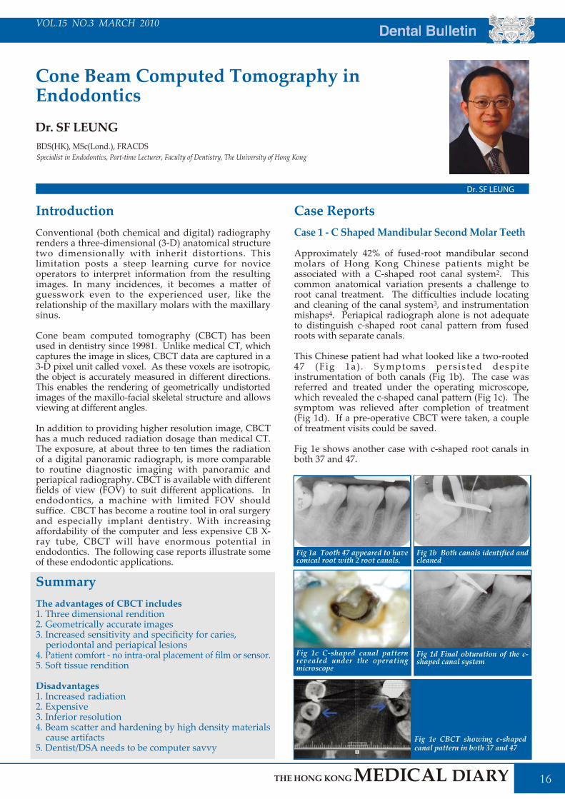
Case ReportsCase 1 - C Shaped Mandibular Second Molar Teeth
Approximately 42% of fused-root mandibular second molars of Hong Kong Chinese patients might be associated with a C-shaped root canal system2. This common anatomical variation presents a challenge to root canal treatment. The difficulties include locating and cleaning of the canal system3, and instrumentation mishaps4. Periapical radiograph alone is not adequate to distinguish c-shaped root canal pattern from fused roots with separate canals.
This Chinese patient had what looked like a two-rooted 47 (Fig 1a) . Symptoms persisted despite instrumentation of both canals (Fig 1b). The case was referred and treated under the operating microscope, which revealed the c-shaped canal pattern (Fig 1c). The symptom was relieved after completion of treatment (Fig 1d). If a pre-operative CBCT were taken, a couple of treatment visits could be saved.
Fig 1e shows another case with c-shaped root canals in both 37 and 47.
SummaryThe advantages of CBCT includes1. Three dimensional rendition2. Geometrically accurate images3. Increased sensitivity and specificity for caries,
periodontal and periapical lesions4. Patient comfort - no intra-oral placement of film or sensor.5. Soft tissue rendition
Disadvantages1. Increased radiation2. Expensive3. Inferior resolution4. Beam scatter and hardening by high density materials
cause artifacts5. Dentist/DSA needs to be computer savvy
Cone Beam Computed Tomography in EndodonticsDr. SF LEUNG
Dr. SF LEUNG
Specialist in Endodontics, Part-time Lecturer, Faculty of Dentistry, The University of Hong KongBDS(HK), MSc(Lond.), FRACDS
Fig 1a Tooth 47 appeared to have conical root with 2 root canals.
Fig 1b Both canals identified and cleaned
Fig 1c C-shaped canal pattern revealed under the operating microscope
Fig 1d Final obturation of the c-shaped canal system
Fig 1e CBCT showing c-shaped canal pattern in both 37 and 47

Dental Bulletin
17
VOL.15 NO.3 MARCH 2010
Case 2 - Extra Root/Canal
This patient complained of persistent discomfort from tooth 24 despite apparently satisfactory root canal treatment. The periapical radiograph revealed satisfactory root canal fillings without periapical change (Fig 2a). As the pain radiated to the cheek and zygoma area, a CBCT was taken to check for missing root canal and possible sinus problem.
The CBCT revealed an untreated MB root canal (Fig 2b). The symptom was relieved after retreatment was performed (Fig 2c).
Maxillary molars, particularly the MB roots, present problems frequently. The MB2 canal should be considered as the norm rather than the exception. They are revealed readily with the CBCT (Fig 2d).
Case 3 - The "Hidden" Radiolucencies
The CBCT gives improved sensitivity and specificity in diagnosis of periapical lesions over conventional radiographs5. The analyses of diagnostic methods showed that apical periodontitis was detected more frequently when CBCT was used, compared with periapical radiograph6.
This patient complained of persistent poorly located discomfort from his lower right posterior teeth. Tooth 47 was heavily restored but responsive to pulp tests. The tooth appeared normal on periapical radiograph (Fig 3a). No crack tooth was suspected in the region and the opposing dentition. There was hesitation to remove the filling for further investigation due to the potential cumulative pulpal injury from repeated operative procedure7.
A CBCT revealed a periapical lesion that was not evident on the periapical radiograph (Fig 3b). Root canal treatment was instituted. The pulp was confirmed necrotic on opening. The treatment was completed uneventfully and the pre-operative symptom was cured (Fig 3d). The confronting post-operative problem is whether CBCT will be required for periodic reviews. This will imply high radiation and cost. A radiologist will be consulted.
Case 4 - Cervical Resorption
This patient was referred by his general dentist for the management of the two non-vital upper central incisors. The teeth suffered traumatic injury more than 20 years ago and became discoloured over the last few years. Both teeth did not respond to pulp tests. The periapical radiograph showed there was pulpal sclerosis, together with small periapical lesions with both teeth (Fig. 4a). There were radiolucent lesions in the root of 11. It was difficult to determine the nature of the resorptive lesions.
A CBCT was acquired and revealed multiple resorptive lacunae inside the pulp chamber of 11 (Fig. 4b). The diagnosis was cervical resorption of 11 and internal resorption of 21.
Treatment of 11 would be challenging due to the co-existence of cervical resorption and total pulpal sclerosis. Substantial tooth tissue has to be removed to gain access to these lacunae. The surgical procedure would be traumatic and destructive. As the tooth has been asymptomatic over these many years and the resorption process was slow, the patient decided not to take treatment but to keep the tooth under periodic reviews. The root treatment of 21 was completed uneventfully (Fig 4c).
Fig 2a Symptomatic tooth 24 despite apparently satisfactory root canal fillings and absence of periapical lesion.
Fig 2b Untreated MB root revealed
Fig 2c Retreatment of all canals Fig 2d Untreated MB2 canals in both 16 and 17. Note 16MB canal was stripped perforated
Fig 3a Normal bone around tooth 47
Fig 4a Non-vital 11 and 21 with pulpal sclerosis and resorption
Fig 4c Root treatment of 21
Fig 4b Multiple resorptive lacunae and widened periodontal ligament space at 11 suggest cervical root resorption. Internal resorption in 21
Fig 3b CBCT shows the periapical lesion at the mesial root
Fig 3c and a large distal lesion Fig 3c and a large distal lesionFig 3d 47 completed root treatment
VOL.11 NO.5 MAY 2006 Dental Bulletin
18
VOL.15 NO.3 MARCH 2010
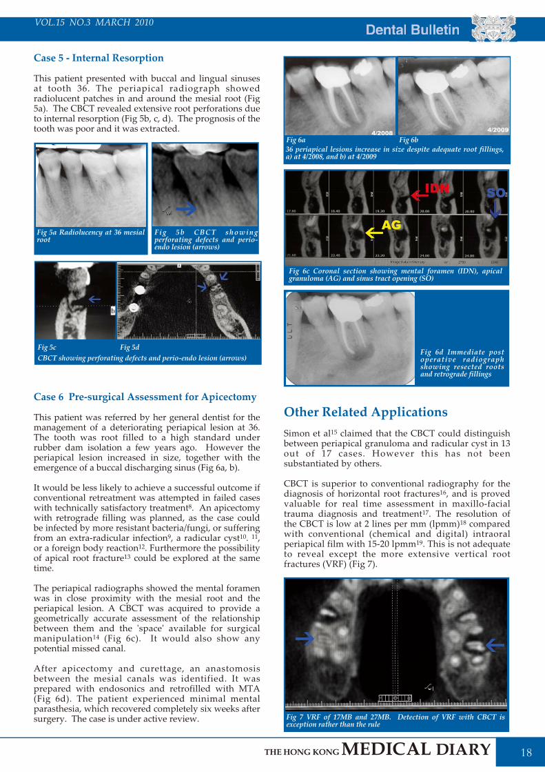
Fig 6d Immediate post operative radiograph showing resected roots and retrograde fillings
Fig 7 VRF of 17MB and 27MB. Detection of VRF with CBCT is exception rather than the rule
Case 5 - Internal Resorption
This patient presented with buccal and lingual sinuses at tooth 36. The periapical radiograph showed radiolucent patches in and around the mesial root (Fig 5a). The CBCT revealed extensive root perforations due to internal resorption (Fig 5b, c, d). The prognosis of the tooth was poor and it was extracted.
Case 6 Pre-surgical Assessment for Apicectomy
This patient was referred by her general dentist for the management of a deteriorating periapical lesion at 36. The tooth was root filled to a high standard under rubber dam isolation a few years ago. However the periapical lesion increased in size, together with the emergence of a buccal discharging sinus (Fig 6a, b).
It would be less likely to achieve a successful outcome if conventional retreatment was attempted in failed cases with technically satisfactory treatment8. An apicectomy with retrograde filling was planned, as the case could be infected by more resistant bacteria/fungi, or suffering from an extra-radicular infection9, a radicular cyst10. 11, or a foreign body reaction12. Furthermore the possibility of apical root fracture13 could be explored at the same time.
The periapical radiographs showed the mental foramen was in close proximity with the mesial root and the periapical lesion. A CBCT was acquired to provide a geometrically accurate assessment of the relationship between them and the 'space' available for surgical manipulation14 (Fig 6c). It would also show any potential missed canal.
After apicectomy and curettage, an anastomosis between the mesial canals was identified. It was prepared with endosonics and retrofilled with MTA (Fig 6d). The patient experienced minimal mental parasthesia, which recovered completely six weeks after surgery. The case is under active review.
Other Related Applications Simon et al15 claimed that the CBCT could distinguish between periapical granuloma and radicular cyst in 13 out of 17 cases. However this has not been substantiated by others.
CBCT is superior to conventional radiography for the diagnosis of horizontal root fractures16, and is proved valuable for real time assessment in maxillo-facial trauma diagnosis and treatment17. The resolution of the CBCT is low at 2 lines per mm (lpmm)18 compared with conventional (chemical and digital) intraoral periapical film with 15-20 lpmm19. This is not adequate to reveal except the more extensive vertical root fractures (VRF) (Fig 7).
Fig 5a Radiolucency at 36 mesial root
Fig 6a
Fig 6c Coronal section showing mental foramen (IDN), apical granuloma (AG) and sinus tract opening (SO)
Fig 5b CBCT showing perforating defects and perio-endo lesion (arrows)
CBCT showing perforating defects and perio-endo lesion (arrows)Fig 5c Fig 5d
Fig 6b36 periapical lesions increase in size despite adequate root fillings, a) at 4/2008, and b) at 4/2009

Dental Bulletin
19
VOL.15 NO.3 MARCH 2010
This lack of resolution, however, does not affect the superiority of CBCT in the assessment of periodontal regeneration, caries and bone lesions20, 21, 22. The image on the scan is well demarcated and provides better sensitivity and specificity than conventional radiograph. However the scatter and beam hardening could significantly affect the image occasionally (Fig 8).
ConclusionThe CBCT is a valuable adjunct to the endodontists' armamentarium. The learning curve is not steep and variability of clinical interpretation is low. However it is a sophisticated tool, requiring special skills for operating the machine and the image manipulation afterwards. Like any equipment in the digital age, continuous evolution and refinement is anticipated. Extra hidden expenses in depreciation and upgrades have to be added to the initial installation cost.
In conclusion the CBCT is a useful tool for the diagnosis and management of endodontic problems. Its use is becoming increasingly popular but some machines are better suited for endodontic purposes than others. The operators should consider their specific needs before making the move to acquiring one in the office.
Fig 8 In this case of an endo perio lesion of tooth 26, beam scattered from the root fillings render this scan useless in revealing the untreated MB2 canal and vertical root fracture
Nair PN, Sjogren U, Schumacher E et al (1993) Radicular cyst affecting a root-filled human tooth: a long-term post-treatment follow-up Int Endodon J;26:225-233.Nair PN, Sjogren U, Krey G et al (1990) Therapy-resistant foreign body giant cell granuloma at the periapex of a root-filled human tooth J Endodon 16;12:589-595.Adorno CG, Yoshioka T, Suda H (2009) The Effect of root preparation technique and instrumentation length on the development of apical root cracks J Endodon 35;3:389-392.Uchida Y, Noguchi N, Goto M et al (2009) Measurement of anterior loop length for the mandibular canal and diameter of the mandibular incisive canal to avoid nerve damage when installing endosseous implants in the interforaminal region: a second attempt introducing cone beam computed tomography. J Oral Maxillofac Surg. 67;4:744-50.Simon JSH, Enciso R, Malfaz J-M et al (2006) Differential diagnosis of large periapical lesions using cone-beam computed tomography measurements and biopsy J Endodon 32;9:833-837.Bornstein MM, Wolner-Hanssen AB, Sendi P (2009) Comparison of intraoral radiography and limited cone beam computed tomography for the assessment of root-fractured permanent teeth Dental Traumatol 25;6:571-577.Chow BKC, Chow JKF (2009) Application of office base three-dimensional technologies including cone-beam computed tomography and rapid prototyping in the management of maxillofacial trauma-literature review and a case report. Hong Kong Dent J 6;2:93-97. Hong Kong Dent Asso, Hong Kong.Yamamoto K, Ueno K, Seo K et al (2003) Development of dento-maxillofacial cone beam X-ray computed tomography system. Orthodon Cranfac Res 6(Suppl. 1) 160-2.Farman AG, Farman TT (2005) A comparison of 18 different x-ray detectors currently used in dentistry Oral Surg Oral Med Oral Pathol Oral Radiol 1Endodontol 99;4:485-489.Grimard BA, Hoidal MJ, Mills MP et al (2009) Comparison of clinical, periapical radiograph, and cone-beam volume tomography measurement techniques for assessing bone level changes following regenerative periodontal therapy J Periodontol 80;1:48-55.Young SM, Lee JT, Hodges RJ et al (2009) A comparative study of high-resolution cone beam computed tomography and charge-coupled device sensors for detecting caries Dentomaxillofac Radiol. 38;7:445-51.Noujeim M, Prihoda T, Langlais R et al (2009) Evaluation of high-resolution cone beam computed tomography in the detection of simulated interradicular bone lesions Dentomaxillofac Radiol. 38;3:156-62.
11.
12.
13.
14.
15.
16.
17.
18.
19.
20.
21.
22.
Mozzo P, Procacci C, Tacconi A et al (1998) A new volumetric CT machine for dental imaging based on the cone-beam technique: preliminary results. Euro Radiol 8;9:1558-64.Cheung LH, Low D, Cheung GS (2006) Root morphology--a study of the mandibular second molar of ethnic Chinese. Ann R Aust Coll Dent Surg 18:47-50.Gao Y, Fan B, Cheung GS et al (2006) C-shaped canal system in mandibular second molars part IV: 3-D morphological analysis and transverse measurement J Endodon 32;11:1062-1065Cheung LH, Cheung GS (2008) Evaluation of a rotary instrumentation method for C-shaped canals with micro-computed tomography J Endodon 34;10:1233-8Wu M-K, Shemesh H, Wesselink PR (2009) Limitations of previously published systematic reviews evaluating the outcome of endodontic treatment Int Endodon J 42;8:656-666.Estrela C, Reis Bueno M, Rodrigues Leles C et al (2008) Accuracy of cone beam computed tomography and panoramic and periapical radiography for detection of apical periodontitis J Endodon 34;10:273-9.Langeland K, Langeland LK (1970) Pulp reactions to cavity and crown preparation Aust Dent J 15;4:261-76.Farzaneh M, Abitbol S, Friedman S (2004) Treatment outcome in endodontics: The Toronto Study. Phases I and II: orthograde retreatment J Endodon 30;9:627-633. Sunde PT, Olsen I, Debelian GJ et al (2002) Microbiota of periapical Lesions Refractory to Endodontic Therapy J Endodon 28;4:304-310.Simon JH . (1980) Incidence of periapical cysts in relation to the root canal . J Endodon 6;11:845-848.
1.
2.
3.
4.
5.
6.
7.
8.
9.
10.
References

Dental Bulletin
21
VOL.15 NO.3 MARCH 2010
Dentine Hypersensitivity in Hong KongDentine hypersensitivity may be defined as short, sharp pain arising from exposed dentine typically in response to chemical, thermal or osmotic stimuli that cannot be explained as arising from any other forms of dental defect or pathology.1 Dentine hypersensitivity is a common problem found in many adult populations. A study on 226 patients attending a dental hospital in Hong Kong in 2003 found about two third of the patients (68%) had dentine hypersensitivity.2 The commonest initiating factor for dentine hypersensitivity among them was cold drinks. While many studies reported the commonest teeth affected are the premolars, the study found the commonest teeth affected were the lower incisors; and the majority of hypersensitive dentine surfaces were present on the facial surface of the teeth. The study also showed that dentine hypersensitivity peaked between 40 and 50 years of age, followed by a decline with age. The probable reason for this drop in dentine hypersensitivity after the fifth decade may be related to the pulpal changes with increasing age, particularly dentinal sclerosis and the laying down of secondary or tertiary dentine.
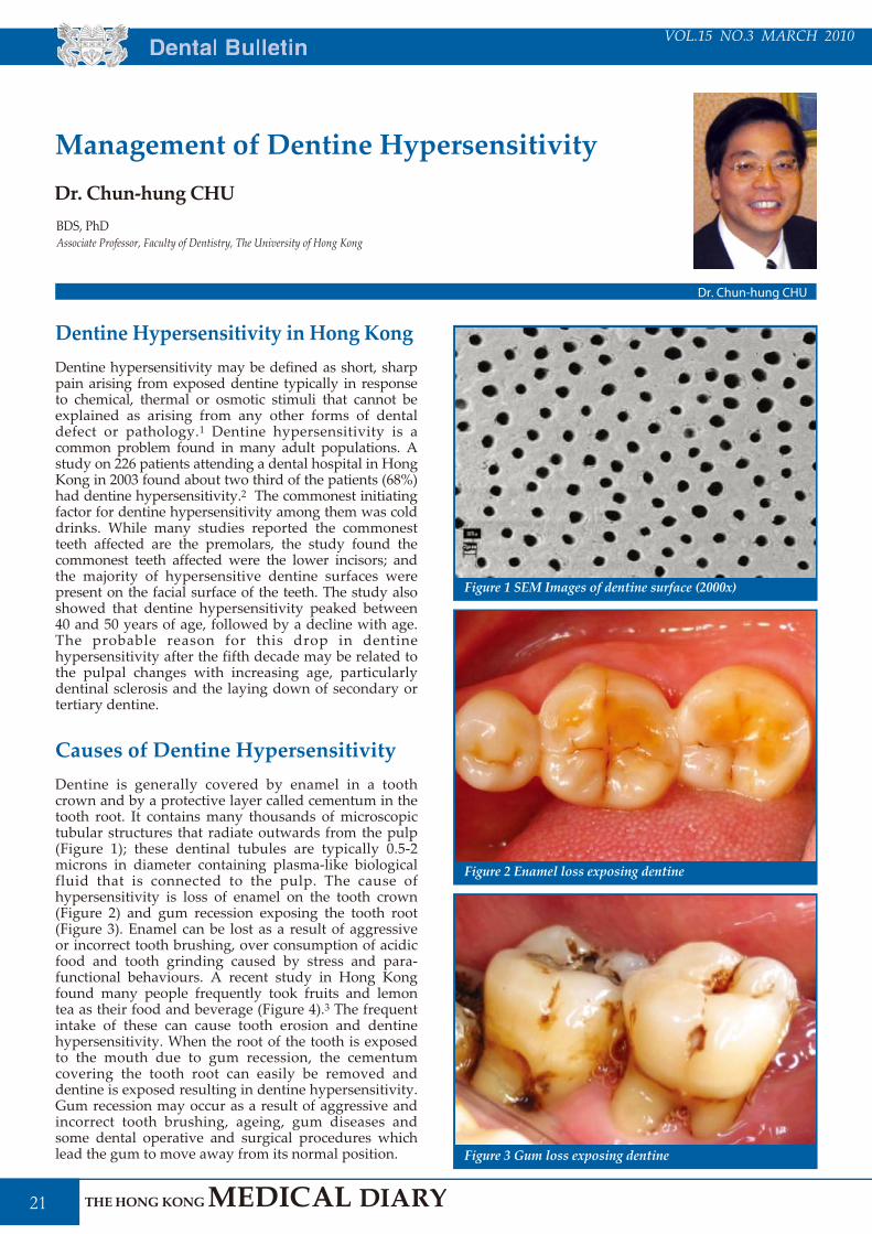
Causes of Dentine HypersensitivityDentine is generally covered by enamel in a tooth crown and by a protective layer called cementum in the tooth root. It contains many thousands of microscopic tubular structures that radiate outwards from the pulp (Figure 1); these dentinal tubules are typically 0.5-2 microns in diameter containing plasma-like biological fluid that is connected to the pulp. The cause of hypersensitivity is loss of enamel on the tooth crown (Figure 2) and gum recession exposing the tooth root (Figure 3). Enamel can be lost as a result of aggressive or incorrect tooth brushing, over consumption of acidic food and tooth grinding caused by stress and para-functional behaviours. A recent study in Hong Kong found many people frequently took fruits and lemon tea as their food and beverage (Figure 4).3 The frequent intake of these can cause tooth erosion and dentine hypersensitivity. When the root of the tooth is exposed to the mouth due to gum recession, the cementum covering the tooth root can easily be removed and dentine is exposed resulting in dentine hypersensitivity. Gum recession may occur as a result of aggressive and incorrect tooth brushing, ageing, gum diseases and some dental operative and surgical procedures which lead the gum to move away from its normal position.
Management of Dentine HypersensitivityDr. Chun-hung CHU
Dr. Chun-hung CHU
Associate Professor, Faculty of Dentistry, The University of Hong KongBDS, PhD
Figure 1 SEM Images of dentine surface (2000x)
Figure 2 Enamel loss exposing dentine
Figure 3 Gum loss exposing dentine

VOL.11 NO.5 MAY 2006 Dental Bulletin
22
VOL.15 NO.3 MARCH 2010
The Hydrodynamic Theory of Dentine HypersensitivityThe exact mechanism of dentine hypersensitivity is still under research. The hydrodynamic theory suggests that changes in the flow of the fluid present in the dentinal tubules can trigger receptors present on nerves located at the pulpal aspect thereby eliciting a pain response (Figure 5).4 This hydrodynamic flow can be increased by changes in temperature, humidity, air pressure and osmotic pressure, or forces acting onto the tooth. Hot or cold food or drinks, and physical pressure are typical triggers in those individuals with dentine hypersensitivity.
M a n a g e m e n t o f D e n t i n e HypersensitivityIt is essential to consult a dentist when a person suffers from pain with nature similar to the symptoms of dentine hypersensitivity. Dentinal hypersensitivity has all the criteria to be considered a true pain syndrome.5 It may share similar symptoms with dental decay and gum disease. In addition, the cause of dentine hypersensitivity should be identified and a diagnosis by exclusion must be made for dentinal hypersensitivity, ruling out other conditions requiring different treatments. Once the diagnosis of dentine hypersensitivity is confirmed, the dentist often needs to
discuss with his/her patient regarding his/her oral hygiene habits and diets. Changes and behaviour modifications such as decreasing the intake of acid-containing foods are often necessary to manage dentine hypersensitivity. The patient should also be shown correct brushing techniques because improper tooth brushing has often been associated with dentine hypersensitivity. It has been shown that both a manual and a power brush used with desensitising toothpaste are almost equivalent in effectiveness.6
Home Management with Desensitising ToothpasteUse of desensitising toothpaste is considered by many as the "first option" recommendation. It is effective but often takes 4 to 8 weeks for pain relief. Two treatment approaches have been used to provide relief of dentine hypersensitivity. The first approach is to interrupt the neural response to pain stimuli (Figure 6); and the other is to occlude open tubules to block the hydrodynamic mechanism (Figure 7). Many desensitising toothpastes contain potassium salts, strontium salts and/or fluoride compounds. Potassium salts such as potassium nitrate and potassium citrate provide potassium ions to decrease the excitability of the nerves that transit pain sensation. Strontium salts such as strontium chloride and strontium acetate form mineralised deposits within the porous dentinal tubules and on the surface of the exposed dentine. Fluoride compounds such as sodium fluoride and silver diamine fluoride form precipitation of insoluble metal compounds, mainly calcium fluoride globules, which promote remineralisation and occlude dentinal openings on exposed dentine surface. Recently available desensitising toothpastes with new chemicals such as amorphous calcium phosphate and casein phosphopeptide-amorphous calcium phosphate (ACP-CPP) and arginine and calcium carbonate (Arginine-CaCO3) are now available in the market. ACP-CPP and Arginine-CaCO3 products have a similar mode of action to occlude and block open dentinal tubules from external s t imuli associated with dent ine hypersensitivity. Studies have used bioactive and biocompatible glasses which are known to induce osteogenesis in physiological systems, and hence could theoretically occlude tubules.7 Toothpaste with calcium sodium phosphosilicate bioactive glass is also introduced. This bioactive glass material has been shown to seal and clog open dentinal tubules and thus reduces dentine hypersensitivity.8
Figure 4 Common acidic food and beverages taken by Hong Kong people
Figure 5 Pain eliciting by movement of dentinal fluid
Figure 6 Interruption of the neural response to pain stimuli with potassium ions

Dental Bulletin
23
VOL.15 NO.3 MARCH 2010
In Office Professional Care Apart from desensitising toothpastes, dentist may apply a variety of professional medicaments to reduce the dentine hypersensitivity. A variety of products has been used to reduce dentine hypersensitivity, including resin-based materials, sodium fluoride varnish, oxalates or an aqueous solution of glutaraldehyde and hydroxyethylmethacrylate (HEMA). These products generally occlude and seal the dentine tubules. Arginine-CaCO3 is also used as an active ingredient in a professionally used prophy-paste to manage dentine hypersensitivity. A clinical study on 390 patients found professional application of arginine and calcium carbonate by dentists and dental hygienists in Hong Kong significantly reduced severity of pain on patients with dentine hypersensitivity9. Furthermore, dentists may apply dental sealants and other desensitising and filling materials to cover the exposed dentine. Lasers can also be used to seal open dentine tubules, either alone or with surface treatments to manage dentine hypersensitivity.10
ConclusionDentine hypersensitivity is a common oral health problem among many adult population groups. Many treatment methods have been proposed and the choice varies according to the clinical presentation. When a patient presents with symptoms that may be attributed to dentine hypersensitivity, a thorough clinical examination should be carried out to rule out the other likely causes before making a diagnosis and embarking on treatment. Depending on the identified cause, a combination of individualised instructions on proper oral health behaviours, use of self-care products, and professional treatment may be required to manage the problem.
Figure 7 Occlusion of the open tubules to prevent pain stimuli
Addy M, Urquart E. Dentine hypersensitivity: its prevalence, aetiology and clinical management. Dental Update 1992; 19: 407,408, 410-412.Rees JS, Jin LJ, Lam S, Kudanowska I, Vowles R. The prevalence of dentine hypersensitivity in a hospital clinic population in Hong Kong. J Dent 2003; 31:453-461.Chu CH, Pang KL, Yip HK. Dietary behaviour and dental erosion symptoms of Hong Kong people. J Dent Res 2008, 87(Spec Issue C):41Brannstrom M. A hydrodynamic mechanism in the transmission of pain producing stimuli through the dentine. In: Anderson DJ, editor. Sensory mechanisms in dentine. Oxford: Pergamon Press; 1963; p. 73-79.Curro FA. Tooth hypersensitivity in spectrum of pain. Dent Clin North Am. 1990; 34:429-437.Sengupta K, Lawrence HP, Limeback H. Comparison of power and manual toothbrushes in dentine sensitivity. J Dent Res 2005; 84(Spec Issue A): 942. Gillam DG, Tang JY, Mordan NJ, Newman HN. The effects of a novel Bioglass dentifrice on dentine sensitivity: a scanning electron microscopy investigation. J Oral Rehabil 2002; 29: 305-313.Burwell AK, Litkowski LJ, Greenspan DC. Calcium sodium phosphosilicate (NovaMin): remineralization potential. Adv Dent Res 2009; 21:35-39.Chu CH, Lui KS, Lau KP, Kwok CM , Huang T. Effects of 8% arginine desensitizing paste on teeth with hypersensitivity. J Dent Res 2010, 89(Spec Issue A) (Submitted on Feb 5, 2010)Schwarz F, Arweiler N, Georg T, Reich E. Desensitizing effects of an Er:YAG laser on hypersensitive dentine. J Clin Periodontol 2002; 29:211-215.
1.
2.
3.
4.
5.
6.
7.
8.
9.
10.
References
Life Style
25
VOL.15 NO.3 MARCH 2010
"This is Tyler and you are listening to Speed at Metro Radio Hong Kong 99.7MHz! Bringing to you the latest in motorsports and car test reports with LIVE in-car recordings........" ( http://www.metroradio.com.hk/997 )
So we finished another recording session for the radio programme which would come on air on the following Saturday morning from 11am to noon. Usually, we would make the recording in the station's studios, but this time we were recording live car tests impressions on track at the Zhuhai International Racing Circuit and testing the luxury grand tourers from Bavarian Motors, the M5 and M6's both sharing the same chassis, V10 engine and drive train. It was a nice day of testing with perfect weather and good organisation. The cars were immaculately prepared and all working as they should. Our co-host, Steven also had a ride for his life in one of the cars whilst recording on the fly, though his colour turned a little green when he came out of the car. I could get used to this very easily, testing new cars in excess of a cool million dollars and driving them as if I had just stolen them as they said (http://www.104mfonline.com.hk/MetroFinance/Leisure/Car.aspx ).
It had been over 20 years since I graduated from Brands Hatch racing stable back in 1987 and what a far cry the BMW M5's was from the school Ford XR3's that we used to throw around that had to be push started because of excess engine heat after each stint. That was a time when I could still push start a car¡K..Since then I had gone through a few more racing schools; driven a host of cars and raced a few more.
Motorsports is just like any other sports. One needs regular practice with proper understanding of the mechanics and physics of the equipment and the necessary mental and physical conditions for enjoyment and safety. The pleasure of motorsports to me is never about the excitement nor glamour as my other genera acquaintances would try to persuade me to agree but instead, the serenity and inner calmness when I can focus completely on the job at hand i.e. driving the vehicle atl
its current configuration to the best I can to find its limits and that of my own.
When one fully concentrates on driving, one reaches a higher level of alertness and your senses are on overdrive reading the machine's behaviour, track conditions and anticipate what is likely to happen so to be prepared to make corrections and adjustments when on the fly. There is a feeling of elation when one can go through a given corner fully committed and slightly faster than before knowing that one could not have done any better on the day nor anyone else. And to be able to do all that under a safe environment of a racing circuit and not on the public roads is a prerequisite instead of a bonus. We also need to learn to be a team player, cooperating with our managers, engineers, technicians, officials etc and learn to communicate effectively and to manage our precious resources effectively with a detailed plan before testing. Driver's improvement and technical knowledge become the two main areas of continued education and one starts to befriend with your trusty mechanic who looks after your cars.
Race cars are categorically built to different specifications to road cars. Most of them are custom built by either ordering a special stripped out version motor car from the factory and then built from scratch again ground up which would require the complete removal of engine, drive train, loom etc. They have extra stiffness in the chassis with welded in roll cage, stiffened suspensions and seam welds for strength. All the interiors are stripped to save weight. There is an emphasis in safety with FIA homologated race seats, race harness, HANS device to protect the driver and passenger from whip-lash injuries, plumb-in fire extinguishers, racing tyres for wet and dry conditions, uprated brakes. Race cars are not suitable for driving on the public roads nor road cars any good for circuits.
1987 Brands Hatch Racing School, UK ( Racing Saloons )1995 John Watson's Racing Centre, Silverstone, UK
( Formula Fords )1996 Private Instructions with Mark Goddard1997 Greater China Classic Race Champion2003 Roadsport Race Macau 50th GP2004 Supercar Challenge Series ZIC, 2nd Runner Up2005 Lotus Challenge ( Asian Zone ) ZIC Race winner2006 Present Private Instructions with Mike Knight2008 Co Host, Speed at Metro Radio Hong Kong
Motor Racing
Dr. Edgar SHIH
Dr. Edgar SHIH
BDS( HK ), MSc in Orthodontics ( London )

VOL.11 NO.5 MAY 2006 Life Style
26
VOL.15 NO.3 MARCH 2010
Over the years, I have had the pleasure of being put into the driver's seat of different types of performance cars. From the German perennial rear engine rear wheel driven 911's with excess power to grip which demands respect to Japanese 2 litre turbo-charged 4WD rally homologation road cars to the ultra modern everything electronic all conquering GTR's which offer exhilarating performance at a bargain. For the past few years it has been a pleasure to learn to drive in the bespoke designed and built carbon fibre chassis formula race cars with sequential gearboxes. All of them bring immense enjoyment when one begins to come to terms with their quirky characteristics and handling properties. Probably, it is because of formula racing cars that I am now subscribing to the great Anthony Colin Bruce Chapman's philosophy of lightweight cars and have started to collect and tune British roadsters with spartan creature comforts and well designed chassis with simple drive trains. They are great little cars to learn to drive in and also for honing driving skills so much so that we have established an internet forum with common enthusiasts to exchange ideas. ( www.trackndrive.com )
I am only an average driver with average abilities and anyone can enjoy motorsports activities. I would recommend that one should go through proper racing school training which is available at ZIC at a very reasonable cost which provides first class training in proper formula cars, race gears, meals and accommodation over an enjoyable weekend. Even my son, who received training in Formula Renault before his 12 year old birthday, had never driven a car nor had any experience in Go-Karting before and became the youngest graduate and certificate holder from the ART Formula Renault Training Course.
I have been fortunate enough to win a few races and make a lot of friends from motorsports. Most notable would be Mike Knight who is our current driving instructor. Mike is still racing Grand Prix cars from the 60's and 70's. He was the operator/instructor of the world renowned Winfield Racing School, where he had more than 20 students who became F1 drivers and others of world class fame as well. Names include Alain Prost, Damon Hill, Jacque Villenueve, Olivier Panis, Jacque Lafite, Rene Arnoux, Jason Plato, Andy Priaulx...
See you on track and drive safely on the public roads!

Advance Directives Consultation Paper
27
VOL.15 NO.3 MARCH 2010
BackgroundIn the Hong Kong context, the term "advance directive" (AD) usually refers to a set of instructions about what kind of life sustaining treatment (LST) that a patient wishes to refuse when he becomes mentally incapacitated under some specified circumstances. In Hong Kong, a proxy directive appointing another person to decide on the patient's behalf does not have legal status. Under the common law framework in countries like UK, Australia, Canada and USA, a valid and applicable AD refusing LST is legally binding. The courts in Hong Kong would very likely take this view. In 2006, the Law Reform Commission published a report on AD recommending promotion of the concept of AD via non-legislative means in Hong Kong. However, there are different views on this in the community, and the Food and Health Bureau has issued a consultation paper in December 2009 asking for comments, among other questions, whether the concept of AD should be more widely promoted in Hong Kong or not.
There is little dispute about the importance of understanding a patient's values and treatment preferences in end of life care. Such an understanding could be achieved through discussions among the patient, the healthcare providers, and the family members in the "advance care planning" (ACP) process. The concept of ACP should be promoted in Hong Kong among patients with advanced incurable diseases. ACP does not necessarily end up in a legally binding AD. A non-legally binding expression of the patient's values and preferences in end of life care may be adequate in many cases, and can guide the doctor and family to make decisions in the patient's best interests when the patient is incompetent.
Role of AD in Clinical PracticeSometimes, AD as a legally binding tool in ACP has its role in clinical practice:
DifficultiesHowever, there are practical difficulties in executing an AD. Unlike contemporaneous refusals, a patient may have difficulty to make a rational advance decision before the actual scenario happens. On the other hand, the clinical team may have difficulty to judge whether the AD is valid or applicable. Also, unlike a contemporaneous refusal, the clinical team may find it difficult to accept an advance refusal which appears not in the best interests of the patient, without the chance of further discussing with the already incompetent patient. Such difficulties should be greater in patients without a terminal illness.
Another problematic area is an AD demanding withdrawal of artificial nutrition and hydration when PVS status is confirmed. This is highly controversial. Common law framework seems to support that such AD should be followed. However, many people think this is euthanasia.
Furthermore, without specific legislation in Hong Kong, there are legal uncertainties. Recently, doubts have been raised whether a valid and applicable AD can really override the best interests principle. For example, can a doctor transfuse a Jehovah Witness patient (with a valid AD refusing transfusion) dying of bleeding when he becomes unconscious, based on the best interests principle according to the Mental Health Ordinance Cap 136 Section 59ZF?
The Way ForwardThere are no easy answers to the above difficulties. In the 2006 report, taking reference mainly from the Singapore model, which has a rigid legislation with statutory forms and procedures, the Law Reform
When it is difficult to judge what is in the best interests of the patient: In some cases, a decision based on the best interests principle for an incompetent patient can be difficult, even if the patient's values and wishes are known. A statement indicating his decision would be helpful.
When the wish of the patient appears not in the best interests of the patient: Occasionally, a patient may have idiosyncratic view on a particular LST, for example, a Jehovah Witness patient would not want transfusion. A statement indicating his decision would be helpful.
Advance Directives Consultation PaperThe Food and Health Bureau published the Concept of Advance Directives Consultation Paper. To enhance the awareness of this consultation, the Federation has invited a doctor, a medical ethicist, and a lawyer to each contribute a short article on this subject. We would like to thank all three authors for writing and sharing their personal views and perspectives in this issue.
Advance Directives: Their Role in Clinical Practice and Their DifficultiesDr. Chun-yan TSEHonorary Advisor of the Hong Kong Society of Palliative Medicine
When there is a need to express the decision of the patient effectively to clinicians not familiar to the patient: For example, a terminally ill patient who plans to stay at home as much as possible may die at home. A statement that he declines CPR would be helpful. Another example is a severe COPD patient with repeated intubation for respiratory failure. If the patient does not want further intubation on his next admission for respiratory failure, a statement indicating his decision would be helpful.

VOL.11 NO.5 MAY 2006 Advance Direrr ctives Consultation Paper
28
VOL.15 NO.3 MARCH 2010
Commission considers that legislation would not help the promotion of AD in Hong Kong. However, there are some recent developments. In 2007, the Mental Capacity Act became operative in England and Wales. The legislation on AD there is just on the basic principles only, allowing flexibility in its execution, but it confirms the legal status of AD and its relationship to the best interests principle. I think Hong Kong should work towards legislation for AD along the UK approach, to confirm the legal status of AD, but to allow flexibility in its execution. At this moment, I think AD should be promoted in selected patients as part of ACP in advanced incurable illnesses. However, it may be prudent to wait till enactment of specific legislation on AD before promoting AD to the general public in Hong Kong. Meanwhile, it would be useful to develop guidelines for clinicians on how to handle an AD.
Regardless of whether AD is promoted in Hong Kong or not, it should be noted that AD is not a panacea for the difficulties faced by dying patients. Firstly, an AD cannot (and should not) cover all possible future scenarios. Scenarios not covered by the AD should be managed along the best interests principle, taking into account the values and wishes of the patient. Secondly, AD is a legal tool only. In patients faced with an advanced incurable disease, AD should be part of ACP, and ACP should be part of the full spectrum of palliative care for a terminally ill patient.

Advance Directives Consultation Paper
29
VOL.15 NO.3 MARCH 2010
In December 23, 2009, the Food and Health Bureau (FHB) issued a Consultation Paper entitled "Introduction of the Concept of Advance Directives in Hong Kong". This consultation paper was issued in response to a report published by the Law Reform Commission (LRC) in August 2006 entitled "Substitute Decision-making and Advance Directives in Relation to Medical Treatment". In the Consultation Paper, the Government agreed with the LRC's view that "it would be premature to attempt to formulate a statutory frame work and to embark on any legislative process for advance directives, without greater public awareness of the issues involved."1 Secondly, the Government made it clear that "it has no intention at this stage to actively advocate or encourage the public to make advance directives." 2 What the Government was prepared to do was (1) To produce and make available to the general public an information package on advance directives (AD); (2) To develop any necessary guidelines and procedures for making and executing AD after consultation with the public and the medical and legal professions; (3) To consult the public about the acceptance of advance care planning (ACP) and the best way to promote this concept in Hong Kong.
In this paper I wish to discuss two points in the Consultation Paper, one related to ACP and the other to AD. I like to argue that conceptually ACP is primary and AD is secondary. This is so because ACP is a process of communication among patients, family members, health care professionals (HCP), spiritual advisers, lawyers and any significant others about the patient's preferred medical care in the future when the patient can no longer make healthcare decisions. ACP takes place in the context not only of a patient-HCP relationship, but also in the context of patient-family-HCP relationship. In the ACP process, patients may speak with any one or all of the participants mentioned above to discuss their own values, beliefs, preferences and attitudes regarding appropriate medical interventions when they become seriously ill and/or lose their decision-making capacity. It is an integral part of medical care for elderly patients that are documented and reviewed regularly. In the case of patients with an illness that may deteriorate into a life-threatening illness, it can be expected that the ACP process will lead to the discussion and drawing up of an AD, but the latter is not a necessary product of the ACP process. The real benefit of the ACP process is two-fold: for patients and their families, it becomes a way to prepare for death, strengthen interpersonal and interfamily relationships, relieve family members and love ones of making decisions on behalf of patients and allow
patients to maintain control over healthcare decisions related to their dying and death. On the other hand, the ACP process assists the HCPs and family members to interpret the AD with greater certainty since the AD is the product of a process in which they are participants.
The consultation paper attributes the Chinese taboo that forbids the discussion of the patient's death as the reason for the infrequent use of ACP process in Hong Kong, and speculates that perhaps when the public has become more familiar with the concept of AD, it may find ACP more acceptable. This is unlikely to be the case, for if the taboo affects the ACP process, it is difficult to see how the making of an AD is immune from it. The real cause(s) of the infrequent practice of ACP in Hong Kong must be sought elsewhere. Firstly, HCPs in Hong Kong are not sufficiently trained to engage patients and families in the ACP process and they feel uncomfortable to raise death and dying issues with them. Secondly, the ACP process is time-consuming and it requires the HCPs to set aside time from their busy schedule to participate in the discussion of their patients' physical, mental, emotional, relational and spiritual concerns about their ageing, dying and death. It is a process that requires sensitivity and patience and HCPs can not participate or perceived by patients to participate in a hurry. Furthermore, since patients' preferences towards end-of-life treatments may be unstable and change with time,3 the ACP process is an ongoing one that may take several sessions to complete and requires patients being able to see the same HCPs each time they visit the clinic. It is therefore reasonable to speculate that the real causes of the infrequent practice of ACP in Hong Kong are due to the deficient training of HCPs in the ACP process, the extreme time constraints under which Hong Kong HCPs operate, and the uncertainty of patients' ability to see the same HCPs over a period of time required by the ACP process. Nonetheless, the ACP concept should be promoted in Hong Kong, with emphasis on the need of ACP training programmes for practitioners and the need to allocate extra resources devoted to ACP. But if AD is the "natural product" of the ACP process, then arguably ACP should be promoted prior to or at the same time when AD is being promoted. There are reasons to be skeptical that "ACP would be[come] a concept acceptable to the public...when the public has become more familiar with the concept of advance directive." 4
A second point in the Consultation Paper that merits further clarification is the definition of "terminally ill", particularly in relation to the activating and revoking an AD. Both the LRC Report and the Consultation Paper state that the AD will become operative (activated)
Introducing the Use of Advance Care Planning and Advance Directives in Hong Kong?Prof. EC HUI
Prof. EC HUI
Professor of Medical Ethics, LKS Faculty of Medicine, The University of Hong KongMD, PhD
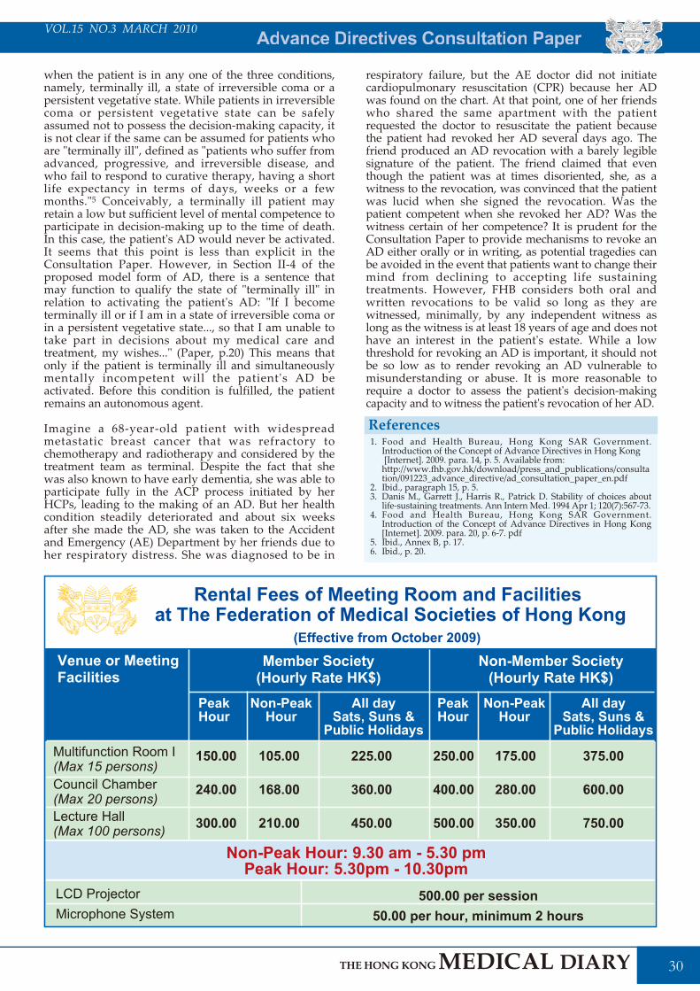
VOL.11 NO.5 MAY 2006 Advance Direrr ctives Consultation Paper
30
VOL.15 NO.3 MARCH 2010
when the patient is in any one of the three conditions, namely, terminally ill, a state of irreversible coma or a persistent vegetative state. While patients in irreversible coma or persistent vegetative state can be safely assumed not to possess the decision-making capacity, it is not clear if the same can be assumed for patients who are "terminally ill", defined as "patients who suffer from advanced, progressive, and irreversible disease, and who fail to respond to curative therapy, having a short life expectancy in terms of days, weeks or a few months."5 Conceivably, a terminally ill patient may retain a low but sufficient level of mental competence to participate in decision-making up to the time of death. In this case, the patient's AD would never be activated. It seems that this point is less than explicit in the Consultation Paper. However, in Section II-4 of the proposed model form of AD, there is a sentence that may function to qualify the state of "terminally ill" in relation to activating the patient's AD: "If I become terminally ill or if I am in a state of irreversible coma or in a persistent vegetative state..., so that I am unable to take part in decisions about my medical care and treatment, my wishes..." (Paper, p.20) This means that only if the patient is terminally ill and simultaneously mentally incompetent will the patient's AD be activated. Before this condition is fulfilled, the patient remains an autonomous agent.
Imagine a 68-year-old patient with widespread metastatic breast cancer that was refractory to chemotherapy and radiotherapy and considered by the treatment team as terminal. Despite the fact that she was also known to have early dementia, she was able to participate fully in the ACP process initiated by her HCPs, leading to the making of an AD. But her health condition steadily deteriorated and about six weeks after she made the AD, she was taken to the Accident and Emergency (AE) Department by her friends due to her respiratory distress. She was diagnosed to be in
respiratory failure, but the AE doctor did not initiate cardiopulmonary resuscitation (CPR) because her AD was found on the chart. At that point, one of her friends who shared the same apartment with the patient requested the doctor to resuscitate the patient because the patient had revoked her AD several days ago. The friend produced an AD revocation with a barely legible signature of the patient. The friend claimed that even though the patient was at times disoriented, she, as a witness to the revocation, was convinced that the patient was lucid when she signed the revocation. Was the patient competent when she revoked her AD? Was the witness certain of her competence? It is prudent for the Consultation Paper to provide mechanisms to revoke an AD either orally or in writing, as potential tragedies can be avoided in the event that patients want to change their mind from declining to accepting life sustaining treatments. However, FHB considers both oral and written revocations to be valid so long as they are witnessed, minimally, by any independent witness as long as the witness is at least 18 years of age and does not have an interest in the patient's estate. While a low threshold for revoking an AD is important, it should not be so low as to render revoking an AD vulnerable to misunderstanding or abuse. It is more reasonable to require a doctor to assess the patient's decision-making capacity and to witness the patient's revocation of her AD.
Food and Health Bureau, Hong Kong SAR Government. Introduction of the Concept of Advance Directives in Hong Kong [Internet]. 2009. para. 14, p. 5. Available from: http://www.fhb.gov.hk/download/press_and_publications/consultation/091223_advance_directive/ad_consultation_paper_en.pdfIbid., paragraph 15, p. 5. Danis M., Garrett J., Harris R., Patrick D. Stability of choices about life-sustaining treatments. Ann Intern Med. 1994 Apr 1; 120(7):567-73.Food and Health Bureau, Hong Kong SAR Government. Introduction of the Concept of Advance Directives in Hong Kong [Internet]. 2009. para. 20, p. 6-7. pdfIbid., Annex B, p. 17. Ibid., p. 20.
1.
2.3.
4.
5.6.
References
Venue or MeetingFacilities
Member Society(Hourly Rate HK$)
PeakHour
Non-PeakHour
All day Sats, Suns &
Public Holidays
Rental Fees of Meeting Room and Facilities at The Federation of Medical Societies of Hong Kong
Non-Member Society(Hourly Rate HK$)
Multifunction Room I (Max 15 persons)Council Chamber (Max 20 persons)Lecture Hall (Max 100 persons)
150.00
240.00
300.00
105.00
168.00
210.00
225.00
360.00
450.00
250.00
400.00
500.00
175.00
280.00
350.00
375.00
600.00
750.00
PeakHour
Non-PeakHour
All day Sats, Suns &
Public Holidays
(Effective from October 2009)
Non-Peak Hour: 9.30 am - 5.30 pmPeak Hour: 5.30pm - 10.30pm
LCD Projector Microphone System
500.00 per session50.00 per hour, minimum 2 hours


VOL.11 NO.5 MAY 2006 Advance Direrr ctives Consultation Paper
32
VOL.15 NO.3 MARCH 2010
What are Advance Directives?These are directions given by an adult, at a time when he is mentally competent, concerning the medical treatment which he wishes to receive or does not wish to receive at a future time when he will no longer be mentally competent. In other words, these are medical directions given by an individual patient in advance, to be put into effect if and when he subsequently becomes (either temporarily or permanently) mentally incompetent.
What are the Relevant Legal Principles?Advance directives are recognised under common law. Currently, there is no statutory framework. It reflects the principle of self-determination1, according to which respect must be given to the wishes of an adult patient of sound mind regarding his medical treatment.
As already stated, at the time of giving his directions, the individual patient has to be mentally competent. His mental capacity should not be diminished by long term illness or medication. Furthermore, there should be no undue influence by any third party.
In the same way that a mentally competent patient can validly refuse treatment (and such refusal must be respected), the patient can communicate his wishes at an earlier time before he becomes incapable of communicating them.
Those involved in looking after the terminally ill (including patients who have become mentally incompetent) have a duty to respect the wishes of the patients. According to the Hospital Authority's Guidelines2, validly executed advance directives, including those refusing life-sustaining treatment, should be respected.
In deciding whether or not to withhold life-sustaining treatment, a medical practitioner should take into account the wishes of the patient (as well as those of his family)3. These wishes would be recorded in an advance directive, if one has been executed. Similarly, where a guardian has been appointed by the Guardianship Board to consent to medical treatment on behalf of a mentally incapacitated person ("MIP"), in deciding whether or not to consent to treatment, the guardian is required to consider what is in the MIP's best interest. In doing so, the guardian should take into account any prior wishes expressed by the patient before he becomes mentally incapacitated.
The Present and the FutureIn August 2006, the Law Reform Commission published a report on advance directives in relation to medical treatment, having collected views from the public through a consultation paper (published in 2004). The Law Reform Commission has made the following recommendations:
More recently, on 23rd December 2009, the Food and Health Bureau published a consultation paper in response to the above-mentioned report. This consultation again addresses the issue of whether the concept of advance directives should be introduced in Hong Kong, the contents of information to be provided to the public as well as the guidance to be provided to the medical profession. It is proposed that procedures and guidelines should be developed for medical and healthcare professionals on the making and handling of advance directives.
The concept of advance directives should be promoted, initially by non-legislative means, until the community has become more widely familiar with the concept (recommendation 1).The Commission put forward a model form of advance directive. This ensures that the directions given are clear and unambiguous. The form should be witnessed by two witnesses, one of whom should be a medical practitioner who is in a position to explain to the patient the nature and implications of the advance directive. Neither of the witnesses should have an interest in the estate of the patient (recommendation 7).The model form does not allow the patient to refuse basic or palliative care necessary to maintain the patient's comfort and dignity, or to relieve pain.If for any reasons the patient is unable to make a written advance directive, then an oral advance directive should be made before a doctor, lawyer or another independent person (recommendation 7). (It is important that the doctor records such directives/instructions in the medical records.)The advance directive can subsequently be revoked in writing or orally. If it is revoked in writing, then the revocation should be witnessed by an independent witness who does not have an interest in the estate. In the case of an oral revocation, it should be made before a doctor, lawyer or another independent person (recommendation 8).
Advance Directives - the Legal Issues
Mr. David KAN
Mr. David KAN
Partner, Richards Butler SolicitorsMA(Med. Law), MBBS, MFFLM, BMed Sci(Hons), PDipCAH
Airedale NHS v. Bland [1993] 1 All ER 821Guidelines on life-sustaining treatment in the terminally ill, published by the Hospital Authority (paragraphs 5.16 to 5.23)Code of Professional Conduct for the Guidance of Registered Medical Practitioners, paragraph 34.3
1.2.
3.
References

The Federation of Medical Societies of Hong Kong

Medical Diaryrr of March
35
VOL.15 NO.3 MARCH 2010
Sunday
Monday
Tuesday
Wednesday
Thursday
Friday
Saturday
1819
20
2122
23
281415
1617
1112
1310
78
24
2930
31
9
43
12
56
2526
27
HK
MA
Tai
Po
Com
mun
ity N
etw
ork
- C
omm
on U
rolo
gy
Prob
lem
in P
rim
ary
Car
e Se
ctor
HK
Neu
rosu
rgic
al S
ocie
ty
Mon
thly
Aca
dem
ic
Mee
ting
- Cer
ebra
l Re
vasc
ular
izat
ion
Sem
inar
on
Gift
ed B
abie
s (C
ode
no: S
E-G
B-10
02)
FMSH
K E
xecu
tive
Com
mitt
ee M
eetin
g
HK
MA
Cen
tral
, Wes
tern
&
Sou
ther
n C
omm
unity
N
etw
ork
- CM
E on
"Adv
ance
in A
sthm
a M
anag
emen
t"
The
Paed
iatr
ic In
fect
ious
D
isea
se a
nd Im
mun
olog
y C
ours
e 20
10 -
Infe
ctio
n an
d Im
mun
ity o
f the
Fet
us
and
New
born
Infa
nt
HK
MA
Cen
tral
, Wes
tern
&
Sout
hern
Com
mun
ity
Net
wor
k - C
ME
on "S
inus
itis
and
Rhin
itis:
Upd
ated
M
anag
emen
t Tip
s fo
r Pri
mar
y C
are
Phys
icia
ns"
HK
MA
Sha
tin D
octo
rs
Net
wor
k - M
anag
emen
t of
Inso
mni
a an
d th
e U
se o
f Hyp
notic
M
edic
atio
n
The
Paed
iatr
ic In
fect
ious
Dis
ease
an
d Im
mun
olog
y C
ours
e 20
10 -
Infe
ctio
n an
d Im
mun
ity o
f the
Fe
tus
and
New
born
In
fant
The
Paed
iatr
ic In
fect
ious
D
isea
se a
nd Im
mun
olog
y C
ours
e 20
10 -
Infe
ctio
n an
d Im
mun
ity o
f the
Fet
us a
nd
New
born
Infa
nt
Hon
g K
ong
Ope
n In
door
Ro
win
g C
ham
pion
ship
s &
C
hari
ty R
owat
hon
HK
MA
Yau
Tsi
m M
ong
Com
mun
ity N
etw
ork
- Pr
even
ting
the
Prev
enta
ble
(Car
diol
ogy)
HK
MA
Foo
tbal
l Day
201
0
App
roac
h to
Acn
e an
d O
ther
Fol
licul
ar L
esio
n
HK
MA
Yau
Tsi
m M
ong
Com
mun
ity N
etw
ork
- Pr
even
ting
the
Prev
enta
ble
(Gen
eral
Su
rger
y)
HK
MA
Cer
tific
ate
Cou
rse
on F
amily
Med
icin
e 20
10A
nnua
l Sci
entif
ic M
eetin
g 20
10
HK
MA
Kow
loon
Wes
t C
omm
unity
Net
wor
k - C
ase
Shar
ing
on D
iabe
tes
Man
agem
ent i
n Pr
actic
eM
idm
ark
Prom
otal
Hon
g K
ong
Show
Roo
m O
peni
ng
HK
MA
Cho
ir F
amily
Con
cert
Wor
ksho
p on
the
Art
of F
etal
an
d N
ewbo
rn D
evel
opm
ent
and
its C
omm
unic
atio
n to
Par
ent (
SE-A
F-10
01 A
&
SE-
AF-
1001
B)
Wor
ksho
p on
the
Art
of F
etal
an
d N
ewbo
rn D
evel
opm
ent
and
its C
omm
unic
atio
n to
Pa
rent
(SE-
AF-
1001
A
& S
E-A
F-10
01 B
)H
KM
A T
ai P
o C
omm
unity
N
etw
ork
- Man
agem
ent o
f A
dult
Ast
hma
Prac
tical
Hea
lth In
form
atic
s C
ours
e of
Doc
tors
(Ses
sion
II
)
Join
t Sur
gica
l Sym
posi
um
- Upd
ate
in V
ascu
lar
Surg
ery
14th
Ann
ual S
cien
tific
M
eetin
g
HK
MA
Sha
tin D
octo
rs
Net
wor
k - C
ertif
icat
e C
ours
e in
Reh
abili
tatio
n M
edic
ine
(I)
The
Fede
ratio
n Sp
ring
G
athe
ring
cum
Ope
n H
ouse
Refr
eshe
r Cou
rse
for H
ealth
C
are
Prov
ider
s 20
09/ 2
010
The
3rd
Ann
ual S
cien
tific
M
eetin
g an
d 4t
h A
nnua
l G
ener
al M
eetin
g of
the
Hon
g K
ong
Soci
ety
for P
aedi
atri
c Im
mun
olog
y an
d In
fect
ious
D
isea
ses,
Bill
Mar
shal
l and
Ro
land
Lev
insk
y M
emor
ial
Lect
ures
HK
MA
Sha
tin D
octo
rs
Net
wor
k - C
ertif
icat
e C
ours
e in
Reh
abili
tatio
n M
edic
ine
(I) Free
Sem
inar
on
Dri
ving
Tal
k
Fire
Dri
llH
KM
A C
ounc
il M
eetin
g
Prac
tical
Hea
lth In
form
atic
s C
ours
e of
Doc
tors
(Ses
sion
III)
HK
MA
Str
uctu
red
CM
E Pr
ogra
mm
e w
ith H
ong
Kon
g Sa
nato
rium
& H
ospi
tal
Year
201
0 - T
he
Cur
rent
Man
agem
ent
of H
BV
HK
MA
Hon
g K
ong
East
C
omm
unity
Net
wor
k - C
ertif
icat
e C
ours
e: P
ract
ical
Psy
chia
try
for
the
Gen
eral
Pra
ctiti
oner
s (I)
HK
MA
Hon
g K
ong
East
C
omm
unity
Net
wor
k - C
ertif
icat
e C
ours
e: P
ract
ical
Psy
chia
try
for
the
Gen
eral
Pra
ctiti
oner
s (II
)
HK
MA
Hon
g K
ong
East
C
omm
unity
Net
wor
k -
Cer
tific
ate
Cou
rse:
Pra
ctic
al
Psyc
hiat
ry fo
r the
Gen
eral
Pr
actit
ione
rs (I
II)
HK
MA
New
Ter
rito
ries
W
est C
omm
unity
Net
wor
k -
Man
agem
ent o
f Ins
omni
a an
d th
e us
e of
H
ypno
tic
Med
icat
ion
Prac
tical
Hea
lth In
form
atic
s C
ours
e of
Doc
tors
(Ses
sion
IV)
FMSH
K O
ffice
rs' M
eetin
g

VOL.11 NO.5 MAY 2006 Medical Diaryrr of March
36
VOL.15 NO.3 MARCH 2010
6 SAT14th Annual Scientific Meeting Organiser: Hong Kong Society Infectious Diseases
Ms. Chloe WONGTel: 2155 8557 / 2116 4348 Fax: 2559 6910Eamil: [email protected]
7 SUNHong Kong Open Indoor Rowing Championships & Charity RowathonOrganiser: The Hong Kong Medical Association, Venue: Kowloon Park Sports Centre
Ms. Dora HOTel: 2527 8285
9 TUEHKMA Kowloon West Community Network - Case Sharing on Diabetes Management in PracticeOrganiser: HKMA Kowloon West Community Network, Chairman: Dr. CHAN Siu Man Bernard, Speaker: Dr. TONG Chun Yip Peter, Venue: Crystal Room I-III, 30/F, Panda Hotel, 3 Tsuen Wah Street, Tsuen Wan, NT
Miss Alice TANGTel: 2527 82851 CME Point for Participant2 CME Points for Speaker
Midmark Promotal Hong Kong Show Room OpeningOrganiser: Midmark Corporation - VP International Business Development: M. Brad WALKER and Promotal President: M. Francois NOGRIX, Venue: 10th Floor, Valiant Industrial Center, 2-12 Au Pui Wan Street, Fo Tan, Sha Tin, N.T. HONG KONG
Miss Heidi LEUNG Tel: 2604 9389
Workshop on the Art of Fetal and Newborn Development and its Communication to Parent (SE-AF-1001 A & SE-AF-1001 B)Organiser: College of Nursing, Hong Kong, Speaker: Ms. CHAN Cheung Lung Sharon
SecretariatTel: 2572 9255 Fax: 2838 62801.5 CNE/ PEM per day
HKMA Choir Family ConcertOrganiser: The Hong Kong Medical Association, Venue: Theatre, Sheung Wan Civic Centre
Ms. Candy YUENTel: 2527 8285
1:30 pm
8:00 pm
4 THUPractical Health Informatics Course of Doctors (Session II, III & IV)Organiser: The Hong Kong Medical Association, Speakers: Various, Venue: Lecture Theatre, G/F, Block M, Queen Elizabeth Hospital, 30 Gascoigne Road, Kowloon
TUEMs. Kaman HOTel: 2963 55241 CME Point for Participant2 CME Points for Speaker
1:45 pm
8:00 pm - 10:00pm
2 HKMA Tai Po Community Network - Common Urology Problem in Primary Care SectorOrganiser: HKMA Tai Po Community Network, Speaker: Dr. SZETO Shek, Venue:
FRIDepartment of Surgery, Hong Kong Sanatorium & HospitalTel: 2835 8698 Fax: 2892 75111 CME Point (Active)
8:00 am - 9:00 am
11:30 am - 12:30 am
5 Joint Surgical Symposium - Update in Vascular SurgeryOrganisers: Department of Surgery, The University of Hong Kong & Hong Kong Sanatorium & Hospital, Chairman: Dr. SIU Wing-tai, Speakers: Dr. TING Chi-Wai & Dr. WONG Chiu-Cheuk, Venue: Hong Kong Sanatorium & Hospital
Ms. Christine WONGTel: 2527 8285
Fire DrillOrganiser: The Hong Kong Medical Association, Venue: Duke of Windsor Social Service Building, 15 Hennessy Road, Wanchai, Hong Kong
1:00 pm
1:00 pm
3:00 pm
7:00 pm - 8:30 pm
8:00 pm
Dr. MAK Wing KinTel: 2649 44661.5 CME Points for Participant
HKMA Shatin Doctors Network - Certificate Course in Rehabilitation Medicine (I)Organiser: HKMA Shatin Doctors Network, Speakers: Dr. LEUNG Siu Lun Arran & Mr. Billy CHEUNG, Venue: 1/F, Jasmine Room, Royal Park Hotel, Shatin, N.T.
Ms. Paulina TANGTel: 2527 8898 Fax: 2865 0345
Miss Carman WONGTel: 2527 82852 CME Points for Participant
HKMA Council MeetingOrganiser: The Hong Kong Medical Association, Chairman: Dr. H.H. TSE, Venue: HKMA Head Office, 5/F., Duke of Windsor Social Service Building, 15 Hennessy Road, Hong Kong
Ms. Christine WONGTel: 2527 8285
FMSHK Officers' MeetingOrganiser: The Federation of Medical Societies of Hong Kong, Venue: Gallop, 2/F., Hong Kong Jockey Club Club House, Shan Kwong Road, Happy Valley, Hong Kong
Date / Time Function Enquiry / Remarks
(11, 18)
(19)
(23)
(18, 25)
Dr. Y.C. PO Tel: 2990 3788Fax: 2990 3789
7:30 am
1:00 pm
10 WEDHK Neurosurgical Society Monthly Academic Meeting - Cerebral Revascularization Organiser: HK Neurosurgical Society, Chairman: Dr. SUN Tin Fung David, Speaker: Dr. C.P. TSANG, Venue: Seminar Room, G/F, Block A, Queen Elizabeth Hospital, Kowloon
Miss Alice TANGTel: 2527 8285
HKMA Central, Western & Southern Community Network - CME on"Advance in Asthma Management"Organiser: HKMA Central, Western & Southern Community Network, Chairman: Dr. YIK Ping Yin, Speaker: Dr. Jane C.K. CHAN, Venue: The HKMA Dr. Li Shu Pui Professional Education Centre, 2/F, Chinese Club Building, 21-22 Connaught Road Central, Hong Kong
Miss Viviane LAMTel: 2527 84523 CME Points
2:00 pm 14 SUNHKMA Certificate Course on Family Medicine 2010Organiser: The Hong Kong Medical Association, Speakers: Dr. LO Kwok Wing & Dr. Michael CHENG, Venue: Lecture Theatre, G/F, Block M, Queen Elizabeth Hospital, 30 Gascoigne Road, Kowloon
Ms. Chloe WONGTel: 2155 8557 / 2116 4348Fax: 2559 6910Email: [email protected]
Annual Scientific Meeting 2010Organiser: Hong Kong Thoracic Society and American College of Chest Physicians (HK & Macau Chapter)
Ms. Paulina TANGTel: 2527 8898 Fax: 2865 0345
6:30 pm - 8:30 pm 12 FRIThe Federation Spring Gathering cum Open HouseOrganiser: The Federation of Medical Societies of Hong Kong, Venue: 4/F, Duke of Windsor Social Service Building, 15 Hennessy Road, Wanchai, Hong Kong
Ms. Jaclyn LEETel: 2877 1106
1:00 pm
2:00 pm
11 THUHKMA Hong Kong East Community Network - Certificate Course: Practical Psychiatry for the General Practitioners (I), (II) & (III) Organiser: HKMA Hong Kong East Community Network, Hong Kong Society of Biological Psychiatry and Lundbeck Institute Hong Kong, Chairman: Dr. Jenny TSANG, Prof. SW TANG, Speaker: Various. Venue: Regus Conference Centre, 35/F., Central Plaza, 18 Harbour Road, Wanchai
Miss Viviane LAMTel: 2527 84521 CME Point for Participant2 CME Points for Speaker
HKMA Structured CME Programme with Hong Kong Sanatorium & Hospital Year 2010 - The Current Management of HBVOrganiser: The Hong Kong Medical Association, Speaker: Dr. CHAN On On Annie, Venue: The HKMA Dr. Li Shu Pui Professional Education Centre, 2/F, Chinese Club Building, 21-22 Connaught Road Central, Hong Kong
Calendar of Events
37
VOL.15 NO.3 MARCH 2010
FRIMs. Karen CHUTel: 2527 8898Fax: 2865 0345
7:30 pm - 9:30 pm 19 Free Seminar on Driving Talk Organiser: The Federation of Medical Societies of Hong Kong, Speakers: Dr. MONG Hoi Keung & Mr. HUNG Wan Shun Stephen, Venue: 4/F, Duke of Windsor Social Service Building, 15 Hennessy Road, Wanchai, Hong Kong
SUNMiss Alice TANGTel: 2527 8285
2:00 pm 21 HKMA Yau Tsim Mong Community Network - Preventing the Preventable (Cardiology)Organiser: HKMA Yau Tsim Mong Community Network, Chairman: Dr. LI Siu Lung Steven, Speakers: Various, Venue: Lecture Theatre, 10/F, TWGHs Yu Chun Keung Memorial Medical Centre, Kwong Wah Hospital, 25 Waterloo Road, Kowloon
TUEMs. Carol LAMTel: 9045 51421.5 CME Points for Participant3 CME Points for Speaker
1:00 pm 23 HKMA Tai Po Community Network - Management of Adult AsthmaOrganiser: HKMA Tai Po Community Network, Speaker: Dr. SHE Hoi Wah, Venue: Chiu Chow Garden Restaurant, 1/F, Uptown Plaza, No. 9 Nam Wan Road, Tai Po
THUMiss Alice TANGTel: 2527 8285
1:00 pm 25 HKMA New Territories West Community Network - Management of Insomnia and the use of Hypnotic MedicationOrganiser: HKMA New Territories West Community Network, Chairman: Dr. LEE Fook Kay Aaron, Speaker: Dr. John SO, Venue: Plentiful Delight Banquet ( ), Yuen Long
WEDMiss Alice TANGTel: 2527 8285
1:00 pm
1:00 pm
24 HKMA Central, Western & Southern Community Network - CME on "Sinusitis and Rhinitis: Updated Management Tips for Primary Care Physicians"Organiser: HKMA Central, Western & Southern Community Network, Chairman: Dr. LAW Yim Kwai, Speaker: Dr. LO Chun Yip Amos, Venue: The HKMA Dr. Li Shu Pui Professional Education Centre, 2/F, Chinese Club Building, 21-22 Connaught Road Central, Hong Kong
SUNMs. Dora HOTel: 2527 8285
12:00 pm
2:00 pm
6:00 pm
28 HKMA Football Day 2010Organiser: The Hong Kong Medical Association, Venue: Sir Philip Hadden Cave Football Field
Miss Alice TANGTel: 2527 8285
HKMA Yau Tsim Mong Community Network - Preventing the Preventable (General Surgery)Organiser: HKMA Yau Tsim Mong Community Network, Chairman: Dr. LAM Tzit Yuen David, Speakers: Various, Venue: Lecture Theatre, 10/F, TWGHs Yu Chun Keung Memorial Medical Centre, Kwong Wah Hospital, 25 Waterloo Road, Kowloon
Ms. Dorothy KWOKTel: 2527 82851 CME Point for Participant2 CME Points for Speaker
Approach to Acne and Other Follicular LesionOrganiser: Hong Kong Medical Association, Speaker: Dr. CHAN Hau Ngai Kingsley, Venue: Regal Riverside Hotel, Kowloon, 34-36 Tai Chung Kiu Road, Shatin, NT
Dr. MAK Wing KinTel: 2649 44661.5 CME Points for Participant
HKMA Shatin Doctors Network - Management of Insomnia and the Use of Hypnotic MedicationOrganiser: HKMA Shatin Doctors Network, Speaker: Dr. WONG Yee Him, Venue: 1/F, Jasmine Room, Royal Park Hotel, Shatin, N.T.
SATMs. Clara TSANGTel: 2354 24402 CME Points for Participant4 CME Points for Speaker
2:30 pm 20 Refresher Course for Health Care Providers 2009/ 2010Organiser: The Hong Kong Medical Association, Speaker: Dr. CHEUNG Kit Ying Andy, Venue: Training Room II, 1/F., OPD Block, Our Lady of Maryknoll Hospital, 118 Shatin Pass Road, Wong Tai Sin, Kowloon, Hong Kong
Ms. YL YEUNGTel:2990 2872Fax: 2990 2875E-mail: [email protected]
The 3rd Annual Scientific Meeting and 4th Annual General Meeting of the Hong Kong Society for Paediatric Immunology and Infectious Diseases, Bill Marshall and Roland Levinsky Memorial LecturesSpeakers: Dr. Nigel CURTIS (Bill Marshall Memorial Lecture) & Prof. David ISAACS (Roland Levinsky Memorial Lecture) and Free Paper Presentation, Venue: Ballroom, The Langham Hong Kong, 8 Peking Road, Tsimshatsui, Hong Kong
6:30pm - 9:30pm
7:00 pm - 8:00 pm
17 WEDThe Paediatric Infectious Disease and Immunology Course 2010 - Infection and Immunity of the Fetus and Newborn InfantOrganiser: Hong Kong Hospital Authority Infectious Disease Centre, Hong Kong Society for Paediatric Immunology and Infectious Diseases and The Hong Kong Society of Neonatal Medicine, Speakers: Various, Venue: Lecture Theatre, 7th Floor, Block H, Princess Margaret Hospital
TUESecretariatTel: 2572 9255 Fax: 2838 62803 CNE/PEM16
Dr. Mike KWANTel:2990 2872Fax: 2990 2875Website: http://haidc.home E-mail: [email protected]
Seminar on Gifted Babies (Code no: SE-GB-1002)Organiser: College of Nursing, Hong Kong, Speaker: Ms. Daisy CHAN
Ms. Paulina TANG Tel: 2527 8898 Fax: 2865 0345
FMSHK Executive Committee Meeting Organiser: The Federation of Medical Societies of Hong Kong, Venue: Council Chambers, 4/F., Duke of Windsor Social Service Building, 15 Hennessy Road, Wanchai, Hong Kong
Date / Time Function Enquiry / Remarks
(18, 19)
MeetingsAnnual Scientific Meeting 2010Organiser: Hong Kong Pain Society, Enquiry: Ms. Chloe WONG, Tel: 2155 8557 / 2116 4348, Fax: 2559 6910, Email: [email protected]
11th Regional Osteoporosis Conference - ISCD Bone Densitometry Courses and Certification Examinations 2010 & 1st ISCD Vertebral Fracture Assessment Course in Hong KongVenue: Novotel Century Hotel Hong Kong and Hong Kong Convention & Exhibition Centre, Enquiry: ROC 2010 Conference Secretariat, c/o International Conference Consultants, Ltd., Tel: (852) 2559 9973, Fax: (852) 2547 9528, Email: [email protected], Websites: www.oshk.org.hk / www.hkgerisoc.org
Annual Scientific Meeting 2010 Organiser: Hong Kong Society of Dermatology and Venerology, Enquiry: Ms. Chloe WONG, Tel: 2155 8557 / 2116 4348, Fax: 2559 6910, Email: [email protected]
24-25/4/2010
13-16/5/2010
20/6/2010

VOL.11 NO.5 MAY 2006
38
VOL.15 NO.3 MARCH 2010
PatronThe HonourableDonald TSANG, GBM 曾蔭權先生
PresidentDr. LO See-kit, Raymond 勞思傑醫生
1st Vice-PresidentDr. CHAN Chi-fung, Godfrey 陳志峰醫生
2nd Vice-PresidentDr. LO Sze-ching, Susanna 盧時楨醫生
Hon. TreasurerMr. LEE Cheung-mei, Benjamin 李祥美先生
Hon. SecretaryDr. CHAN Sai-kwing 陳世炯醫生
Executive Committee MembersDr. CHAN Chi-wing, Timmy 陳智榮醫生Dr. CHAN Hau-ngai, Kingsley 陳厚毅醫生Dr. CHIM Chor-sang, James 詹楚生醫生Dr. CHOI Kin 蔡堅醫生Dr. HUNG Che-wai, Terry 洪致偉醫生Ms. KU Wai-yin, Ellen 顧慧賢女士Dr. LEUNG Ka-kit, Gilberto 梁嘉傑醫生Dr. MAN Chi-wai 文志衛醫生Dr. MOK Chun-on 莫鎮安醫生Dr. NG Yin-kwok 吳賢國醫生Dr. YU Chau-leung, Edwin 余秋良醫生Dr. WONG Mo-lin, Maureen 黃慕蓮醫生
The Federation of Medical Societies of Hong Kong4/F Duke of Windsor Social Service Building,15 Hennessy Road, Wanchai, Hong KongTel: 2527 8898 Fax: 2865 0345
PresidentDr. TSE Hung-hing 謝鴻興醫生
Vice- PresidentsDr. CHAN Yee-shing, Alvin 陳以誠醫生Dr. CHOW Pak-chin 周伯展醫生
Hon. SecretaryDr. LO Chi-fung, Ernie 羅智峰醫生
Hon. TreasurerDr. LEUNG Chi-chiu 梁子超醫生
Council RepresentativesDr. CHAN Yee-shing 陳以誠醫生Dr. CHOI Kin 蔡堅醫生
Chief ExecutiveMrs. LEUNG, Yvonne 梁周月美女士Tel: 2527 8285 (General Office)
2527 8324 / 2536 9388 (Club House in Wanchai / Central)Fax: 2865 0943 (Wanchai), 2536 9398 (Central)Email: [email protected] Website: http://www.hkma.org
PresidentDr. WU, Adrian 鄔揚源醫生
Vice-PresidentDr. LO See-kit, Raymond 勞思傑醫生
Hon. SecretaryDr. LI, Anthony 李志毅醫生
Hon. TreasurerDr. LEUNG, Clarence 梁顯信醫生
Council RepresentativesDr. LO See-kit, Raymond 勞思傑醫生Dr. CHEUNG Tse-ming 張子明醫生Tel: 2527 8898 Fax: 2865 0345
Board of DirectorsPresident
Dr. LO See-kit, Raymond 勞思傑醫生1st Vice-President Dr. CHAN Chi-fung, Godfrey 陳志峰醫生2nd Vice-President
Dr. LO Sze-ching, Susanna 盧時楨醫生Hon. Treasurer
Mr. LEE Cheung-mei, Benjamin 李祥美先生Hon. Secretary
Dr. CHAN Sai-kwing 陳世炯醫生Directors
Mr. CHAN Yan-chi, Samuel 陳恩賜先生Dr. CHIM Chor-sang, James 詹楚生醫生Ms. KU Wai-yin, Ellen 顧慧賢女士Dr. WONG Mo-lin, Maureen 黃慕蓮醫生Dr. YU Chak-man, Aaron 余則文醫生
Founder Members
The Federation Spring Gathering cum Open House
We have the pleasure to announce that The Federation of Medical Societies of Hong Kong (FMSHK) will host a Spring Gathering cum Open House on 12 March 2010, from 6.30pm to 8.30pm, at the Federation Premises at 4/F Duke of Windsor Social Service Building, 15 Hennessy Road, Wanchai.
For quite a while we were aware that the Federation premises had seen her age, and badly needed a renovation. Seeing the importance of up-keeping our service to members, we proceeded with a renovation in 2009. The renovated premises are now complete, with a bright new Lecture Hall equipped with modern audio-visual equipment; additional meeting room and storage spaces for members; a hospitality corner where memorabilia are on display; and a lift lobby totally redesigned not only to house our Federation emblem and flags but also to serve as a gallery for artistic pieces of our members.
While welcoming the Year of the Tiger, we cordially invite you to join us at the Spring Gathering and to visit the newly renovated premises. This would be a perfect occasion for our professional fraternity. We would like to take the opportunity to introduce our various services too.
Kindly contact the secretariat on 2527 8898 to advise your attendance.
Wishing you a prosperous Year of the Tiger and looking forward to seeing you at the Spring Gathering.
