the hpa axis in bipolar disorder: systematic review and
TRANSCRIPT
Accepted Manuscript
Title: The HPA axis in Bipolar Disorder: systematic reviewand meta-analysis
Author: Martino Belvederi Murri Davide Prestia ValeriaMondelli Carmine Pariante Sara Patti Benedetta OlivieriCostanza Arzani Mattia Masotti Matteo Respino MarcoAntonioli Linda Vassallo Gianluca Serafini Giampaolo PernaMaurizio Pompili Mario Amore
PII: S0306-4530(15)00962-2DOI: http://dx.doi.org/doi:10.1016/j.psyneuen.2015.10.014Reference: PNEC 3112
To appear in:
Received date: 3-7-2015Revised date: 9-10-2015Accepted date: 19-10-2015
Please cite this article as: Murri, Martino Belvederi, Prestia, Davide, Mondelli,Valeria, Pariante, Carmine, Patti, Sara, Olivieri, Benedetta, Arzani, Costanza,Masotti, Mattia, Respino, Matteo, Antonioli, Marco, Vassallo, Linda, Serafini,Gianluca, Perna, Giampaolo, Pompili, Maurizio, Amore, Mario, The HPA axis inBipolar Disorder: systematic review and meta-analysis.Psychoneuroendocrinologyhttp://dx.doi.org/10.1016/j.psyneuen.2015.10.014
This is a PDF file of an unedited manuscript that has been accepted for publication.As a service to our customers we are providing this early version of the manuscript.The manuscript will undergo copyediting, typesetting, and review of the resulting proofbefore it is published in its final form. Please note that during the production processerrors may be discovered which could affect the content, and all legal disclaimers thatapply to the journal pertain.
1
The HPA axis in Bipolar Disorder: systematic review and meta-analysis
Martino Belvederi Murri 1,2, Davide Prestia 1, Valeria Mondelli 2, Carmine Pariante 2, Sara Patti 1, Benedetta Olivieri 1, Costanza Arzani 1, Mattia Masotti 1, Matteo Respino 1, Marco Antonioli 3, Linda Vassallo 1, Gianluca Serafini1, Giampaolo Perna 4, Maurizio Pompili 5, Mario Amore 1
Affiliations
1. Section of Psychiatry, Department of Neuroscience, Oftalmology, Genetics and Infant-Maternal Science, University of Genoa, Italy
2. Institute of Psychiatry, Psychology and Neuroscience, Department of Psychological Medicine, King’s College London, London, UK
3. Section of Psychiatry, Department of Neuroscience and Infant-Maternal Science, University of Sassari, Italy
4. San Benedetto Hospital, Hermanas Hospitalarias, Department of Clinical Neuroscience, Albese con Cassano, Como, Italy
5. Suicide Prevention Center, Department of Neurosciences, Mental Health and Sensory Organs, Suicide Prevention Center, Sant’Andrea Hospital, Sapienza University of Rome, Italy
Corresponding author
Surname: Belvederi Murri
First name: Martino
Section of Psychiatry, Department of Neuroscience, Ophthalmology, Genetics and Infant-Maternal Science (DINOGMI), University of Genoa.
Address: Largo Rosanna Benzi, 10, Genova, 16132, Italy;
Email: [email protected];; Telephone: +39 333 5248720; Fax: +39 10 3537669
Running head: Bipolar disorder and HPA axis
Word count: 7862 (was 7880)

2
Highlights
‐ Bipolar disorder is associated with state and trait hyperactivity of the HPA axis ‐ Abnormalities of glucocorticoid signaling are found in several key brain areas ‐ Cortisol levels are associated with structural and functional neuroimaging indices in BD ‐ HPA axis dysregulation is not a proper endophenotype of bipolar disorder ‐ HPA axis dysfunction can increase the risk of relapses and cognitive deterioration

3
Abstract
Objectives. To provide a quantitative and qualitative synthesis of the available evidence on the role
of Hypothalamic-Pituitary-Adrenal (HPA) axis in the pathophysiology of Bipolar Disorder (BD).
Methods. Meta-analysis and meta-regression of case-control studies examining the levels of
cortisol, ACTH, CRH levels. Systematic review of stress reactivity, genetic, molecular and
neuroimaging studies related to HPA axis activity in BD.
Results. Forty-one studies were included in the meta-analyses. BD was associated with
significantly increased levels of cortisol (basal and post-dexamethasone) and ACTH, but not of
CRH. In the meta-regression, case-control differences in cortisol levels were positively associated
with the manic phase (p=0.005) and participants’ age (p=0.08), and negatively with antipsychotics
use (p=0.001). Reviewed studies suggest that BD is associated with abnormalities of stress-related
molecular pathways in several brain areas. Variants of HPA axis-related genes seem not associated
with a direct risk of developing BD, but with different clinical presentations. Also, studies on
unaffected relatives suggest that HPA axis dysregulation is not an endophenotype of BD, but seems
related to environmental risk factors, such as childhood trauma. Progressive HPA axis dysfunction
is a putative mechanism that might underlie the clinical and cognitive deterioration of patients with
BD.
Conclusions. BD is associated with dysfunction of HPA axis activity, with important
pathophysiological implications. Targeting HPA axis dysfunctions might be a novel strategy to
improve the outcomes of BD.
Keywords: bipolar disorder; mania; depression; HPA axis; cortisol; glucocorticoid receptor
4
1. Introduction
Bipolar disorder (BD) is associated with abnormalities of Hypothalamic-Pituitary-Adrenal (HPA)
axis activity, with unclear pathophysiological role (Daban et al. 2005).
The HPA axis is one of the main biological systems involved in the response to stress: its main
byproduct, cortisol, exerts fundamental homeostatic and allostatic effects on cognitive and affective
processes in responses to environmental stimuli, ultimately shaping the central nervous system
(CNS) structures along the lifespan (McEwen ,2007). For example, the hyperproduction of cortisol
in Cushing’s disease is responsible for the onset of depressive, manic symptoms and neurocognitive
deficits, and directly influences the function and structure of various CNS areas (Sonino and Fava
,2001; Marques et al. 2009; Andela et al. 2015). In turn, the HPA axis is regulated by top-down
influences from various corticolimbic structures (Dedovic et al. 2009; Pruessner et al. 2010).
Given these premises, it is not surprising that individuals suffering from BD display abnormalities
of the HPA axis activity, although their entity and role in BD pathophysiology is not clear yet
(Daban et al. 2005; Girshkin et al. 2014). In particular, a recent meta-analysis found that BD was
associated with significant, small increases of cortisol levels; the pooled estimates, however, were
based only on studies that measured cortisol in the morning hours (Girshkin et al. 2014). Moreover,
several lines of evidence suggest that HPA-axis dysregulation might be a key element in the
pathogenesis and in the pathophysiology of BD: 1) the expression of HPA axis-related genes is
associated with different clinical features of BD (Colasanti et al. 2013; Spijker et al. 2011; Chen et
al. 2010); 2) unaffected relatives of patients with BD display abnormal HPA axis activity
(Ellenbogen et al. 2006; Krieg et al. 2001); 3) BD is associated with altered stress-related molecular
signaling in CNS areas that are directly involved in the pathophysiology of BD (Webster et al.
2002; Qi et al. 2013; Sinclair et al. 2013); 4) indices of HPA axis function are associated with the
severity of symptoms of BD (Belvederi Murri et al. 2014; Valiengo et al. 2012); 5) drugs targeting
the HPA axis can improve BD symptoms (Young et al. 2004; Young ,2014; Juruena et al. 2009).
Although the knowledge on this topic has dramatically increased in the last years, literature is still
characterized by conflicting findings, and it is still unclear whether HPA axis dysfunction could
represent an endophenotype of BD, a risk factor, a pathophysiological mechanism or simply a
consequence of the bipolar illness. Hence, we undertook a comprehensive review of HPA axis
activity in BD. Our aims were: 1) to quantify the magnitude of HPA axis abnormalities in BD,
using meta-analytic techniques, and 2) to put in context the role of HPA axis abnormalities in BD
by integrating the findings of genetic, molecular and neuroimaging studies.

5
2. Methods
2.1. Search strategy, screening and selection procedure
The Pubmed, Psycinfo and Embase databases were searched for abstracts in English language up to
July 2014, using the following search terms: (bipolar disorder, mania, manic-depressive) AND
(cortisol, HPA, ACTH, CRH, dexamethasone, glucocorticoid, mineralocorticoid). Reference lists of
original articles were screened for additional relevant citations.
The retrieved citations were screened to select the following types of studies: 1) case-control studies
examining indices of HPA axis activity. In this regard, meta-analyses and meta-regressions were
used to quantify the magnitude of HPA axis dysfunction in BD and to identify moderating factors.
To be eligible for the meta-analyses, studies had to report data on the most commonly used indices
of HPA axis activity (basal cortisol (CORT), post-dexamethasone cortisol (PDEX), ACTH and
CRH); 2) case-control studies examining HPA axis reactivity during intrinsic tests of HPA axis
activity (ACTH test, CRH stimulation test or combined dexamethasone/CRH test (DEX/CRH)); 3)
studies reporting HPA axis reactivity to psychosocial stress, such as the Trier Social Stress Test
(TSST); 4) case-control studies examining the levels of molecular markers of HPA axis activity,
such as glucocorticoid receptors (GR), mineralocorticoid receptors (MR) or others, either measured
from in vivo or postmortem biological samples; 5) studies on genes related to the HPA axis; 6)
studies on the association between indices of HPA axis activity and neuroimaging data in BD. Of
note, a recent meta-analysis on pituitary volumes in BD is available (Clark et al. 2014), thus we
excluded studies of this kind; 7) case-control studies examining HPA axis activity among first-
degree relatives of patients with BD.
2.2 Data extraction and coding of moderator variables
The values of HPA axis indices and standard deviation were retrieved from each study to calculate
the value of Hedges’ weighted effect sizes (g), calculated as the difference between group means,
divided by the pooled standard deviation. If studies reported multiple comparisons of HPA axis
indices (e.g. both saliva and plasma CORT, or both morning and evening levels) an effect size was
calculated for each comparison in order to retain the highest amount of information possible. If
studies did not report sufficient data to calculate an effect size, the corresponding author was
contacted via email to retrieve such data. In case of nonresponse, studies were however considered
for the discussion.
To identify factors associated with between-study heterogeneity (moderators), data on predefined
methodological and clinical factors were coded (Stetler and Miller ,2011; Belvederi Murri et al.

6
2014). These were: operational definition of the HPA axis index (mean value, area under the curve
and slope), type of sampling fluid (plasma, saliva, urine and CSF), assay (radioimmunoassay (RIA),
Enzyme-linked Immuno-Sorbent Assay (ELISA), high performance liquid chromatography
(HPLC)) and time of sampling (awakening, morning: 0700h – 1159h; afternoon: 1200h – 0659h;
night: 0700h – 0659h; continuous: 12 to 24 hours). More specifically, continuous measurement was
defined if a study collected the samples across more than one of these time periods (for instance, 12
or 24 hours urinary cortisol levels). Sociodemographic and clinical variables were also coded,
including sample mean age, percentage of females, hospitalization status (in- vs. outpatients), type
of bipolar illness (type I, II, both), illness phase (manic, hypomanic, depressive, mixed episode),
severity of depressive symptoms and manic symptoms (z-scores of rating scales mean scores),
prevalence of patients drug-free since at least a week, treated with lithium, mood stabilizers (MS),
antidepressants (AD) and antipsychotics (AP). When a study reported data of cortisol in different
subgroups (e.g. manic, euthymic and depressed), we calculated both the effect size for the total
group, to be included in the general meta-analysis, and the effect sizes for the subgroups, that were
used for subgroup analyses. Study methodological quality was rated according to an adapted
version of a recently developed assessment tool, specifically designed for studies on HPA axis
activity (Tak et al. 2011). The tool evaluates study methodology in the selection of participants,
quantification and reporting of HPA axis function and adequate control for confounding variables
(see Table S6, supplementary material).
2.3 Statistical analyses
Separate meta-analyses were conducted for indices of HPA axis activity at each time of assessment
(awakening, morning, afternoon, night, continuously) (Belvederi Murri et al. 2014). This strategy
has the advantage of retaining data from all available comparisons, but prevents to include inter-
correlated effect sizes in the same analysis. Substantial heterogeneity could be expected, hence
meta-analyses were based on the random effect model, calculating both Q-statistics and I2 as
indicators of significance and entity of the heterogeneity.
To explore the role of moderators, subgroup and meta-regression analyses were performed. In
subgroup analyses, studies are subdivided on the basis of the most significant categorical
moderators: if this is associated with a substantial decrease of heterogeneity (I2), it can be inferred
the moderator contributes to heterogeneity. Subsequently, in meta-regression analyses, the effect
size is used as the dependent variable and the moderator as the predictor. For meta-regressions,
predictors were entered first one at a time, then those significantly associated with the effect size
were entered in a multivariate analysis. For each model, regression coefficients, 95% CI,

7
significance level and the proportion of explained variance of heterogeneity (R2) are reported. Data
were tested for publication bias by visual examination of the funnel plot, conducting Egger’s test
and a trim and fill procedure. The STATA 12.0 package (StataCorp, College Station, Texas, USA)
was used for all analyses.
3. Results of the meta-analysis
3.1. Search results and study characteristics
The detailed breakdown of the selection procedure for the meta-analyses is reported in Figure S1
and Table S1 of supplementary material. From the retrieved 736 citations, seven studies used the
CRH and the DEX/CRH test, three studies examined HPA axis reactivity to psychosocial stress, 15
studies examined cellular and molecular markers of HPA axis activity, 15 studies examined the role
of genes related to the HPA axis, seven examined neuroimaging data and 14 studies examined HPA
axis activity among relatives of patients with BD.
The meta-analyses of HPA axis activity in BD was based on 41 studies examining CORT, ACTH,
CRH and PDEX in case-control studies (Amsterdam et al. 1983; Banki et al. 1992; Bei et al. 2013;
Belvederi Murri et al. 2012; Berrettini et al. 1985; Berrettini et al. 1987; Cervantes et al. 2001;
Cousins et al. 2010; Colla et al. 2009; Deshauer et al. 2003; Deshauer et al. 2006; Dewan et al.
1988; Dinan et al. 1994; El Khoury et al. 2003; Gallagher et al. 2007; Garfinkel et al. 1979; Hardoy
et al. 2006; Jabben et al. 2011; Judd et al. 1981; Linkowski et al. 1994; Lu et al. 1988; Macritchie et
al. 2013; Maj et al. 1984; Maripuu et al. 2014; Meltzer et al. 1984; Nugent et al. 2013; Perini et al.
1984; Pruessner et al. 2013; Rasgon et al. 2007; Schmider et al. 1995; Shiah et al. 1998; Stokes et
al. 1984; Thakore et al. 1996; Thompson et al. 2005; Valiengo et al. 2012; Vieta et al. 1999; Watson
et al. 2004; Watson et al. 2012; Whalley et al. 1985; Wieck et al. 2013; Yatham ,1996). Additional
data were extracted from four other studies that examined the same subjects (Thakore and Dinan
,1996; Vieta et al. 1997a; Vieta et al. 1997b; Yatham et al. 1999). These studies comprised a total of
1069 bipolar patients and 1836 healthy controls. The sample mean age was 39.0 (range 23.3 –
54.4), the mean percentage of females among cases was 49.0% (range 0-100). The majority of
studies (n=23) examined outpatients, while 13 were on inpatients and 5 studies did not report
information on patient status. In nine studies BD was diagnosed based on RDC criteria, in 9 studies
on DSM-III, in 21 studies on DSM-IV, in one on ICD-10 criteria and in one study diagnoses were
based on clinical consensus. Eighteen studies examined patients in euthymic phase, 16 in mania,
eleven in depressed phase, one study examined patients in mixed phases, while two studies did not

8
report information on the illness phase. Only seven studies provided data on the severity of manic
symptoms using the Young Mania Rating Scale (YMRS), 17 studies on the severity of depressive
symptoms using the Hamilton Depression Rating Scale (n=13), BDI (n=1) or MADRS (n=3).
Thirty-seven studies provided data on basal cortisol, four on PDEX, four on basal ACTH and two
CRH. The majority of studies measured such indices from plasma (n=27), followed by saliva
(n=11), CSF (n=1) or from multiple biological fluids (n=2). To detect the levels of these indices, 34
studies used RIA, 5 ELISA, one immunoassay with fluorescence detection and one
chemiluminescence. HPA axis activity was assessed from samples collected at awakening (n=1), in
the morning hours (n=22), in the afternoon hours (n=4), in the night hours (n=2), continuously
(n=3) or from multiple time points (n=9). The median score of methodological quality was 7 points
(range 2-10). Overall, these studies allowed the calculation of 98 effect sizes.
3.2. Basal cortisol
Thirty-seven studies examining basal cortisol levels provided data that allowed the calculation of 53
effect sizes. We removed data from two studies with outlier values (Thakore et al. 1996; Valiengo
et al. 2012) leaving with 51 effect sizes. The meta-analyses showed that bipolar patients had higher
basal cortisol than controls at awakening (k=5; g= 0.27, 95% CI = 0.09 – 0.44, p = 0.003; Q test χ2=
3.20, df = 4, p= 0.52, I2 = 0%), morning (k=23; g= 0.40, 95% CI = 0.23 – 0.58, p < 0.001; Q test
χ2= 41.96, df = 22, p= 0.006, I2 = 48%), afternoon (k =7; g = 0.23, 95% CI = -0.02 – 0.47, p = 0.07;
Q test χ2= 13.43, df = 6, p= 0.04, I2 = 55%) and night hours (k =9; g = 0.27, 95% CI = 0.12 – 0.43,
p = 0.001; Q test χ2= 16.20, df = 8, p= 0.04, I2 = 51%). Furthermore, when cortisol was assessed
over the 12 or 24 hours, it was again significantly higher than controls (k =7; g = 0.38, 95% CI =
0.19 – 0.57, p < 0.001; Q test χ2= 16.68, df = 6, p< 0.001, I2 = 64%). The forest plots are shown in
figure S2, additional material. Significant levels of heterogeneity were evident in all the analyses,
except in that of awakening cortisol. Considering publication bias, visual asymmetry was not
apparent in the funnel plot of basal cortisol (Figure S3, additional material) and Egger tests was not
significant (p=0.74).
3.2.1. Subgroup analyses
Using subgroup analyses, we explored the role of factors that commonly affect between-study
heterogeneity in meta-analyses on CORT (see Table 1 and Table S2, additional material). First,
studies were subdivided on the basis of the type of body fluid used to measure cortisol (saliva vs.
plasma, excluding one study using urine samples). Awakening CORT was only sampled from
saliva, therefore no change was observed in results. Compared with controls, nighttime cortisol

9
levels were significantly higher in BD patients only when they was sampled from saliva, while
cortisol measured continuously was only significantly higher when sampled from plasma. However,
the levels of heterogeneity were similar to those observed in the general meta-analyses, indicating
that other factors needed to be examined.
Studies were also subdivided according to the illness phase: studies on bipolar depression showed
no difference in CORT between patients and controls, except for cortisol measured continuously
(g=0.44). Instead, studies conducted in the manic and euthymic phase yielded significant effect
sizes almost at all time points, except in the afternoon. Heterogeneity was lowest in the meta-
analyses of studies conducted in the euthymic phase, while it remained higher in those examining
the manic and depressed phases.
Additional subgroup analyses based on method of cortisol assay are reported in additional material
(Table S2). Briefly, studies using radioimmunoassay yielded effect sizes for CORT that were
similar to those of the general meta-analyses, while studies using other assays yielded non-
significant effect sizes.
3.2.2. Meta-regression analyses
A series of meta-regressions was conducted to explore the potential moderating role of
methodological and clinical factors on basal CORT (see Table 2). Among univariate predictors, the
year of study publication (beta= -0.01, SE= 0.004, p=0.02) and the percentage of patients receiving
antipsychotics (beta= -0.005, SE= 0.002, p=0.002) were associated with a reduction of the effect
size. Instead, the use of RIA to measure cortisol (beta= 0.36, SE= 0.17, p=0.04), a higher participant
mean age (beta= 0.01, SE= 0.007, p=0.04) and assessing patients in the manic phase (beta= 0.39,
SE= 0.13, p=0.003) were associated with higher effect sizes. The percentage of patients taking
antidepressants showed a trend for a reduction in the effect size (beta= -0.004, SE= 0.002, p=0.09),
but given a high number of missing values (only 35 observations) it was not included in the
multivariate analysis. The multivariate model showed that the manic phase predicted a higher
difference in CORT between BD patients and controls (beta= 0.50, SE= 0.17, p=0.005), while the
use of antipsychotics predicted a reduced difference (beta= -0.004, SE= 0.002, p=0.01).
Furthermore, using RIA assay (beta= 0.30, SE= 0.15, p=0.06) and a higher participant mean age
(beta= 0.01, SE= 0.007, p=0.08) were associated with a trend for an increased difference in CORT.
While retaining the majority of study results (71% of the effect sizes), this model explained a high
proportion of between-study heterogeneity (Adj. R2=97%).

10
3.3. Basal ACTH
Four studies measured ACTH, three in the morning (Berrettini et al. 1985; Vieta et al. 1997a;
Schmider et al. 1995) and one in the night hours (Rasgon et al. 2007). Patients with BD had higher
ACTH levels than controls (g = 0.42, 95% CI = 0.09 – 0.76, p < 0.001) with significant
heterogeneity (Q test χ2= 10.55, df =3, p= 0.01, I2 = 72%; see figure S4, additional material).
Removing the study conducted on night hours did not significantly change results (g = 0.49, 95%
CI = 0.15 – 0.84, p = 0.006; Q test χ2= 8.45, df =2, p= 0.02, I2 = 76%).
3.4 Basal CRH
Only two studies compared CRH levels in BD patients and controls (Berrettini et al. 1987; Banki et
al. 1992). The meta-analysis showed non-significant difference in CRH levels (g = 0.19, 95% CI = -
0.18 – 0.56, p = 0.31; Q test χ2= 5.74, df =1, p= 0.02, I2 = 83%). Another study compared plasma
levels of CRH among patients with BD and controls, finding that, irrespective of suicide risk,
patients with BD had higher CRH levels (Monfrim et al. 2014); however, data were not normally
distributed, therefore it could not be included in the meta-analysis.
3.5. Post-dexamethasone cortisol
Four studies provided data on PDEX, which was measured at awakening (Jabben et al. 2011),
morning (Maripuu et al. 2014), afternoon hours (Watson et al. 2012) and at multiple time-points,
including morning (Stokes et al. 1984). Therefore, we conducted a single meta-analysis of morning
PDEX levels (figure S5, additional material). Patients with BD had a small, but significant effect for
higher PDEX than controls (g = 0.24, 95% CI = 0.11 – 0.37, p < 0.001). Between-study
heterogeneity was virtually absent (Q test χ2= 1.56, df = 4, p= 0.82, I2 = 0%).
4. Results of the systematic review
4.1 Studies on dynamic HPA axis reactivity
Table 3 shows the results of studies examining the activity of the HPA axis using the CRH
stimulation test, the combined DEX/CRH test or psychosocial stress paradigms. Two groups
provided results of the CRH stimulation test: in one study six patients were compared with 15
controls, found no difference in ACTH and CORT responses to CRH (Gold et al. 1986), while the
second found BD displayed higher peak ACTH than controls following CRH administration, but no
significant differences in unbound CORT (Vieta et al. 1997a). Results from the CRH stimulation

11
test were predictive of subsequent depressive and manic relapses (Vieta et al. 1999; Vieta et al.
1997a).
Both studies on combined DEX/CRH test showed a higher response in ACTH and CORT levels in
BD; this result was observed both in the active phases of the illness and in remission (Schmider et
al. 1995; Watson et al. 2004; Watson et al. 2005; Watson et al. 2007a).
Only one study was based on the TSST, and found that patients with BD exhibited a blunted
cortisol response compared to controls (Wieck et al. 2013); instead, Havermans and colleagues did
not find significant case-control differences in cortisol responses to negative events. However, they
detected a positive association between the number of previous mood episodes and cortisol
reactivity to negative events (Havermans et al. 2011). Another study examined the pre-post changes
in CORT during neuropsychological testing, but failed to find significant case-control differences
(Steen et al. 2011a).
4.2 Studies on molecular mechanisms of HPA axis activity
Table 4 summarizes the results of studies examining molecular mechanisms of HPA axis activity in
BD. Seven studies assessed in vivo GR expression or function from peripheral blood. Evidence
indicated lower GR function in BD than in healthy controls, inferred either from reduced levels of
GR protein (Bei et al. 2009), mRNA (Matsubara et al. 2006) or from indirect assays of GR function
(Wieck et al. 2013; Fries et al. 2014). In one study, the levels of GR protein were higher in BD than
among healthy controls, but BD was associated with impaired intracellular signaling, including a
reduced binding of the GR to the DNA (Spiliotaki et al. 2006). Further studies suggested that
reduced GR function was related to structurally altered co-chaperones (such as heath shock
proteins), FKBP5 or abnormal GR transcription or splicing (Bei et al. 2009; Bei et al. 2013; Fries et
al. 2014; Watanuki et al. 2008). Two studies found that similar alterations were also evident among
first-degree relatives of BD patients (Matsubara et al. 2006; Fries et al. 2014).
Eight studies used post-mortem samples of CNS tissues. Findings indicated a reduction of the levels
of GR mRNA in the hippocampus and amygdala, but not in the dorsolateral prefrontal cortex
(DLPFC), the inferior temporal gyrus (ITG) or the orbitofrontal cortex (OFC); however, BD was
associated with the presence of abnormal GR mRNA isoforms in the DLPFC and OFC, and
differences in the levels of intracellular stress signaling molecules in the DLPFC (Perlman et al.
2004; Webster et al. 2002; Sinclair et al. 2012a; Sinclair et al. 2012b; Sinclair et al. 2013; Qi et al.
2013). Two studies examined the levels of MR mRNA, and found reduced levels in the DLPFC and
ACC (Xing et al. 2004; Qi et al. 2013).
12
4.3 Studies on HPA axis-related genes
Fifteen studies examined the role of genes related to the HPA axis in patients with BD (Table S3,
additional material). Two studies failed to find significant associations between the CRH or CRH
receptor genes and the risk of BD (Stratakis et al. 1997; Ceulemans et al. 2011). Instead, two CRH
gene polymorphisms were associated with the presence and with the severity of psychotic
symptoms in BD (Leszczynska-Rodziewicz et al. 2013a; Leszczynska-Rodziewicz et al. 2012), but
not with suicidality (De Luca et al. 2007).
While two studies found significant associations between GR gene polymorphisms and BD (Spijker
et al. 2009; Ceulemans et al. 2011) one did not (Szczepankiewicz et al. 2011a). Different GR gene
polymorphisms were instead associated with the number of manic episodes (Spijker et al. 2009),
seasonal patterns or earlier onset of mania (Spijker et al. 2011), predominance of depression
(Szczepankiewicz et al. 2011a), lithium response (Szczepankiewicz et al. 2011b), but not psychosis
(Leszczynska-Rodziewicz et al. 2012; Leszczynska-Rodziewicz et al. 2013a) or suicidality
(Leszczynska-Rodziewicz et al. 2013b). In another study, the degree of GR-gene methylation was
associated with childhood maltreatment (Perroud et al. 2014). Only two studies examined MR gene
polymorphisms, and found no association with BD or BD clinical features (Ceulemans et al. 2011;
Spijker et al. 2011).
AVP gene polymorphisms were not investigated in their possible association with the risk of BD. In
contrast, one polymorphism was associated with the presence of psychosis (Leszczynska-
Rodziewicz et al. 2013a; Leszczynska-Rodziewicz et al. 2012) and none with suicidality
(Leszczynska-Rodziewicz et al. 2013b). Two studies failed to find significant associations between
the FKBP5 gene polymorphisms and the presence of BD (Ceulemans et al. 2011; Szczepankiewicz
et al. 2014). Lastly, one study found an association between BD and a polymorphisms of the TPSO
gene, which product is involved in steroid biosynthesis (Colasanti et al. 2013).
4.4 Studies on the association between HPA axis activity and neuroimaging data
Six studies examined the correlations between neuroimaging data and indices of HPA axis activity
in BD (see Table S4). Three studies used data obtained from CT scans: one found a positive,
significant association between ventricle-brain ratio (VBR), used as an index of brain atrophy, and
24-hour urinary free cortisol (Kellner et al. 1983), whereas the others failed to replicate this finding
(Dewan et al. 1988; Mukherjee et al. 1993). However, in manic patients, third ventricle width
correlated significantly with PDEX (Mukherjee et al. 1993). In a structural MRI study, pituitary
volume and third ventricle width did not correlate with basal CORT (Cousins et al., 2010). More
recently, BD was associated with higher levels of white matter hyperintensities: among controls, the
13
levels of cortisol correlated positively with the degree of periventricular fractional anisotropy (a
measure of white matter fiber disruption), but this association was absent in patients with BD
(Macritchie et al. 2013). All studies examining drug-free patients found significant associations,
while those examining medicated patients failed to do so.
Studies examining functional neuroimaging were based on FDG-PET and MR spectroscopy. The
first found that depressed, but not euthymic patients with BD displayed an increase of left amygdala
metabolism, and this correlated with CORT (Drevets et al. 2002). The second examined the
concentrations of glutamate in the hippocampi of BD patients in long-term remission after lithium
treatment (Colla et al. 2009). The authors found that BD had increased levels of glutamate: these
correlated positively with lithium levels, and negatively with CORT. Of note, spectroscopy does not
assess glutamate neurotransmission, but rather the level of metabolic activity, which was considered
a proxy for neuroplaticity (Colla et al. 2009).
4.5 Studies on HPA axis activity in first degree relatives
Fourteen studies compared HPA axis activity among relatives or offspring of patients with BD and
healthy controls (reported in Table S5, additional materials). Three studies assessed CORT and did
not find significant differences (Sobczak et al. 2002; Aydin et al. 2013; Fries et al. 2014), as did one
study measuring PDEX (Fries et al. 2014) and one using the DEX/CRH test (Modell et al. 2003). A
larger study instead found that first degree-relatives displayed higher HPA axis reactivity than
controls with the DEX/CRH test (Krieg et al. 2001).
Four research groups examined HPA axis activity among unaffected offspring of patients with BD
and healthy controls. One found that offspring displayed higher HPA axis activity expressed by
higher basal CORT and the CAR but not by responses to the TSST. Higher HPA axis activity was
associated with less structured parenting style, and was a significant predictor of later onset of BD
(Ellenbogen et al. 2013; Ellenbogen et al. 2011; Ellenbogen et al. 2010; Ellenbogen and Hodgins
,2009; Ellenbogen et al. 2006; Ellenbogen et al. 2004). Similarly, another study found higher CORT
among offspring of BD (Ostiguy et al. 2011), while two studies failed to find significant differences
in basal CORT (Deshauer et al. 2006) or in ACTH response to CRH (Ronsaville et al. 2006).
5. Discussion
This meta-analytic review summarized the available evidence on the status of the HPA axis in
bipolar disorder, using both quantitative and qualitative methods. The following sections present a
discussion of the meta-analytic findings, followed by the discussion of reviewed studies.

14
5.1 Findings from the meta-analyses: the nature of HPA axis abnormalities in bipolar
disorder
The results of the meta-analyses suggest that BD is associated with hyperactivity of the HPA axis,
as evident from higher CORT, PDEX, ACTH and increased response to the DEX/CRH test. HPA
axis hyperactivity seems to be more prominent among patients assessed in the manic phase, but is
also evident in euthymia.
The finding of HPA axis hyperactivity in patients with BD is consistent with, and extends the
findings of another recent meta-analysis (Girshkin et al. 2014). Girshkin and colleagues
summarized the results of 19 studies examining morning (8 am) CORT in patients with BD and
healthy controls: they found that BD was associated with a small effect size (Hedges’ g = 0.21) for
higher morning CORT, following Cohen’s conventions (Cohen ,1988). The choice of limiting the
analyses on a single hormone, using the 8 am time frame, had the advantage of yielding lower
degrees of heterogeneity and precise estimates, but limits the representativity and the interpretation
of the findings. In fact, by including a larger set of studies, this review showed that the degree of
HPA axis hyperactivity in BD could be more pronounced than previously reported (g ranging from
0.23 to 0.40 depending on the time of assessment). Moreover, we reported on several other aspects
that are necessary to fully comprehend the activity of the HPA axis: diurnal variability (Kudielka et
al. 2012; Kalsbeek et al. 2012), ACTH and CRH levels (Bornstein et al. 2008), dynamic tests such
as dexamethasone suppression (Pariante and Lightman ,2008) and the CRH/DEX test (Watson et al.
2006a). In fact, the basal activity of the HPA axis follows both circadian and ultradian rhythms, that
are regulated by “clock” genes (Nicolaides et al. 2014) and multiple intrinsic and extrinsic
pacemakers. The main intrinsic pacemakers of the HPA axis include the pituitary gland and the
hypothalamus (Conway-Campbell et al. 2012; Gudmand-Hoeyer et al. 2014): these structures
abundantly express the GR and MR, that constitute the molecular basis of feedback and
feedforward regulation (Evanson et al. 2010; Berardelli et al. 2013; Kalsbeek et al. 2012). Another
important driver of pituitary ACTH release, besides CRH, is Arginine-Vasopressin (AVP), which is
synthesized in the hypothalamus and seems to act as a compensatory mechanism to CRH during
chronic stress (O'Keane et al. 2012).
Results from the meta-analysis showed that individuals suffering from BD display increased ACTH
levels with an effect size in the moderate range (g=0.49) and increased PDEX with an effect size in
the small range (g=0.24). Moreover, the review of studies using the DEX/CRH test suggest that BD
is characterized by a disinhibition of the pituitary responses to CRH stimulation (Watson et al.
2006a). Taken together, these evidence suggests the possible presence of pituitary dysfunction in
BD, which is in line with the finding of increased pituitary volume from another recent meta-

15
analysis (Clark et al. 2014). Instead, data are still inconclusive regarding abnormalities at the
hypothalamic level: on the basis of few existing studies, CRH levels were not significantly higher in
BD patients than controls. Whereas, AVP was examined in only one study, which found elevated
levels among lithium-treated, but not other BD patients (Watson et al. 2007a). Given the absence of
such alteration in patients who were not treated with lithium, elevated AVP was interpreted as a
consequence of treatment, rather than the illness (Watson et al. 2007a). Another putative
mechanisms of HPA axis dysfunction in BD is the increase of cortisol peripheral metabolism,
which would lead to a compensatory central hyperproduction (Steen et al. 2011b; Steen et al. 2014).
Whereas, no study has yet examined the presence of adrenal hypersensitivity to ACTH (Bornstein
et al. 2008; Kalsbeek et al. 2012). In summary, BD is associated with significant HPA axis
hyperactivity in the whole circadian rhythm, characterized by impairments of both intrinsic
feedback mechanisms (Pariante and Lightman ,2008) and altered cortisol peripheral metabolism.
5.2 Findings from meta-regression and subgroup analyses: moderators of HPA axis activity in
bipolar disorder
Results from the subgroup and meta-regression analyses suggest that abnormalities of the HPA axis
activity in BD might change according to the phase of the illness. In subgroup analyses, studies on
the manic, depressed and euthymic phase were characterized by different profiles of HPA axis
activity, and this could explain previous inconsistencies. Studies assessing CORT in the manic
phase yielded the highest effect sizes: these were larger in the morning than in the night hours. This
finding is consistent with a recent study showing that manic symptomatology predicted higher
values of the cortisol diurnal slope, i.e. a steeper diurnal decline of CORT (Jabben et al. 2011).
Studies on euthymic patients were, too, associated with significant effect sizes for higher CORT:
this supports the hypothesis that abnormal HPA axis activity is not merely an epiphenomenon of the
illness, but persists during clinical remission. Whereas, studies on bipolar depression yielded
significant effect sizes only when CORT was measured continuously during the day, but not in
specific time points. The reason for this might lie on the clinical heterogeneity that characterizes
bipolar depression: both melancholic or atypical features are quite common, and are associated with
opposite patterns of neurovegetative symptoms and HPA axis activity. Atypical depression is
particularly frequent in BD, and presents with fatigue, hypersomnia and/or hyperphagia (Blanco et
al. 2012; Lee et al. 2009; Benazzi ,2006). Atypical features are associated with normal or even low
cortisol levels, whereas melancholic depression consistently displays higher CORT and flatter
circadian rhythm (O'Keane et al. 2012; Lamers et al. 2013; Gold ,2015; Gudmand-Hoeyer et al.
2014; Stetler and Miller ,2011). By examining samples that encompassed both subtypes, the
16
differences in HPA axis activity between cases and controls might be partly leveled off. Lastly, only
few studies investigated HPA axis activity during mixed states, and found they might be associated
with degrees of HPA axis hyperactivity that are even higher than “pure” forms of mania or
depression (Evans and Nemeroff ,1983; Swann et al. 1992; Swann et al. 1994; Krishnan et al. 1983;
Valiengo et al. 2012). Prior to recent changes in diagnostic criteria, several patients with mixed
states might have been diagnosed with manic, rather than depressive episodes (Swann et al. 2013),
thus possibly contributing to the higher HPA dysfunction observed in studies on manic patients.
The use of psychotropic drugs seems to be another moderator of HPA axis hyperactivity in BD. In
the meta-regression analyses, the percentage of patients receiving antipsychotics predicted a smaller
effect size of CORT. Case-control studies generally failed to find significant associations between
the use of antipsychotics and CORT, but this might be related to a type II error. In fact,
experimental data suggest that antipsychotics can indeed reduce cortisol levels (Walker et al. 2008).
In our meta-regression data did not allow to discriminate between first- and second-generation
compounds, but literature suggests that atypicals are associated with a greater reduction of cortisol
than haloperidol, possibly reducing CRH levels through 5HT2 receptor antagonism, or by
histaminergic and noradrenergic antagonism (Cohrs et al. 2006). Moreover, first- and second-
generation compounds might possess differential abilities to protect from cortisol detrimental
effects on synaptic plasticity (Dupin et al. 2006). We also found a trend for an association between
the percentage of patients taking antidepressants and a reduced effect size for cortisol levels;
however this finding was based on a reduced number of studies, therefore should be interpreted
with caution. The effect of antidepressants on the HPA axis are still partly unclear (McKay and
Zakzanis ,2010), and most available data come from studies on MDD (Anacker et al. 2011),
therefore might not be generalizable to BD (Strawn et al. 2014; Valenti et al. 2011). Moreover,
antidepressants have very heterogeneous receptor profiles, their effects on the HPA axis seem partly
independent from therapeutical actions (Horstmann et al. 2009) and time-dependent (McKay and
Zakzanis ,2010; Schule ,2007; Lai et al. 2003). Further studies are needed to clarify this issue. The
meta-regression analyses did not show significant moderating effects of lithium or other mood
stabilizers’ use on the HPA axis. Consistently, in previous reports, valproate and carbamazepine
were not associated with changes in HPA axis activity in epileptic patients (Hill et al. 2010).
Instead, lithium was shown to increase CORT within few weeks (Bschor et al. 2011), possibly
through changes in AVP levels (Watson et al. 2007a). Instead, it might contribute to normalize the
HPA axis activity after years of its use (Colla et al. 2009). Further studies are needed to understand
the effects of mood stabilizers and antidepressants on HPA axis activity.
17
Among other investigated moderators, the meta-regression showed that case-control differences in
CORT tended to increase with age, similar to findings in unipolar depression (Stetler and Miller
,2011; Belvederi Murri et al. 2014); however this effect was reduced to a statistical trend when
adjusted for other factors. Unlike previous meta-analyses on bipolar disorder (Girshkin et al. 2014).
and unipolar depression (Stetler and Miller ,2011) we did not find that hospitalization status, length
of the illness or severity of symptoms influenced the magnitude of effect sizes; however, this might
be due to a reduced availability of data, hence these factors should be accounted for in future
studies.
In summary, HPA axis abnormalities in BD might possess both trait-like (observed in the euthymic
phase) and state-like properties (showing differences according to the illness phase); however,
longitudinal studies are warranted to confirm this hypothesis. Among patients with BD,
antipsychotics seem to counteract HPA axis hyperactivity, while aging might exacerbate it. Future
studies should account for the use of psychotropic drugs, including benzodiazepines (Manthey et al.
2010).
5.3 Review findings: molecular, neuroimaging and stress-reactivity studies
Abnormalities of the HPA axis might have important implications for the pathophysiology of BD,
both at the neurobiological and clinical level.
As for the first point, it needs to be considered that HPA axis homeostatic function is tightly and bi-
directionally inter-regulated with that of the CNS (McEwen ,2007). In addition to intrinsic
pacemakers, the activity of the HPA axis depends on complex top-down regulatory mechanisms
exerted by CNS areas that are directly connected with the paraventricular nucleus (PVN) of the
hypothalamus. These include the hippocampi, the amygdalae, prefrontal (PFC), orbitofrontal (OFC)
and anterior cingulate (ACC) cortices (Dedovic et al. 2009). Since BD is associated with structural
and functional alterations of these structures (Kupferschmidt and Zakzanis ,2011; Maletic and
Raison ,2014), HPA axis abnormalities could be considered, at least in part, as a consequence of
primary alterations of the CNS that are associated with BD. Indeed, most neuroimaging studies
showed that patients with BD and healthy controls displayed different patterns of association
between HPA axis activity and functional or structural indices of CNS functioning (Drevets et al.
2002; Macritchie et al. 2013; Colla et al. 2009). However, cortisol can also modulate the activity of
neural structures through genomic and non-genomic bottom-up actions on GR and MR (Evanson et
al. 2010). Studies examining molecular markers of HPA axis activity suggest that these regulatory
mechanisms might be, too, disrupted: in particular, BD is associated with abnormal GR signaling in
the DLPFC and reduced transcription of the MR gene in the DLPFC and OFC (Xing et al. 2004; Qi

18
et al. 2013). These abnormalities might be the consequence of chronic exposure to high levels of
CORT, and can also constitute the basis of abnormal neural responses to glucocorticoids (Evanson
et al. 2010) in key structures for the pathophysiology of BD (Kupferschmidt and Zakzanis ,2011;
Maletic and Raison ,2014). Therefore, a large body of evidence suggests that BD is characterized by
a disruption of the reciprocal interactions between the HPA axis and the CNS. Further studies, aided
by the use of functional connectivity methodologies (Sudheimer et al. 2015; Alexander et al. 2012)
and longitudinal designs, might help to gain further insights into this issue.
Considering the clinical level, HPA axis hyperactivity might have relevant consequences for the
physical and mental health of patients with BD. Glucocorticoids predispose to immune and
metabolic abnormalities, increasing the risk for cardiovascular diseases (Straub et al. 2011),
structural CNS changes (Andela et al. 2015). and cognitive dysfunction (Lupien et al. 2007). In
particular, HPA axis hyperactivity has neurotoxic effects on the hippocampus, and this can
determine a progressive disinhibition of CRH release (Lupien et al. 2007) paving the way to
dementia, which is dramatically frequent in BD (Popp et al. 2015; Lupien et al. 1999; Wu et al.
2013). At present few, but promising evidence is available to corroborate this hypothesis. One study
found a negative association between CORT and hippocampal metabolism, supporting the
neurotoxic role of CORT in BD (Colla et al. 2009). Others found positive associations between
CORT and the degree of cerebral atrophy (Kellner et al. 1983; Mukherjee et al. 1993); however,
negative findings are available as well (Dewan et al. 1988; Cousins et al. 2010). Studies on HPA
axis activity and neurocognitive function are instead of difficult interpretation, being cross-sectional
and confounded by psychotropic drug use. These studies yielded conflicting results: CORT was
associated with better performance in few indices of neurocognitive performance (Thompson et al.
2005), PDEX predicted worse working memory (Watson et al. 2006b) and, in another report, there
were no significant associations between cognitive performance and CORT (van der Werf-Eldering
et al. 2012). More compelling evidence comes from a recent randomized trial showing that
mifepristone, a GR antagonist, improved neurocognitive performance in BD (Watson et al. 2012).
Further investigations are needed to clarify the relationship between HPA axis dysfunction and
neurocognition in BD (Lupien et al. 2007; Popp et al. 2015).
Another important point is that HPA axis abnormalities might influence the clinical course of BD.
In fact, prolonged hypercortisolemia can determine the onset of mood or psychotic symptoms
(Marques et al. 2009; Belvederi Murri et al. 2012), thus, HPA axis dysregulation might partly
mediate the increased risk of BD relapse following intense psychosocial stress (Weiss et al. 2015).
Indeed, HPA axis hyperactivity predicted clinical relapses in different studies (Vieta et al. 1999;
Vieta et al. 1997a; Ellenbogen et al. 2011) and, conversely, a higher number of mood episodes was

19
associated with increased HPA responses to negative daily events (Havermans et al. 2011). This
seems consistent with an increased sensitivity of the HPA axis to psychosocial stress (Ostiguy et al.
2011), which might even increase over the illness course (Weiss et al. 2015), In conflict with these
evidence, laboratory-based studies seem not to indicate that BD is associated with increased cortisol
responses to standardized psychosocial stress (Steen et al. 2011a; Wieck et al. 2013). However, the
validity of such findings could be questioned, since patients were medicated, and laboratory-based
tasks might not to be sufficiently representative of real-life stress (Ostiguy et al. 2011). Further
longitudinal studies are needed to clarify the extent to which abnormal HPA axis reactivity
contributes to clinical relapses in BD.
In summary, an extensive body of literature suggests that HPA axis hyperactivity represents an
important physiopathological mechanism that mediates the detrimental effects of stress, both at the
neurobiological and clinical level among patients with BD. This mechanism might also underlie the
increased risk of cognitive deficits and contribute to worsen the illness course, but further
longitudinal studies are warranted in order to clarify this issue.
5.4 Review findings: genetic and family studies of HPA axis activity in BD
The review of genetic and family studies might help clarifying if HPA axis dysfunctions could be
considered among the etiological factors, or as an endophenotype of BD. Bipolar disorder has a
multifactorial etiology, depending on both genetic and environmental factors: recent estimates
indicate a monozygotic twin concordance between 40-70%, and a heritability of around 90%
(Craddock and Sklar ,2013). The search for genetic risk factors of BD is still ongoing, but is
complicated by the intrinsic difficulties of characterizing BD as a phenotype (Craddock and Sklar
,2013). A similar case could be made for HPA axis activity, although notable progress has been
made (Gudmand-Hoeyer et al. 2014). With few exceptions (Ceulemans et al. 2011; Colasanti et al.
2013), genes that are directly related to HPA axis activity were not found to be significant risk
factors for BD (see Table S3). Instead, common polymorphisms of HPA-related genes were
associated with different clinical features of BD, namely between the number of manic episodes
(Spijker et al. 2009), seasonal pattern (Spijker et al. 2011), lithium response (Szczepankiewicz et al.
2011b), suicidality (De Luca et al. 2007) and psychotic symptoms (Leszczynska-Rodziewicz et al.
2012). Therefore, the genetic basis of HPA axis activity does not seem to directly influence the risk
of developing BD, but might contribute to its clinical presentation among subjects who suffer from
BD. In this regard, a recent line of research might open a novel framework to extend the knowledge
on the role of HPA axis in the genetic background of BD. Variants of “clock” genes confer a
vulnerability to circadian rhythms instability and to BD itself (McCarthy et al. 2012; Gonzalez

20
,2014). These pathways are tightly and bi-directionally linked with HPA axis activity (Lee et al.
2013; Nicolaides et al. 2014), therefore would deserve further investigation.
Several studies have investigated whether HPA axis abnormalities are also found among unaffected
first-degree relatives of patients with BD. However, findings are partly conflicting (see Table S5).
Consequently, there is only partial support to consider HPA axis hyperactivity as a properly-defined
endophenotype of BD (Hasler et al. 2006). Interestingly, studies conducted on the offspring of
patients with BD are more consistent revealing HPA axis hyperactivity than studies on first-degree
relatives. This suggests that environmental stressors, such as parental neglect, might be responsible
for a vertical, intergenerational transmission of HPA axis abnormalities. Indeed, among the
offspring of patients with BD, the degree of HPA axis activity was associated with measures of
childhood trauma (Watson et al. 2007b) and dysregulated parenting style (Ellenbogen and Hodgins
,2009), and increased the risk for the subsequent transition to full-blown affective episodes
(Ellenbogen et al. 2011). Moreover, it was recently shown that childhood traumatic experiences can
determine epigenetic modifications of the GR gene in patients with BD (Perroud et al. 2014; Fish et
al. 2004). Since childhood maltreatment is a known risk factor for the onset (Aas et al. 2014; Etain
et al. 2008) and for unfavorable outcomes of BD (ruy-Filho et al. 2011), HPA axis dysfunction
might act as a mediator, on the basis of gene-environment interactions (Ostiguy et al. 2011).
In summary, HPA axis hyperactivity should not, at present, be considered as an endophenotype or
as an etiological factor of BD, but rather as a pathogenetic mechanism that can contribute to shape
the clinical presentation of the disorder, on the basis of genetic - environmental interplay.
5.5 Limitations, indications for future research and conclusions.
The present study must be considered in light of its limitations. First, in meta-regression analyses
data were not sufficient to account for intra-study correlation of the effect sizes. Hence, they should
be regarded as exploratory. However, a similar method was used in a recent work by our group
(Belvederi Murri et al. 2014), and yielded results that were consistent with a larger study using
mixed models (Stetler and Miller ,2011). Second, most of the studies that were included in the
meta-analyses did not consider the ultradian variability of the HPA axis (Andersen et al. 2013), or
the presence of non-linear associations between HPA axis activity and clinical features of BD
(Penninx et al. 2007). Third, we only included data from published studies: this might have
contributed to observe larger effects in the meta-analyses, although we did not observe a significant
publication bias in the Egger test.
On the basis of this review, future studies on this topic should: 1) examine the role of hypothalamic dysfunction in BD, measuring both CRH and AVP levels and assessing the role of clock genes; 2) examine HPA axis reactivity to psychosocial stress and its relationship with the illness course; 3)
21
compare HPA axis activity between different clinical subtypes of bipolar depression; 4) increase the knowledge on the effects of psychotropic drugs on HPA axis activity; 5) examine structural and functional neuroimaging correlates of HPA axis activity in BD; 6) use longitudinal study designs; 7) take into account both circadian and ultradian HPA axis variability and non-linear associations. In conclusion, bipolar disorder is associated with a significant degree of HPA axis hyperactivity which is most prominent in the manic phase, but also persists in remission. While HPA axis abnormalities are likely to respond, at least in part, to known pharmacological treatments for BD, they might persist or even worsen along the illness course. Overall, the available evidence suggest that HPA axis abnormalities should not be considered as an etiological factor or endophenotype of BD, but rather as a pathogenetic and pathophysiological mechanism that contributes to shape BD clinical presentation, while increasing the risk of clinical relapses and cognitive deterioration. Thus, targeting the HPA axis pharmacologically (Watson et al. 2012; Juruena et al. 2009) might be a fruitful strategy to improve the outcomes of bipolar disorder in the long term.
Funding body agreements and policies This was an independent study carried out thanks to the kind support and the resources of the University of Genoa, Italy. The authors declare the study did not require funding. The supporting sources had no involvement in the collection, analysis and interpretation of data; in the writing of the report; and in the decision to submit the paper for publication. Contributors
All authors had a role in designing this study, have contributed to and have approved the final manuscript. Dr. Belvederi Murri and Dr. Prestia designed the study, conducted statistical analyses and drafted the manuscript. Drs. Patti, Olivieri, Arzani, Respino, Masotti, Antonioli, Vassallo and Serafini contributed to bibliographic searches, data extraction and drafting of the manuscript. Professors Mondelli, Pariante, Perna, Pompili and Amore oversaw the statistical analysis and contributed to the drafting of the manuscript.
Conflict of Interest.
All authors declare that they have no conflicts of interest.
Acknowledgements The authors would like to thank all authors who helped to develop our study by sending original data and providing precious suggestions.
22
Reference List
Aas, M., Aminoff, S.R., Vik, L.T., Etain, B., Agartz, I., Andreassen, O.A., Melle, I., 2014. Affective lability in patients with bipolar disorders is associated with high levels of childhood trauma. Psychiatry Res 218, 252-255.
Alexander, N., Klucken, T., Koppe, G., Osinsky, R., Walter, B., Vaitl, D., Sammer, G., Stark, R., Hennig, J., 2012. Interaction of the serotonin transporter-linked polymorphic region and environmental adversity: increased amygdala-hypothalamus connectivity as a potential mechanism linking neural and endocrine hyperreactivity. Biol Psychiatry 72, 49-56.
Amsterdam, J.D., Winokur, A., Lucki, I., Caroff, S., Snyder, P., Rickels, K., 1983. A neuroendocrine test battery in bipolar patients and healthy subjects. Arch Gen Psychiatry. 40(5), 515-521.
Anacker, C., Zunszain, P.A., Cattaneo, A., Carvalho, L.A., Garabedian, M.J., Thuret, S., Price, J., Pariante, C.M., 2011. Antidepressants increase human hippocampal neurogenesis by activating the glucocorticoid receptor. Mol.Psychiatry 16, 738-750.
Andela, C.D., van Haalen, F.M., Ragnarsson, O., Papakokkinou, E., Johannsson, G., Santos, A., Webb, S.M., Biermasz, N.R., van der Wee, N.J., Pereira, A.M., 2015. Cushing's syndrome causes irreversible effects on the human brain: a systematic review of structural and functional magnetic resonance imaging studies. Eur.J Endocrinol. 173, R1-R14.
Andersen, M., Vinther, F., Ottesen, J.T., 2013. Mathematical modeling of the hypothalamic-pituitary-adrenal gland (HPA) axis, including hippocampal mechanisms. Math.Biosci. 246, 122-138.
Aydin, A., Selvi, Y., Besiroglu, L., Boysan, M., Atli, A., Ozdemir, O., Kilic, S., Balaharoglu, R., 2013. Mood and metabolic consequences of sleep deprivation as a potential endophenotype' in bipolar disorder. Journal of Affective Disorders 150, 284-294.
Banki, C.M., Karmacsi, L., Bissette, G., Nemeroff, C.B., 1992. Cerebrospinal fluid neuropeptides in mood disorder and dementia. J Affect.Disord. 25(1), 39-45.
Bei, E., Salpeas, V., Pappa, D., Anagnostara, C., Alevizos, V., Moutsatsou, P., 2009. Phosphorylation status of glucocorticoid receptor, heat shock protein 70, cytochrome c and Bax in lymphocytes of euthymic, depressed and manic bipolar patients. Psychoneuroendocrinology. 34(8), 1162-1175.
Bei, E.S., Salpeas, V., Alevizos, B., Anagnostara, C., Pappa, D., Moutsatsou, P., 2013. Pattern of heat shock factor and heat shock protein expression in lymphocytes of bipolar patients: increased HSP70-glucocorticoid receptor heterocomplex. J Psychiatr Res 47, 1725-1736.
Belvederi Murri, M., Pariante, C., Mondelli, V., Masotti, M., Atti, A.R., Mellacqua, Z., Antonioli, M., Ghio, L., Menchetti, M., Zanetidou, S., Innamorati, M., Amore, M., 2014. HPA axis and aging in depression: systematic review and meta-analysis. Psychoneuroendocrinology 41, 46-62.
Belvederi Murri, M., Pariante, C.M., Dazzan, P., Hepgul, N., Papadopoulos, A.S., Zunszain, P., Di, F.M., Murray, R.M., Mondelli, V., 2012. Hypothalamic-pituitary-adrenal axis and clinical symptoms in first-episode psychosis. Psychoneuroendocrinology 37, 629-644.
Benazzi, F., 2006. Various forms of depression. Dialogues Clin Neurosci. 8, 151-161.
Berardelli, R., Karamouzis, I., D'Angelo, V., Zichi, C., Fussotto, B., Giordano, R., Ghigo, E., Arvat, E., 2013. Role of mineralocorticoid receptors on the hypothalamus-pituitary-adrenal axis in humans. Endocrine 43, 51-58.

23
Berrettini, W.H., Nurnberger, J.I., Jr., Chan, J.S., Chrousos, G.P., Gaspar, L., Gold, P.W., Seidah, N.G., Simmons-Alling, S., Goldin, L.R., Chretien, M., ., 1985. Pro-opiomelanocortin-related peptides in cerebrospinal fluid: a study of manic-depressive disorder. Psychiatry Res. 16(4), 287-302.
Berrettini, W.H., Nurnberger, J.I., Jr., Zerbe, R.L., Gold, P.W., Chrousos, G.P., Tomai, T., 1987. CSF neuropeptides in euthymic bipolar patients and controls. Br J Psychiatry. 150, 208-212.
Blanco, C., Vesga-Lopez, O., Stewart, J.W., Liu, S.M., Grant, B.F., Hasin, D.S., 2012. Epidemiology of major depression with atypical features: results from the National Epidemiologic Survey on Alcohol and Related Conditions (NESARC). J Clin Psychiatry 73, 224-232.
Bornstein, S.R., Engeland, W.C., Ehrhart-Bornstein, M., Herman, J.P., 2008. Dissociation of ACTH and glucocorticoids. Trends Endocrinol.Metab 19, 175-180.
Bschor, T., Ritter, D., Winkelmann, P., Erbe, S., Uhr, M., Ising, M., Lewitzka, U., 2011. Lithium monotherapy increases ACTH and cortisol response in the DEX/CRH test in unipolar depressed subjects. A study with 30 treatment-naive patients. PLoS One 6, e27613.
Cervantes, P., Gelber, S., Kin, F.N., Nair, V.N., Schwartz, G., 2001. Circadian secretion of cortisol in bipolar disorder. J Psychiatry Neurosci. 26(5), 411-416.
Ceulemans, S., De, Z.S., Heyrman, L., Norrback, K.F., Nordin, A., Nilsson, L.G., Adolfsson, R., Del-Favero, J., Claes, S., 2011. Evidence for the involvement of the glucocorticoid receptor gene in bipolar disorder in an isolated northern Swedish population. Bipolar Disord. 13(7-8), 614-623.
Chen, G., Henter, I.D., Manji, H.K., 2010. Translational research in bipolar disorder: emerging insights from genetically based models. Mol.Psychiatry 15, 883-895.
Clark, I.A., Mackay, C.E., Goodwin, G.M., 2014. Pituitary gland volumes in bipolar disorder. J Affect.Disord 169, 197-202.
Cohen, J., 1988. Statistical power analysis for the behavioral sciences. 2nd ed.
Cohrs, S., Roher, C., Jordan, W., Meier, A., Huether, G., Wuttke, W., Ruther, E., Rodenbeck, A., 2006. The atypical antipsychotics olanzapine and quetiapine, but not haloperidol, reduce ACTH and cortisol secretion in healthy subjects. Psychopharmacology (Berl) 185, 11-18.
Colasanti, A., Owen, D.R., Grozeva, D., Rabiner, E.A., Matthews, P.M., Craddock, N., Young, A.H., 2013. Bipolar Disorder is associated with the rs6971 polymorphism in the gene encoding 18kDa Translocator Protein (TSPO). Psychoneuroendocrinology 38, 2826-2829.
Colla, M., Schubert, F., Bubner, M., Heidenreich, J.O., Bajbouj, M., Seifert, F., Luborzewski, A., Heuser, I., Kronenberg, G., 2009. Glutamate as a spectroscopic marker of hippocampal structural plasticity is elevated in long-term euthymic bipolar patients on chronic lithium therapy and correlates inversely with diurnal cortisol. Mol.Psychiatry. 14(7), 696-704, 647.
Conway-Campbell, B.L., Pooley, J.R., Hager, G.L., Lightman, S.L., 2012. Molecular dynamics of ultradian glucocorticoid receptor action. Mol.Cell Endocrinol. 348, 383-393.
Cousins, D.A., Moore, P.B., Watson, S., Harrison, L., Ferrier, I.N., Young, A.H., Lloyd, A.J., 2010. Pituitary volume and third ventricle width in euthymic patients with bipolar disorder. Psychoneuroendocrinology. 35(7), 1074-1081.
Craddock, N., Sklar, P., 2013. Genetics of bipolar disorder. Lancet 381, 1654-1662.
Daban, C., Vieta, E., Mackin, P., Young, A.H., 2005. Hypothalamic-pituitary-adrenal axis and bipolar disorder. Psychiatr Clin North Am 28, 469-480.
24
De Luca, V., Tharmalingam, S., Kennedy, J.L., 2007. Association study between the corticotropin-releasing hormone receptor 2 gene and suicidality in bipolar disorder. Eur.Psychiatry. 22(5), 282-287.
Dedovic, K., Duchesne, A., Andrews, J., Engert, V., Pruessner, J.C., 2009. The brain and the stress axis: the neural correlates of cortisol regulation in response to stress. Neuroimage 47, 864-871.
Deshauer, D., Duffy, A., Alda, M., Grof, E., Albuquerque, J., Grof, P., 2003. The cortisol awakening response in bipolar illness: a pilot study. Can.J Psychiatry. 48(7), 462-466.
Deshauer, D., Duffy, A., Meaney, M., Sharma, S., Grof, P., 2006. Salivary cortisol secretion in remitted bipolar patients and offspring of bipolar parents. Bipolar Disord. 8(4), 345-349.
Dewan, M.J., Haldipur, C.V., Boucher, M., Major, L.F., 1988. Is CT ventriculomegaly related to hypercortisolemia? Acta Psychiatr Scand. 77(2), 230-231.
Dinan, T.G., O'Keane, V., Thakore, J., 1994. Pyridostigmine induced growth hormone release in mania: focus on the cholinergic/somatostatin system. Clin Endocrinol.(Oxf). 40(1), 93-96.
Drevets, W.C., Price, J.L., Bardgett, M.E., Reich, T., Todd, R.D., Raichle, M.E., 2002. Glucose metabolism in the amygdala in depression: Relationship to diagnostic subtype and plasma cortisol levels. [References]. Pharmacology, Biochemistry and Behavior Vol.71, 431-447.
Dupin, N., Mailliet, F., Rocher, C., Kessal, K., Spedding, M., Jay, T.M., 2006. Common efficacy of psychotropic drugs in restoring stress-induced impairment of prefrontal plasticity. Neurotox Res 10, 193-198.
El Khoury, A., Tham, A., Mathe, A.A., berg-Wistedt, A., Stain-Malmgren, R., 2003. Decreased plasma prolactin release in euthymic lithium-treated women with bipolar disorder. Neuropsychobiology. 48(1), 14-18.
Ellenbogen, M.A., Hodgins, S., 2009. Structure provided by parents in middle childhood predicts cortisol reactivity in adolescence among the offspring of parents with bipolar disorder and controls. Psychoneuroendocrinology. 34(5), 773-785.
Ellenbogen, M.A., Hodgins, S., Linnen, A.M., Ostiguy, C.S., 2011. Elevated daytime cortisol levels: a biomarker of subsequent major affective disorder? J Affect.Disord. 132(1-2), 265-269.
Ellenbogen, M.A., Hodgins, S., Walker, C.D., 2004. High levels of cortisol among adolescent offspring of parents with bipolar disorder: a pilot study. Psychoneuroendocrinology. 29(1), 99-106.
Ellenbogen, M.A., Hodgins, S., Walker, C.D., Couture, S., Adam, S., 2006. Daytime cortisol and stress reactivity in the offspring of parents with bipolar disorder. Psychoneuroendocrinology. 31(10), 1164-1180.
Ellenbogen, M.A., Santo, J.B., Linnen, A.M., Walker, C.D., Hodgins, S., 2010. High cortisol levels in the offspring of parents with bipolar disorder during two weeks of daily sampling. Bipolar Disord. 12(1), 77-86.
Ellenbogen, M.A., Linnen, A.M., Santo, J.B., aan het Rot, M., Hodgins, S., Young, S.N., 2013. Salivary cortisol and interpersonal functioning: An event-contingent recording study in the offspring of parents with bipolar disorder. [References]. Psychoneuroendocrinology Vol.38, 997-1006.
Etain, B., Henry, C., Bellivier, F., Mathieu, F., Leboyer, M., 2008. Beyond genetics: childhood affective trauma in bipolar disorder. Bipolar Disord 10, 867-876.
Evans, D.L., Nemeroff, C.B., 1983. The dexamethasone suppression test in mixed bipolar disorder. Am J Psychiatry. 140(5), 615-617.

25
Evanson, N.K., Herman, J.P., Sakai, R.R., Krause, E.G., 2010. Nongenomic actions of adrenal steroids in the central nervous system. J Neuroendocrinol. 22, 846-861.
Fish, E.W., Shahrokh, D., Bagot, R., Caldji, C., Bredy, T., Szyf, M., Meaney, M.J., 2004. Epigenetic programming of stress responses through variations in maternal care. Ann.N.Y Acad.Sci. 1036, 167-180.
Fries, G.R., Vasconcelos-Moreno, M.P., Gubert, C., dos Santos, B.T., Sartori, J., Eisele, B., Ferrari, P., Fijtman, A., Ruegg, J., Gassen, N.C., Kapczinski, F., Rein, T., Kauer-Sant'anna, M., 2014. Hypothalamic-pituitary-adrenal axis dysfunction and illness progression in bipolar disorder. Int J Neuropsychopharmacol. 18.
Gallagher, P., Watson, S., Smith, M.S., Young, A.H., Ferrier, I.N., 2007. Plasma cortisol-dehydroepiandrosterone (DHEA) ratios in schizophrenia and bipolar disorder. Schizophr.Res. 90(1-3), 258-265.
Garfinkel, P.E., Brown, G.M., Warsh, J.J., Stancer, H.C., 1979. Neuroendocrine responses to carbidopa in primary affective dosorders. Psychoneuroendocrinology 4, 13-20.
Girshkin, L., Matheson, S.L., Shepherd, A.M., Green, M.J., 2014. Morning cortisol levels in schizophrenia and bipolar disorder: a meta-analysis. Psychoneuroendocrinology 49, 187-206.
Gold, P.W., 2015. The organization of the stress system and its dysregulation in depressive illness. Mol.Psychiatry 20, 32-47.
Gold, P.W., Calabrese, J.R., Kling, M.A., Avgerinos, P., Khan, I., Gallucci, W.T., Tomai, T.P., Chrousos, G.P., 1986. Abnormal ACTH and cortisol responses to ovine corticotropin releasing factor in patients with primary affective disorder. Prog.Neuropsychopharmacol.Biol Psychiatry. 10(1), 57-65.
Gonzalez, R., 2014. The relationship between bipolar disorder and biological rhythms. J Clin Psychiatry 75, e323-e331.
Gudmand-Hoeyer, J., Timmermann, S., Ottesen, J.T., 2014. Patient-specific modeling of the neuroendocrine HPA-axis and its relation to depression: Ultradian and circadian oscillations. Math.Biosci. 257, 23-32.
Hardoy, M.C., Serra, M., Carta, M.G., Contu, P., Pisu, M.G., Biggio, G., 2006. Increased neuroactive steroid concentrations in women with bipolar disorder or major depressive disorder. J Clin Psychopharmacol. 26(4), 379-384.
Hasler, G., Drevets, W.C., Gould, T.D., Gottesman, I.I., Manji, H.K., 2006. Toward constructing an endophenotype strategy for bipolar disorders. Biol Psychiatry 60, 93-105.
Havermans, R., Nicolson, N.A., Berkhof, J., deVries, M.W., 2011. Patterns of salivary cortisol secretion and responses to daily events in patients with remitted bipolar disorder. Psychoneuroendocrinology. 36(2), 258-265.
Hill, M., Zarubova, J., Marusic, P., Vrbikova, J., Velikova, M., Kancheva, R., Kancheva, L., Kubatova, J., Duskova, M., Zamrazilova, L., Kazihnitkova, H., Simunkova, K., Starka, L., 2010. Effects of valproate and carbamazepine monotherapy on neuroactive steroids, their precursors and metabolites in adult men with epilepsy. J Steroid Biochem.Mol.Biol 122, 239-252.
Horstmann, S., Dose, T., Lucae, S., Kloiber, S., Menke, A., Hennings, J., Spieler, D., Uhr, M., Holsboer, F., Ising, M., 2009. Suppressive effect of mirtazapine on the HPA system in acutely depressed women seems to be transient and not related to antidepressant action. Psychoneuroendocrinology. 34(2), 238-248.

26
Jabben, N., Nolen, W.A., Smit, J.H., Vreeburg, S.A., Beekman, A.T., Penninx, B.W., 2011. Co-occurring manic symptomatology influences HPA axis alterations in depression. J Psychiatr Res. 45(9), 1208-1213.
Judd, L.L., Janowsky, D.S., Zettner, A., Huey, L.Y., Takahashi, K.I., 1981. Effects of naloxone-HCl on cortisol levels in patients with affective disorder and normal controls. Psychiatry Res 4, 277-283.
Juruena, M.F., Gama, C.S., Berk, M., Belmonte-de-Abreu, P.S., 2009. Improved stress response in bipolar affective disorder with adjunctive spironolactone (mineralocorticoid receptor antagonist): case series. J Psychopharmacol. 23, 985-987.
Kalsbeek, A., van der, S.R., Lei, J., Endert, E., Buijs, R.M., Fliers, E., 2012. Circadian rhythms in the hypothalamo-pituitary-adrenal (HPA) axis. Mol.Cell Endocrinol. 349, 20-29.
Kellner, C.H., Rubinow, D.R., Gold, P.W., Post, R.M., 1983. Relationship of cortisol hypersecretion to brain CT scan alterations in depressed patients. Psychiatry Res. 8(3), 191-197.
Krieg, J.C., Lauer, C.J., Schreiber, W., Modell, S., Holsboer, F., 2001. Neuroendocrine, polysomnographic and psychometric observations in healthy subjects at high familial risk for affective disorders: the current state of the 'Munich vulnerability study'. J Affect.Disord. 62(1-2), 33-37.
Krishnan, R.R., Maltbie, A.A., Davidson, J.R., 1983. Abnormal cortisol suppression in bipolar patients with simultaneous manic and depressive symptoms. Am J Psychiatry. 140(2), 203-205.
Kudielka, B.M., Gierens, A., Hellhammer, D.H., Wust, S., Schlotz, W., 2012. Salivary cortisol in ambulatory assessment--some dos, some don'ts, and some open questions. Psychosom.Med 74, 418-431.
Kupferschmidt, D.A., Zakzanis, K.K., 2011. Toward a functional neuroanatomical signature of bipolar disorder: quantitative evidence from the neuroimaging literature. Psychiatry Res 193, 71-79.
Lai, M., McCormick, J.A., Chapman, K.E., Kelly, P.A., Seckl, J.R., Yau, J.L., 2003. Differential regulation of corticosteroid receptors by monoamine neurotransmitters and antidepressant drugs in primary hippocampal culture. Neuroscience 118, 975-984.
Lamers, F., Vogelzangs, N., Merikangas, K.R., de, J.P., Beekman, A.T., Penninx, B.W., 2013. Evidence for a differential role of HPA-axis function, inflammation and metabolic syndrome in melancholic versus atypical depression. Mol.Psychiatry 18, 692-699.
Lee, H.J., Son, G.H., Geum, D., 2013. Circadian rhythm hypotheses of mixed features, antidepressant treatment resistance, and manic switching in bipolar disorder. Psychiatry Investig. 10, 225-232.
Lee, S., Ng, K.L., Tsang, A., 2009. Prevalence and correlates of depression with atypical symptoms in Hong Kong. Aust.N.Z J Psychiatry 43, 1147-1154.
Leszczynska-Rodziewicz, A., Maciukiewicz, M., Szczepankiewicz, A., Poglodzinski, A., Hauser, J., 2013a. Association between OPCRIT dimensions and polymorphisms of HPA axis genes in bipolar disorder. Journal of Affective Disorders 151, 744-747.
Leszczynska-Rodziewicz, A., Szczepankiewicz, A., Dmitrzak-Weglarz, M., Skibinska, M., Hauser, J., 2012. Association between functional polymorphism of the AVPR1b gene and polymorphism rs1293651 of the CRHR1 gene and bipolar disorder with psychotic features. J Affect.Disord. 138(3), 490-493.
Leszczynska-Rodziewicz, A., Szczepankiewicz, A., Pawlak, J., Dmitrzak-Weglarz, M., Hauser, J., 2013b. Association, haplotype, and gene-gene interactions of the HPA axis genes with suicidal behaviour in affective disorders. ScientificWorldJournal. 2013, 207361.

27
Linkowski, P., Kerkhofs, M., Van, O.A., Hubain, P., Copinschi, G., L'Hermite-Baleriaux, M., Leclercq, R., Brasseur, M., Mendlewicz, J., Van, C.E., 1994. The 24-hour profiles of cortisol, prolactin, and growth hormone secretion in mania. Arch Gen Psychiatry. 51(8), 616-624.
Lu, R.B., Ho, S.L., Huang, H.C., Lin, Y.T., 1988. The specificity of the dexamethasone suppression test in endogenous depressive patients. Neuropsychopharmacology. 1(2), 157-162.
Lupien, S.J., Maheu, F., Tu, M., Fiocco, A., Schramek, T.E., 2007. The effects of stress and stress hormones on human cognition: Implications for the field of brain and cognition. Brain Cogn 65, 209-237.
Lupien, S.J., Nair, N.P., Briere, S., Maheu, F., Tu, M.T., Lemay, M., McEwen, B.S., Meaney, M.J., 1999. Increased cortisol levels and impaired cognition in human aging: implication for depression and dementia in later life. Rev Neurosci. 10, 117-139.
Macritchie, K.A.N., Gallagher, P., Lloyd, A.J., Bastin, M.E., Vasudev, K., Marshall, I., Wardlaw, J.M., Ferrier, I.N., Moore, P.B., Young, A.H., 2013. Periventricular white matter integrity and cortisol levels in healthy controls and in euthymic patients with bipolar disorder: An exploratory analysis. [References]. Journal of Affective Disorders Vol.148, 249-255.
Maj, M., Ariano, M.G., Arena, F., Kemali, D., 1984. Plasma cortisol, catecholamine and cyclic AMP levels, response to dexamethasone suppression test and platelet MAO activity in manic-depressive patients. A longitudinal study. Neuropsychobiology. 11(3), 168-173.
Maletic, V., Raison, C., 2014. Integrated neurobiology of bipolar disorder. Front Psychiatry 5, 98.
Manthey, L., Giltay, E.J., van, V.T., Neven, A.K., Vreeburg, S.A., Penninx, B.W., Zitman, F.G., 2010. Long-term benzodiazepine use and salivary cortisol: the Netherlands Study of Depression and Anxiety (NESDA). J Clin Psychopharmacol. 30, 160-168.
Maripuu, M., Wikgren, M., Karling, P., Adolfsson, R., Norrback, K.F., 2014. Relative hypo- and hypercortisolism are both associated with depression and lower quality of life in bipolar disorder: a cross-sectional study. PLoS One 9, e98682.
Marques, A.H., Silverman, M.N., Sternberg, E.M., 2009. Glucocorticoid dysregulations and their clinical correlates. From receptors to therapeutics. Ann.N.Y Acad.Sci. 1179, 1-18.
Matsubara, T., Funato, H., Kobayashi, A., Nobumoto, M., Watanabe, Y., 2006. Reduced Glucocorticoid Receptor alpha Expression in Mood Disorder Patients and First-Degree Relatives. Biol Psychiatry 59, 689-695.
McCarthy, M.J., Nievergelt, C.M., Kelsoe, J.R., Welsh, D.K., 2012. A survey of genomic studies supports association of circadian clock genes with bipolar disorder spectrum illnesses and lithium response. PLoS One 7, e32091.
McEwen, B.S., 2007. Physiology and neurobiology of stress and adaptation: central role of the brain. Physiol Rev 87, 873-904.
McKay, M.S., Zakzanis, K.K., 2010. The impact of treatment on HPA axis activity in unipolar major depression. J Psychiatr Res 44, 183-192.
Meltzer, H.Y., Umberkoman-Wiita, B., Robertson, A., Tricou, B.J., Lowy, M., Perline, R., 1984. Effect of 5-hydroxytryptophan on serum cortisol levels in major affective disorders. I. Enhanced response in depression and mania. Arch Gen Psychiatry. 41(4), 366-374.
Modell, S., Huber, J., Holsboer, F., Lauer, C.J., 2003. The Munich Vulnerability Study on Affective Disorders: risk factors for unipolarity versus bipolarity. J Affect.Disord. 74(2), 173-184.

28
Monfrim, X., Gazal, M., De Leon, P.B., Quevedo, L., Souza, L.D., Jansen, K., Oses, J.P., Pinheiro, R.T., Silva, R.A., Lara, D.R., Ghisleni, G., Spessato, B., Kaster, M.P., 2014. Immune dysfunction in bipolar disorder and suicide risk: is there an association between peripheral corticotropin-releasing hormone and interleukin-1beta? Bipolar Disord 16, 741-747.
Mukherjee, S., Schnur, D.B., Lo, E.S., Sackeim, H.A., Cooper, T.B., 1993. Post-dexamethasone cortisol levels and computerized tomographic findings in manic patients. Acta Psychiatr Scand. 88(3), 145-148.
Nicolaides, N.C., Charmandari, E., Chrousos, G.P., Kino, T., 2014. Circadian endocrine rhythms: the hypothalamic-pituitary-adrenal axis and its actions. Ann.N.Y Acad.Sci. 1318, 71-80.
Nugent, A.C., Bain, E.E., Carlson, P.J., Neumeister, A., Bonne, O., Carson, R.E., Eckelman, W., Herscovitch, P., Zarate, C.A.J., Charney, D.S., Drevets, W.C., 2013. Reduced post-synaptic serotonin type 1A receptor binding in bipolar depression. European Neuropsychopharmacology Vol.23, 822-829.
O'Keane, V., Frodl, T., Dinan, T.G., 2012. A review of Atypical depression in relation to the course of depression and changes in HPA axis organization. Psychoneuroendocrinology 37, 1589-1599.
Ostiguy, C.S., Ellenbogen, M.A., Walker, C.D., Walker, E.F., Hodgins, S., 2011. Sensitivity to stress among the offspring of parents with bipolar disorder: a study of daytime cortisol levels. Psychol Med. 41(11), 2447-2457.
Pariante, C.M., Lightman, S.L., 2008. The HPA axis in major depression: classical theories and new developments. Trends Neurosci. 31, 464-468.
Penninx, B.W., Beekman, A.T., Bandinelli, S., Corsi, A.M., Bremmer, M., Hoogendijk, W.J., Guralnik, J.M., Ferrucci, L., 2007. Late-life depressive symptoms are associated with both hyperactivity and hypoactivity of the hypothalamo-pituitary-adrenal axis. Am J Geriatr Psychiatry. 15(6), 522-529.
Perini, G.I., Fava, G.A., Morphy, M.A., Carson, S.W., Molnar, G., Jusko, W.J., 1984. The metyrapone test in manic patients and healthy subjects. Pharmacopsychiatry. 17(3), 94-97.
Perlman, W.R., Webster, M.J., Kleinman, J.E., Weickert, C.S., 2004. Reduced glucocorticoid and estrogen receptor alpha messenger ribonucleic acid levels in the amygdala of patients with major mental illness. Biol Psychiatry 56, 844-852.
Perroud, N., Dayer, A., Piguet, C., Nallet, A., Favre, S., Malafosse, A., Aubry, J.M., 2014. Childhood maltreatment and methylation of the glucocorticoid receptor gene NR3C1 in bipolar disorder. Br J Psychiatry 204, 30-35.
Popp, J., Wolfsgruber, S., Heuser, I., Peters, O., Hull, M., Schroder, J., Moller, H.J., Lewczuk, P., Schneider, A., Jahn, H., Luckhaus, C., Perneczky, R., Frolich, L., Wagner, M., Maier, W., Wiltfang, J., Kornhuber, J., Jessen, F., 2015. Cerebrospinal fluid cortisol and clinical disease progression in MCI and dementia of Alzheimer's type. Neurobiol.Aging 36, 601-607.
Pruessner, J.C., Dedovic, K., Pruessner, M., Lord, C., Buss, C., Collins, L., Dagher, A., Lupien, S.J., 2010. Stress regulation in the central nervous system: evidence from structural and functional neuroimaging studies in human populations - 2008 Curt Richter Award Winner. Psychoneuroendocrinology 35, 179-191.
Pruessner, M., Vracotas, N., Joober, R., Pruessner, J.C., Malla, A.K., 2013. Blunted cortisol awakening response in men with first episode psychosis: relationship to parental bonding. Psychoneuroendocrinology 38, 229-240.
Qi, X.R., Kamphuis, W., Wang, S., Wang, Q., Lucassen, P.J., Zhou, J.N., Swaab, D.F., 2013. Aberrant stress hormone receptor balance in the human prefrontal cortex and hypothalamic paraventricular nucleus of depressed patients. Psychoneuroendocrinology 38, 863-870.

29
Rasgon, N.L., Kenna, H.A., Wong, M.L., Whybrow, P.C., Bauer, M., 2007. Hypothalamic-pituitary-end organ function in women with bipolar depression. Psychoneuroendocrinology. 32(3), 279-286.
Ronsaville, D.S., Municchi, G., Laney, C., Cizza, G., Meyer, S.E., Haim, A., Radke-Yarrow, M., Chrousos, G., Gold, P.W., Martinez, P.E., 2006. Maternal and environmental factors influence the hypothalamic-pituitary-adrenal axis response to corticotropin-releasing hormone infusion in offspring of mothers with or without mood disorders. Dev.Psychopathol. 18(1), 173-194.
ruy-Filho, L., Brietzke, E., Lafer, B., Grassi-Oliveira, R., 2011. Childhood maltreatment and clinical outcomes of bipolar disorder. Acta Psychiatr Scand 124, 427-434.
Schmider, J., Lammers, C.H., Gotthardt, U., Dettling, M., Holsboer, F., Heuser, I.J., 1995. Combined dexamethasone/corticotropin-releasing hormone test in acute and remitted manic patients, in acute depression, and in normal controls: I. Biol Psychiatry. 38(12), 797-802.
Schule, C., 2007. Neuroendocrinological mechanisms of actions of antidepressant drugs. J Neuroendocrinol. 19, 213-226.
Shiah, I.S., Yatham, L.N., Lam, R.W., Tam, E.M., Zis, A.P., 1998. Cortisol, hypothermic, and behavioral responses to ipsapirone in patients with bipolar depression and normal controls. Neuropsychobiology. 38(1), 6-12.
Sinclair, D., Fillman, S.G., Webster, M.J., Weickert, C.S., 2013. Dysregulation of glucocorticoid receptor co-factors FKBP5, BAG1 and PTGES3 in prefrontal cortex in psychotic illness. Sci.Rep 3, 3539.
Sinclair, D., Fullerton, J.M., Webster, M.J., Shannon, W.C., 2012a. Glucocorticoid receptor 1B and 1C mRNA transcript alterations in schizophrenia and bipolar disorder, and their possible regulation by GR gene variants. PLoS One. 7(3), e31720.
Sinclair, D., Webster, M.J., Fullerton, J.M., Weickert, C.S., 2012b. Glucocorticoid receptor mRNA and protein isoform alterations in the orbitofrontal cortex in schizophrenia and bipolar disorder. BMC Psychiatry. %20;12, 84-12.
Sobczak, S., Honig, A., Nicolson, N.A., Riedel, W.J., 2002. Effects of acute tryptophan depletion on mood and cortisol release in first-degree relatives of type I and type II bipolar patients and healthy matched controls. Neuropsychopharmacology. 27(5), 834-842.
Sonino, N., Fava, G.A., 2001. Psychiatric disorders associated with Cushing's syndrome. Epidemiology, pathophysiology and treatment. [Review] [84 refs]. CNS Drugs 15, 361-373.
Spijker, A.T., Giltay, E.J., van Rossum, E.F., Manenschijn, L., DeRijk, R.H., Haffmans, J., Zitman, F.G., Hoencamp, E., 2011. Glucocorticoid and mineralocorticoid receptor polymorphisms and clinical characteristics in bipolar disorder patients. Psychoneuroendocrinology. 36(10), 1460-1469.
Spijker, A.T., van Rossum, E.F., Hoencamp, E., DeRijk, R.H., Haffmans, J., Blom, M., Manenschijn, L., Koper, J.W., Lamberts, S.W., Zitman, F.G., 2009. Functional polymorphism of the glucocorticoid receptor gene associates with mania and hypomania in bipolar disorder. Bipolar Disord. 11(1), 95-101.
Spiliotaki, M., Salpeas, V., Malitas, P., Alevizos, V., Moutsatsou, P., 2006. Altered glucocorticoid receptor signaling cascade in lymphocytes of bipolar disorder patients. Psychoneuroendocrinology. 31(6), 748-760.
Steen, N.E., Lorentzen, S., Barrett, E.A., Lagerberg, T.V., Hope, S., Larsson, S., Berg, A.O., Agartz, I., Melle, I., Berg, J.P., Andreassen, O.A., 2011a. Sex-specific cortisol levels in bipolar disorder and schizophrenia during mental challenge--relationship to clinical characteristics and medication. Prog.Neuropsychopharmacol.Biol Psychiatry. 35(4), 1100-1107.
30
Steen, N.E., Methlie, P., Lorentzen, S., Dieset, I., Aas, M., Nerhus, M., Haram, M., Agartz, I., Melle, I., Berg, J.P., Andreassen, O.A., 2014. Altered systemic cortisol metabolism in bipolar disorder and schizophrenia spectrum disorders. J Psychiatr Res 52, 57-62.
Steen, N.E., Methlie, P., Lorentzen, S., Hope, S., Barrett, E.A., Larsson, S., Mork, E., Almas, B., Lovas, K., Agartz, I., Melle, I., Berg, J.P., Andreassen, O.A., 2011b. Increased systemic cortisol metabolism in patients with schizophrenia and bipolar disorder: a mechanism for increased stress vulnerability? J Clin Psychiatry. 72(11), 1515-1521.
Stetler, C., Miller, G.E., 2011. Depression and hypothalamic-pituitary-adrenal activation: a quantitative summary of four decades of research. Psychosom.Med 73, 114-126.
Stokes, P.E., Stoll, P.M., Koslow, S.H., Maas, J.W., Davis, J.M., Swann, A.C., Robins, E., 1984. Pretreatment DST and hypothalamic-pituitary-adrenocortical function in depressed patients and comparison groups. A multicenter study. Arch Gen Psychiatry. 41(3), 257-267.
Stratakis, C.A., Sarlis, N.J., Berrettini, W.H., Badner, J.A., Chrousos, G.P., Gershon, E.S., tera-Wadleigh, S.D., 1997. Lack of linkage between the corticotropin-releasing hormone (CRH) gene and bipolar affective disorder. Mol.Psychiatry. 2(6), 483-485.
Straub, R.H., Buttgereit, F., Cutolo, M., 2011. Alterations of the hypothalamic-pituitary-adrenal axis in systemic immune diseases - a role for misguided energy regulation. Clin Exp.Rheumatol 29, S23-S31.
Strawn, J.R., Adler, C.M., McNamara, R.K., Welge, J.A., Bitter, S.M., Mills, N.P., Barzman, D.H., Cerullo, M.A., Chang, K.D., Strakowski, S.M., DelBello, M.P., 2014. Antidepressant tolerability in anxious and depressed youth at high risk for bipolar disorder: a prospective naturalistic treatment study. Bipolar Disord 16, 523-530.
Sudheimer, K., Keller, J., Gomez, R., Tennakoon, L., Reiss, A., Garrett, A., Kenna, H., O'Hara, R., Schatzberg, A.F., 2015. Decreased hypothalamic functional connectivity with subgenual cortex in psychotic major depression. Neuropsychopharmacology 40, 849-860.
Swann, A.C., Lafer, B., Perugi, G., Frye, M.A., Bauer, M., Bahk, W.M., Scott, J., Ha, K., Suppes, T., 2013. Bipolar mixed states: an international society for bipolar disorders task force report of symptom structure, course of illness, and diagnosis. Am J Psychiatry 170, 31-42.
Swann, A.C., Stokes, P.E., Casper, R., Secunda, S.K., Bowden, C.L., Berman, N., Katz, M.M., Robins, E., 1992. Hypothalamic-pituitary-adrenocortical function in mixed and pure mania. Acta Psychiatr Scand. 85(4), 270-274.
Swann, A.C., Stokes, P.E., Secunda, S.K., Maas, J.W., Bowden, C.L., Berman, N., Koslow, S.H., 1994. Depressive mania versus agitated depression: biogenic amine and hypothalamic-pituitary-adrenocortical function. Biol Psychiatry 35, 803-813.
Szczepankiewicz, A., Leszczynska-Rodziewicz, A., Pawlak, J., Narozna, B., Rajewska-Rager, A., Wilkosc, M., Zaremba, D., Maciukiewicz, M., Twarowska-Hauser, J., 2014. FKBP5 polymorphism is associated with major depression but not with bipolar disorder. J Affect.Disord 164, 33-37.
Szczepankiewicz, A., Leszczynska-Rodziewicz, A., Pawlak, J., Rajewska-Rager, A., Dmitrzak-Weglarz, M., Wilkosc, M., Skibinska, M., Hauser, J., 2011a. Glucocorticoid receptor polymorphism is associated with major depression and predominance of depression in the course of bipolar disorder. J Affect.Disord. 134(1-3), 138-144.
Szczepankiewicz, A., Rybakowski, J.K., Suwalska, A., Hauser, J., 2011b. Glucocorticoid receptor polymorphism is associated with lithium response in bipolar patients. Neuro Endocrinol.Lett. 32(4), 545-551.

31
Tak, L.M., Cleare, A.J., Ormel, J., Manoharan, A., Kok, I.C., Wessely, S., Rosmalen, J.G., 2011. Meta-analysis and meta-regression of hypothalamic-pituitary-adrenal axis activity in functional somatic disorders. Biol Psychol 87, 183-194.
Thakore, J.H., Dinan, T.G., 1996. Blunted dexamethasone-induced growth hormone responses in acute mania. Psychoneuroendocrinology. 21(8), 695-701.
Thakore, J.H., O'Keane, V., Dinan, T.G., 1996. d-fenfluramine-induced prolactin responses in mania: evidence for serotonergic subsensitivity. Am J Psychiatry. 153(11), 1460-1463.
Thompson, J.M., Gallagher, P., Hughes, J.H., Watson, S., Gray, J.M., Ferrier, I.N., Young, A.H., 2005. Neurocognitive impairment in euthymic patients with bipolar affective disorder. Br J Psychiatry. 186, 32-40.
Valenti, M., Pacchiarotti, I., Rosa, A.R., Bonnin, C.M., Popovic, D., Nivoli, A.M., Murru, A., Grande, I., Colom, F., Vieta, E., 2011. Bipolar mixed episodes and antidepressants: a cohort study of bipolar I disorder patients. Bipolar Disord 13, 145-154.
Valiengo, L.L., Soeiro-de-Souza, M.G., Marques, A.H., Moreno, D.H., Juruena, M.F., Andreazza, A.C., Gattaz, W.F., hado-Vieira, R., 2012. Plasma cortisol in first episode drug-naive mania: differential levels in euphoric versus irritable mood. J Affect.Disord. 138(1-2), 149-152.
van der Werf-Eldering, M.J., Riemersma-van der Lek, R.F., Burger, H., Holthausen, E.A., Aleman, A., Nolen, W.A., 2012. Can variation in hypothalamic-pituitary-adrenal (HPA)-axis activity explain the relationship between depression and cognition in bipolar patients? PLoS One. 7(5), e37119.
Vieta, E., Gasto, C., Martinez de Osaba, M.J., Nieto, E., Canto, T.J., Otero, A., Vallejo, J., 1997a. Prediction of depressive relapse in remitted bipolar patients using corticotrophin-releasing hormone challenge test. Acta Psychiatr Scand. 95(3), 205-211.
Vieta, E., Gasto, C., Osaba, M.M., Otero, A., Nieto, E., Pintor, L., Blanch, J., Vallejo, J., 1997b. Cortisol-binding globulin levels in bipolar disorder. European Psychiatry: the Journal of the Association of European Psychiatrists 12, 11-15.
Vieta, E., Martinez-De-Osaba, M.J., Colom, F., Martinez-Aran, A., Benabarre, A., Gasto, C., 1999. Enhanced corticotropin response to corticotropin-releasing hormone as a predictor of mania in euthymic bipolar patients. Psychol Med. 29(4), 971-978.
Walker, E., Mittal, V., Tessner, K., 2008. Stress and the hypothalamic pituitary adrenal axis in the developmental course of schizophrenia. Annu.Rev Clin Psychol 4, 189-216.
Watanuki, T., Funato, H., Uchida, S., Matsubara, T., Kobayashi, A., Wakabayashi, Y., Otsuki, K., Nishida, A., Watanabe, Y., 2008. Increased expression of splicing factor SRp20 mRNA in bipolar disorder patients. J Affect.Disord 110, 62-69.
Watson, S., Gallagher, P., Porter, R.J., Smith, M.S., Herron, L.J., Bulmer, S., Young, A.H., Ferrier, I.N., 2012. A randomized trial to examine the effect of mifepristone on neuropsychological performance and mood in patients with bipolar depression. Biol Psychiatry. 72(11), 943-949.
Watson, S., Gallagher, P., Ritchie, J.C., Ferrier, I.N., Young, A.H., 2004. Hypothalamic-pituitary-adrenal axis function in patients with bipolar disorder. Br J Psychiatry. 184, 496-502.
Watson, S., Gallagher, P., Smith, M.S., Ferrier, I.N., Young, A.H., 2006a. The dex/CRH test--is it better than the DST? Psychoneuroendocrinology. 31(7), 889-894.
Watson, S., Gallagher, P., Smith, M.S., Young, A.H., Ferrier, I.N., 2007a. Lithium, arginine vasopressin and the dex/CRH test in mood disordered patients. Psychoneuroendocrinology. 32(5), 464-469.
32
Watson, S., Owen, B.M., Gallagher, P., Hearn, A.J., Young, A.H., Ferrier, I.N., 2007b. Family history, early adversity and the hypothalamic-pituitary-adrenal (HPA) axis: Mediation of the vulnerability to mood disorders. Neuropsychiatric Disease & Treatment 3, 647-653.
Watson, S., Thompson, J.M., Malik, N., Ferrier, I.N., Young, A.H., 2005. Temporal stability of the dex/CRH test in patients with rapid-cycling bipolar I disorder: a pilot study. Aust.N.Z J Psychiatry. 39(4), 244-248.
Watson, S., Thompson, J.M., Ritchie, J.C., Nicol, F., I, Young, A.H., 2006b. Neuropsychological impairment in bipolar disorder: the relationship with glucocorticoid receptor function. Bipolar Disord. 8(1), 85-90.
Webster, M.J., Knable, M.B., O'Grady, J., Orthmann, J., Weickert, C.S., 2002. Regional specificity of brain glucocorticoid receptor mRNA alterations in subjects with schizophrenia and mood disorders. Mol.Psychiatry. 7(9), 985-94, 924.
Weiss, R.B., Stange, J.P., Boland, E.M., Black, S.K., LaBelle, D.R., Abramson, L.Y., Alloy, L.B., 2015. Kindling of life stress in bipolar disorder: comparison of sensitization and autonomy models. J Abnorm.Psychol 124, 4-16.
Whalley, L.J., Christie, J.E., Bennie, J., Dick, H., Blackburn, I.M., Blackwood, D., Sanchez, W.G., Fink, G., 1985. Selective increase in plasma luteinising hormone concentrations in drug free young men with mania. Br Med J (Clin Res Ed). 290(6462), 99-102.
Wieck, A., Grassi-Oliveira, R., do Prado, C.H., Rizzo, L.B., de Oliveira, A.S., Kommers-Molina, J., Viola, T.W., Teixeira, A.L., Bauer, M.E., 2013. Differential neuroendocrine and immune responses to acute psychosocial stress in women with type 1 bipolar disorder. Brain, Behavior, & Immunity 34, 47-55.
Wu, K.Y., Chang, C.M., Liang, H.Y., Wu, C.S., Chia-Hsuan, W.E., Chen, C.H., Chau, Y.L., Tsai, H.J., 2013. Increased risk of developing dementia in patients with bipolar disorder: a nested matched case-control study. Bipolar Disord 15, 787-794.
Xing, G.Q., Russell, S., Webster, M.J., Post, R.M., 2004. Decreased expression of mineralocorticoid receptor mRNA in the prefrontal cortex in schizophrenia and bipolar disorder. Int J Neuropsychopharmacol. 7(2), 143-153.
Yatham, L.N., 1996. Prolactin and cortisol responses to fenfluramine challenge in mania. Biol Psychiatry. 39(4), 285-288.
Yatham, L.N., Shiah, I.S., Lam, R.W., Tam, E.M., Zis, A.P., 1999. Hypothermic, ACTH, and cortisol responses to ipsapirone in patients with mania and healthy controls. J Affect.Disord. 54(3), 295-301.
Young, A.H., 2014. The effects of HPA axis function on cognition and its implications for the pathophysiology of bipolar disorder. Harv.Rev Psychiatry 22, 331-333.
Young, A.H., Gallagher, P., Watson, S., Del-Estal, D., Owen, B.M., Ferrier, I.N., 2004. Improvements in neurocognitive function and mood following adjunctive treatment with mifepristone (RU-486) in bipolar disorder. Neuropsychopharmacology. 29(8), 1538-1545.

33
Table 1. Subgroup analyses of basal cortisol levels
Total (k =51) Plasma (k =30) a Saliva (k =20) k g 95%CI I2 k g 95%CI I2 k g 95%CI I2 Awakening 5 0.27* 0.09; 0.44 0% - - - - 5 0.27* 0.09; 0.44 0 Morning 23 0.40* 0.23; 0.58 48% 19 0.39* 0.25; 0.52 39% 4 0.44* 0.02; 0.85 76% Afternoon 7 0.23¥ -0.02; 0.47 55% 4 0.25 -0.05; 0.56 55% 3 0.18 -0.22; 0.58 66% Night 9 0.27* 0.12; 0.43 51% 5 0.12 -0.16; 0.40 62% 4 0.33* 0.15; 0.52 29% Continuous 7 0.38* 0.19; 0.57 64% 2 1.22* 0.51; 1.92 0% 4 0.21 -0.03; 0.44 46% Manic phase (k =19) Depressed phase (k =14) Euthymic phase (k =19) k g 95%CI I2 k g 95%CI I2 k g 95%CI I2 Awakening - - - - - - - - 2 0.59* 0.14; 1.03 0% Morning 12 0.66* 0.42; 0.89 68% 6 0.09 -0.15; 0.34 0% 9 0.41* 0.16; 0.65 0% Afternoon 1 0.74 -0.14; 1.62 - 2 0.03 -0.35; 0.41 47% 3 0.31¥ -0.03; 0.65 0% Night 3 0.15* -0.29; 0.58 69% 3 0.01 -0.33; 0.34 52% 2 0.20 -0.23; 0.62 67% Continuous 3 0.64* 0.16; 1.12 30% 3 0.44* 0.15; 0.72 64% 3 0.28* 0.001; 0.55 0% The subgroup analyses report the results of meta-analyses conducted within subgroups of studies when they are divided according to the type of fluid used to assess cortisol levels (plasma vs. saliva), study methodological quality (above vs. below 6 points) and illness phase (mania, depression, euthymia). k indicates the number of comparisons (effect sizes) included in each subgroup for each time of the day.
* p<0.05, ¥ p<0.08; a one study was based on urine samples

34
Table 2. Factors influencing the difference in basal cortisol levels between BD patients and controls k coefficient SE p-value Adj. R2 Study year 66 -0.01 0.004 0.02 * 21% Study size 62 0.00007 0.00003 0.86 Cortisol assay a 66 0.36 0.17 0.04 * 10% Plasma b 66 0.14 0.12 0.23 Morning sampling (8am -noon) 66 0.03 0.12 0.81 Afternoon sampling (noon-8pm) 66 -0.15 0.18 0.43 Nighttime sampling (8pm -8am) 66 -0.12 0.15 0.44 Continuous sampling 66 0.18 0.15 0.25 Methodological quality points 66 -0.02 0.03 0.50 Mean age 63 0.01 0.007 0.04 * 26% Length of illness (months) 16 0.001 0.002 0.44 Percentage of females 62 -0.004 0.002 0.11 Inpatients 55 0.20 0.14 0.17 Manic phase c 63 0.39 0.13 0.003 * 28% Depressive phase c 63 -0.22 0.14 0.12 Euthymic phase c 63 0.04 0.13 0.73 Severity of manic symptoms d 15 -0.12 0.16 0.46 Severity of depressive symptoms d 26 0.47 0.05 0.46 Percentage of medication-free 65 0.002 0.001 0.19 Percentage of mood stabilizers (any) 51 0.0005 0.002 0.74 Percentage of lithium 46 0.002 0.002 0.16 Percentage of antidepressants 35 -0.004 0.002 0.09 ¥ 76% Percentage of antipsychotics 51 -0.005 0.002 0.002 * 81% Multivariate model 47 coefficient SE p Adj. R2 Method of cortisol measurement a 0.302 0.15 0.06 ¥ 97% Mean age 0.013 0.007 0.08 ¥ Manic phase 0.50 0.17 0.005 * Percentage on antipsychotics -0.004 0.002 0.01 * Meta-regression analyses. k, number of effects; Adj. R2, proportion of between-study variance explained by the model. Characteristics of the multivariate model: F4,42= 6.52; p<0.001; tau2=0.002; I2 res.= 23%. * p<0.05 ¥ p<0.08; a radioimmunoassay vs. others b Plasma vs. others (saliva or urine) c vs. other phases d z-scores of rating scales for the severity of depressive or manic symptoms

35
Table 3. Studies on HPA axis reactivity to CRH stimulation test, Dexamethasone – CRH and psychosocial stress in BD Study Population
characteristics (setting, number, %F, mean age)
Clinical characteristics (Bipolar type, illness phase, medications)
DEX and CRH administration (dosage, route, time) - Stressor / experimental paradigm
Assessment of HPA index, fluid, time a, outcome
Main results
CRH-ST (Gold et al., 1986)
6 PT with BD, nr, nr vs. 15 HC
nr, 100% Mania, nr CRH: 1 µg/Kg, 8pm, day 1
Plasma CORT, ACTH and CRH from -15 mins to +180 mins (10 samples) day 1; nr
CORT, ACTH and CRH responses to CRH were not different in BD patients compared with HC
(Vieta et al., 1997; Vieta et al., 1999)
42 OUTPT with BD, 66% F, mean age 37,3 vs. 21 gender and age matched HC
Type I, 100 % euthymic in remission since 6 months, 100% lithium. 12 months follow up.
CRH: 100 µg, 8am, day 1
Plasma free CORT and ACTH, from 15 mins to 120 mins (5 samples), peak value, delta and total response
Baseline ACTH, but not free CORT, was higher in BD than in HC. Peak ACTH, but not peak CORT, was higher in BD patients than HC. Patients with subsequent depressive relapses had lower ACTH responses (peak, delta, total) than those maintaining remission and HC. Patients with subsequent manic relapses had higher ACTH levels (baseline, peak and total) than those maintaining remission and HC. Differences between BD and controls disappeared when patients with subsequent relapses were excluded.
DEX/CRH (Schmider et al., 1995)
11 OUTPT with BD, 36% F, mean age 39 vs. 11 gender- and age-matched HC
Type I, 100 % Mania. Six patients were reevaluated in remission
DEX: 1,5 mg, oral, 11pm, day 0. CRH: 100 µg, 3pm, day 1
Plasma CORT and ACTH, 2pm - 6pm (15 samples), day 1, AUC corrected for baseline levels
BD had significantly higher CORT and ACTH responses than HC. Patients who were reevaluated in remission showed lower CORT and ACTH responses compared with their manic state, but still higher than HC.
(Watson et al., 2004; Watson et al., 2005; Watson et al., 2007b; Watson et al., 2007a)
53 OUTPT with BD, 55% F, mean age 46 vs. 28 gender and age matched HC
nr, 51% in remission DEX: 1,5 mg, oral, 11pm, day 0. CRH: 100 µg 3pm, day 1
Plasma CORT, 3pm - 5pm, (8 samples), delta CORT
CORT response was significantly greater in BD patients (either remitted, non-remitted or depressed) than HC. Results from 5 patients with rapid cycling BD showed stable DEX/CRH outcomes across different illness phases. Patients taking carbamazepine and lithium showed higher CORT output than other patients with BD. Patients taking lithium also had higher post-dexamethasone arginine-vasopressin levels than HC and patients not on lithium.
Psychosocial stress (Havermans et al., 2011)
36 OUTPT with BD, 50% F, mean age 46
Type I and II, remission, 100% on
Experience Sampling Method
Saliva CORT sampling in parallel to ESM;
BD was associated with a flatter diurnal slope, but not significantly higher levels of CORT than HC. Cortisol reactivity to negative daily events was not different

36
vs. 38 HC MS, 14% on AP, 14% on AD
(ESM): recording occurrence of daily events (10 times/day, for 6 days) at random times
CORT between BD and HC. However, a higher number of previous illness episodes was associated with higher cortisol levels, flatter diurnal slope and higher reactivity to negative events.
(Steen et al., 2011a)
81 OUTPT with BD, 59% F, mean age 34 vs. 98 HC
Type I and II, nr, nr Neuropsychological test (NT) battery (morning hours, duration 1h)
Saliva CORT: 1) at arrival at research center and 2) after breakfast, medication intake and NT; delta CORT
No significant difference in CORT between BD and HC or between BD and SCZ. Males had reduced CORT decline than females.
(Wieck et al., 2013)
13 OUTPT with BD, 100% F, mean age 46 vs. 15 gender and age matched HC
Type I, euthymic, 100% on MS, 62% on AP, 38% on AD
Trier Social Stress Test (TSST)
Saliva CORT at -5 and +20 mins from the TSST;
Blunted cortisol response in BD compared to HC.
a time is considered relative to the CRH infusion List of abbreviations: INPBD, bipolar disorder; T, inpatients; OUTPT, outpatients; F, female; Dex, dexamethasone; CRH; corticotropin releasing hormone; ACTH, adenocorticotropic hormone; BD, bipolar disorder; HC, healthy controls; HPA, hypothalamic-pituitary-adrenal axis; nr, not reported; DST: dexamethsone suppression test. RC-BD, rapid-cycling BD

37
Table 4. Studies examining the molecular mechanisms of HPA axis functioning in patients with bipolar disorder Study Population
characteristics, comparison group
Molecule/assessment Source Main results; interpretation
Peripheral in vivo markers Spiliotaki et al., 2006
15 depressed BD treated with AD and 15 euthymic BD treated with lithium vs. 25 HC
GR protein and related signaling cascade proteins: cFOS, AP-1, JNK, NFKB (whole cell and nuclear levels, binding activity)
lymphocytes Depressed BD showed: higher levels of GR (both whole cell and nuclear), but reduced DNA-binding activity; reduced levels of nuclear JNK and c-fos; higher whole cell NFKB; impaired AP-1 binding Euthymic BD showed higher levels of GR (only nuclear) but no difference in binding activity; lower nuclear JNK, no difference in NFKB, c-FOS and AP-1 signaling. Findings suggest that despite a higher number of GR, GR signaling is impaired in BD. Possible influences of illness phase and drug treatment.
Bei et al., 2009 48 BD treated with AD, AP, MS (depressed, manic, euthymic) vs. 22HC
Whole cell GR protein; phosphorylated GR (GRp): total and at serine 211 in the nucleus (GRpS211); factors regulating apoptosis (HSP70, Cytocrome C, BAX)
lymphocytes BD, irrespective of illness phase, displayed lower GR, lower GRp, higher GRpS211 and pro-apoptotic state. Findings suggest reduced GR content and activation in BD, irrespective of drug treatment and illness phase.
Matsubara et al., 2006
48 BD in depressed phase and remission vs. 31 HC
mRNA of GR mononuclear cells
Both depressed and remitted BD patients showed reduced GRα mRNA but no difference in GRβ mRNA (also shown in first-degree relatives). Different from controls, BD patients did not show an inverse correlation between GRα and GRβ mRNA levels. Findings suggest reduced transcription of GR, irrespective of illness phase.
Bei et al., 2013 42 medicated BD (depressed, manic, euthymic) vs. 17 HC
Levels of HSP (70 and 90), HSF (1 and 4), GR-HSP heterocomplex; DNA-HSF binding
lymphocytes BD displayed higher HSP70-GR heterocomplex and reduced nuclear HSP70. While HC displayed significant correlations between HSFs, HSPs, GR levels and HSP70-GR heterocomplex, BD did not. Findings suggest abnormalities in GR protein translocation/folding in BD, which might results in reduced functioning.
Watanuki et al., 2008
13 BD in depressed phase, 37 BP in euthymic phase vs. 28 HC
mRNA of splicing factors related to different GR isoforms (SRp30c, SRp20 and others)
white blood cells
BD showed no difference in SRp30c but higher SRp20 mRNA levels. While HC displayed a significant inverse correlation between SRp30c and GRβ/GRα ratio, BD did not. Findings suggest abnormalities in GR mRNA splicing in BD, which might cause reduced GR functioning.
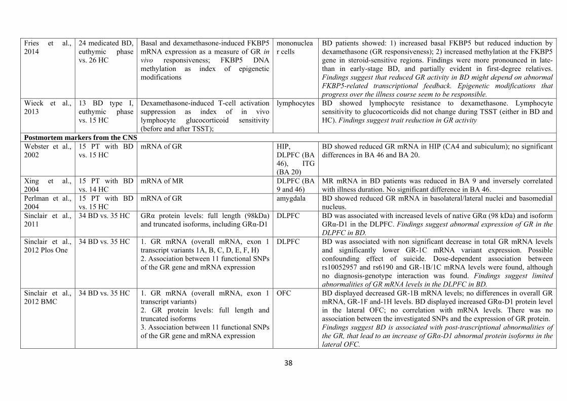
38
Fries et al., 2014
24 medicated BD, euthymic phase vs. 26 HC
Basal and dexamethasone-induced FKBP5 mRNA expression as a measure of GR in vivo responsiveness; FKBP5 DNA methylation as index of epigenetic modifications
mononuclear cells
BD patients showed: 1) increased basal FKBP5 but reduced induction by dexamethasone (GR responsiveness); 2) increased methylation at the FKBP5 gene in steroid-sensitive regions. Findings were more pronounced in late- than in early-stage BD, and partially evident in first-degree relatives. Findings suggest that reduced GR activity in BD might depend on abnormal FKBP5-related transcriptional feedback. Epigenetic modifications that progress over the illness course seem to be responsible.
Wieck et al., 2013
13 BD type I, euthymic phase vs. 15 HC
Dexamethasone-induced T-cell activation suppression as index of in vivo lymphocyte glucocorticoid sensitivity (before and after TSST);
lymphocytes BD showed lymphocyte resistance to dexamethasone. Lymphocyte sensitivity to glucocorticoids did not change during TSST (either in BD and HC). Findings suggest trait reduction in GR activity
Postmortem markers from the CNS Webster et al., 2002
15 PT with BD vs. 15 HC
mRNA of GR HIP, DLPFC (BA 46), ITG (BA 20)
BD showed reduced GR mRNA in HIP (CA4 and subiculum); no significant differences in BA 46 and BA 20.
Xing et al., 2004
15 PT with BD vs. 14 HC
mRNA of MR DLPFC (BA 9 and 46)
MR mRNA in BD patients was reduced in BA 9 and inversely correlated with illness duration. No significant difference in BA 46.
Perlman et al., 2004
15 PT with BD vs. 15 HC
mRNA of GR amygdala BD showed reduced GR mRNA in basolateral/lateral nuclei and basomedial nucleus.
Sinclair et al., 2011
34 BD vs. 35 HC GRα protein levels: full length (98kDa) and truncated isoforms, including GRα-D1
DLPFC BD was associated with increased levels of native GRα (98 kDa) and isoform GRα-D1 in the DLPFC. Findings suggest abnormal expression of GR in the DLPFC in BD.
Sinclair et al., 2012 Plos One
34 BD vs. 35 HC 1. GR mRNA (overall mRNA, exon 1 transcript variants 1A, B, C, D, E, F, H) 2. Association between 11 functional SNPs of the GR gene and mRNA expression
DLPFC BD was associated with non significant decrease in total GR mRNA levels and significantly lower GR-1C mRNA variant expression. Possible confounding effect of suicide. Dose-dependent association between rs10052957 and rs6190 and GR-1B/1C mRNA levels were found, although no diagnosis-genotype interaction was found. Findings suggest limited abnormalities of GR mRNA levels in the DLPFC in BD.
Sinclair et al., 2012 BMC
34 BD vs. 35 HC 1. GR mRNA (overall mRNA, exon 1 transcript variants) 2. GR protein levels: full length and truncated isoforms 3. Association between 11 functional SNPs of the GR gene and mRNA expression
OFC BD displayed decreased GR-1B mRNA levels; no differences in overall GR mRNA, GR-1F and-1H levels. BD displayed increased GRα-D1 protein level in the lateral OFC; no correlation with mRNA levels. There was no association between the investigated SNPs and the expression of GR protein. Findings suggest BD is associated with post-trascriptional abnormalities of the GR, that lead to an increase of GRα-D1 abnormal protein isoforms in the lateral OFC.

39
Sinclair et al., 2013
34 BD vs. 35 HC Assessment of intracellular stress signaling pathways and correlation with GR mRNA levels. 1. mRNA of GR co-factors (FKB4, FKB5, PTGES3, BAG1) and chaperones (HSPA-1A, HSP90AA1, DNAJB1, HSPB1) 2. FKB5-1 protein levels 3. eight functional SNPs of the FKB5 gene
DLPFC BD displayed increased FKB5 and decreased BAG1 mRNA, while no difference was found in chaperones mRNA or FKB5 protein levels. Significant correlations were found between GR-1B mRNA and HSPs and cofactors mRNA levels. No genotype-diagnosis interaction was found in the association between FKB5 SNPs and FKB5 mRNA. Findings suggest BD is associated with widespread abnormalities in the stress-signaling pathways.
Qi et al., 2013 10 elderly INPT with BD, depressed phase vs. 12 HC
mRNA levels of 17 stress-related genes (GR, MR, CRH, CRHR1-2, CRHBP, AVPR1α and others)
ACC, DLPFC
BD patients showed reduced levels of MR mRNA and increased ratio of GRα/MR mRNA in the ACC and the DLPFC.
BA, Brodmann Area; GR (α,β, γ), glucocorticoid receptor (subunits α,β, γ); MR, mineralocorticoid receptor; TSST, Trier Social Stress Test; HIP, hippocampus; DLPFC, dorsolateral prefrontal cortex; ITG, inferior temporal gyrus; OFC, orbitofrontal cortex; ACC, anterior cingulate cortex. CRHR1,2, CRH receptor 1,2; CRHBP, CRH binding protein; AVPR1α vasopressin receptor-1α; NF-kB, Nuclear factor kappa B; JNK, C-jun N-terminal kinase HSP heat shock proteins; HSF, heat shock transcription factors; CRH Corticotrophin - releasing hormone