the il-33/st2 pathway controls coxsackievirus …...the journal of immunology the il-33/st2 pathway...
TRANSCRIPT

of November 13, 2017.This information is current as
PancreatitisInduced Experimental−Coxsackievirus B5
The IL-33/ST2 Pathway Controls
Alves-Filho, Eurico Arruda, Foo Y. Liew and João S. SilvaProença-Módena, Daniela Carlos, Maria L. Silva, José C. Renata Sesti-Costa, Grace K. Silva, José L.
http://www.jimmunol.org/content/191/1/283doi: 10.4049/jimmunol.12028062013;
2013; 191:283-292; Prepublished online 3 JuneJ Immunol
MaterialSupplementary
6.DC1http://www.jimmunol.org/content/suppl/2013/06/03/jimmunol.120280
average*
4 weeks from acceptance to publicationSpeedy Publication! •
Every submission reviewed by practicing scientistsNo Triage! •
from submission to initial decisionRapid Reviews! 30 days* •
?The JIWhy
Referenceshttp://www.jimmunol.org/content/191/1/283.full#ref-list-1
, 22 of which you can access for free at: cites 47 articlesThis article
Subscriptionhttp://jimmunol.org/subscription
is online at: The Journal of ImmunologyInformation about subscribing to
Permissionshttp://www.aai.org/About/Publications/JI/copyright.htmlSubmit copyright permission requests at:
Email Alertshttp://jimmunol.org/alertsReceive free email-alerts when new articles cite this article. Sign up at:
Print ISSN: 0022-1767 Online ISSN: 1550-6606. Immunologists, Inc. All rights reserved.Copyright © 2013 by The American Association of1451 Rockville Pike, Suite 650, Rockville, MD 20852The American Association of Immunologists, Inc.,
is published twice each month byThe Journal of Immunology
by guest on Novem
ber 13, 2017http://w
ww
.jimm
unol.org/D
ownloaded from
by guest on N
ovember 13, 2017
http://ww
w.jim
munol.org/
Dow
nloaded from

The Journal of Immunology
The IL-33/ST2 Pathway Controls Coxsackievirus B5–InducedExperimental Pancreatitis
Renata Sesti-Costa,* Grace K. Silva,* Jose L. Proenca-Modena,† Daniela Carlos,*
Maria L. Silva,† Jose C. Alves-Filho,‡ Eurico Arruda,† Foo Y. Liew,x and
Joao S. Silva*
Coxsackievirus B (CVB) is a common cause of acute and chronic infectious myocarditis and pancreatitis. Th1 cells producing IFN-
g and TNF-a are important for CVB clearance, but they are also associated with the pathogenesis of inflammatory lesions,
suggesting that the modulation of Th1 and Th2 balance is likely important in controlling CVB-induced pancreatitis. We inves-
tigated the role of IL-33, which is an important recently discovered cytokine for induction of Th2-associated responses, in
experimental CVB5 infection. We found that mice deficient in IL-33R, T1/ST2, significantly developed more severe pancreatitis,
had greater weight loss, and contained higher viral load compared with wild-type (WT) mice when infected with CVB5. Con-
versely, WT mice treated with rIL-33 developed significantly lower viral titers, and pancreatitis was attenuated. Mechanistic
studies demonstrated that IL-33 enhances the degranulation and production of IFN-g and TNF-a by CD8+ T and NK cells, which
is associated with viral clearance. Furthermore, IL-33 triggers the production of IL-4 from mast cells, which results in enhanced
differentiation of M2 macrophages and regulatory T cells, leading to the attenuation of inflammatory pancreatitis. Adoptively
transferred mast cells or M2 macrophages reversed the heightened pancreatitis in the T1/ST22/2 mice. In contrast, inhibition of
regulatory T cells exacerbated the disease in WT mice. Together, our findings reveal an unrecognized IL-33/ST2 functional pathway
and a key mechanism for CVB5-induced pancreatitis. These data further suggest a novel approach in treating virus-induced pan-
creatitis, which is a major medical condition with unmet clinical needs. The Journal of Immunology, 2013, 191: 283–292.
Coxsackieviruses are nonenveloped, icosahedral viruseswith a single-stranded positive RNA genome that belongto the Picornaviridae family and enterovirus genus. Cox-
sackieviruses are highly diverse and circulate worldwide. Thereare 23 known serotypes of coxsackieviruses A (CVA1-23) and6 serotypes of coxsackieviruses B (CVB1-6) (1).CVBs are highly cytolytic and are one of the most common causes
of acute infectious myocarditis (2, 3). CVBs also infect pancreaticacinar cells, often leading to pancreatic deficiency (4). Both mildand severe forms of acute pancreatitis are characterized by in-flammation and edema of the exocrine pancreas, which constitutes∼85% of the pancreas, and in the most severe form, tissue necrosis
is also observed (5). A single attack of acute pancreatitis, whichresults in tissue damage and the loss of acinar cells, is normallyfollowed by the recovery of the structure and function of the exo-crine pancreas, suggesting that the pancreas has regenerative ca-pacity after limited damage and tissue loss. If pancreatitis is notcompletely resolved, however, the disease may progress to a chronicstate, causing pancreatic deficiency and resulting in malnutritionand weight loss (4). Coxsackievirus persistence has been related tochronic cardiomyopathy and pancreatitis. Although the morpho-logical damage is reversible in acute pancreatitis as a result of tissuerepair, the changes observed in chronic disease are irreversible. Cur-rently, there is no treatment for chronic pancreatitis.The role of T lymphocytes in protection and disease induced by
coxsackieviruses is complex and not well understood. AlthoughT lymphocyte responses are required for survival in the early stagesof acute pancreatitis, CD4+ T cells can also exacerbate pancreatitis(6). Thus, Th1 cells that produce IFN-g have been associated withthe pathogenesis of CVB5-induced pancreatitis (7). In CVB3 in-fection, the resistance of female mice to myocarditis is associatedwith the induction of type II cytokines and alternatively activatedmacrophages (M2) (7). We have previously reported that the re-cently discovered cytokine, IL-33, is a potent inducer of type IIcytokines and M2 (8, 9). We therefore investigated the role of IL-33 in CVB5-induced pancreatitis.IL-33 is a member of the IL-1 family and promotes CD4+ T cell
differentiation to an atypical Th2 phenotype that is characterizedby the expression of IL-5 and IL-13 (10). IL-33 also stimulatesmast cell (11–14), basophil (15, 16), and eosinophil (15, 17, 18)responses. IL-33 is mainly produced by epithelial and endothelialcells in response to tissue damage, viral infection, allergens, andinflammation. IL-33 binds to the receptor T1/ST2, which exists intwo isoforms: a soluble form (sST2) that acts as a decoy receptor,and a membrane-bound form presented as a heterodimer with IL-
*Department of Biochemistry and Immunology, University of Sao Paulo-RibeiraoPreto School of Medicine, Monte Alegre, Ribeirao Preto-Sao Paulo 14049-900,Brazil; †Department of Cellular and Molecular Biology and Pathogenic Bioagents,University of Sao Paulo-Ribeirao Preto School of Medicine, Monte Alegre, RibeiraoPreto-Sao Paulo 14049-900, Brazil; ‡Department of Pharmacology, University of SaoPaulo-Ribeirao Preto School of Medicine, Monte Alegre, Ribeirao Preto-Sao Paulo14049-900 Brazil; and xDivision of Infection, Immunity, and Inflammation, Univer-sity of Glasgow, Glasgow G12 8TA, United Kingdom
Received for publication October 9, 2012. Accepted for publication May 2, 2013.
This work was supported by Fundacao de Amparo a Pesquisa do Estado de SaoPaulo, Coordenacao de Aperfeicoamento de Pessoal de Nıvel Superior, and the Well-come Trust and the Medical Research Council, United Kingdom (F.Y.L.).
Address correspondence and reprint requests to Prof. Joao S. Silva or Foo Y. Liew,Avenida Bandeirantes 3900, Monte Alegre, Ribeirao Preto-Sao Paulo 14049-900,Brazil (J.S.S.) or Division of Infection, Immunity, and Inflammation, University ofGlasgow, 120 University Place, Glasgow G12 8TA, U.K. (F.Y.L.). E-mail addresses:[email protected] (J.S.S.) or [email protected] (F.Y.L.)
The online version of this article contains supplemental material.
Abbreviations used in this article: Areg, amphiregulin; CVA, coxsackievirus A; CVB,coxsackievirus B; GITR, glucocorticoid-induced TNF receptor; ILC, innate lymphoidtype 2 cell; p.i., postinfection; qPCR, quantitative PCR; sST2, soluble form of ST2;TCID50, tissue culture infectious dose 50; Treg, regulatory T cell.
Copyright� 2013 by The American Association of Immunologists, Inc. 0022-1767/13/$16.00
www.jimmunol.org/cgi/doi/10.4049/jimmunol.1202806
by guest on Novem
ber 13, 2017http://w
ww
.jimm
unol.org/D
ownloaded from

1R–associated protein (IL-1RAcP) in the membrane of mast cells,Th2 cells, dendritic cells, basophils, and macrophages (19, 20).We demonstrated that the production of IL-33 in the pancreas is
markedly elevated post CVB5 infection, whereas the production ofsST2 is reduced. T1/ST22/2 mice developed an exacerbated form ofpancreatitis, whereas the treatment of wild-type (WT) mice with IL-33 significantly attenuated the disease and reduced the viral load.Mechanistic analysis revealed that the IL-33/ST2 pathway activatesmast cells resulting in the production of IL-4, which polarizesmacrophages to the M2 phenotype, and the enhancement of Foxp3+
regulatory T cells (Tregs) in a Stat6-dependent manner. T1/ST2signaling also led to an increase in CD8+ and NK cell populations.Furthermore, adoptively transferred M2 and mast cells suppressedinflammatory pancreatitis. Our findings therefore reveal an unrec-ognized role of the IL-33/ST2 pathway in attenuating CVB5-inducedpancreatitis and further suggest that IL-33 may be a potential thera-peutic agent against Coxsackievirus-induced pathology.
Materials and MethodsMice
BALB/c and Stat62/2 mice on the BALB/c background were purchasedfrom The Jackson Laboratory. T1/ST22/2 mice on the BALB/c back-ground were generated as described previously (21).
Ethics statement
The animal protocol used was approved by the Ethical Commission ofEthics in Animal Research of the University of Sao Paulo, Ribeirao PretoSchool of Medicine (protocol no. 029/2011). This commission is part ofthe National Brazilian College of Animal Experimentation.
Virus titration and infection
The original stock of CVB5, kindly provided by Prof. Roger Loria (Medi-cal College of Virginia, Richmond, VA), was propagated in a Vero cellmonolayer in MEM (Life Technologies) supplemented with 2% FBS andantibiotics at 37˚C. The virus was titrated by limiting dilutions and inocu-lated in quadruplicates to determine the 50% cytopathic effect of thecellular monolayer (TCID50). BALB/c, ST2
2/2, and Stat62/2 mice wereinjected i.p. with either 107 TCID50 CVB5 in 100 ml medium or Vero cellsupernatants alone as a negative control. The animals were sacrificed at3 or 7 d postinfection (p.i.).
Histology and scores
The pancreas from mice was fixed and embedded in paraffin. The slideswere prepared and stained with H&E. The inflammatory and edema/necrosis scores were quantified as follows: 1, ,25% of the tissue affected;2, 25–50% of the tissue affected; and 3, .50% of the tissue affected.
Treatment of mice and cell transfer
Mice were treated i.p. with 200 ng/day IL-33 (BioLegend) on days 21, 0,1, and 2 p.i. Mast cell degranulation was inhibited by the i.p. injection of100 mg/kg cromolyn sodium (Sigma Aldrich) in PBS twice a day (22). Theanti–glucocorticoid-induced TNF receptor (GITR) Ab was obtained fromhybridomas (DTA-1) as previously described (23). The mice were injectedi.p. with 500 mg of the Ab 2 d p.i. The control mice received 500 mgnormal mouse IgG. Bone marrow–derived macrophages were differenti-ated in culture for 7 d with 20% L929 supernatant and polarized to M2 for2 d with 5 ng/ml M-CSF (BioLegend), 30 ng/ml IL-33, and 30 ng/ml IL-4(Invitrogen). The polarization to M2 was assessed by CD206 (mannosereceptor) expression by FACS. M2 macrophages (1 3 106) were trans-ferred i.v. to the recipient mice 2 d p.i. Mast cells were obtained by cul-turing the bone marrow cells with 100 ng/ml stem cell factor and 20 ng/mlIL-3 (PeproTech) for 21 d (24). The cells were analyzed by FACS for theexpression of CD117e and FcεRI. The mast cells (5 3 105) were trans-ferred i.v. to the recipient mice 2 d p.i.
Macrophage infection
Macrophages were differentiated from bone marrow cells for 7 d in mediacontaining 20% of L929 supernatants. The cells were infected with CVB5at a multiplicity of infection of 5 for 24 h. The cytokines in the super-natants were measured by ELISA.
Detection of cytokines by ELISA
The culture supernatants and pancreas homogenates were examined forTNF-a, IL-12p40, IL-4, IL-5, IL-13, IL-10, IFN-g, IL-33, and sST2 ac-cording to the manufacturer’s instructions (R&D Systems).
RNA extraction and real-time PCR
Total RNA from the pancreas was extracted using the illustra RNAspin Kit(GE Healthcare) according to the manufacturer’s instructions and reversetranscribed using the High Capacity cDNA Reverse Transcription Kit(Applied Biosystems). Quantitative PCR (qPCR) was performed targetingthe 59 untranslated region of CVB5, which is a region that is conservedin all enteroviruses. In brief, the reaction was performed in triplicatesin a final volume of 10 ml containing 150 ng template cDNA, 5 mlTaqMan universal PCR Master Mix (Applied Biosystems), 300 nmol/lof each primer, and 150 nmol/l of probe. The primer and probe se-quences are as follows: HEV-F: 59-GCGGAACCGACTACTTTGGG-39,HEV-R: 59-CTCAATTG TCACCATAAGCAGCC-39, HEV-probe: Fam-TCCGTGTTTCCTTTTATTCTTATA-MGB. The amplification reactionswere performed on a 7300 Real Time PCR system (Applied Biosystems).A standard curve was produced using serial 10-fold dilutions of the plas-mid pGEM-HEV that contained a 200-nt fragment from the 59 untranslatedregion of CVB5. Viral loads were determined as the number of copies ofviral RNA per gram of pancreas tissue. The expression of iNos, Arginase-1, Fizz-1, Tnfa, Foxp3, Id2, Areg, and Gapdh were analyzed by qPCRusing the SYBR Green PCR Master Mix (Applied Biosystems). The fol-lowing primers were used: iNos forward: 59-CGAAACGCTTCACTTCCAA-39, reverse: 59-TGAGCCTATATTGCTGTGGCT-39; Arginase-1 forward: 59-GTTCCCAGATGTACCAGGATTC-39, reverse: 59-CGATGTCTTTGGCA-GATATGC-39; Fizz-1 forward: 59-TCCCAGTGAATACTGATGAGA-39, re-verse: 59-CCACTCTGGATCTCCCAAGA-39; Tnfa forward: 59-TGTGCT-CAGAGCTTTCAACAA-39, reverse: 59-CTTGATGGTGGTGCATGAGA-39;Foxp3 forward: 59-ACAACCTGAGCCTGCACAAGT-39, reverse: 59-GCC-CACCTTTTCTTGGTTTTG-39; Id2 forward: 59-CCTGAACACGGACA-TCAGC-39, reverse: 59-CACAGAGTACTTTGCTATCATTCG-39; Aregforward: 59-GCGAATGCAGATACATCGAGAA-39, reverse: 59-TCCAC-ACCGTTCACCAAAGTAA-39; Gapdh forward: 59-TGCAGTGGCAA-AGTGGAGAT-39, reverse: 59-CGTGAGTGGAGTCATACTGGAA-39. Theresults were analyzed using the DCT method (cycle threshold of test2 cyclethreshold of endogen control) (25).
Flow cytometry
To measure intracellular cytokine production, we incubated pancreaticlymph node cells with PMA (500 mg/ml), ionomycin (50 mg/ml), andbrefeldin (GolgiStop; BD Biosciences) for 6 h at 37˚C. Both stimulatedand unstimulated cell suspensions were incubated with Fc block (BDBiosciences) for 45 min. PE-, PE-Cy7–, FITC-, allophycocyanin-, allo-phycocyanin-Cy7–, or PerCP-conjugated mAbs against CD3, CD49b,CD19, CD4, CD8, CD25, CD107a, CD117, FcεRI, and ST2 (BD Bio-sciences) were added to the cell suspension and incubated for 30 min at4˚C. The cells were then fixed and permeabilized with the Mouse Foxp3Buffer Set (BD Biosciences) followed by incubation with Alexa Fluor647–anti-Foxp3, or fixed and permeabilized with Cytoperm/Cytofix (BDBiosciences) followed by incubation with PE- or allophycocyanin-conjugated mAbs against IFN-g and TNF-a according to the manu-facturer’s instructions. The following Abs were used to evaluate innatelymphoid cells: FITC-conjugated mAbs against CD3, CD19, CD11b,CD11c, CD49b, allophycocyanin–c-kit, PE–Sca-1, allophycocyanin-Cy-CD90, PE-Cy7-CD25, and PE-CF594-CD127. The cells were analyzedon a flow cytometer (FACSCantoII; BD Biosciences). Data analysis wasperformed using the FlowJo software (Tree Star, Ashland, OR).
Urea quantification
The urea concentration was determined by adding 25 a-isonitroso–propiophenone dissolved in 100% ethanol to the pancreas homogenateand incubating at 100˚C for 45 min. After 10 min in the dark, the OD at540 nm was determined in a microplate reader (BioRad) using 200-lali-quotsin nonsterile microculture plates. A calibration curve was preparedwith increasing amounts of urea between 15 and 30 mg and 400 ml of theacid mixture. A total of 25 ml a-isonitroso–propiophenone was added to100 ml of the urea solution.
Ab titrations
The concentrations of serum anti-CVB5 IgG and IgM Abs were determinedby ELISA. In brief, 96-well plates were coated with 8 mg inactivated CVB5overnight at 4˚C. The plates were blocked with PBS containing 1% BSA
284 IL-33 ATTENUATES VIRAL PANCREATITIS
by guest on Novem
ber 13, 2017http://w
ww
.jimm
unol.org/D
ownloaded from

for 2 h; then serum (1:100 dilution) was added and incubated for 3 h atroom temperature. Anti-mouse IgG or IgM (Zymed) was added with thesecondary Abs conjugated to peroxidase and incubated at 37˚C for 1 h. Thereaction was developed with tetramethylbenzidine (BD Bioscience) for 10min and stopped with H2SO4 2N. The absorbance at 450 nm was mea-sured. Neutralizing Abs were analyzed in Vero cell cultures. Sera werecomplement inactivated at 56˚C for 1 h and then incubated with 100TCID50 CVB5 for 1 h at 37˚C. CVB5 alone or preincubated with serumwas inoculated in quadruplicates to Vero cell cultures. The cytopathiceffect in 50% of cellular monolayers (TCID50) was then determined.
Statistical analysis
The data were analyzed using the Student t test or the ANOVA followedby the Tukey–Kramer test. A p value ,0.05 was considered statisticallysignificant.
ResultsIL-33 is produced post CVB5 infection and is essential for therecovery of pancreatitis
We first investigated whether IL-33 and T1/ST2 expression wereinduced by CVB5 infection. BALB/c mice were infected i.p. with107 TCID50 of CVB5, and the production of IL-33 and sST2 in thepancreas was determined by ELISA. IL-33 levels were significantlyelevated on day 7 p.i., whereas sST2, the decoy receptor for IL-33,was markedly reduced by day 3 (Fig. 1A). These results suggest thatIL-33 may be associated with CVB5 infection. Infection also in-
duced the emergence of T1/ST2+ cells in the pancreatic lymphnodes, with numbers reaching ∼4 3 104 cells/organ. Total T1/ST2+
lymphocytes, T1/ST2+CD3+CD4+ T cells, T1/ST2+CD3+CD8+
T cells, and T1/ST2+CD19+ B cells were also increased in thepancreas p.i. (Fig. 1B). However, T1/ST2+CD49b+ NK cells and T1/ST2+CD3+CD49b+ NKT cells were present in low numbers and didnot significantly change. We then investigated the potential role ofIL-33 by comparing the disease development in T1/ST22/2 and WT
mice infected with CVB5. WT and T1/ST22/2 mice had similarlevels of acinar cell destruction 3 d p.i. However, although the acinarcells of the WT mice began to regenerate and recover their mor-phology by day 7 p.i., T1/ST22/2 mice contained even more severe
pancreatic damage and tissue necrosis on day 7 p.i. than on day 3(Fig. 1C). Pancreatitis in the T1/ST22/2mice persisted for at least21 d p.i. (data not shown). T1/ST22/2 mice also had a significantlyreduced weight gain and pancreas weight as a percentage of bodyweight as compared with WT mice (Fig. 1D). Viral RNA analysis
revealed that the viral load peaked at day 3. By day 7, ST22/2 micehad an increased viral load in the pancreas compared with WT mice(Fig. 1E). We next investigated the effect of rIL-33 on CVB5 in-fection in WT mice. Mice treated with IL-33 showed markedly re-
duced acinar cell destruction by day 3 (Fig. 1F) and a significantlyreduced viral titer (Fig. 1G). Together, these results indicate that IL-33 plays a protective role against CVB5-induced pathology.
FIGURE 1. T1/ST2 protects mice from CVB5-
induced pancreatitis. BALB/c mice were infected
i.p. with 107 TCID50 of CVB5. (A) IL-33 and sST2
levels in the pancreas were determined by ELISA
up to 7 d p.i. (B) The number of T1/ST2+ cells in
the pancreatic lymph nodes was analyzed by flow
cytometry (FACS) 7 d p.i. (C) The pancreatic his-
tology of WT and T1/ST22/2 mice on days 0, 3,
and 7 p.i. was analyzed by H&E staining. Scale
bars, 100 mm. Inflammatory and edema/necrosis
scores were quantified. (D) The body weight of WT
and T1/ST22/2 mice during CVB5 infection, and
the ratio of the pancreas to body weight on day 7
p.i. (E) Pancreatic viral RNA was determined by
qPCR on days 3 and 7 p.i. (F and G) WT BALB/c
mice were infected with CVB5 and injected i.p.
with IL-33 or PBS on days21, 0, 1, and 2 p.i. Mice
were sacrificed on day 3 p.i. (F) The pancreatic
histology (H&E) was analyzed, and inflammatory
and edema/necrosis scores were quantified. Scale
bars, 100 mm. (G) The viral titer and RNA levels in
the pancreas were determined by virus titration and
qPCR, respectively. All of the data represent the
mean6 SE, n = 6–7, from two to three independent
experiments. Asterisks indicate statistically signifi-
cant differences with p , 0.05. NI, Not infected;
ST2, T1/ST2.
The Journal of Immunology 285
by guest on Novem
ber 13, 2017http://w
ww
.jimm
unol.org/D
ownloaded from

Because Abs are essential for viral clearance during enteroviralinfection (26, 27), we first determined the levels of CVB5-specificAbs in infected WT and T1/ST22/2 mice. We found that T1/ST22/2 mice produced more serum anti-CVB5 IgG and IgM thanWT mice (Supplemental Fig. 1A). However, there was no differ-ence in the concentration of serum neutralizing Abs against CVB5between the two strains of mice (Supplemental Fig. 1B). There-fore, the protective effect of the IL-33/ST2 pathway is unlikely tobe due to viral neutralizing Abs.
The role of T cells and NK cells during CVB5 infection
We next examined the cellular composition of the pancreatic lymphnodes. Seven days p.i., T1/ST22/2 mice had a decreased percentageof CD4+ T and CD49b+ NK cells compared with the WT mice, butboth strains of mice had an equal frequency of CD8+ T, B, andNKT cells (Supplemental Fig. 2). However, T1/ST22/2 mice hada significantly reduced percentage and total number of multifunc-tional CD8+ T cells, as indicated by the decreased percentage ofCD107a+IFN-g+, CD107a+TNF-a+, and CD107a+TNF-a+IFN-g+
cells in the pancreatic lymph nodes (Fig. 2A). Moreover, degra-nulating NK cells, as well as double-positive CD107a+IFN-g+ andCD107a+TNF-a+ cells, were also diminished in the absence of T1/ST2 (Fig. 2B). These results therefore suggest that the antiviraleffect of IL-33 may be mediated via the induction of CD8+ and NKcells, which have well-established antiviral roles.
Mast cells are associated with protection againstCVB5-induced pancreatitis
We then investigated the mechanism by which IL-33 attenuatespancreatitis. Mast cells express a high density of cell-surface T1/ST2 (28), and we have previously reported that mast cells play animportant role in IL-33–mediated rheumatic disease (13, 29). Wetherefore examined the potential role of mast cells in CVB5-induced pancreatitis. At 7 d p.i., T1/ST22/2 mice infected withCVB5 contained markedly less FcεRI+CD117+ mast cells andproduced significantly reduced levels of IL-4 in the pancreascompared with the WT mice (Fig. 3A). To directly demonstratea role for mast cells in this model of pancreatitis, we passivelytransferred mast cells into WT or T1/ST22/2 mice. The mast cellswere generated from the bone marrow of naive WT mice (Fig. 3B)and were adoptively transferred (5 3 105 cells) into WT or T1/ST22/2 mice on day 2 p.i. Although viral load was not changed
after mast cell transfer (Fig. 3C), the T1/ST22/2 mice that receivedthe mast cells recovered from pancreatitis by 7 d p.i. (Fig. 3D, 3E).Moreover, the T1/ST22/2 recipients of the mast cells containedincreased RNA levels of Foxp3 (a Treg marker; Fig. 3G), Arginase-1, and Fizz-1 (M2 markers), but decreased levels of iNos and Tnfa(M1 markers) in the pancreas at 7 d p.i. compared with the T1/ST22/2 mice that did not receive mast cells (Fig. 3F). To confirmthe role of mast cells in the impairment of pancreatitis, we inhibitedmast cell degranulation by treating WT mice with cromolyn so-dium, as previously described (22). Cromolyn induced more severeedema and necrosis in the pancreas (Fig. 3H, 3I). In addition, Foxp3expression was reduced in the pancreas of treated mice (Fig. 3J),whereas the expression of M1 and M2 markers was modestly butnot significantly changed (Fig. 3K). Neither the transfer of mastcells to the T1/ST22/2 mice nor the treatment of WT mice withcromolyn changed the number of CD8+ T cells or viral clearance(Fig. 3D, 3L). These results demonstrate that mast cells play animportant protective role in IL-33/ST2–mediated protection againstCVB5-induced pancreatitis. These results also suggest a potentialrole for Foxp3+ Tregs and M2 macrophages in this protection.
Stat6 is associated with the protection against CVB5-inducedpancreatitis
Because IL-4 is a major product of mast cells activated with IL-33(30), and Stat6 phosphorylation is a key downstream event for IL-4–mediated signaling, we investigated the potential role of Stat6in CVB5-induced pancreatitis. p.i., T1/ST22/2 mice expressedlower levels of Stat6 mRNA in the pancreas compared with WTmice. In addition, the treatment of WT mice with cromolyn so-dium also reduced the expression of this molecule (Fig. 4A).Therefore, Stat62/2 and WT mice were infected with CVB5, andpancreatic histology was examined 7 d p.i. Whereas the pancreasfrom the WT mice had recovered from the pancreatitis, the pan-creas from the Stat62/2 mice remained highly inflamed with in-tense edema and necrosis (Fig. 4B, 4C). There was no differencein the number of mast cells between the infected WT and Stat62/2
mice (Fig. 4D). However, the pancreas from the infected Stat62/2
mice contained significantly higher levels of iNos RNA and re-duced levels of Arginase-1 and Fizz-1 RNA compared with theWT mice (Fig. 4E). The pancreas from the Stat62/2 mice alsocontained less CD4+Foxp3+ Tregs than the WT mice (Fig. 4F).Furthermore, the percentage of IFN-g–producing and the total
FIGURE 2. T1/ST2 induces CD8+ T and NK
cells after CVB5 infection. BALB/c and T1/ST22/2
mice were infected i.p. with 107 TCID50 of CVB5.
Pancreatic lymph node cells were harvested 7 d p.i.,
and stimulated with PMA and ionomycin in the
presence of brefeldin. The number of CD8+ T (A)
and NK cells (B) that were degranulating, CD107a+
and producing IFN-g and TNF-a was analyzed by
flow cytometry (FACS). All of the data represent
the mean 6 SE of seven mice per group and are
representative of two experiments. ST2, T1/ST2.
286 IL-33 ATTENUATES VIRAL PANCREATITIS
by guest on Novem
ber 13, 2017http://w
ww
.jimm
unol.org/D
ownloaded from

number of CD8+ cells were reduced in the pancreatic lymph nodesfrom Stat62/2 mice (Fig. 4G). These reductions correlated witha significant increase in pancreatic CVB5 RNA (Fig. 4H). Thepercentages of IFN-g–producing and degranulating NK cells werenot changed in the absence of Stat6 (data not shown). Together,these results demonstrate that Stat62/2 mice recapitulate the phe-notype of the T1/ST22/2 mice during CVB5 infection. Further-more, Stat6 not only acts after mast cell activation, leading toincreased levels of M2 and Tregs cells, but also acts to increaseCD8+ T cells and reduce viral load.
IL-33 stimulates M2 macrophages and innate lymphoid cellsafter CVB5 infection
IL-33 is a potent inducer of M2 (8), most likely via IL-4 and Stat6.The results with mast cells and Stat62/2 mice strongly indicatethat M2 could play an important role in the IL-33–mediatedprotection against CVB5-induced pancreatitis. We therefore ana-lyzed the pancreas of T1/ST22/2 and WT mice infected withCVB5 for M1 and M2 markers. The pancreas from T1/ST22/2
mice expressed more Tnfa RNA, the same levels of iNos RNA, butless Arginase-1 and Fizz-1 RNA compared with the WT mice(Fig. 5A). The pancreatic cells from the infected T1/ST22/2 micealso produced less urea, which is a marker of M2 activity (Fig. 5B).In addition, bone marrow–derived macrophages from T1/ST22/2
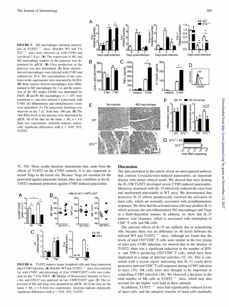
mice produced more TNF-a and IL-12p40 than cells from the WTmice 24 h after CVB5 infection in vitro (Fig. 5C). These resultsindicate that the IL-33/ST2 pathway is important for the inductionof M2 by CVB5 in vivo and in vitro. We then directly tested therole of M2 in CVB5-induced pancreatitis. Bone marrow–derivedmacrophages were differentiated to M2 in vitro with IL-33, IL-4,and M-CSF (Fig. 5D), and adoptively transferred to WT or T1/ST22/2 mice 2 d p.i. Whereas the T1/ST22/2 mice developedmassive pancreatitis 7 d p.i., the T1/ST22/2 mice given the M2cells were protected from pancreatitis (Fig. 5E, 5F), althoughviral load was not changed (Fig. 5G). These results thereforedemonstrate that the induction of M2 by the IL-33/ST2 path-way plays a key role in protecting mice against CVB5-inducedpancreatitis.
FIGURE 3. Mast cells are required for the
protection against pancreatitis. (A) BALB/c WT
and T1/ST22/2 mice were infected i.p. with 107
TCID50 of CVB5, and the presence of mast
cells and the concentration of IL-4 in the pan-
creas on day 7 were determined by FACS and
ELISA, respectively. (B) Mast cells were dif-
ferentiated in vitro by culturing bone marrow
cells for 21 d and analyzed by FACS. (C) The
viral RNA levels in the pancreas were deter-
mined by qPCR. (D) The histology (H&E) of
the pancreas was examined on day 7 p.i. Scale
bars, 100 mm. (E) Inflammatory and edema/
necrosis scores were quantified. The expression
of M1 (iNos, Tnfa) and M2 (Arginase-1, Fizz-1)
markers (F) and Foxp3 (G) in the pancreas
of T1/ST22/2 recipients were determined by
qPCR 7 d p.i. BALB/c mice were infected i.p.
with 107 TCID50 of CVB5 and treated twice
a day with cromolyn sodium to inhibit mast cell
function. (H) The histology (H&E) of the pan-
creas was examined on day 7 p.i. Scale bars,
100 mm. (I) Inflammatory and edema/necrosis
scores were quantified. The expression of Foxp3
(J), iNos, and Arginase-1 (K) in the pancreas
was determined by qPCR 7 d p.i. (L) The
number of CD8+ T and NK cells that were
degranulating, CD107a+, and producing IFN-g
and TNF-a was analyzed by flow cytometry
(FACS). All of the data represent the mean 6SE of five to seven mice per group and are
representative of two experiments. Asterisks
indicate statistically significant differences with
p , 0.05. NT, Not transferred; ST2, T1/ST2.
The Journal of Immunology 287
by guest on Novem
ber 13, 2017http://w
ww
.jimm
unol.org/D
ownloaded from

IL-33 was also reported to induce tissue repair through stimu-lation of innate lymphoid type 2 cells (ILCs) (31). We then in-vestigated whether these cells are induced by IL-33 after CVB5infection. We found that CVB5 infection of WT mice induced anincrease on the percentage of lineage-negative (Lin2) cells that donot express markers of T cells, B cells, NK cells, macrophages,and dendritic cells, but express CD90 and CD25. T1/ST22/2 naivemice presented lower percentage of these cells than WT, and nochange was observed after CVB5 infection (Fig. 6A), showing thatthe induction of ILCs after CVB5 infection is dependent on IL-33signaling. Lin2CD90+CD25+ cells expressed the same quantity ofSca-1, c-kit, and CD127 in the absence of T1/ST2 (Fig. 6B), andthe main transcription factor expressed by ILCs (Id2) was inducedby infection; however, it was not dependent of IL-33 signalingbecause its expression was similar in T1/ST22/2 mice (Fig. 6C).Thus, we quantified the expression of amphiregulin (Areg), whichis produced by ILCs and is responsible for lung remodeling duringinfluenza virus infection (31). We were not able to detect Aregexpression in pancreatic lymph nodes from naive mice; however,after CVB5 infection, Areg was stimulated, and in the absence of
T1/ST2 this expression was significantly diminished (Fig. 6C).These findings indicate that ILCs are induced by CVB5 infectionin an IL-33 signaling–dependent mechanism.
Tregs are associated with resistance to CVB5-inducedpancreatitis
M2 macrophages have been associated with an increase in thenumber of Tregs at the site of CVB infection and are implicatedin the protection against CVB3-induced myocarditis (7). Our re-sults strongly suggest that Tregs also play an important role in theIL-33–mediated protection against CVB5-induced pancreatitis.We therefore determined whether Tregs played a direct role in ourexperimental model. The pancreas from T1/ST22/2 mice infectedwith CVB5 expressed less Foxp3 mRNA compared with the WTmice (Fig. 7A). The pancreatic lymph nodes also contained asignificantly lower frequency of CD4+Foxp3+ Tregs (Fig. 7B).Furthermore, CVB5-infected WT mice treated with an anti-GITRAb, which inhibits subsets of Tregs (32), developed massive cel-lular infiltration and inflammation compared with the IgG-treatedcontrol mice, which had normal pancreas histology 7 d p.i. (Fig.
FIGURE 4. Stat6 is required for the protection
against CVB5-induced pancreatitis. (A) The ex-
pression of Stat6 in the pancreas of WT, T1/ST22/2,
and WT treated with cromolyn sodium mice. (B
and C) The histology (H&E) of the pancreas from
BALB/c WT and Stat62/2 mice 7 d p.i. with CVB5.
(B) Inflammatory and edema/necrosis scores were
quantified. (C) Representative images. Scale bars,
100 mm. (D) The percentage of mast cells (FcεRI+
CD117+) in the pancreatic lymph nodes 7 d p.i.
was analyzed by FACS. (E) M1 (iNos) and M2
(Arginase-1, Fizz-1) markers in the pancreas were
quantified by qPCR day 7 p.i. (F) The frequency of
CD4+Foxp3+ T cells in the pancreatic lymph nodes
7 d p.i. was analyzed by FACS. (G) Lymph node
cells were harvested 7 d p.i. and stimulated with
PMA and ionomycin in the presence of brefeldin
for 6 h at 37˚C. The number of CD8+ T degranu-
lating, CD107a+, and IFN-g– and TNF-a–produc-
ing cells were analyzed by flow cytometry (FACS).
(H) Viral RNA load in the pancreas 3 d p.i. All of
the data are the mean 6 SE of five to six mice per
group and are from two independent experiments.
Asterisks indicate statistically significant differences
with p , 0.05. NI, Not infected; ST2, T1/ST2.
288 IL-33 ATTENUATES VIRAL PANCREATITIS
by guest on Novem
ber 13, 2017http://w
ww
.jimm
unol.org/D
ownloaded from
7C, 7D). These results therefore demonstrate that, aside from theeffects of T1/ST2 on the CVB5 controls, it is also important torecruit Tregs to the lesion site. Because Tregs are essential for theprotection against pancreatic lesions, they may contribute to the IL-33/ST2–mediated protection against CVB5-induced pancreatitis.
DiscussionThe data presented in this article reveal an unrecognized pathwaythat controls Coxsackievirus-induced pancreatitis, an importantdisease with unmet clinical needs. We showed that mice lackingthe IL-33R T1/ST2 developed severe CVB5-induced pancreatitis.Moreover, treatment with rIL-33 effectively reduced the virus loadand ameliorated pancreatitis in WT mice. We demonstrated thatprotective IL-33 effects paradoxically involved the activation ofmast cells, which are normally associated with proinflammatoryresponses. We show that the activated mast cells may produce IL-4,which activates the anti-inflammatory M2 macrophages and Tregsin a Stat6-dependent manner. In addition, we show that IL-33induces viral clearance, which is associated with stimulation ofCD8+ T cells and NK cells.The antiviral effects of IL-33 are unlikely due to neutralizing
Abs, because there was no difference in Ab levels between theinfected WT and T1/ST22/2 mice. Although we found that thelevels of total CD3+CD8+ T cells were similar in the two strainsof mice post CVB5 infection, we showed that in the absence ofT1/ST2, there was a significant reduction in the number of IFN-g– and TNF-a–producing CD3+CD8+ T cells, which have beenimplicated in a range of antiviral activities (33, 34). This is con-sistent with a recent report indicating that IL-33 could driveprotective antiviral CD8+ T cell responses during LCMV infectionin mice (35). NK cells were also thought to be important incontrolling CVB5 infection (36). We observed a decrease in thetotal number of NK cells in T1/ST22/2 mice, which may alsoaccount for the higher viral load in these animals.In addition, T1/ST22/2 mice had significantly reduced levels
of mast cells, and the adoptive transfer of mast cells markedly
FIGURE 5. M2 macrophages attenuate pancrea-
titis in T1/ST22/2 mice. BALB/c WT and T1/
ST22/2 mice were infected i.p. with CVB5 and
sacrificed 7 d p.i. (A) The expression of M1 and
M2 macrophage markers in the pancreas was de-
termined by qPCR. (B) Urea production in the
pancreas was also determined. (C) Bone marrow–
derived macrophages were infected with CVB5 and
cultured for 24 h. The concentrations of the cyto-
kines in the supernatants were measured by ELISA.
(D) Bone marrow–derived macrophages were differ-
entiated to M2 macrophages for 2 d, and the expres-
sion of the M2 marker CD206 was determined by
FACS. (E and F) M2 macrophages (1 3 106) were
transferred i.v. into mice infected 2 d previously with
CVB5. (E) Inflammatory and edema/necrosis scores
were quantified. (F) The pancreatic histology was
observed on day 7 p.i. Scale bars, 100 mm. (G) The
viral RNA levels in the pancreas were determined by
qPCR. All of the data are the mean 6 SE, n = 5–6
from two experiments. Asterisks indicate statisti-
cally significant differences with p , 0.05. ST2,
T1/ST2.
FIGURE 6. T1/ST2 induces innate lymphoid cells and Areg expression
after CVB5 infection. (A) BALB/c WT and T1/ST22/2 mice were infected
i.p. with CVB5, and percentage of (Lin2CD90+CD25+) cells was evalu-
ated on day 7 d by FACS. (B) Median of fluorescence intensity of Sca-1,
c-kit, and CD127 was analyzed on Lin2CD90+CD25+ gate. (C) The ex-
pression of Id2 and Areg were quantified by qPCR. All of the data are the
mean 6 SE, n = 6 from two experiments. Asterisks indicate statistically
significant differences with p , 0.05. ST2, T1/ST2.
The Journal of Immunology 289
by guest on Novem
ber 13, 2017http://w
ww
.jimm
unol.org/D
ownloaded from

protected WT mice from pancreatitis. Mast cells express a highdensity of T1/ST2 (23) and are normally associated with proin-flammatory functions (29). However, in the present system, mastcells play an important anti-inflammatory role in protecting miceagainst CVB5-induced inflammatory pancreatitis, as indicatedby enhanced pancreatitis after mast cell inhibition. Because theadoptive transfer of mast cells into T1/ST22/2 mice attenuatedpancreatitis, it is likely that mast cells function as a result of IL-33treatment. Our findings are consistent with a number of recentreports indicating a protective role for mast cells against acuteinflammation (37, 38). In particular, mast cells have been shown toinduce Tregs to mediate their anti-inflammatory functions (37). Inour model, T1/ST22/2 mice that contained adoptively transferredmast cells developed increased levels of Foxp3 RNA in the pan-creas, which is consistent with the notion that mast cells directlyinduce Tregs. Furthermore, CVB5-infected WT mice treated withcromolyn expressed significantly reduced levels of Foxp3 RNAin the pancreas. Moreover, an anti-GITR Ab markedly increasedpancreatitis in CVB5-infected WT mice. Together, these resultssuggest that IL-33–activated mast cells may suppress inflamma-tory pancreatitis via the induction of Tregs.The pancreas from T1/ST22/2 mice that were given mast cells
also expressed reduced levels of iNos and Tnfa RNA, which istypical of M1 macrophages, and elevated levels of Arginase-1 andFizz-1 RNA, which is typical of M2 macrophages. Our findingsare consistent with an earlier report (39) that analyzed gene ex-pression in the pancreas from mice infected with CoxsackievirusB4. The levels of M2/Th2 RNA were altered in these mice. Wenow indicate one molecular and cellular mechanism for thepreferential induction of M2 macrophages and the potentialtherapeutic control of viral-induced pancreatitis. M2 are associ-ated with anti-inflammatory functions (40, 41). We have previ-ously reported that the preferential induction of M2 in vivocontributed significantly to the anti-inflammatory functions ofIL-33 (8, 42). Furthermore, a recent report shows that M2 mac-rophages are able to promote Treg differentiation in CVB3-induced myocarditis (7). Moreover, it has been shown that Tregscan also induce M2 maturation (43). Thus, it is likely that in oursystem, mast cells play a central role in the IL-33–mediated at-tenuation of pancreatitis via the induction of M2 and Tregs. Tregsseem to inhibit inflammatory pancreatitis, whereas M2 macro-
phages may induce acinar cell proliferation and tissue repair,because M2 are known to be involved in tissue regeneration inseveral models (44–46). In addition, ILCs have also been reportedto respond to IL-33 through T1/ST2 expression. ILCs are alsoimportant for containing lung injury (31) and airway hyperreac-tivity (47) induced by influenza virus infection. We found thatCVB5 increases the percentage of ILCs in pancreatic lymph nodeand stimulates the expression of Areg in an IL-33 signaling–dependent manner. Therefore, it is likely that these cells may alsobe involved in the protection against CVB5-induced pancreatitisby stimulating tissue remodeling.We have also explored the mechanism by which mast cells
induce the differentiation of M2 and Tregs. Cytokine profileanalysis indicated that IL-4 was the major cytokine substantiallyreduced in the CVB5-infected T1/ST22/2 mice compared withthe similarly infected WT mice. We therefore investigated therole of Stat6, the major downstream transcription factor for IL-4. Stat6 expression p.i. was reduced in the absence of T1/ST2and after the inhibition of mast cell degranulation. In addition,Stat62/2 mice recapitulated the phenotypes of the T1/ST22/2
mice during CVB5 infection. Thus, Stat62/2 mice developedmarkedly more severe pancreatitis and had reduced frequencyof Tregs and M2 macrophages. Moreover, Stat62/2 mice hadreduced numbers of CD8+ T cells and a higher viral load. Im-portantly, the Stat6-mediated increase in M2 macrophages andTregs is likely to depend on mast cells, whereas the Stat6-mediated induction of multifunctional CD8+ T cells and viralclearance is mast cell independent. Therefore, it is possible thatIL-33 may act directly on CD8+ T cells as previously shown inan LCMV model (35).The mouse model of CVB5-induced pancreatitis shares many
features with human diseases, which are often debilitating and maynecessitate organ transplant. Chronic pancreatitis is also a majorrisk factor for pancreatic cancer, one of the most intractableconditions (48, 49). Our findings revealing a molecular and cel-lular mechanism behind the reduction in CVB5 load and attenu-ation of pancreatitis upon IL-33 treatment represent a significantadvance in our knowledge in this important medical condition.Our findings also reveal an unrecognized role of IL-33 in a keydisease area. A recent report shows that patients with acute pan-creatitis exhibited elevated levels of sST2 (the decoy receptor of
FIGURE 7. Tregs are associated with resistance to
CVB5-induced pancreatitis. BALB/c WT and T1/
ST22/2 mice were infected i.p. with CVB5, and (A)
the expression of Foxp3 RNA in the pancreas 3 and
7 d p.i. was determined by qPCR. (B) The frequency
of CD4+Foxp3+ T cells in the pancreatic lymph nodes
was analyzed by FACS. (C and D) WT BALB/c mice
were injected i.p. with 500 mg anti-GITR Ab or
normal IgG as a control 2 d p.i. (C) Inflammatory and
edema/necrosis scores were quantified. (D) The
pancreatic histology (H&E) was examined 7 d p.i.
Scale bars, 100 mm. All of the data are the mean 6SE, n = 5–6 from two experiments. Asterisks indicate
statistically significant differences compared to non-
infected (NI) with p , 0.05. ST2, T1/ST2.
290 IL-33 ATTENUATES VIRAL PANCREATITIS
by guest on Novem
ber 13, 2017http://w
ww
.jimm
unol.org/D
ownloaded from

IL-33) early during pancreatitis, and these elevated levels corre-lated with the parameters of severity (50), suggesting that IL-33may also play an important protective role in clinical pancreatitis.Therefore, our findings indicate that IL-33 may be an importantpotential option for treating this debilitating disease.
AcknowledgmentsWe thank Cristiane M. Milanezi, Wander C.R. da Silva, and Maria Elena
Riul for technical support.
DisclosuresThe authors have no financial conflicts of interest.
References1. Racaniello, V. R. 2007. Picornaviridae: The viruses and their replication. In
Field’s Virology. D. M. Knipe, P. M. Howley, D. E. Griffin, R. A. Lamb, M. A.Martin, B. Roizman, and S. E. Straus, eds. Lippincott, Williams & Wilkins,Philadelphia, PA, p. 796–838.
2. Ward, C. 1978. Severe arrhythmias in Coxsackievirus B3 myopericarditis. Arch.Dis. Child. 53: 174–176.
3. Whitton, J. L., C. T. Cornell, and R. Feuer. 2005. Host and virus determinants ofpicornavirus pathogenesis and tropism. Nat. Rev. Microbiol. 3: 765–776.
4. Huber, S., and A. I. Ramsingh. 2004. Coxsackievirus-induced pancreatitis. ViralImmunol. 17: 358–369.
5. Mena, I., C. Fischer, J. R. Gebhard, C. M. Perry, S. Harkins, and J. L. Whitton.2000. Coxsackievirus infection of the pancreas: evaluation of receptor expres-sion, pathogenesis, and immunopathology. Virology 271: 276–288.
6. Ramsingh, A. I., W. T. Lee, D. N. Collins, and L. E. Armstrong. 1999. T cellscontribute to disease severity during coxsackievirus B4 infection. J. Virol. 73:3080–3086.
7. Li, K., W. Xu, Q. Guo, Z. Jiang, P. Wang, Y. Yue, and S. Xiong. 2009. Differ-ential macrophage polarization in male and female BALB/c mice infected withcoxsackievirus B3 defines susceptibility to viral myocarditis. Circ. Res. 105:353–364.
8. Kurowska-Stolarska, M., B. Stolarski, P. Kewin, G. Murphy, C. J. Corrigan,S. Ying, N. Pitman, A. Mirchandani, B. Rana, N. van Rooijen, et al. 2009. IL-33amplifies the polarization of alternatively activated macrophages that contributeto airway inflammation. J. Immunol. 183: 6469–6477.
9. Zaiss, M. M., M. Kurowska-Stolarska, C. Bohm, R. Gary, C. Scholtysek,B. Stolarski, J. Reilly, S. Kerr, N. L. Millar, T. Kamradt, et al. 2011. IL-33shifts the balance from osteoclast to alternatively activated macrophage dif-ferentiation and protects from TNF-alpha-mediated bone loss. J. Immunol.186: 6097–6105.
10. Kurowska-Stolarska, M., P. Kewin, G. Murphy, R. C. Russo, B. Stolarski,C. C. Garcia, M. Komai-Koma, N. Pitman, Y. Li, W. Niedbala, et al. 2008. IL-33induces antigen-specific IL-5+ T cells and promotes allergic-induced airwayinflammation independent of IL-4. [Published erratum appears in 2008 JImmunol. 181: 8170.] J. Immunol. 181: 4780–4790.
11. Allakhverdi, Z., D. E. Smith, M. R. Comeau, and G. Delespesse. 2007. Cuttingedge: The ST2 ligand IL-33 potently activates and drives maturation of humanmast cells. J. Immunol. 179: 2051–2054.
12. Moulin, D., O. Donze, D. Talabot-Ayer, F. Mezin, G. Palmer, and C. Gabay.2007. Interleukin (IL)-33 induces the release of pro-inflammatory mediators bymast cells. Cytokine 40: 216–225.
13. Xu, D., H. R. Jiang, P. Kewin, Y. Li, R. Mu, A. R. Fraser, N. Pitman,M. Kurowska-Stolarska, A. N. McKenzie, I. B. McInnes, and F. Y. Liew. 2008.IL-33 exacerbates antigen-induced arthritis by activating mast cells. Proc. Natl.Acad. Sci. USA 105: 10913–10918.
14. Enoksson, M., K. Lyberg, C. Moller-Westerberg, P. G. Fallon, G. Nilsson, andC. Lunderius-Andersson. 2011. Mast cells as sensors of cell injury through IL-33recognition. J. Immunol. 186: 2523–2528.
15. Pecaric-Petkovic, T., S. A. Didichenko, S. Kaempfer, N. Spiegl, andC. A. Dahinden. 2009. Human basophils and eosinophils are the direct targetleukocytes of the novel IL-1 family member IL-33. Blood 113: 1526–1534.
16. Suzukawa, M., M. Iikura, R. Koketsu, H. Nagase, C. Tamura, A. Komiya,S. Nakae, K. Matsushima, K. Ohta, K. Yamamoto, and M. Yamaguchi. 2008. AnIL-1 cytokine member, IL-33, induces human basophil activation via its ST2receptor. J. Immunol. 181: 5981–5989.
17. Cherry, W. B., J. Yoon, K. R. Bartemes, K. Iijima, and H. Kita. 2008. A novel IL-1 family cytokine, IL-33, potently activates human eosinophils. J. Allergy Clin.Immunol. 121: 1484–1490.
18. Stolarski, B., M. Kurowska-Stolarska, P. Kewin, D. Xu, and F. Y. Liew. 2010. IL-33 exacerbates eosinophil-mediated airway inflammation. J. Immunol. 185:3472–3480.
19. Schmitz, J., A. Owyang, E. Oldham, Y. Song, E. Murphy, T. K. McClanahan,G. Zurawski, M. Moshrefi, J. Qin, X. Li, et al. 2005. IL-33, an interleukin-1-likecytokine that signals via the IL-1 receptor-related protein ST2 and induces Thelper type 2-associated cytokines. Immunity 23: 479–490.
20. Barksby, H. E., S. R. Lea, P. M. Preshaw, and J. J. Taylor. 2007. The expandingfamily of interleukin-1 cytokines and their role in destructive inflammatorydisorders. Clin. Exp. Immunol. 149: 217–225.
21. Brint, E. K., D. Xu, H. Liu, A. Dunne, A. N. McKenzie, L. A. O’Neill, andF. Y. Liew. 2004. ST2 is an inhibitor of interleukin 1 receptor and Toll-likereceptor 4 signaling and maintains endotoxin tolerance. Nat. Immunol. 5:373–379.
22. Hepworth, M. R., E. Daniłowicz-Luebert, S. Rausch, M. Metz, C. Klotz,M. Maurer, and S. Hartmann. 2012. Mast cells orchestrate type 2 immunity tohelminths through regulation of tissue-derived cytokines. Proc. Natl. Acad. Sci.USA 109: 6644–6649.
23. Mariano, F. S., F. R. Gutierrez, W. R. Pavanelli, C. M. Milanezi, K. A. Cavassani,A. P. Moreira, B. R. Ferreira, F. Q. Cunha, C. R. Cardoso, and J. S. Silva. 2008.The involvement of CD4+CD25+ T cells in the acute phase of Trypanosomacruzi infection. Microbes Infect. 10: 825–833.
24. Carlos, D., F. G. Frantz, D. A. Souza-Junior, M. C. Jamur, C. Oliver,S. G. Ramos, V. F. Quesniaux, B. Ryffel, C. L. Silva, M. T. Bozza, andL. H. Faccioli. 2009. TLR2-dependent mast cell activation contributes to thecontrol of Mycobacterium tuberculosis infection. Microbes Infect. 11: 770–778.
25. Pfaffl, M. W. 2001. A new mathematical model for relative quantification in real-time RT-PCR. Nucleic Acids Res. 29: e45.
26. Misbah, S. A., G. P. Spickett, P. C. Ryba, J. M. Hockaday, J. S. Kroll,C. Sherwood, J. B. Kurtz, E. R. Moxon, and H. M. Chapel. 1992. Chronic en-teroviral meningoencephalitis in agammaglobulinemia: case report and literaturereview. J. Clin. Immunol. 12: 266–270.
27. Mena, I., C. M. Perry, S. Harkins, F. Rodriguez, J. Gebhard, and J. L. Whitton.1999. The role of B lymphocytes in coxsackievirus B3 infection. Am. J. Pathol.155: 1205–1215.
28. Moritz, D. R., H. R. Rodewald, J. Gheyselinck, and R. Klemenz. 1998. The IL-1receptor-related T1 antigen is expressed on immature and mature mast cells andon fetal blood mast cell progenitors. J. Immunol. 161: 4866–4874.
29. Xu, D., H. R. Jiang, Y. Li, P. N. Pushparaj, M. Kurowska-Stolarska, B. P. Leung,R. Mu, H. K. Tay, A. N. McKenzie, I. B. McInnes, et al. 2010. IL-33 exacerbatesautoantibody-induced arthritis. J. Immunol. 184: 2620–2626.
30. Hsu, C. L., C. V. Neilsen, and P. J. Bryce. 2010. IL-33 is produced by mast cellsand regulates IgE-dependent inflammation. PLoS ONE 5: e11944.
31. Monticelli, L. A., G. F. Sonnenberg, M. C. Abt, T. Alenghat, C. G. Ziegler,T. A. Doering, J. M. Angelosanto, B. J. Laidlaw, C. Y. Yang, T. Sathaliyawala,et al. 2011. Innate lymphoid cells promote lung-tissue homeostasis after infec-tion with influenza virus. Nat. Immunol. 12: 1045–1054.
32. Scumpia, P. O., M. J. Delano, K. M. Kelly-Scumpia, J. S. Weinstein, J. L. Wynn,R. D. Winfield, C. Xia, C. S. Chung, A. Ayala, M. A. Atkinson, et al. 2007.Treatment with GITR agonistic antibody corrects adaptive immune dysfunctionin sepsis. Blood 110: 3673–3681.
33. Ehtisham, S., N. P. Sunil-Chandra, and A. A. Nash. 1993. Pathogenesis of mu-rine gammaherpesvirus infection in mice deficient in CD4 and CD8 T cells. J.Virol. 67: 5247–5252.
34. Fung-Leung, W. P., T. M. Kundig, R. M. Zinkernagel, and T. W. Mak. 1991.Immune response against lymphocytic choriomeningitis virus infection in micewithout CD8 expression. J. Exp. Med. 174: 1425–1429.
35. Bonilla, W. V., A. Frohlich, K. Senn, S. Kallert, M. Fernandez, S. Johnson,M. Kreutzfeldt, A. N. Hegazy, C. Schrick, P. G. Fallon, et al. 2012. The alarmininterleukin-33 drives protective antiviral CD8⁺ T cell responses. Science 335:984–989.
36. Huhn, M. H., M. Hultcrantz, K. Lind, H. G. Ljunggren, K. J. Malmberg, andM. Flodstrom-Tullberg. 2008. IFN-gamma production dominates the early hu-man natural killer cell response to Coxsackievirus infection. Cell. Microbiol. 10:426–436.
37. Lu, L. F., E. F. Lind, D. C. Gondek, K. A. Bennett, M. W. Gleeson, K. Pino-Lagos, Z. A. Scott, A. J. Coyle, J. L. Reed, J. Van Snick, et al. 2006. Mast cellsare essential intermediaries in regulatory T-cell tolerance. Nature 442: 997–1002.
38. Galli, S. J., J. Kalesnikoff, M. A. Grimbaldeston, A. M. Piliponsky,C. M. Williams, and M. Tsai. 2005. Mast cells as “tunable” effector and im-munoregulatory cells: recent advances. Annu. Rev. Immunol. 23: 749–786.
39. Ostrowski, S. E., A. A. Reilly, D. N. Collins, and A. I. Ramsingh. 2004. Pro-gression or resolution of coxsackievirus B4-induced pancreatitis: a genomicanalysis. J. Virol. 78: 8229–8237.
40. Gordon, S. 2003. Alternative activation of macrophages. Nat. Rev. Immunol. 3:23–35.
41. Benoit, M., B. Desnues, and J. L. Mege. 2008. Macrophage polarization inbacterial infections. J. Immunol. 181: 3733–3739.
42. Jiang, H. R., M. Milovanovic, D. Allan, W. Niedbala, A. G. Besnard,S. Y. Fukada, J. C. Alves-Filho, D. Togbe, C. S. Goodyear, C. Linington,et al. 2012. IL-33 attenuates EAE by suppressing IL-17 and IFN-g productionand inducing alternatively activated macrophages. Eur. J. Immunol. 42:1804–14.
43. Liu, G., H. Ma, L. Qiu, L. Li, Y. Cao, J. Ma, and Y. Zhao. 2011. Phenotypic andfunctional switch of macrophages induced by regulatory CD4+CD25+ T cells inmice. Immunol. Cell Biol. 89: 130–142.
44. Zhang, M. Z., B. Yao, S. Yang, L. Jiang, S. Wang, X. Fan, H. Yin, K. Wong,T. Miyazawa, J. Chen, et al. 2012. CSF-1 signaling mediates recovery from acutekidney injury. J. Clin. Invest. 122: 4519–4532.
45. Villalta, S. A., B. Deng, C. Rinaldi, M. Wehling-Henricks, and J. G. Tidball.2011. IFN-g promotes muscle damage in the mdx mouse model of Duchennemuscular dystrophy by suppressing M2 macrophage activation and inhibitingmuscle cell proliferation. J. Immunol. 187: 5419–5428.
46. Schwartz, M. 2010. “Tissue-repairing” blood-derived macrophages are essentialfor healing of the injured spinal cord: from skin-activated macrophages to in-filtrating blood-derived cells? Brain Behav. Immun. 24: 1054–1057.
The Journal of Immunology 291
by guest on Novem
ber 13, 2017http://w
ww
.jimm
unol.org/D
ownloaded from

47. Chang, Y. J., H. Y. Kim, L. A. Albacker, N. Baumgarth, A. N. McKenzie,D. E. Smith, R. H. Dekruyff, and D. T. Umetsu. 2011. Innate lymphoid cellsmediate influenza-induced airway hyper-reactivity independently of adaptiveimmunity. Nat. Immunol. 12: 631–638.
48. Lowenfels, A. B., P. Maisonneuve, and P. G. Lankisch. 1999. Chronic pancre-atitis and other risk factors for pancreatic cancer. Gastroenterol. Clin. North Am.28: 673–685, x.
49. Talamini, G., M. Falconi, C. Bassi, N. Sartori, R. Salvia, E. Caldiron, L. Frulloni,V. Di Francesco, B. Vaona, P. Bovo, et al. 1999. Incidence of cancer in the courseof chronic pancreatitis. Am. J. Gastroenterol. 94: 1253–1260.
50. Ouziel, R., Gustot, T., Moreno, C., Arvanitakis, M., Degre, D., Trepo, E.,Quertinmont, E., Vercruysse, V., Demetter, P., Le Moine, O., et al. 2012. TheST2 pathway is involved in acute pancreatitis: a translational study in humansand mice. Am. J. Pathol. 180: 2330–9.
292 IL-33 ATTENUATES VIRAL PANCREATITIS
by guest on Novem
ber 13, 2017http://w
ww
.jimm
unol.org/D
ownloaded from