the impact of outward remodeling on vasodilation in skeletal
TRANSCRIPT

THE IMPACT OF OUTWARD REMODELING ON VASODILATION IN SKELETAL
MUSCLE RESISTANCE ARTERIES
A Thesis
presented to
the Faculty of California Polytechnic State University
San Luis Obispo
In Partial Fulfillment
of the Requirements for the Degree
Master of Science in Biomedical Engineering
by
Ryan Gallagher
December 2012

ii
© 2012 Ryan Gallagher
ALL RIGHTS RESERVED

iii
COMMITTEE MEMBERSHIP
TITLE: The Impact of Outward Remodeling on Vasodilation in Skeletal Muscle Resistance Arteries
AUTHOR: Ryan Gallagher
DATE SUBMITTED: December 2012
COMMITTEE CHAIR: Dr. Trevor Cardinal, Assistant Professor
COMMITTEE MEMBER: Dr. Jason Blank, Assistant Professor
COMMITTEE MEMBER: Dr. Lily Laiho, Associate Professor

iv
ABSTRACT
The Impact of Outward Remodeling on Vasodilation in
Skeletal Muscle Resistance Arteries
Ryan Gallagher
Peripheral arterial occlusive disease (PAOD) is an ischemic disease characterized
by narrowing of the peripheral arteries due to the accumulation of atherosclerotic plaque
in the inner lining of the vessels, which disrupts blood flow to downstream tissues. Blood
can be redirected into collateral vessels, natural bypasses around arterial occlusions,
causing shear-induced outward remodeling of the vessels. The enlarged vessels facilitate
transfer of increased blood flow to downstream tissues. The remodeling process,
however, may impair vasodilation, which in turn may cause or contribute to intermittent
claudication- transient pain brought on by locomotion. To stimulate the growth of
collateral arteries, the femoral arteries of young, otherwise healthy mice were ligated
distally to the profunda femoris, the stem to the gracilis collateral circuit. The diameter of
the profunda femoris artery was measured at rest and following gracilis muscle
contraction 7 and 28 days post-surgery using intravital microscopy. Enlarged resting
diameter, consistent with collateral enlargement, and impaired vasodilation was observed
at day 7, but not at day 28. To determine if impaired functional vasodilation is due to
impaired endothelial- or smooth muscle-dependent responses during outward remodeling,
cell-dependent vasodilators were applied to the hindlimb. Endothelial- and smooth
muscle-dependent vasodilation was significantly impaired 7 days post-ligation, but not 28
days after. This data supports the hypothesis that smooth muscle dysfunction causes
impaired functional vasodilation in the early stages of collateral enlargement.
Keywords: Outward Remodeling, Vasodilation, Arteriogenesis, Collateral, Peripheral
Arterial Occlusive Disease, Ischemia, Gracilis

v
ACKOWLEDGMENTS
I would like to thank Dr. Trevor Cardinal for his continued support and guidance, the
other members of the Microcirculation and Tissue Repair Lab, and my parents for their
endless love and support.
“The more that I see the less that I know for sure.”
John Lennon

vi
TABLE OF CONTENTS
LIST OF FIGURES ...................................................................................................... vii Chapter 1: INTRODUCTION ......................................................................................1
PERIPHERAL ARTERIAL OCCLUSIVE DISEASE ..................................................1 Overview ..................................................................................................................1 Atherogenesis ...........................................................................................................5 Current Treatment for PAOD ...................................................................................6
VASCULAR REMODELING AND REACTIVITY ....................................................8 Arteriogenesis ..........................................................................................................8 Mechanoadaptation................................................................................................. 10 Therapeutic Arteriogenesis ..................................................................................... 11 Project Rationale .................................................................................................... 16
SPECIFIC AIMS AND HYPOTHESES..................................................................... 18 Chapter 2. FUNCTIONAL VASODILATION PROTOCOL VERIFICATION ......... 20
INTRODUCTION ..................................................................................................... 20 METHODS ................................................................................................................ 21
ANIMAL HOUSING AND CARE ......................................................................... 21 FUNCTIONAL VASODILATION PROTOCOL ................................................... 21
IMAGING AND STATISTICAL ANALYSIS ........................................................... 23 RESULTS .................................................................................................................. 24 DISCUSSION ............................................................................................................ 26
Chapter 3: FUNCTIONAL VASODILATION FOLLOWING FEED ARTERY OUTWARD REMODELING ........................................................................................ 27
INTRODUCTION ..................................................................................................... 27 METHODS ................................................................................................................ 29
ANIMAL HOUSING AND CARE ......................................................................... 29 FEMORAL ARTERY LIGATION PROTOCOL.................................................... 29 FUNCTIONAL VASODILATION WITH INTRAVITAL MICROSCOPY ........... 32
GRACILIS COLLATERAL STAINING ................................................................... 34 IMAGING AND STATISTICAL ANALYSIS ........................................................... 34 RESULTS .................................................................................................................. 35 DISCUSSION ............................................................................................................ 41
Chapter 4: VASCULAR REACTIVITY WITH INTRAVITAL MICROSCOPY ....... 46 INTRODUCTION ..................................................................................................... 46 METHODS ................................................................................................................ 47
ANIMAL HOUSING AND CARE ......................................................................... 47 FEMORAL ARTERY LIGATION PROTOCOL.................................................... 47 VASCULAR REACTIVITY WITH INTRAVITAL MICROSCOPY ..................... 48
IMAGING AND STATISTICAL ANALYSIS ........................................................... 50 RESULTS .................................................................................................................. 51 DISCUSSION ............................................................................................................ 59
Chapter 5: DISCUSSION .......................................................................................... 63 REFERENCES .............................................................................................................. 69 APPENDIX ................................................................................................................... 73

vii
LIST OF FIGURES
Figure 1: Peripheral Arterial Occlusive Disease. ..............................................................2
Figure 2: Hemodynamic Changes Induced by Arterial Occlusion ....................................5
Figure 3: Arteriogenesis in a Collateral Circuit ................................................................9
Figure 4: Structure of a Large Artery. ............................................................................ 12
Figure 5: Vasodilatory Mechanisms. .............................................................................. 14
Figure 6: Vasodilation in Normal and Inflammatory Conditions. ................................... 15
Figure 7: The Mouse Hindlimb Collateral Circuit. ......................................................... 17
Figure 8: Stimulating Microelectrode Placement. ........................................................... 23
Figure 9: Functional Vasodilation Protocol .................................................................... 25
Figure 10: Typical Surgical Field Preparation ................................................................ 30
Figure 11: Mouse Hindlimb with Ligation Site .............................................................. 31
Figure 12: Progression of the Femoral Ligation Surgery ................................................ 32
Figure 13: Day 7 Functional Vasodilation ...................................................................... 36
Figure 14: Day 28 Functional Vasodilation .................................................................... 38
Figure 15: Collateral Arteriole Diameter ........................................................................ 40
Figure 16: Superfusion Experimental Setup ................................................................... 49
Figure 17: Day 7 Acetylcholine Reactivity. ................................................................... 52
Figure 18: Day 7 Sodium Nitroprusside Reactivity ........................................................ 54
Figure 19: Day 28 Acetylcholine Reactivity................................................................... 56
Figure 20: Day 28 Sodium Nitroprusside Reactivity ...................................................... 58
Figure 21: ECM Remodeling. ........................................................................................ 60

1
Chapter 1: INTRODUCTION
PERIPHERAL ARTERIAL OCCLUSIVE DISEASE
Overview
Peripheral arterial occlusive disease (PAOD) is an ischemic disease characterized
by narrowing of the peripheral arteries due to the accumulation of atherosclerotic plaque
in the inner lining of the vessels. The accumulated plaque obstructs blood flow to
downstream tissues, causing ischemia, an insufficient blood flow to match metabolic
demand, which results in tissue hypoxia and potentially necrosis.
There are an estimated 5 to 12 million adults in the United States affected by
PAOD and this number will likely increase as the population ages [1, 2]. Blockages
within the peripheral arteries of these individuals lead to diminished blood flow to the
extremities (Figure 1). Ischemia-induced claudication (ischemic pain induced by
locomotion) can result and if the disease progresses further, blood flow can diminish
enough to necessitate amputation. The presence of PAOD in patients suggests a decrease
in overall cardiovascular health as they are more likely to develop blockages in blood
vessels elsewhere in the body. PAOD is a reliable predictor of coronary heart disease
(CHD) and cerebrovascular disease (CBVD),which can lead to myocardial infarction and
stroke [3]. The manifestation of atherosclerotic occlusions in other vascular beds is the
major determinant of mortality in patients with PAOD; the ten-year mortality rate for
cardiovascular events is three- to six-fold greater in patients with PAOD [4].

2
Figure 1: Peripheral Arterial Occlusive Disease. PAOD is characterized by arterial narrowing due to atherosclerotic plaque buildup [5].
The most common symptom of PAOD is intermittent claudication, or transient
limb pain associated with exercise that is relieved by rest, which affects 10% to 35% of
patients [1]. Evaluating the distance a patient can walk until experiencing pain is the
gold-standard method of quantifying the severity of intermittent claudication. Intermittent
claudication is associated with a 2- to 4-fold increased risk of mortality, mainly from
cardiovascular disease [6, 7]. Forty to fifty percent of patients experience critical limb
ischemia, a more severe form of PAOD in which leg pain does not correlate with exertion
and is not relieved by rest due to severe blockage of vessels [6]. Gangrene or ulceration

3
present in approximately 2% of patients as a result of critical limb ischemia [1]. In
extreme cases, the blockage of blood flow and nutrients to downstream vessels can be
severe enough to necessitate amputation. Interestingly, most individuals with PAOD
experience no symptoms relating to the disease, which further exacerbates the risk of
mortality from PAOD and CHD as individuals are unaware of the need for treatment [1].
Furthermore, diagnosis of PAOD is difficult as unlike CHD, which typically presents as
angina, asymptomatic PAOD diagnosis requires uncommon instrumentation.
The Ankle Brachial Index (ABI) is the most commonly used diagnostic tool for
evaluating a patient for PAOD as it is simple and noninvasive. Using a continuous wave
Doppler device, the ratio of blood pressure in the posterior tibial artery with respect to the
brachial artery is determined [1]. Occlusion of the peripheral arteries results in a decrease
in blood pressure in the legs. By comparing the ratio of blood pressure in the lower leg to
that in the arm, the severity of arterial blockage can be quantified. A normal ABI is 0.9 to
1.3, suggesting blood pressure in the leg is approximately the same or greater than the
pressure in the arm. ABI values of 0.41 to 0.90 indicate mild to moderate PAOD while
values under 0.40 indicate severe PAOD [1]. In addition to being used to diagnose
PAOD, abnormal ABI values are independent predictors of morbidity and mortality
[8]. Treadmill exercise testing is coupled with pre- and post-exercise ABI measurements
in patients who are highly suspect to have PAOD, but exhibit normal ABI values [1].
Other methods of diagnosing PAOD include non-invasive techniques such as
Doppler ultrasonography, computed tomographic angiography, and magnetic resonance
angiography [1]. A more invasive technique, catheter based angiography, involves the

4
injection of a contrast agent into the vasculature. Catheter based angiography is helpful in
analyzing the impact of PAOD on hemodynamics [1].
The major risk factors for PAOD are similar to the risk factors for coronary heart
disease and include advanced age, a family history of the disease, African-American
heritage, and male sex [1]. Controllable risk factors include hypertension, hyperlipidemia,
diabetes, and smoking [1], of which smoking and diabetes are the strongest and may
accelerate PAOD [4].
The etiology of PAOD is similar to other ischemic diseases, such as coronary
heart disease, and involves atherogenesis induced by endothelial injury and inflammation
[1]. The risk factors for atherogenesis in the peripheral arteries induce endothelial
damage, which initiates the buildup of atherosclerotic plaque. This buildup restricts the
flow of blood from the heart to the periphery, resulting in ischemic injury in downstream
tissues, which become dependent on the collateral circulation (Figure 2).

5
Figure 2: Hemodynamic Changes Induced by Arterial Occlusion. Illustrations of an artery and the properties of the downstream vasculature and tissue in the absence (top)
and presence of (bottom) peripheral arterial occlusion. Adapted from [9].
Atherogenesis
Atherosclerotic lesions are comprised of three major components: cellular
material (primarily smooth muscle cells and macrophages), connective tissue (primarily
extracellular matrix), and an intracellular component comprised of lipids within
macrophages and foam cells [10]. The pathogenesis of atherosclerosis begins with
turbulent flow within a vessel as illustrated by the observation that atheromas tend to
occur at vessel bifurcations, areas of disturbed flow patterns [10, 11]. In response to this
turbulence, endothelial cells become activated, form intercellular gaps, and present
adhesion molecules on their surface through degranulation of intracellular vesicles [10].
Molecules such as VCAM-1 are presented on endothelial cell surfaces and bind to
monocytes and T-cells [11], which extravasate through the gaps between the activated

6
endothelial cells. Macrophage-colony stimulating factor induces monocytes to transform
into macrophages and begin to phagocytose oxidized LDL (ox-LDL) [11]. This process is
thought to be a protective measure to minimize endothelial exposure to inflammatory ox-
LDL, but chronically elevated ox-LDL stimulates T-cell/macrophage interactions and a
positive feedback cycle of inflammation. T-cells, activated by ox-LDL antigens presented
by the macrophages, secrete inflammatory cytokines such as interferon-γ, which further
activates endothelial cells and macrophages [11]. Macrophages secrete factors that
initiate the proliferation and migration of smooth muscle cells that eventually form the
neointima [11]. Continual phagocytosis of oxidized LDL by macrophages transforms
them into foam cells, which also inhabit the neointima [11]. The neointima gradually
enlarges to become an atherosclerotic plaque, and occludes the vessel, resulting in
ischemia downstream of the occlusion.
Current Treatment for PAOD
As patients with PAOD are likely to experience atherosclerosis in vascular beds
other than those in the extremities, the current treatment approach focuses on both
improvement of lower limb ischemia and reduction of overall cardiovascular risk, and
includes lifestyle changes, drug therapy, and revascularization procedures [4]. The first
course of action in treatment of PAOD is risk factor modification through lifestyle
changes such as exercise, diet change, and cessation of smoking. When PAOD is
diagnosed early, lifestyle changes can prevent disease progression.
Exercise and smoking cessation cause the greatest improvement in patients with
PAOD. Exercise therapy improves risk factors for PAOD, including lipid profile, glucose
metabolism, and blood pressure [12]. Walking distance until the presentation of

7
claudication is increased with exercise [12] and increased peripheral artery wall thickness
(a predictor of atherosclerosis) can be reversed through exercise training [13]. The
mechanism for this effect may be due to restoration of eNOS signaling and improved
endothelial structural integrity [14]. Some limitations of exercise are that only supervised
therapy has been shown to be effective [12]; patients may not strictly adhere to the
training regimen, and insurance companies may deny exercise training coverage. The fact
that exercise often elicits claudication may explain poor patient adherence to exercise
therapy. Smoking cessation is another lifestyle change that can prevent disease
progression. Smoking increases an individual’s risk of PAOD by sevenfold and is a more
reliable predictor for PAOD than for coronary artery disease [15]. Stopping smoking
leads to a ten-year reduction in mortality rate from 54% to 18% [16], however, cessation
of smoking rates following written or oral recommendation by a doctor are as low as 13%
after two years [17].
If lifestyle changes are not enough to improve symptoms in patients, additional
treatments are implemented. Antiplatelet therapy to reduce the likelihood of clotting,
such as aspirin or Clopidrogel, reduces vascular death in patients with any form of
atherosclerosis by approximately 25% and is equally as effective in treating patients with
PAOD as with patients with coronary artery disease [16]. Angiotensin converting enzyme
inhibitors and angiotensin receptor blockers, which prevent the production and function
of a potent vasoconstrictor, reduce cardiovascular morbidity and mortality in PAOD
patients by approximately 25% [16].
If lifestyle changes or pharmacologic therapies do not sufficiently arrest the
progression of PAOD, revascularization procedures are performed. The most commonly

8
used endovascular treatment is percutaneous transluminal angioplasty, with or without
stent placement [18]. Bypass surgery is performed when long sections of a vessel are
atherosclerotic or if a plaque is not suitable for angioplasty. There is no significant
difference in outcome of percutaneous angioplasty and bypass surgery after four years
[19].
While there are many treatment options available for the management of PAOD,
no one course of action is comprehensive to alleviate symptoms. Furthermore, many
patients with PAOD are unsuitable candidates for revascularization therapies. Those with
heavily calcified lesions, fully occluded arteries, long occlusions (>5cm), and lack of
suitable vessels for bypass grafting are unable to receive intervention [18]. There is thus
significant motivation to develop novel therapeutic strategies to improve blood flow, such
as by stimulating the growth or formation of collateral arteries- natural bypass vessels.
VASCULAR REMODELING AND REACTIVITY
Arteriogenesis
PAOD is characterized by the occlusion of arterial vessels which disrupts blood
flow to downstream tissues. When a conduit artery becomes occluded by an
atherosclerotic plaque, there is a decrease in arterial pressure distal to the occlusion,
Figure 3. The disparity between the high arterial pressure proximal to the occlusion and
the low arterial pressure distally causes blood flow to be redirected into collateral
arterioles connecting the two regions, providing an alternate blood flow path to the
circulation downstream of the occlusion. The redistribution of blood flow into the
collateral arterioles causes increased flow velocity and shear stress on the vessel wall,
which initiates outward remodeling. Outward remodeling of the vessel restores the shear
9
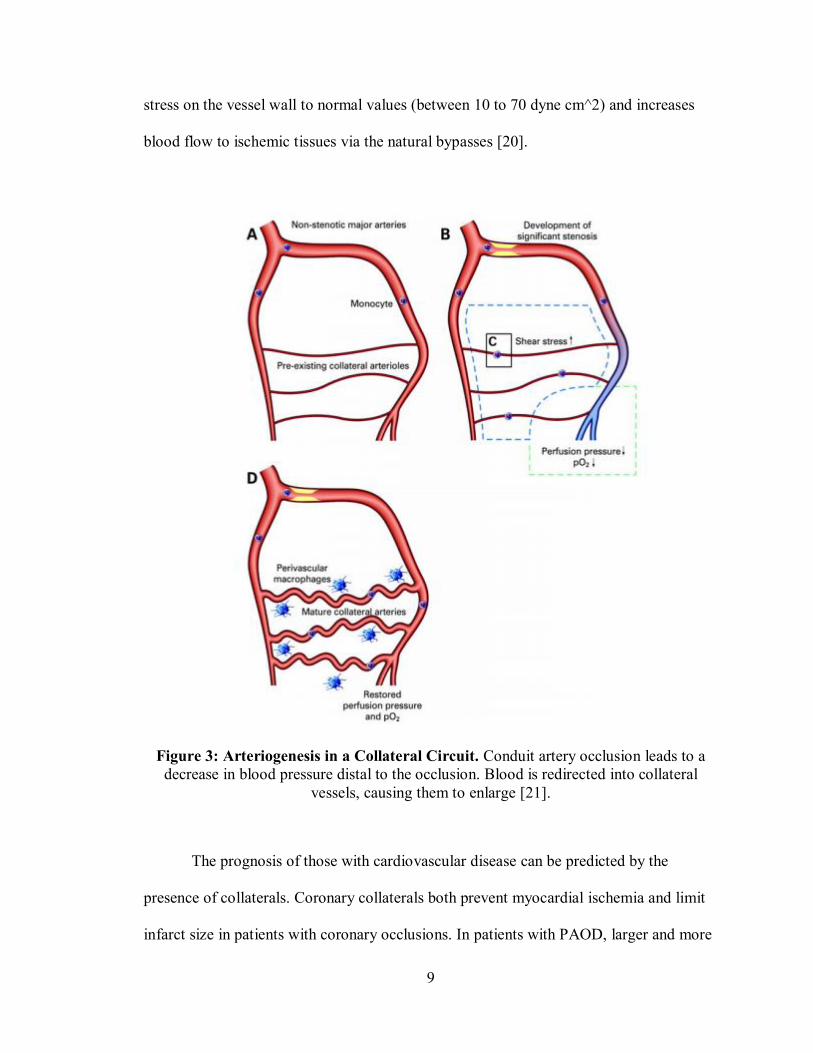
stress on the vessel wall to normal values (between 10 to 70 dyne cm^2) and increases
blood flow to ischemic tissues via the natural bypasses [20].
Figure 3: Arteriogenesis in a Collateral Circuit. Conduit artery occlusion leads to a decrease in blood pressure distal to the occlusion. Blood is redirected into collateral
vessels, causing them to enlarge [21].
The prognosis of those with cardiovascular disease can be predicted by the
presence of collaterals. Coronary collaterals both prevent myocardial ischemia and limit
infarct size in patients with coronary occlusions. In patients with PAOD, larger and more

10
numerous collateral vessels are associated with better ABI values and a lower prevalence
of asymptomatic PAOD [22], making the stimulation of collateral artery growth a
promising and potentially beneficial treatment option for patients with ischemic disease.
Outward vessel remodeling can be achieved through two mechanisms, arteriogenesis and
mechanoadaptation. Arteriogenesis, discussed above, involves the proliferation of
endothelial and smooth muscles cells and degradation of extracellular matrix, while
mechanoadaptation involves the non-proliferative reorganization of smooth muscle cells
within the vessel wall. It is likely that these processes, as well as vasodilation, are part of
remodeling continuum that contributes to blood flow control both acutely and chronically
[23].
Mechanoadaptation
The simplest mechanism of outward remodeling involves the reorientation of
smooth muscle cells (SMCs) within the vessel wall. SMCs are capable of changing their
attachments to other cells and extracellular matrix to move within the media [23].
Cellular repositioning allows wall shear stress to equilibrate without adding additional
cells to the vessel wall through proliferation. When continually exposed to a
vasoconstrictor, SMCs migrate to increase their overlap with adjacent cells to decrease
baseline vessel diameter [24]. While not as well-documented, continual exposure to
vasodilatory agents leads to acute outward remodeling, likely due to smooth muscle
migration [23]. Due to our poor understanding of the mechanisms of mechanoadaptation,
it has not been investigated as a therapeutic strategy.

11
Therapeutic Arteriogenesis
While mechanoadaptation is the simplest mechanism of outward remodeling,
arteriogenesis, a much more complex process, is better understood and more widely
studied. Arteriogenesis is initiated by an increase in shear stress on the vessel wall, which
causes activation of the endothelial cells lining the vessel lumen. A signaling cascade
involving various cytokines and growth factors is carried out to provide the inflammatory
environment needed for outward growth. Endothelial cells and smooth muscle cells
divide and matrix metalloproteinases (MMPs) degrade the extracellular matrix. In the
later stages of arteriogenesis, remodeling beings [25]. SMCs begin to synthesize a new
elastic lamina and the downregulation of MMPs facilitates formation of the intima and
remodeling of the media, Figure 4. SMCs begin to migrate into the intima. Some SMCs
in the media begin to apoptose and are replaced by new ones. The SMCs that begin to
proliferate change their phenotype from a contractile one to a synthetic one.
Arteriogenesis results in an enlarged vessel capable of increasing blood flow to
downstream tissues.

12
Figure 4: Structure of a large artery. During arteriogenesis the vessel wall is remodeled to normalize the shear stress brought on by increased fluid flow [26].
Therapeutic stimulation of collateral vessel growth is a promising therapy to
improve blood flow to ischemic tissue. A variety of factors have been identified as targets
to stimulate arteriogenesis, with some reaching clinical testing, and others abandoned in
the pre-clinical phase due to adverse side effects. Granulocyte-macrophage colony-
stimulating factor (GM-CSF), a factor shown to be pro-arteriogenic in the rabbit
hindlimb, increased collateral blood flow in clinical trials, but did not improve walking
distance to claudication [27]. Fibroblast growth factor, an endothelial and smooth muscle
cell mitogen [28], administration increased walking distance to claudication [29] and
MCP-1, which stimulates monocyte recruitment, induced collateral growth, but is
unsuitable as a therapeutic as it is increases atherosclerotic plaque formation.
Transforming growth factor beta-1 (TGFβ-1), a monocyte chemoattractant, is a
promising therapeutic as it is both pro-arteriogenic and atheroprotective. In order for pro-
13
arteriogenic therapies to be effective, the enlarged vessels need to maintain vasoreactivity
to properly match tissue nutrient demand. During arteriogenesis, however, vascular
smooth muscle cells are thought to be poorly reactive due to their shift in phenotype,
resulting in impaired vasodilation. In order to develop effective therapies, the
mechanisms controlling vasodilation during outward remodeling of vessels need to be
better understood.
Remodeling and Vasoreactivity
Although outward remodeling in collateral arteries could produce a natural bypass
around an occluded conduit artery, the remodeling process may impair vascular
reactivity- potentially worsening symptoms of intermittent claudication. The ability of a
vessel to dilate or constrict is essential to maintain homeostasis by matching the supply of
oxygen and nutrients to downstream cellular demand. Without effective blood flow
control to tissues, tissue damage can occur due to insufficient nutrient supply and waste
product removal. This is especially critical for vessels supplying peripheral skeletal
muscle, as increased muscle fiber activity results in increased metabolic demand that is
orders of magnitude greater than at rest. In skeletal muscle, hyperemia through
vasodilatory mechanisms can manifest from a variety of sources including skeletal
muscle fibers, erythrocytes, endothelial cells, and smooth muscle cells, Figure 5.
Endothelial cells can also transmit vasodilation signals to adjacent cells via endothelial
gap junctions, through a process termed conducted vasodilation [30]. Outward
remodeling of the vessels, however, may impair these vasodilatory mechanisms, as
previously observed in our lab [31].

14
Figure 5: Vasodilatory Mechanisms. Vasodilatory mechanisms originated from (A) skeletal muscle cells, (B) endothelial cells, (C) erythrocytes. Interaction between these
systems is shown in (D) [32].
A possible explanation for impaired vasodilation during outward remodeling is
the process of cellular repositioning during mechanoadaptation. Mechanoadaptation
involves the concerted remodeling of the extracellular matrix-integrin-cytoskeletal axis,
which may impair the ability of smooth muscle cells to relax, resulting in smaller
dynamic range of the vessel. Another possible explanation for impaired vasodilation
during arteriogenesis is the transition of smooth muscle cells from a contractile to a
synthetic phenotype, as described above. The loss of contractile proteins and the
accumulation of free ribosomes and endoplasmic reticulum associated with this

15
phenotype switch may impair the ability of the SMCs to control blood flow via
contraction and relaxation. This is likely not the cause of impaired vasodilation in small
arteries as no smooth muscle cell proliferation (a trademark of arteriogenesis) is present
during outward remodeling of these vessels [33].
Another explanation for impaired vasodilation during outward remodeling is that
inflammation, a critical component of both PAOD and arteriogenesis, causes endothelial
dysfunction and significantly impacts blood flow. Under normal conditions, endothelial
cells induce vasodilation by releasing nitric oxide (NO) in response to receptor-dependent
dilators and increased shear stress [34]. During inflammation, however, cytokines
activate endothelial NADPH oxidase which produces superoxide [34]. Superoxide
inactivates NO and inhibits the ability of receptor-dependent dilators and shear stress to
induce vasodilation, Figure 6.
Figure 6: Vasodilation in normal and inflammatory conditions. Vasodilation is impaired during inflammation [34].
It is possible that the mechanisms discussed above are involved in the impaired
vasoactivity seen in patients with PAOD, as atherosclerosis and arteriogenesis are

16
inflammatory processes. This hypothesis is supported by the observation that non-
occluded vessels in patients with PAOD also exhibit impaired vasoreactivity, suggesting
the disease elicits a systemic response. Other factors associated with inflammation such
as C-reactive protein, cytokines and oxidized low density lipoprotein (oxLDL) may also
be involved in this impairment [35]. Determining which cell types are affected by these
factors is critical to developing strategies to improve vasodilation.
Vascular reactivity studies aimed at determining the cell type responsible for the
impairment in vasodilation observed in outwardly remodeling vessels are inconclusive,
with some studies suggesting impaired endothelial dependent dilation [36, 37] and others
implicating smooth muscle cells [38].
Project Rationale
Baseline diameter of the profunda femoris feed artery, the stem of the gracilis
collateral circuit, increases following femoral artery ligation (Figure 7). Further,
functional vasodilation has been observed in our laboratory to be both impaired and
normal in the profunda femoris artery following femoral ligation. If functional
vasodilation is impaired following femoral artery ligation, it suggests that the outward
remodeling process of the profunda impairs its ability to dilate. Previous studies observed
no smooth muscle proliferation in the vessel, suggesting that the impairment in
vasodilation during outward vascular remodeling (if confirmed to be present) is due to
the reorientation of smooth muscle cells (mechanoadaptation) [33]. An analogous
impairment in vasoreactivity occurs with inward remodeling following prolonged
exposure to a vasoconstrictor [24].

17
Figure 7: The Mouse Hindlimb Collateral Circuit. Collateral vessels originating from the profunda femoris artery anastomose with the femoral artery.

18
SPECIFIC AIMS AND HYPOTHESES
The overall goal of this research is to determine how outward arterial remodeling
impacts vasodilation in the stem region of the collateral circuit. Understanding the effect
of outward remodeling on vasodilation could eventually lead to the development of more
effective therapeutic agents for patients with peripheral arterial occlusive disease by
determining if endogenous collateral enlargement or exogenous stimulation of vessel
growth impairs vasodilation.
Specific Aim 1: To test the hypothesis that functional vasodilation of the
profunda femoris artery is due to increased metabolism in contracting muscles.
The purpose of this aim is to ensure that any vasodilation observed in the
profunda femoris artery is a result of an increase in gracilis metabolism with
electrical stimulation of muscle contraction, and not due to direct electrical
stimulation of the vessel itself, as was suggested by reviewers of a grant submitted
by our lab.
Specific Aim 2: Test the hypothesis that outward vascular remodeling results in
a transient impairment in functional vasodilation.
The purpose of this aim is to validate previous research. I expect that vasodilation
will be impaired at the early stages of outward remodeling, 7 days post-femoral
ligation, but will be restored following the outward remodeling process, 28 days
post-ligation.
19
Specific Aim 3: Test the hypothesis that outward vascular remodeling results in
a transient impairment in smooth muscle-dependent vasodilation.
The purpose of this aim is to determine if impaired functional vasodilation is due
to impaired endothelial- or smooth muscle-dependent responses during outward
remodeling.

20
Chapter 2. FUNCTIONAL VASODILATION PROTOCOL VERIFICATION
INTRODUCTION
Vasodilation transiently increases the diameter of a vessel, resulting in increased
blood flow to downstream tissues, and is critical for maintaining homeostasis. Without
proper vasodilation, metabolically active tissues are unable to receive sufficient oxygen
and nutrients to match cellular demand, resulting in tissue damage. The effects of
impaired vasodilation manifest as intermittent claudication in patients with PAOD and a
better understanding of this impairment is needed to identify therapeutic targets to treat
this dysfunction.
In this study, the gracilis muscle is stimulated to induce endogenous vasodilation
of the profunda femoris. Stimulation of the motor end plates innervating the gracilis
muscle leads to muscle contraction, which causes an increase in metabolic activity and
subsequent vasodilation of the muscle feed artery due to conducted vasodilation, the
transmission of vasoactive signals to adjacent cells through gap junctions. Vasodilation
results from relaxation of the smooth muscle cells within the vascular wall due to
decreased intracellular calcium concentration.
To ensure that vasodilation of the profunda femoris, or alterations to that
vasodilation response following arterial occlusion, are a result of endogenous
vasodilation and not direct electrical stimulation of the vessel, vasoreactivity of the
profunda was assessed following quadriceps stimulation, a muscle near, but not fed by
the profunda. It is hypothesized that stimulation of a muscle not fed by the profunda
femoris will have no effect on the vasoactivity of the vessel due to the highly directional
conducted vasodilation response.

21
METHODS
ANIMAL HOUSING AND CARE
Male C57Bl/6 mice from Taconic Farms aged 7-9 weeks were used for all
experiments according to protocols reviewed and approved by the Cal Poly State
University Institutional Animal Care and Use Committee. Mice were housed in a
temperature controlled room within the University Vivarium, with four mice per micro-
isolator cage. Nesting material, a “mouse house”, and a plastic tube were provided as
enrichment. Mice were monitored daily, given water and rodent chow ad libitum, and
maintained on a 12 hour light- 12 hour dark cycle.
FUNCTIONAL VASODILATION PROTOCOL
Mice were anesthetized in an induction chamber with 5% isoflurane in oxygen
flowing at ~3 l∙min-1. The animal was then weighed and transferred to a preparatory
bench where anesthesia was reduced to ~1-3% isoflurane in oxygen flowing at 0.8-1.0
l∙min-1. Hindlimb hair was removed with clippers and depilatory cream. The mouse was
then placed supine on a heated surgical stage. Body temperature was monitored by a
rectal thermistor and maintained at ~35oC with a heat pad.
A skin incision was made on the middle, medial aspect of the hindlimb and
extended proximally to the abdominal wall. Phosphate buffered saline (PBS) was applied
to the tissue to prevent desiccation and connective tissue was gently blunt dissected to
maximize the tissue exposed. The epigastric fat pad and neurovascular bundle were
resected using a heat cautery. Two tungsten microelectrodes (one stimulating and one
ground) were rested on either the gracilis muscle (functional vasodilation) or the

22
quadriceps muscle (sham) (Figure 8). To ensure correct placement of the microelectrodes
to induce strong muscle contraction, a brief set of muscle contractions was performed
with 1mA square waves 200µs in duration at 1Hz using a PowerLab acquisition system.
Excess PBS was removed with a KimWipe and mineral oil was placed over the tissue
with a pipette to prevent tissue desiccation and oxygen diffusion into the muscle. Plastic
wrap was placed over the mineral oil to prevent runoff. An intravital microscope
(Olympus BXFM) was positioned over the distal profunda femoris, just before the artery-
vein pair passes under the gracilis anterior. The preparation was given 30 minutes to
equilibrate. After equilibration, the muscle was stimulated for 90 seconds with 1mA
square wave 200µs in duration at 8Hz. Images of the profunda femoris were captured
before stimulation, immediately following stimulation, and every subsequent minute until
the vessel returned to baseline diameter. The gracilis and quadriceps muscles in both
hindlimbs were stimulated in random order. Following the experiment, mice were
euthanized by cervical dislocation.

23
Figure 8: Stimulating Microelectrode Placement. (A) Placement of tungsten microelectrodes on the gracilis muscle and (B) location of electrode placement (X) on the
quadriceps during electrical stimulation.
IMAGING AND STATISTICAL ANALYSIS
Infinity imaging software was used to capture images, ImageJ was used to
measure vessel diameters, and Microsoft Excel was used to perform statistical analysis.
Data are presented as mean ±SEM with p ≤ 0.05 representing statistical significance.
Differences in profunda diameter between muscle groups and pre-/post-contraction were
assessed by independent t-test and paired t-test, respectively.
B
A
B

24
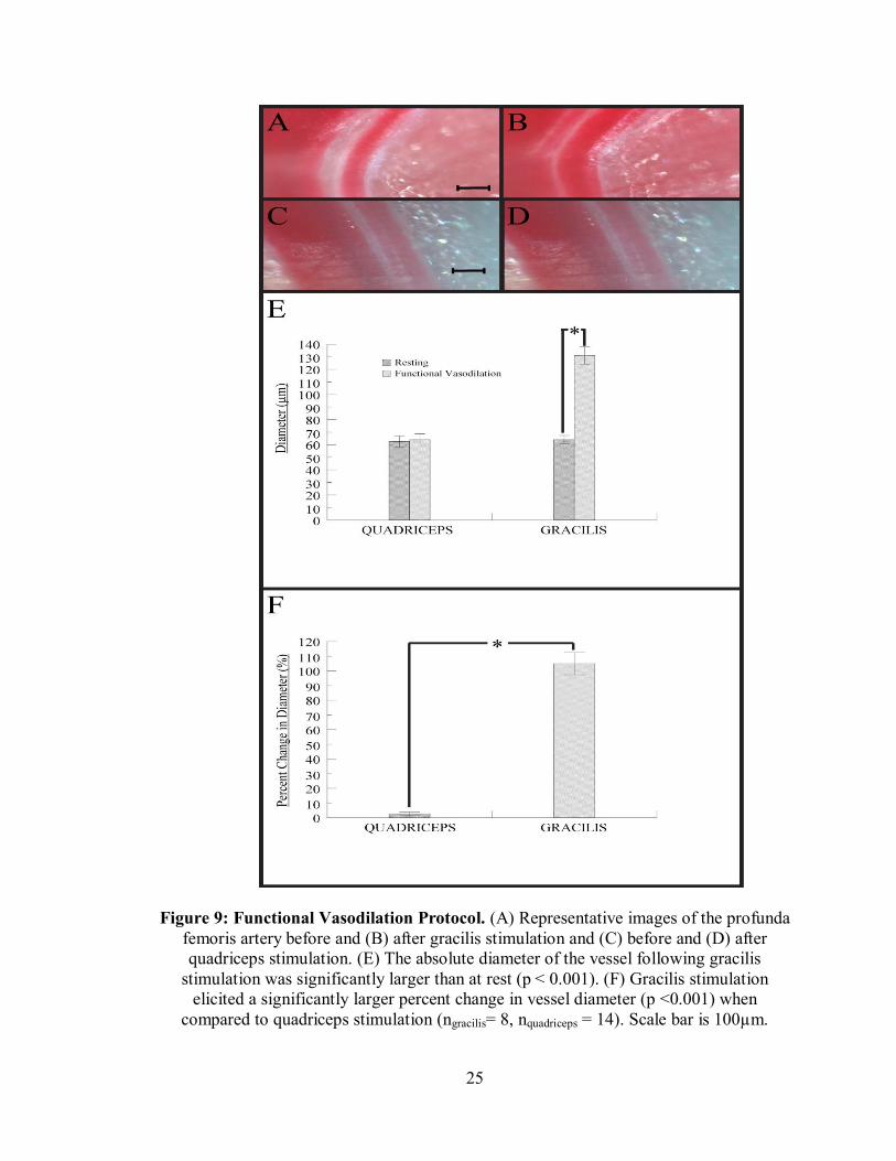
RESULTS
Data obtained from the right and left hindlimbs was combined into a single data
set after confirming they were not statistically different (p > 0.40). The absolute diameter
of the profunda femoris artery after gracilis stimulation was significantly higher than at
rest, while the quadriceps diameter remained unchanged (Figure 9C). The percent
change of the vessel following gracilis stimulation was significantly higher than after
quadriceps stimulation (Figure 9D).
25
Figure 9: Functional Vasodilation Protocol. (A) Representative images of the profunda femoris artery before and (B) after gracilis stimulation and (C) before and (D) after quadriceps stimulation. (E) The absolute diameter of the vessel following gracilis
stimulation was significantly larger than at rest (p < 0.001). (F) Gracilis stimulation elicited a significantly larger percent change in vessel diameter (p <0.001) when
compared to quadriceps stimulation (ngracilis= 8, nquadriceps = 14). Scale bar is 100µm.

26
DISCUSSION
To verify that vasodilation of the profunda femoris following gracilis stimulation
is due to increased metabolism associated with muscle contraction and not direct
stimulation of the vessel wall, the quadriceps muscle, a muscle near, but not fed by the
profunda was stimulated. The diameter of the profunda femoris was unaffected by
quadriceps stimulation, indicating that any impairment in vasodilation is not due to direct
electrical stimulation of vasoconstriction by the electrical field (Figure 9). The diameter
of the vessel following gracilis stimulation, however, increased dramatically (Figure 9).
This indicates that vasodilation of the profunda femoris is due to an increase in muscle
metabolism. If stimulation of the muscles did directly affect the cells within the vascular
wall, the profunda femoris would have changed diameter in response to quadriceps
stimulation. These results suggest that the functional vasodilation protocol employed in
the lab is a valid, physiologically relevant method of assessing vasoreactivity of the
profunda femoris. This protocol was used for all subsequent functional vasodilation
studies (Chapter 3).

27
Chapter 3: FUNCTIONAL VASODILATION FOLLOWING FEED ARTERY OUTWARD REMODELING
INTRODUCTION
Peripheral arterial occlusive disease is associated with the buildup of plaque
within the vascular wall, which not only structurally impedes blood flow, but also impairs
the downstream vasculature’s ability to dilate, resulting in ischemic injury in downstream
tissues. Collateral vessels can act as natural bypasses around the occluded vessel, and the
redistribution of blood flow into these vessels causes an increase in blood velocity and
wall shear stress, which initiates outward remodeling of the vessel. The enlarged vessel
facilitates transport of an additional amount of blood to the ischemic tissues.
The ability of collateral vessels to outward remodel, however, is often impaired in
patients with diabetes, hypercholesterolemia, and old age (co-morbidities of PAOD) [39-
41]. Furthermore, vasodilation may be impaired during outward remodeling as a result of
the transition of smooth muscle cell phenotype from contractile to synthetic during
arteriogenesis, the repositioning of the cells during mechanoadaptation, or shear-induced
inflammation. Impaired vasodilation could then manifest as intermittent claudication,
transient pain during exercise, in patients with PAOD. Stimulating outward vascular
remodeling via biologic or pharmacologic agents is a promising therapy for patients with
PAOD. However, for these therapies to be effective, their effects on vasoreactivity of the
vessels must be understood.
This study focuses on the impact of arterial occlusion and subsequent outward
collateral remodeling on vasodilation in skeletal muscle resistance arteries. Using the
functional vasodilation protocol developed in Chapter 2, the reactivity of the profunda

28
femoris was assessed. It was hypothesized that vasodilation would be impaired 7 days
post-femoral ligation (early in the remodeling process), but not 28 days after (once
remodeling is expected to be roughly complete).
29
METHODS
ANIMAL HOUSING AND CARE
Male C57Bl/6 mice from Taconic Farms aged 7-9 weeks were used for all
experiments according to protocols reviewed and approved by the Cal Poly State
University Institutional Animal Care and Use Committee. Mice were housed in a
temperature controlled room within the University Vivarium, with four mice per micro-
isolator cage. Nesting material, a “mouse house”, and a plastic tube were provided as
enrichment. Mice were monitored daily, given water and rodent chow ad libitum, and
maintained on a 12 hour light- 12 hour dark cycle.
FEMORAL ARTERY LIGATION PROTOCOL
To stimulate the outward remodeling of collateral arteries in the mouse hindlimb,
the femoral artery was ligated. Mice were anesthetized and prepared as previously
described. Prior to surgery, the mouse was given buprenorphrine analgesic (0.075
mg∙kg-1, subQ) and veterinary ophthalmic ointment was placed over the eyes to prevent
corneal desiccation. Surgical field preparation is shown in Figure 10.
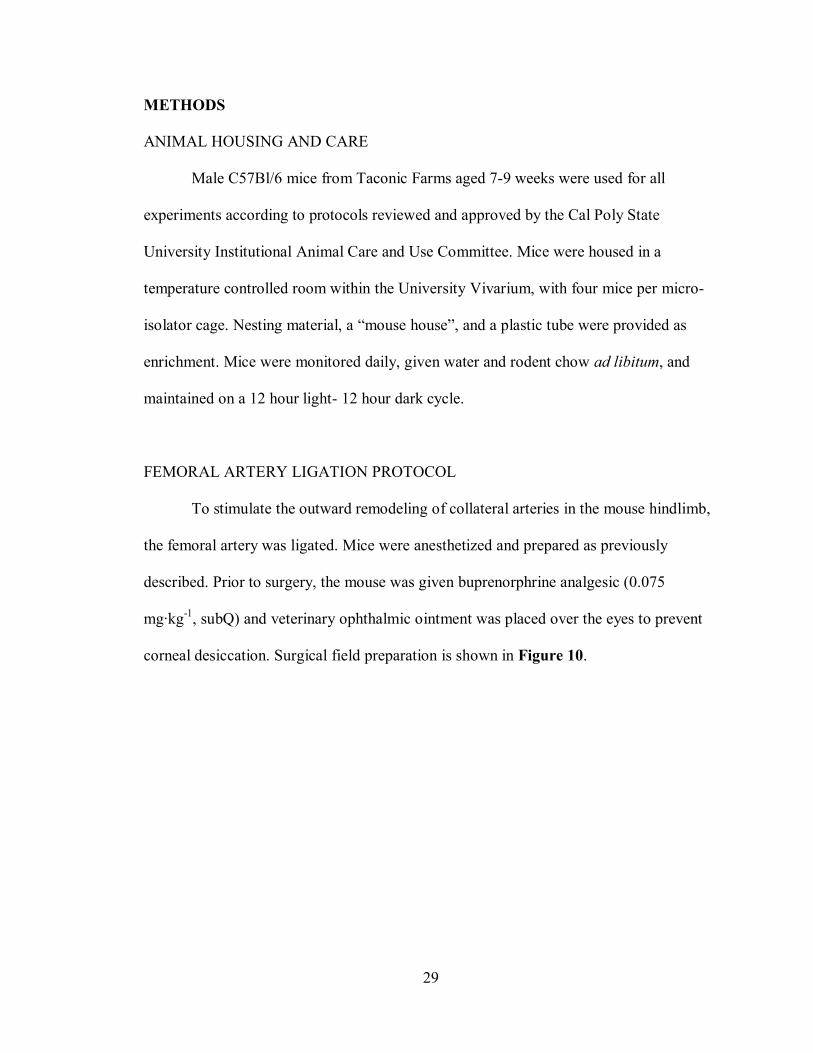
30
Figure 10: Typical Surgical Field Preparation. The drape, cotton swabs, suture, ligature, and surgical instruments are sterilized prior to the procedure.
Using aseptic technique, a small incision was made on the middle, medial aspect
of the left hindlimb, directly over the femoral neurovascular bundle. The incision was
extended proximally to the abdominal wall. The area was continually irrigated with
sterile saline throughout the procedure to prevent desiccation. Connective tissue was
gently blunt dissected and a heat cautery was used to remove the epigastric fat pad and
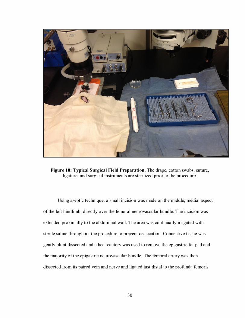
the majority of the epigastric neurovascular bundle. The femoral artery was then
dissected from its paired vein and nerve and ligated just distal to the profunda femoris
31
branch using 6.0 silk suture (Figure 11). The skin incision was closed using 7.0
polypropylene suture. A diagram of the surgery is shown in Figure 12.
Figure 11: Mouse Hindlimb with Ligation Site. Structure of typical hindlimb vasculature with femoral artery ligation site shown.
A skin incision of similar length and placement was made on the contralateral
limb. Sterile saline was applied to the tissue, connective tissue was gently blunt dissected,
and the incision was closed with 7.0 polypropylene suture. The contralateral limb was
treated as a sham in all experiments to control for the impact of surgical trauma on
vasodilation.

32
A second dose of buprenorphrine analgesic was given immediately following
surgery, as described above, and the animal was allowed to recover with warming until
ambulatory.
Figure 12: Progression of the Femoral Ligation Surgery. (A) The initial skin incision is made, (B) connective tissue is blunt dissected, and the epigastric fat pad and
neurovascular bundle removed. (C) The femoral artery is then ligated distal to the profunda femoris and (D) the incision is closed with 7.0 suture.
FUNCTIONAL VASODILATION WITH INTRAVITAL MICROSCOPY
At day-7 or 28 after femoral artery ligation animals were anesthetized and the
hindlimbs were prepared as described above. A skin incision was made on the medial
hindlimb and was extended proximally to the abdominal wall. Phosphate buffered saline
(PBS) was applied to the tissue to prevent tissue desiccation and connective tissue was
gently blunt dissected. Any newly formed adipose was resected using a heat cautery. Two
tungsten microelectrodes (one stimulating and one ground) were rested on the gracilis
muscle. To ensure correct placement of the microelectrodes to induce strong muscle
contractions, a brief set of muscle contractions was performed with 1mA square waves
200µs in duration at 1Hz using a PowerLab acquisition system. Excess PBS was removed
with a KimWipe and mineral oil was pipetted onto the tissue to prevent tissue desiccation
and oxygen diffusion into the muscle. Plastic wrap was placed over the mineral oil to
33
prevent runoff. The same procedure was followed on the contralateral limb, with the
exception that the epigastric fat pad and neurovascular bundle were resected using a heat
cautery.
An intravital microscope (Olympus BXFM) was positioned above the distal
profunda femoris, just before the artery-vein pair passes under the gracilis anterior, and
the preparation was given 30 minutes to equilibrate. After equilibration, the gracilis
muscle was stimulated for 90 seconds with 1mA square wave 200µs in duration at 8Hz.
Images of the profunda femoris were captured before stimulation, immediately following
stimulation, and each subsequent minute until the vessel returned to baseline diameter.
Measurement order between the hindlimbs was randomized. Following the experiment,
mice were euthanized by cervical dislocation.
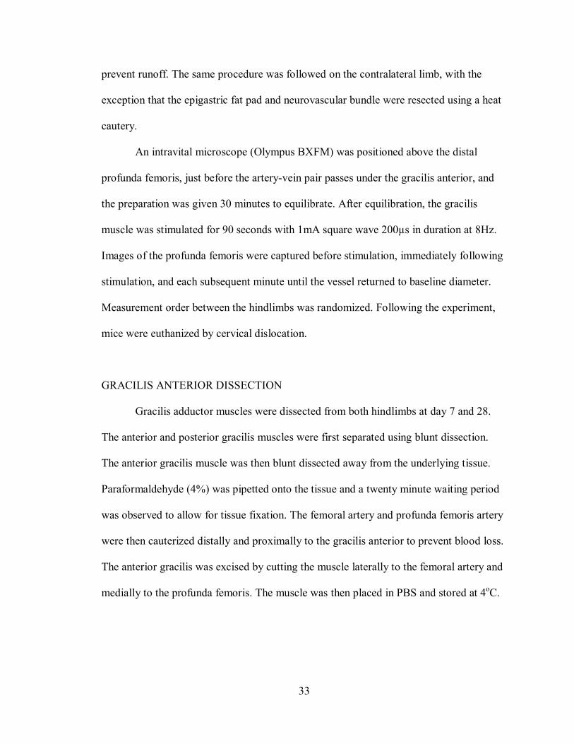
GRACILIS ANTERIOR DISSECTION
Gracilis adductor muscles were dissected from both hindlimbs at day 7 and 28.
The anterior and posterior gracilis muscles were first separated using blunt dissection.
The anterior gracilis muscle was then blunt dissected away from the underlying tissue.
Paraformaldehyde (4%) was pipetted onto the tissue and a twenty minute waiting period
was observed to allow for tissue fixation. The femoral artery and profunda femoris artery
were then cauterized distally and proximally to the gracilis anterior to prevent blood loss.
The anterior gracilis was excised by cutting the muscle laterally to the femoral artery and
medially to the profunda femoris. The muscle was then placed in PBS and stored at 4oC.
34
GRACILIS COLLATERAL STAINING
Sylgard-coated plates were prepared at least three days prior to staining. A 10:1
sylgard elastomer: curing agent solution was mixed in a 50 mL conical tube. From this
solution, 500-750 µL was added to each well of a 24 well plate. The mixture was allowed
to harden at room temperature for 48 hours.
Each excised gracilis anterior muscle was removed from PBS and placed in one
of the wells. An antibody solution containing 1:200 1A4 clone (alpha-smooth muscle
actin, Cy3 conjugate) in 0.1% saponin, 2% BSA in PBS was prepared. The muscles were
incubated in the antibody solution for 72 hours at 4oC. Muscles were washed in 0.1%
saponin in PBS three times for 20 minutes then with plain PBS for 30 minutes. The plate
was wrapped in Parafilm and covered with foil during each wash to prevent evaporation
and exposure to light. The muscles were then placed on a slide and cover slipped with
mounting media. The slides were stored at 4oC in foil prior to imaging with a fluorescent
widefield microscope (Olympus BX41).
IMAGING AND STATISTICAL ANALYSIS
Infinity imaging software was used to capture images from functional
vasodilation experiments, QCapture software was used to image dissected and stained
muscles, ImageJ was used to measure vessel diameters, and Microsoft Excel was used to
perform statistical analysis. Data are presented as mean ±SEM with p ≤ 0.05 representing
statistical significance. Differences in sham and experimental vessel diameter were
assessed by an independent t-test.

35
RESULTS
Day 7
Baseline diameter of the profunda femoris artery was significantly larger 7 days
post-ligation when compared to control limbs, indicating outward remodeling of the
vessel (Figure 13C). The absolute diameter of the vessel was significantly larger, while
the percent change in diameter was significantly smaller in the operated limb after
gracilis stimulation (Figure 13C), indicating that the dynamic range of the vessel is
reduced following arterial occlusion.

36
Figure 13: Day 7 Functional Vasodilation. (A) Representative images of the profunda femoris artery in the operated limb before and (B) after gracilis stimulation. (C) The
diameter of the profunda femoris in the operated limb was larger at rest and after stimulation (p < 0.001 and p = 0.008, respectively). (D) Percent change in vessel
diameter was significantly reduced after femoral ligation (p = 0.02) (n= 6). Scale bar is 100µm.

37
Day 28
Since functional vasodilation was reduced 7 days post-ligation, profunda femoris
vasoreactivity was assessed after 28 days (a time when vascular remodeling is expected
to be complete). Vessel diameter of the profunda femoris at rest and after gracilis
stimulation was not significantly different 28 days post-ligation when compared to
control limbs (Figure 14C). Percent change in vessel diameter was also not significantly
different following femoral ligation (Figure 14D). It was surprising that vessel diameter
returned to control values as we expected the diameter to remain larger in order to supply
sufficient blood flow to downstream tissue. To assess if the ligation protocol used in
these experiments indeed caused long-term outward remodeling of the collateral circuit,
the diameters of the collaterals within the midzone of the gracilis circuit were measured.
Since profunda diameter returned to normal values, we expected the diameter of the
midzone collaterals to increase following ligation.

38
Figure 14: Day 28 Functional Vasodilation. (A) Representative images of the profunda femoris artery in the operated limb before and (B) after gracilis stimulation. (C) The
diameter of the profunda femoris in the operated limb was not significantly different at rest or after stimulation (p > 0.05). (D) Percent change in vessel diameter was not
significantly different (p > 0.05) (n= 7). Scale bar is 100µm.
39
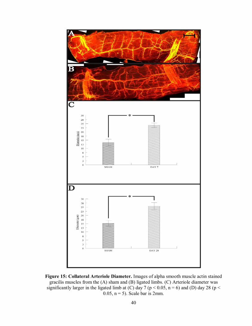
Midzone Collaterals
At both day 7 (Figure 15C) and 28 (Figure 15D), the diameter of the collateral
arteriole in the midzone of the gracilis collateral circuit was significantly larger in the
ligated limb. This was expected after observing profunda femoris diameter return to
control values at day 28. The increased shear stress within these vessels caused outward
remodeling likely via arteriogenesis, allowing blood flow to tissue distal to the ligation
site.
40
Figure 15: Collateral Arteriole Diameter. Images of alpha smooth muscle actin stained gracilis muscles from the (A) sham and (B) ligated limbs. (C) Arteriole diameter was
significantly larger in the ligated limb at (C) day 7 (p < 0.05, n = 6) and (D) day 28 (p < 0.05, n = 5). Scale bar is 2mm.

41
DISCUSSION
Baseline diameter of the profunda in the ligated limb was significantly larger than
in the sham, indicating that outward remodeling occurred (Figure 13C). The impairment
in functional vasodilation 7 days after femoral ligation indicates that the outward
remodeling process impairs the vessel’s ability to dilate (Figure 13D). Vessels can
enlarge via proliferation or reorientation of the cells within the vessel wall.
Arteriogenesis, the best-documented process of vessel enlargement, is associated with
increased wall shear stress and cellular proliferation. No proliferation, however, is present
in the profunda at this time point, indicating that a different process of enlargement, such
as mechanoadaptation, may be involved [33]. Mechanoadaptation involves the non-
proliferative reorientation of smooth muscle cells within the vessel wall. Smooth muscle
cells may reorient themselves to decrease their overlap with adjacent cells in response to
increased blood flow and shear stress, resulting in enlarged vessel diameter. The
reorientation process itself may impair vasodilation as smooth muscle cells migrate
throughout the vessel wall. Mechanoadaptation likely involves remodeling of the
extracellular matrix-integrin-cytoskeletal axis, which may disrupt cellular communication
and relaxation, resulting in impaired dilation.
Interestingly, the diameter of the profunda femoris returned to control values by
day 28, indicating that outward remodeling in the stem region of the collateral circuit is a
transient process following artery occlusion (Figure 14C). The midzone collaterals,
which are known to undergo arteriogenesis (as evidenced by increased diameter and
cellular proliferation 7 days after ligation [33]), maintained their larger diameter at this
time point (Figure 15D). Furthermore, functional vasodilation was not impaired at day
42
28, indicating that even though baseline diameter of the profunda femoris was smaller
than at day 7, the dynamic range of the vessel, and its ability to respond to increased
metabolic demand via vasodilation, was restored (Figure 14D).
The outward remodeling process a vessel undergoes is determined by shear stress
and vessel size. The profunda femoris likely remodels by mechanoadaptation, indicating
that small arteries are able to normalize increased wall shear stress via reorientation of
smooth muscle cells within the vessel wall. The smaller midzone collaterals, however,
undergo arteriogenesis to normalize increased shear stress via cellular proliferation. Wall
shear stress is inversely proportional to the third power of vessel radius. Hence,
normalization of increased shear stress in a small artery would require a smaller degree of
remodeling when compared to smaller collateral arterioles. By day 28, however, the
profunda femoris returns to control diameter while the collateral arterioles remain
enlarged, indicating that shear stress is only transiently elevated in the profunda, but
remains elevated in the arterioles. Following femoral occlusion, the pressure gradient
across the collateral circuit is increased, resulting in higher blood velocity and shear
stress within the vessels. In response to the increased wall shear stress, both the profunda
femoris and the collateral arterioles outwardly remodel. As the arterioles undergo
arteriogenesis to stabilize wall shear stress, blood pressure is partially restored distal to
the occlusion, resulting in a lower pressure gradient across (and lower shear stress within)
the arterioles. A “threshold” of shear stress may be needed to cause outward remodeling
[42]. The shear stress in the midzone arterioles is likely still above threshold at day 28.
The pressure gradient across the profunda femoris also decreases following arteriole
enlargement, likely reducing shear stress levels enough to induce inward remodeling.

43
In order to better characterize the factors that determine whether a vessel
undergoes arteriogenesis or mechanoadaptation, experiments should be performed to
assess blood velocity and shear stress in the stem and midzone regions of the collateral
circuit at day 7 and 28. In order to determine velocity and shear stress profiles, the streak
length of jugular-injected microspheres within the collateral circuit would be measured
using fluorescent microscopy.
Once the shear stress profiles within the stem region of the collateral circuit are
quantified, the next logical experiment to perform would be to study mechanoadaptation
in the profunda femoris with confocal microscopy 7 and 28 days after femoral ligation to
assess smooth muscle cell orientation. If mechanoadaptation is occurring, orientation of
the cells in the ligated limb would differ from those in the sham. A decrease in overlap
between smooth muscle cells would likely be observed at day 7 in response to increased
shear stress brought on by the redistribution of blood flow through the vessel. By day 28,
the orientation of the smooth muscle cells in the ligated limb would likely be similar to
those in the sham.
Once the remodeling mechanism in the stem region of the collateral circuit is
understood, the functionality of the midzone and reentry regions of the circuit should be
assessed 7 and 28 days after arterial ligation. Multiphoton microscopy in conjunction
with a jugular injection of fluorescent compounds would be used to image the vessels
deep within the gracilis. The vasodilatory function of collateral arterioles in response to
electrical stimulation of the gracilis would likely be hindered at day 7 as the vessels
undergo arteriogenesis. If impairment is observed, it is likely attributed to the change in
phenotype of the smooth muscle cells from contractile to synthetic. By day 28,
44
arteriogenesis would be complete [33] and there would likely be no impairment in
functional vasodilation.
The results of these studies agree with those obtained by previous members of our
lab. Significant profunda femoris enlargement was previously observed 14 days after
femoral ligation [43], and a trend toward enlargement was observed at day 7 [33]. Studies
assessing vasodilation of the profunda femoris are inconsistent, with some indicating
significant impairment [44], and others observing a trend toward impairment [43]. Other
research groups observed impaired functional vasodilation in chronically ischemic rat
hindlimbs [45, 46].
The experiments performed do have some limitations. Firstly, the mouse model
employed in these experiments does not accurately mimic the progression of
atherosclerotic plaque in humans. Arterial occlusion in patients with PAOD occurs
chronically, with the exception of thrombosis, as plaque gradually builds up to occlude
the vessel, while the femoral artery of mice used in these studies was fully occluded
almost instantly. This difference likely causes different flow patterns within the collateral
circuit. Secondly, while care was taken when placing the stimulating microelectrodes to
ensure strong muscle contraction, variation in contraction strength was observed. Weak
muscle contraction could make it appear as though functional vasodilation is impaired
due to reduced vasodilatory stimulus. Thirdly, in some cases, adipose tissue obstructed
measurement of the profunda femoris diameter, requiring diameter measurements to be
taken from small visible sections. The amount of adipose tissue present decreased after
mice were switched to a lower protein diet.

45
The impairment in functional vasodilation is likely due to dysfunction of
endothelial or smooth muscle cells. The next step in determining if these cell types
played a role in the observed impairment in vasodilation was to apply cell-dependent
dilators to the hindlimb and measure diameter change of the profunda femoris.

46
Chapter 4: VASCULAR REACTIVITY WITH INTRAVITAL MICROSCOPY
INTRODUCTION
Following arterial occlusion, blood can be redirected into collateral vessels,
resulting in increased vessel wall shear stress and outward remodeling. During outward
remodeling of the collateral circuit, vasodilation is impaired (Chapter 3), likely due to
endothelial or smooth muscle dysfunction. Patients with PAOD often exhibit impaired
vasodilation, which may result in intermittent claudication, ischemic-induced pain during
locomotion. Some hypotheses to explain impaired vasodilation include the repositioning
of smooth muscle cells during mechanoadaptation or the decrease in endothelial-derived
nitric oxide due to shear-induced inflammation. Dysfunction of these cell types would
impair the ability of a vessel to dilate and constrict normally to supply adequate blood
flow to downstream tissues.
To determine which cell-type is responsible for the impaired functional
vasodilation in the profunda femoris (Chapter 3), cell specific vasodilators were
superfused over the tissue. Both acetylcholine, an endothelial-dependent dilator, and
sodium nitroprusside, a smooth muscle-dependent dilator, were used. Acetylcholine is a
neurotransmitter released by nerve endings that binds to receptors on endothelial cells,
causing the release of vasodilators (such as nitric oxide, endothelial derived
hyperpolarizing factor, and prostacyclin [47]), which bind to adjacent smooth muscle
cells, resulting in vasodilation. Sodium nitroprusside acts independently of the
endothelium by directly donating nitric oxide to smooth muscle cells to cause
vasodilation. Knowing which cell-type is dysfunctional is critical to eventually
identifying therapeutic targets to improve vasodilation in patients with PAOD.

47
METHODS
ANIMAL HOUSING AND CARE
Male C57Bl/6 mice from Taconic Farms aged 7-9 weeks were used for all
experiments according to protocols reviewed and approved by the Cal Poly State
University Institutional Animal Care and Use Committee. Mice were housed in a
temperature controlled room within the University Vivarium, with four mice per micro-
isolator cage. Nesting material, a “mouse house”, and a plastic tube were provided as
enrichment. Mice were monitored daily, given water and rodent chow ad libitum, and
maintained on a 12 hour light- 12 hour dark cycle.
FEMORAL ARTERY LIGATION PROTOCOL
Mice were anesthetized and prepared as previously described. Prior to surgery,
the mouse was given buprenorphrine analgesic (0.075 mg∙kg-1, subQ) and veterinary
ophthalmic ointment was placed over the eyes to prevent desiccation.
Using aseptic technique, a small incision was made on the middle, medial aspect
of the left hindlimb, directly over the femoral neurovascular bundle. The incision was
extended proximally to the abdominal wall. The area was continually irrigated with
sterile saline throughout the procedure to prevent desiccation. Connective tissue was
gently blunt dissected and a heat cautery was then used to remove the epigastric fat pad
and the majority of the epigastric neurovascular bundle. The femoral artery was then
dissected from its paired vein and nerve and ligated just distal to the profunda femoris
branch using 6.0 silk suture. The skin incision was closed using 7.0 polypropylene suture.

48
A skin incision of similar length and placement was made on the contralateral
limb. Sterile saline was applied to the tissue, connective tissue was gently blunt dissected,
and the incision was closed with 7.0 polypropylene suture. The contralateral limb was
treated as a sham in all experiments.
A second dose of buprenorphrine analgesic was given immediately following
surgery, as described above, and the animal was allowed to recover with warming until
ambulatory.
VASCULAR REACTIVITY WITH INTRAVITAL MICROSCOPY
On day-7 or 28 post-surgery, mice were anesthetized and hair was removed as
described above. A physiological salt solution (PSS) comprised of (in mM) NaCl 131.9,
KCl 4.7, MgSO4 1.17, CaCl2 2 and NaHCO3 18 was prepared in a 1L volumetric flask.
The flask was placed into a 50oC water bath and deoxygenated by bubbling with 5%
CO2-95% N2; a pH meter was used to confirm a pH of ~7.4. A portion of the PSS was
transferred to a heated 60 mL syringe. The solution exited the syringe through
approximately one foot of insulated tubing at a flow rate of ~2 mL∙min-1 and temperature
of ~35ºC. A skin incision was made on the medial hindlimb and was extended proximally
to the abdominal wall. Phosphate buffered saline (PBS) was applied to the tissue to
prevent tissue desiccation and connective tissue was gently blunt dissected and any newly
formed adipose was resected using a heat cautery. The tubing was positioned so that the
PSS flowed over the mouse hindlimb (Figure 16) and a KimWipe was placed on the
distal end of the hindlimb to wick excess solution to a reservoir. An intravital microscope
(Olympus BXFM) was positioned above the distal profunda femoris, just before the
49
artery-vein pair passes under the gracilis anterior, and the preparation was given 30
minutes to equilibrate.
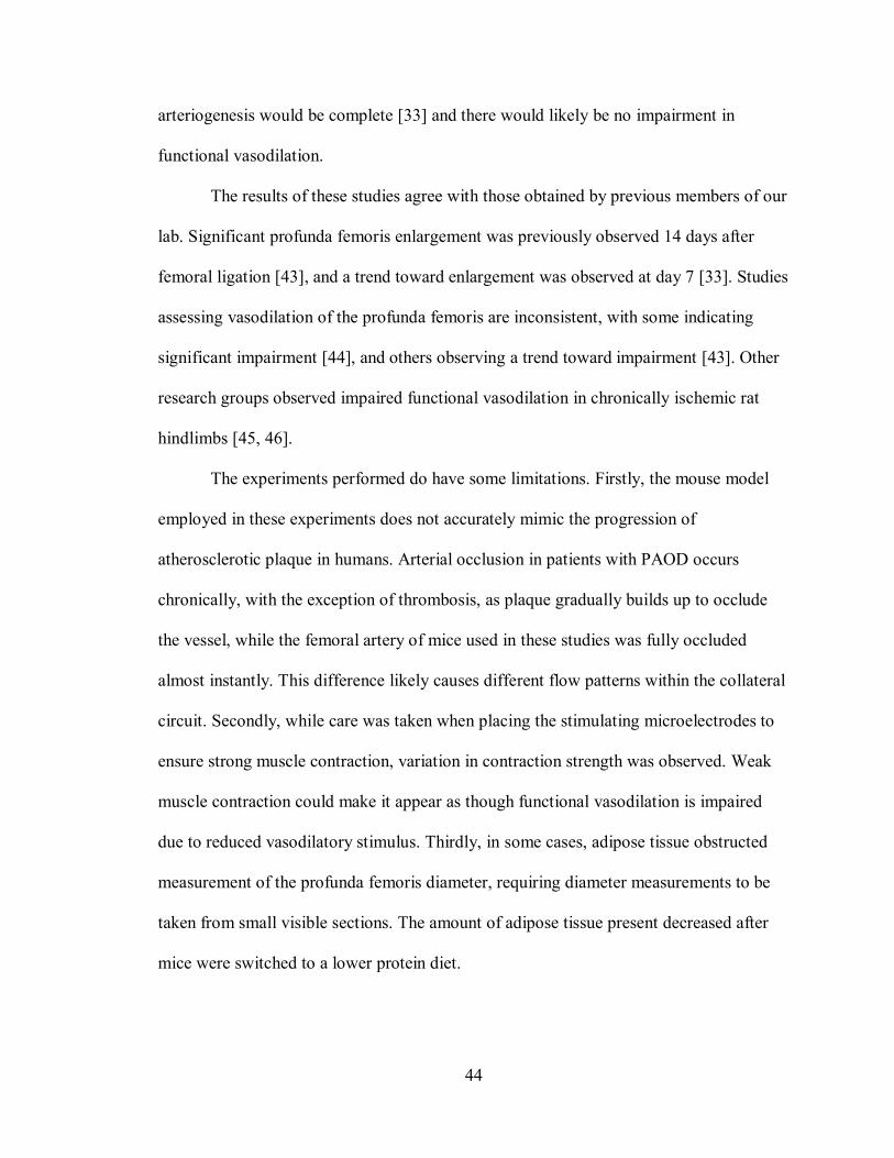
Figure 16: Superfusion experimental setup. Solution flows from a heated syringe, through insulated tubing, and over the mouse hindlimb.
Following equilibration, a baseline image of the profunda femoris was captured.
A dosage response curve (10-9 - 10-5 M) was obtained for acetylcholine and sodium
nitroprusside. Micropipettes were used to transfer the agents to the heated syringe. The
arteries were given 5 minutes to equilibrate to the dose administration before images
were captured. The process was repeated on the contralateral limb. Measurement order

50
between hindlimbs and pharmacologic agents was randomized. Following the
experiment, mice were euthanized by cervical dislocation.
IMAGING AND STATISTICAL ANALYSIS
Infinity imaging software was used to capture images, ImageJ was used to
measure vessel diameters, and Microsoft Excel and Minitab were used to perform
statistical analysis. Data are presented as mean ±SEM with p ≤ 0.05 representing
statistical significance. Baseline diameter differences were assessed by independent t-test
and diameter differences after vasodilator application were assessed by general linear
model ANOVA using Tukey interval comparison.

51
RESULTS
Day 7
Acetylcholine
Resting profunda femoris diameter in the operated limb was significantly larger
than in the sham 7 days post-ligation (Figure 17B). No significant difference was
observed between the diameters of the ligated and sham limbs at all doses of the
endothelial-dependent vasodilator, acetylcholine (Figure 17B), although there was a
clear trend toward a larger diameter in the ligated limb. Percent change in vessel
diameter, a more precise indicator of vascular reactivity when comparing vessels of
different resting diameters, was significantly lower in the ligated limb at high
acetylcholine doses (Figure 17C), indicating that vasodilation is impaired during the
early stages of outward remodeling. These results, however, do not confirm dysfunction
of the endothelium, as although acetylcholine acts directly on endothelial cells,
vasodilation is still dependent on smooth muscle cell activity. Smooth muscle-dependent
vasodilation needs to be assessed in order to determine the dysfunction.

52
Figure 17: Day 7 Acetylcholine Reactivity. (A) Representative images of the profunda femoris artery in the operated limb at rest and 5 minutes after application of each dose
(10-9 – 10-5 M) of acetylcholine. (B) The diameter of the profunda femoris in the operated limb was significantly larger at rest (p < 0.05), but was not significantly different after
application of each Ach dose (p > 0.05). (C) Percent change in vessel diameter was significantly different at the 10-6 and 10-5M dose (p < 0.05) (n= 7). Scale bar is 100µm.
53
Sodium Nitroprusside
The results observed with SNP application were very similar to those with
acetylcholine. Resting vessel diameter was significantly larger in the ligated limb and no
significant difference was observed between the diameters of the ligated and sham limbs
at all doses of SNP (Figure 18B), although a clear trend toward a larger diameter in the
ligated limb was observed. Percent change in vessel diameter was significantly lower in
the ligated limb at high SNP doses (Figure 18C). Both endothelial- and smooth muscle-
dependent vasodilation was impaired at high doses, indicating dysfunction of the smooth
muscle cells as both vasodilators ultimately rely on smooth muscle cell function.

54
Figure 18: Day 7 Sodium Nitroprusside Reactivity. (A) Representative images of the profunda femoris artery in the operated limb at rest and 5 minutes after application of
each dose (10-9 – 10-5 M) of SNP. (B) The diameter of the profunda femoris in the operated limb was significantly larger at rest (p < 0.05), but not after application of each SNP dose (p > 0.05). (C) Percent change in vessel diameter was significantly different at
the 10-6 and 10-5M dose (p < 0.05) (n= 7). Scale bar is 100µm.

55
Day 28
Acetylcholine
No significant difference was observed between the diameters of the ligated and
sham limbs at all doses of acetylcholine (Figure 19B). Percent change in vessel diameter
was also not significantly different at all doses (Figure 19C), indicating restoration of
vasodilation.

56
Figure 19: Day 28 Acetylcholine Reactivity. (A) Representative images of the profunda femoris artery in the operated limb at rest and 5 minutes after application of each dose
(10-9 – 10-5 M) of acetylcholine. (B) The diameter of the profunda femoris in the operated limb was not significantly different at rest or after application of each dose (p > 0.05). (C)
Percent change in vessel diameter was not significantly different at any dose (p > 0.05) (n= 7). Scale bar is 100µm.

57
Sodium Nitroprusside
There was a significant difference between the vessel diameters in the ligated and
sham limbs at high doses of SNP (10-6 and 10-5 M), indicating greater sensitivity to nitric
oxide following transient vessel enlargement (Figure 20B). Percent change in vessel
diameter, however, was not significantly different between the limbs at any dose (Figure
20C).

58
Figure 20: Day 28 Sodium Nitroprusside Reactivity. (A) Representative images of the profunda femoris artery in the operated limb at rest and 5 minutes after application of
each dose (10-9 – 10-5 M) of SNP. (B) The diameter of the profunda femoris in the operated limb was significantly different at the 10-6 and 10-5 M dose (p < 0.05). (C)
Percent change in vessel diameter was not significantly different at any dose (p > 0.05) (n= 7). Scale bar is 100µm.

59
DISCUSSION
Resting vessel diameter was significantly larger in the ligated limb at day 7, as
was observed in the functional vasodilation experiments (Chapter 3) (Figures 17B &
18B). At day 7, vasodilation to both acetylcholine and sodium nitroprusside was impaired
at high vasodilator concentration, suggesting smooth muscle dysfunction, likely due to
cellular repositioning within the vessel wall (Figures 17C & 18C). The ability of smooth
muscle cells to relax may be impaired as they reorient themselves to increase vessel
diameter due to changes in physical cellular connections or responsiveness to
vasodilators.
It is likely that smooth muscle dysfunction is caused by alterations to the
extracellular matrix-integrin-cytoskeletal axis during flow-induced remodeling. The
sequence of events from vasodilation, to intracellular reorganization of the cytoskeleton,
to cellular repositioning, and eventually to a change in passive vessel diameter should be
thought of as a continuum as they likely rely on overlapping pathways and depend on the
same structural elements [23]. Most of the work done on non-proliferative vascular
remodeling has involved chronic vasoconstriction and subsequent inward remodeling.
Inward arterial remodeling depends on the concerted activation of enzymes such as
tissue-type transglutaminase (thought to induce collagen cross-linking) and focal
adhesion kinase (involved in actin polymerization), as well as integrins [23]. Similar
interactions are likely involved in outward remodeling brought on by chronic vasodilator
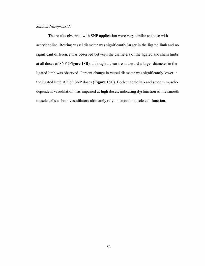
stimulus. Figure 21 shows a proposed explanation for impaired vasodilation following
chronic vasoconstriction involving the cross-linking of extracellular matrix elements. An
60
analogous mechanism may occur during outward remodeling, which could explain the
impaired functional and pharmacologic vasodilation observed at day 7.
Figure 21: ECM Remodeling. (A) A cross section of a relaxed vessel wall showing a SMC (yellow), matrix fibers (black), and cross-links (blue). (B) SMC
contraction leads to matrix unloading. (C) New matrix elements (brown) are formed and new cross-links (orange) are formed between new and existing elements. (D) The new
cross-links impair dilation. Adapted from [48].
By day 28, the only difference between the experimental and sham limb was
absolute diameter in response to high SNP doses (Figure 20B). Once the smooth muscle
cells (presumably) return to their original positions of increased cellular overlap, they
exhibit increased sensitivity to nitric oxide. This altered sensitivity may be explained by
the deposition of new extracellular matrix fibers during mechanoadaptation. Synthesis of
some ECM fibers, such as collagen, may result in altered ECM/integrin interaction and

61
subsequent downregulation of soluble guanylyl cyclase (a factor involved in nitric oxide-
mediated vasodilation) [49]. Increased sensitivity to nitric oxide donors can occur
following periods of decreased nitric oxide availability [50]. Hence, at day 28, vessels
may exhibit increased sensitivity to nitric oxide following downregulation of soluble
guanylyl cyclase during transient outward remodeling. Percent change in diameter of the
vessel was not different between groups (Figures 19C & 20C). Larger baseline diameter
was no longer present in the ligated limb, indicating a reversal in outward remodeling
from day 7 (as seen in Chapter 3). The smooth muscle cells likely return to their original
positions in response to decreased shear stress and restore their vasodilatory function.
The results of this study are consistent with previous research. Both endothelial-
and smooth muscle-dependent dilation is impaired in adults at risk of developing
atherosclerotic plaques [51, 52]. Other vascular reactivity studies aimed at determining
the cell type responsible for the impairment in vasodilation in outwardly remodeling
vessels observed impaired endothelial- [36, 37] and smooth muscle-dependent dilation,
indicating smooth muscle dysfunction [36, 38].
One of the limitations to this study is that only two vasodilators were used,
acetylcholine and sodium nitroprusside. The next study to perform would be to apply
additional vasodilators to the hindlimb following arterial ligation. To better assess what is
causing impaired vasodilation, the effect of other vasodilators such as prostanoids and
hyperpolarizing factors on vasodilation should be investigated. Different vasodilators are
dependent on different cell types and involve different cellular receptors, so by assessing
the impact of many dilators, we can better understand the dysfunction.

62
Once vasodilation to different vasodilators is assessed in the profunda femoris, the
reactivity of the gracilis collateral arterioles in response to these dilators should be
assessed. The mechanism of remodeling, and likely the cause of impaired vasodilation, is
different in these two types of vessels. Arteriogenesis in the collateral arterioles likely
causes dysfunction of the smooth muscle cells as they switch their phenotype. Assessing
the reactivity of these vessels to different vasodilators would help determine the cause of
impaired vasodilation, if present, during outward remodeling. Multiphoton microscopy in
conjunction with fluorescent compounds would need to be used to image the deep
vessels.
Some modifications could be made to the superfusion protocol to improve
experiment consistency and reproducibility. The flow of gas and superfusion solution
varied slightly during each experiment. Flow meters and pumps could be used to provide
a controlled gas bubbling rate and solution flow rate. The time needed to perform the
experiment could also be reduced by splitting the tip of the insulated tubing to superfuse
solution over both hindlimbs at the same time.

63
Chapter 5: DISCUSSION
PAOD is characterized by the buildup of plaque with the vascular wall which
physically impedes blood flow and reduces the reactivity of downstream resistance
vessels, resulting in ischemic injury in downstream tissues. Blood can be redistributed
into collateral vessels, causing increased shear stress and subsequent outward remodeling
of the vessels. During outward remodeling, however, vasodilation may be impaired,
potentially resulting in ischemic limb pain during ambulation. Impaired vasodilation may
be caused by the transition of smooth muscle cell phenotype from contractile to synthetic
during arteriogenesis, the repositioning of the cells during mechanoadaptation, or the
decrease in endothelial-derived nitric oxide due to shear-induced inflammation.
Stimulating outward vascular remodeling via biologic or pharmacologic agents is a
promising therapy for patients with PAOD. However, for these therapies to be effective,
their impact on vasoreactivity of the vessels must be understood.
The functional vasodilation and vascular reactivity studies demonstrated that the
diameter of the profunda femoris 7 days after femoral occlusion was significantly larger
than the sham limb, indicating outward remodeling of the vessel (Figures 13, 17 &18).
Similar observations were documented previously in our lab [33, 53]. The vasodilatory
ability of the profunda femoris in response to gracilis stimulation was impaired at the day
7 time point, likely due to the remodeling process (Figure 13). It is not likely that this
impairment is a result of changes in smooth muscle cell phenotype as previous studies
observed no cellular proliferation in the profunda femoris following arterial occlusion
[33].
64
The enlarged diameter and impaired vasodilatory response of the profunda
femoris 7 days after ligation could be attributed to a variety of factors. It is possible that
flow-mediated dilation led to increased resting diameter measurements. Occlusion of the
femoral artery increases the pressure gradient across the gracilis collateral circuit,
resulting in increased blood flow and shear stress in the profunda femoris. Increased
shear stress causes the release of vasodilators from the endothelium, which relax the
adjacent smooth muscle cells. If the vessel was partially dilated during resting diameter
measurement, it would appear as though the vessel had outwardly remodeled, when in
actuality, the vessel was just transiently enlarged. This could explain why there is no
difference in absolute profunda femoris diameter following vasodilator application
(Figures 17 & 18). Flow-mediated dilation would also make it appear as though
vasodilation was impaired as resting diameter would be inflated.
However, it is unlikely that flow-mediated dilation would persist for 7 days
without triggering a remodeling response. Outward remodeling in response to application
of a vasodilator can occur in as little as 3 days [54]. To test whether flow-mediated
dilation or remodeling mechanisms caused the increase in profunda femoris diameter at
day 7, pressure myography or pharmacologic superfusion experiments could be
performed. Profunda femoris arteries could be excised at day 7 from both hindlimbs and
isolated in a pressure myograph system. The passive diameter of the vessels would be
measured at physiologic pressure in the absence of calcium to eliminate the myogenic
response. If structural remodeling of the vessel occurred, the diameter in the ligated limb
would be significantly larger. A similar experiment could be performed in vivo using the
pharmacologic superfusion protocol employed in Chapter 3. However, instead of
65
applying vasodilators to the hindlimb at day 7, a vasoconstrictor would be used. If
outward remodeling occurred, the vessel in the ligated limb would likely constrict to a
smaller degree.
If outward remodeling does occur, the increased vessel diameter and impairment
in vasodilation is likely due to non-proliferative remodeling, a process termed acute
mechanoadaptation. While mechanoadaptation is better documented in inwardly
remodeling vessels, an analogous situation likely occurs during outward remodeling.
Smooth muscle cells may reorient themselves to decrease their overlap with adjacent
cells in response to continual vasodilation stimuli, resulting in an enlarged diameter. The
reorientation process itself may impair vasodilation due to changing physical cellular
connections or responsiveness to vasodilators. Mechanoadaptation likely involves
remodeling of the extracellular matrix-integrin-cytoskeletal axis, which may disrupt
cellular relaxation and communication, resulting in impaired dilation. This hypothesis is
supported by observations from the vascular reactivity studies, in which vasodilation at
day 7 was impaired in response to both endothelial- and smooth muscle- dependent
dilators, indicating smooth muscle dysfunction (Figures 17 & 18).
Not only did the profunda femoris artery enlarge at day 7, the collateral arterioles
in the midzone of the gracilis collateral circuit enlarged in response to the arterial ligation
(Figure 15C). The outward remodeling of these vessels is likely attributed to
arteriogenesis as proliferating smooth muscle cells are present in these vessels at day 7
[33].
By day 28 post-ligation, profunda femoris artery diameter returned to control
resting values and no impairment to functional or pharmacologic vasodilation was

66
observed, indicating that vasodilation in feed arteries is transiently impaired following
artery occlusion in young, healthy mice (Figures 19 & 20). The collateral arterioles
within the gracilis, however, remained enlarged (Figure 15D). A possible explanation for
these observations is that the pressure gradient across the gracilis collateral circuit is
reduced as the arterioles, which provide the greatest resistance to flow, undergo
arteriogenesis. When the collateral arterioles have enlarged enough to normalize shear
stress, the pressure gradient across the profunda femoris is reduced, resulting in reduced
shear stress and return to control diameter of ~65µm. Shear stress in the arterioles likely
remains elevated at day 28 as indicated by their maintained increased diameter, while
shear stress in the profunda femoris is likely decreased compared to day 7 values. In
order for a vessel to outwardly remodel, a “threshold” of shear stress may be needed [42].
It is possible that the shear stress in the profunda femoris decreases below this threshold
following collateral arteriole growth, causing the vessel to return to control diameter.
Even though the baseline diameter of the profunda femoris was smaller than at day 7, the
dynamic range of the vessel, and its ability to respond to increased metabolic demand via
vasodilation, was restored. Furthermore, vasodilation to high doses of SNP was
significantly greater in the ligated limb, indicating increased sensitivity to nitric oxide
following enlargement.
The outward remodeling process a vessel undergoes is determined by shear stress
and vessel size. Normalization of shear stress within a small artery likely requires a
smaller degree of remodeling than within collateral arterioles as wall shear stress is
inversely proportional to the third power of vessel radius. The transient enlargement and
impaired vasodilation of the profunda femoris is likely attributed to mechanoadaptation,

67
indicating that small arteries are able to normalize increased wall shear stress via
reorientation of smooth muscle cells within the vessel wall. The smaller midzone
collaterals, however, undergo arteriogenesis to normalize the increased shear stress via
cellular proliferation. The next logical experiment to perform would be to characterize
what factors determine if a vessel undergoes arteriogenesis or mechanoadaptation by
measuring blood velocity and shear stress in the stem and midzone regions of the
collateral circuit at day 7 and 28 by assessing the streak length of jugular-injected
microspheres using fluorescent microscopy.
Once the shear stress profiles within the collateral circuit are determined, the next
logical study to perform would be to assess mechanoadaptation in the profunda femoris
with confocal microscopy 7 and 28 days after femoral ligation. If mechanoadaptation is
occurring, a decrease in smooth muscle cell overlap would likely be observed in the
ligated limb 7 days after ligation, but overlap would return to control values by day 28.
Regardless of the position of the smooth muscle cells, the cellular mechanism
responsible for impaired vasodilation needs to be determined to identify therapeutic
targets. This could be achieved by applying other vasodilators, such as hyperpolarizing
factors and prostanoids, to the hindlimb. Changes in vasodilation in response to these
factors would help to better identify understand the dysfunction.
Once the remodeling mechanism in the stem region of the collateral circuit is
understood, the functionality of the midzone and reentry regions of the circuit should be
assessed 7 and 28 days after arterial ligation. Vasodilation in the gracilis feed artery is
impaired, but little is known about the intramuscular collaterals. The function of

68
collateral arterioles would likely be hindered at day 7 as the vessels undergo
arteriogenesis, but would be restored by day 28 following completion of arteriogenesis.
Overall, impaired vasodilation in the mouse hindlimb following arterial ligation
was modest and transient, suggesting that young, otherwise healthy animals are capable
of rapid and effective remodeling responses. To better understand the mechanism of
impaired vasodilation in patients with PAOD, future studies should employ mouse
disease models. The majority of patients with PAOD have other comorbidities. Hence,
aged, diabetic, hypertensive or hypercholesterolemic mice should be used to better assess
how collateral enlargement affects vasodilation in an environment more consistent with
the human patient. It is possible that patients with these ailments are able to initiate the
outward remodeling process (during which vasodilation is impaired), but are unable to
fully remodel [55, 56]. The findings of this study could potentially aid in developing
therapies guided toward restoring activity in collateral vessels. It is crucial to understand
how comorbidities impact the impairment observed with arterial occlusion alone. This
insight will allow the identification of potential therapeutic targets and support the
development of novel therapies to aid these patients.

69
REFERENCES
1. Shanmugasundaram, M., V.K. Ram, U.C. Luft, M. Szerlip, and J.S. Alpert, Peripheral arterial disease--what do we need to know? Clin Cardiol. 34(8): p. 478-82.
2. Selvin, E. and T.P. Erlinger, Prevalence of and Risk Factors for Peripheral Arterial Disease in the United States. Circulation, 2004. 110(6): p. 738-743.
3. Golomb, B.A., T.T. Dang, and M.H. Criqui, Peripheral arterial disease: morbidity and mortality implications. Circulation, 2006. 114(7): p. 688-99.
4. Kasapis, C. and H.S. Gurm, Current approach to the diagnosis and treatment of femoral-popliteal arterial disease. A systematic review. Curr Cardiol Rev, 2009. 5(4): p. 296-311.
5. Health, N.I.o., What Is Peripheral Arterial Disease? http://www.nhlbi.nih.gov/health/health-topics/topics/pad/.
6. Kannel, W.B., D. McGee, and T. Gordon, A general cardiovascular risk profile: the Framingham Study. Am J Cardiol, 1976. 38(1): p. 46-51.
7. Haring, R., T.G. Travison, S. Bhasin, R.S. Vasan, H. Wallaschofski, M.N. Davda, A. Coviello, and J.M. Murabito, Relation between Sex Hormone Concentrations, Peripheral Arterial Disease, and Change in Ankle-Brachial Index: Findings from the Framingham Heart Study. J Clin Endocrinol Metab. 96(12): p. 3724-32.
8. Ostergren, J., P. Sleight, G. Dagenais, K. Danisa, J. Bosch, Y. Qilong, and S. Yusuf, Impact of ramipril in patients with evidence of clinical or subclinical peripheral arterial disease. Eur Heart J, 2004. 25(1): p. 17-24.
9. Hiatt, W., Pathophysiology of Intermittent Claudication in Peripheral Arterial Disease. CardiologyRounds, 2006. 10(1).
10. Crowther, M.A., Pathogenesis of atherosclerosis. Hematology Am Soc Hematol Educ Program, 2005: p. 436-41.
11. Kumar V., A.A., Fausto N., Mitchell R., Robbins Basic Pathology. 8 ed. 2007, Philadelphia: Saunders.
12. Leng, G.C., B. Fowler, and E. Ernst, Exercise for intermittent claudication. Cochrane Database Syst Rev, 2000(2): p. CD000990.
13. Thijssen, D.H., N.T. Cable, and D.J. Green, Impact of exercise training on arterial wall thickness in humans. Clin Sci (Lond). 122(7): p. 311-22.
14. Wang, Y., S. Wang, W.G. Wier, Q. Zhang, H. Jiang, Q. Li, S. Chen, Z. Tian, Y. Li, X. Yu, M. Zhao, J. Liu, J. Yang, J. Zhang, and W. Zang, Exercise improves the dilatation function of mesenteric arteries in postmyocardial infarction rats via a PI3K/Akt/eNOS pathway-mediated mechanism. Am J Physiol Heart Circ Physiol. 299(6): p. H2097-106.
15. Lu, J.T. and M.A. Creager, The relationship of cigarette smoking to peripheral arterial disease. Rev Cardiovasc Med, 2004. 5(4): p. 189-93.
16. Burns, P., S. Gough, and A.W. Bradbury, Management of peripheral arterial disease in primary care. BMJ, 2003. 326(7389): p. 584-588.

70
17. Hirsch, A.T., D. Treat-Jacobson, H.A. Lando, and D.K. Hatsukami, The role of tobacco cessation, antiplatelet and lipid-lowering therapies in the treatment of peripheral arterial disease. Vasc Med, 1997. 2(3): p. 243-51.
18. Almahameed, A. and D.L. Bhatt, Contemporary management of peripheral arterial disease: III. Endovascular and surgical management. Cleve Clin J Med, 2006. 73 Suppl 4: p. S45-51.
19. Wolf, G.L., S.E. Wilson, A.P. Cross, R.H. Deupree, and W.B. Stason, Surgery or balloon angioplasty for peripheral vascular disease: a randomized clinical trial. Principal investigators and their Associates of Veterans Administration Cooperative Study Number 199. J Vasc Interv Radiol, 1993. 4(5): p. 639-48.
20. Grundmann, S., J.J. Piek, G. Pasterkamp, and I.E. Hoefer, Arteriogenesis: basic mechanisms and therapeutic stimulation. Eur J Clin Invest, 2007. 37(10): p. 755-66.
21. Schirmer, S.H., F.C. van Nooijen, J.J. Piek, and N. van Royen, Stimulation of collateral artery growth: travelling further down the road to clinical application. Heart, 2009. 95(3): p. 191-7.
22. Keeling, A.N., T.J. Carroll, M.M. McDermott, K. Liu, Y. Liao, C.T. Farrelly, W.H. Pearce, and J. Carr, Clinical correlates of size and number of collateral vessels in peripheral artery disease. Vasc Med.
23. Martinez-Lemus, L.A., M.A. Hill, and G.A. Meininger, The plastic nature of the vascular wall: a continuum of remodeling events contributing to control of arteriolar diameter and structure. Physiology (Bethesda), 2009. 24: p. 45-57.
24. Martinez-Lemus, L.A., M.A. Hill, S.S. Bolz, U. Pohl, and G.A. Meininger, Acute mechanoadaptation of vascular smooth muscle cells in response to continuous arteriolar vasoconstriction: implications for functional remodeling. FASEB J, 2004. 18(6): p. 708-10.
25. Schaper, W. and D. Scholz, Factors regulating arteriogenesis. Arterioscler Thromb Vasc Biol, 2003. 23(7): p. 1143-51.
26. Lusis, A.J., Atherosclerosis. Nature, 2000. 407(6801): p. 233-241. 27. van Royen, N., S.H. Schirmer, B. Atasever, C.Y. Behrens, D. Ubbink, E.E.
Buschmann, M. Voskuil, P. Bot, I. Hoefer, R.O. Schlingemann, B.J. Biemond, J.G. Tijssen, C. Bode, W. Schaper, J. Oskam, D.A. Legemate, J.J. Piek, and I. Buschmann, START Trial: a pilot study on STimulation of ARTeriogenesis using subcutaneous application of granulocyte-macrophage colony-stimulating factor as a new treatment for peripheral vascular disease. Circulation, 2005. 112(7): p. 1040-6.
28. van Royen, N., J.J. Piek, I. Buschmann, I. Hoefer, M. Voskuil, and W. Schaper, Stimulation of arteriogenesis; a new concept for the treatment of arterial occlusive disease. Cardiovasc Res, 2001. 49(3): p. 543-53.
29. Lederman, R.J., F.O. Mendelsohn, R.D. Anderson, J.F. Saucedo, A.N. Tenaglia, J.B. Hermiller, W.B. Hillegass, K. Rocha-Singh, T.E. Moon, M.J. Whitehouse, and B.H. Annex, Therapeutic angiogenesis with recombinant fibroblast growth factor-2 for intermittent claudication (the TRAFFIC study): a randomised trial. Lancet, 2002. 359(9323): p. 2053-8.
30. Bagher, P. and S.S. Segal, Regulation of blood flow in the microcirculation: role of conducted vasodilation. Acta Physiol (Oxf). 202(3): p. 271-84.

71
31. Struthers, K.R., ISCHEMIA IMPAIRS VASODILATION IN SKELETAL MUSCLE RESISTANCE ARTERY in Biomedical Engineering. 2011, California Polytechnic State University, San Luis Obispo San Luis Obispo p. 94.
32. Clifford, P.S. and Y. Hellsten, Vasodilatory mechanisms in contracting skeletal muscle. J Appl Physiol, 2004. 97(1): p. 393-403.
33. Bynum, A.J., Impact of Collateral Enlargement on Smooth Muscle Phenotype, in Biomedical Engineering. 2011, California Polytechnic State University San Luis Obispo: San Luis Obispo. p. 87.
34. D. Neil Granger, E.S., Inflammation and the Microcirculation. 2010, San Rafael: Morgan & Claypool Life Sciences.
35. Zhang, C., The role of inflammatory cytokines in endothelial dysfunction. Basic Res Cardiol, 2008. 103(5): p. 398-406.
36. Kelsall, C.J., M.D. Brown, and O. Hudlicka, Alterations in reactivity of small arterioles in rat skeletal muscle as a result of chronic ischaemia. J Vasc Res, 2001. 38(3): p. 212-8.
37. Takeshita, S., T. Isshiki, M. Ochiai, K. Eto, H. Mori, E. Tanaka, K. Umetani, and T. Sato, Endothelium-dependent relaxation of collateral microvessels after intramuscular gene transfer of vascular endothelial growth factor in a rat model of hindlimb ischemia. Circulation, 1998. 98(13): p. 1261-3.
38. Takeshita, S., T. Isshiki, H. Mori, E. Tanaka, A. Tanaka, K. Umetani, K. Eto, Y. Miyazawa, M. Ochiai, and T. Sato, Microangiographic assessment of collateral vessel formation following direct gene transfer of vascular endothelial growth factor in rats. Cardiovasc Res, 1997. 35(3): p. 547-52.
39. Waltenberger, J., J. Lange, and A. Kranz, Vascular endothelial growth factor-A-induced chemotaxis of monocytes is attenuated in patients with diabetes mellitus: A potential predictor for the individual capacity to develop collaterals. Circulation, 2000. 102(2): p. 185-90.
40. Van Belle, E., A. Rivard, D. Chen, M. Silver, S. Bunting, N. Ferrara, J.F. Symes, C. Bauters, and J.M. Isner, Hypercholesterolemia attenuates angiogenesis but does not preclude augmentation by angiogenic cytokines. Circulation, 1997. 96(8): p. 2667-74.
41. Nakae, I., M. Fujita, K. Miwa, K. Hasegawa, Y. Kihara, R. Nohara, S. Miyamoto, K. Ueda, S. Tamaki, and S. Sasayama, Age-dependent impairment of coronary collateral development in humans. Heart Vessels, 2000. 15(4): p. 176-80.
42. Scholz, D. and W. Schaper, Preconditioning of arteriogenesis. Cardiovasc Res, 2005. 65(2): p. 513-23.
43. Yocum, M., ARTERIOGENIC REVASCULARIZATION DOES NOT INDUCE VASCULAR FUNCTION IMPAIRMENT, in Biomedical Engineering. 2009, California Polytechnic State University San Luis Obispo: San Luis Obispo.
44. Kesler, T., Impaired Vascular Reactivity Following Chronic Ischemia in the Arteries of the Mouse Hindlimb, California Polytechnic State University San Luis Obispo.
45. Hudlicka, O., M.D. Brown, S. Egginton, and J.M. Dawson, Effect of long-term electrical stimulation on vascular supply and fatigue in chronically ischemic muscles. J Appl Physiol, 1994. 77(3): p. 1317-24.

72
46. Dawson, J.M., I. Okyayuz-Baklouti, and O. Hudlicka, Skeletal muscle microcirculation: the effects of limited blood supply and treatment with torbafylline. Int J Microcirc Clin Exp, 1990. 9(4): p. 385-400.
47. Quyyumi, A.A., D. Mulcahy, N.P. Andrews, S. Husain, J.A. Panza, and R.O. Cannon, 3rd, Coronary vascular nitric oxide activity in hypertension and hypercholesterolemia. Comparison of acetylcholine and substance P. Circulation, 1997. 95(1): p. 104-10.
48. van den Akker, J., M.J. Schoorl, E.N. Bakker, and E. Vanbavel, Small artery remodeling: current concepts and questions. J Vasc Res. 47(3): p. 183-202.
49. De Frutos, S., M. Saura, M. Griera, F.J. Rivero-Vilches, C. Zaragoza, D. Rodriguez-Puyol, and M. Rodriguez-Puyol, Differential regulation of soluble guanylyl cyclase expression and signaling by collagens: involvement of integrin-linked kinase. J Am Soc Nephrol, 2005. 16(9): p. 2626-35.
50. Friebe, A. and D. Koesling, Regulation of nitric oxide-sensitive guanylyl cyclase. Circ Res, 2003. 93(2): p. 96-105.
51. Adams, M.R., J. Robinson, R. McCredie, J.P. Seale, K.E. Sorensen, J.E. Deanfield, and D.S. Celermajer, Smooth muscle dysfunction occurs independently of impaired endothelium-dependent dilation in adults at risk of atherosclerosis. J Am Coll Cardiol, 1998. 32(1): p. 123-7.
52. Celermajer, D.S., K.E. Sorensen, C. Bull, J. Robinson, and J.E. Deanfield, Endothelium-dependent dilation in the systemic arteries of asymptomatic subjects relates to coronary risk factors and their interaction. J Am Coll Cardiol, 1994. 24(6): p. 1468-74.
53. Machado, M., Impaired Resistance Artery Reactivity Following Arteriogenesis, in Biomedical Engineering. 2011, California Polytechnic State University: San Luis Obispo.
54. Sorop, O., E.N. Bakker, A. Pistea, J.A. Spaan, and E. VanBavel, Calcium channel blockade prevents pressure-dependent inward remodeling in isolated subendocardial resistance vessels. Am J Physiol Heart Circ Physiol, 2006. 291(3): p. H1236-45.
55. Tirziu, D., K.L. Moodie, Z.W. Zhuang, K. Singer, A. Helisch, J.F. Dunn, W. Li, J. Singh, and M. Simons, Delayed arteriogenesis in hypercholesterolemic mice. Circulation, 2005. 112(16): p. 2501-9.
56. Yang, H.T., Effect of Aging On Angiogenesis and Arteriogenesis Cardiology, 2007. 3: p. 65-74.
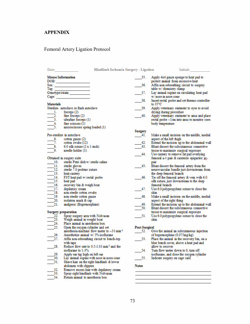
73
APPENDIX
Femoral Artery Ligation Protocol

74
Functional Vasodilation Protocol

75

76
Superfusion Protocol

77

78

79
Gracilis Anterior Dissection Protocol

80
Gracilis Whole Mount Immunostaining Protocol