the impact of retrotransposons on human genome evolution
TRANSCRIPT
Transposable elements (TEs; also known as ‘jumping
genes’) are discrete pieces of DNA that can move within
(and sometimes between) genomes. Although they were
discovered in the 1940s1, it was approximately half a
century before scientists began to understand how TEs
interact with their genomic environment. Crucially, the
completion of the first human genome sequence revealed
that nearly half of our genome is derived from TEs2,3
(FIG. 1a). This is likely to be an underestimate, as many
ancient TEs in the human genome have probably diverged
beyond recognition3. The scale of the contribution of TEs
to the human genome is all the more remarkable when
one considers that protein-coding regions account for
just 1.5% of the human genome3.
TEs can be separated into two major classes: DNA
transposons and retrotransposons. DNA transposons,
which constitute ~3% of the human genome (FIG. 1a),
can excise themselves from the genome, move as DNA
and insert themselves into new genomic sites4. Although
DNA transposons are currently not mobile in the human
genome, they were active during early primate evolution
until ~37 million years (Myr) ago5. Retrotransposons
duplicate through RNA intermediates that are reverse
transcribed and inserted at new genomic locations4.
Retrotransposons can be subdivided into two groups
distinguished by the presence or absence of long terminal
repeats (LTRs). Human LTR elements are endogenous
retroviruses (HERVs), which along with related elements
account for ~8% of the genome (FIG. 1a). Most HERVs
inserted into the human genome >25 Myr ago, and their
activity is presently very limited in humans, if it occurs at
all3,6. By contrast, the majority of human TEs result from
the present and past activity of non-LTR retrotransposons,
including the LINE-1 (long interspersed element 1,
abbreviated here to L1), Alu and SVA elements, which
collectively account for approximately one-third of the
human genome3 (FIG. 1a). L1, Alu and SVA non-LTR
retrotransposons are the only TEs that have unequivo-
cally been shown to be currently active in humans, as
indicated by the more than 60 reported cases of de novo
insertions that are responsible for genetic disorders7–11.
The extremely high density of TEs in our genome
poses the question: what impact have they had on
human evolution? The development of innovative
molecular methodologies — such as retrotransposition
assays in cultured cells12,13 and computational techniques
for comparative genomics — in conjunction with the
sequencing of multiple primate genomes (such as the
human3, chimpanzee14 and rhesus macaque15 genomes)
has resulted in a progressive shift in the focus of TE
research towards investigating the extent of the impact of
TE activity on genomic evolution. Important discoveries
regarding how TEs affect human genome evolution have
recently made it possible to quantify the overall impact
that TE activity has had on our genome. For example,
although it has long been recognized that recombination
between TEs can trigger genomic deletions in humans
(these deletions have caused several genetic disorders8),
only recently have genome-wide comparisons of human
and other primate genomes allowed us to determine the
*Université de Poitiers, CNRS UMR 6556 Ecologie, Evolution, Symbiose, 40 Avenue du Recteur Pineau, 86022 Poitiers, France.‡Department of Biological Sciences, Louisiana State University, 202 Life Sciences Building, Baton Rouge, Louisiana 70803, USA.Correspondence to M.A.B. e-mail: [email protected]:10.1038/nrg2640
Long terminal repeatsSequences of 300–1,000 bp
that are directly repeated
at the 5 and 3 ends of
long terminal repeat
retrotransposons
and retroviruses.
SVA elementAn element that is made
up of a short interspersed
element (SINE) region, a
variable number of tandem
repeats (VNTR) region and
an Alu-like region.
The impact of retrotransposons on human genome evolutionRichard Cordaux* and Mark A. Batzer‡
Abstract | Their ability to move within genomes gives transposable elements an intrinsic propensity to affect genome evolution. Non-long terminal repeat (LTR) retrotransposons — including LINE-1, Alu and SVA elements — have proliferated over the past 80 million years of primate evolution and now account for approximately one-third of the human genome. In this Review, we focus on this major class of elements and discuss the many ways that they affect the human genome: from generating insertion mutations and genomic instability to altering gene expression and contributing to genetic innovation. Increasingly detailed analyses of human and other primate genomes are revealing the scale and complexity of the past and current contributions of non-LTR retrotransposons to genomic change in the human lineage.
R E V I E W S
NATURE REVIEWS | GENETICS VOLUME 10 | OCTOBER 2009 | 691
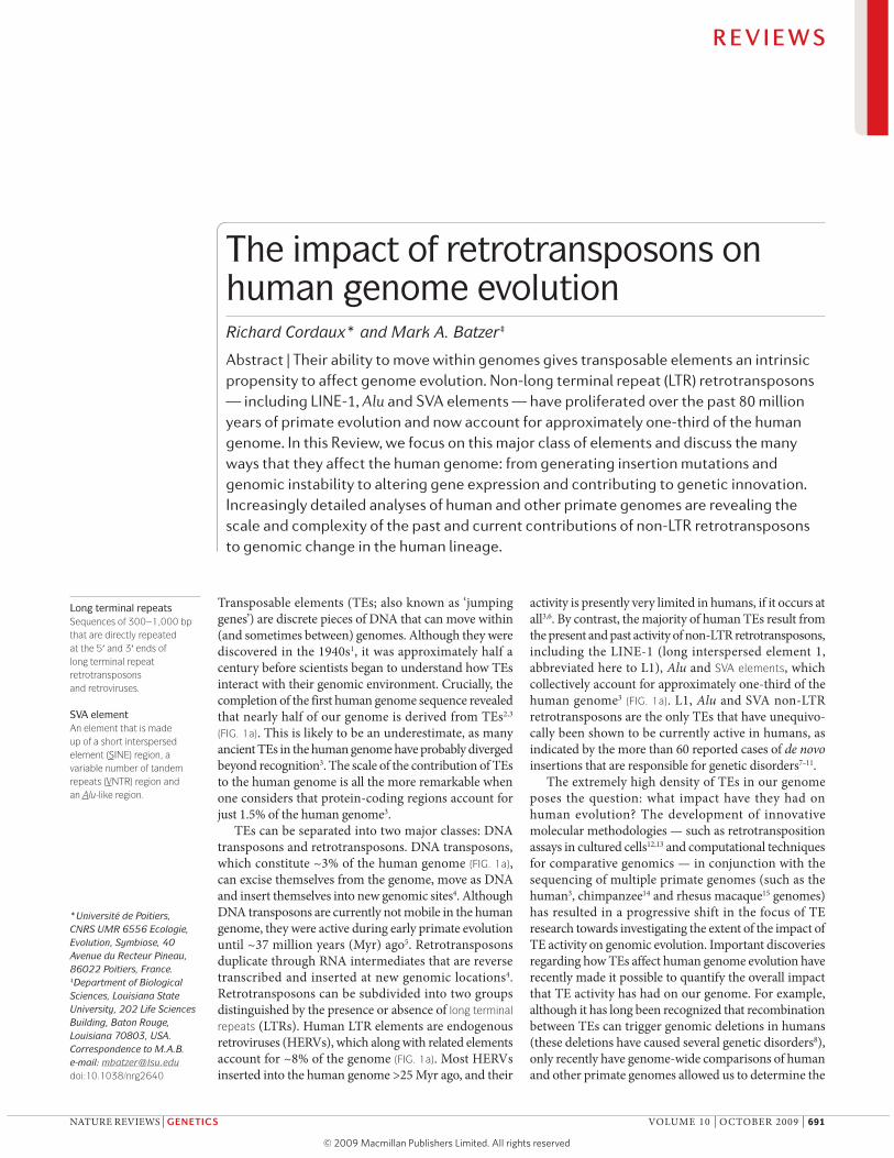
Non-LTRretrotransposons
DNA transposons2.8%
Non-transposable elements (~55%)
L116.9%
Alu10.6%
SVA0.2%
Others6.0%
LTR retrotransposons8.3%
VNTR HERV-K10-like AAA
Alu
3 UTRORF1 ORF25 UTR AAA
Right monomerLeft monomer A-rich AAA
Alu-like(CCCTCT)n
a
b
L1 pA
SVA pA
Figure 1 | The transposable element content of the human genome. a | Approximately 45% of the human genome can currently be recognized as being derived from transposable elements, the majority of which are non-long terminal repeat (LTR) retrotransposons, such as LINE-1 (L1), Alu and SVA elements. b | The canonical L1 element consists of two open reading frames (ORF1 and ORF2) flanked by 5 and 3 UTRs. The 5 UTR possesses an internal RNA polymerase II promoter (blue box). The element ends with an oligo(dA)-rich tail (AAA) preceded by a polyadenylation signal (pA). The canonical Alu element consists of two related monomers separated by an A-rich linker region (with consensus sequence A
5TACA
6). The left monomer contains A and B boxes
(blue boxes), which are transcriptional promoters for RNA polymerase III. The element ends with an oligo(dA)-rich tail (AAA) that can be up to 100 bp long. The canonical SVA element has a composite structure consisting of (from the 5 end to 3 end): a (CCCTCT)
n hexamer repeat
region; an Alu-like region consisting of two antisense Alu fragments and an additional sequence of unknown origin; a variable number of tandem repeats (VNTR) region made of units 35–50 bp in length; and a region derived from the envelope polyprotein (env) gene and the 3 LTR of human endogenous retrovirus (HERV)-K10. The element ends with an oligo(dA)-rich tail preceded by a polyadenylation signal. L1, Alu and SVA elements are typically flanked by target site duplications (black arrows) that are generated upon integration. Elements are not drawn to scale.
magnitude and significance of TE recombination-mediated
deletions at an evolutionary scale16–18.
In this Review, we focus on how the abundance and
activity of non-LTR retrotransposons has affected recent
human evolution. First, we describe the structure of non-
LTR retrotransposons and the mechanisms by which they
move. Second, we explore the evolutionary dynamics of
non-LTR retrotransposons — that is, what has made
them so evolutionarily successful in the human genome.
Addressing this question may help us to understand how
and to what extent TEs — and non-LTR retrotrans-
posons in particular — have affected human genome
evolution. The effects of TEs range from local instability
to large-scale structural variation. TEs not only contrib-
ute to genetic innovation but also alter gene expression.
We conclude with potential future research directions.
Human non-LTR retrotransposonsL1 elements. There are >500,000 L1 copies in the human
genome as a result of their continued mobilization activ-
ity over the past 150 Myr3. L1 elements constitute ~17%
of the human genome, which makes them the most suc-
cessful TEs in the human genome by mass (FIG. 1a). The
canonical, full-length L1 element is ~6 kb long and con-
sists of a 5 UTR containing an internal RNA polymer-
ase II (RNAPII) promoter19, two open reading frames
(ORF1 and ORF2) and a 3 UTR containing a poly-
adenylation signal ending with an oligo(dA)-rich tail of
variable length20 (FIG. 1b). ORF1 encodes an RNA-binding
protein and ORF2 encodes a protein with endonuclease
and reverse-transcriptase activities20. This molecular
machinery allows the retrotransposition process known
as target-primed reverse transcription (TPRT) to occur
(BOX 1), therefore making L1 elements the only autono-
mous TEs in the human genome. However, not all L1
copies are competent for retrotransposition. Indeed, as a
result of TPRT and decay over time, most L1 copies are
inactivated by truncations, internal rearrangements and
mutations3,21. Of the >500,000 L1 elements in the human
genome, less than 100 copies are functional22.
Alu elements. There are >1 million Alu copies in the
human genome3 as a result of their continued mobiliza-
tion activity over the past ~65 Myr23. This makes Alu
elements the most successful TEs in the human genome
in terms of copy number. The typical full-length Alu
element is ~300 bp long and has a dimeric structure
formed by the fusion of two monomers derived from
the 7SL RNA gene24 (a component of the signal recogni-
tion particle). The monomers are separated by an A-rich
linker region (FIG. 1b). The 5 region contains an inter-
nal RNA polymerase III (RNAPIII) promoter (A and
B boxes) and the element ends with an oligo(dA)-rich
tail of variable length23. As Alu elements do not possess
RNAPIII termination signals, Alu transcripts extend into
the downstream flanking sequence until a terminator
(typically a run of four or more consecutive thymines) is
found25,26. Alu elements have no coding capacity and are
therefore non-autonomous TEs. Instead, they make use
of the retrotransposition molecular machinery encoded
by L1 elements12, which is the reason why Alu elements
are sometimes referred to as ‘a parasite’s parasite’ (REF. 27).
However, L1 ORF1 and ORF2 proteins show a strong
cis-preference for L1 RNA28 (BOX 1).
R E V I E W S
692 | OCTOBER 2009 | VOLUME 10 www.nature.com/reviews/genetics

TSD
AATTTT 5
b Priming and reverse transcription
d DNA synthesis
e TSD formation
5
5
TTTTA
AAA
5
TTTTA
AAA
HOHO
5
5
5a First strand nick
5
5
c Second strand cleavage 5
TSD
SVA elements. SVA elements have been active throughout
the ~25 Myr of hominoid evolution, and there are now
~3,000 copies in the human genome29,30. A typical full-
length SVA element is ~2 kb long and is composed of a
hexamer repeat region, an Alu-like region, a region con-
sisting of a variable number of tandem repeats, a HERV-
K10-like region and a polyadenylation signal ending
with an oligo(dA)-rich tail of variable length29,30 (FIG. 1b).
Several lines of evidence suggest that SVA elements are
transcribed by RNAPII29,30. However, they apparently
contain no internal promoter and might rely, at least
in part, on promoter activity in flanking regions29,30.
Like Alu elements, SVA elements are non-autonomous
TEs that are presumably trans-mobilized by the L1
retrotransposition machinery29,30 (BOX 1).
Other non-LTR retrotransposons. In addition to the L1,
Alu and SVA elements, which are currently active, there
are families of old, inactive non-LTR retrotransposons
that comprise ~6% of the human genome (FIG. 1a).
Although they are far less numerous than L1 and Alu
elements, these elements provide a rich molecular ‘fos-
sil record’ that testifies to the long relationship between
TEs and the human genome3. This record indicates, for
example, that before the autonomous L1 element and its
Alu parasite expansions, the genome experienced retro-
transposition of the autonomous LINE-2 element and its
mammalian-wide interspersed repeat (MIR) parasite3.
These old elements might have substantially affected
human genome evolution31–34.
Evolutionary dynamicsThe impact of non-LTR retrotransposons on human
genome evolution largely results from their extremely
high copy numbers (for example, there is one Alu inser-
tion every 3 kb on average3) and their continued activ-
ity over tens of millions of years. These two features
are particularly striking when considering the various
cellular processes that control retrotransposon activity
(BOX 2). At an evolutionary scale, the vertical persistence
of non-LTR retrotransposons, not only in primates but
also in mammals in general, sets them apart from most
other TEs in mammals and other eukaryotes3,5,35. In
this section, we discuss the evolutionary dynamics that
have made non-LTR retrotransposons so prolific during
primate genome evolution.
Subfamily structure and source elements. A key con-
cept that is relevant to the evolutionary dynamics of L1,
Alu and SVA sequences is that they can all be divided
into subfamilies or ‘clades’ of related elements based
on diagnostic nucleotide substitutions and insertions
or deletions that are exclusively shared by all subfamily
members. For example, more than 200 Alu subfamilies
are currently recognized in the human genome36 but only
6 subfamilies of the younger SVA family exist30. Not
only are subfamilies different in age, but the diagnostic
sequence mutations or changes that define subfamilies
tend to accumulate hierarchically23,37; that is, instead
of two subfamilies being independently derived from
an ancestral subfamily, most subfamilies represent an
Box 1 | The retrotransposition cycle
The increase in copy numbers of non-long terminal repeat (LTR) retrotransposons occurs through an RNA-based duplication process termed retrotransposition. The first step in LINE-1 (L1) retrotransposition involves RNA polymerase II-mediated transcription of a genomic L1 locus from an internal promoter that directs transcription initiation at the 5 boundary of the L1 element19,129; an internal promoter allows a retrotransposon to generate autonomous duplicate copies at multiple locations in the genome. The L1 RNA is exported to the cytoplasm, in which ORF1 (which encodes an RNA-binding protein) and ORF2 (which encodes a protein with endonuclease and reverse-transcriptase activities) are translated. Both proteins show a strong cis-preference28; consequently, they preferentially associate with the L1 RNA transcript that encoded them to produce a ribonucleoprotein (RNP) particle. The RNP is then transported back into the nucleus by a mechanism that is poorly understood.
The integration of the L1 element into the genome is likely to occur through a process termed target-primed reverse transcription (TPRT)13,130,131, which was originally described for the R2 non-LTR retrotransposon of the silkworm Bombyx mori132. During TPRT, it is thought that the L1 endonuclease cleaves the first strand of target DNA, generally between T and A at 5 -TTTTAA-3 consensus sites133 (see the figure, part a). The free 3 hydroxyl (OH) generated by the nick is then used to prime reverse transcription of L1 RNA (red) by the L1 reverse transcriptase (b). The second strand of the target DNA is cleaved (c) and used to prime second-strand synthesis (d) through poorly understood mechanisms. Hallmarks of the integration process include frequent 5 truncations, the presence of an oligo(dA)-rich tail at the 3 end and target site duplications (TSDs) of between 2 and 20 base pairs in length3,21 (e).
Alu and SVA retrotransposition is also likely to occur through TPRT using the L1 retrotransposition machinery12,29,30. The mechanism of Alu and SVA trans-mobilization by L1 proteins remains elusive. RNA polymerase III-mediated Alu transcripts are exported to the cytoplasm and bound to signal recognition particle 9 kDa protein (SRP9) or SRP14 to form stable RNPs134,135. It has been suggested that Alu RNPs interact with ribosomes, thereby positioning Alu transcripts in close proximity to nascent L1 ORF2 proteins12,42 (the ORF1 protein enhances, but is not strictly required for, Alu retrotransposition12,136). However, it remains unclear whether Alu RNPs gain access to the L1 retrotransposition machinery in the cytoplasm or in the nucleus, as Alu RNPs might recruit L1 ORF2 proteins in the nucleus and immediately proceed with TPRT137.
R E V I E W S
NATURE REVIEWS | GENETICS VOLUME 10 | OCTOBER 2009 | 693

Box 2 | Cellular factors that influence retrotransposition
Transposable elements (TEs) can be seen as selfish genetic entities, the spread of which can be deleterious to the host cell due to the genomic instability that is induced by a massive increase in copy number. As a result of the conflicting interests of TEs and the host genome, the cell has developed various processes to control retrotransposon activity, as predicted by the Red Queen hypothesis138. Below we provide examples of how LINE-1 (L1) and Alu retrotransposition activity is regulated in host cells (for more detailed discussions, see REFS 11,139).
Regulation of L1 retrotransposition can occur at the transcriptional level — for example, new regulatory regions have frequently been recruited during the evolution of L1 (REF. 38). The current L1 5 UTR contains several transcription factor-binding sites that are important for transcription activation or initiation140–142. In addition, DNA methylation at the promoter is known to repress L1 expression124,143. L1 elements are also subject to post-transcriptional regulation. For example, RNA-induced silencing through RNA interference has been suggested to reduce L1 retrotransposition in cultured cells144,145. The A-rich coding strand of the full-length human L1 contains 19 potential canonical and non-canonical polyadenylation signals that lead to truncation of full-length L1 transcripts by premature polyadenylation, therefore contributing to the attenuation of L1 activity110. Furthermore, cells produce proteins, such as those of the apolipoprotein B mRNA-editing complex 3 (APOBEC3) family, that can inhibit L1 and Alu retrotransposition146.
Alu activity is influenced by its primary sequence in that the accumulation of mutations can alter important motifs, such as the internal RNA polymerase III promoter or signal recognition particle 9 kDa protein (SRP9)- or SRP14-binding motifs26,42. The accumulation of mutations is facilitated by the high density of CpG dinucleotides, which are prone to mutation as a result of the deamination of 5-methylcytosine residues125. Overall, it has been estimated that when an Alu copy reaches ~10% divergence from its subfamily consensus sequence, the likelihood that it will continue to be active is remote42. The length and homogeneity of the oligo(dA)-rich tail also seem to be important for activity147,148. The genomic environment into which Alu copies are inserted is crucial for retrotranspositional activity149–151, and the distance between the oligo(dA)-rich tail at the 3 end of the Alu sequence and the RNA polymerase III terminator, which is located in the downstream sequence and determines the overall length of Alu transcripts26, is also important.
HominoidsThe group of primates
comprised of humans and
apes. Hominoids diverged
from Old World monkeys
approximately 25 million
years ago.
Trans-mobilizationThe process by which
non-autonomous
retrotransposons, such as Alu
and SVA elements, borrow the
LINE-1 retrotransposition
machinery to perform their
own retrotransposition.
Red Queen hypothesisProposed by Van Valen in
1973, this hypothesis states
that, for an evolutionary
system, continuing
development is needed to
maintain its fitness relative
to the systems it is
co-evolving with.
ongoing linear sequential evolution pattern in which
one subfamily is derived successively from another. For
example, it has been shown that all L1 subfamilies in
the human genome were derived sequentially from a
single lineage over the past ~40 Myr38. Similar patterns
of subfamily evolution have been reported for Alu23 and
SVA30 elements. These observations can be explained if
one assumes that only a few elements (so-called ‘source’
or ‘master’ elements) are involved in the retrotransposi-
tion process and are responsible for the formation of all
other subfamily members37.
The original ‘master gene’ model of retrotransposon
amplification37 has been refined, in particular by quan-
tifying the number of retrotransposition-competent ele-
ments in the human genome. Analysis of the >200 Alu
subfamilies in the human genome suggested the exist-
ence of at least 143 Alu source elements36, and it has been
estimated that an average human genome carries 80–100
retrotransposition-competent L1 copies, 6 of which —
known as ‘hot L1s’ — are probably responsible for the
majority of L1 retrotransposition22,39,40. These results
further indicate that several source elements may exist
within a subfamily, because all 6 hot L1 elements belong
to the L1-Ta subfamily22. A network-based analysis also
revealed that human-specific Alu subfamilies typically
contain secondary source elements in addition to a mas-
ter element. Secondary source elements comprise ~15%
of subfamily members and give rise to approximately
30% of all subfamily copies41. Therefore, there may be
hundreds of active Alu ‘core’ sequences in the human
genome42. Although they only represent a tiny fraction of
all human non-LTR retrotransposons, source elements
can be considered as the ultimate drivers of evolutionary
change in the human genome because they are respon-
sible for most of the L1, Alu and SVA elements that have
been inserted into our genome.
Stealth drivers and long-term evolution. Another dis-
tinguishing feature of human non-LTR retrotransposons
is their persistent activity over tens of millions of years
of evolution. How have active retrotransposons been
maintained over this time? Reconstruction of the evo-
lutionary history of the Alu Yb lineage showed that it
originated during early hominoid evolution, 18–25 Myr
ago43. Strikingly, the Alu Yb lineage has dramatically
expanded to ~2,000 copies over the past few million
years specifically in the human genome, as shown by
the finding that non-human hominoid primates carry
only a few Alu Yb elements43–45. Therefore, the Alu Yb
lineage remained in the genome with little or no retro-
transposition for 15–20 Myr while preserving the ability
to generate a high number of new copies in a species-
specific manner. These results suggest that long-lived,
low-activity source elements may act as ‘stealth drivers’
that occasionally produce copies, some of which can
become highly active. Whereas highly active master
elements might be deleterious and negatively selected,
low-activity stealth drivers might allow the Alu lineage
to persist in the long term43. Attenuation of mobiliza-
tion activity might be a common evolutionary strategy
of various retrotransposons46,47; therefore, the abil-
ity to maintain low to moderate levels of retrotrans-
position activity might be an important feature that
allowed human non-LTR retrotransposons to maintain
long-term activity.
Impact on genome evolutionAmplification rates. Because of their continued activity
and accumulation in the genome over tens of millions of
years, L1, Alu and SVA elements have had a tremendous
impact on the evolution of primate genomes in terms of
both structure and function. To assess this impact, one
can consider how frequently retrotransposition occurs
in the germ line. The current rate of Alu retrotransposi-
tion has been estimated as approximately 1 insertion for
every 20 births in humans, which is based both on the
frequency of disease-causing de novo insertions com-
pared with nucleotide substitutions48 and on compari-
sons between the human and chimpanzee genomes48
and between multiple human genome sequences49. The
current rate of L1 retrotransposition has also been esti-
mated as approximately 1 insertion for every 20 births
in humans based on disease-causing de novo inser-
tions50, but as approximately 1 insertion for every 200
births based on genome comparisons49. The difference
between the two estimates might lie in the underlying
assumptions of the methods used, but no such bias is
observed for Alu element insertion estimates based on
R E V I E W S
694 | OCTOBER 2009 | VOLUME 10 www.nature.com/reviews/genetics

Box 3 | Retrotransposon insertion polymorphisms as genetic markers
As revealed by pioneering studies on humans152–154, primates155 and non-primate groups156,157, retrotransposons provide several advantages that make them very powerful tools as genetic markers for studying human and non-human primate evolutionary history23,157,158. They are essentially homoplasy-free markers, as individuals that share retrotransposon copies at orthologous sites are almost certain to have inherited them from a common ancestor (the precise excision of retrotransposons is extremely rare)158,159. When comparing genomes, the absence of an element at a locus indicates that the individual carries an ancestral version of that locus, and this makes it possible to include hypothetical ancestors to root phylogenetic trees153. As there are only two possible character states for each locus — the presence or absence of the element — genotyping of individuals for retrotransposon insertions is technically straightforward. Furthermore, the vast majority of retrotransposon insertions are neutral residents of the genome160, and the gradual accumulation of elements over time makes it possible to determine the loci that are most suitable for investigating evolutionary relationships at a range of time points in primate history. As a result, retrotransposon insertion polymorphisms (most notably Alu elements) have been used to decipher the phylogenetic relationships of various primate groups161,162, including the resolution of the human–chimpanzee–gorilla trichotomy that demonstrated the close relationship between humans and chimpanzees163.
Some retrotransposons were inserted so recently that they are polymorphic for presence or absence among human populations and individuals23,49,164. In particular, Alu elements have proved highly informative for the study of human origins by providing strong evidence for an African origin of anatomically modern humans153,154. More recently, Alu element insertion polymorphisms have been used to investigate human population structure and demography154,165,166. Retrotransposon insertion polymorphisms are also being used as forensic tools — for example, for species-specific DNA detection and quantification, for the analysis of complex biomaterials, for human gender determination and for the inference of geographic origin of human samples167.
HomoplasySimilarity due to independent
evolutionary change — that is,
not inherited from a common
ancestor.
X inactivationThe process by which, in
female mammals, one of
the two copies of the
X chromosome is inactivated
during early embryogenesis.
The inactive X chromosome is
silenced by being packaged
into transcriptionally inactive
heterochromatin.
the same approaches. Alternatively, the difference might
reflect recent variation in the L1 retrotransposition rate
or intense negative selection against L1 insertions. The
current SVA retrotransposition rate has tentatively
been estimated as approximately 1 insertion for every
900 births based on genome comparisons49; this rate is
more uncertain owing to the smaller data sets available
for analysis. Although new heritable retrotransposition
events take place in the germ line, retrotransposition also
occurs in somatic tissues and has been implicated
in processes ranging from cancer to brain develop-
ment8,51,52. Retrotransposon-induced somatic variation
is a fascinating area of investigation that is likely to
provide new insights into the biology of TEs and their
impact on humans.
The amplification rates of TEs have not been uniform
over time. For example, the majority of Alu elements
were inserted ~40 Myr ago following a peak of amplifi-
cation during which there was approximately one new
Alu insertion in every birth53. Similarly, over the past
~70 Myr of evolution, variation in the L1 amplification
rate has been observed, and the most prolific L1 sub-
families were amplified 12–40 Myr ago38. Genome-wide
comparisons of the human and chimpanzee genomes
have provided additional evidence for recent variation
in L1, Alu and SVA retrotransposition rates, as judged by
the different numbers of species-specific elements that
have been inserted since the divergence of the two spe-
cies ~6 Myr ago14,54,55. Such fluctuation in amplification
rates over a short timescale suggests influences at the
host population level40,54.
Changes in copy number. Perhaps one of the most intuitive
consequences of TE accumulation is their contribu-
tion to increases in genome size56: L1 and Alu elements
alone have contributed ~750 Mb to the human genome3
(FIG. 1a). This increase in genome size is an ongoing proc-
ess, as the human genome has accumulated ~2,000 L1,
~7,000 Alu and ~1,000 SVA copies over the past ~6 Myr
of human evolution, which is a combined addition of
>8 Mb14. Equally importantly, the ongoing expansion
of non-LTR retrotransposons has also created sig-
nificant inter-individual variation in retrotransposon
content; several hundred new mobile element inser-
tions have been detected in multiple human genomic
sequences49,57–59. These human-specific retrotransposon
insertions are often polymorphic (present or absent) at
orthologous loci among human individuals, and they
constitute highly informative genetic markers that are
being used to investigate human evolutionary history,
population structure and demography (BOX 3).
Local genomic instabilityRetrotransposons can generate genomic instability in
many ways. In this section, we consider the effects of
retrotransposons at a local genomic scale.
Insertion mutagenesis. The most straightforward way a
retrotransposon can alter genome function, and thereby
potentially influence genome evolution, is by inserting
into protein-coding or regulatory regions (FIG. 2a). Owing
to the immediate phenotypic impact of such insertions,
they were the first to be detected7. Examples of human
genetic disorders caused by de novo L1, Alu and SVA
insertions continue to accumulate, and 65 cases have been
shown to cause heritable diseases, such as haemophilia,
cystic fibrosis, Apert syndrome, neurofibromatosis,
Duchenne muscular dystrophy, β-thalassaemia,
hypercholesterol aemia and breast and colon cancers8,9,11.
Overall, it has been estimated that ~0.3% of all human
mutations are attributable to de novo L1, Alu and SVA
insertions10. Interestingly, L1 (and to a lesser extent Alu
and SVA) disease-causing insertions seem to be enriched
on the X chromosome8,9,11. This might partly be attrib-
utable to ascertainment bias, as X-linked genetic dis-
orders are often dominant in males and are thus more
easily detected. Alternatively, L1 elements might pref-
erentially insert into the X chromosome. One possible
explanation is that the preference might be linked to a
proposed involvement of L1 elements in X inactivation60,61,
in which they might help to spread silencing signals.
DNA double-strand breaks. It has recently been shown
in mammalian cell lines that the number of DNA
double-strand breaks (DSBs) generated by L1 ORF2 pro-
teins, which have endonuclease activity, is much higher
than the number associated with actual L1 insertions62
(FIG. 2b). The extent to which these DSBs contribute to
human genomic instability remains unknown because
levels of L1 expression under these experimental con-
ditions were much higher than those expected under
normal cellular conditions. However, the repair of
L1-mediated DSB lesions would leave no signature of L1
R E V I E W S
NATURE REVIEWS | GENETICS VOLUME 10 | OCTOBER 2009 | 695

(TA)n
b Creating and repairing DNA double-strand breaks
c Microsatellite seeding d Gene conversiona Insertional mutagenesis
e Insertion-mediated deletions f Ectopic recombination g Transduction
Figure 2 | Impact of retrotransposons on human genome structure. a | Typical insertion of a LINE-1 (L1), Alu or SVA retrotransposon (red box) at a new genomic site (dark grey). If the new genomic site is a genic region, the retrotransposon may cause insertional mutagenesis. b | The protein product (green oval) of an L1 element may create DNA double-strand breaks (broken dark grey area). Alternatively, an existing double-strand break may be repaired by non-classical endonuclease-independent insertion of a retrotransposon. c | Microsatellites (for example, (TA)
n) may arise from the homopolymeric tracts that are endogenous to retrotransposons. d | Gene
conversion may alter the sequence compositions of homologous retrotransposon copies (red and blue boxes). e | The insertion of a retrotransposon is sometimes associated with the concomitant deletion of a target genomic sequence (light grey box). f | Ectopic recombination (double arrowhead) between non-allelic homologous retrotransposons may result in genomic rearrangements, such as deletions (left) or duplications (right) of intervening genomic sequences. g | During the duplication of a retrotransposon, the downstream 3 flanking sequence or the upstream 5 flanking sequence (dark grey boxes) may also be duplicated (known as 3 or 5 transduction, respectively). This results in the retrotransposition of the 3 flanking sequence (left) or the 5 flanking sequence (right) along with the retrotransposon.
Homopolymeric tractA DNA sequence made of the
same nucleotide repeated in
tandem.
MicrosatelliteA class of repetitive DNA made
up of tandem repeats that are
1–8 bp in length.
involvement, so it is possible that a substantial fraction
of the genomic instability associated with DSBs, which
are highly mutagenic and prone to recombination, is
attributable to L1 activity.
L1 and Alu elements have been linked to DSB repair.
Evidence from L1 retrotransposition assays in cul-
tured cells showed that L1 insertions can occur inde-
pendently of endonuclease in mammalian cell lines
that cannot perform non-homologous end joining,
which is a major mechanism of DSB repair63 (FIG. 2b).
Endonuclease-independent (ENi) L1 insertions lack
the hallmarks of TPRT (BOX 1), which suggests that L1
elements can integrate into and repair DSBs63. In addi-
tion, dysfunctional telomeres can serve as substrates
for ENi L1 retrotransposition, and endonuclease-
deficient LINE-like (Penelope) elements are present at
the telomeres of several eukaryotes, which suggests that
ENi retrotransposition might be an ancestral mecha-
nism of RNA-mediated DNA repair that was used before
non-LTR retrotransposons acquired an endonuclease
domain64,65. Recent analyses of the human genome have
shown that 0.5–0.7% of all L1 and Alu insertions
have non-canonical structures and might have resulted
from ENi retrotransposition66,67, which suggests that
non-LTR retrotransposons in general, not just L1 ele-
ments, might provide an additional mechanism for
maintaining human genome integrity.
Sources of microsatellites. Because of their abundance
in the genome and because they contain homopolymeric
tracts, non-LTR retrotransposons can generate microsat-
ellites at many loci in the genome (FIG. 2c). In particular,
this has been studied for Alu elements68,69, each new
copy of which provides two potential sources of micro-
satellites: the linker region in the middle of the element
and the 3 oligo(dA)-rich tail (FIG. 1b). These homopoly-
meric repeats can be subject to mutations, such as
nucleotide substitutions and replication slippage, which
can produce microsatellites of varying length and com-
plexity. Consequently, it is not surprising that ~20% of
all microsatellites (including ~50% of mononucleotide
R E V I E W S
696 | OCTOBER 2009 | VOLUME 10 www.nature.com/reviews/genetics

Identical by stateAlleles that have the same
character state as a result of
independent evolutionary
changes (that is, the alleles
were not inherited from a
common ancestor).
Identical by descentAlleles that have the same
character state as a result
of being directly inherited
from a common ancestor.
microsatellites) shared by the human and chimpanzee
genomes lie within Alu elements70. In addition, there
are at least two examples of genetic disorders that are
caused by the expansion of microsatellites that arose
from A-rich regions of Alu elements71,72.
Gene conversion. Several studies indicate that Alu
elements undergo gene conversion73,74 (FIG. 2d), which
is a type of recombination that is defined as the non-
reciprocal transfer of information between homologous
sequences. Gene conversion might play a part in the
evolution of Alu elements by inactivating active copies
or reactivating inactivated copies23. For example, it has
recently been shown that the master element of the Alu
Yh3a3 subfamily has been inactivated by gene conver-
sion in humans, therefore preventing further ampli-
fication of this subfamily75. In addition, because Alu
elements make up >10% of the human genome,
Alu-mediated gene conversion might have a substan-
tial impact on the overall nucleotide diversity of our
genome. Also, it might impair the use of SNPs located
within Alu sequences as genetic markers, as gene con-
version would make these SNPs identical by state rather
than identical by descent23. However, the significance of
this phenomenon has not been tested formally; next-
generation sequencing and personal genomics will open
new avenues for resolving this question.
Genomic rearrangementsIn addition to generating local genomic instability,
retrotransposons can generate genomic rearrangements,
such as deletions, duplications and inversions. In this
section, we discuss three ways in which retrotransposons
can create structural variation in the genome.
Insertion-mediated deletions. The insertion of L1 and
Alu elements at new genomic sites sometimes results
in the concomitant deletion of an adjacent genomic
sequence (FIG. 2e). This phenomenon was first observed
through the analysis of L1 integrations in cultured
human cells: ~20% of L1 insertions were associated
with structural rearrangements, including concomitant
deletions at the insertion site that ranged in size from
1 bp to possibly >130 kb76–78. These deletions can arise
by endonuclease-dependent and ENi mechanisms78. L1
and Alu insertion-mediated deletions have subsequently
been shown to occur naturally in the human and chim-
panzee genomes, although the deletions are usually
shorter (<800 bp on average) and occur at a much
lower frequency than in cultured cells (a frequency of
~2% and ~0.3% for L1 and Alu insertion events, respec-
tively)79,80. This might reflect, at least partly, negative
selection against large, disruptive, insertion-mediated
deletions. Consistent with these observations, a 46 kb
L1 insertion-mediated deletion event in the pyruvate
dehydrogenase complex, component X (PDHX) gene
has recently been implicated in pyruvate dehydrogenase
complex deficiency81, and human–chimpanzee genome
comparisons have identified a single insertion-mediated
deletion event that caused loss of a functional gene in
the past ~6 Myr79.
It has also been noted that ~90% of non-classical
ENi L1 and Alu insertions are associated with deletions
of flanking sequences that range in size from 1 bp to
14 kb, including one deletion that removed an olfac-
tory receptor gene from the human and chimpanzee
genomes66,67. Altogether, it has been estimated that
during primate evolution, as many as 45,000 insertion-
mediated deletions might have removed >30 Mb of
genomic sequence18.
Ectopic recombination. Due to their extremely high
copy numbers, L1 and Alu elements can also create
structural genomic variation at the post-insertion stage
through recombination between non-allelic homo-
logous elements (FIG. 2f), including between elements
that have been present in the genome for a long time.
Ectopic recombination can result in various types of
genomic rearrangements, such as deletions, duplications
and inversions.
It has long been recognized that Alu recombination-
mediated deletions (RMDs) occur in the human
genome: there are >70 reported cases of Alu RMDs being
responsible for various cancers and genetic disorders8,10.
By contrast, only three disease-causing L1 RMD events
have been reported17. Genome-wide comparisons have
identified 492 Alu RMD events and 73 L1 RMD events
that have taken place in the human genome since the
human–chimpanzee divergence16,17. L1 RMDs are larger
on average than Alu RMDs and occur more frequently
in gene-poor regions of the genome. These results
suggest that there might be negative selection against
long, deleterious L1 RMDs in gene-rich regions of the
genome18,82,83. Therefore, Alu and L1 RMD events that are
detectable by comparative genomics approaches
are likely to represent the fraction of RMDs that have
escaped negative selection. However, based on human
and chimpanzee genome comparisons, these events
have collectively removed nearly 1 Mb of genomic
sequence from the human genome over the past few
million years16–18, thereby underscoring their important
evolutionary impact.
The human genome contains many large (>10 kb)
and highly similar (>90% sequence identity) duplicated
genomic regions, which are termed segmental duplica-
tions. Interestingly, the boundaries of human segmental
duplications are significantly enriched in Alu elements
— that is, they comprise ~24% of boundary sequences
but only ~11% of the total human genome84. Considering
that ~5% of the human genome has been duplicated in
the past ~40 Myr, recombination between Alu elements
might represent an important mechanism for the origin
and expansion of human segmental duplications84.
The contribution of L1 and Alu elements to
chromosomal inversions has also been investigated
recently by comparative genomics. Nearly half of the
inversions that have taken place in the human and
chimpanzee genomes since their divergence have
involved L1 and Alu elements, and ~20% of all inver-
sions can clearly be identified as products of L1–L1 or
Alu–Alu recombination events85. Although this type of
rearrangement does not result in gain or loss of genomic
R E V I E W S
NATURE REVIEWS | GENETICS VOLUME 10 | OCTOBER 2009 | 697

(AAA)n
SVA insertion
AMAC1
Transduced sequence
SVA
(AAA)nAMAC1L1SVA
(AAA)nAMAC1L2SVA
(AAA)nSVA
SVA
SVA
(AAA)n
(AAA)n
Retrotransposition
b
Chr8
Chr18
Chr17_B
Chr17_Aa
Transcription
RNA splicing
Source AMAC geneAMAC1L3
sequence, it contributes to genomic variation and can
be of functional importance — for example, by causing
the inversion of exons85.
Transduction of flanking sequences. In addition to
duplicating themselves, L1 and SVA elements some-
times carry upstream or downstream flanking genomic
sequences with them (termed 5 and 3 transduction,
respectively) (FIG. 2g). In 3 transduction, the RNA tran-
scription machinery skips the weak retrotransposon
polyadenylation signal and terminates transcription
by using an alternative polyadenylation signal located
downstream in the 3 flanking sequence. Similarly, 5
transduction occurs when a promoter located upstream
of the retrotransposon is used to transcribe the sequence
down to the retrotransposon86,87. The transcript contain-
ing the retrotransposon, along with the extra genomic
sequence, is integrated into the genome through retro-
transposition. Initially characterized using cell culture-
based methods88, 3 transduction has subsequently
been shown to occur frequently in the human genome:
~10% of L1 and SVA insertions are associated with 3
transduction events30,89–91.
Genetic innovationVariation in the number of genes among species
indicates that new genes are continuously generated
over evolutionary time. Comparative genomic studies
have confirmed the notion of ‘evolutionary tinkering’92,
according to which new genes most commonly arise
by rearrangements between pre-existing genetic struc-
tures. In this section, we explore mechanisms by which
retrotransposons have fostered genetic innovations in
the human lineage.
Transduction-mediated gene formation. The process
of retrotransposon-mediated transduction (discussed
above) can lead to the duplication of coding sequences
that are located in the transduced flanking genomic
sequence. The potential of L1 retrotransposons to
mediate exon shuffling through 3 transduction has
been experimentally confirmed using cell culture
assays88. This mechanism has subsequently been shown
to have mediated the formation of a new gene family
during recent human evolution through multiple SVA-
mediated transduction events of the acyl-malonyl
condensing enzyme 1 (AMAC1) gene89 (BOX 4).
Gene retrotransposition. In contrast to transduction,
gene retrotransposition only duplicates gene sequences
and no retrotransposon sequence is co-duplicated in
the process. This is because gene retrotransposition is
based on the hijacking of the L1 retrotransposition
machinery by host mRNA transcripts93 in a similar way to
Alu and SVA retrotransposition. As a result, gene retro-
transposition generally does not duplicate upstream
regulatory regions; therefore, duplicated genes must
fortuitously acquire new regulatory regions to become
functional. Gene retrotransposition was long thought to
generate non-functional duplicate gene copies termed
retropseudogenes. However, genome-wide searches
Box 4 | Birth of a gene family by retrotransposon-mediated transduction
It has been experimentally shown using cell culture assays that LINE-1 (L1) retrotransposons can mediate exon shuffling by 3 transduction88. Subsequent analyses of the human genome have confirmed that L1-mediated transduction took place during human genome evolution and that it may account for 0.6–1% of human DNA3,90,91. However, whether it contributes to the evolution of new gene functions remains an open question. A recent analysis of SVA retrotransposons has demonstrated the evolutionary significance of retrotransposon-mediated 3 transduction by showing that SVA-mediated transduction is responsible for the creation of the acyl-malonyl condensing enzyme 1 (AMAC1) gene family, which has four members in the human genome89.
As part of a genome-wide analysis of SVA-mediated transduction, Xing et al.89 identified 143 events that transduced sequences ranging in size from a few dozen base pairs to almost two kilobase pairs. Interestingly, 3 transduced sequences located on chromosomes 8, 17 and 18 were found to originate from the same source locus that was located elsewhere on chromosome 17 (see the figure, part a). The flanking sequences of the original locus are shown as dark grey boxes and the sequences flanking the transduced loci are shown as light grey boxes. Target site duplications are shown as green arrows. SVA elements are shown as red bars, and the coding regions are shown as purple bars. SVA element oligo(dA)-rich tails are shown as ‘(AAA)n’. Analysis of the four paralogous sequences identified four copies of the AMAC1 gene. The ancestral AMAC1L3 gene copy at the source locus consisted of two exons separated by an intron. By contrast, the three transduced copies of AMAC1L3 (AMAC1, AMAC1L1 and AMAC1L2) were intronless as a result of the splicing of the intron during the retrotransposition process (b). Evolutionary analyses indicated that the three transduction events all took place ~7–14 Myr ago, as humans and African great apes share all four AMAC1 copies, whereas orangutans and other primate and non-primate species that have been analysed only possess the ancestral AMAC1L3 gene. Experimental studies indicated that, in addition to AMAC1L3, at least two of the three transduced AMAC1 genes are expressed in human tissues. RNA transcript sequence analyses of the expressed AMAC1 duplicates further revealed that the promoter sequence had been duplicated along with the AMAC1 coding sequence as part of the 3 transduction process. This indicates that retrotransposon-mediated gene transduction can duplicate not only coding regions of genes but also their regulatory regions; therefore, genes retain their functional potential after duplication, and retrotransposon-mediated duplication can lead to the rapid generation of functional gene families. Figure modified, with permission, from REF. 89 © 2006 National Academy of Sciences, USA.
R E V I E W S
698 | OCTOBER 2009 | VOLUME 10 www.nature.com/reviews/genetics

Nuclear retention
AAAA to I A to I
AAA
e RNA editing
c Modulation of gene expression
Attenuation
AAA Premature polyadenylation
f Epigenetic regulation
d Sense and antisense promoter effects
a Exonization and alternative splicing b Transcription elongation defects
Figure 3 | Impact of retrotransposons on human gene expression. a | A retrotransposon sequence (red box) can be recruited as a coding sequence and be integrated into a gene (made up here of two exons, grey boxes). This is often associated with alternative splicing (dashed lines). b | The presence of a retrotransposon in the intron of a gene (the sequence between the two grey boxes, which represent exons) can result in transcription elongation defects, such as attenuation or premature polyadenylation. c | Retrotransposons carry transcription factor-binding sites. A transcription factor (green oval) carried by a retrotransposon can upregulate or downregulate (curved arrow) the expression (horizontal arrow) of neighbouring genes (grey boxes). d | A retrotransposon carries sense and antisense promoters (horizontal arrows) that can initiate downstream and upstream transcription. e | The presence of two Alu elements in the opposite orientation in gene transcripts can lead to adenosine to inosine (A to I) editing, which can result in suppression of expression through nuclear retention of edited RNA transcripts. f | A retrotransposon sequence can be methylated, which may initiate and spread the formation of heterochromatin (blue ovals), thereby altering the expression (horizontal arrow) of neighbouring genes (grey boxes).
RetrogeneAn expressed and functional
gene that is generated by
retrotransposition and that
usually has an intact ORF that
is consistent with that of the
parental gene.
Molecular domesticationThe recruitment of a
transposable element-derived
sequence into a new functional
role by the genome.
have confirmed the importance of gene retrotranspo-
sition in the emergence of new primate genes94–96, and
it has been estimated that at least one new retrogene
has emerged every million years in the human lineage
over the past ~65 Myr97 (for a more detailed discussion,
see REF. 96).
Exonization. Alternative splicing is a widespread
mechanism that occurs in 40–60% of human genes3,98.
By producing more than one type of mRNA from a sin-
gle gene, alternative splicing substantially contributes
to human proteome variation98. Interestingly, retro-
transposon sequences are sometimes recruited as exons
that become integrated into genes in a process termed
exonization (FIG. 3a). It was initially estimated, based on
transcript sequence data, that ~4% of human protein-
coding sequences contained TEs (mostly Alu and L1)99.
However, a recent analysis at the protein level suggested
that this proportion is closer to ~0.1%100.
Exonization is thought to be facilitated by the fact
that many TEs carry cryptic donor and acceptor splice
sites. For example, a typical Alu sequence contains 9 GT
dinucleotides and 14 AG dinucleotides that represent
the same numbers of cryptic donor and acceptor splice
sites, respectively101,102. Alu exonization has occurred
repeatedly and consistently during primate evolution103.
It has been estimated that ~5% of alternatively spliced
exons are derived from Alu elements in humans and
that most — if not all — Alu exons are alternatively
spliced, presumably because constitutively expressed
Alu exons are deleterious and negatively selected101.
Consistent with this assumption, the three reported
cases of exonized Alu elements becoming constitutively
expressed are all associated with genetic disorders98.
Non-LTR retrotransposons have also been involved
in facilitating the molecular domestication of other TEs.
This is exemplified by the SET domain and mariner
transposase fusion gene (SETMAR), a chimeric pri-
mate gene that resulted from the fusion of a SET his-
tone methyltransferase gene to the transposase gene of
an Hsmar1 DNA transposon104. The birth of SETMAR
might never have occurred without the contribution of
an Alu element that inserted into and partially deleted
the 5 -terminal inverted repeat of the Hsmar1 element104.
Because both of the terminal inverted repeats of DNA
transposons are necessary for transposition, the Alu
insertion might have contributed to the recruitment of
the Hsmar1 transposon as part of SETMAR by immobi-
lizing it at a period when the Hsmar1 family was experi-
encing high levels of transposition in primate genomes5.
Overall, it is striking that non-LTR retrotransposons
seem to directly contribute a disproportionately small
number of domesticated genes to genomes compared
with other TEs (such as DNA transposons), despite
the fact that they are the most numerous TEs in the
human genome105,106.
Impact on gene expressionAs described above, retrotransposons have dramatically
affected human evolution at the DNA level. Evidence is
also accumulating that retrotransposons have substan-
tially shaped human evolution at the RNA level through
various mechanisms, which we discuss in this section.
Modulation of gene expression. Non-LTR retro transposons
affect the expression of nearby genes through a range
of mechanisms. Similar to Alu elements, L1 sequences
can provide new splice sites that might promote exoni-
zation and alternative splicing107,108 (FIG. 3a). In addition,
intronic L1 elements can interfere with transcriptional
elongation of the host gene owing to RNAPII having a
reduced ability to read through L1 sequences109 (FIG. 3b).
Furthermore, retrotransposon sequences can provide
polyadenylation signals that induce the termination of
gene transcripts110–112 (FIG. 3b). It has also been shown
that Alu elements carry transcription factor-binding
sites that might modulate gene expression113,114 (FIG. 3c).
The functional promoter sequences of L1 and Alu ele-
ments can also initiate sense or antisense transcription
through other genes115–117 (FIG. 3d).
R E V I E W S
NATURE REVIEWS | GENETICS VOLUME 10 | OCTOBER 2009 | 699

ImprintingAn epigenetic phenomenon
in which certain genes are
expressed in a parent-of-
origin-specific manner.
The potential of L1 endogenous promoter and
polyadenylation signals to create transcriptome diver-
sity in humans is shown by 15 human genes that were
apparently split by L1 elements, which were inserted into
intronic sequences in antisense orientations118. In each of
these genes, a transcript containing exons upstream
of the insertion site terminates at the L1 3 antisense poly-
adenylation signal; a second transcript derived from the
L1 5 antisense promoter drives the expression of a tran-
script that includes the downstream exons of the gene.
These observations provide a mechanistic basis for the
emergence of new gene structures by gene fission.
RNA editing. RNA editing is a process by which RNA
nucleotide sequences are co- or post-transcriptionally
modified, such as by the conversion of adenosine to
inosine (A to I) in dsRNA (FIG. 3e). A to I editing is
widespread in humans, and >90% of all A to I substitu-
tions occur in Alu sequences embedded in mRNA tran-
scripts119–122. Editing in Alu elements might be favoured
by the dimeric structure of these elements and the occa-
sional occurrence of pairs of Alu elements in inverted
orientations. A to I editing can eliminate splice sites and
therefore might affect the alternative splicing of exonized
Alu sequences. Furthermore, it has recently been shown
that A to I editing of pairs of inverted Alu elements in
3 UTRs can suppress expression through the nuclear
retention of mRNA transcripts123.
Epigenetic regulation. The epigenetic silencing of retro-
transposon activity through DNA methylation is an
important defence mechanism for the cell (BOX 2). The L1
promoter CpG island is typically highly methylated124, and
Alu and SVA elements have a high density of CpG sites30,125,
to the extent that one-third of all human CpG sites
are contained within Alu sequences126. Because L1, Alu
and SVA elements are frequently found in or near genes,
heterochromatin formed at retrotransposons could
spread and repress the transcription of nearby genes
(FIG. 3f). Consistent with this is the observation that Alu ele-
ments might be excluded from human imprinted regions
owing to their potential negative effect on methylation,
which is associated with imprinting127. The proposed
involvement of L1 elements in X inactivation (discussed
above) is also linked to methylation. However, the formal
demonstration of retrotransposon-mediated epigenetic
control of neighbouring genes in humans and the eval-
uation of the extent of this phenomenon at a genome-
wide scale are active topics of investigation in the field.
Conserved non-coding elements. Recent genome compari-
sons have revealed the occurrence of numerous conserved
non-coding elements (CNEs) in the human genome.
Strikingly, many CNEs seem to be derived from ancient
TE sequences and in particular from a class of non-LTR
retrotransposons known as short interspersed elements
(SINEs), to which Alu elements belong31–33. These ancient
SINE-derived sequences are currently evolving under
strong negative selection and have apparently taken on
regulatory functions31–33. It remains unclear whether
the frequent recruitment of SINEs as CNEs indicates an
endogenous functional property of these elements, is a
by-product of their high copy numbers in mammalian
genomes or results from their distinctive sequence archi-
tecture, which makes them more readily identifiable as
old retrotransposons106. In any event, the genome-wide
contribution of this phenomenon to human evolution
remains to be determined but is likely to be important.
Conclusions and future directionsFor tens or even hundreds of millions of years, TEs have
shaped the evolution of the genomes in which they
reside128. The maintenance of activity over extended
periods of time is a distinguishing feature of non-LTR
retrotransposons that has been instrumental in their evo-
lutionary success in the human lineage. Our understand-
ing of the factors underlying this evolutionary success
is still incomplete, and new insights into this intriguing
topic are likely to emerge over the next few years. The
intricate relationship of non-LTR retrotransposons with
the human genome does not mean that they have been
maintained because they confer evolutionary advantages.
On the contrary, we believe that the profound impact of
retrotransposons on genome evolution is a by-product
of, not the reason for, the evolutionary success of these
selfish genetic elements.
This view is supported by the notion that retrotrans-
posons often pose a threat to human health. Although
it has long been established that retrotransposons can
cause genetic diseases through insertional mutagenesis
as a result of their sustained mobilization activity, other
mechanisms are less well understood. For example, inves-
tigating the contribution of L1 endonuclease to the gen-
eration of DSBs in germline and somatic tissues might
provide insights into the L1 integration process and the
interaction of L1 with DNA repair mechanisms, as well as
into chromosomal damage and human health more gen-
erally. Although the contribution of retrotransposons to
genomic deletions, such as insertion-mediated deletions
and RMDs, is well established, other types of genomic
rearrangements, such as retrotransposon recombination-
mediated duplications, are less well understood, partly
because they are more difficult to characterize through
computational comparisons of genome sequences. Given
that duplications are a key contributor to genetic inno-
vation, the extent to which retrotransposons have con-
tributed to the formation of new genes in the human
genome might still be underestimated. This is also true
for many aspects of the impact of retrotransposons on
gene expression. For example, there is growing evidence
that TEs in general, not just non-LTR retrotransposons,
have been a rich source of material for the assembly
and evolution of regulatory networks106. The increased
genomic and transcriptomic sequence data provided by
next-generation sequencing are likely to shed new light
on the dynamic roles that TEs have in shaping within-
and inter-individual variation and will allow research-
ers to dissect retrotransposon-induced variation at an
ever-increasing resolution. Such information is crucial
if we are to better understand the overall impact of
TEs on human health and genome evolution and their
contribution to the unique traits that make us human.
R E V I E W S
700 | OCTOBER 2009 | VOLUME 10 www.nature.com/reviews/genetics
1. McClintock, B. Controlling elements and the gene. Cold Spring Harb. Symp. Quant. Biol. 21, 197–216 (1956).
2. Smit, A. F. Interspersed repeats and other mementos of transposable elements in mammalian genomes. Curr. Opin. Genet. Dev. 9, 657–663 (1999).
3. Lander, E. S. et al. Initial sequencing and analysis of the human genome. Nature 409, 860–921 (2001).A landmark study that analysed the entire human
genome sequence and revealed that TEs make up
nearly half of our genome.
4. Craig, N. L., Craigie, R., Gellert, M. & Lambowitz, A. M. Mobile DNA II (American Society for Microbiology, Washington, 2002).
5. Pace, J. K. & Feschotte, C. The evolutionary history of human DNA transposons: evidence for intense activity in the primate lineage. Genome Res. 17, 422–432 (2007).
6. Mills, R. E., Bennett, E. A., Iskow, R. C. & Devine, S. E. Which transposable elements are active in the human genome? Trends Genet. 23, 183–191 (2007).
7. Kazazian, H. H. Jr et al. Haemophilia A resulting from de novo insertion of L1 sequences represents a novel mechanism for mutation in man. Nature 332, 164–166 (1988).
8. Deininger, P. L. & Batzer, M. A. Alu repeats and human disease. Mol. Genet. Metab. 67, 183–193 (1999).
9. Chen, J. M., Stenson, P. D., Cooper, D. N. & Ferec, C. A systematic analysis of LINE-1 endonuclease-dependent retrotranspositional events causing human genetic disease. Hum. Genet. 117, 411–427 (2005).
10. Callinan, P. A. & Batzer, M. A. Retrotransposable elements and human disease. Genome Dyn. 1, 104–115 (2006).
11. Belancio, V. P., Hedges, D. J. & Deininger, P. Mammalian non-LTR retrotransposons: for better or worse, in sickness and in health. Genome Res. 18, 343–358 (2008).
12. Dewannieux, M., Esnault, C. & Heidmann, T. LINE-mediated retrotransposition of marked Alu sequences. Nature Genet. 35, 41–48 (2003).In this paper, the authors establish an experimental
test of Alu retrotransposition in cultured cells and
show that the L1 ORF2 protein is required for Alu retrotransposition.
13. Moran, J. V. et al. High frequency retrotransposition in cultured mammalian cells. Cell 87, 917–927 (1996).A landmark study that presents the development
and characterization of an in vitro assay to measure
L1 retrotransposition in cultured cells.
14. Chimpanzee Sequencing and Analysis Consortium. Initial sequence of the chimpanzee genome and comparison with the human genome. Nature 437, 69–87 (2005).
15. Gibbs, R. A. et al. Evolutionary and biomedical insights from the rhesus macaque genome. Science 316, 222–234 (2007).
16. Sen, S. K. et al. Human genomic deletions mediated by recombination between Alu elements. Am. J. Hum. Genet. 79, 41–53 (2006).
17. Han, K. et al. L1 recombination-associated deletions generate human genomic variation. Proc. Natl Acad. Sci. USA 105, 19366–19371 (2008).References 16 and 17 report genome-wide
analyses showing that L1 and Alu recombination-
mediated deletions have greatly affected human
genome evolution.
18. Cordaux, R. The human genome in the LINE of fire. Proc. Natl Acad. Sci. USA 105, 19033–19034 (2008).
19. Swergold, G. D. Identification, characterization, and cell specificity of a human LINE-1 promoter. Mol. Cell. Biol. 10, 6718–6729 (1990).
20. Babushok, D. V. & Kazazian, H. H. Jr. Progress in understanding the biology of the human mutagen LINE-1. Hum. Mutat. 28, 527–539 (2007).
21. Szak, S. T. et al. Molecular archeology of L1 insertions in the human genome. Genome Biol. 3, research0052 (2002).
22. Brouha, B. et al. Hot L1s account for the bulk of retrotransposition in the human population. Proc. Natl Acad. Sci. USA 100, 5280–5285 (2003).This study indicates that most L1
retrotransposition events in humans may result
from the activity of just six highly active L1
elements.
23. Batzer, M. A. & Deininger, P. L. Alu repeats and human genomic diversity. Nature Rev. Genet. 3, 370–379 (2002).
24. Kriegs, J. O., Churakov, G., Jurka, J., Brosius, J. & Schmitz, J. Evolutionary history of 7SL RNA- derived SINEs in Supraprimates. Trends Genet. 23, 158–161 (2007).
25. Shaikh, T. H., Roy, A. M., Kim, J., Batzer, M. A. & Deininger, P. L. cDNAs derived from primary and small cytoplasmic Alu (scAlu) transcripts. J. Mol. Biol. 271, 222–234 (1997).
26. Comeaux, M. S., Roy-Engel, A. M., Hedges, D. J. & Deininger, P. L. Diverse cis factors controlling Alu retrotransposition: what causes Alu elements to die? Genome Res. 19, 545–555 (2009).
27. Weiner, A. M. SINEs and LINEs: the art of biting the hand that feeds you. Curr. Opin. Cell Biol. 14, 343–350 (2002).
28. Wei, W. et al. Human L1 retrotransposition: cis preference versus trans complementation. Mol. Cell. Biol. 21, 1429–1439 (2001).
29. Ostertag, E. M., Goodier, J. L., Zhang, Y. & Kazazian, H. H. Jr. SVA elements are nonautonomous retrotransposons that cause disease in humans. Am. J. Hum. Genet. 73, 1444–1451 (2003).
30. Wang, H. et al. SVA elements: a hominid-specific retroposon family. J. Mol. Biol. 354, 994–1007 (2005).
31. Bejerano, G. et al. A distal enhancer and an ultraconserved exon are derived from a novel retroposon. Nature 441, 87–90 (2006).
32. Nishihara, H., Smit, A. F. & Okada, N. Functional noncoding sequences derived from SINEs in the mammalian genome. Genome Res. 16, 864–874 (2006).
33. Kamal, M., Xie, X. & Lander, E. S. A large family of ancient repeat elements in the human genome is under strong selection. Proc. Natl Acad. Sci. USA 103, 2740–2745 (2006).
34. Krull, M., Petrusma, M., Makalowski, W., Brosius, J. & Schmitz, J. Functional persistence of exonized mammalian-wide interspersed repeat elements (MIRs). Genome Res. 17, 1139–1145 (2007).
35. Cantrell, M. A., Scott, L., Brown, C. J., Martinez, A. R. & Wichman, H. A. Loss of LINE-1 activity in the megabats. Genetics 178, 393–404 (2008).
36. Price, A. L., Eskin, E. & Pevzner, P. A. Whole-genome analysis of Alu repeat elements reveals complex evolutionary history. Genome Res. 14, 2245–2252 (2004).
37. Deininger, P. L., Batzer, M. A., Hutchison, C. A. & Edgell, M. H. Master genes in mammalian repetitive DNA amplification. Trends Genet. 8, 307–311 (1992).
38. Khan, H., Smit, A. & Boissinot, S. Molecular evolution and tempo of amplification of human LINE-1 retrotransposons since the origin of primates. Genome Res. 16, 78–87 (2006).
39. Lutz, S. M., Vincent, B. J., Kazazian, H. H. Jr, Batzer, M. A. & Moran, J. V. Allelic heterogeneity in LINE-1 retrotransposition activity. Am. J. Hum. Genet. 73, 1431–1437 (2003).
40. Seleme Mdel, C. et al. Extensive individual variation in L1 retrotransposition capability contributes to human genetic diversity. Proc. Natl Acad. Sci. USA 103, 6611–6616 (2006).
41. Cordaux, R., Hedges, D. J. & Batzer, M. A. Retrotransposition of Alu elements: how many sources? Trends Genet. 20, 464–467 (2004).The authors refine the master gene model of
Alu amplification by showing that in a typical
human-specific Alu subfamily, 10–20% of the Alu
copies are retrotransposition competent.
42. Bennett, E. A. et al. Active Alu retrotransposons in the human genome. Genome Res. 18, 1875–1883 (2008).
43. Han, K. et al. Under the genomic radar: the stealth model of Alu amplification. Genome Res. 15, 655–664 (2005).
44. Carter, A. B. et al. Genome-wide analysis of the human Alu Yb-lineage. Hum. Genomics 1, 167–178 (2004).
45. Gibbons, R. et al. Distinguishing humans from great apes with AluYb8 repeats. J. Mol. Biol. 339, 721–729 (2004).
46. Li, T. H. & Schmid, C. W. Alu’s dimeric consensus sequence destabilizes its transcripts. Gene 324, 191–200 (2004).
47. Han, J. S. & Boeke, J. D. A highly active synthetic mammalian retrotransposon. Nature 429, 314–318 (2004).
48. Cordaux, R., Hedges, D. J., Herke, S. W. & Batzer, M. A. Estimating the retrotransposition rate of human Alu elements. Gene 373, 134–137 (2006).
49. Xing, J. et al. Mobile elements create structural variation: analysis of a complete human genome. Genome Res. 13 May 2009 (doi:10.1101/gr.091827.109).The first genome-wide comparison of inter-
individual structural variation due to TEs in humans.
50. Kazazian, H. H. Jr. An estimated frequency of endogenous insertional mutations in humans. Nature Genet. 22, 130 (1999).
51. Muotri, A. R. et al. Somatic mosaicism in neuronal precursor cells mediated by L1 retrotransposition. Nature 435, 903–910 (2005).
52. Kano, H. et al. L1 retrotransposition occurs mainly in embryogenesis and creates somatic mosaicism. Genes Dev. 23, 1303–1312 (2009).
53. Shen, M. R., Batzer, M. A. & Deininger, P. L. Evolution of the master Alu gene(s). J. Mol. Evol. 33, 311–320 (1991).
54. Hedges, D. J. et al. Differential Alu mobilization and polymorphism among the human and chimpanzee lineages. Genome Res. 14, 1068–1075 (2004).
55. Mills, R. E. et al. Recently mobilized transposons in the human and chimpanzee genomes. Am. J. Hum. Genet. 78, 671–679 (2006).
56. Liu, G. et al. Analysis of primate genomic variation reveals a repeat-driven expansion of the human genome. Genome Res. 13, 358–368 (2003).
57. Badge, R. M., Alisch, R. S. & Moran, J. V. ATLAS: a system to selectively identify human-specific L1 insertions. Am. J. Hum. Genet. 72, 823–838 (2003).
58. Bennett, E. A., Coleman, L. E., Tsui, C., Pittard, W. S. & Devine, S. E. Natural genetic variation caused by transposable elements in humans. Genetics 168, 933–951 (2004).
59. Boissinot, S., Entezam, A., Young, L., Munson, P. J. & Furano, A. V. The insertional history of an active family of L1 retrotransposons in humans. Genome Res. 14, 1221–1231 (2004).
60. Lyon, M. F. X-chromosome inactivation: a repeat hypothesis. Cytogenet. Cell Genet. 80, 133–137 (1998).
61. Bailey, J. A., Carrel, L., Chakravarti, A. & Eichler, E. E. Molecular evidence for a relationship between LINE-1 elements and X chromosome inactivation: the Lyon repeat hypothesis. Proc. Natl Acad. Sci. USA 97, 6634–6639 (2000).
62. Gasior, S. L., Wakeman, T. P., Xu, B. & Deininger, P. L. The human LINE-1 retrotransposon creates DNA double-strand breaks. J. Mol. Biol. 357, 1383–1393 (2006).
63. Morrish, T. A. et al. DNA repair mediated by endonuclease-independent LINE-1 retrotransposition. Nature Genet. 31, 159–165 (2002).
64. Morrish, T. A. et al. Endonuclease-independent LINE-1 retrotransposition at mammalian telomeres. Nature 446, 208–212 (2007).
65. Gladyshev, E. A. & Arkhipova, I. R. Telomere-associated endonuclease-deficient Penelope-like retroelements in diverse eukaryotes. Proc. Natl Acad. Sci. USA 104, 9352–9357 (2007).
66. Sen, S. K., Huang, C. T., Han, K. & Batzer, M. A. Endonuclease-independent insertion provides an alternative pathway for L1 retrotransposition in the human genome. Nucleic Acids Res. 35, 3741–3751 (2007).
67. Srikanta, D. et al. An alternative pathway for Alu retrotransposition suggests a role in DNA double-strand break repair. Genomics 93, 205–215 (2009).
68. Arcot, S. S., Wang, Z., Weber, J. L., Deininger, P. L. & Batzer, M. A. Alu repeats: a source for the genesis of primate microsatellites. Genomics 29, 136–144 (1995).
69. Jurka, J. & Pethiyagoda, C. Simple repetitive DNA sequences from primates: compilation and analysis. J. Mol. Evol. 40, 120–126 (1995).
70. Kelkar, Y. D., Tyekucheva, S., Chiaromonte, F. & Makova, K. D. The genome-wide determinants of human and chimpanzee microsatellite evolution. Genome Res. 18, 30–38 (2008).
71. Justice, C. M. et al. Phylogenetic analysis of the Friedreich ataxia GAA trinucleotide repeat. J. Mol. Evol. 52, 232–238 (2001).
72. Kurosaki, T., Ninokata, A., Wang, L. & Ueda, S. Evolutionary scenario for acquisition of CAG repeats in human SCA1 gene. Gene 373, 23–27 (2006).
73. Kass, D. H., Batzer, M. A. & Deininger, P. L. Gene conversion as a secondary mechanism of short interspersed element (SINE) evolution. Mol. Cell. Biol. 15, 19–25 (1995).
R E V I E W S
NATURE REVIEWS | GENETICS VOLUME 10 | OCTOBER 2009 | 701

74. Roy, A. M. et al. Potential gene conversion and source genes for recently integrated Alu elements. Genome Res. 10, 1485–1495 (2000).
75. Styles, P. & Brookfield, J. F. Y. Source gene composition and gene conversion of the AluYh and AluYi lineages of retrotransposons. BMC Evol. Biol. 9, 102 (2009).
76. Gilbert, N., Lutz-Prigge, S. & Moran, J. V. Genomic deletions created upon LINE-1 retrotransposition. Cell 110, 315–325 (2002).
77. Symer, D. E. et al. Human l1 retrotransposition is associated with genetic instability in vivo. Cell 110, 327–338 (2002).References 76 and 77 show that L1
retrotransposition can be associated with various
forms of genomic instability in cultured cells.
78. Gilbert, N., Lutz, S., Morrish, T. A. & Moran, J. V. Multiple fates of L1 retrotransposition intermediates in cultured human cells. Mol. Cell. Biol. 25, 7780–7795 (2005).
79. Callinan, P. A. et al. Alu retrotransposition-mediated deletion. J. Mol. Biol. 348, 791–800 (2005).
80. Han, K. et al. Genomic rearrangements by LINE-1 insertion-mediated deletion in the human and chimpanzee lineages. Nucleic Acids Res. 33, 4040–4052 (2005).
81. Miné, M. et al. A large genomic deletion in the PDHX gene caused by the retrotranspositional insertion of a full-length LINE-1 element. Hum. Mutat. 28, 137–142 (2007).
82. Boissinot, S., Davis, J., Entezam, A., Petrov, D. & Furano, A. V. Fitness cost of LINE-1 (L1) activity in humans. Proc. Natl Acad. Sci. USA 103, 9590–9594 (2006).
83. Song, M. & Boissinot, S. Selection against LINE-1 retrotransposons results principally from their ability to mediate ectopic recombination. Gene 390, 206–213 (2007).
84. Bailey, J. A., Liu, G. & Eichler, E. E. An Alu transposition model for the origin and expansion of human segmental duplications. Am. J. Hum. Genet. 73, 823–834 (2003).
85. Lee, J., Han, K., Meyer, T. J., Kim, H. S. & Batzer, M. A. Chromosomal inversions between human and chimpanzee lineages caused by retrotransposons. PLoS ONE 3, e4047 (2008).
86. Hancks, D., Ewing, A., Chen, J. E., Tokunaga, K. & Kazazian, H. Exon-trapping mediated by the human retrotransposon SVA. Genome Res. 27 Jul 2009 (doi:10.1101/gr.093153.109).
87. Dament, A. et al. 5 -transducing SVA retrotransposon groups spread efficiently throughout the human genome. Genome Res. 3 Aug 2009 (doi:10.1101/gr.093435.109).
88. Moran, J. V., DeBerardinis, R. J. & Kazazian, H. H. Jr. Exon shuffling by L1 retrotransposition. Science 283, 1530–1534 (1999).This paper shows that retrotransposon-
mediated transduction can create new genes
in cultured cells.
89. Xing, J. et al. Emergence of primate genes by retrotransposon-mediated sequence transduction. Proc. Natl Acad. Sci. USA 103, 17608–17613 (2006).This study shows the evolutionary significance of
retrotransposon-mediated transduction during
human evolution.
90. Goodier, J. L., Ostertag, E. M. & Kazazian, H. H. Jr. Transduction of 3 -flanking sequences is common in L1 retrotransposition. Hum. Mol. Genet. 9, 653–657 (2000).
91. Pickeral, O. K., Makalowski, W., Boguski, M. S. & Boeke, J. D. Frequent human genomic DNA transduction driven by LINE-1 retrotransposition. Genome Res. 10, 411–415 (2000).
92. Jacob, F. Evolution and tinkering. Science 196, 1161–1166 (1977).
93. Esnault, C., Maestre, J. & Heidmann, T. Human LINE retrotransposons generate processed pseudogenes. Nature Genet. 24, 363–367 (2000).
94. Sayah, D. M., Sokolskaja, E., Berthoux, L. & Luban, J. Cyclophilin A retrotransposition into TRIM5 explains owl monkey resistance to HIV-1. Nature 430, 569–573 (2004).
95. Babushok, D. V. et al. A novel testis ubiquitin-binding protein gene arose by exon shuffling in hominoids. Genome Res. 17, 1129–1138 (2007).
96. Kaessmann, H., Vinckenbosch, N. & Long, M. RNA-based gene duplication: mechanistic and evolutionary insights. Nature Rev. Genet. 10, 19–31 (2009).
97. Marques, A. C., Dupanloup, I., Vinckenbosch, N., Reymond, A. & Kaessmann, H. Emergence of young human genes after a burst of retroposition in primates. PLoS Biol. 3, e357 (2005).
98. Kreahling, J. & Graveley, B. R. The origins and implications of Aluternative splicing. Trends Genet. 20, 1–4 (2004).
99. Nekrutenko, A. & Li, W. H. Transposable elements are found in a large number of human protein-coding genes. Trends Genet. 17, 619–621 (2001).
100. Gotea, V. & Makalowski, W. Do transposable elements really contribute to proteomes? Trends Genet. 22, 260–267 (2006).
101. Sorek, R., Ast, G. & Graur, D. Alu-containing exons are alternatively spliced. Genome Res. 12, 1060–1067 (2002).
102. Lev-Maor, G., Sorek, R., Shomron, N. & Ast, G. The birth of an alternatively spliced exon: 3 splice-site selection in Alu exons. Science 300, 1288–1291 (2003).
103. Krull, M., Brosius, J. & Schmitz, J. Alu-SINE exonization: en route to protein coding function. Mol. Biol. Evol. 22, 1702–1711 (2005).
104. Cordaux, R., Udit, S., Batzer, M. A. & Feschotte, C. Birth of a chimeric primate gene by capture of the transposase gene from a mobile element. Proc. Natl Acad. Sci. USA 103, 8101–8106 (2006).
105. Volff, J. N. Turning junk into gold: domestication of transposable elements and the creation of new genes in eukaryotes. Bioessays 28, 913–922 (2006).
106. Feschotte, C. Transposable elements and the evolution of regulatory networks. Nature Rev. Genet. 9, 397–405 (2008).
107. Belancio, V. P., Hedges, D. J. & Deininger, P. LINE-1 RNA splicing and influences on mammalian gene expression. Nucleic Acids Res. 34, 1512–1521 (2006).
108. Belancio, V. P., Roy-Engel, A. M. & Deininger, P. The impact of multiple splice sites in human L1 elements. Gene 411, 38–45 (2008).
109. Han, J. S., Szak, S. T. & Boeke, J. D. Transcriptional disruption by the L1 retrotransposon and implications for mammalian transcriptomes. Nature 429, 268–274 (2004).
110. Perepelitsa-Belancio, V. & Deininger, P. RNA truncation by premature polyadenylation attenuates human mobile element activity. Nature Genet. 35, 363–366 (2003).This study shows that the L1 element contains
many polyadenylation signals, which result in
truncated transcripts and attenuated L1 activity.
111. Lee, J. Y., Ji, Z. & Tian, B. Phylogenetic analysis of mRNA polyadenylation sites reveals a role of transposable elements in evolution of the 3 -end of genes. Nucleic Acids Res. 36, 5581–5590 (2008).
112. Chen, C., Ara, T. & Gautheret, D. Using Alu elements as polyadenylation sites: a case of retroposon exaptation. Mol. Biol. Evol. 26, 327–334 (2009).
113. Shankar, R., Grover, D., Brahmachari, S. K. & Mukerji, M. Evolution and distribution of RNA polymerase II regulatory sites from RNA polymerase III dependant mobile Alu elements. BMC Evol. Biol. 4, 37 (2004).
114. Polak, P. & Domany, E. Alu elements contain many binding sites for transcription factors and may play a role in regulation of developmental processes. BMC Genomics 7, 133 (2006).
115. Speek, M. Antisense promoter of human L1 retrotransposon drives transcription of adjacent cellular genes. Mol. Cell. Biol. 21, 1973–1985 (2001).
116. Borchert, G. M., Lanier, W. & Davidson, B. L. RNA polymerase III transcribes human microRNAs. Nature Struct. Mol. Biol. 13, 1097–1101 (2006).
117. Faulkner, G. J. et al. The regulated retrotransposon transcriptome of mammalian cells. Nature Genet. 41, 563–571 (2009).
118. Wheelan, S. J., Aizawa, Y., Han, J. S. & Boeke, J. D. Gene-breaking: a new paradigm for human retrotransposon-mediated gene evolution. Genome Res. 15, 1073–1078 (2005).This paper shows how the antisense promoter and
polyadenylation signals of L1 elements can lead to
the formation of new genes by fission of
pre-existing genes.
119. Kim, D. D. et al. Widespread RNA editing of embedded Alu elements in the human transcriptome. Genome Res. 14, 1719–1725 (2004).
120. Athanasiadis, A., Rich, A. & Maas, S. Widespread A-to-I RNA editing of Alu-containing mRNAs in the human transcriptome. PLoS Biol. 2, e391 (2004).
121. Levanon, E. Y. et al. Systematic identification of abundant A-to-I editing sites in the human transcriptome. Nature Biotechnol. 22, 1001–1005 (2004).
122. Li, J. B. et al. Genome-wide identification of human RNA editing sites by parallel DNA capturing and sequencing. Science 324, 1210–1213 (2009).
123. Chen, L. L., DeCerbo, J. N. & Carmichael, G. G. Alu element-mediated gene silencing. EMBO J. 27, 1694–1705 (2008).
124. Hata, K. & Sakaki, Y. Identification of critical CpG sites for repression of L1 transcription by DNA methylation. Gene 189, 227–234 (1997).
125. Xing, J. et al. Alu element mutation spectra: molecular clocks and the effect of DNA methylation. J. Mol. Biol. 344, 675–682 (2004).
126. Rubin, C. M., VandeVoort, C. A., Teplitz, R. L. & Schmid, C. W. Alu repeated DNAs are differentially methylated in primate germ cells. Nucleic Acids Res. 22, 5121–5127 (1994).
127. Greally, J. M. Short interspersed transposable elements (SINEs) are excluded from imprinted regions in the human genome. Proc. Natl Acad. Sci. USA 99, 327–332 (2002).
128. Jurka, J., Kapitonov, V. V., Kohany, O. & Jurka, M. V. Repetitive sequences in complex genomes: structure and evolution. Annu. Rev. Genomics Hum. Genet. 8, 241–259 (2007).
129. Lavie, L., Maldener, E., Brouha, B., Meese, E. U. & Mayer, J. The human L1 promoter: variable transcription initiation sites and a major impact of upstream flanking sequence on promoter activity. Genome Res. 14, 2253–2260 (2004).
130. Feng, Q., Moran, J. V., Kazazian, H. H. Jr & Boeke, J. D. Human L1 retrotransposon encodes a conserved endonuclease required for retrotransposition. Cell 87, 905–916 (1996).
131. Cost, G. J., Feng, Q., Jacquier, A. & Boeke, J. D. Human L1 element target-primed reverse transcription in vitro. EMBO J. 21, 5899–5910 (2002).
132. Luan, D. D., Korman, M. H., Jakubczak, J. L. & Eickbush, T. H. Reverse transcription of R2Bm RNA is primed by a nick at the chromosomal target site: a mechanism for non-LTR retrotransposition. Cell 72, 595–605 (1993).
133. Jurka, J. Sequence patterns indicate an enzymatic involvement in integration of mammalian retroposons. Proc. Natl Acad. Sci. USA 94, 1872–1877 (1997).
134. Sinnett, D., Richer, C., Deragon, J. M. & Labuda, D. Alu RNA secondary structure consists of two independent 7 SL RNA-like folding units. J. Biol. Chem. 266, 8675–8678 (1991).
135. Chang, D. Y., Hsu, K. & Maraia, R. J. Monomeric scAlu and nascent dimeric Alu RNAs induced by adenovirus are assembled into SRP9/14-containing RNPs in HeLa cells. Nucleic Acids Res. 24, 4165–4170 (1996).
136. Wallace, N., Wagstaff, B. J., Deininger, P. L. & Roy-Engel, A. M. LINE-1 ORF1 protein enhances Alu SINE retrotransposition. Gene 419, 1–6 (2008).
137. Kroutter, E. N., Belancio, V. P., Wagstaff, B. J. & Roy-Engel, A. M. The RNA polymerase dictates ORF1 requirement and timing of LINE and SINE retrotransposition. PLoS Genet. 5, e1000458 (2009).
138. Van Valen, L. A new evolutionary law. Evol. Theor. 1, 1–30 (1973).
139. Goodier, J. L. & Kazazian, H. H. Jr. Retrotransposons revisited: the restraint and rehabilitation of parasites. Cell 135, 23–35 (2008).
140. Tchenio, T., Casella, J. F. & Heidmann, T. Members of the SRY family regulate the human LINE retrotransposons. Nucleic Acids Res. 28, 411–415 (2000).
141. Yang, N., Zhang, L., Zhang, Y. & Kazazian, H. H. Jr. An important role for RUNX3 in human L1 transcription and retrotransposition. Nucleic Acids Res. 31, 4929–4940 (2003).
142. Athanikar, J. N., Badge, R. M. & Moran, J. V. A YY1-binding site is required for accurate human LINE-1 transcription initiation. Nucleic Acids Res. 32, 3846–3855 (2004).
143. Bourc’his, D. & Bestor, T. H. Meiotic catastrophe and retrotransposon reactivation in male germ cells lacking Dnmt3L. Nature 431, 96–99 (2004).
144. Soifer, H. S., Zaragoza, A., Peyvan, M., Behlke, M. A. & Rossi, J. J. A potential role for RNA interference in controlling the activity of the human LINE-1 retro-transposon. Nucleic Acids Res. 33, 846–856 (2005).
145. Yang, N. & Kazazian, H. H. Jr. L1 retrotransposition is suppressed by endogenously encoded small interfering RNAs in human cultured cells. Nature Struct. Mol. Biol. 13, 763–771 (2006).
R E V I E W S
702 | OCTOBER 2009 | VOLUME 10 www.nature.com/reviews/genetics

146. Schumann, G. G. APOBEC3 proteins: major players in intracellular defence against LINE-1-mediated retrotransposition. Biochem. Soc. Trans. 35, 637–642 (2007).
147. Roy-Engel, A. M. et al. Active Alu element ‘A-tails’: size does matter. Genome Res. 12, 1333–1344 (2002).
148. Dewannieux, M. & Heidmann, T. Role of poly(A) tail length in Alu retrotransposition. Genomics 86, 378–381 (2005).
149. Chesnokov, I. & Schmid, C. W. Flanking sequences of an Alu source stimulate transcription in vitro by interacting with sequence-specific transcription factors. J. Mol. Evol. 42, 30–36 (1996).
150. Aleman, C., Roy-Engel, A. M., Shaikh, T. H. & Deininger, P. L. Cis-acting influences on Alu RNA levels. Nucleic Acids Res. 28, 4755–4761 (2000).
151. Roy, A. M. et al. Upstream flanking sequences and transcription of SINEs. J. Mol. Biol. 302, 17–25 (2000).
152. Perna, N. T., Batzer, M. A., Deininger, P. L. & Stoneking, M. Alu insertion polymorphism: a new type of marker for human population studies. Hum. Biol. 64, 641–648 (1992).
153. Batzer, M. A. et al. African origin of human-specific polymorphic Alu insertions. Proc. Natl Acad. Sci. USA 91, 12288–12292 (1994).
154. Stoneking, M. et al. Alu insertion polymorphisms and human evolution: evidence for a larger population size in Africa. Genome Res. 7, 1061–1071 (1997).References 153 and 154 show how Alu insertion
polymorphisms can be used to study human
evolutionary history and demography.
155. Ryan, S. C. & Dugaiczyk, A. Newly arisen DNA repeats in primate phylogeny. Proc. Natl Acad. Sci. USA 86, 9360–9364 (1989).
156. Shimamura, M. et al. Molecular evidence from retroposons that whales form a clade within even-toed ungulates. Nature 388, 666–670 (1997).
157. Shedlock, A. M. & Okada, N. SINE insertions: powerful tools for molecular systematics. Bioessays 22, 148–160 (2000).
158. Ray, D. A., Xing, J., Salem, A. H. & Batzer, M. A. SINEs of a nearly perfect character. Syst. Biol. 55, 928–935 (2006).
159. van de Lagemaat, L. N., Gagnier, L., Medstrand, P. & Mager, D. L. Genomic deletions and precise removal of transposable elements mediated by short identical DNA segments in primates. Genome Res. 15, 1243–1249 (2005).
160. Cordaux, R., Lee, J., Dinoso, L. & Batzer, M. A. Recently integrated Alu retrotransposons are essentially neutral residents of the human genome. Gene 373, 138–144 (2006).
161. Roos, C., Schmitz, J. & Zischler, H. Primate jumping genes elucidate strepsirrhine phylogeny. Proc. Natl Acad. Sci. USA 101, 10650–10654 (2004).
162. Ray, D. A. et al. Alu insertion loci and platyrrhine primate phylogeny. Mol. Phylogenet. Evol. 35, 117–126 (2005).
163. Salem, A. H. et al. Alu elements and hominid phylogenetics. Proc. Natl Acad. Sci. USA 100, 12787–12791 (2003).
164. Cordaux, R., Srikanta, D., Lee, J., Stoneking, M. & Batzer, M. A. In search of polymorphic Alu insertions with restricted geographic distributions. Genomics 90, 154–158 (2007).
165. Watkins, W. S. et al. Genetic variation among world populations: inferences from 100 Alu insertion polymorphisms. Genome Res. 13, 1607–1618 (2003).
166. Bamshad, M. J. et al. Human population genetic structure and inference of group membership. Am. J. Hum. Genet. 72, 578–589 (2003).
167. Ray, D. A., Walker, J. A. & Batzer, M. A. Mobile element-based forensic genomics. Mutat. Res. 616, 24–33 (2007).
AcknowledgementsWe apologize to colleagues whose work could not be dis-cussed or cited owing to space constraints. Our research on various aspects of mobile elements is supported by a Young Investigator ATIP Award from the Centre National de la Recherche Scientifique to R.C. and by grants from the Louisiana Board of Regents Governor’s Biotechnology Initiative (GBI 2002-005), the National Science Foundation (BCS-0218338) and the National Institutes of Health (PO1 AG022064 and RO1 GM59290) to M.A.B.
DATABASESEntrez Gene: http://www.ncbi.nlm.nih.gov/entrez/query.fcgi?db=geneAMAC1 | PDHX | RN7SL1 | SETMARUniProtKB: http://www.uniprot.orgSRP9 | SRP14
FURTHER INFORMATIONRichard Cordaux’s homepage: http://site.voila.fr/rcordauxMark A. Batzer’s homepage: http://batzerlab.lsu.edudbRIP, a database of retrotransposon insertion polymorphisms: http://dbrip.brocku.caDolan DNA Learning Center, Alu insertion polymorphism module: http://www.geneticorigins.org/pv92/aluframeset.htmRepbase, a database of eukaryotic TEs: http://www.girinst.org/repbase/index.html
ALL LINKS ARE ACTIVE IN THE ONLINE PDF
R E V I E W S
NATURE REVIEWS | GENETICS VOLUME 10 | OCTOBER 2009 | 703