the impact of the centrifuge characteristics and ... · the impact of the centrifuge...
TRANSCRIPT

POSEIDO. 2014;2(2) Impact of centrifuge & protocol on L-‐PRF biology. Part 3
155
ISSN 2307-5295, Published by the POSEIDO Organization & Foundation
under a Creative Commons Attribution-NonCommercial-NoDerivatives 4.0 International (CC BY-NC-ND 4.0) License
Research article The impact of the centrifuge characteristics and centrifugation protocols on the cells, growth factors and fibrin architecture of a Leukocyte- and Platelet-Rich Fibrin (L-PRF) clot and membrane. Part 3: comparison of the growth factors content and slow release between the original L-PRF and the modified A-PRF (Advanced Platelet-Rich Fibrin) membranes David M. Dohan Ehrenfest,1,* Marco Del Corso,2 Byung-Soo Kang,3 Nicole Lanata,4 Marc Quirynen,5 Hom-Lay Wang,6 and Nelson R. Pinto.5,7 1 LoB5 research unit, School of Dentistry & Research Center for Biomineralization Disorders, Chonnam National University, Gwangju, South Korea 2 Private Practice, Turin, Italy 3 Department of Physics, Seoul National University, Seoul, South Korea 4 Research Center of Tissue Engineering and Regenerative Medicine, Concepcion, Chile 5 Department of Oral Health Sciences, Katholieke Universiteit Leuven (KUL) & Periodontology, University Hospitals Leuven, Leuven, Belgium 6 Department of Periodontics and Oral Medicine, The University of Michigan, School of Dentistry, Ann Arbor, Michigan, USA 7 Graduate School of Periodontics and Implant Dentistry, University of the Andes (UANDES), Santiago, Chile *Corresponding author: David M. Dohan Ehrenfest, [email protected] Submitted May 19th, 2014; accepted after minor corrections on June 4th, 2014.
Abstract Background and Objectives. L-PRF (Leukocyte- and Platelet-Rich Fibrin) is one of the 4 families of platelet concentrates for surgical use and is widely used in oral and maxillofacial regenerative therapies. The objective of this third article was to evaluate how the changes of the L-PRF protocol may influence its biological signature, independently from the characteristics of the centrifuge. Materials and Methods. In each volunteer donor, veinous blood was taken in 2 groups, respectively Intra-Spin 9ml glass-coated plastic tubes (Intra-Lock, Boca-Raton, FL, USA) and A-PRF 10ml glass tubes (Process, Nice, France). Tubes were immediately centrifuged at 2700 rpm (around 400g) during 12 minutes to produce L-PRF clots, or at 1500 rpm during 14 minutes to produce A-PRF clots. All centrifugations were done using the original L-PRF centrifuge (Intra-Spin system, Intra-Lock), as recommended by the 2 manufacturers. All clots were collected into a sterile surgical box (Xpression kit) and compressed into membranes. Half of the membranes were placed individually in culture media and transferred in a new tube at 7 experimental times: 20 minutes, 1 hour, 4h, 24h, 72h, 120h and 168h. The releases of Transforming Growth Factor β-1 (TGFβ-1), Platelet Derived Growth Factor AB (PDGF-AB), Vascular Endothelial Growth Factor (VEGF) and Bone Morphogenetic Protein 2 (BMP-2) were quantified using ELISA kits at these 7 experimental times. The remaining membranes were used to evaluate the initial quantity of growth factors of the L-PRF and A-PRF membranes, through forcible extraction.
156 Research article: Dohan Ehrenfest DM, et al. (2014)
ISSN 2307-5295, Published by the POSEIDO Organization & Foundation
under a Creative Commons Attribution-NonCommercial-NoDerivatives 4.0 International (CC BY-NC-ND 4.0) License
Results. The slow release of the 3 tested growth factors (TGFβ-1, PDGF-AB and VEGF) from original L-PRF membranes was significantly much stronger (more than twice stronger, p<0.001) at all experimental times than the release from A-PRF membranes. No trace of BMP2 could be detected in the A-PRF membrane. A slow release of BMP2 was detected during at least 7 days in the original L-PRF. Moreover, the original L-PRF clots and membranes (produced with 9mL blood) were always significantly larger than the A-PRF clots and membranes (produced with 10mL blood). The A-PRF membranes dissolved in vitro after less than 3 days, while the L-PRF membrane remained in good shape during at least 7 days. Discussion and Conclusion. The cumulative curves are defining the biological signatures of the tested product. The original L-PRF signature is always more than twice stronger than the A-PRF signature. The same centrifuge was used for both products in this study; only the protocol (particularly the centrifugation forces) was different. The original L-PRF protocol allowed producing larger clots and membranes and a more intense release of growth factors than the modified A-PRF protocol. The exact impact of the tubes should also be investigated in the future. Both protocols are therefore very significantly different, and the clinical and experimental results from the original L-PRF shall not be extrapolated to the A-PRF. Finally, the comparison between the total released amounts and the initial content of the membrane (after forcible extraction) highlighted that the leukocytes living in the fibrin matrix are involved in the production of significant amounts of growth factors. Keywords. Blood platelets, growth factors, leukocytes, platelet-rich plasma, regenerative medicine, wound healing.
1. Introduction Leukocyte- and Platelet-Rich Fibrin (L-PRF) is one of the 4 main families of platelet concentrates for surgical use [1-4], and it is frequently used in oral and maxillofacial surgery as a surgical adjuvant to improve healing and promote tissue regeneration[5-12]. The original L-PRF protocol was open-access, very simple and inexpensive [1]: blood is taken in 9ml tubes without anticoagulant and immediately centrifuged at 2700 rpm during 12 minutes. The blood coagulation during centrifugation allows the formation of the L-PRF clot in the middle of each tube [13]. The L-PRF clot can be then used directly [14,15] or compressed into a membrane [16,17] or a cylinder plug [18,19], depending on the expected application. The original system is nowadays marketed in only one form as a CE and FDA cleared inexpensive material under the name Intra-Spin L-PRF (Intra-Lock, Boca-Raton, FL, USA). In the first description of the architecture and properties of this regenerative material, it was shown that the L-PRF clot or membrane contains most platelets and half of the leukocytes from the initial blood harvest, with an increased ratio of lymphocytes [13]. Because of this specific cell composition within a strong fibrin network, the L-PRF acronym was justified and L-PRF started to be described as a real autologous tissue and an optimized blood clot [5].
The release of growth factors is one of the main objectives justifying the use of platelet concentrates in regenerative medicine [20-22], even if other parameters shall not be neglected (matrix and cell content)[23-25]. In previous works, it was shown that the L-PRF membrane presents in vitro during at least 7 days a very intense slow release of many growth factors (particularly the growth factors released by the platelets) and related molecules [18,26,27], all involved in the acceleration and control of healing and tissue regeneration: Transforming Growth Factor β1 (TGFβ1), Platelet-Derived Growth Factor AB (PDGF-AB), Vascular Endothelial Growth Factor (VEGF), Thrombospondin-1 (TSP-1), Fibronectin and

POSEIDO. 2014;2(2) Impact of centrifuge & protocol on L-‐PRF biology. Part 3
157
ISSN 2307-5295, Published by the POSEIDO Organization & Foundation
under a Creative Commons Attribution-NonCommercial-NoDerivatives 4.0 International (CC BY-NC-ND 4.0) License
Vitronectin. This intensity and pattern of release were compared with other forms of platelet concentrates (particularly with some Platelet-Rich Plasma gel)[27], and it was claimed that this growth factors slow release profile can be considered as a biological signature of each platelet concentrate gel as a regenerative healing biomaterial [27,28]. From these studies, it was shown than the natural combination of growth factors, platelets and adequate fibrin network was the key to obtain a strongly sustained release of these factors. It was also advocated that the presence of cells in L-PRF was participating to some supplementary growth factors production [26]. With the development of this method worldwide, some authors tried to prepare L-PRF using different centrifuges or even protocols. The protocols, even when they may appear often identical (same g force and time) did not use the same hardware (centrifuge and tubes). Using different centrifuges and kits, L-PRF clots were not identical in terms of weight and volume. The main reason of these changes of hardware was economic, the main idea behind was to use a cheaper centrifuge and to try to get an acceptable result with it. Unfortunately, these changes of materials and/or methods are now a source of considerable confusion and bias in the literature [29-31], as these changes are affecting considerably the architecture and cell content of the L-PRF material [18].
The original L-PRF was developed as an open-access protocol, but the material was initially selected carefully, using some high-quality very stable centrifuge in order to reach the best possible clot and result. In the first 2 parts of this series of articles, we demonstrated that 3 low-cost laboratory centrifuges (A-PRF, Salvin, LW) frequently encountered for the production of L-PRF (even if not CE/FDA cleared for this application) presented in fact a much higher level of vibrations than the original L-PRF system (Intra-Spin). These vibrations were directly responsible of the perturbation of the fibrin architecture and the critical damage of the cell content of the L-PRF clots and membranes. Moreover, size and weights of the clots and membranes were very different with these centrifuges in comparison to the original L-PRF. It was concluded that A-PRF, LW and Salvin centrifuges were not suitable for the production of original L-PRF clots and membranes through the standard 400g protocol developed initially for the L-PRF.
To conclude logically this series of studies, the objective of this third article was to evaluate how the changes of the protocol alone (for example reduction of the g forces) may influence the biological signature of the L-PRF clots and membranes, independently from the characteristics of the centrifuge. To reach this objective, the slow release of some growth factors from an original L-PRF membrane was compared with the slow release of an A-PRF (Advanced Platelet-Rich Fibrin) membrane, as both products can be prepared using the original L-PRF machine.
2. Materials and methods 2.1. Preparation of L-PRF and A-PRF
For the production of L-PRF and A-PRF clots and membranes, blood collection was carried out on six volunteer donors, 3 males and 3 females, non-smokers, aged between 30 and 40 years old, with no history of recent aspirin intake or any medication neither disease correlated with the coagulation process. For each volunteer, eight tubes of blood were obtained without anticoagulant from the antecubital vein, respectively 4 Intra-Spin 9ml glass-coated plastic tubes (Intra-Lock International Inc., Boca-Raton, FL, USA) and 4 A-PRF 10ml glass tubes (Process, Nice, France).

158 Research article: Dohan Ehrenfest DM, et al. (2014)
ISSN 2307-5295, Published by the POSEIDO Organization & Foundation
under a Creative Commons Attribution-NonCommercial-NoDerivatives 4.0 International (CC BY-NC-ND 4.0) License
The blood was collected quickly (17 seconds mean value, less than 20 seconds per tube) and immediately (before 1 minute) centrifuged at room temperature at 2700 rpm (around 400g) during 12 minutes to produce L-PRF clots, or at 1500 rpm during 14 minutes to produce A-PRF clots. All centrifugations were done using the original L-PRF centrifuge (Intra-Spin system, Intra-Lock, Boca Raton, FL, USA), as recommended by the manufacturers of both A-PRF (Process) and Intra-Spin L-PRF (Intra-Lock). The A-PRF was initially developed on the original centrifuge (Intra-Spin), before to become an independent technique with its own centrifuge. The use of the same centrifuge allowed to neutralize the parameter related to the quality of the centrifuge, as it was shown in the first part of this research that the Intra-Spin centrifuge was by far the most stable machine and the only one without significant vibrations.
Four Intra-Spin L-PRF clots were produced for each donor: 2 were used to quantify the release of molecules during the experiment, and 2 were used for immediate extraction by force and quantification. Four Process A-PRF clots were produced for each donor: 2 were used to quantify the release of molecules during the experiment, and 2 were used for immediate extraction by force and quantification. The clots were finally collected carefully into a sterile adapted surgical box (Xpression kit, Intra-Lock, Boca-Raton, FL, USA) and compressed into membranes for the next step of the study. 2.2. Sample preparation
In the release quantification group, each L-PRF or A-PRF clot was gently pressed into a membrane, and placed in a 10 mL tube with 4 mL of sterile DMEM (Dulbecco's Modified Eagle's Medium). Then, at each experimental time, the membrane was transferred in a new tube of 4 mL sterile DMEM, and the previous 4 mL were stored at -80°C before ELISA quantification. The membrane transfer was done at 7 experimental times: 20 minutes, 1 hour, 4 hours, 24 hours (day 1), 72 hours (day 3), 120 hours (day 5) and 168 hours (day 7). This procedure was done separately for the 4 membranes (2 L-PRF, 2 A-PRF) of each donor, thus 24 membranes were separately treated, in order to calculate means and standard deviations.
In the group for immediate extraction by force, each L-PRF or A-PRF membrane was cut in small pieces and homogenized in 1 mL sterile DMEM using a Polytron extraction-dispersing machine (Polytron, Kinematica AG, Lucerne, Switzerland). Then, a final centrifugation (15000rpm during 10 minutes) was performed in order to remove residual particulates. About 1 mL of solution was then collected and stored at -80°C before ELISA quantification. This procedure was done separately for the 4 membranes (2 L-PRF, 2 A-PRF) of each donor, thus 24 membranes were separately treated, in order to calculate means and standard deviations.
2.3. ELISA quantification and data collection
When all the samples were collected, quantifications of 4 molecules were performed by using classically available ELISA kits (Quantikine, R&D Systems, Minneapolis, MN, USA): Transforming Growth Factor β-1 (TGFβ-1), Platelet Derived Growth Factor AB (PDGF-AB), Vascular Endothelial Growth Factor (VEGF) and Bone Morphogenetic Protein 2 (BMP-2). Absorbances were read using a microplate reader ELISA X500, then concentrations were calculated. For intra- and inter-study comparisons, all the results were finally referred to a 1 mL volume, and then expressed as total weight of molecules (nanograms for TGFβ-1 and
POSEIDO. 2014;2(2) Impact of centrifuge & protocol on L-‐PRF biology. Part 3
159
ISSN 2307-5295, Published by the POSEIDO Organization & Foundation
under a Creative Commons Attribution-NonCommercial-NoDerivatives 4.0 International (CC BY-NC-ND 4.0) License
PDGF-AB, picograms for VEGF, picograms for BMP2). For each molecule and each experimental period, means and standard deviations were calculated. Differences at each time between L-PRF and A-PRF data were assessed using a paired t-test (p<0.01).
Finally, for each tested molecule, the total released amounts were calculated and these results were then compared to the initial amount forcibly extracted from the membrane soon after L-PRF and A-PRF preparation. The ratio between the total released quantity and the initial extracted quantity was calculated.
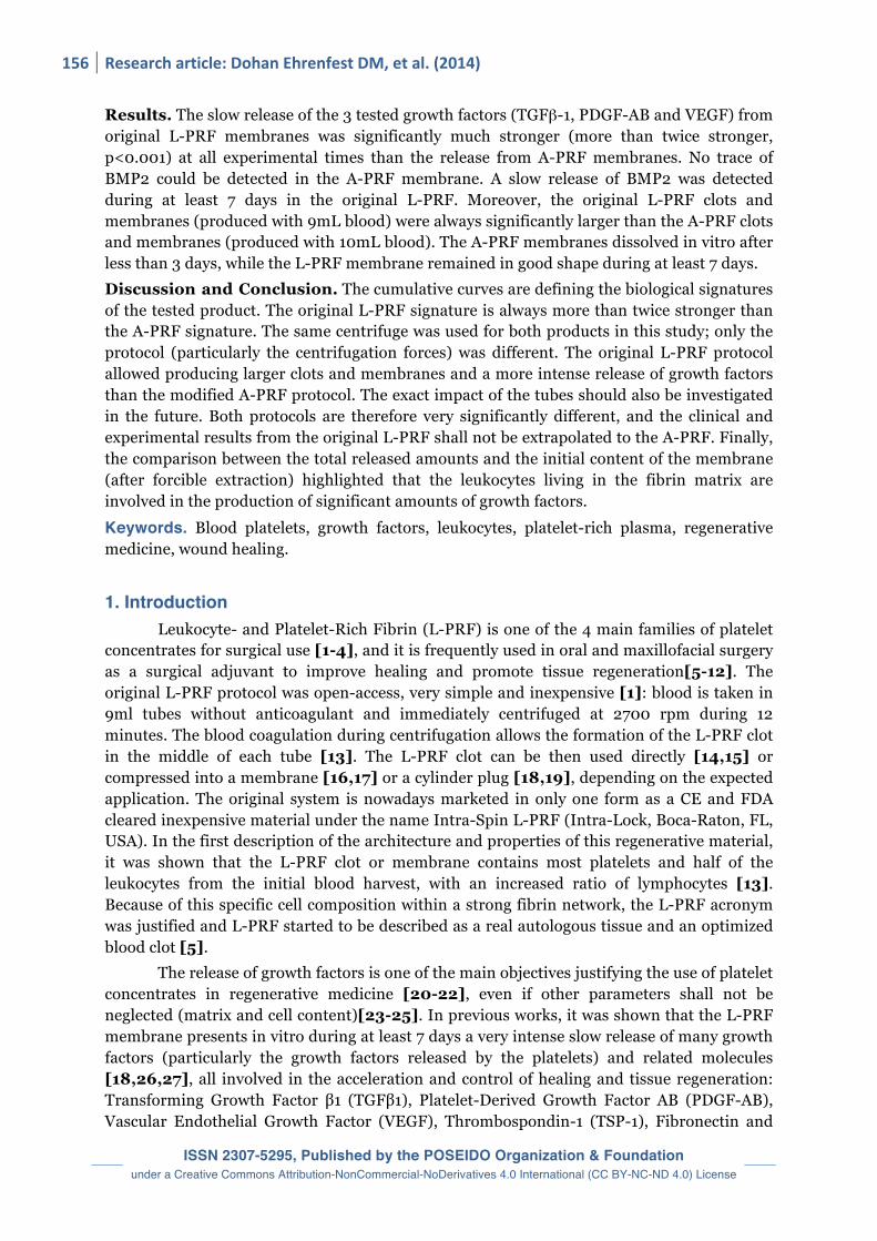
3. Results As a first macroscopic observation, the original L-PRF clots and membranes produced with 9ml blood were always much larger than the A-PRF clots and membranes produced with 10ml blood (Figure 1). The difference was not calculated, but it appeared systematically that the L-PRF were at least 30% bigger than the A-PRF clots and membranes. Figure 1. Original Intra-Spin L-PRF (A) and A-PRF (B) clots in their respective official tubes just after centrifugation. The aspect of the 2 kinds of clots in the tube was since the beginning very different. Clots were then collected and placed in the PRF surgical box (Xpression, Intra-Lock) for compression into membranes (C) to be used for the slow release test. The original Intra-Spin L-PRF membrane was produced with the original L-PRF protocol (2700rpm, 12 minutes) and 9mL blood. The A-PRF membrane was produced with the specific A-PRF protocol (1500rpm, 14 minutes) and 10mL blood. However, in comparison to the original L-PRF membrane, the A-PRF clots and membranes appeared obviously much smaller, more fragile and not so clearly separated from the red blood cell part.
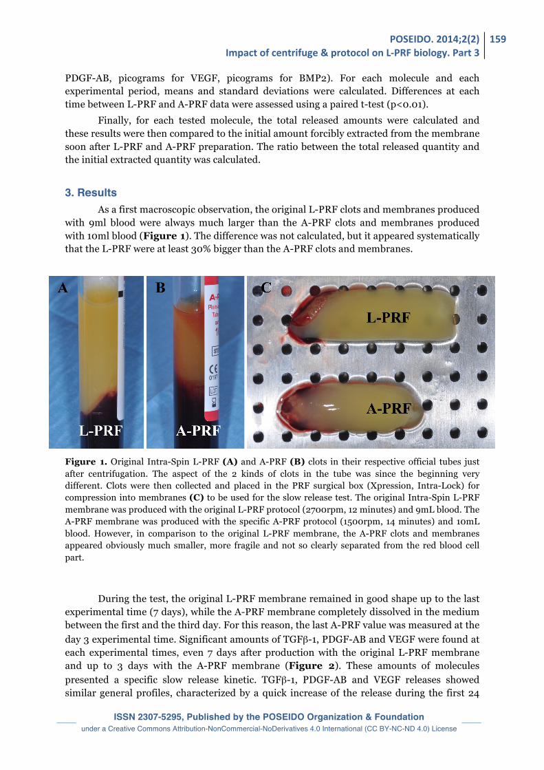
During the test, the original L-PRF membrane remained in good shape up to the last experimental time (7 days), while the A-PRF membrane completely dissolved in the medium between the first and the third day. For this reason, the last A-PRF value was measured at the day 3 experimental time. Significant amounts of TGFβ-1, PDGF-AB and VEGF were found at each experimental times, even 7 days after production with the original L-PRF membrane and up to 3 days with the A-PRF membrane (Figure 2). These amounts of molecules presented a specific slow release kinetic. TGFβ-1, PDGF-AB and VEGF releases showed similar general profiles, characterized by a quick increase of the release during the first 24
160 Research article: Dohan Ehrenfest DM, et al. (2014)
ISSN 2307-5295, Published by the POSEIDO Organization & Foundation
under a Creative Commons Attribution-NonCommercial-NoDerivatives 4.0 International (CC BY-NC-ND 4.0) License
hours. For original L-PRF, a significant but slower release until day 5 (120 h) was observed; during the last 2 days of the experiment, membranes continued to release significant amounts of these molecules, but very slowly. For the A-PRF, the release also slowed down after the first day, but the release stopped quickly thereafter with the complete dissolution of the A-PRF membrane. Figure 2. Slow release of TGFβ1 (A), PDGF-AB (B), VEGF (C) and BMP2 (D) from an original L-PRF membrane and from an A-PRF membrane during 7 days in vitro. Values are expressed as the cumulative mean quantity of molecules at 20 minutes, 1 hours, 4 hours, 24 hours, 72 hours (3 days), 120 hours (5 days) and 168 hours (7 days). The slow release of TGFβ-1, PDGF-AB and VEGF from an original L-PRF membrane (Intra-Spin) was always significantly much stronger (p<0.001) at all experimental times than the release from an A-PRF membrane. All results were presented as graphs (Figure 2) to follow the cumulative released mean amounts of each molecule during the first 168 hours after L-PRF and A-PRF membrane preparation respectively. The gradient of the curves revealed the force of the slow release during the experimental periods. These curves were defining the biological signatures of the original L-PRF membrane produced with Intra-Spin system and of the A-PRF membrane. The original L-PRF signature was always more than twice stronger than the A-PRF signature. For the evaluation of BMP2, no traces of BMP2 could be detected in the A-PRF membrane, and the values represented in the Figure 2 are considered as the unavoidable experimental background noise. On the contrary, a slow release of BMP2 was clearly detected during at least 7 days in the original L-PRF, even if the quantities remained quite small.
For the original L-PRF, the total quantity of released factors was in all cases significantly higher than the total amounts extracted just after membrane preparation (Table). However the ratios between these values (total slow release/initial quantity) were

POSEIDO. 2014;2(2) Impact of centrifuge & protocol on L-‐PRF biology. Part 3
161
ISSN 2307-5295, Published by the POSEIDO Organization & Foundation
under a Creative Commons Attribution-NonCommercial-NoDerivatives 4.0 International (CC BY-NC-ND 4.0) License
very different according to the molecule: TGFβ-1, VEGF and BMP2 following the same high ratio (around 7), while PDGF-AB ratio was much closer to 1.
For the A-PRF, the total quantity of released factors and the total amounts extracted just after membrane preparation were significantly smaller than for the L-PRF membrane (Table). In A-PRF, the ratios between these values (total slow release/initial quantity) were also very different according to the molecule: TGFβ-1 and VEGF following the same high ratio (around 4.5 or 5, lower than for L-PRF), while PDGF-AB ratio was much closer to 1 (similar to L-PRF).
Tested molecule TGFβ-1 (nanog)
VEGF (picog)
PDGF-AB (nanog)
BMP-2 (picog)
Total released after 168h (sum of the amounts measured at each experimental time)
L-PRF 315.5 (±21.1) 6602 (±704) 67.1 (±9.8) 580 (±73)
A-PRF 92.1 (±25.4) 2445 (±782) 20.9 (±8.1) NA
Total extracted at T0 from the membrane
L-PRF 44.4 (±3.7) 994 (±159) 44.4 (±4.2) 79 (±10)
A-PRF 18.6 (±4.8) 514 (±188) 15 (±5.8) NA
Ratio between slow released and extracted molecules
L-PRF 7.1 6.64 1.51 7.34
A-PRF 4.95 4.76 1.39 NA Table. Comparison between the total released quantity at the end of the experimental time (after 168 hours) and the initial extracted quantity of each tested molecule in an original L-PRF membrane and in an A-PRF membrane. Results are expressed as means and standard deviations.
4. Discussion This study compared accurately the biological signatures of 2 kinds of L-PRF
materials, the original L-PRF (Intra-Spin) and the modified protocol A-PRF. A-PRF is in fact a variation of the original L-PRF using a much lower centrifugation speed, a slightly longer centrifugation time, and glass tubes [32]. This technique was initially proposed on the original L-PRF centrifuge (using 1500 rpm) before it was definitively associated with the specific A-PRF centrifuge (using 1300 rpm) tested in the 2 previous parts of this series of articles. It was therefore a perfect model to compare the impact of the change of protocol alone on the biological signature of a PRF membrane, as both original L-PRF and A-PRF can be produced using the exact same centrifuge.
The main observation of this experiment was that A-PRF clots showed a much lower release of growth factors and a weaker biological signature than the original L-PRF. Moreover, the A-PRF clots dissolved quickly in the tubes, while the original L-PRF remained in good shape even after 7 days in vitro. The second observation was that all A-PRF clots and membranes (produced with 10ml blood) were at least 30% smaller than the original L-PRF clots and membranes (produced with 9ml blood). It was previously proven in this series of articles that the vibrations of the A-PRF centrifuge were leading to the formation of an even much smaller clot and membrane. In this study, the same stable original centrifuge (Intra-
162 Research article: Dohan Ehrenfest DM, et al. (2014)
ISSN 2307-5295, Published by the POSEIDO Organization & Foundation
under a Creative Commons Attribution-NonCommercial-NoDerivatives 4.0 International (CC BY-NC-ND 4.0) License
Spin) was used to produce both L-PRF and A-PRF clots to neutralize the centrifuge vibrations variable, and the main difference that could explain these differences of size of the clots and of biological signature of membranes was the change of the protocol, mostly the forces of centrifugation, but also the proprietary type of tubes and the time of centrifugation.
In a previous work, it was shown that the production of L-PRF clots did not seem to be affected by the use of glass tubes or glass coated plastic tubes [13], therefore the differences of tubes between A-PRF and L-PRF may not explain the observed differences. However, this shall be confirmed in future research, as there are many kinds of glass tubes and glass coated plastic tubes. The Intra-Spin tubes were selected very specifically following the long L-PRF experience to fulfill CE and FDA clearance, while nothing is known about the source of the A-PRF tubes (except they are for in vitro diagnostic and made in China). This difference may have an impact in the results and should be investigated. Moreover, it was shown that there is very little impact in using longer centrifugation time with an original L-PRF, as it is common to centrifuge during 18 minutes when patients are under anticoagulant treatment [19]. The increase of centrifugation time mostly gives a bit more time for a fibrin clot to polymerize. It was not needed to last longer than 12 minutes for the original L-PRF in most cases, but A-PRF seems to need this supplementary time to finish its gel polymerization (14 minutes in total).
As a conclusion, these differences of size, aspect and biological signature of the clots and membranes between the original L-PRF and A-PRF can be probably associated with the change in the centrifugation forces. It confirms the need for using forces around 400g (2700 rpm in the original centrifuge), in order to do a proper separation of the blood constituents with an adequate gradient of centrifugation and collect a large and proper L-PRF clot. The use of a lower g force and speed (1500 rpm for A-PRF) did not seem enough for a proper separation of the blood constituents and lead to the preparation of a clot (A-PRF) of much smaller size, weaker biological signature and lower fibrin polymerization, even when the tubes were larger (10ml) and if an adequate stable centrifuge was used.
In previous publications, it was advocated that the cell population of a L-PRF membrane was responsible of the production of new growth factors [26]. Indeed the total release quantities after 7 days of many growth factors were always much higher than the total quantities detected after forcible extraction from the whole membrane just after preparation [26]. In this new study, the same observation can be done for L-PRF, and the result obtained with A-PRF somehow confirmed it. The comparison of the results between L-PRF and A-PRF also highlighted that cells must be placed in a specific environment to massively produce more molecules. TGFβ-1, VEGF and BMP2 presented the same high ratio (around 7 for L-PRF) between total slow release / initial quantity, revealing somehow the activity of production of these molecules by the cells within the clots. The lower ratios of A-PRF revealed also a lower production activity. On the contrary, PDGF-AB ratio was quite stable around 1 for both products, as this molecule is mostly contained and released by platelets initially collected in the sample. Therefore this study also confirmed the need to protect the viability of the cells and even pointed out the need for an activation of the cells by the centrifugation process. This notion of activation is the most logical explanation for the very strong differences of biological signatures between original L-PRF and A-PRF, particularly the interesting result with BMP2.
BMP-2 is an important osteoinductive molecule belonging to the TGF-β superfamily of proteins and playing particularly an important function in bone development. For this reason, recombinant forms of this molecule were marketed in a few countries for the
POSEIDO. 2014;2(2) Impact of centrifuge & protocol on L-‐PRF biology. Part 3
163
ISSN 2307-5295, Published by the POSEIDO Organization & Foundation
under a Creative Commons Attribution-NonCommercial-NoDerivatives 4.0 International (CC BY-NC-ND 4.0) License
treatment of bony defects in orthopaedic and maxillofacial surgery, with mixed or controversial results [33] related to the difficult control of the effects of this molecule in a direct therapeutical approach. The release of small quantities of BMP2 from the original L-PRF probably contributed in some way to the stimulation of bone cell proliferation and differentiation observed in vitro by the L-PRF [34,35] and to the positive clinical effects of L-PRF during bone regeneration [14]. However, its importance in the global equation of the L-PRF (combining, many cells, many growth factors into a specific fibrin matrix) is impossible to point out at this time [23,27,34].
It is interesting to notice that BMP2 was not detected with A-PRF (detected as a noise, probably under the detection threshold of 29 pg/mL), while the company marketing it used the release of BMP2 as a commercial argument for the A-PRF protocol. The exact origin of the BMP2 detected in the L-PRF is difficult to point out, as BMP2 is a molecule specific to bone cells, the ELISA kit itself was designed mostly for bone tissue extracts and bone cell culture supernatants. Small quantities of BMP2 can be detected in the blood in some conditions [36], but its overexpression blood is often associated with various pathologies [37,38]. As BMP2 is not supposed to be released by platelets, consequently the different cell populations (mostly leukocytes) living in the L-PRF clot released BMP2. As BMP2 levels were quite low after forcible extraction from the initial L-PRF clot, consequently the L-PRF cells released and produced this molecule step by step during the experiment. The combination of these observations in both L-PRF and A-PRF supported the conclusion that the quantity and state of the cell population within the L-PRF clot defines a large part of its biological signature. Finally, the evaluation of the slow release of key growth factors from a PRP gel or a L-PRF membrane appeared again as a quite simple method of characterization of the biological signature of an activated platelet concentrate gel. It was already used in several publications [27,28], and it illustrated quite well the differences of growth factor content, cell content and fibrin architecture of different products. The slow release pattern is so characteristic from a product, that it should be evaluated systematically in all kinds of products to define their exact biological signature prior to compare them or analyze their clinical effects.
5. Conclusion The slow release of the 4 tested growth factors from original L-PRF membranes was much stronger than the release from A-PRF membranes. Moreover, the original L-PRF clots and membranes (produced with 9mL blood) were always significantly larger than the A-PRF clots and membranes (produced with 10mL blood). The A-PRF membranes dissolved in vitro after less than 3 days, while the L-PRF membrane remained in good shape during at least 7 days. The same centrifuge was used for both products in this study; only the protocol (particularly the centrifugation forces) was different between the original L-PRF and the A-PRF. Consequently, it can be concluded that the original L-PRF protocol allows to produce larger clots and membranes and a more intense release of growth factors than the modified A-PRF protocol. The exact impact of the tubes should also be investigated in the future. As a general conclusion for this series of articles, it was clearly proven that the centrifuge characteristics and centrifugation protocols have a very significant impact on the cell, growth factors and fibrin architecture of a L-PRF clot and membrane, and that any modification of the original L-PRF material and method shall be clearly investigated and identified separately from the original methods, in order to avoid to create confusion and inaccurate results in the literature.
164 Research article: Dohan Ehrenfest DM, et al. (2014)
ISSN 2307-5295, Published by the POSEIDO Organization & Foundation
under a Creative Commons Attribution-NonCommercial-NoDerivatives 4.0 International (CC BY-NC-ND 4.0) License
Disclosure of interests The authors have no conflict of interest to report.
Acknowledgements
This research work on new biotechnologies and therapeutic strategies in regenerative medicine was supported by the PACT (Platelet & Advanced Cell Therapies) Forum Civitatis of the POSEIDO Academic Consortium (Periodontology, Oral Surgery, Esthetic & Implant Dentistry Organization), by a grant from the National Research Foundation of Korea (NRF) funded by the Korean government-MEST (No. 2011-0030121) and by the LoB5 Foundation for Research, France.
Author Contributions
All authors participated to the technical design and organization of the study, the treatment of data and to the elaboration of the manuscript. DDE, NP, MDC and BSK were in charge of the collection of the materials, samples and raw data.
References [1] Dohan Ehrenfest DM, Rasmusson L, Albrektsson T. Classification of platelet concentrates: from pure platelet-rich plasma (P-PRP) to leucocyte- and platelet-rich fibrin (L-PRF). Trends Biotechnol. 2009;27(3):158-67. [2] Dohan Ehrenfest DM, Bielecki T, Mishra A, Borzini P, Inchingolo F, Sammartino G, Rasmusson L, Evert PA. In search of a consensus terminology in the field of platelet concentrates for surgical use: platelet-rich plasma (PRP), platelet-rich fibrin (PRF), fibrin gel polymerization and leukocytes. Curr Pharm Biotechnol. 2012;13(7):1131-7. [3] Dohan Ehrenfest DM, Sammartino G, Shibli JA, Wang HL, Zou DR, Bernard JP. Guidelines for the publication of articles related to platelet concentrates (Platelet-Rich Plasma - PRP, or Platelet-Rich Fibrin - PRF): the international classification of the POSEIDO. POSEIDO. 2013;1(1):17-27. [4] Dohan Ehrenfest DM, Andia I, Zumstein MA, Zhang CQ, Pinto NR, Bielecki T. Classification of platelet concentrates (Platelet-Rich Plasma-PRP, Platelet-Rich Fibrin-PRF) for topical and infiltrative use in orthopedic and sports medicine: current consensus, clinical implications and perspectives. Muscles Ligaments Tendons J. 2014;4(1):3-9. [5] Del Corso M, Vervelle A, Simonpieri A, Jimbo R, Inchingolo F, Sammartino G, Dohan Ehrenfest DM. Current knowledge and perspectives for the use of platelet-rich plasma (PRP) and platelet-rich fibrin (PRF) in oral and maxillofacial surgery part 1: Periodontal and dentoalveolar surgery. Curr Pharm Biotechnol. 2012;13(7):1207-30. [6] Simonpieri A, Del Corso M, Vervelle A, Jimbo R, Inchingolo F, Sammartino G, Dohan Ehrenfest DM. Current knowledge and perspectives for the use of platelet-rich plasma (PRP) and platelet-rich fibrin (PRF) in oral and maxillofacial surgery part 2: Bone graft, implant and reconstructive surgery. Curr Pharm Biotechnol. 2012;13(7):1231-56. [7] Del Corso M, Dohan Ehrenfest DM. Immediate implantation and peri-implant Natural Bone Regeneration (NBR) in the severely resorbed posterior mandible using Leukocyte- and Platelet-Rich Fibrin (L-PRF): a 4-year follow-up. POSEIDO. 2013;1(2):109-16. [8] Simonpieri A, Del Corso M, Sammartino G, Dohan Ehrenfest DM. The relevance of Choukroun's platelet-rich fibrin and metronidazole during complex maxillary rehabilitations using bone allograft. Part I: a new grafting protocol. Implant Dent. 2009;18(2):102-11. [9] Simonpieri A, Del Corso M, Sammartino G, Dohan Ehrenfest DM. The relevance of Choukroun's platelet-rich fibrin and metronidazole during complex maxillary rehabilitations using bone allograft. Part II: implant surgery, prosthodontics, and survival. Implant Dent. 2009;18(3):220-9. [10] Toeroek R, Dohan Ehrenfest DM. The concept of Screw-Guided Bone Regeneration (S-GBR). Part 2: S-GBR in the severely resorbed preimplant posterior mandible using bone xenograft and Leukocyte- and Platelet-Rich Fibrin (L-PRF): a 5-year follow-up. POSEIDO. 2013;1(2):85-92. [11] Toeroek R, Dohan Ehrenfest DM. The concept of Screw-Guided Bone Regeneration (S-GBR). Part 3: Fast Screw-Guided Bone Regeneration (FS-GBR) in the severely resorbed preimplant posterior mandible using allograft and Leukocyte- and Platelet-Rich Fibrin (L-PRF): a 4-year follow-up. POSEIDO. 2013;1(2):93-100.
POSEIDO. 2014;2(2) Impact of centrifuge & protocol on L-‐PRF biology. Part 3
165
ISSN 2307-5295, Published by the POSEIDO Organization & Foundation
under a Creative Commons Attribution-NonCommercial-NoDerivatives 4.0 International (CC BY-NC-ND 4.0) License
[12] Toeroek R, Mazor Z, Del Corso M, Dohan Ehrenfest DM. The concept of Screw-Guided Bone Regeneration (S-GBR). Part 1: from sinus-lift to general applications in the resorbed maxilla and mandible. POSEIDO. 2013;1(2):69-84. [13] Dohan Ehrenfest DM, Del Corso M, Diss A, Mouhyi J, Charrier JB. Three-dimensional architecture and cell composition of a Choukroun's platelet-rich fibrin clot and membrane. J Periodontol. 2010;81(4):546-55. [14] Mazor Z, Horowitz RA, Del Corso M, Prasad HS, Rohrer MD, Dohan Ehrenfest DM. Sinus floor augmentation with simultaneous implant placement using Choukroun's platelet-rich fibrin as the sole grafting material: a radiologic and histologic study at 6 months. J Periodontol. 2009;80(12):2056-64. [15] Simonpieri A, Choukroun J, Del Corso M, Sammartino G, Dohan Ehrenfest DM. Simultaneous sinus-lift and implantation using microthreaded implants and leukocyte- and platelet-rich fibrin as sole grafting material: a six-year experience. Implant Dent. 2011;20(1):2-12. [16] Del Corso M, Mazor Z, Rutkowski JL, Dohan Ehrenfest DM. The use of leukocyte- and platelet-rich fibrin during immediate postextractive implantation and loading for the esthetic replacement of a fractured maxillary central incisor. J Oral Implantol. 2012;38(2):181-7. [17] Del Corso M, Sammartino G, Dohan Ehrenfest DM. Re: "Clinical evaluation of a modified coronally advanced flap alone or in combination with a platelet-rich fibrin membrane for the treatment of adjacent multiple gingival recessions: a 6-month study". J Periodontol. 2009;80(11):1694-7; author reply 7-9. [18] Dohan Ehrenfest DM. How to optimize the preparation of leukocyte- and platelet-rich fibrin (L-PRF, Choukroun's technique) clots and membranes: introducing the PRF Box. Oral Surg Oral Med Oral Pathol Oral Radiol Endod. 2010;110(3):275-8; author reply 8-80. [19] Sammartino G, Dohan Ehrenfest DM, Carile F, Tia M, Bucci P. Prevention of hemorrhagic complications after dental extractions into open heart surgery patients under anticoagulant therapy: the use of leukocyte- and platelet-rich fibrin. J Oral Implantol. 2011;37(6):681-90. [20] Bielecki T, Dohan Ehrenfest DM. Leukocyte- and platelet-rich Plasma (L-PRP)/fibrin (L-PRF) in medicine - past, present, future. Curr Pharm Biotechnol. 2012;13(7):i-ii. [21] Bielecki T, Dohan Ehrenfest DM. Platelet-rich plasma (PRP) and Platelet-Rich Fibrin (PRF): surgical adjuvants, preparations for in situ regenerative medicine and tools for tissue engineering. Curr Pharm Biotechnol. 2012;13(7):1121-30. [22] Cieslik-Bielecka A, Choukroun J, Odin G, Dohan Ehrenfest DM. L-PRP/L-PRF in esthetic plastic surgery, regenerative medicine of the skin and chronic wounds. Curr Pharm Biotechnol. 2012;13(7):1266-77. [23] Bielecki T, Dohan Ehrenfest DM, Everts PA, Wiczkowski A. The role of leukocytes from L-PRP/L-PRF in wound healing and immune defense: new perspectives. Curr Pharm Biotechnol. 2012;13(7):1153-62. [24] Cieslik-Bielecka A, Dohan Ehrenfest DM, Lubkowska A, Bielecki T. Microbicidal properties of Leukocyte- and Platelet-Rich Plasma/Fibrin (L-PRP/L-PRF): new perspectives. J Biol Regul Homeost Agents. 2012;26(2 Suppl 1):43S-52S. [25] Dohan Ehrenfest DM, Bielecki T, Del Corso M, Inchingolo F, Sammartino G. Shedding light in the controversial terminology for platelet-rich products: platelet-rich plasma (PRP), platelet-rich fibrin (PRF), platelet-leukocyte gel (PLG), preparation rich in growth factors (PRGF), classification and commercialism. J Biomed Mater Res A. 2010;95(4):1280-2. [26] Dohan Ehrenfest DM, de Peppo GM, Doglioli P, Sammartino G. Slow release of growth factors and thrombospondin-1 in Choukroun's platelet-rich fibrin (PRF): a gold standard to achieve for all surgical platelet concentrates technologies. Growth Factors. 2009;27(1):63-9. [27] Dohan Ehrenfest DM, Bielecki T, Jimbo R, Barbe G, Del Corso M, Inchingolo F, Sammartino G. Do the fibrin architecture and leukocyte content influence the growth factor release of platelet concentrates? An evidence-based answer comparing a pure platelet-rich plasma (P-PRP) gel and a leukocyte- and platelet-rich fibrin (L-PRF). Curr Pharm Biotechnol. 2012;13(7):1145-52. [28] Zumstein MA, Bielecki T, Dohan Ehrenfest DM. The Future of Platelet Concentrates in Sports Medicine: Platelet-Rich Plasma, Platelet-Rich Fibrin, and the Impact of Scaffolds and Cells on the Long-term Delivery of Growth Factors. Operative Techniques in Sports Medicine. 2011;19(3):190-7. [29] Dohan Ehrenfest DM, Del Corso M, Inchingolo F, Charrier JB. Selecting a relevant in vitro cell model for testing and comparing the effects of a Choukroun's platelet-rich fibrin (PRF) membrane and a platelet-rich plasma (PRP) gel: tricks and traps. Oral Surg Oral Med Oral Pathol Oral Radiol Endod. 2010;110(4):409-11; author reply 11-3. [30] Dohan Ehrenfest DM, Del Corso M, Inchingolo F, Sammartino G, Charrier JB. Platelet-rich plasma (PRP) and platelet-rich fibrin (PRF) in human cell cultures: growth factor release and contradictory results. Oral Surg Oral Med Oral Pathol Oral Radiol Endod. 2010;110(4):418-21; author reply 21-2.
166 Research article: Dohan Ehrenfest DM, et al. (2014)
ISSN 2307-5295, Published by the POSEIDO Organization & Foundation
under a Creative Commons Attribution-NonCommercial-NoDerivatives 4.0 International (CC BY-NC-ND 4.0) License
[31] Dohan Ehrenfest DM, Lemo N, Jimbo R, Sammartino G. Selecting a relevant animal model for testing the in vivo effects of Choukroun's platelet-rich fibrin (PRF): rabbit tricks and traps. Oral Surg Oral Med Oral Pathol Oral Radiol Endod. 2010;110(4):413-6; author reply 6-8. [32] Ghanaati S, Booms P, Orlowska A, Kubesch A, Lorenz J, Rutkowski J, Landes C, Sader R, Kirkpatrick C, Choukroun J. Advanced platelet-rich fibrin: a new concept for cell-based tissue engineering by means of inflammatory cells. J Oral Implantol. 2014;40(6):679-89. [33] Carragee EJ, Hurwitz EL, Weiner BK. A critical review of recombinant human bone morphogenetic protein-2 trials in spinal surgery: emerging safety concerns and lessons learned. Spine J. 2011;11(6):471-91. [34] Dohan Ehrenfest DM, Diss A, Odin G, Doglioli P, Hippolyte MP, Charrier JB. In vitro effects of Choukroun's PRF (platelet-rich fibrin) on human gingival fibroblasts, dermal prekeratinocytes, preadipocytes, and maxillofacial osteoblasts in primary cultures. Oral Surg Oral Med Oral Pathol Oral Radiol Endod. 2009;108(3):341-52. [35] Dohan Ehrenfest DM, Doglioli P, de Peppo GM, Del Corso M, Charrier JB. Choukroun's platelet-rich fibrin (PRF) stimulates in vitro proliferation and differentiation of human oral bone mesenchymal stem cell in a dose-dependent way. Arch Oral Biol. 2010;55(3):185-94. [36] Kruger JP, Freymannx U, Vetterlein S, Neumann K, Endres M, Kaps C. Bioactive factors in platelet-rich plasma obtained by apheresis. Transfus Med Hemother. 2013;40(6):432-40. [37] Tosovsky M, Bradna P, Andrys C, Andrysova K, Cermakova E, Soukup T. The VEGF and BMP-2 levels in patients with ankylosing spondylitis and the relationship to treatment with tumour necrosis factor alpha inhibitors. Acta Medica (Hradec Kralove). 2014;57(2):56-61. [38] Fei ZH, Yao CY, Yang XL, Huang XE, Ma SL. Serum BMP-2 up-regulation as an indicator of poor survival in advanced non-small cell lung cancer patients. Asian Pac J Cancer Prev. 2013;14(9):5293-9. This article can be cited as: Dohan Ehrenfest DM, Del Corso M, Kang BS, Lanata N, Quirynen M, Wang HL, Pinto NR. The impact of the centrifuge characteristics and centrifugation protocols on the cells, growth factors and fibrin architecture of a Leukocyte- and Platelet-Rich Fibrin (L-PRF) clot and membrane. Part 3: comparison of the growth factors content and slow release between the original L-PRF and the modified A-PRF (Advanced Platelet-Rich Fibrin) membranes. POSEIDO. 2014;2(2):155-66.