the injury response process exss 380: therapeutic modalities hoffman
TRANSCRIPT
THE INJURY RESPONSE PROCESSTHE INJURY RESPONSE PROCESS
EXSS 380: Therapeutic ModalitiesHoffman

STRESSES PLACED ON THE CELLSTRESSES PLACED ON THE CELL
– Body/Cells respond to stress in one of three ways:• Adapt• Becomes injured, but recovers• Dies
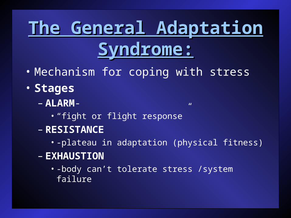
The General Adaptation The General Adaptation Syndrome:Syndrome:
• Mechanism for coping with stress• Stages
– ALARM-• “fight or flight response”
– RESISTANCE• -plateau in adaptation (physical fitness)
– EXHAUSTION• -body can’t tolerate stress /system failure
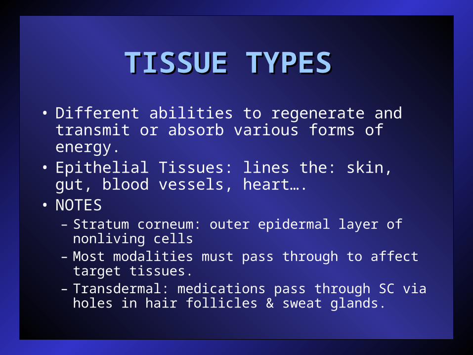
TISSUE TYPES TISSUE TYPES
• Different abilities to regenerate and transmit or absorb various forms of energy.
• Epithelial Tissues: lines the: skin, gut, blood vessels, heart….
• NOTES– Stratum corneum: outer epidermal layer of
nonliving cells– Most modalities must pass through to affect
target tissues.– Transdermal: medications pass through SC via
holes in hair follicles & sweat glands.
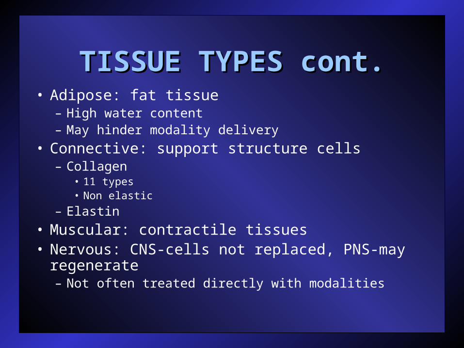
TISSUE TYPES cont.TISSUE TYPES cont.• Adipose: fat tissue
– High water content– May hinder modality delivery
• Connective: support structure cells– Collagen
• 11 types• Non elastic
– Elastin
• Muscular: contractile tissues• Nervous: CNS-cells not replaced, PNS-may
regenerate– Not often treated directly with modalities

PreviewPreview
• Injury Process• Phases of Healing
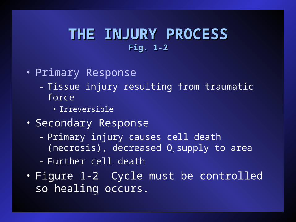
THE INJURY PROCESSTHE INJURY PROCESSFig. 1-2Fig. 1-2
• Primary Response– Tissue injury resulting from traumatic force
• Irreversible
• Secondary Response– Primary injury causes cell death (necrosis),
decreased O2 supply to area– Further cell death
• Figure 1-2 Cycle must be controlled so healing occurs.
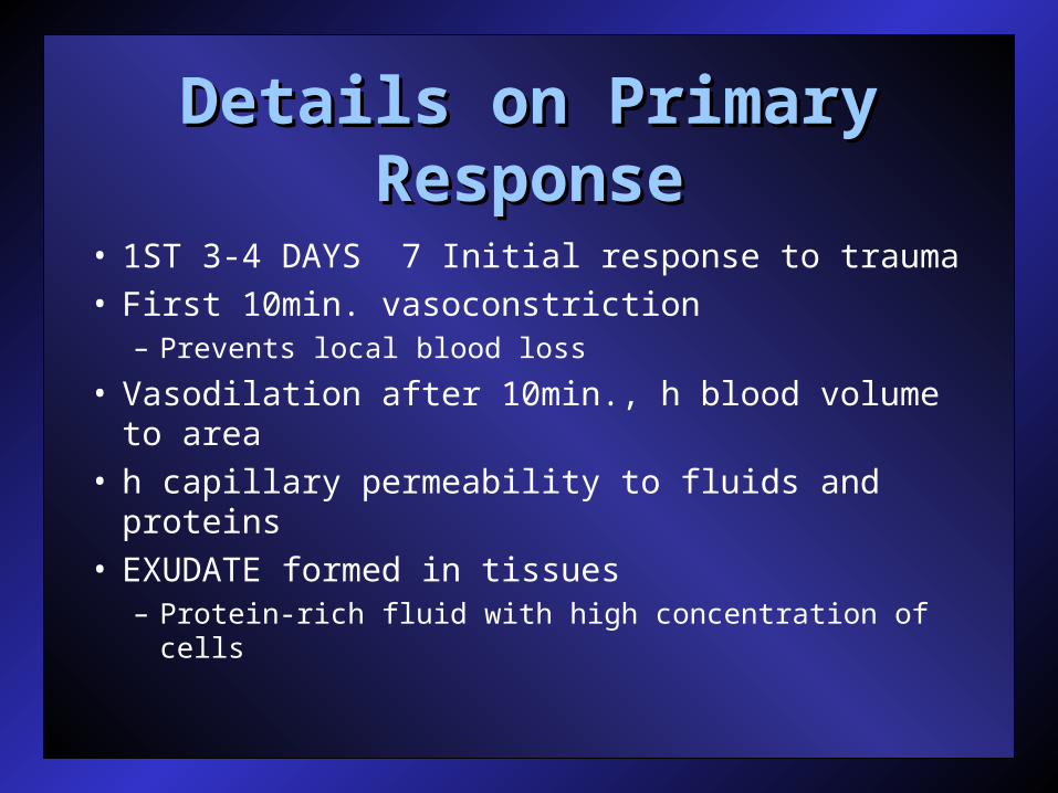
Details on Primary Details on Primary ResponseResponse
• 1ST 3-4 DAYS 7 Initial response to trauma• First 10min. vasoconstriction
– Prevents local blood loss
• Vasodilation after 10min., h blood volume to area
• h capillary permeability to fluids and proteins• EXUDATE formed in tissues
– Protein-rich fluid with high concentration of cells

• Prostaglandin (PGE1) increase vascular permeability
• Prostaglandin (PGE2) attracts leukocytes to area– WBC’s that act as scavengers
• Fluids/proteins leak into interstitial space through capillaries and deposit leukocytes to remove dead material (swelling)
• Venous & lymphatic flow blocked-further increased blood flow
• Chemical mediators control inflammation.

Figure 1-2Figure 1-2

PHASES OF HEALINGPHASES OF HEALING
• Inflammatory Phase: – Attempt to isolate and localize trauma
• Proliferation Phase:– Preparation to rebuild damaged tissue
• Remodeling (Maturation) Phase:– Collagen/fibroblasts align and attempt
to replicate original structure/function

OUTLINEOUTLINE
• Healing Phases– Inflammation
• Levels– Circulation and Cellular
• Phases– ACUTE, SUBACUTE, CHRONIC
– Proliferation• Regeneration vs Replacement• Revascularation• Wound contraction• Remodeling
– Maturation

ACUTE INFLAMMATORY ACUTE INFLAMMATORY RESPONSE (Table 1-8)RESPONSE (Table 1-8)
• INFLAMMATION: essential to healing process– Natural, physiological reaction to injury
which mobilizes the body’s defense systems.
• Purpose: control effects of injury and restore tissue homeostasis.
• Triggered by chemical irritation, heat, mechanical trauma, or bacteria.
• Hemodynamic (blood flow) and cellular changes.• 3 Phases: Acute (initial reaction), Sub-acute (2-
4wks), Chronic (>1month.)

CARDINAL SIGNS OF CARDINAL SIGNS OF INFLAMMATIONINFLAMMATION
• HEAT: blood flow, metabolic rate• REDNESS: histamine release• SWELLING:
– Inflammatory agents into area, blocked venous return
• PAIN:– Chemical irritants released (bradykinin,
histamine, prostaglandins, etc.).– Mechanical & chemical pressure on nerve
receptors• LOSS OF FUNCTION:
– Primary tissue damage,muscle guarding…
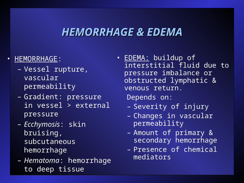
HEMORRHAGE & EDEMAHEMORRHAGE & EDEMA
• HEMORRHAGE: – Vessel rupture,
vascular permeability– Gradient: pressure in
vessel > external pressure
– Ecchymosis: skin bruising, subcutaneous hemorrhage
– Hematoma: hemorrhage to deep tissue
• EDEMA: buildup of interstitial fluid due to pressure imbalance or obstructed lymphatic & venous return.Depends on:– Severity of injury– Changes in vascular
permeability– Amount of primary &
secondary hemorrhage– Presence of chemical
mediators

EDEMA CONT.EDEMA CONT.• Formation & removal dependent on:
– Vascular hydrostatic pressure: capillary to tissues– Plasma osmotic pressure: tissues to capillaries– Limb hydrostatic pressure: dependent on limb
position
• Normal: vascular = plasma pressure• Injury: capillary permeability, hydrostatic pressure
forces material out.– Edema exacerbates inflammatory process
• Prevents blood & O2 into area• Inhibits venous & lymphatic return by clogging
pathway• Necrosis, pain, ROM
• GOAL: reduce edema through venous & lymphatic return.

VENOUS & LYMPHATIC RETURNVENOUS & LYMPHATIC RETURN
• Thoracic duct: connects lymphatic system to venous
• Venous flow not affected by blood pressure
• Muscle contraction – vessel diameter, blood forced through 1-way valve, valve closes (Fig. 1-5)
• Gravity, passive motion, E-stim., massage all venous flow.

PROLIFERATION PHASEPROLIFERATION PHASE 72HRS.72HRS.3WKS3WKS..
• Growth of new tissue, permanently replaces exudate produced during inflammatory phase.
• Regeneration vs. replacement (Table 1-1)• Replacement: different cell than original
– Skin cells regenerate, muscle cells replace with fibrous scar tissue
– Uncontrolled causes function

PROLIFERATION PHASE:PROLIFERATION PHASE:
• Soft-tissue repair: proliferation of granulation tissue– Fibroblast formation– Collagen synthesis– Tissue remodeling– Tissue alignment
• ATP regulates rate & quality of healing– Provides energy to restore cell-membrane
properties
• Fibroblasts, platelets & macrophages contribute.
PROLIFERATION PHASE:PROLIFERATION PHASE:
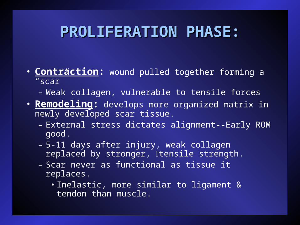
• Contraction: wound pulled together forming a “scar”– Weak collagen, vulnerable to tensile forces
• Remodeling: develops more organized matrix in newly developed scar tissue.– External stress dictates alignment--Early ROM
good.– 5-11 days after injury, weak collagen replaced by
stronger, tensile strength.– Scar never as functional as tissue it replaces.
• Inelastic, more similar to ligament & tendon than muscle.

MATURATION PHASEMATURATION PHASE
• Final phase of injury response process—”clean-up”.– May last 1 year or more.
• Decreased vascularity, capillaries, water content.– Fading redness of scar, normalizing skin color,
texture.
• Macrophages, fibroblasts, myofibroblasts reduced to preinjury state.
• Type I collagen increases, tensile strength increases.
• GOAL:– Insure adequate tissue tensile strength by
placing increased stresses on replaced tissue.
THERAPEUTIC MODALITIESTHERAPEUTIC MODALITIES
• THERAPEUTIC MODALITY:• The application of thermal, mechanical,
electrical, or chemical stress to the body for the purpose of healing it from injury.
• Balance between protection from further injury and returning to normal function.