the innate immune dna sensor cgas is a negative regulator ... · innate immune system to the...
TRANSCRIPT

The innate immune DNA sensor cGAS is a negative regulator of DNA
repair hence promotes genome instability and cell death
Hui Jiang1, Swarupa Panda1, Xiaoyu Xue2, Fengshan Liang2, Patrick Sung2 & Nelson O.
Gekara1,*.
1The Laboratory for Molecular Infection Medicine Sweden (MIMS), The Departments of
Molecular Biology, Umeå Centre for Microbial Research (UCMR), Umeå University, 90 187,
Umeå, Sweden.
2Department of Molecular Biophysics and Biochemistry, Yale University School of
Medicine, New Haven, Connecticut 06520, USA.
*Lead contact: N.O.G. ([email protected]).
Short title: cGAS is an inhibitor of DNA repair
was not certified by peer review) is the author/funder. All rights reserved. No reuse allowed without permission. The copyright holder for this preprint (whichthis version posted November 8, 2018. . https://doi.org/10.1101/465401doi: bioRxiv preprint

ABSTRACT
Stringent regulation of DNA repair is essential for organismal integrity, but the mechanisms
are not fully understood. Cyclic cGMP-AMP synthase (cGAS), the DNA sensor that alerts the
innate immune system to the presence of foreign or damaged self-DNA in the cytoplasm is
critical for the outcome of infections, inflammatory diseases and cancer. Besides this
cytoplasmic function as an innate immune sensor, whether cGAS fulfills other biological roles
remains unknown. Here we report that cGAS has a distinct role in the nucleus: it inhibits
homologous recombination DNA repair (HR) thereby promoting genome instability and
associated micronuclear generation and mitotic death. We show that cGAS-mediated inhibition
of HR requires its DNA binding and oligomerization but not its catalytic activity or the
downstream innate immune signaling events. Mechanistically, we show that cGAS impede
RAD51-mediated DNA strand invasion, a key step in HR. These results uncover a new function
of cGAS relevant for understanding its involvement in genome instability- associated
disorders.
was not certified by peer review) is the author/funder. All rights reserved. No reuse allowed without permission. The copyright holder for this preprint (whichthis version posted November 8, 2018. . https://doi.org/10.1101/465401doi: bioRxiv preprint

INTRODUCTION
The DNA Damage Response (DDR) that senses threats to our genome, and the immune system
- the inherent ability to sense and respond to infections, both function as surveillance systems
essential for the preservation of organismal integrity. Emerging evidence indicates that these
two systems are interdependent 1.
DDR constitutes a complex set of signalling pathways that mediate the elimination of DNA
lesions, but could also trigger senescence or cell death to prevent the transmission of damaged
genetic material 2, 3. Besides these classical outcomes of DDR, it is increasingly becoming
apparent that DNA damage has a major impact on the immune system. DNA damage releases
DNA fragments into the cytosol resulting in innate immune induction of cytokines such as type
I interferons (IFN-Is) 1. These cytokines in turn play an important role in priming the immune
system for enhanced antimicrobial 1 and antitumor immunity 4-7, but, if produced in excessive
amounts, could lead to autoimmunity 1, 8 and tumorigenesis 9-11. Moreover, DNA damage-
induced cytokines have also been found to be essential for classical DDR cellular outcomes
including senescence 12-15.
Altogether, these recent findings highlighting the symbiotic partnership between DDR and the
immune system have opened new avenues for understanding the biology of several disorders
such as cancer and autoimmune diseases, caused by genome instability and immune defects 10.
However, it remains unclear how these two biological systems are cross-regulated and what
signalling molecules are involved.
From recent reports, there is a growing consensus that a key player linking DNA damage and
immunity is the cyclic cGMP-AMP synthase (cGAS). cGAS surveys the cytoplasm for the
presence of microbial DNA 16, 17, self-DNA from damaged chromatin 1, or DNA released from
distressed mitochondria 18. Upon recognition of double stranded DNA (dsDNA), cGAS
catalyzes the cyclization of ATP and GTP into the second messenger cyclic GMP–AMP (2′3′-
was not certified by peer review) is the author/funder. All rights reserved. No reuse allowed without permission. The copyright holder for this preprint (whichthis version posted November 8, 2018. . https://doi.org/10.1101/465401doi: bioRxiv preprint

cGAMP) 16, 17, 19-24. Subsequently, cGAMP binds to its adaptor Stimulator of Interferon Genes
(STING) 25 leading to the activation of downstream transcription factors: Interferon regulatory
factor 3 (IRF3) and nuclear factor kB (NF-kB) 26, 27, and these together, coordinate the
induction IFN-Is and other inflammatory cytokines. Dysfunctions in the cGAS-STING
pathway have been implicated in many disorders including infections, inflammatory diseases,
neurodegeneration and cancer 10, 28.
Thus far, all the functions ascribed to cGAS are linked to its cytoplasmic role in innate immune
system activation. Beyond this, whether cGAS also serves other biological functions is
unknown.
DNA double-strand DNA breaks (DSB) are potentially highly deleterious lesions. If
improperly repaired, DSB results in chromosomal deletions or translocations culminating in
genome instability-associated disorders including tumorigenesis, accelerated aging and other
diseases 3. For a healthy outcome, the DNA damage signaling is carefully calibrated to ensure
timely removal of damaged DNA breaks, but if the DNA damage is excessive, to promote cell
death to eradicate genetically defective cells 29. The regulatory molecules involved in this
delicate balance are not fully known. In this study, we report our studies identifying a new
regulator of DNA repair: cGAS. We demonstrate that aside from its function in the cytoplasm
as an innate immune sensor, cGAS is also present in the nucleus where it acts as a negative
regulator of homologous recombination-mediated DNA repair and thereby promotes
micronuclear generation and mitotic catastrophic death of genetically stressed cells.
RESULTS
cGAS is presence in the nuclear and cytosolic compartments
cGAS is generally considered a cytosolic protein. However, while monitoring the subcellular
localization of endogenous cGAS in bone morrow differentiating monocytes (BMDMos) or
was not certified by peer review) is the author/funder. All rights reserved. No reuse allowed without permission. The copyright holder for this preprint (whichthis version posted November 8, 2018. . https://doi.org/10.1101/465401doi: bioRxiv preprint

exogenously expressed GFP-tagged human cGAS (GFP-hcGAS) in HEK293 cells, we noted
that cGAS is also abundant in the nucleus (Figure 1 and Figure S1). Curiously, and consistent
with recent observations 13, when cells were enriched at G0/G1 cell cycle phase by serum
starvation or by cell contact inhibition at high cell density, or at G1/early S phase by the DNA
polymerase inhibitor aphidicolin, cGAS showed an increase in the cytosol (Figure 1 and
Figure S1). These data demonstrate that cGAS is constitutively present in the cytosol and
nucleus and that the relative abundance of cGAS in these subcellular compartments can vary
with the cell cycle. To better understand the cGAS features essential for its nuclear localization
we analysed different cGAS mutants. The nuclear localization of the catalytic dead
E225A/D227A mutant (GFP-hcGASΔcGAMP)30 and the oligomerization defective K394A
mutant (GFP-hcGASΔOligo)21 were comparable to that of full length GFP-hcGAS. In contrast,
DNA binding C396A/C397A mutant (GFP-hcGASΔDNA)19, 31 did not show a strong
accumulation in the nucleus and was unaffected by changes in cycle phase (Figure S2A). On
further analysis we found that nuclear cGAS was not within soluble nuclear fractions but
mostly chromatin-bound (Figure S2B). Together with previous observations13, these findings
indicated that the localization of cGAS in the nucleus is due to its binding to DNA following
dissolution of the nuclear membrane during cell division. To interrogate this further, we asked
how introduction a strong nuclear export or import signals (NES or NLS respectively) would
impact cGAS localization. The NLS localized cGAS almost entirely into the nucleus. In
contrast, the NES substantially increased hcGAS localization in the cytosol but did not
eliminate its presence from the nucleus (Figure S2C, D). Taken together with the behavior of
the cGASΔDNA mutant (Figure S2A), these results confirm that the localization and retention
of cGAS in the nucleus is due to its avid binding to DNA and hence even a strong NES is not
sufficient to not completely dislodge it from the nucleus.
was not certified by peer review) is the author/funder. All rights reserved. No reuse allowed without permission. The copyright holder for this preprint (whichthis version posted November 8, 2018. . https://doi.org/10.1101/465401doi: bioRxiv preprint

Nuclear cGAS promotes genome destabilization, micronuclei generation and mitotic
death
We wished to determine the biological role of nuclear cGAS. Micronuclei are hallmarks of
genome instability. Micronuclei arise following the mis-segregation of broken chromosomes
during mitosis 32, 33 and have recently been proposed as platforms for cGAS-mediated innate
immune activation following DNA damage 6, 33, 34. While studying the impact of cGAS in the
cellular response to genotoxic stress, we noted that in response to γ-irradiation, HEK293 cells
expressing GFP-hcGAS exhibit a higher incidence of micronuclei than cells expressing a GFP
control containing a nuclear localization sequence (GFP-NLS) (Figure. 2A, B). As expected,
GFP-hcGAS, but not GFP-NLS, restored IFNB1 response to transfected DNA (Figure. 2C).
This observation led us to hypothesize that the presence of cGAS in the nucleus and
micronuclei generation were causally related. Hence we tested whether endogenous cGAS does
promote micronuclei generation in BMDMos. To induce micronuclei generation, BMDMos
were arrested at the G2 phase using the microtubule-depolymerizing agent nocodazole
followed by γ-irradiation and release into mitosis (Figure 2D). BMDMos from WT mice
exhibited more micronuclei compared to those from cGAS-/- mice (Figure 2D-F),
demonstrating that in addition to innate immune activation, cGAS promotes genome
destabilization.
Mitotic catastrophe is a form of cell death, which, similar to micronuclei generation, occurs
when damaged chromosomes mis-segregate during mitosis. Hence mitotic catastrophe is
considered to be an important mechanism that prevents replication of genetically damaged cells
29. To further elucidate the biological relevance of nuclear cGAS, we tested the impact of cGAS
on mitotic catastrophe. BMDMos from cGAS-/- mice were found to be less susceptible to mitotic
catastrophic death compared to those from WT mice (Figure 2G). This demonstrates that by
was not certified by peer review) is the author/funder. All rights reserved. No reuse allowed without permission. The copyright holder for this preprint (whichthis version posted November 8, 2018. . https://doi.org/10.1101/465401doi: bioRxiv preprint

accelerating genomic destabilization, the concomitant chromosome mis-segregation and
mitotic catastrophic death, cGAS contributes to the elimination of cells with severely damaged
genomes.
cGAS impedes DNA repair independently of STING signalling
Next we sought to determine whether cGAS contributes to genome instability by inhibiting
DNA double strand break (DSB) repair. For that, we monitored DSB at different time points
following γ-irradiation by comet and pulsed-field gel electrophoresis assays. We found that γ-
irradiated BMDMos from cGAS-/- mice resolved DSB faster than those from WT mice (Figure
S3A, Figure 3A, B), confirming that cGAS is a negative regulator of DSB repair. Curiously,
while γ-irradiated BMDMos from cGAS-/- mice exhibited a faster resolution of DSB, those
from Sting-/- mice were comparable to WT BMDMos in this regard (Figure 3A, B). Further,
expression of GFP-hcGAS in the HEK293T cells that lack endogenous expression of both
cGAS and STING 17 also impaired DSB repair in these cells (Figure S3B), but, as expected,
failed to restore the IFN-1 response (Figure S3C). Thus, in contrast to innate immune
activation, cGAS-mediated inhibition of DSB repair is STING-independent.
To further interrogate how cGAS affects genome stability, we considered whether inhibition
of DSB repair was mediated by cGAMP via a hitherto undefined STING-independent
mechanism. However, treatment of HEK293 cells with cGAMP before or during γ-irradiation
did not lead to increased fragmentation of genomic DNA (Figure S3D), but, as expected,
activated STING-dependent Interferon Regulatory Factor (IRF3) (Figure S3E). Accordingly,
the full length and catalytically dead GFP-hcGASΔcGAMP comparably inhibited DSB repair
in HEK293 cells, as assessed by comet tail length (Figure S3F, G) and as expected, unlike
GFP-hcGAS, GFP-hcGASΔcGAMP failed to restore the IFN-I response (Figure S3H).
was not certified by peer review) is the author/funder. All rights reserved. No reuse allowed without permission. The copyright holder for this preprint (whichthis version posted November 8, 2018. . https://doi.org/10.1101/465401doi: bioRxiv preprint

Curiously, on further analysis we noted that although not essential for cGAS-mediated
suppression of DNA repair, cGAMP-STING was partly required for optimal generation of
micronuclei and induction mitotic cell death (Figure 3C-D and Figure S3I, J). This is
consistent with previous findings indicating a role for interferons in mitotic stress35, 36 – an
ingredient for mitotic death and micronuclei generation. Thus, although not primarily required
for cGAS-mediated inhibition of DSB repair, STING-IFN-I signaling does contribute to
secondary cellular outcomes of DNA damage such as micronuclei generation and mitotic death.
Finally, when we compared hcGAS and mcGAS, we found them to inhibit DNA repair
comparably (Figure S3K, L).
cGAS attenuates DSB repair in a cell cycle and ATM-dependent manner
DSB repair occur via two major pathways: non-homologous end-joining (NHEJ) and
homologous recombination (HR)37, 38. NHEJ is an error-prone repair pathway active
throughout the cell cycle, and entails the ligation of DNA ends, often leading to deletion or
insertional mutations 37, 38. HR is on the other hand an accurate repair process active in
proliferating cells and occurs mainly during the S and G2 cell cycle phases wherein it engages
the undamaged sister chromatid to template break repair to restore the original DNA sequence
37, 38.
To determine which of the two major DSB repair pathways is impeded by cGAS, we monitored
γ-irradiation-induced DNA breaks under different conditions permissive or not permissive for
either of these DNA repair pathways. We found that arresting cells at the G1/early S phase
using Aphidicolin abolished the cGAS phenotype. In contrast, arresting cells at the G2 phase
by nocodazole did not (Figure 4A-D). ATM and DNA-PKc are proximal kinases that play
critical roles in HR and NHEJ, respectively. Inhibition of ATM abrogated the cGAS-associated
was not certified by peer review) is the author/funder. All rights reserved. No reuse allowed without permission. The copyright holder for this preprint (whichthis version posted November 8, 2018. . https://doi.org/10.1101/465401doi: bioRxiv preprint

increase in DNA breaks, while the DNA-PKc inhibitor had no effect (Figure 4E-G, Figure
S4A), implying that cGAS mainly interferes with the ATM-dependent HR pathway.
cGAS attenuates HR-DNA repair via DNA binding and oligomerisation
To independently verify the above findings, we examined the repair of a site-specific DSB
induced by the I-SceI endonuclease using the direct repeat-GFP (DR-GFP) 39 and the total-
NHEJ-GFP (EJ5-GFP) 40 reporter systems for HR and NHEJ, respectively. siRNA knockdown
of endogenous cGAS in U2OS cells increased HR efficiency but minimally affected NHEJ
repair (Figure 5A-D). In contrast, expression of hcGAS in HEK293T cells strongly reduced
the efficiency of HR but not the NHEJ repair (Figure 5E, F).
To elucidate the cGAS features mediating inhibition of HR, we tested different cGAS mutants.
The catalytically dead hcGASΔcGAMP inhibited HR to a similar degree as hcGAS. In contrast,
the DNA binding (hcGASΔDNA) and oligomerization (hcGASΔOligo) mutants respectively,
exhibited a complete or severe lack of such inhibitory effect (Figure 5E, F). These data
conclusively demonstrate that cGAS specifically blocks HR-mediated DNA repair via its DNA
binding and oligomerization but not catalytic activity. To further interrogate these findings, we
addressed the importance of cGAS nuclear localization by testing the impact of hcGAS-NLS
and hcGAS-NES on HR-DNA repair. Consistent with its increased accumulation in the nucleus
(Figure S2C) cGAS-NLS had a stronger inhibitory effect on HR-DNA repair than hcGAS. In
contrast, hcGAS-NES exhibited a weaker inhibitory effects (Figure 5G, H) consistent with its
reduced presence in the nucleus (Figure S2C, D). Together, these results demonstrate that the
ability of nuclear cGAS to impede HR-DNA repair depends on its DNA binding and
oligomerization but not its enzymatic function.
cGAS interferes with RAD51-mediated DNA strand invasion
was not certified by peer review) is the author/funder. All rights reserved. No reuse allowed without permission. The copyright holder for this preprint (whichthis version posted November 8, 2018. . https://doi.org/10.1101/465401doi: bioRxiv preprint

To understand the specific signaling step in HR-DNA pathway targeted by cGAS we asked
whether cGAS was inhibiting activation of the HR kinase ATM. However, expression of GFP-
hcGAS or hcGASΔcGAMP in HEK293T or HEK293 cells had no effect on γ-irradiation-
induced phosphorylation of ATM (Figure S4B, C). Similarly, WT, cGAS-/- and Sting-/-
BMDMos showed comparable γ-irradiation-induced ATM activation (Figure S4D). To
determine whether cGAS is recruited to DSB sites, we employed a DSB reporter system
based on a mCherry-LacI-FokI nuclease fusion protein to create DSBs within a single
genomic locus in U2OS cells (U2OS-DSB reporter) 41. GFP-hcGAS and mCherry-LacI-
FokI did not co-localize at the DSB sites (Figure S5A). Moreover, GFP-hcGAS and γ-H2AX
foci were not colocalized in γ-irradiated HEK293 cells (Figure S5B). Moreover,
biochemical fractionation studies confirmed that cGAS was constantly in the nucleus and
remained unchanged upon γ-irradiation (Figure S5C) indicating that observed phenotype
was not via a cGAS effect on proximal signaling events at DSB sites but most likely due to
inhibition of a critical downstream process.
The RAD51 recombinase acts downstream of ATM to catalyse HR-mediated DSB repair.
Specifically, protomers of RAD51 form a protein filament on 3′ single-stranded DNA (ssDNA)
tails stemming from the DSB end resection process 37, 38, 42. The RAD51-ssDNA filament, also
referred to as the presynaptic filament, searches for and invades a homologous duplex target
and exchanges ssDNA strands with the latter to generate a displacement loop (D-loop). This is
followed by DNA synthesis and resolution of DNA intermediates to complete repair 37, 38, 42.
Given the above data suggesting that cGAS inhibits a step downstream of ATM, we
investigated whether cGAS might inhibit a RAD51-dependent process, first, by using the
RAD51 Inhibitor B02, a small molecule that attenuates DNA binding by RAD51. As expected,
treatment of cells with B02 caused an overall increase in fragmentation of genomic DNA.
was not certified by peer review) is the author/funder. All rights reserved. No reuse allowed without permission. The copyright holder for this preprint (whichthis version posted November 8, 2018. . https://doi.org/10.1101/465401doi: bioRxiv preprint

Notably, the cGAS-mediated increase in fragmentation of genomic DNA was not detected in
B02-treated in BMDMos and HEK293 cells (Figure 6A, B and Figure S6A, B), lending
credence to the idea that cGAS might be interfering with a RAD51-controlled step in DSB
repair. However, GFP-hcGAS was found not to colocalize with or impede the formation of
RAD51 foci in γ-irradiated HEK293 cells (Figure S6C), again supporting the premise
that cGAS was not inhibiting proximal signaling events at DSB sites.
Condensation of DNA into higher-ordered structures is a barrier to HR 43, 44. cGAS induces the
formation of higher-ordered ladder structures via its DNA binding and subsequent
oligomerization 45-47. In view of this and the above data showing that inhibition of HR by cGAS
was dependent on its DNA binding and oligomerization, we tested whether cGAS was
hindering RAD51 filaments from invading target homologous dsDNA template. To test this
premise, we examined the effect of purified mouse cGAS (Figure S7A) in the RAD51-
mediated D-loop reaction with ssDNA and supercoiled dsDNA as substrates (schematics,
Figure 6C, E). Incubation of the supercoiled DNA template with cGAS led to a strong
inhibition of D-loop formation (Figure 6D compare lane 2 vs 4-6). However, if RAD51
filaments were pre-bound to template dsDNA (schematic Figure 6C (ii)) then cGAS did not
interfere with RAD51-mediated D-loop formation (Figure 6D, compare lane 7 vs 8). This
demonstrates that cGAS-mediated attenuation of D-loop formation was not due to inhibition
of the enzymatic activity of RAD51 but by hindering the RAD51 filaments from accessing the
dsDNA template. Accordingly, when pre-incubated with linear dsDNA template, cGAS also
inhibited RAD51-mediated DNA strand exchange between ssDNA and the linear dsDNA
(Figure S7B-D). Noteworthy, in both assay systems, inhibition occurred regardless of whether
ATP and GTP, the precursors required for cGAS-mediated cGAMP synthesis, were present in
the reaction, thus confirming that the inhibitory activity of cGAS is not cGAMP-mediated
was not certified by peer review) is the author/funder. All rights reserved. No reuse allowed without permission. The copyright holder for this preprint (whichthis version posted November 8, 2018. . https://doi.org/10.1101/465401doi: bioRxiv preprint

(Figure 6E, F). Of note, cGAS also inhibited DNA strand exchange mediated by yeast Rad51
protein (Figure S7E, F).
Finally, we asked whether inhibition of RAD51-mediated D-loop formation is a universal
feature of dsDNA binding proteins. First, we tested for a protein that binds dsDNA with a
similar affinity as cGAS and identified MHF, a component of the Fanconi anemia (FA) core
complex 48 as protein fitting this criteria (Figure S8A, B). Remarkably, in spite of comparable
binding to dsDNA, in sharp contrast to cGAS, we found that MHF does not inhibit D-loop
formation by human RAD51 (Figure S8C-E), thus demonstrating that observed inhibition of
D-loop formation is not non-specific but an inherent feature of cGAS owed to both its DNA
binding and oligomerization.
All together these results demonstrate that cGAS is a negative regulator of HR-mediated DSB
repair, and we have provided biochemical evidence that cGAS acts by inhibiting RAD51-
mediated DNA strand invasion.
DISCUSSION
Whereas accurate repair of DNA lesions via HR is indispensable for organismal integrity,
unrestrained HR may lead to undesired endpoints including chromosomal translocation,
deletion, inversion or loss of heterozygosity. Therefore, to match specific cellular needs, HR is
subject to careful regulation via mechanisms not fully understood. Since its discovery a few
years ago 17, cGAS has been described as a cytoplasmic sensor that alerts the innate immune
system to the presence of foreign or self DNA, leading to not only antimicrobial and antitumor
immunity, but also autoimmunity, neurodegeneration and cancer 10, 28. Here we have uncovered
a new function for cGAS in the negative regulation of HR, and that this property contributes to
genome destabilization. We show that this nuclear property of cGAS is uncoupled from its
was not certified by peer review) is the author/funder. All rights reserved. No reuse allowed without permission. The copyright holder for this preprint (whichthis version posted November 8, 2018. . https://doi.org/10.1101/465401doi: bioRxiv preprint

enzymatic activity or the STING-IFN-I pathway. A model incorporating this new function and
the known cytoplasmic role of cGAS in innate immunity is shown in Figure 7.
Mechanistically, we show that, cGAS hinders RAD51-mediated strand invasion, a critical step
in HR. This mechanism of regulation is perhaps analogous to that by DNA compaction proteins
such as the linker histone H143, 44, 49 proposed to inhibit HR by limiting access of RAD51
filaments to template DNA 50. What is the biological relevance of cGAS-mediated attenuation
of HR? We posit that under homeostatic conditions, cGAS may generally function as a negative
regulator to suppress unscheduled genome rearrangement to prevent chromosomal aberrations.
On the other hand, by inhibiting HR-DNA repair in cells under severe genotoxic stress, cGAS
promotes mitotic catastrophic death thereby limiting the transfer of defective genomes to the
next generation.
Defects in DNA repair and immune pathways are relevant for many human afflictions
including infections, autoimmunity, neurodegeneration, cancer and aging-associated disorders.
Recently, there is a growing appreciation that the DNA damage response and the immune
system are interconnected, and that a key molecule at the intersections of these biological
systems is cGAS. All the biological effects linked to cGAS has been ascribed to its role as an
innate immune sensor of DNA. We have now established that independent of its canonical
function in immune activation, cGAS is a negative the regulator of DNA repair hence
accelerates the cellular outcomes of DNA damage.
This study presents a new entry point towards a better understanding of how cGAS may impact
upon a variety of diseases, in particular those associated with genome instability and immune
dysfunctions. For example, cGAS is increasingly acknowledged to promote both tumorigenesis
11, 51, 52 and anti-tumor immunity 4-6, 53. Understanding the extent and biological context in
which these distinct subcellular functions of cGAS contribute to anti-tumor immunity or drive
was not certified by peer review) is the author/funder. All rights reserved. No reuse allowed without permission. The copyright holder for this preprint (whichthis version posted November 8, 2018. . https://doi.org/10.1101/465401doi: bioRxiv preprint

tumorigenesis, and how they can be manipulated will be beneficial for achieving the desired
outcome of DNA damage- and immune- based anti-tumor therapies.
was not certified by peer review) is the author/funder. All rights reserved. No reuse allowed without permission. The copyright holder for this preprint (whichthis version posted November 8, 2018. . https://doi.org/10.1101/465401doi: bioRxiv preprint

MATERIALS AND EXPERIMENTAL PROCEDURES
Mice
All the mice in this study were on pure C57BL/6 background. Sting-/- (C57BL/6J-
Tmem173gt/J) 54 and cGAS-/- (B6(C)-Mb21d1tm1d (EUCOMM) Hmgu/J) 55 were from
Jackson Laboratory. Mice were bred in specific pathogen-free animal facility of Umeå center
for comparative Biology (UCCB) and experiments carried out according to the guidelines set
out by the Umeå Regional Animal Ethic Committee (Umeå Regionala Djurförsöksetiska
Nämnd), Approval no. A53-14.
Antibodies and Reagents
The anti-α-Tubulin antibody, aphidicolin, nocodazole, the ATM inhibitor (KU-55933), DNA-
PK inhibitor (NU7026) and the Rad51 inhibitor (B02) were purchased from Sigma-Aldrich.
The anti p-ATM (Ser1981) and GFP antibody was from Santa Cruz. Antibodies against ATM,
Flag, mouse cGAS, human cGAS, STING, H2A, H2A.X, γ-H2A.X p-IRF3 and IRF3 were
from Cell Signaling Technology. 2’,3’-cGAMP and Immunostimulatory DNA (ISD) were
from InvivoGen. ATP was from New England Biology while GTP, Rad51 and Lamin B1
antibody was from Abcam.
Plasmids and constructs cloning
pTRIP-SFFV-EGFP-NLS(GFP-NLS), pTRIP-CMV-GFP-FLAG-hcGAS(GFP-hcGAS) and
pTRIP-CMV-GFP-FLAG-hcGAS E225A-D227A(GFP-hcGAS(ΔcGAMP) (Addgene plasmid
#86677, #86675,and #86674) have been described previously 30. pTRIP-CMV-GFP-FLAG-
hcGAS C396A-C397A(GFP-hcGAS(ΔDNA)) and pTRIP-CMV-GFP-FLAG-hcGAS K394E
were generated by site directed mutagenesis from pTRIP-CMV-GFP-FLAG-hcGAS(GFP-
was not certified by peer review) is the author/funder. All rights reserved. No reuse allowed without permission. The copyright holder for this preprint (whichthis version posted November 8, 2018. . https://doi.org/10.1101/465401doi: bioRxiv preprint

hcGAS). Flag-hcGAS was cloned into pcDNA3.1+ to generate the pcDNA-hcGAS plasmid.
pcDNA-hcGAS E225A-D227A(pcDNA-hcGAS(ΔcGAMP), pcDNA-hcGAS C396A-
C397A(GFP-hcGAS(ΔDNA) and pcDNA-hcGAS E394A were generated by site directed
mutagenesis from the pcDNA-hcGAS plasmid. The SV40 NLS (nuclear localization signal)
sequence (5'-CCAAAAAAGAAGAGAAAGGTA-3') was cloned separately into C terminal of
pTRIP-CMV-GFP-FLAG-hcGAS and pcDNA-hcGAS to generate pTRIP-CMV-GFP-FLAG-
hcGAS-NLS and pcDNA-hcGAS-NLS. NES (nuclear export signal) sequence (5’-
CTGCCCCCCCTGGAGCGCCTGACCCTG-3’) was cloned separately into C terminal of
pTRIP-CMV-GFP-FLAG-hcGAS and pcDNA-hcGAS to generate pTRIP-CMV-GFP-FLAG-
hcGAS-NES and pcDNA-hcGAS-NES. pHPRT-DRGFP and pCBASceI were gifts from
Maria Jasin (Addgene plasmid # 26476 and # 26477) 39. pimEJ5GFP was a gift from Jeremy
Stark (Addgene plasmid # 44026) 40.
Cells and cell culture
HEK293, HEK293T, U2OS cells were cultured under 5% CO2 at 37 °C in Dulbecco’s
modified Eagle medium (DMEM, high glucose, GlutaMAX) (Life Technologies) containing
10% (v/v) fetal calf serum (FCS, GIBCO), 1% (v/v) penicillin (100 IU/ ml)+streptomycin
(100 μg/ ml). Bone-marrow-differentiating monocytes (BMDMos) were generated by culturing
the mouse bone marrow cells in IMDM medium (GIBCO, Life Technologies) supplemented
with 10% (v/v) FCS (GIBCO, Life Technologies), 1% (v/v) penicillin
(100 IU ml−1)/streptomycin (100 μg/ml), 2 mM glutamine (Sigma-Aldrich) and 20% (v/v)
L929 conditional medium and maintained with 5% CO2 at 37 °C. The cells were used for
experiment on 4 days after start of differentiation.
Generation of stable cell line
was not certified by peer review) is the author/funder. All rights reserved. No reuse allowed without permission. The copyright holder for this preprint (whichthis version posted November 8, 2018. . https://doi.org/10.1101/465401doi: bioRxiv preprint

HEK 293T cells were transfected with psPAX2, pMD2.G plasmids and the lentiviral vector
pTRIP containing the open reading frame of GFP-NLS or GFP-cGAS or GFP-cGAS mutants
by using Lipofectamine LTX. The supernatants containing lentiviral particles were harvested
at 48 h. HEK293 and HEK293T cells were then transduced with the lentiviral vectors by
directly adding supernatant together with polybrene (5ug/ml) to cells. 2 days later, GFP positive
cells were sorted by flow cytometry and propagated further. To generate stable HEK293T
DNA damage reporters HEK293T cells were transfected separately with pHPRT-DRGFP (to
monitor HR) and pimEJ5GFP (to monitor NHEJ), 2 days later cells were put under puromycin
(2ug/ml) selection. Single clones were picked and expanded for the reporter assays.
Immunofluorescence
Cells were seeded and cultured on glass coverslips in 12 well plate and fixed in 4%
paraformaldehyde (PFA) in PBS for 20 min at room temperature. Cells were permeabilized in
0.5% Triton X-100 for 10 min. Slides were blocked in 5% normal goat serum (NGS), and
incubated with primary antibodies diluted in 1% NGS overnight at 4°C. Samples were then
incubated with secondary antibodies labeled with Alexa Fluor 488 (Invitrogen) diluted in 1%
NGS at RT for 1 h.
Thereafter they were stained with DAPI (or plus Phalloidin) for 15 min at room temperature.
Coverslips were mounted using Dako fluorescence mounting medium (Agilent) and imaged
using Nikon confocal (Eclipse C1 plus). All scoring was performed under blinded conditions.
Subcellular fractionation and immunoblotting
For cytoplasmic and nuclear extracts were prepared using the nuclear extraction kit (Abcam)
according to the manufacturer's instructions. For chromatin bound fraction, we use the
Subcellular Protein Fractionation Kit (Thermo Fisher) according to the manufacturer's
was not certified by peer review) is the author/funder. All rights reserved. No reuse allowed without permission. The copyright holder for this preprint (whichthis version posted November 8, 2018. . https://doi.org/10.1101/465401doi: bioRxiv preprint

instructions. For other assays, cells grown in culture were trypsinized, pelleted, washed, and
resuspended in a mild Nonidet P-40 lysis buffer (1% NP-40, 50 mM Tris-HCl, 150 mM NaCl
pH 7.5, 1 mM NaF, 2 mM PMSF, protease inhibitor cocktail [Roche Applied Science], 1 mM
sodium orthovanadate and 10 mM sodium pyrophosphate). The lysates were centrifuged at
13,000 rpm for 15 min, and proteins in supernatants were quantified by BCA reagent (Thermo
Fisher Scientific, Rockford, IL). Proteins were resolved in SDS-PAGE, transferred to
nitrocellulose membrane (Amersham protan 0.45µm NC) and immunoblotted with specific
primary antibodies followed by HRP-conjugated secondary antibodies. Protein bands were
detected by Supersignal West Pico or Femto Chemiluminescence kit (Thermo Fisher
Scientific).
Mitotic catastrophic death
BMDMos were arrested at G2 by incubation with 100 nM nocodazole for 12 hours. Thereafter
they were γ-irradiated then released into mitosis and evaluated for cell death at indicated time
points. Irradiation-induced mitotic catastrophic death was determined by XTT assay (Sigma-
Aldrich) according to the manufacturer's instructions. Absorbency was measured with a
spectrophotometer (Tecan Infinite M200 Microplate reader) at 450 nm with a reference
wavelength at 650 nm. Relative number of dead cells as compared to the number of cells
without treatment was expressed as percent mitotic cell death using the following formula:
mitotic cell death (%)=100% - 100%(A450 of treated cells/A450 of untreated cells).
Comet assay
Cells were γ-irradiated in a 137Cs gamma-ray source (Gammacell 40 irradiator, MDS Nordion)
with indicated dose and chromosome fragmentation was determined by comet assay. Briefly,
during irradiation all the cells are keep in ice to stop the DNA repair process. Thereafter, cells
was not certified by peer review) is the author/funder. All rights reserved. No reuse allowed without permission. The copyright holder for this preprint (whichthis version posted November 8, 2018. . https://doi.org/10.1101/465401doi: bioRxiv preprint

were transfer to 37℃ to allow DNA repair and then harvested at indicated time points for
analysis. 1 × 10⁵ cells/ml in cold PBS were resuspended in 1% low-melting agarose at 40°C
at a ratio of 1:3 vol/vol and pipetted onto a CometSlide. Slides were then immersed in
prechilled lysis buffer (1.2 M NaCl, 100 mM EDTA, 0.1% sodium lauryl sarcosinate, 0.26M
NaOH PH>13) for overnight (18-20 h) lysis at 4°C in the dark. Slides were then carefully
removed and submerged in room temperature rinse buffer (0.03M NaOH and 2mM EDTA, pH
> 12) for 20 min in the dark. This washing step was done 2 times.
Slides were transferred to a horizontal electrophoresis chamber containing rinse buffer and
separated for 25 min at voltage (0.6V/cm). Finally, slides were washed with distilled water
and stained with 10 µg/ml propidium iodide and analyzed by fluorescence microscopy. 20
fields with about 200 cells in each sample were evaluated and quantified by the Fiji software
to determine the tail length (tail moment).
Pulsed-field gel electrophoresis
BMDMos from WT and cGAS-/- mice were irradiated (20 Gy or 30 Gy) on ice (time 0) then
incubated at 37 ̊C to allow DNA repair. At different time points post irradiation (15, 45 min)
cells were washed twice with ice-cold phosphate-buffered saline (PBS). Cell pellets (plugs)
were immediately placed in 10x volume of lysis buffer (0. 5MEDTA (pH 9.5), 1 % sarkosyl,
and 1 mg/ml proteinase K (Sigma) for a 48-h digestion at 50°C with one buffer change after
24 h. Following lysis, the plugs were washed for at least 24 h with 10x volume of TE buffer
containing 10mM phenylmethylsulphonyl fluoride (PMSF) at room temperature. After PMSF
treatment, the plugs were washed three times for at least 2 h in each case in 10x volume of TE
buffer without PMSF at room temperature.
Electrophoresis was performed in a CHEF-DR II apparatus (Bio-Rad) with a hexagonal array
of 24 electrodes, which produce a field reorientation angle of 120°. The plugs were inserted
was not certified by peer review) is the author/funder. All rights reserved. No reuse allowed without permission. The copyright holder for this preprint (whichthis version posted November 8, 2018. . https://doi.org/10.1101/465401doi: bioRxiv preprint

into 1% gels made from high tensile strength agarose (pulsed field grade agarose; Bio-Rad) in
0.5x TBE. The gel was run at 13°C in 0.5x TBE (the buffer was recirculated through a
refrigeration unit to keep the temperature constant and to avoid ion build up at the electrodes)
for 36 h. The pulse time was increased during the run linearly from 50 to 150 s at a field strength
of 6 V/cm. After electrophoresis, the gels were stained for 1 h in 200 ml staining buffer, (TE
containing 10 µg/ml ethidium bromide) and de-stained for 3 h in the same buffer in the absence
of ethidium bromide. After that, the signals were detected by a Gel-DOC system.
Determination of micronuclei
HEK293 cells exposed (or not) to γ-irradiation and cultured for 24 hours. BMDMos arrested
at G2 by incubating with Aphidicolin were to γ-irradiation then cultured for 48 hours. Cells
were fixed, permeabilized in 0.5% Triton X-100, stained with the DNA dye DAPI, then
analysed by microscopy for the presence of micronuclei. Micronuclei were defined as discrete
DNA aggregates separate from the primary nucleus in cells where interphase primary nuclear
morphology was normal. Cells with an apoptotic or necrotic appearance were excluded.
HR and NHEJ reporter assays
Homologous recombination (HR) and NHEJ repair in HEK293T cells was measured as
described previously using the DR-GFP stable cells 39 and EJ5-GFP stable cells 40. Briefly,
0.5 × 10⁶ HEK293T stable reporter cells were seeded in 6-well plates. co-transfected with 2 μg
I-SceI expression plasmid (pCBASce) and either 4 μg pcDNA-hcGAS, pcDNA-hcGAS-
ΔcGAMP pcDNA-hcGAS-ΔDNA or empty pcDNA vector. 48 hours post transfection, cells
were harvested and analysed by flow cytometry analysis for GFP expression. Means were
obtained from three independent experiments. U2OS cells silenced for cGAS using were
was not certified by peer review) is the author/funder. All rights reserved. No reuse allowed without permission. The copyright holder for this preprint (whichthis version posted November 8, 2018. . https://doi.org/10.1101/465401doi: bioRxiv preprint

transfected with 2 μg I-SceI expression plasmid (pCBASce) for 2 days then harvested and
analysed by flow cytometry analysis for GFP expression.
Protein purification:
Purified mouse cGAS (mcGAS aa 141–507) was a gift from Karl-Peter Hopfner, and the
method for purification has been described 47. Human RAD51, Hop2/Mnd1, MHF and budding
yeast Rad51 and Rad54 were purified as described previously 48, 56-58.
D-loop formation:
The D-loop reaction was conducted as described previously 59. Briefly, mouse cGAS protein
(0.2-1.0 μM) was pre-incubated with pBluescript dsDNA (36 μM base pairs) on ice for 10 min.
Human RAD51 (0.6 μM) was incubated with 32P-labeled 90-mer ssDNA (2.4 μM nucleotides)
at 37°C for 10 min to allow RAD51 filament formation. Hop2/Mnd1 complex (0.5 μM) was
then added to the mixture, followed by a 2-min incubated at 37°C. The reaction was initiated
by adding the cGAS-pBluescript dsDNA mixture and further incubated at 37°C for 5 min. The
reaction mixtures were deproteinized before being resolved in 0.9% agarose gels in TBE buffer.
Gels were dried and the radiolabeled DNA species were revealed and quantified by
phosphorimaging analysis. cGAS protein was also added after D-loop formation, followed by
a further 5-min incubation at 37°C.
DNA strand exchange assay
The assay was conducted at 37°C and reaction mixtures were resolved by electrophoresis in
non-denaturing 10% polyacrylamide gels in TAE buffer (45 mM Tris-acetate, pH 7.5, 0.5 mM
EDTA) as described previously 60. Briefly, the 150-mer oligo (6 μM nucleotides, P1 in Table
S1) was incubated with human RAD51 (2 μM) in 10 μl of buffer G (25 mM Tris-HCl, pH 7.5,
was not certified by peer review) is the author/funder. All rights reserved. No reuse allowed without permission. The copyright holder for this preprint (whichthis version posted November 8, 2018. . https://doi.org/10.1101/465401doi: bioRxiv preprint

60 mM KCl, 1 mM DTT, 100 μg/ml BSA, 1 mM ATP/1 mM GTP, and 2 mM MgCl2)
containing an ATP-regenerating system consisting of 20 mM creatine phosphate and 20 μg/ml
creatine kinase for 5 min. cGAS was premixed with 32P-labeled homologous dsDNA (6 μM
base pairs, P2/P3 in Table S1) on ice for 10 min. The two reaction mixtures were combined,
followed by the addition of 4 mM spermidine hydrochloride to 12.5 μl final volume. After 30
min of incubation, the reactions were stopped by adding an equal volume of 1% SDS
containing proteinase K (1 mg/ml) and a 5-min incubation. Gels in which the deproteinized
reaction mixtures had been resolved were dried and subject to phosphorimaging analysis.
DNA mobility shift assay
cGAS (20 to 200 nM) or MHF hetero-tetramer 48 (20 to 200 nM) was incubated with an 80 mer
double strand DNA substrate (dsDNA, 10 nM each) at 37°C for 10 min in 10 μl of buffer B
(25 mM Tris-HCl, pH 7.5, 1 mM DTT, 100 μg/ml BSA, 1 mM MgCl2, and 45 mM KCl). The
reaction mixtures were resolved in 7% polyacrylamide gels in TAE buffer (40mM Tris, 20mM
Acetate and 1mM EDTA) at 4°C. Gels were dried onto Whatman DE81 paper (Whatman
International Limited) and subject to phosphorimaging analysis.
RT-qPCR
Total RNA was extracted using the Trizol (Thermo Fisher) according to the manufacturer’s
protocol. cDNA was prepared using Maxima H Minus First Strand cDNA Synthesis Kit and
random oligomer primers (Thermo Fisher Scientific). qRT–PCR was performed using SYBR
Select Master Mix (Thermo Fisher Scientific) on an QuantStudio 5 Real-Time PCR System
(Thermo Fisher). The IFNB1 transcript levels were normalized to the housekeeping gene 18S
rRNA.
was not certified by peer review) is the author/funder. All rights reserved. No reuse allowed without permission. The copyright holder for this preprint (whichthis version posted November 8, 2018. . https://doi.org/10.1101/465401doi: bioRxiv preprint

Table S1 Oligonucleotides used in this study
Name Sequence Assay
IFNB1 5’-CAGCAATTTTCAGTGTCAGAAGCT-3’
5’-TCATCCTGTCCTTGAGGCAGTA-3’
RT-PCR
18S
rRNA
5’GATATGCTCATGTGGTGTTG-3’
5’- AATCTTCTTCAGTCGCTCCA-3’
RT-PCR
P1 TCTTATTTATGTCTCTTTTATTTCATTTCCTATA
TTTATTCCTATTATGTTTTATTCATTTACTTATT
CTTTATGTTCATTTTTTATATCCTTTACTTTATT
TTCTCTGTTTATTCATTTACTTATTTTGTATTAT
CCTTATCTTATTTA
DNA strand
exchange
P2 AGAATAAGTAAATGAATAAAACATAATAGGA
ATAAATATAGGAAATGAAATAAAAGAGAC
DNA strand
exchange
P3 GTCTCTTTTATTTCATTTCCTATATTTATTCCTA
TTATGTTTTATTCATTTACTTATTCT
DNA strand
exchange
siRNA-mediated cGAS silencing
To silence cGAS, U2OS cells were transfected with a pool of the following siRNA from
Thermofischer (sicGAS-1: 5′-GGAAGAAAUUAACGACAUU-3′;sicGAS-2: 5′-GAAGA
AACAUGGCGGCUAU-3′).
FokI induced double strand breaks system
was not certified by peer review) is the author/funder. All rights reserved. No reuse allowed without permission. The copyright holder for this preprint (whichthis version posted November 8, 2018. . https://doi.org/10.1101/465401doi: bioRxiv preprint

The U2OS-FokI DSB reporter cells contain a stably integrated LacO array and an mCherry-
LacI-FokI fusion protein fused to a destabilization domain (DD) and a modified estradiol
receptor (ER) (ER-mCherry-LacI-FokI-DD)41. This enables inducible nuclear expression of
ER-mCherry-LacR-FokI-DD after administration of the small molecule Shield-1 ligand
(stabilizes the DD-domain) and 4-hydroxytamoxifen (4-OHT; induce nuclear translocation of
ER-mCherry-LacR-FokI-DD). To induce site-specific double-strand breaks by FokI, these
cells were incubated with 1 µM Shield-1 (cat. no. 632189, Clontech) and 1 µM 4-OHT (cat.
no. H7904, Sigma-Aldrich) for about 5h.
Author contributions
H.J and N.O.G conceived the study. H.J., S. P., X.X., F.L., P.S., N.O.G, designed experiments
and interpreted data. H.J., S. P., X.X, F.L. performed experiments. N.O.G supervised the
research and together with H.J. wrote the paper which other authors commented on.
Acknowledgments
We are grateful to Karl-Peter Hopfner, Ludwig-Maximilians-University, Munich, for
providing recombinant murine cGAS. Funding: This work was funded by the Laboratory for
Molecular Infection Medicine Sweden (MIMS), the Medical Faculty, Umeå University, the
Swedish Research Council (grants 2015-02857 and 2016-00890 to N.O.G), the Swedish
Cancer Foundation (grant, CAN 2017/421 to N.O.G) and the National Institutes of Health
(NIH) (grant RO1 CA220123 to P.S.).
Declaration of interest
The authors declare no competing interests.
was not certified by peer review) is the author/funder. All rights reserved. No reuse allowed without permission. The copyright holder for this preprint (whichthis version posted November 8, 2018. . https://doi.org/10.1101/465401doi: bioRxiv preprint

REFERENCES
1. Hartlova, A. et al. DNA damage primes the type I interferon system via the cytosolic
DNA sensor STING to promote anti-microbial innate immunity. Immunity 42, 332-343
(2015).
2. Ciccia, A. & Elledge, S.J. The DNA damage response: making it safe to play with
knives. Mol Cell 40, 179-204 (2010).
3. Jackson, S.P. & Bartek, J. The DNA-damage response in human biology and disease.
Nature 461, 1071-1078 (2009).
4. Deng, L. et al. STING-Dependent Cytosolic DNA Sensing Promotes Radiation-
Induced Type I Interferon-Dependent Antitumor Immunity in Immunogenic Tumors.
Immunity 41, 843-852 (2014).
5. Woo, S.R. et al. STING-dependent cytosolic DNA sensing mediates innate immune
recognition of immunogenic tumors. Immunity 41, 830-842 (2014).
6. Harding, S.M. et al. Mitotic progression following DNA damage enables pattern
recognition within micronuclei. Nature 548, 466-470 (2017).
7. Lau, L., Gray, E.E., Brunette, R.L. & Stetson, D.B. DNA tumor virus oncogenes
antagonize the cGAS-STING DNA-sensing pathway. Science 350, 568-571 (2015).
8. Gall, A. et al. Autoimmunity initiates in nonhematopoietic cells and progresses via
lymphocytes in an interferon-dependent autoimmune disease. Immunity 36, 120-131
(2012).
9. Ahn, J., Gutman, D., Saijo, S. & Barber, G.N. STING manifests self DNA-dependent
inflammatory disease. Proc Natl Acad Sci U S A 109, 19386-19391 (2012).
10. Barber, G.N. STING: infection, inflammation and cancer. Nat Rev Immunol 15, 760-
770 (2015).
11. Bakhoum, S.F. et al. Chromosomal instability drives metastasis through a cytosolic
DNA response. Nature (2018).
12. Gluck, S. et al. Innate immune sensing of cytosolic chromatin fragments through cGAS
promotes senescence. Nat Cell Biol 19, 1061-1070 (2017).
13. Yang, H., Wang, H., Ren, J., Chen, Q. & Chen, Z.J. cGAS is essential for cellular
senescence. Proc Natl Acad Sci U S A 114, E4612-E4620 (2017).
14. Yu, Q. et al. DNA-damage-induced type I interferon promotes senescence and inhibits
stem cell function. Cell Rep 11, 785-797 (2015).
15. Bartek, J., Hodny, Z. & Lukas, J. Cytokine loops driving senescence. Nat Cell Biol 10,
887-889 (2008).
16. Gao, D. et al. Cyclic GMP-AMP synthase is an innate immune sensor of HIV and other
retroviruses. Science 341, 903-906 (2013).
17. Sun, L., Wu, J., Du, F., Chen, X. & Chen, Z.J. Cyclic GMP-AMP synthase is a cytosolic
DNA sensor that activates the type I interferon pathway. Science 339, 786-791 (2013).
18. West, A.P. et al. Mitochondrial DNA stress primes the antiviral innate immune
response. Nature 520, 553-557 (2015).
19. Civril, F. et al. Structural mechanism of cytosolic DNA sensing by cGAS. Nature 498,
332-337 (2013).
20. Diner, E.J. et al. The innate immune DNA sensor cGAS produces a noncanonical cyclic
dinucleotide that activates human STING. Cell Rep 3, 1355-1361 (2013).
21. Li, X. et al. Cyclic GMP-AMP synthase is activated by double-stranded DNA-induced
oligomerization. Immunity 39, 1019-1031 (2013).
22. Zhang, X. et al. Cyclic GMP-AMP containing mixed phosphodiester linkages is an
endogenous high-affinity ligand for STING. Mol Cell 51, 226-235 (2013).
was not certified by peer review) is the author/funder. All rights reserved. No reuse allowed without permission. The copyright holder for this preprint (whichthis version posted November 8, 2018. . https://doi.org/10.1101/465401doi: bioRxiv preprint

23. Ablasser, A. et al. cGAS produces a 2'-5'-linked cyclic dinucleotide second messenger
that activates STING. Nature 498, 380-384 (2013).
24. Gao, P. et al. Cyclic [G(2',5')pA(3',5')p] is the metazoan second messenger produced
by DNA-activated cyclic GMP-AMP synthase. Cell 153, 1094-1107 (2013).
25. Ishikawa, H. & Barber, G.N. STING is an endoplasmic reticulum adaptor that facilitates
innate immune signalling. Nature 455, 674-678 (2008).
26. Abe, T. & Barber, G.N. Cytosolic-DNA-mediated, STING-dependent proinflammatory
gene induction necessitates canonical NF-kappaB activation through TBK1. J Virol 88,
5328-5341 (2014).
27. Ishikawa, H., Ma, Z. & Barber, G.N. STING regulates intracellular DNA-mediated,
type I interferon-dependent innate immunity. Nature 461, 788-792 (2009).
28. Chen, Q., Sun, L. & Chen, Z.J. Regulation and function of the cGAS-STING pathway
of cytosolic DNA sensing. Nat Immunol 17, 1142-1149 (2016).
29. Vitale, I., Galluzzi, L., Castedo, M. & Kroemer, G. Mitotic catastrophe: a mechanism
for avoiding genomic instability. Nat Rev Mol Cell Biol 12, 385-392 (2011).
30. Raab, M. et al. ESCRT III repairs nuclear envelope ruptures during cell migration to
limit DNA damage and cell death. Science 352, 359-362 (2016).
31. Kranzusch, P.J., Lee, A.S., Berger, J.M. & Doudna, J.A. Structure of human cGAS
reveals a conserved family of second-messenger enzymes in innate immunity. Cell Rep
3, 1362-1368 (2013).
32. Crasta, K. et al. DNA breaks and chromosome pulverization from errors in mitosis.
Nature 482, 53-58 (2012).
33. Mackenzie, K.J. et al. cGAS surveillance of micronuclei links genome instability to
innate immunity. Nature 548, 461-465 (2017).
34. Bartsch, K. et al. Absence of RNase H2 triggers generation of immunogenic
micronuclei removed by autophagy. Hum Mol Genet 26, 3960-3972 (2017).
35. Panniers, L.R. & Clemens, M.J. Inhibition of cell division by interferon: changes in cell
cycle characteristics and in morphology of Ehrlich ascites tumour cells in culture. J Cell
Sci 48, 259-279 (1981).
36. O'Shaughnessy, M.V., Lee, S.H. & Rozee, K.R. Interferon inhibition of DNA synthesis
and cell division. Can J Microbiol 18, 145-151 (1972).
37. Ceccaldi, R., Rondinelli, B. & D'Andrea, A.D. Repair Pathway Choices and
Consequences at the Double-Strand Break. Trends Cell Biol 26, 52-64 (2016).
38. Chapman, J.R., Taylor, M.R. & Boulton, S.J. Playing the end game: DNA double-
strand break repair pathway choice. Mol Cell 47, 497-510 (2012).
39. Pierce, A.J., Hu, P., Han, M., Ellis, N. & Jasin, M. Ku DNA end-binding protein
modulates homologous repair of double-strand breaks in mammalian cells. Genes Dev
15, 3237-3242 (2001).
40. Bennardo, N., Cheng, A., Huang, N. & Stark, J.M. Alternative-NHEJ is a
mechanistically distinct pathway of mammalian chromosome break repair. PLoS Genet
4, e1000110 (2008).
41. Shanbhag, N.M., Rafalska-Metcalf, I.U., Balane-Bolivar, C., Janicki, S.M. &
Greenberg, R.A. ATM-dependent chromatin changes silence transcription in cis to
DNA double-strand breaks. Cell 141, 970-981 (2010).
42. San Filippo, J., Sung, P. & Klein, H. Mechanism of eukaryotic homologous
recombination. Annu Rev Biochem 77, 229-257 (2008).
43. Downs, J.A., Kosmidou, E., Morgan, A. & Jackson, S.P. Suppression of homologous
recombination by the Saccharomyces cerevisiae linker histone. Mol Cell 11, 1685-1692
(2003).
was not certified by peer review) is the author/funder. All rights reserved. No reuse allowed without permission. The copyright holder for this preprint (whichthis version posted November 8, 2018. . https://doi.org/10.1101/465401doi: bioRxiv preprint

44. Murga, M. et al. Global chromatin compaction limits the strength of the DNA damage
response. J Cell Biol 178, 1101-1108 (2007).
45. Du, M. & Chen, Z.J. DNA-induced liquid phase condensation of cGAS activates innate
immune signaling. Science 361, 704-709 (2018).
46. Hooy, R.M. & Sohn, J. The allosteric activation of cGAS underpins its dynamic
signaling landscape. Elife 7 (2018).
47. Andreeva, L. et al. cGAS senses long and HMGB/TFAM-bound U-turn DNA by
forming protein-DNA ladders. Nature 549, 394-398 (2017).
48. Zhao, Q. et al. The MHF complex senses branched DNA by binding a pair of crossover
DNA duplexes. Nat Commun 5, 2987 (2014).
49. Hashimoto, H. et al. Histone H1 variant, H1R is involved in DNA damage response.
DNA Repair (Amst) 6, 1584-1595 (2007).
50. Machida, S. et al. Nap1 stimulates homologous recombination by RAD51 and RAD54
in higher-ordered chromatin containing histone H1. Sci Rep 4, 4863 (2014).
51. Dou, Z. et al. Cytoplasmic chromatin triggers inflammation in senescence and cancer.
Nature 550, 402-406 (2017).
52. Ahn, J. et al. Inflammation-driven carcinogenesis is mediated through STING. Nat
Commun 5, 5166 (2014).
53. Wang, H. et al. cGAS is essential for the antitumor effect of immune checkpoint
blockade. Proc Natl Acad Sci U S A 114, 1637-1642 (2017).
54. Sauer, J.D. et al. The N-ethyl-N-nitrosourea-induced Goldenticket mouse mutant
reveals an essential function of Sting in the in vivo interferon response to Listeria
monocytogenes and cyclic dinucleotides. Infect Immun 79, 688-694 (2011).
55. Schoggins, J.W. et al. Pan-viral specificity of IFN-induced genes reveals new roles for
cGAS in innate immunity. Nature 505, 691-695 (2014).
56. Chi, P., San Filippo, J., Sehorn, M.G., Petukhova, G.V. & Sung, P. Bipartite stimulatory
action of the Hop2-Mnd1 complex on the Rad51 recombinase. Genes Dev 21, 1747-
1757 (2007).
57. Sung, P. Catalysis of ATP-dependent homologous DNA pairing and strand exchange
by yeast RAD51 protein. Science 265, 1241-1243 (1994).
58. Petukhova, G., Stratton, S. & Sung, P. Catalysis of homologous DNA pairing by yeast
Rad51 and Rad54 proteins. Nature 393, 91-94 (1998).
59. Raynard, S. & Sung, P. Assay for human Rad51-mediated DNA displacement loop
formation. Cold Spring Harb Protoc 2009, pdb prot5120 (2009).
60. Zhao, W. & Sung, P. Significance of ligand interactions involving Hop2-Mnd1 and the
RAD51 and DMC1 recombinases in homologous DNA repair and XX ovarian
dysgenesis. Nucleic Acids Res 43, 4055-4066 (2015).
was not certified by peer review) is the author/funder. All rights reserved. No reuse allowed without permission. The copyright holder for this preprint (whichthis version posted November 8, 2018. . https://doi.org/10.1101/465401doi: bioRxiv preprint

FIGURES AND FIGURE LEGENDS
Figure 1. cGAS is constantly present in the cytosol and nucleus and is impacted by cell cycle. (A-C),
Immunofluorescence images of cGAS in the nucleus (DAPI) and cytosol (phalloidin) in BMDMos cultured at
low/ high density (A), with/without serum (B), or with/without Aphidicolin (Aphi) (C). Scale bar: 50 μm. (D-F).
Immunoblot estimation of cGAS in nuclear/cytosolic subcellular fraction of BMDMos cultured under indicated
conditions. Lamin B and α-Tubulin are nuclear and cytosolic markers respectively. (G-I) Flow cytometric analysis
of cell cycle of BMDMos depicted in D-F. See also Figure S1.
was not certified by peer review) is the author/funder. All rights reserved. No reuse allowed without permission. The copyright holder for this preprint (whichthis version posted November 8, 2018. . https://doi.org/10.1101/465401doi: bioRxiv preprint

Figure 2. cGAS promotes micronuclei generation and mitotic cell death. (A-B), Micronuclei (MN) form
more frequently in HEK293 cells expressing GFP-hcGAS. (A) Micronuclei (indicated by arrow head) in GFP-
NLS- or GFP-hcGAS- expressing HEK293 cells before (0 h) or 24 h after γ-irradiation (IR; 10 Gy). Scale bar: 10
μm. (B) The average MNs/cell. (C), IFNB1 response in HEK293 cells stimulated with transfected plasmid DNA.
Mean ± SEM. of n=3 independent experiments; unpaired two-tailed Student’s t-test. **** P ≤ 0.0001. (D),
Experimental outline for micronuclei generation and mitotic cell death in BMDMos. (E), Micronuclei (indicated
by arrow head) and cGAS staining in WT and cGAS-/- BMDMos exposed to γ-irradiation (10 Gy). scale bar10
μm. (F), Average MNs/cell in corresponding representative images. MN graphs show Mean ± SEM independent
experiments (n=3) representing eight different microscopic fields with over 200 cells; unpaired two-tailed
Student’s t-test. *** P ≤ 0.001. (G), WT and cGAS-/- BMDMos arrested at G2, γ-irradiated (10 Gy) then evaluated
for cell death at indicated time points after release into mitosis.
was not certified by peer review) is the author/funder. All rights reserved. No reuse allowed without permission. The copyright holder for this preprint (whichthis version posted November 8, 2018. . https://doi.org/10.1101/465401doi: bioRxiv preprint

Figure 3. STING is dispensable for cGAS-mediated inhibition of DNA repair but boosts mitotic death. (A-
B), BMDMos from WT and Sting-/- mice exhibit lower DNA repair efficiency than those from cGAS-/- mice. (A),
Representative comet tails of WT, cGAS-/- and Sting-/- BMDMos exposed to γ-irradiation (IR: 10 Gy) on ice then
incubated at 37 ̊C for indicated duration. (B) Corresponding quantification of the comet tail moments from 20
different fields with n > 200 comets. Unpaired two-tailed Student’s t-test. *P ≤ 0.05, ***P ≤ 0.001. (C-D), cGAS
promotes micronuclei generation in BMDMos. (D), Micronuclei (indicated by arrow head) and cGAS staining in
WT, cGAS-/- and Sting-/- BMDMos exposed to γ-irradiation (10 Gy). Scale bar10 μm (D) Average MNs/cell in
corresponding representative images. Bar graphs show mean values from eight different microscopic fields with
over 200 cells. Mean ± SEM. of n=3 independent experiments; unpaired two-tailed Student’s t-test. *** P ≤ 0.001.
(E), Mitotic cell death in WT, cGAS-/- and Sting-/- BMDMos.
was not certified by peer review) is the author/funder. All rights reserved. No reuse allowed without permission. The copyright holder for this preprint (whichthis version posted November 8, 2018. . https://doi.org/10.1101/465401doi: bioRxiv preprint

Fig. 4. cGAS impedes DNA repair in a cell cycle and ATM-dependent manner. (A-D), cGAS suppresses
DNA repair at G2 but not at G1/early S cell cycle phase. Representative images (upper) and corresponding
quantification of comet tails in WT and cGAS-/- BMDMos that were either treated with (A) DMSO (control), (B),
Aphidicolin (+Aphi) or (C) Nocodazole (+Noc) then γ-irradiated (IR: 10 Gy) and analyzed 15 min later. (D) Flow
cytometric cell cycle analysis or cells in A-C. (E-G), Microscopic images (upper) and corresponding
quantification of comet tails in WT and cGAS-/- BMDMos in (A) DMSO (control) or treated with (F) ATM
inhibitor or (G) DNA-PKc inhibitor then γ-irradiated (IR: 10 Gy) and analyzed 15 min later. Each data set in the
graphs represent mean score from 20 different fields with n > 200 comets; unpaired two-tailed Student’s t-test.
NS: P >0.05, **P ≤ 0.01. For all figures, experiments were repeated at least three times in triplicates. Data are
represented as Mean ± SEM.
was not certified by peer review) is the author/funder. All rights reserved. No reuse allowed without permission. The copyright holder for this preprint (whichthis version posted November 8, 2018. . https://doi.org/10.1101/465401doi: bioRxiv preprint

Figure. 5. cGAS inhibits homologous recombination DNA (HR) via its DNA binding and oligomerization.
(A-B), Schematics of HR and NHEJ reporter assays. (C-D) Obtained results showing the effect of silencing cGAS
on HR (C) and NHEJ (D) in U2OS cells. Immunoblot inserts in C and D depict knockdown efficiency of cGAS.
Tubulin: loading control. (E-F), Results showing the effect of hcGAS, hcGAS△cGAMP, or hcGAS△DNA or
hcGAS△Oligo on HR (E) or (F) NHEJ in HEK293 cells. Data are means with S.E.M., n=3. Data are means with
SEM., n=3, unpaired two-tailed Student’s t-test. **** P ≤ 0.0001. NS: P>0.05. (G-H), Reporter assays showing
the effect of NLS and NES on cGAS-mediated inhibition of DNA repair.
was not certified by peer review) is the author/funder. All rights reserved. No reuse allowed without permission. The copyright holder for this preprint (whichthis version posted November 8, 2018. . https://doi.org/10.1101/465401doi: bioRxiv preprint

Figure 6. cGAS inhibits the HR DNA repair pathway by blocking RAD51-mediated D-loop formation.
(A-B), RAD51 inhibition abrogates cGAS-mediated suppression of DNA repair. Comet tail moments of WT and
cGAS-/- BMDMos(A) or GFP-NLS-, or GFP-hCGAS-expressing HEK293 cells (B) pretreated with the RAD51
inhibitor B02 (20 μM) 12 hour prior to γ-irradiation (10 Gy). Comet tail moments were quantified from 20
different fields with n > 200 comets; unpaired two-tailed Student’s t-test. NS: P >0.05, ***P ≤ 0.001. (C),
Schematics of the D-loop formation assay, including pre-incubation of template dsDNA with cGAS (i), or with
cGAS being added after RAD51 was bound to dsDNA (ii). (D) Pre-incubation of dsDNA with cGAS prevents D-
loop formation by human RAD51, but does not affect the RAD1 activity once RAD51 filaments are bound to
dsDNA. The percentage of D-loop formed in each reaction (left) was graphed as the average of triplicates ± SD.,
unpaired two-tailed Student’s t-test. NS P >0.05, ****P ≤ 0.0001. (E), Schematics of the D-loop assay. (F) Pre-
incubation of template dsDNA with cGAS blocks subsequent D-loop formation regardless of the presence of
cGAMP precursors (ATP+GTP).
.
was not certified by peer review) is the author/funder. All rights reserved. No reuse allowed without permission. The copyright holder for this preprint (whichthis version posted November 8, 2018. . https://doi.org/10.1101/465401doi: bioRxiv preprint

Figure 7. Model of the distinct subcellular functions of cGAS. Aside from the canonical function in the
cytoplasm in sensing microbial- or self-DNA to activate the innate immune system, our work reveals that in the
nucleus cGAS inhibits homologous DNA repair thereby promotes genome destabilization. This later function
requires the DNA binding of cGAS but not its enzymatic activity and the downstream STING signaling events.
was not certified by peer review) is the author/funder. All rights reserved. No reuse allowed without permission. The copyright holder for this preprint (whichthis version posted November 8, 2018. . https://doi.org/10.1101/465401doi: bioRxiv preprint

Supplemental Information
The innate immune DNA sensor cGAS is a negative regulator of DNA
repair hence contributes to genome instability
Hui Jiang, Swarupa Panda, Xiaoyu Xue, Fengshan Liang, Patrick Sung & Nelson O. Gekara.
was not certified by peer review) is the author/funder. All rights reserved. No reuse allowed without permission. The copyright holder for this preprint (whichthis version posted November 8, 2018. . https://doi.org/10.1101/465401doi: bioRxiv preprint

Figure S1. cGAS is constantly present in the cytosol and nucleus and is impacted by cell cycle. (A-C),
Fluorescence images of GFP-hcGAS in HEK293 cells cultured at (A) low/high density, (B) with/without serum,
(C) with/without Aphidicolin (Aphi). DAPI (DNA). Scale bar: 100 μm. (D-F), Immunoblots estimation of GFP-
hcGAS in nuclear/cytosolic fractions of HEK293 cells cultured as in A-C. Lamin B and α-Tubulin are nuclear
and cytosolic markers respectively. (G-I), Flow cytometric analysis of cell cycle of HEK293 cells depicted in D-
F. (J), Estimation of endogenous cGAS in nuclear/cytosolic fractions of Bone marrow derived macrophages
(BMDMs), RAW 264.7 macrophages, HeLa cells and THP1 monocytes. Related to Figure1.
was not certified by peer review) is the author/funder. All rights reserved. No reuse allowed without permission. The copyright holder for this preprint (whichthis version posted November 8, 2018. . https://doi.org/10.1101/465401doi: bioRxiv preprint

Figure S2. Localization and retention of cGAS in the nucleus is due to is avid binding to DNA. (A)
Fluorescence images of GFP-hcGAS, GFP-hcGASΔcGAMP, GFP-hcGASΔDNA and GFP-hcGASΔOligo in
HEK293 cells cultured with or without Aphidicolin. (B) Most of the nuclear cGAS is bound to chromatin:
Cytosolic (cyto), soluble nuclear and chromatin fractions from BMDMos were immunoblotted for cGAS, (C-D)
A nuclear export signaling (NES) is not sufficient to dislodge chromatin-bound cGAS from the nucleus. (C)
Fluorescence images of GFP-hCGAS, GFP-hCGAS-NLS and GFP-hCGAS-NES in HEK293 cells. (D)
Immunoblots of subcellular fractions of GFP-hCGAS-, GFP-hCGAS-NLS- and GFP-hCGAS-NES-expressing
HEK293 cells.
was not certified by peer review) is the author/funder. All rights reserved. No reuse allowed without permission. The copyright holder for this preprint (whichthis version posted November 8, 2018. . https://doi.org/10.1101/465401doi: bioRxiv preprint

Figure S3. STING signaling is dispensable for cGAS-mediated inhibition of DNA repair but boosts
micronuclei generation. (A), Pulsed-field gel electrophoresis analysis of WT and cGAS-/- BMDMos γ-irradiated
(10 Gy) on ice then incubated at 37 ̊C to allow DNA repair for indicated duration. (B), Comet tail moments in
GFP-NLS- and GFP-hcGAS- expressing HEK293T cells γ-irradiated (IR: 10 Gy) on ice then incubated at
incubated at 37 ̊C for indicated duration. (C), RT-PCR analysis of IFNB1 response in GFP-NLS- or GFP-hcGAS-
expressing HEK293T cells stimulated with transfected DNA for 6 hours. (D), Comet tail moments of HEK293
cells stimulated with 10 ug/ml cGAMP for indicate periods then γ-irradiated and incubated at 37 ̊C for indicated
duration. (E), Immunoblots of IRF3 phosphorylation in HEK293 cells treated as in (D). (F-G), Microscopic
images (F) and quantifications (G) of comet tails 15 min after irradiation of GFP-NLS-, GFP-hcGAS-, GFP-
hcGASΔcGAMP) expressing HEK293 cells. (H), RT-PCR analysis of IFNB1 response in GFP-NLS- or GFP-
hcGAS- expressing HEK293 cells stimulated with transfected DNA for 6 hours. (I), Micronuclei in GFP-NLS-,
GFP-hcGAS-, GFP-hcGAS(ΔcGAMP)- expressing HEK293 cells 24 hours after γ-irradiation (IR; 10 Gy). DAPI
(DNA). Scale bar:10 μm. (J), Correspondingq uantification of micronuclei (MN)/cell. (K-L), Microscopic images
(F) and quantifications of comet tails (K) 15 min after irradiation (10Gy) of GFP-NLS-, GFP-hcGAS-, or GFP-
mcGAS-expressing HEK293 cells. Each data set bar comet graph was calculated from six different microscopic
fields with over 200 cells; unpaired two-tailed Student’s t-test. **** P ≤ 0.0001. Mean ± SEM. of n=3 independent
experiments; unpaired two-tailed Student’s t-test. NS P >0.05.
was not certified by peer review) is the author/funder. All rights reserved. No reuse allowed without permission. The copyright holder for this preprint (whichthis version posted November 8, 2018. . https://doi.org/10.1101/465401doi: bioRxiv preprint

Figure S4. cGAS suppresses DNA repair in an ATM-dependent manner but not ATM activation. (A)
Inhibition of the ATM kinase but not DNA-PKc abrogates cGAS-mediated suppression of DNA repair. Comet
tail moments in GFP-NLS-, or GFP-hCGAS- expressing HEK293 cells pretreated for 12 hours with ATM
inhibitor KU55933 (ATM inh.), 200 nM DNA-PK inhibitor NU7026 (DNA-PK inh.) then γ-irradiated (IR: 10Gy)
and analyzed 15 min later. Data set represent mean score from 40 different fields with n > 200 comets; unpaired
two-tailed Student’s t-test. NS: P >0.05, ***P ≤ 0.001. (B-D), cGAS does not impeded ATM activation. ATM
phosphorylation in γ-irradiated (10 Gy) (B), GFP-NLS-, GFP-hcGAS-expressing HEK293T cells, or (C), GFP-
NLS-, GFP-hcGAS-, GFP-hcGASΔcGAMP-expressing HEK293 cells or (D), γ-irradiated (2.5 Gy) WT, cGAS-
/- and Sting-/- BMDMos.
was not certified by peer review) is the author/funder. All rights reserved. No reuse allowed without permission. The copyright holder for this preprint (whichthis version posted November 8, 2018. . https://doi.org/10.1101/465401doi: bioRxiv preprint

Figure 5. cGAS is not recruited to DSB sites. (A), Fluorescence images of GFP-NLS- or GFP-hcGAS-
expressing U2OS-DSB reporter cells that were incubated (or not) with Shield-1 and 4-OHT to induce the
expression and translocation of mCherry-LacI-FokI (red) to specific DSB sites. (B), Fluorescence images of
GFP-NLS- or GFP-hcGAS-expressing HEK293 cells exposed (or not) to γ-irradiation (IR: 10 Gy) then stained
for γ-H2A-X. (C), Cytosolic and nuclear pools of cGAS remain unaltered upon γ-irradiation. Immunoblots of
cGAS, γ-H2A-X, H2A and Tubulin in cytosolic and nuclear fractions of γ-irradiated (10 Gy) BMDMos.
was not certified by peer review) is the author/funder. All rights reserved. No reuse allowed without permission. The copyright holder for this preprint (whichthis version posted November 8, 2018. . https://doi.org/10.1101/465401doi: bioRxiv preprint

Figure S6. cGAS suppresses DNA repair in a RAD51-dependent manner but does not colocalize with or
impede RAD51 foci formation. (A-B), RAD51 inhibition abrogates cGAS-mediated suppression of DNA repair.
(A) Representative images of comet tail in (A) WT and cGAS-/- BMDMos or (B) GFP-NLS-, GFP-hcGAS-
expressing HEK293 cells pretreated with the RAD51 inhibitor B02 (20 μM) 12 hour prior to γ-irradiation (10
Gy). (C) γ-irradiated GFP-NLS-, GFP-hcGAS-expressing HEK293 cells) stained for RAD51 (red).
was not certified by peer review) is the author/funder. All rights reserved. No reuse allowed without permission. The copyright holder for this preprint (whichthis version posted November 8, 2018. . https://doi.org/10.1101/465401doi: bioRxiv preprint
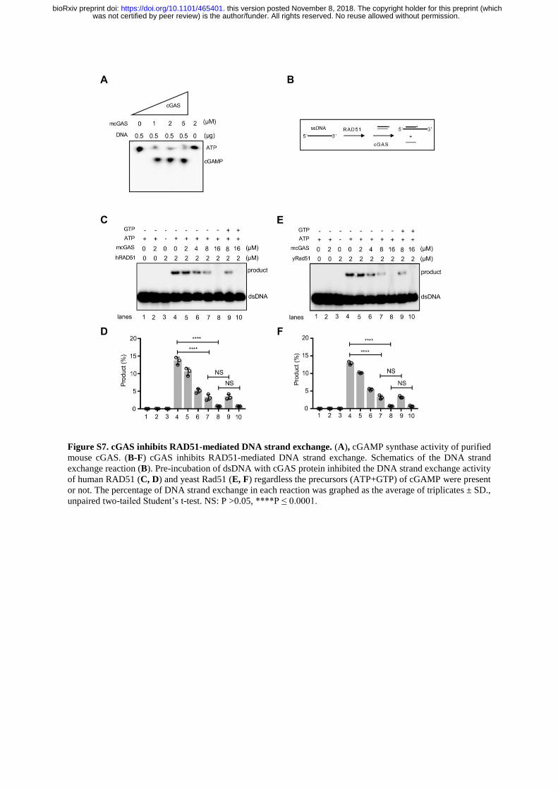
Figure S7. cGAS inhibits RAD51-mediated DNA strand exchange. (A), cGAMP synthase activity of purified
mouse cGAS. (B-F) cGAS inhibits RAD51-mediated DNA strand exchange. Schematics of the DNA strand
exchange reaction (B). Pre-incubation of dsDNA with cGAS protein inhibited the DNA strand exchange activity
of human RAD51 (C, D) and yeast Rad51 (E, F) regardless the precursors (ATP+GTP) of cGAMP were present
or not. The percentage of DNA strand exchange in each reaction was graphed as the average of triplicates ± SD.,
unpaired two-tailed Student’s t-test. NS: P >0.05, ****P ≤ 0.0001.
was not certified by peer review) is the author/funder. All rights reserved. No reuse allowed without permission. The copyright holder for this preprint (whichthis version posted November 8, 2018. . https://doi.org/10.1101/465401doi: bioRxiv preprint

Figure S8. Inhibition of RAD51-mediated D-loop formation is not a ubiquitous feature of all dsDNA binding
proteins. a, b, cGAS and MHF binds to dsDNA with similar affinity. 20-200 nM of cGAS or MHF were incubated
with dsDNA at 37oC and protein-dsDNA complexes analyzed by gel separation (a) densitometric quantification
were graphed as the average of triplicates ± SD (b). c, Schematics of the D-loop assay. d, Pre-incubation of
template dsDNA with cGAS blocks subsequent D-loop by RAD51 while pre-incubation with the DNA binding
protein MHF does not. e, The percentage of D-loop formed in each reaction (d) was graphed as the average of
triplicates ± SD., unpaired two-tailed Student’s t-test. ****P ≤ 0.0001.
was not certified by peer review) is the author/funder. All rights reserved. No reuse allowed without permission. The copyright holder for this preprint (whichthis version posted November 8, 2018. . https://doi.org/10.1101/465401doi: bioRxiv preprint