the innovative viscoelastic cp esp cervical disk prosthesis...
TRANSCRIPT
ORIGINAL ARTICLE • CERVICAL SPINE - PROSTHESIS
The innovative viscoelastic CP ESP cervical disk prosthesiswith six degrees of freedom: biomechanical concepts, developmentprogram and preliminary clinical experience
Jean-yves Lazennec1 • Alain Aaron2 • Olivier Ricart3 • Jean Patrick Rakover4
Received: 19 August 2015 / Accepted: 21 August 2015
� The Author(s) 2015. This article is published with open access at Springerlink.com
Abstract The viscoelastic cervical disk prosthesis ESP is
an innovative one-piece deformable but cohesive interbody
spacer. It is an evolution of the LP ESP lumbar disk
implanted since 2006. CP ESP provides six full degrees of
freedom about the three axes including shock absorbtion.
The prosthesis geometry allows limited rotation and
translation with resistance to motion (elastic return prop-
erty) aimed at avoiding overload of the posterior facets.
The rotation center can vary freely during motion. The
concept of the ESP prosthesis is fundamentally different
from that of the devices currently used in the cervical
spine. The originality of the concept of the ESP� prosthesis
led to innovative and intense testing to validate the adhe-
sion of the viscoelastic component of the disk on the tita-
nium endplates and to assess the mechanical properties of
the PCU cushion. The preliminary clinical and radiological
results with 2-year follow-up are encouraging for pain,
function and kinematic behavior (range of motion and
evolution of the mean centers of rotation). In this series, we
did not observe device-related specific complications,
misalignment, instability or ossifications. Additional stud-
ies and longer patient follow-up are needed to assess long-
term reliability of this innovative implant.
Keywords Cervical disk prosthesis � Artificial disk �Viscoelastic disk replacement � Cervical spine mobility �CP ESP � Rotation center � Degenerative cervical disk
Introduction
Anterior cervical discectomy and fusion (ACDF) is a
proven intervention for patients with radiculopathy and
myelopathy. A major concern related to the treatment of
cervical degenerative disk disease and spondylosis with
ACDF is the issue of adjacent segment degeneration [1].
Radiographic evidence of adjacent-level disease (ALD)
has been reported to occur in as many as 92 % of patients
at 5-year follow-up [2]. Hilibrand et al. [3] calculated a
2.9 % annual risk of symptomatic ALD; survivorship
analysis projected that a 25.6 % of the patients who
underwent an ACDF would develop symptomatic ALD
within 10 years after. There is clinical evidence to support
the postsurgical nature of adjacent segment disease. Goffin
et al. [2] were able to demonstrate a similar rate of ALD in
younger patients with trauma compared with older patients
with degeneration following anterior cervical arthrodesis.
The concept that adjacent levels need to compensate for
loss of motion in the fused segment is supported by
biomechanical studies. Cadaveric testing has demonstrated
the finding that arthrodesis generally results in increased
adjacent-level intradiscal pressures [4], and statistically
significant changes in adjacent-level motion compared with
arthroplasty under the same experimental conditions [5].
Studies additionally reveal that the degree of lordosis
achieved during fusion significantly alters adjacent-level
range of motion [6] and that in some cases, segmental
mobility may be maximally increased at levels distant from
the index level [7]. In addition, kyphotic deformity
& Jean-yves Lazennec
1 Pitie Salpetriere Hospital, UPMC, Paris, France
2 FHI, Quimper, France
3 Kirchberg Hospital, Luxembourg, Luxembourg
4 Clinique du Pre, Le Mans, France
123
Eur J Orthop Surg Traumatol
DOI 10.1007/s00590-015-1695-1

following ACDF has been implicated in the development
of segmental instability, clinically significant ALD and
poor functional recovery. It has been speculated that fusion
in kyphosis increases posterior slipping forces onto adja-
cent vertebral levels and may cause higher loads on the
posterior column than a fusion in lordosis [8].
Cervical disk arthroplasty cannot systematically sup-
plant arthrodesis [9], but it has emerged as a promising
alternative to fusion in appropriately selected patients [10]
to reduce or eliminate ALD by preserving motion at the
treated level.
The primary goals of the procedure are to preserve or to
restore normal spinal kinematics. Nevertheless, although
the range of motion (ROM) is an important feature of an
artificial disk, it is only a single aspect of spinal biome-
chanics: elastic resistance to movement, twisting potential
and elastic resistance to load bearing are major properties
of the anatomical disk. Normal kinematics should not be
analyzed only as a movement on the three planes, but also
as elastic resistance to dynamic stress on these three planes.
The aim of motion preservation is also to neutralize
excessive movements while preserving the physiologic
biomechanical properties of the functional spinal unit
(FSU) involved to interrupt the progression of degenerative
processes and to prevent ALD. Elastic resistance of the
FSU is a biomechanical property often underestimated but
crucial for the stability of the spine. The biomechanics of
cervical implants takes into account only the ROM of the
devices and not the elastic resistance: the risk is a greater
ROM in comparison with a normal disk, especially in
rotation, with a potential overloading of the facet joints
[11].
In terms of the quality of vertebral motion, the instan-
taneous center of rotation (ICR) during flexion–extension is
considered as a major parameter. The ICR location depends
on the cervical FSU level. In addition, Liu et al. described a
correlation with the age-related degeneration [12]. Cervical
arthroplasty should be optimized or adaptive enough for
attempting an ICR location close to physiologic kinematic
conditions of the motion segment according to physiologic
aging, normal degenerative changes and local potential
evolution.
The effect of this motion-sparing alternative on angu-
lation at the treated level and on the overall spinal align-
ment may be important to long-term clinical outcomes and
rates of adjacent segment disease. The control of stability
and ROM is critical to optimize and maintain local and
regional balance.
In addition, the impact on axial loading and shock
absorption of the FSU needs to be addressed [13]. Suc-
cessful reproduction of physiologic kinematics and long-
term viability of the disk replacement must consider mul-
tiple factors, including facet loading, disk height, device
design and biomaterials, and implant position [14]. The
goal is to obtain the potential to approximate the axes of
rotation of the native segment in flexion–extension, lateral
bending and axial rotation, and a graded resistance to
motion.
Total disk arthroplasty devices can be classified
according to modular versus nonmodular design, fixation
properties, articular design and composition (uniarticular,
biarticular and nonarticular), and kinematics (constrained,
semiconstrained and unconstrained) [15, 16].
The viscoelastic cervical disk prosthesis ESP is an
innovative one-piece deformable but cohesive interbody
spacer. It is an evolution of the LP ESP lumbar disk
implanted since 2006 [17].
Recognizing that the human disk does not work like a
joint but as a ‘‘silent bloc,’’ this technology meets a critical
need: in the cervical spine, the shock absorption and the
control of stability are very important to avoid side-level
degeneration, hypermobility and rotational or sagittal
imbalance. In addition, the biomechanical constrains are
very different from a disk level to each other as far as
mobility is concerned. Moreover, one-third of the asymp-
tomatic population has cervical kyphosis [18], and recent
literature has pointed out significant modifications of the
ICR according to the age and the degree of cervical
degeneration [12]. The complexity of these anatomical and
functional data shows the limitations of conventional
mechanical prostheses and highlights the potential interest
of viscoelastic concept for cervical prostheses.
CP ESP provides six full degrees of freedom about the
three axes including shock absorption.
The prosthesis geometry allows limited rotation and
translation with resistance to motion (elastic return prop-
erty) aimed at avoiding overload of the posterior facets.
The rotation center can vary freely during motion. It thus
differs substantially from current prostheses.
The goal of this paper is to present this innovative
concept and the preliminary clinical results and radiologi-
cal outcomes. This study reports the results of a prospective
pilot evaluation of the first patients implanted since 2012.
In addition to measuring ROM, we were specifically
interested in the quality of the kinematics and thus we
investigated the mean center of rotation at both the
instrumented and adjacent levels.
The implant
Because the healthy human intervertebral disk has a
deformable elastic structure with six degrees of freedom,
elastomeric one-piece intervertebral prostheses might be
the most physiologic implant for mimicking physiologic
levels of shock absorption and flexural stiffness. The ESP�
Eur J Orthop Surg Traumatol
123

concept (currently lumbar and cervical disk prostheses
marketed as the elastic spine pad) was in development for
20 years. With the technological advancements of the
ESP�, the problem of how to bond the elastic component
and the titanium endplates of the disk is solved. Following
promising in vitro and in vivo testing, the LP ESP gained
clinical approval for use in Europe in 2005 for the lumbar
levels [19]. More recently, the cervical version has been
CE approved in 2012 for the first implantations.
The design of the CP ESP� prosthesis is based on the
principle of the silent block bush (Fig. 1). The CP ESP�
is a one-piece deformable implant including a central
core made of polycarbonate urethane (PCU) securely
fixed to titanium endplates. The endplates have anchor-
ing pegs to provide primary fixation and are covered by
a textured T 40 titanium layer and hydroxyapatite to
improve bone ongrowth. This center cushion is bonded
to the titanium alloy endplates via adhesion molding and
employs a peripheral groove without using glue for
reinforcement. This type of fixation prevents fluid infil-
tration and fatigue fractures of the interface, despite the
disparate mechanical properties of the polymer and metal
endplates. The shape of the cushion has been studied to
obtain an optimization of the mobility as well as the
control of translation and shear movements during cer-
vical spine mobility.
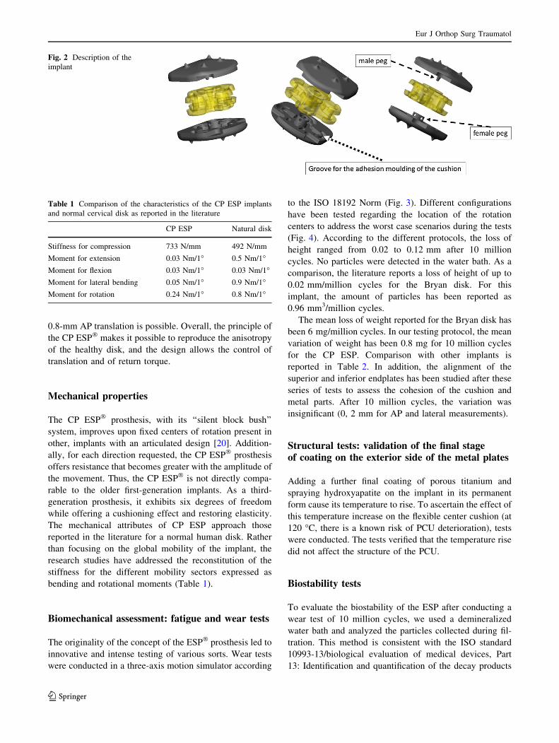
The PCU annulus is stabilized by supplementary
‘‘male’’ and ‘‘female’’ inner pegs located on the internal
surface of both metal endplates. The geometry and position
of the pegs, between the peripheral groove and the central
area of the endplates, were planned to control compression
and translation (Fig. 2). These two pegs, with their con-
tactless fit of the male and female caps, serve to limit
shearing during anteroposterior and medio-lateral transla-
tion. Through this mechanism, as well as crushing the
annulus between the metal plates, the inner pegs also
provide cushioning and compressing. These pegs also limit
shearing when the endplates are inclined to the horizontal.
The prostheses are available in three thicknesses (5, 6
and 7 mm), each with three sizes in AP and lateral
dimensions (13 mm 9 15 mm, 14 mm 9 17 mm and
15 mm 9 20 mm). Regardless of the model, however, the
mechanically active cushion and the mechanical properties
of the prosthesis are the same: the differences in thickness
do not affect the prosthesis’s mobility or its cushioning,
even shock-absorbing, effect. The design of the prosthesis
allows a range of 14� for flexion–extension, 12� for right-left lateral bending and 8� for rotation. In addition, a
Fig. 1 Radiological images with CP ESP disk implant
Eur J Orthop Surg Traumatol
123
0.8-mm AP translation is possible. Overall, the principle of
the CP ESP� makes it possible to reproduce the anisotropy
of the healthy disk, and the design allows the control of
translation and of return torque.
Mechanical properties
The CP ESP� prosthesis, with its ‘‘silent block bush’’
system, improves upon fixed centers of rotation present in
other, implants with an articulated design [20]. Addition-
ally, for each direction requested, the CP ESP� prosthesis
offers resistance that becomes greater with the amplitude of
the movement. Thus, the CP ESP� is not directly compa-
rable to the older first-generation implants. As a third-
generation prosthesis, it exhibits six degrees of freedom
while offering a cushioning effect and restoring elasticity.
The mechanical attributes of CP ESP approach those
reported in the literature for a normal human disk. Rather
than focusing on the global mobility of the implant, the
research studies have addressed the reconstitution of the
stiffness for the different mobility sectors expressed as
bending and rotational moments (Table 1).
Biomechanical assessment: fatigue and wear tests
The originality of the concept of the ESP� prosthesis led to
innovative and intense testing of various sorts. Wear tests
were conducted in a three-axis motion simulator according
to the ISO 18192 Norm (Fig. 3). Different configurations
have been tested regarding the location of the rotation
centers to address the worst case scenarios during the tests
(Fig. 4). According to the different protocols, the loss of
height ranged from 0.02 to 0.12 mm after 10 million
cycles. No particles were detected in the water bath. As a
comparison, the literature reports a loss of height of up to
0.02 mm/million cycles for the Bryan disk. For this
implant, the amount of particles has been reported as
0.96 mm3/million cycles.
The mean loss of weight reported for the Bryan disk has
been 6 mg/million cycles. In our testing protocol, the mean
variation of weight has been 0.8 mg for 10 million cycles
for the CP ESP. Comparison with other implants is
reported in Table 2. In addition, the alignment of the
superior and inferior endplates has been studied after these
series of tests to assess the cohesion of the cushion and
metal parts. After 10 million cycles, the variation was
insignificant (0, 2 mm for AP and lateral measurements).
Structural tests: validation of the final stageof coating on the exterior side of the metal plates
Adding a further final coating of porous titanium and
spraying hydroxyapatite on the implant in its permanent
form cause its temperature to rise. To ascertain the effect of
this temperature increase on the flexible center cushion (at
120 �C, there is a known risk of PCU deterioration), tests
were conducted. The tests verified that the temperature rise
did not affect the structure of the PCU.
Biostability tests
To evaluate the biostability of the ESP after conducting a
wear test of 10 million cycles, we used a demineralized
water bath and analyzed the particles collected during fil-
tration. This method is consistent with the ISO standard
10993-13/biological evaluation of medical devices, Part
13: Identification and quantification of the decay products
Fig. 2 Description of the
implant
Table 1 Comparison of the characteristics of the CP ESP implants
and normal cervical disk as reported in the literature
CP ESP Natural disk
Stiffness for compression 733 N/mm 492 N/mm
Moment for extension 0.03 Nm/1� 0.5 Nm/1�Moment for flexion 0.03 Nm/1� 0.03 Nm/1�Moment for lateral bending 0.05 Nm/1� 0.9 Nm/1�Moment for rotation 0.24 Nm/1� 0.8 Nm/1�
Eur J Orthop Surg Traumatol
123

of polymer-based medical devices. We used a scanning
electron microscope (SEM LEO I455VP), equipped with
an energy-selective spectrometer (EDS OXFORD). No
particles from the component materials of the prosthesis
were found. These results were consistent with previous
studies on the LP ESP disk.
PCU aging test
The specific PCU used for the LP ESP prosthesis is not
oxidized during storage (bionate 80A (DSNM Biomedical,
The Netherlands) according to master file MAF844) [28].
To evaluate this claim, the PCU was artificially aged in
80 �C water, in accordance with recommendations for
aging plastics such as the UHMWPE (ASTM standard F
2003: Accelerated aging of ultra-high molecular weight
polyethylene after gamma irradiation in air). The aged
PCU was then subjected to compression loads from 150 N
to 1250 N, consistent with ISO standard 18192 (interver-
tebral spinal disk prostheses Part 1: Loading and dis-
placement parameters for wear testing and corresponding
environmental conditions for test) for wear tests, for 10
million cycles.
No modification of the Fournier transform infrared
spectrum or any modification of the mean molecular
weight (ASTM standard D 5296) was observed. The PCU
aging test results were comparable to those reported in the
literature [29].
Biocompatibility tests
Biomatech (Chasse-sur-Rhone, France) conducted the
biocompatibility tests (Table 3). The material components
were evaluated individually and as a finished assembly, as
Fig. 3 Description of the cycle
testing according to ISO 18192
Norm
Fig. 4 Different configurations
have been tested regarding the
location of the rotation centers
to address the worst case
scenarios during the tests
Eur J Orthop Surg Traumatol
123

Table
2Literature
summaryofbiomechanical
simulationsforwear
References
Device
Sim
ulation
Massloss
Heightloss
Volumeloss
Andersonet
al.
[21]
Bryan
6disks:10millioncycles;150N
forceflexion/extension,
lateralbendingandaxialrotationin
37�C
calfserum
sample;bending?
10millioncycles
rotationat
4Hz;
4disks:
tested
untilandplate
toendplate
contact
1.75%
(after
20millioncycles
in6disks)
0.48%
(after
20million
cycles
in6disks)
0.57mm
3/m
illioncycles
Andersonet
al.
[21]
Prestige
10millioncycles
of148N
flexion/extension,5million
cycles
of49N
lateralbendingand5millioncycles
of
49N
axialrotationat
2Hzin
37�C
calfserum
sample;
number
ofdiskstested
notreported
NR
NR
0.18mm
3/m
illioncycles
(0,
19%
loss
after20million
cycles)
FDA
[22]
ProDiscC
10millioncycles
of150N
flexion/extension,lateral
bendingandaxialrotationat
1Hzin
37�C
calfserum
sample;debrissampledeverymillioncycles
2.59mg/m
illioncycles
NR
NR
Gruppet
al.[23]
ActiveC
6diskstested
withaISO
18192-1:2008(E)
process:flexion,extension,lateralbendingandaxial
rotationmovem
entsin
sinusoidal
wavefrom
with150N
flexionforceand50N
extensionforce,10millioncycles
at1Hzin
37�C
calfserum
sample
1.0
mg/m
illioncycles
0.03mg/m
illioncycles
for
polymer
inlay
1.0
mm
3/m
illioncycles
FDA
[24]
PCM
5diskstested
under
DraftASTM
F243-05conditions(±
10�
fullyreversinglateralbending,±6�fullyreversingaxial)
Averagecumulativewearat
10
millioncycles
was
71.22±
17.56mg.Wearrate
0.042mg/m
illioncycles
between3and10million
cycles
NR
NR
FDA
[25]
Secure-C
150N
constantcompressiveload
for10millioncycles.
Calfserum
anddeionized
water
solutionwithEDTA,
maintained
at37�C
Stage1:6disksfor10millioncycles
ofcomplexloadingat
afrequency
of2Hzusingcombined
flexion/extension
(±7�),lateralbending(±
7�)
andaxialrotation(±
1.5�)
Stage2:tested
withincreasedaxialrotation(±
6�)
Stage1:2.57mg±
1.21mgper
millioncycles
Stage2:0.89mg±
0.3
mgper
millioncycles
NR
NR
FDA
[26]
MobiC
6disks:10millioncycles;frequency
of1Hz
Combined
flexion/extension(±
7.5�),lateralbending(±
6�)
androtation(±
4�)
under
axialcompression(50–150N)
1.456±
0.075mg/m
illioncycles
NR
NR
FDA
[27]
PrestigeLP
Stage1:6diskstested
inaccordance
withASTM
2423
(lateral
bendingcoupledwithaxialrotationfollowed
by
flexion/extension)
Stage2:6diskstested
inaccordance
withISO
18192-1
(lateral
bendingcombined
withaxialrotationandflexion/
extension)
NR
NR
Stage1:0.35±
0.03
mm
3/m
illioncycles
totalwearat
20million
cycles:4.22±
0.21mm
3
Stage2:steady-state
wear
rate
0.25±
0.04
mm
3/m
illioncycles
totalaccumulatedwear
2.74±
0.38mm
3
CPESP
3-A
xismotionsimulatoraccordingto
theISA
18192Norm
Meanvariationofweight0.8
mg
for10millioncycles
Loss
ofheightranged
from
0.02to
0.12mm
after
10millioncycles
NR
indicates
notreported
Eur J Orthop Surg Traumatol
123
specified in ISO standard 10993 (Biological Evaluation of
Medical Devices): Cytotoxicity was assessed using ISO
standard 10993-5. Testing for sensitization conformed to
ISO standard 10993-10.
Employing ISO standard 10993-10, the components and
assembly were tested for irritation or intradermal reaction.
In addition, acute systemic toxicity was examined using
ISO standard 10993-11 Chromosomal genotoxicity (Hearts
test), and chromosomal anomalies were tested in accor-
dance with ISO standard I 0993-3.
The Biomatech biocompatibility testing revealed that
the ESP devices also satisfy the criteria of the FDA’s
subacute sensitization test (FDA—Guidelines for Toxicity
Tests Chapter IV).
Clinical study
Our evaluation program was organized in two stages:
• a preliminary study with 1-year follow-up to detect
potential dysfunctions and early postoperative stabi-
lization problems or adjustment difficulties
• a 2-year analysis focused on radiological progression in
terms of mobility and centers of rotation of analysis
The cases
We analyzed prospectively 62 consecutive cases included
in a preliminary study according to the classical indications
and contraindications for cervical disk replacement. The
aim of this work is to provide a snapshot of the evolution of
our series since the first implantations.
The surgeries were performed by two senior surgeons.
There were 39 women and 23 men in this group. The
average age was 45 ± 8 years (29–60). The implantation
was on a single level in 74 % of the CP ESP and on two
levels in 26 %. Globally, 71 CP ESP prostheses were
analyzed (Table 4).
Clinical data and X-rays were collected at the preoper-
ative time and at 3, 6, 12 and 24 months post-op.
The functional results were measured using Neck and
Arm VAS, NDI, SF-36 (physical component PCS and
mental component MCS). The analysis was performed by a
single observer who was independent from the selection of
patients and from the surgical procedure.
One-year follow-up results
The mean operative time was 48 min (40–75 min) (SD
9 min). The hospital stay was 3.2 days (SD 0.8)
(1–5 days).
We did not observe device-related specific complica-
tions. In this series, we did not face misalignment, insta-
bility or ossifications.
All clinical outcomes significantly improved at every
time points when compared to the preoperative status
(Table 5). Table 6 summarizes the changes in the varia-
tions of ROM over time.
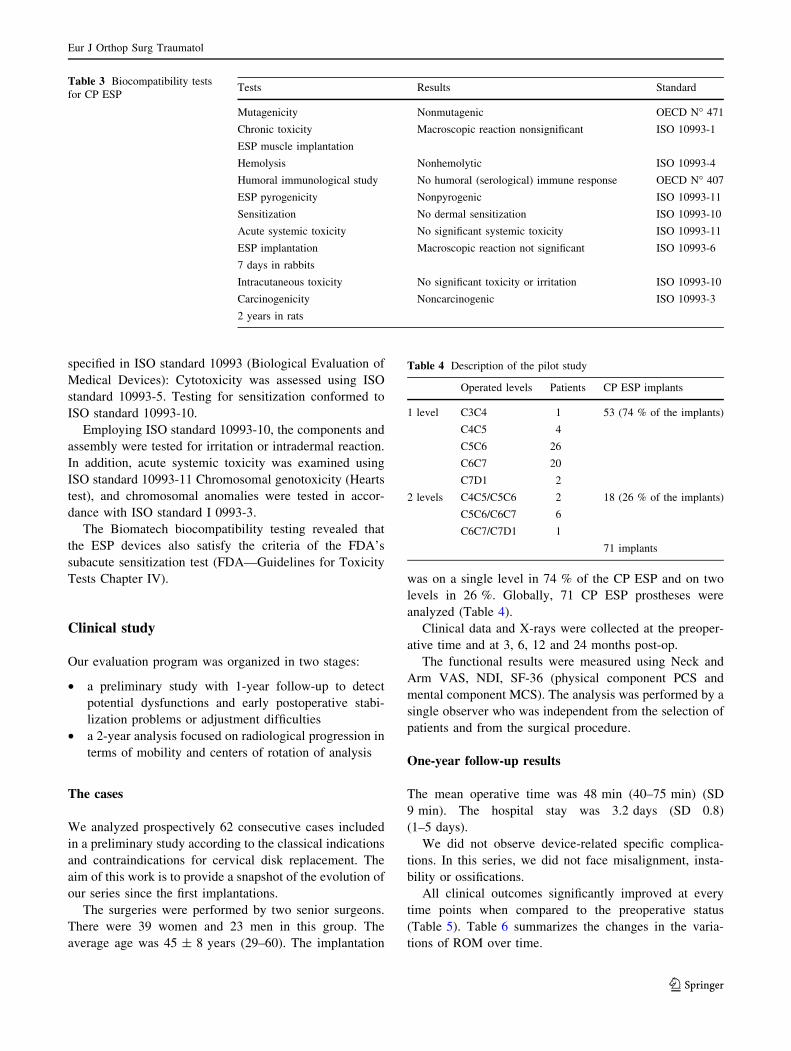
Table 3 Biocompatibility tests
for CP ESPTests Results Standard
Mutagenicity Nonmutagenic OECD N� 471Chronic toxicity
ESP muscle implantation
Macroscopic reaction nonsignificant ISO 10993-1
Hemolysis Nonhemolytic ISO 10993-4
Humoral immunological study No humoral (serological) immune response OECD N� 407ESP pyrogenicity Nonpyrogenic ISO 10993-11
Sensitization No dermal sensitization ISO 10993-10
Acute systemic toxicity No significant systemic toxicity ISO 10993-11
ESP implantation
7 days in rabbits
Macroscopic reaction not significant ISO 10993-6
Intracutaneous toxicity No significant toxicity or irritation ISO 10993-10
Carcinogenicity
2 years in rats
Noncarcinogenic ISO 10993-3
Table 4 Description of the pilot study
Operated levels Patients CP ESP implants
1 level C3C4 1 53 (74 % of the implants)
C4C5 4
C5C6 26
C6C7 20
C7D1 2
2 levels C4C5/C5C6 2 18 (26 % of the implants)
C5C6/C6C7 6
C6C7/C7D1 1
71 implants
Eur J Orthop Surg Traumatol
123

The mean centers of rotation (MCR) at the instrumented
and at the upper and lower adjacent levels were measured
in flexion/extension using Spineview� software (3-, 6- and
12-month follow-up) (Fig. 5). Their evolution illustrates
the versatility and the forgiveness of the CP ESP implants
according to the levels and the various sagittal balance.
Table 5 Clinical outcomes of
the preliminary studyMean ± SD Pre-op 3 Months 6 Months 12 Months
(a)
VAS neck (/10) 6 2.65 1.74 2.65
VAS arm (/10) 6.3 2.7 1.8 2.4
NDI (%) 56 ± 16 32.7 ± 17 22.1 ± 16 24 ± 17
NDI points 27.4 ± 8.8 16.2 ± 8.6 10.9 ± 7.9 11.9 ± 8.5
SF-36 PCS score 31 48 61 56
SF-36 MCS score 32 50 63 62
Pre-op/3
Months
Pre-op/6
Months
Pre-op/12
Months
(b) Mann–Whitney test for the evolution of the clinical tests (VAS, NDI, SF-36 P, SF-36 M)
VAS neck Conclusion
P value 0.01 0.01 0.01
VAS arm Conclusion
P value 0.01 0.01 0.01
NDI Conclusion
P value 0.01 0.01 0.01
SF-36 PCS Conclusion
P value 0.01 0.01 0.01
SF-36 MCS Conclusion
P value 0.01 0.01 0.01
Table 6 Evolution of the ROM
of the implanted and adjacent
levels
Degrees (�) 3 Months 6 Months 12 Months
ROM of the instrumented level 6.8 ± 4.1 10.3 ± 5.0 8.4 ± 4.3
ROM of the upper adjacent level 9.7 ± 4.9 11.7 ± 5.5 12.9 ± 6.8
ROM of the lower adjacent level 6 ± 4.1 10 ± 5.0 9.4 ± 5.7
Fig. 5 Mean centers of rotation (MCR) at the instrumented and at the upper and lower adjacent levels measured for flexion/extension using
Spineview� software; we can observe the adaptation of the implant including a translation for flexion
Eur J Orthop Surg Traumatol
123

Two-year follow-up results
The evolution of clinical and radiological parameters
confirms the good results already observed at 12 months of
follow-up (Table 7; Fig. 6a–f).
Conclusion
The design of the CP ESP� prosthesis provides stability by
limiting rotation and translation, which prevents overload
of the posterior facet joints. At the same time, the ESP
center of rotation can fluctuate during motion. While
achieving six degrees of freedom including vertical trans-
lation, this viscoelastic prosthesis also supplies cushioning,
which offers shock absorption. The CP ESP thus distin-
guishes itself substantially from other current two- or three-
piece prostheses that contain l or 2 bearing surfaces and
provide only 3 or 5 degrees of freedom. These other
devices also offer no or very little resistance and provide no
elastic return. This study reports encouraging clinical
results about pain, function, kinematic behavior and ROM.
We concede that additional clinical studies and patient
follow-up are needed to assess long-term reliability.
However, the results we describe here suggest the out-
comes that surgeons and patients might anticipate.
Pre op 3M 6M 12M
ROM of the instrumented level 4° 9° 13° 10°
ROM of the upper adjacent level 4° 10° 11° 11°
ROM of the lower adjacent level 2° 5° 14° 12°
24M
14°
14°
10°
Fig. 6 a–f Mean centers of rotation (MCR) and mobility at the instrumented and at the upper and lower adjacent levels in a patient with global
cervical kyphosis: adaptation during the follow-up from 3 to 24 months
Table 7 Clinical and radiological outcomes after 2-year follow-up
Mean ± SD 24 Months
VAS neck (/10) 2.9
VAS arm (/10) 1.6
NDI (%) 19 ± 17
NDI points 9.3 ± 8
SF-36 PCS score 64.2
SF-36 MCS score 68.6
ROM of the instrumented level 10.7� ± 4.2�ROM of the upper adjacent level 13.8� ± 6.5�ROM of the lower adjacent level 11.1� ± 8.2�
Eur J Orthop Surg Traumatol
123

The optimal ROM after TDR for limiting adjacent
segmental disease has not yet been established. The radi-
ological evaluation of the results is classically focused on
the ROM, but the quality of movement is also an issue,
especially at the cervical levels as coupled motions are a
key point. The CP ESP acts as a deformable but cohesive
interbody spacer that provides six full degrees of freedom
about the three axes. This allows instantaneous axis of
rotation change freely, as in the normal disk, which can
optimize a functional coherence with the facet joints
mechanics. It is also a significant benefit for the adaptation
to the various disk levels as the biomechanical constrains
are different from C3 to T1. Our preliminary experience
did not point out complications as sagittal misalignment,
ossifications and instability. An interesting point is the
evolution of the MCR in the postoperative course. This
adaptation ability is one of the main features of this
promising implant as we need to consider the mean and
long-term evolution of the global cervical posture and
mobility after a cervical disk replacement.
Compliance with ethical standards
Conflict of interest No funding was received for this study. All of
the authors are patent owners for disk replacement.
Human and animal rights All procedures performed in this study
involving human participants were in accordance with the ethical
standards of the institutional and/or national research committee and
with the 1964 Helsinki Declaration and its later amendments or
comparable ethical standards. Informed consent was obtained from all
individual participants included in the study.
Open Access This article is distributed under the terms of the
Creative Commons Attribution 4.0 International License (http://crea
tivecommons.org/licenses/by/4.0/), which permits unrestricted use,
distribution, and reproduction in any medium, provided you give
appropriate credit to the original author(s) and the source, provide a
link to the Creative Commons license, and indicate if changes were
made.
References
1. Bartolomei JC, Theodore N, Sonntag VK (2005) Adjacent level
degeneration after anterior cervical fusion: a clinical review. Neu-
rosurg Clin N Am 16(4):575–587. doi:10.1016/j.nec.2005.07.004
2. Goffin J, Geusens E, Vantomme N, Quintens E, Waerzeggers Y,
Depreitere B, Van Calenbergh F, van Loon J (2004) Long-term
follow-up after interbody fusion of the cervical spine. J Spinal
Disord Tech 17(2):79–85
3. Hilibrand AS, Carlson GD, Palumbo MA, Jones PK, Bohlman
HH (1999) Radiculopathy and myelopathy at segments adjacent
to the site of a previous anterior cervical arthrodesis. J Bone Joint
Surg Am 81(4):519–528
4. Dmitriev AE, Cunningham BW, Hu N, Sell G, Vigna F, McAfee
PC (2005) Adjacent level intradiscal pressure and segmental
kinematics following a cervical total disc arthroplasty: an in vitro
human cadaveric model. Spine (Phila Pa 1976)
30(10):1165–1172
5. Chang UK, Kim DH, Lee MC, Willenberg R, Kim SH, Lim J
(2007) Range of motion change after cervical arthroplasty with
ProDisc-C and prestige artificial discs compared with anterior
cervical discectomy and fusion. J Neurosurg Spine 7(1):40–46.
doi:10.3171/SPI-07/07/040
6. Hwang SH, Kayanja M, Milks RA, Benzel EC (2007) Biome-
chanical comparison of adjacent segmental motion after ventral
cervical fixation with varying angles of lordosis. Spine J
7(2):216–221. doi:10.1016/j.spinee.2006.05.018
7. Fuller DA, Kirkpatrick JS, Emery SE, Wilber RG, Davy DT
(1998) A kinematic study of the cervical spine before and
after segmental arthrodesis. Spine (Phila Pa 1976)
23(15):1649–1656
8. Katsuura A, Hukuda S, Saruhashi Y, Mori K (2001) Kyphotic
malalignment after anterior cervical fusion is one of the factors
promoting the degenerative process in adjacent intervertebral
levels. Eur Spine J 10(4):320–324
9. Auerbach JD, Jones KJ, Fras CI, Balderston JR, Rushton SA,
Chin KR (2008) The prevalence of indications and contraindi-
cations to cervical total disc replacement. Spine J 8(5):711–716.
doi:10.1016/j.spinee.2007.06.018
10. Albert TJ, Eichenbaum MD (2004) Goals of cervical disc
replacement. Spine J 4(6 Suppl):292S–293S. doi:10.1016/j.spi
nee.2004.07.023
11. Landi A (2013) Elastic resistance of the spine: why does motion
preservation surgery almost fail? World J Clin Cases
1(4):134–139. doi:10.12998/wjcc.v1.i4.134
12. Liu B, Liu Z, VanHoof T, Kalala J, Zeng Z, Lin X (2014)
Kinematic study of the relation between the instantaneous center
of rotation and degenerative changes in the cervical intervertebral
disc. Eur Spine J 23(11):2307–2313. doi:10.1007/s00586-014-
3431-7
13. Kim SW, Shin JH, Arbatin JJ, Park MS, Chung YK, McAfee PC
(2008) Effects of a cervical disc prosthesis on maintaining
sagittal alignment of the functional spinal unit and overall sagittal
balance of the cervical spine. Eur Spine J 17(1):20–29. doi:10.
1007/s00586-007-0459-y
14. Galbusera F, Bellini CM, Brayda-Bruno M, Fornari M (2008)
Biomechanical studies on cervical total disc arthroplasty: a lit-
erature review. Clin Biomech (Bristol, Avon) 23(9):1095–1104.
doi:10.1016/j.clinbiomech.2008.06.002
15. Fekete TF, Porchet F (2010) Overview of disc arthroplasty—past,
present and future. Acta Neurochir (Wien) 152(3):393–404.
doi:10.1007/s00701-009-0529-5
16. Stanton P, Eck JC (2010) Materials and design characteristics of
cervical arthroplasty devices. Techniques in Orthopaedics
25(2):93–96. doi:10.1097/BTO.0b013e3181df2ef3
17. Lazennec JY, Aaron A, Brusson A, Rakover JP, Rousseau MA
(2013) The LP-ESP((R)) lumbar disc prosthesis with 6 degrees of
freedom: development and 7 years of clinical experience. Eur J
Orthop Surg Traumatol 23(2):131–143. doi:10.1007/s00590-012-
1166-x
18. Le Huec JC, Demezon H, Aunoble S (2015) Sagittal parameters
of global cervical balance using EOS imaging: normative values
from a prospective cohort of asymptomatic volunteers. Eur Spine
J 24(1):63–71. doi:10.1007/s00586-014-3632-0
19. Lazennec JY, Even J, Skalli W, Rakover JP, Brusson A, Rous-
seau MA (2014) Clinical outcomes, radiologic kinematics, and
effects on sagittal balance of the 6 df LP-ESP lumbar disc
prosthesis. Spine J 14(9):1914–1920. doi:10.1016/j.spinee.2013.
11.016
20. Rousseau MA, Cottin P, Levante S, Nogier A, Lazennec JY,
Skalli W (2008) In vivo kinematics of two types of ball-and-
socket cervical disc replacements in the sagittal plane: cranial
versus caudal geometric center. Spine (Phila Pa 1976) 33(1):E6–
E9. doi:10.1097/BRS.0b013e31815e5dce
Eur J Orthop Surg Traumatol
123
21. Anderson PA, Rouleau Jeffrey P, Toth Jeffrey M, Daniel Riew K
(2004) A comparison of simulator-tested and -retrieved cervical
disc prostheses. J Neurosurg Spine 1(2):202–210. doi:10.3171/
spi.2004.1.2.0202
22. Food and Drug Administration FDA (2009) Summary of safety
and effectiveness data, ProDisc-C total disc replacement,
P070001, 17 Dec 2007. http://www.accessdata.fda.gov/cdrh_
docs/pdf7/P070001b.pdf
23. Grupp TM, Meisel HJ, Cotton JA, Schwiesau J, Fritz B, Blomer
W, Jansson V (2010) Alternative bearing materials for interver-
tebral disc arthroplasty. Biomaterials 31(3):523–531. doi:10.
1016/j.biomaterials.2009.09.064
24. Food andDrugAdministration FDA (2012) Summary of safety and
effectiveness data, PCM disc replacement, P100012, 26 Oct 2012.
http://www.accessdata.fda.gov/cdrh_docs/pdf10/P100012b.pdf
25. Food and Drug Administration FDA (2012) Summary of safety and
effectiveness data, Secure-C total disc replacement, P100003, 28
Sept 2012. http://www.globusmedical.com/pdf/secure-c/SSED.pdf
26. Food and Drug Administration FDA (2013) Summary of safety
and effectiveness data, Mobi C total disc replacement, P110002,
7 Aug 2013. http://www.accessdata.fda.gov/cdrh_docs/pdf11/
P110002b.pdf
27. Food and Drug Administration FDA (2014) Summary of safety
and effectiveness data, Prestige LP cervical disc replacement,
P090029, 24 July 2014. http://www.accessdata.fda.gov/cdrh_
docs/pdf9/P090029b.pdf
28. Kurtz SM, Siskey R, Reitman M (2010) Accelerated aging, nat-
ural aging, and small punch testing of gamma-air sterilized
polycarbonate urethane acetabular components. J Biomed Mater
Res B Appl Biomater 93(2):442–447. doi:10.1002/jbm.b.31601
29. Christenson EM, Dadsetan M, Wiggins M, Anderson JM, Hiltner
A (2004) Poly(carbonate urethane) and poly(ether urethane)
biodegradation: in vivo studies. J Biomed Mater Res A
69(3):407–416. doi:10.1002/jbm.a.30002
Eur J Orthop Surg Traumatol
123