the integument, musculoskeletal, and neurological systems integument, musculoskeletal... · plantar...
TRANSCRIPT
The Integument,
Musculoskeletal,
and Neurological
Systems Prepared and Presented by:
Ms. Sohad Noorsaeed, RN. MSN
Chapter 8 Health Assessment

The Integument (skin, hair, and nails): 1. Identify techniques used to assess the Integument.
2. Define most common terms when assessing the integument.
3. Explore the normal and abnormal findings of integumentary assessment.
4. Differentiate between primary and secondary skin lesions with examples.
Musculoskeletal System: 1. Identify techniques used to assess the MS.
2. Define most common terms when assessing the MS.
3. Explore the normal and abnormal findings of MS assessment.
Neurological System: 1. Identify techniques used to assess the neurological system.
2. Define most common terms when assessing the neurological system.
3. Explore the normal and abnormal findings of neurological assessment.
Learning Outcomes
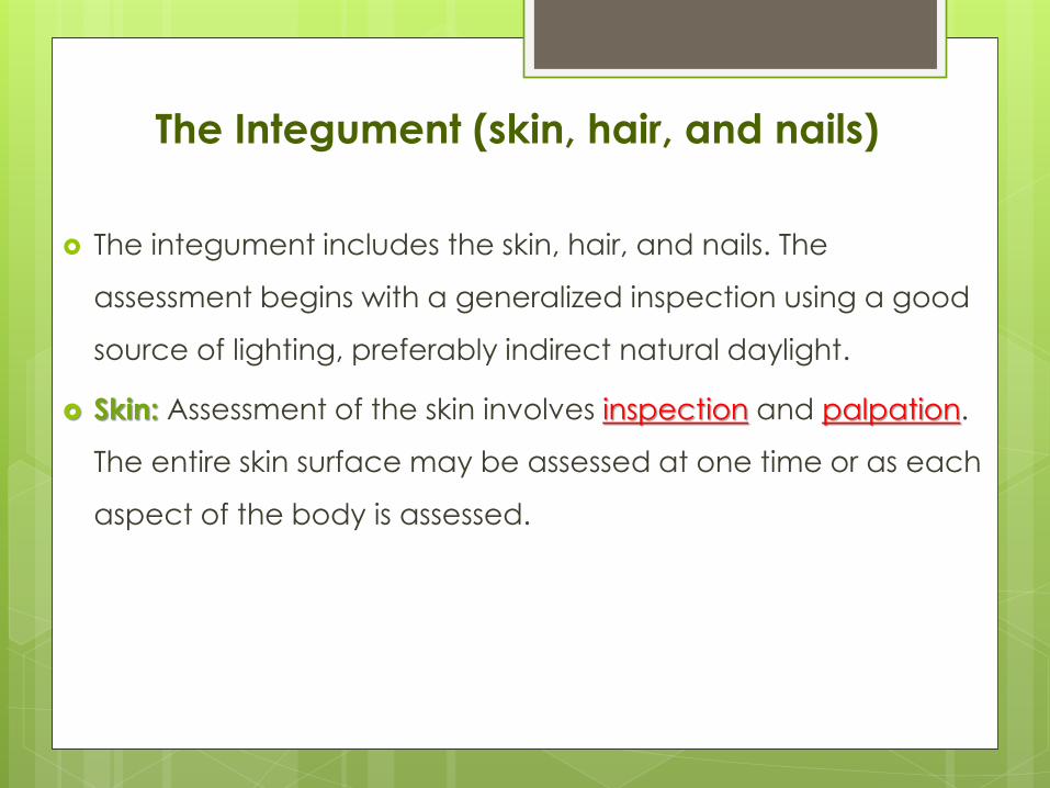
The integument includes the skin, hair, and nails. The
assessment begins with a generalized inspection using a good
source of lighting, preferably indirect natural daylight.
Skin: Assessment of the skin involves inspection and palpation.
The entire skin surface may be assessed at one time or as each
aspect of the body is assessed.
The Integument (skin, hair, and nails)

1-Inspecting Skin Color:
Pallor (decrease in skin color) is the result of inadequate
circulating blood or hemoglobin. Pallor in all people is usually
most evident in the conjunctiva, oral mucous membranes, nail
beds, palms of the hand, and soles of the feet.
Cyanosis (a bluish tinge) is most evident in the nail beds, lips,
and buccal mucosa.
The Integument (skin, hair, and nails)

Jaundice (a yellowish tinge) may first be evident in the sclera of
the eyes and then in the mucous membranes and the skin. If
jaundice is suspected, the posterior part of the hard palate
should also be inspected for a yellowish color tone.
Erythema is a redness associated with a variety of rashes. Other
localized color changes may indicate a problem such as edema
or a localized infection.
Edema is the presence of excess interstitial fluid. An area of
edema appears swollen, shiny, and taut and tends to blanch the
skin color or, if accompanied by inflammation, may redden the
skin.
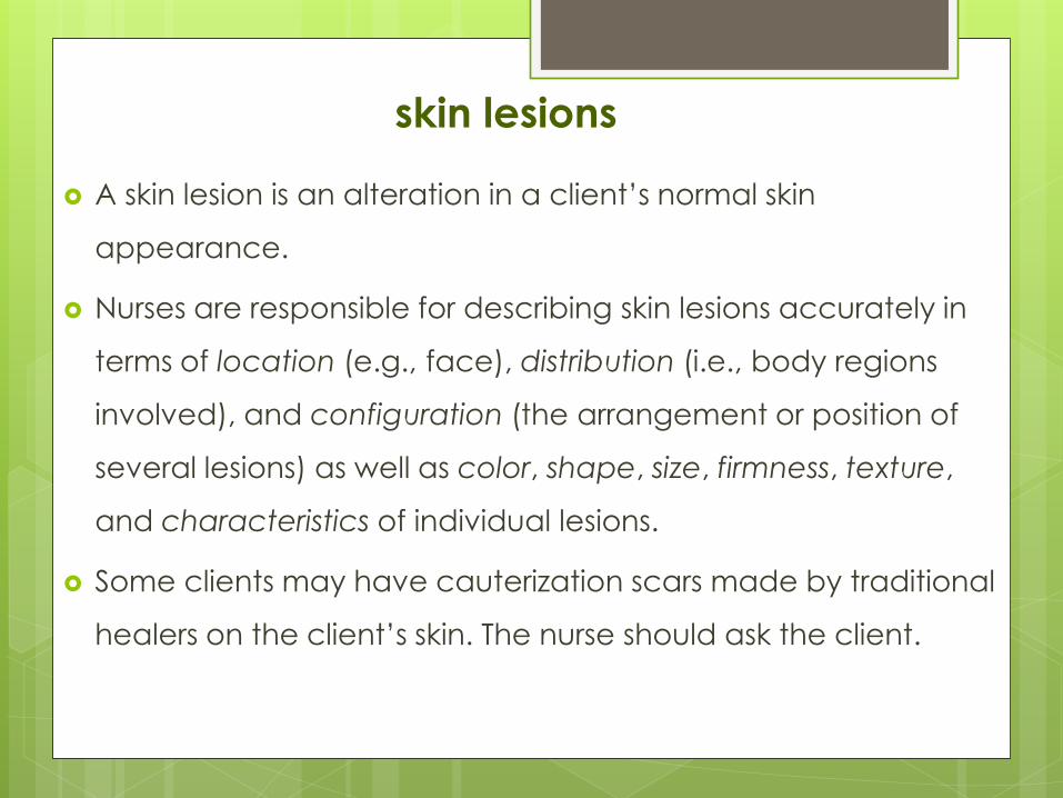
A skin lesion is an alteration in a client’s normal skin
appearance.
Nurses are responsible for describing skin lesions accurately in
terms of location (e.g., face), distribution (i.e., body regions
involved), and configuration (the arrangement or position of
several lesions) as well as color, shape, size, firmness, texture,
and characteristics of individual lesions.
Some clients may have cauterization scars made by traditional
healers on the client’s skin. The nurse should ask the client.
skin lesions
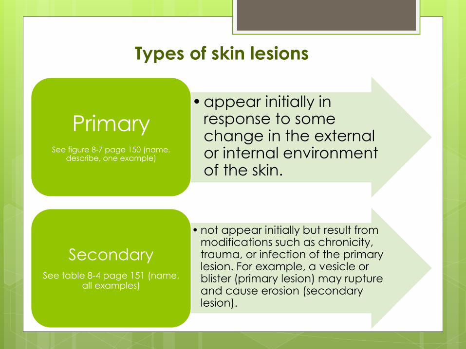
• appear initially in response to some change in the external or internal environment of the skin.
Primary See figure 8-7 page 150 (name,
describe, one example)
• not appear initially but result from modifications such as chronicity, trauma, or infection of the primary lesion. For example, a vesicle or blister (primary lesion) may rupture and cause erosion (secondary lesion).
Secondary See table 8-4 page 151 (name,
all examples)
Types of skin lesions
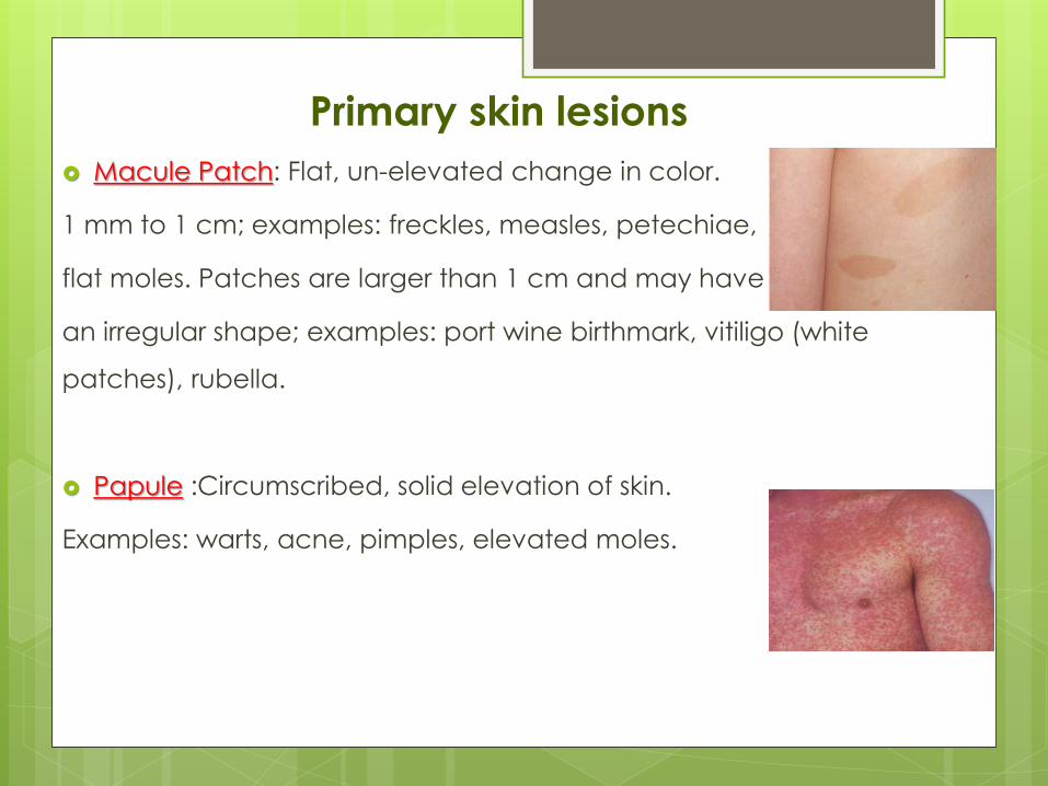
Macule Patch: Flat, un-elevated change in color.
1 mm to 1 cm; examples: freckles, measles, petechiae,
flat moles. Patches are larger than 1 cm and may have
an irregular shape; examples: port wine birthmark, vitiligo (white
patches), rubella.
Papule :Circumscribed, solid elevation of skin.
Examples: warts, acne, pimples, elevated moles.
Primary skin lesions

Primary skin lesions
Plaque :Plaques are larger than 1 cm. Examples: psoriasis, rubeola.
Nodule: Tumor Elevated, solid, hard mass that extends deeper into the
dermis than a papule. circumscribed border 0.5 to 2 cm. Examples:
squamous cell carcinoma, fibroma. Tumors are larger than 2 cm and
may have an irregular border. Examples: malignant melanoma,
hemangioma.
Primary skin lesions
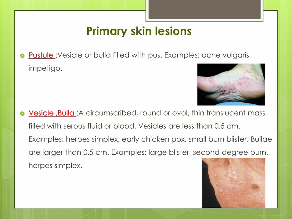
Pustule :Vesicle or bulla filled with pus. Examples: acne vulgaris,
impetigo.
Vesicle ,Bulla :A circumscribed, round or oval, thin translucent mass
filled with serous fluid or blood. Vesicles are less than 0.5 cm.
Examples: herpes simplex, early chicken pox, small burn blister. Bullae
are larger than 0.5 cm. Examples: large blister, second degree burn,
herpes simplex.
Primary skin lesions
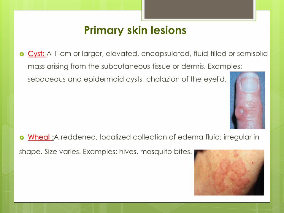
Cyst: A 1-cm or larger, elevated, encapsulated, fluid-filled or semisolid
mass arising from the subcutaneous tissue or dermis. Examples:
sebaceous and epidermoid cysts, chalazion of the eyelid.
Wheal :A reddened, localized collection of edema fluid; irregular in
shape. Size varies. Examples: hives, mosquito bites.

Secondary skin lesions
2-Palpating Skin:
Moisture, temperature, and turgor. (fullness or elasticity) by
lifting and pinching the skin.
Skin returns back to previous position and shape; may be slower in
elders.
Skins stays pinched or tented or moves
back slowly (e.g., in dehydration)

Hair: Assessing a client’s hair includes inspecting the hair,
considering developmental changes and ethnic differences,
and determining the individual’s hair care practices. Much of
the information about hair can be obtained by questioning the
client.
Normal hair is moist, flexible and evenly distributed.
Some therapies cause alopecia (hair loss), and some disease
conditions and medications affect the coarseness of hair.
Alopecia vs. baldness.
The Integument (skin, hair, and nails)
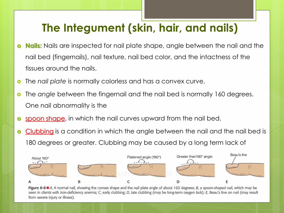
Nails: Nails are inspected for nail plate shape, angle between the nail and the
nail bed (fingernails), nail texture, nail bed color, and the intactness of the
tissues around the nails.
The nail plate is normally colorless and has a convex curve.
The angle between the fingernail and the nail bed is normally 160 degrees.
One nail abnormality is the
spoon shape, in which the nail curves upward from the nail bed.
Clubbing is a condition in which the angle between the nail and the nail bed is
180 degrees or greater. Clubbing may be caused by a long term lack of
oxygen.
The Integument (skin, hair, and nails)

Nail texture is normally smooth. Excessively thick nails can appear in elders,
in the presence of poor circulation, or in relation to a chronic fungal
infection. Beau’s lines are horizontal depressions in the nail that can result
from injury or severe illness. The nail bed is highly vascular, a characteristic
that accounts for its color. A bluish or purplish color to the nail bed may
reflect cyanosis, and pallor may reflect poor arterial circulation. The tissue
surrounding the nails is normally intact epidermis.
Perform capillary refill test: Press two nails between your thumb and
index finger; look for return of pink color to nail bed. Rapid return of pink or
usual color (generally less than 3 seconds). Delayed return of pink or usual
color (may indicate circulatory impairment)
The musculoskeletal system encompasses the muscles, bones, and joints.
Assessment of the MS involves inspection and palpation.
The nurse usually assesses the musculoskeletal system for muscle strength,
tone, size, and symmetry of muscle development, and for tremors
(involuntary twitching of body parts, mostly the hands).
Bones are assessed for normal form.
Joints are assessed for tenderness, swelling, thickening, crepitation (the
sound of bone grating on bone), presence of nodules, and range of
motion (ROM).
Body posture is assessed for normal standing and sitting positions.
Musculoskeletal System
Muscles: inspect the muscles for size. Compare the muscles on one side
of the body. Atrophy (a decrease in size) or hypertrophy
(an increase in size).
Inspect the muscles and tendons for contractures (shortening).
Inspect the muscles for tremors.
Palpate muscles at rest to determine muscle tonicity. Normally firm;
Atonic (lacking tone).
Palpate muscles for flaccidity(weakness or laxness) , spasticity(sudden
involuntary muscle contraction)and smoothness of movement.
Normally; Smooth coordinated movements.
Test muscle strength.

Examination of the neurologic system includes assessment of
(a) mental status including level of consciousness (LOC), (b)
the cranial nerves (CN), (c) reflexes, (d) motor function, and
(e) sensory function.
Assessment of mental status reveals the client’s general
cerebral function. These functions include intellectual
(cognitive) as well as emotional (affective) functions.
Major areas of mental status assessment include language,
orientation, memory, and attention span and calculation.
Neurological System

Language: If the client displays difficulty speaking..
Ask the client to point to common objects, and to name them.
Ask the client to read some words and to match the printed and
written words with pictures.
Ask the client to respond to simple verbal and written commands, e.g.,
“raise your arm.”
Orientation: Determine the client’s orientation to person, time, and
place.
Ask the client direct questions, e.g., “Where are you now?,” “What day
is it today?,” “Who am I?”
Ask the client to state his or her full name to assess the client’s ability to
recognize self.
1-Mental status

Memory: Assess the client’s three categories of memory: immediate recall, recent
memory, and remote memory.
To assess immediate recall:
Ask the client to repeat a series of three digits, e.g., 7–4–3, spoken slowly.
Gradually increase the number of digits until the client fails to repeat the series correctly.
Start again with a series of three digits, but this time ask the client to repeat them backward.
The average person can repeat a series of five to eight digits in sequence and four to six
digits in reverse order.
To assess recent memory:
Ask the client to recall the recent events of the day, such as how the client got to the clinic.
Ask the client to recall information given early in the interview, e.g., the name of a doctor.
Provide the client with three facts to recall, e.g., a color, an object, and an address; or a
three-digit number, and ask the client to repeat all three. Later in the interview, ask the client
to recall all three items.
To assess remote memory:
ask the client to describe a previous illness or surgery, e.g., 5 years ago, or a birthday or
anniversary.
1-Mental status
Attention Span and Calculation:
Test the ability to concentrate or maintain attention span by
asking the client to recite the alphabet or to count backward
from 100.
Test the ability to calculate by asking the client
to subtract 7 or 3 progressively from 100, i.e., 100, 93, 86,
79, or 100, 97, 94, 91. Normally, an adult can complete the
serial sevens test in about 90 seconds with three or fewer
errors.
1-Mental status
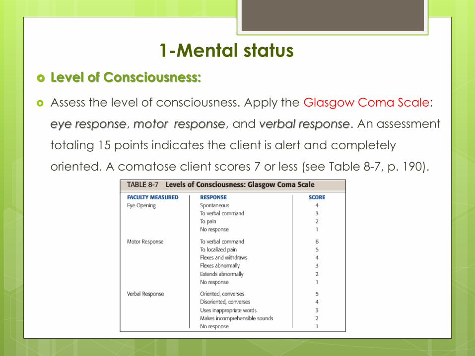
Level of Consciousness:
Assess the level of consciousness. Apply the Glasgow Coma Scale:
eye response, motor response, and verbal response. An assessment
totaling 15 points indicates the client is alert and completely
oriented. A comatose client scores 7 or less (see Table 8-7, p. 190).
1-Mental status
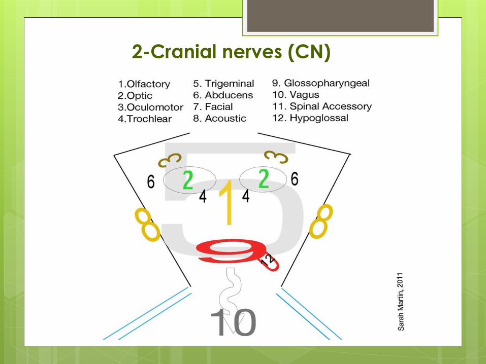
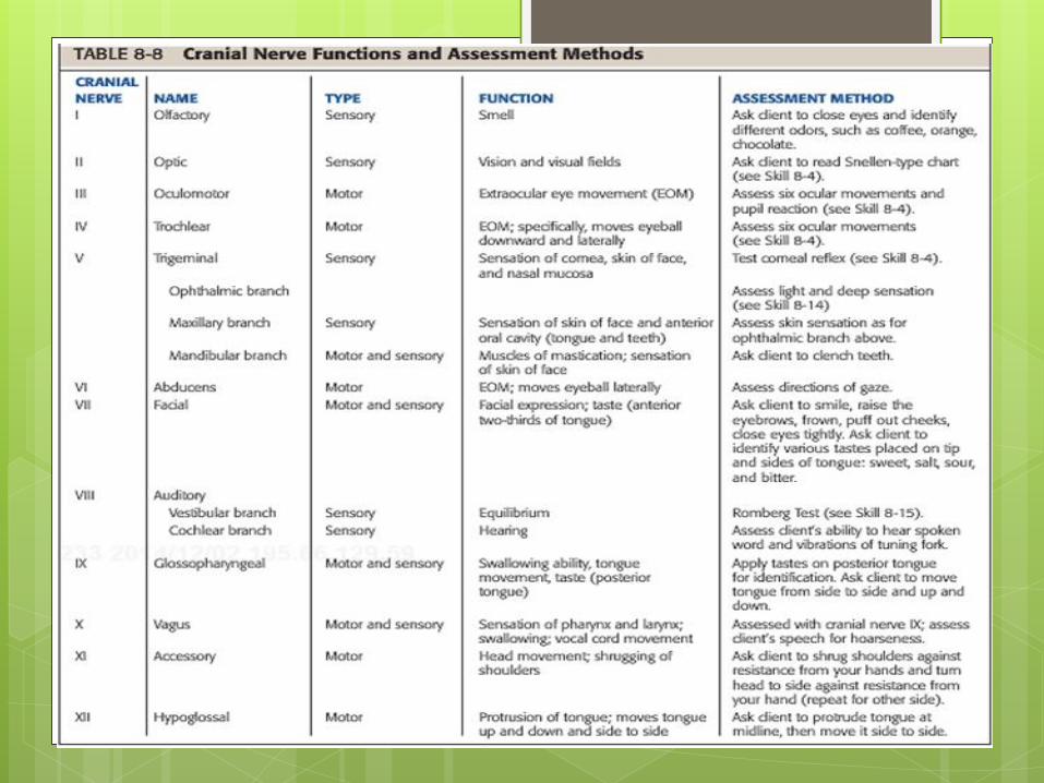
2-Cranial nerves (CN)
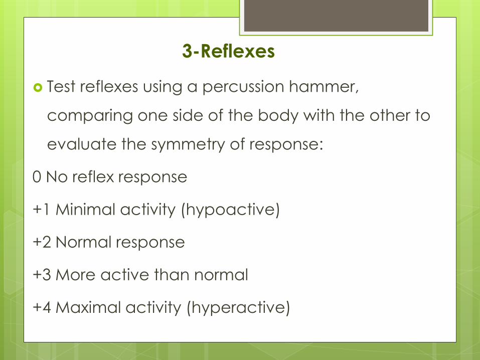
3-Reflexes
Test reflexes using a percussion hammer,
comparing one side of the body with the other to
evaluate the symmetry of response:
0 No reflex response
+1 Minimal activity (hypoactive)
+2 Normal response
+3 More active than normal
+4 Maximal activity (hyperactive)

3-Reflexes
Biceps Reflex :tests the spinal cord level C-5, C-6.
Observe the normal slight flexion of the elbow,
and feel the bicep’s contraction through your thumb.
Triceps Reflex :tests the spinal cord level C-7, C-8.
Observe the normal slight extension of the elbow.
3-Reflexes
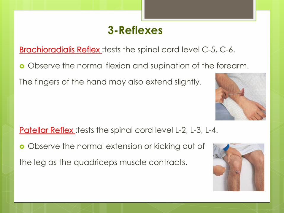
Brachioradialis Reflex :tests the spinal cord level C-5, C-6.
Observe the normal flexion and supination of the forearm.
The fingers of the hand may also extend slightly.
Patellar Reflex :tests the spinal cord level L-2, L-3, L-4.
Observe the normal extension or kicking out of
the leg as the quadriceps muscle contracts.

3-Reflexes
Achilles Reflex :tests the spinal cord level S-1, S-2.
Observe and feel the normal plantar flexion
(downward jerk) of the foot.
Plantar (Babinski) Reflex : it is superficial. It may be absent in adults
without pathology or overridden by voluntary control.
Observe the response. Normally, all five toes bend downward; this
reaction is negative Babinski. In an abnormal (positive) Babinski
response the toes spread outward and the big toe moves upward.
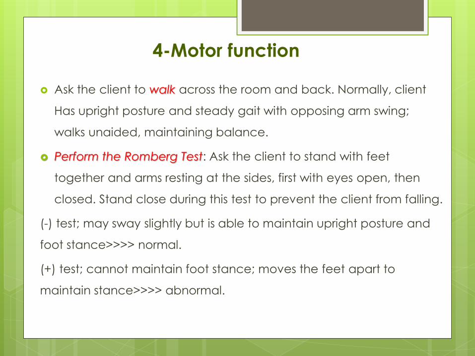
Ask the client to walk across the room and back. Normally, client
Has upright posture and steady gait with opposing arm swing;
walks unaided, maintaining balance.
Perform the Romberg Test: Ask the client to stand with feet
together and arms resting at the sides, first with eyes open, then
closed. Stand close during this test to prevent the client from falling.
(-) test; may sway slightly but is able to maintain upright posture and
foot stance>>>> normal.
(+) test; cannot maintain foot stance; moves the feet apart to
maintain stance>>>> abnormal.
4-Motor function
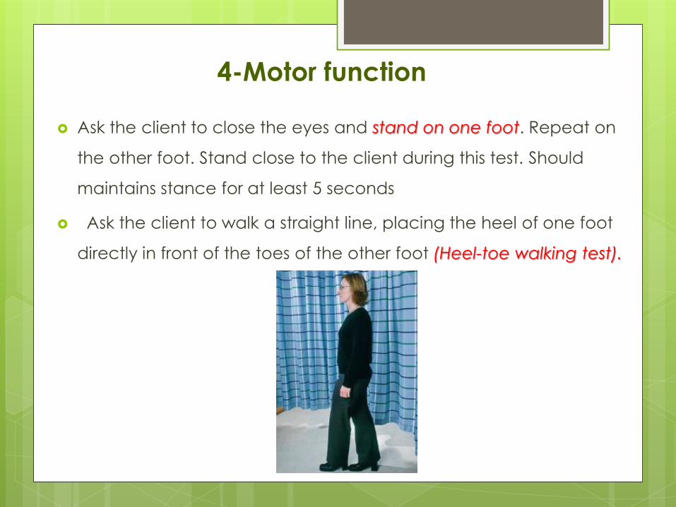
Ask the client to close the eyes and stand on one foot. Repeat on
the other foot. Stand close to the client during this test. Should
maintains stance for at least 5 seconds
Ask the client to walk a straight line, placing the heel of one foot
directly in front of the toes of the other foot (Heel-toe walking test).
4-Motor function
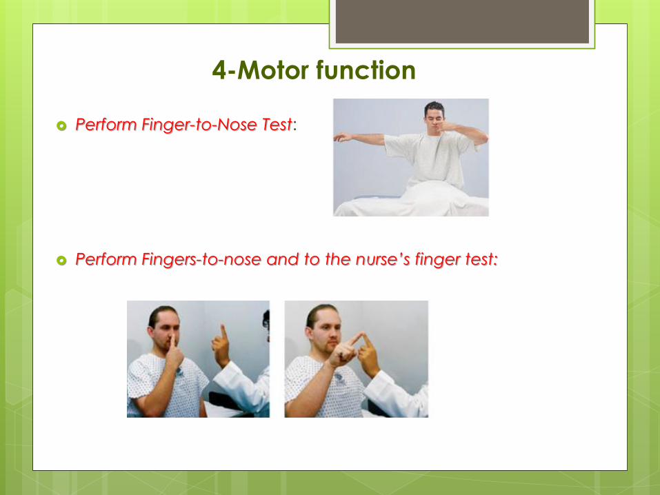
Perform Finger-to-Nose Test:
Perform Fingers-to-nose and to the nurse’s finger test:
4-Motor function

4-Motor function
Perform Fingers-to-thumb (same hand) test:
Perform Heel down opposite shin test:
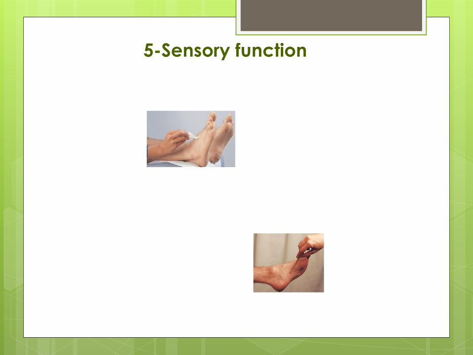
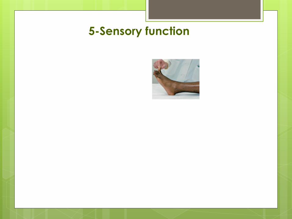
5-Sensory function
5-Sensory function