the interactomes of influenza virus ns1 and ns2 proteins … · 2017-10-03 · influenza virus ns1...
TRANSCRIPT

The Interactomes of Influenza Virus NS1 and NS2Proteins Identify New Host Factors and Provide Insightsfor ADAR1 Playing a Supportive Role in Virus ReplicationBenoıt de Chassey1, Anne Aublin-Gex2,3,4,5,6, Alessia Ruggieri7, Laurene Meyniel-Schicklin2,3,4,5,6,
Fabrine Pradezynski2,3,4,5,6, Nathalie Davoust2,3,4,5,6, Thibault Chantier2,3,4,5,6, Lionel Tafforeau2,3,4,5,6,
Philippe-Emmanuel Mangeot2,3,4,5,6, Claire Ciancia2,3,4,5,6, Laure Perrin-Cocon2,3,4,5,6,
Ralf Bartenschlager7, Patrice Andre1,2,3,4,5,6, Vincent Lotteau2,3,4,5,6*
1 Hospices Civils de Lyon, Hopital de la Croix Rousse, Laboratory of Virology, Lyon, France, 2 CIRI, International Center for Infectiology Research, EVIR Team, Universite de
Lyon, Lyon, France, 3 Inserm, U1111, Lyon, France, 4 Ecole Normale Superieure de Lyon, Lyon, France, 5 Universite Lyon 1, Centre International de Recherche en
Infectiologie, Lyon, France, 6 CNRS, UMR5308, Lyon, France, 7 Department for Infectious Diseases, Molecular Virology, University of Heidelberg, Heidelberg, Germany
Abstract
Influenza A NS1 and NS2 proteins are encoded by the RNA segment 8 of the viral genome. NS1 is a multifunctional proteinand a virulence factor while NS2 is involved in nuclear export of viral ribonucleoprotein complexes. A yeast two-hybridscreening strategy was used to identify host factors supporting NS1 and NS2 functions. More than 560 interactions between79 cellular proteins and NS1 and NS2 proteins from 9 different influenza virus strains have been identified. These interactingproteins are potentially involved in each step of the infectious process and their contribution to viral replication was testedby RNA interference. Validation of the relevance of these host cell proteins for the viral replication cycle revealed that 7 ofthe 79 NS1 and/or NS2-interacting proteins positively or negatively controlled virus replication. One of the main factorstargeted by NS1 of all virus strains was double-stranded RNA binding domain protein family. In particular, adenosinedeaminase acting on RNA 1 (ADAR1) appeared as a pro-viral host factor whose expression is necessary for optimal viralprotein synthesis and replication. Surprisingly, ADAR1 also appeared as a pro-viral host factor for dengue virus replicationand directly interacted with the viral NS3 protein. ADAR1 editing activity was enhanced by both viruses through denguevirus NS3 and influenza virus NS1 proteins, suggesting a similar virus-host co-evolution.
Citation: de Chassey B, Aublin-Gex A, Ruggieri A, Meyniel-Schicklin L, Pradezynski F, et al. (2013) The Interactomes of Influenza Virus NS1 and NS2 ProteinsIdentify New Host Factors and Provide Insights for ADAR1 Playing a Supportive Role in Virus Replication. PLoS Pathog 9(7): e1003440. doi:10.1371/journal.ppat.1003440
Editor: Sumit K. Chanda, Burnham Institute for Medical Research, United States of America
Received September 3, 2012; Accepted May 6, 2013; Published July 4, 2013
Copyright: � 2013 de Chassey et al. This is an open-access article distributed under the terms of the Creative Commons Attribution License, which permitsunrestricted use, distribution, and reproduction in any medium, provided the original author and source are credited.
Funding: This work was funded by ANR, ANRS, Inserm and the FUI from the French Ministry of Industry. RB was supported by the DeutscheForschungsgemeinschaft (FOR1202, TP1). RB and VL are also supported by the European Union’s 7th program (FP7/2007-2013) under grant agreement no 267429(SysPatho). The funders had no role in study design, data collection and analysis, decision to publish, or preparation of the manuscript.
Competing Interests: The authors have declared that no competing interests exist.
* E-mail: [email protected]
Introduction
Influenza A viruses are the causative agents of seasonal and
pandemic infections and are responsible for the death of at least
half a million people worldwide each year. The genome of
influenza A viruses is composed of eight negative-sense single-
stranded RNAs encoding 13 proteins. NS1 and NS2 are derived
from alternatively spliced RNAs that are transcribed from the
eighth RNA segment. The segments are encapsidated by binding
to nucleoproteins (NP) and the polymerase complex (PA, PB1 and
PB2) forming the viral ribonucleoproteins (vRNPs). The viral
particle contains eight vRNPs, the surface glycoproteins haemag-
glutinin (HA) and neuraminidase (NA), the matrix proteins (M1
and M2) and the NS2 protein. Some strains express the pro-
apoptotic PB1-F2 protein and two additional virulence factors,
PB1-N40 and PA-X, have been recently identified [1–3].
The NS1 protein is not incorporated in the virus. It exerts a large
spectrum of functions through interactions with a variety of cellular
components residing either in the cytoplasm or in the nucleus. NS1
is a pleiotropic virulence factor repressing innate antiviral mecha-
nisms e.g. by interfering with the type I interferon system through
direct interaction with PKR and TRIM25, or through the
sequestration of double-stranded RNA [4–8]. NS1 is also known
to perturb the mRNA processing by interacting with CPSF4 and
PABPN1 to inhibit nuclear export of cellular mRNA [9] and is
suspected to hijack the RNA translation machinery in favor of
translation of viral protein e.g. by interacting with STAU1 [10–11].
In contrast to NS1, NS2 protein is a structural component of the
viral particle and it associates with the viral matrix M1 protein
[12]. NS2 mediates the export of vRNPs from the nucleus to the
cytoplasm through export signal [13] via its interaction with
XPO1 [14]. In addition, NS2 interacts with nucleoporins and was
suggested to serve as an adaptor between vRNPs and the nuclear
pore complex [13]. A role of NS2 in the regulation of influenza
virus transcription and replication has also been proposed [15].
However, many functions of NS2, in particular its transit through
the cytoplasm and its incorporation into the viral particle, are not
understood.
PLOS Pathogens | www.plospathogens.org 1 July 2013 | Volume 9 | Issue 7 | e1003440

Several screens have been performed to identify host factors
involved in the influenza virus replication cycle, mainly focusing
on interactors of vRNPs or of the polymerase by using affinity
purification or yeast two-hybrid techniques [16–18]. A proteome-
wide screen of virus-host protein-protein interactions has provided
an important resource of 135 interactions [19]. However, the weak
overlap of the public datasets suggests that they are far from being
complete.
The impact of cellular proteins on the influenza virus replication
has been extensively studied using RNAi screens [19–24].
Although poorly overlapping at the gene level, these screens
better converge at the level of biological processes [25–27]. Hence,
more than the identification of host factors, these studies
highlighted major cellular functions that are essential for the virus
replication. However, for the majority of identified host factors,
the mode of action remains to be determined. Furthermore,
comparisons of strain-specific virus-host interactomes are clearly
missing, which is required to reveal general principles governing
infection mechanisms and to identify common therapeutic targets
as well as broad-spectrum antivirals.
In the present study we conducted stringent yeast two-hybrid
screens to identify human proteins interacting with NS1 and NS2
from 9 influenza A virus strains representative of the variability in
nature. The functional impact of all NS1 and NS2 interactors on
viral replication was systematically addressed by RNA interfer-
ence. In combination with published datasets, our new results offer
a comprehensive view of NS1 and NS2 interactomes and
corresponding targeted cellular functions. The global analysis of
the NS1 and NS2 host cell targets reveals an enrichment of
double-stranded RNA binding domain (DRBD) containing
proteins for the 9 tested influenza virus strains. A focus was put
on ADAR1 since this protein is critical for the replication of other
viruses [28], is highly expressed in human lung cells [29], is
induced by type I interferon [30], is interfering with interferon
signalling production [31] and is interacting with all tested NS1
proteins. In addition, we also observed in another screen that
ADAR1 interacts with the dengue virus NS3 protein which is a
bifunctional enzyme containing protease and helicase activity [32].
We show that ADAR1 is a pro-viral host factor favoring
replication of influenza virus and dengue virus and that these
viral proteins can control ADAR1 editing activity.
Results
Cellular interactors of influenza virus NS1 and NS2To identify all cellular proteins interacting with influenza virus
NS1 and/or NS2 proteins, yeast two-hybrid screens (Y2H) were
carried out using NS1 and NS2 proteins from 9 different virus
strains as baits (Table S1) and three cDNA libraries (from human
spleen, fetal brain and respiratory epithelium). Key features of the
virus strains are provided in Text S1. NS1 and NS2 proteins
selected for this study are representative of the natural diversity
since they are distributed all along the phylogenetic trees of known
NS1 and NS2 sequences (Text S1, Figures S1 and S2 in Text S1,
Alignments of NS1 and NS2 protein sequences are presented in
Figures S3 and S4 in Text S1). Seventy nine non-redundant
cellular proteins were identified to interact with NS1, NS2 or both
and were individually retested in a pairwise array (Figure 1A).
From a total of 1422 possible interactions tested (79 cellular
proteins tested against 9 NS1 and 9 NS2 proteins), 562 tested
positive. In this way, we identified 33 cellular proteins interacting
exclusively with NS1, 28 exclusively with NS2, and 18 with both
NS1 and NS2.
The vast majority (97.5%) of the NS1 and NS2 interactors are
known to be expressed in the respiratory epithelium (Table S2).
Twelve out of the 79 host interactors have already been reported
(AIMP2, SCRIB, CPSF4, the kinases PIK3R1, PIK3R2, MAPK9,
CRK and proteins with a double-stranded RNA-binding domain
STAU1, PRKRA, ADAR1, TARBP2, ILF3) [9,11,19,33–38].
21.5% of host interactors are targeted by all virus strains
(Figure 1B) and 5% appear to be strain specific. 80% of the
cellular interactors bind to more than 50% of the tested NS1 and
NS2 proteins indicating that the dataset is more appropriate to the
identification of common rather than differential interaction
profiles. Together with previously published data available in the
VirHostNet database [39], we now provide a list of 111 non-
redundant cellular proteins interacting exclusively with NS1, 32
exclusively with NS2 and 18 with both proteins (a complete list of
influenza virus interactors is given in Table S3).
Consistent with observations from previous virus-host interactome
studies, NS1 and NS2 proteins tend to interact with highly central
proteins in the human interactome [40–43]. Indeed, the degree
distribution of targeted human proteins was significantly higher than
the degree distribution in the human interactome (U-test, p-
value,2.2610216) (Figure 1C). Similarly, the betweenness distribu-
tion of targeted human proteins was significantly higher than the
betweenness distribution in the human interactome (U-test, p-
value,2.2610216) (Figure 1D). This suggests that influenza NS1 and
NS2 proteins preferentially target pleiotropic cellular proteins [44].
Finally, an assessment of Gene Ontology categories revealed a
significant enrichment (p-value = 3.3610214) for DRBD-contain-
ing proteins (DRBPs) in the interaction dataset. Strikingly, DRBPs
were exclusively targeted by NS1 proteins. All virus strains
interacted with most of the DRBPs suggesting that the direct
targeting of DRBDs is of special importance for influenza A
viruses.
Impact of NS1 and NS2 cellular targets on influenza virusreplication
Among the 79 NS1 and NS2 interactors identified here, 12 have
been previously identified in recent genome-wide siRNA screens
Author Summary
Viruses are obligate intracellular parasites that rely oncellular functions for efficient replication. As most biolog-ical processes are sustained by protein-protein interac-tions, the identification of interactions between viral andhost proteins can provide a global overview about thecellular functions engaged during viral replication. Influ-enza viruses express 13 viral proteins, including NS1 andNS2, which are translated from an alternatively spliced RNAderived from the same genome segment. We present herea comprehensive overview of possible interactions ofcellular proteins with NS1 and NS2 from 9 viral strains.Seventy nine cellular proteins were identified to interactwith NS1, NS2 or both NS1 and NS2. These interacting hostcell proteins are potentially involved in many steps of thevirus life cycle and 7 can directly control the viralreplication. Most of the cellular targets are shared by themajority of the virus strains, especially the double-strandedRNA binding domain protein family that is strikinglytargeted by NS1. One of its members, ADAR1, is essentialfor influenza virus replication. ADAR1 colocalizes with NS1in nuclear structures and its editing activity is enhanced byNS1 expressed on its own and during virus infection. Asimilar phenomenon is observed for dengue virus whoseNS3 protein also interacts with ADAR1, suggesting aparallel virus-host co-evolution.
Interactomic of Influenza Non Structural Proteins
PLOS Pathogens | www.plospathogens.org 2 July 2013 | Volume 9 | Issue 7 | e1003440

Figure 1. Interactions between NS1 and NS2 proteins and human host factors. (A) Yeast two-hybrid array. The 33 NS1-specific interactorsare indicated in blue, 28 NS2-specific interactors in grey and shared interactors in yellow. The 11 NS1 and the single NS2 interactors described earlierare highlighted with bold letters. DRBD-containing proteins (DRBPs) are indicated with a star. (B) Frequency of interactions between individual hostcell factors and NS1 and/or NS2 proteins of the 9 different influenza virus strains. (C) Degree distribution of human proteins and human proteinstargeted by NS1 and/or NS2 proteins in the human interactome. P(k) is the probability of a node to connect k other nodes in the network. Solid linesrepresent linear regression fits. Vertical dashed lines indicate the mean degree of each distribution. (D) Betweenness distribution of human proteinsand human proteins targeted by NS1 and/or NS2 proteins in the human interactome. P(b) is the probability for a node to have a betweenness valueof b in the network. Solid lines represent linear regression fits. Vertical dashed lines indicate the mean betweenness value for each distribution.doi:10.1371/journal.ppat.1003440.g001
Interactomic of Influenza Non Structural Proteins
PLOS Pathogens | www.plospathogens.org 3 July 2013 | Volume 9 | Issue 7 | e1003440
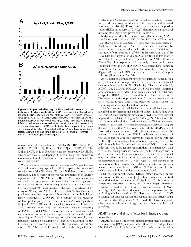
as modulators of viral replication - ATP6V1G1, RPL13A [21,23],
GMEB1, PIK3R2 [19], SON, EEF1A1 [23], CHCHD5, RPL23A
[24] and NUP214 [22]. However, since these genome-wide siRNA
screens are weakly overlapping, it is very likely that numerous
modulators of viral replication have been missed or remain to be
confirmed [25–27].
We have therefore performed a systematic siRNA-based screen
in A549 human lung epithelial cells to explore the functional
contribution of the 79 cellular NS1 and NS2 interactors to virus
replication. The silencing phenotype was first tested by measuring
replication of the A/H1N1/Puerto Rico/8/34 virus strain which
was used in the yeast two- hybrid screen. The complete replication
cycle was first probed by measuring the neuraminidase activity in
the supernatant 48 h post-infection. The assay was calibrated by
using siRNAs against ATP6V1G1 and CSNK2B that have been
previously described as pro-viral host factor and anti-viral host
factor respectively. ATP6V1G1 is a subunit of the vacuolar
ATPase proton pump required for influenza A virus replication
[21] while CSNK2B gene silencing increases virus replication in
A549 infected cells [45]. As expected, siRNAs targeting
ATP6V1G1 and CSNK2B respectively reduced and increased
the neuraminidase activity in the supernatant, thus validating the
assay (Figure 2A and 2B). By comparison with these controls, virus
replication should be altered by at least 35% according to the
threshold defined by Konig et al. in their genome-wide siRNA
screen [22]. This threshold together with a silencing efficiency
greater than 60% for each siRNAs without detectable cytotoxicity
were used for a stringent selection of the pro-viral and anti-viral
host factors (Table S4). These criteria are in the range applied in
earlier siRNA-based screens [23,46,47] (information on individual
silencing efficiency is also provided in Table S4).
In this way, we identified the two pro-viral host factors, ADAR1
and RPSA, and confirmed ATP6V1G1, RPL13A, EEF1A1 and
SON (Figure 2A). In addition, one new anti-viral host factor, N-
PAC, was identified (Figure 2A). These results were confirmed by
using plaque assays, revealing a broader range of inhibition or
activation of virus replication (Table S4). In conclusion, out of the
79 cellular interactors of NS1 and NS2 identified in this study, 7
were identified as possible direct modulators of A/H1N1/Puerto
Rico/8/34 virus replication. Importantly, these results were
confirmed with the A/H1N1/New Caledonia/2006 influenza
virus strain that was not used in the yeast two-hybrid screen
(Figure 2B), although RPL13A only scored positive 72 h post
infection (Figure S5 in Text S1).
As it is a critical component of virus-host interaction, production
of type I interferon was quantified in the supernatant of infected
cells transfected with siRNAs (Table S4). Silencing of ADAR1,
ATP6V1G1, BCLAF1, RPL13A and SON increased interferon
production in infected cells. These proteins interact with NS1 and,
except for BCLAF1, are pro-viral host factors for the virus.
Therefore, 4 of the 6 pro-viral host factors are implicated in
interferon production. This is consistent with the role of NS1 in
interfering with the type I interferon system.
The function and subcellular localization of cellular interactors
identified in the present and published studies indicate that both
NS1 and NS2 are pleiotropic proteins required for several essential
steps of the viral life cycle (Figure 3). Although NS2 function in the
cytoplasm remains elusive, it is shown here that NS2 mostly targets
proteins of the cytoskeleton and involved in intracellular transport
(Figure 3). Given that NS2 also interacts with the vRNPs, it might
also mediate their transport to the plasma membrane or to the
nucleus. In case of the latter, NS2 is implicated in the export of
vRNPs, consistent with the observed interaction of NS2 with the
NPC [13]. The targeting of transcription-regulating proteins by
NS2 is much less documented. A role of NS2 in regulating
influenza virus RNA genome transcription via its interaction with
vRNPs has been previously proposed [15,48]. Although such a
direct interaction with the components of the vRNPs is not ruled
out, our data indicate a direct targeting of the cellular
transcriptional machinery by NS2 (Figure 3, box regulation of
transcription). Interestingly, since NS1 also targets this process, a
potential cooperation between NS1 and NS2 for the control of the
cellular transcription machinery can be speculated.
NS1 proteins target several DRBPs either localized in the
nucleus or in the cytoplasm [49]. These proteins are critical
transcriptional or translational checkpoints. NS1 is known to
inhibit the activation of PKR, one of the major interferon-
inducible antiviral effectors, through direct interaction [6]. More
recently, SON has been described to be important for the
trafficking of influenza virions [23]. Here, we confirmed that SON
is essential for viral replication and suggest that this activity could
be related to the NS1 protein. ADAR1 and PKR have an opposite
effect on virus replication although they are both induced by type I
interferon.
ADAR1 is a pro-viral host factor for influenza A virusreplication
ADAR1 is a type I interferon-induced protein that is expressed
in human lung [29,30] and interacts with all tested NS1 proteins.
The 150 kDa interferon-inducible ADAR1 isoform is expressed in
Figure 2. Impact of silencing of NS1 and NS2 interactors oninfluenza A virus replication. A549 cells were transfected withindicated siRNAs, infected at a MOI of 0.5 with A/H1N1/Puerto Rico/8/34virus strain (A) or A/H1N1/New Caledonia/2006 virus strain (B) and theneuraminidase activity (NA) was measured in the supernatant 48 h postinfection. Values are normalized to neuraminidase activity measured insupernatants of control siRNA-transfected cells and represent the mean+/2 standard deviation (triplicates). ATP6V1G1 is a host dependencyfactor, CSNK2B is an anti-viral host factor. Both served as controls.doi:10.1371/journal.ppat.1003440.g002
Interactomic of Influenza Non Structural Proteins
PLOS Pathogens | www.plospathogens.org 4 July 2013 | Volume 9 | Issue 7 | e1003440

A549 cells upon influenza A virus infection and by type I
interferon. The constitutive 110 kDa ADAR1 isoform was only
induced upon infection indicating that ADAR1 expression can
also be controlled by an interferon-independent mechanism, at
least in the setting of an influenza A virus infection (Figure 4A).
ADAR1-specific siRNAs efficiently reduced the expression of
ADAR1 isoforms and blocked their induction upon infection
(Figure 4B). The silencing of ADAR1 inhibited virus release from
15% at 8 h post-infection to 90% at 48 h post infection
(Figure 4C). Expression of viral proteins (here HA, NP, M1 and
NS1) was also significantly reduced as early as 8 h post infection.
NS1, NP and M1 expression was delayed while HA expression
remained very low until 24 h post infection (Figure 4B). Thus,
ADAR1 is a pro-viral host factor for virus protein expression and
virus production.
Mapping of the ADAR1-NS1 interaction sitesImmunofluorescence revealed that ADAR1 is diffusely distrib-
uted in the nucleus and relocalized in nuclear structures in
influenza virus-infected cells (Figure 5A). In these structures
ADAR1 colocalized with NS1 but not with HA for which no
interaction with ADAR1 could be detected. As NS1 interacts with
several DRBD-containing proteins, the NS1 binding site in
ADAR1 could be a DRBD. Amino acid sequence alignment of
DRBDs revealed a conserved region of 47 amino acid residues
within the two firsts DRBD of ADAR1 (Figure S6 in Text S1). A
set of 4 ADAR1 deletion mutants, differing in their number of
DRBDs, and a plasmid encoding the 47 amino acid residues of the
first DRBD were constructed (Figure 5B). In a yeast two-hybrid
array, ADAR1 interacted with NS1 even in the absence of its first
DRBD while interaction was completely abrogated when the first
two DRBDs were deleted. The peptide of 47 amino acid residues
also interacted with NS1 (Figure 5C) in the array and in GST pull-
down assays (Figure 5D). Thus, ADAR1 displays two potential
NS1 interaction sites located on the first two double-stranded
RNA-binding domains.
To validate these results the NS1 RNA-binding domain (RBD)
and effector domain fused to GST were used in pull-down
experiments for the mapping of NS1 interaction with 36Flag
tagged ADAR1 after co-expression in HEK293T cells (Figure 5E).
Figure 3. Overview of NS1 and NS2 interactors on the influenza A virus life cycle. The framework of this picture is from Brass et al. [21]. NS1and NS2 interactors identified in the present study and described in the literature were placed according to information from Gene Ontology, HumanProtein Atlas and HPRD. When multiple annotations were available, the most relevant for the virus was chosen. Circle node: NS1 interactor; Squarenode: NS2 interactor; Diamond-shaped node: NS1 and NS2 interactor. A red node is a pro-viral host factor and a green node is an anti-viral host factoraccording to the siRNA data from this study and genome-wide screens. The node size is proportional to the number of virus strains interacting withthe host factors (interactors from the literature are often tested against a single virus strain hence appearing with a small node size).doi:10.1371/journal.ppat.1003440.g003
Interactomic of Influenza Non Structural Proteins
PLOS Pathogens | www.plospathogens.org 5 July 2013 | Volume 9 | Issue 7 | e1003440

Interactomic of Influenza Non Structural Proteins
PLOS Pathogens | www.plospathogens.org 6 July 2013 | Volume 9 | Issue 7 | e1003440
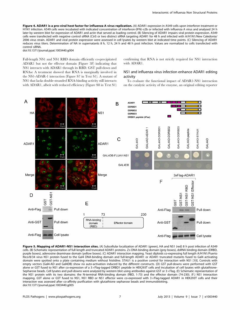
Full-length NS1 and NS1 RBD domain efficiently co-precipitated
ADAR1 but not the effector domain (Figure 5F) indicating that
NS1 interacts with ADAR1 through its RBD. GST pull-down and
RNAse A treatment showed that RNA is marginally involved in
the NS1-ADAR-1 interaction (Figure S7 in Text S1). A mutant of
NS1 that lacks double-stranded RNA-binding activity still interacts
with ADAR1, albeit with reduced efficiency (Figure S8 in Text S1)
confirming that RNA is not strictly required for NS1 interaction
with ADAR1.
NS1 and influenza virus infection enhance ADAR1 editingactivity
To evaluate the functional impact of ADAR1-NS1 interaction
on the catalytic activity of the enzyme, an original editing reporter
Figure 4. ADAR1 is a pro-viral host factor for influenza A virus replication. (A) ADAR1 expression in A549 cells upon interferon treatment orH1N1 infection. A549 cells were incubated with indicated concentration of interferon-(IFN)-a2b or infected with influenza A virus and analysed 24 hlater by western blot for expression of ADAR1 and actin that served as loading control. (B) Silencing of ADAR1 impairs viral protein expression. A549cells were transfected with negative control siRNA (Ctrl) or two distinct siRNA targeting ADAR1 for 48 h and infected with A/H1N1/New Caledonia/2006 virus strain. ADAR1 and viral protein expression were assessed in cell lysates by western blot at indicated time points. (C) Silencing of ADAR1reduces virus titers. Determination of NA in supernatants 8 h, 12 h, 24 h and 48 h post infection. Values are normalized to cells transfected withcontrol siRNA.doi:10.1371/journal.ppat.1003440.g004
Figure 5. Mapping of ADAR1-NS1 interaction sites. (A) Subcellular localization of ADAR1 (green), HA and NS1 (red) 8 h post infection of A549cells. (B) Schematic representation of full-length and truncated ADAR1 proteins. Za DNA-binding domain (grey boxes), dsRNA binding domain (DRBD,purple boxes), adenosine deaminase domain (yellow boxes). (C) ADAR1 interaction mapping. Yeast diploids co-expressing full length A/H1N1/PuertoRico/8/34 virus NS1 protein fused to the Gal4 DNA-binding domain and full-length ADAR1 or ADAR1 truncated mutants fused to Gal4 activatingdomain were spotted onto a plate containing medium without histidine. STAU1 is a positive control for interaction with NS1 [10]. Controls withempty vectors (Gal4-AD and Gal4DB) show no auto-activation induced by the different constructs. (D) GST pull-downs were performed with GSTalone or GST fused to NS1 after co-expression of a 36Flag-tagged DRBD1 peptide in HEK293T cells and incubation of cell lysates with glutathione-Sepharose beads. Cell lysates and pull-downs were analyzed by western blot using antibodies against GST or 36Flag. (E) Schematic representation ofthe NS1 protein with its two domains: the N-terminal RNA-binding domain (RBD, 1-73) and the effector domain (74-230). (F.) NS1 interactionmapping. GST alone or GST fused to NS1, NS1 RBD or NS1 effector were co-expressed with 36Flag-tagged ADAR1 in HEK293T cells and theirinteraction was assessed after co-affinity purification with glutathione sepharose beads and immunoblotting.doi:10.1371/journal.ppat.1003440.g005
Interactomic of Influenza Non Structural Proteins
PLOS Pathogens | www.plospathogens.org 7 July 2013 | Volume 9 | Issue 7 | e1003440

system was constructed. This reporter system consists of a 24
nucleotide-long minimal ADAR1 substrate derived from the
sequence of the antigenome of the hepatitis delta virus that is
edited by this enzyme [50]. In this sequence, ADAR1 editing
activity changes a stop codon into a tryptophane codon (Figure 6A)
[51]. The reporter plasmid contains the ADAR1 substrate
sequence inserted in frame in-between the Renilla and the Firefly
luciferase genes (Figure 6A). In this configuration, the Firefly
luciferase activity reflects the extend of editing and thus ADAR1
activity, leading to the conversion of the stop codon into the
tryptophane codon. ADAR1 was co-expressed in HEK293T cells
with NS1 or its RBD and with the editing reporter construct. The
NS1 effector domain or DLG4, which does not bind to ADAR1
(data not shown), was used as negative control in analogous co-
transfection experiments. NS1 RBD and full-length NS1 increased
the Firefly luciferase signal by 30% and 60% respectively
(Figure 6B) suggesting that NS1 can cooperatively interact with
ADAR1 via its RNA-binding domain to promote ADAR1 editing
activity (Figure 6B). Editing activity was also analyzed in the
context of influenza virus infection after expression of the editing
reporter construct (Figure 6C). H1N1 influenza virus infection
increased the editing activity of ADAR1 by 70% and this was
completely reversed when ADAR1 expression was silenced by
RNA interference.
To validate these observations, a catalytically inactive ADAR1
(E912A) mutant was constructed [52]. Unfortunately, A549 cells
became refractory to plasmid DNA transfection after siRNA
transfection, thus precluding functional tests of the mutant in this
cell line (not shown). As an alternative, we tested a potential
transdominant negative effect of the ADAR1 mutant on influenza
virus growth. The catalytically inactive ADAR1 (E912A) mutant
construct was therefore transfected into A549 cells and virus
growth in these cells was compared to the one achieved with
mock-transfected cells or in wild type ADAR1-transfected cells.
Viral protein expression was reduced in A549 cells expressing the
ADAR1 mutant compared to control cells (Figure 6D). Consistent
with this result, neuraminidase activity in the supernatant was also
significantly reduced (Figure 6E). Importantly, since influenza A
virus infection induces endogenous ADAR1 expression, the impact
of the ADAR1 mutant is most likely underestimated in this
Figure 6. Enhancement of ADAR1 editing activity by influenza virus NS1. (A) The RNA editing reporter system is composed of the hepatitisD virus minimal sequence edited by ADAR1 and positioned in-between the Renilla luciferase and the Firefly luciferase coding sequence, respectively.The unedited reporter has a stop codon that is converted into Trp codon upon A to I editing by ADAR. Hence, editing is correlated with Fireflyluciferase expression while Renilla luciferase expression is used as an internal control. (B) HEK293T were co-transfected with the editing reporter,ADAR1 and NS1 (full-length, RNA-binding domain or effector domain) or the control protein DLG4. Two days post transfection, luciferase activitieswere determined by luminescence measurement. Data are expressed as percentage of the luciferase activity detected in cells expressing the NS1effector domain (relative light unit, RLU). (C) Editing activity in HEK293T expressing or not ADAR1, transfected with the editing reporter and infectedwith influenza virus H1N1. (D, E) A549 cells were transfected with plasmids encoding for wild type or catalytically inactive ADAR1 (ADAR1 E912A, TextS1). Forty eight hours later, cells were infected with the A/H1N1/New Caledonia/2006 virus strain at a MOI of 0.5. After an additional 48 h incubationperiod, expression of ADAR1 and viral proteins was assessed in cell lysates by using western blot (D) and neuraminidase activities were measured insupernatants (E). Values are normalized to mock-transfected cells.doi:10.1371/journal.ppat.1003440.g006
Interactomic of Influenza Non Structural Proteins
PLOS Pathogens | www.plospathogens.org 8 July 2013 | Volume 9 | Issue 7 | e1003440

experimental system. We therefore concluded that the RNA
editing function is required for the pro-viral activity of ADAR1.
Dengue virus NS3 cooperatively interacts with ADAR1During the course of a systematic screening for virus-host
protein-protein interactions with a yeast two-hybrid system, we
also identified ADAR1 as an interactant of the NS3 protein of
dengue virus type 2. This interaction was confirmed in a yeast
two-hybrid pairwise array (Figure 7A). As for NS1 of influenza A
virus, interaction between NS3 and ADAR1 was validated by
GST pull-down experiments (Figure 7B) and also in this case,
RNA contributed to this interaction only to a very minor extent
(Figure S9 in Text S1). Both ADAR1 isoforms were induced
upon dengue virus infection as well as upon type I interferon
treatment of Huh-7 cells (Figure 7C). Silencing of ADAR1
expression by RNA interference (Figure S10 in Text S1) resulted
in a strong decrease of dengue virus replication (Figure 7D). This
result was confirmed with a subgenomic dengue virus replicon
stably replicating in Huh-7 cells, indicating that ADAR1 acts at a
post-entry step in the dengue virus life cycle (Figure S11 in Text
S1). Likewise, as observed for influenza virus, dengue virus
infection strongly increased the editing activity of ADAR1
(Figure 7E). In fact, full-length NS3 and the helicase domain
increased the Firefly signal by 24% and 44% respectively,
suggesting that NS3 cooperatively interacts with ADAR1 to
enhance its editing activity (Figure 7F).
In conclusion, both influenza virus and dengue virus (i) induce
over-expression of ADAR1, (ii) interact with ADAR1 through the
RNA-binding domain of influenza virus NS1 and the helicase
domain of dengue virus NS3, (iii) enhance the editing activity of
ADAR1 and (iv) are dependent on ADAR1 expression for efficient
virus replication.
Figure 7. Dengue virus NS3 protein also interacts with ADAR1. (A) Pairwise Yeast diploids co-expressing dengue virus type 2 NS3 helicasefused to Gal4 DNA-binding domain and ADAR1 fused to Gal4 transactivation domain were plated onto a selective medium lacking histidine todetermine interaction-dependent transactivation of the HIS3 reporter gene. Negative controls are vectors without insert (pPC97 and pPC86 for baitand prey respectively). (B) HEK293T cells were co-transfected with 36Flag-tagged ADAR1 and GST-tagged full-length NS3 of dengue virus (GST-DV-NS3) or its helicase domain (GST-DV-NS3 helicase) or GST-tagged full-length NS1 of influenza virus (GST-FLUAV-NS1) as a positive control. Proteinsbound to glutathione sepharose beads were analyzed by western blot using antibodies against GST or 36Flag. (C) ADAR1 expression in Huh-7 cellsupon interferon treatment or dengue virus infection. Huh-7 cells were incubated with 0, 100 or 1000 IU/ml of interferon- (IFN)-a2b or infected withdengue virus and analyzed 24 h later for expression of ADAR1, NS3 and GAPDH. (D) Impact of ADAR1 silencing on dengue virus replication in Huh-7.Data are expressed as the percentage of virus titer obtained with control siRNA-transfected cells. A siRNA targeting dengue virus NS1-coding region(siDV-NS1) was used as positive control for the silencing. (E) ADAR editing activity in Huh-7 cells infected with dengue virus. (F) Dengue virus NS3contribution to ADAR1 editing activity. The NS1 effector domain of influenza virus, full-length NS3 of dengue virus or its helicase domain were co-expressed with ADAR1 and the editing reporter in HEK293T cells. Luminescence reflecting Firefly and Renilla luciferase activities was measured 48 hpost-transfection. The influenza virus NS1 effector domain does not interact with ADAR1 and was used as negative control. Data are normalized tothe values obtained with the NS1 effector domain. RLU, relative light unit.doi:10.1371/journal.ppat.1003440.g007
Interactomic of Influenza Non Structural Proteins
PLOS Pathogens | www.plospathogens.org 9 July 2013 | Volume 9 | Issue 7 | e1003440

Discussion
This study describes an exhaustive interaction profile for NS1
and NS2 proteins of 9 influenza virus strains. More than 560
interactions between 79 cellular proteins and NS1 and NS2 were
identified. Thirty-three cellular proteins interacted exclusively with
NS1, 28 exclusively with NS2, and 18 with both NS1 and NS2.
Since NS1 and NS2 are the products of alternatively spliced
RNAs, shared interactions may reflect binding to the common N-
terminal 10 amino-acid residues long sequence. This result
suggests that influenza viruses have evolved two proteins to
interact with cellular proteins that are potentially essential for
them.
Twelve out of the 79 NS1 and NS2 cellular interactors have
already been reported in the literature, demonstrating the
reliability and robustness of our screening approach. For NS1,
there is a strong overlap with hits published by others (11 of the 51
interactors identified in the present study, which is well above the
average overlap) [41,53], suggesting that the NS1 interactome
dataset is now close to completion. In case of NS2, only 4 cellular
interactors have been published and one of them, AIMP2, has
been confirmed in our screens. Although 46 new NS2 interactors
have been identified, it is difficult at this stage to estimate the
completion level of the NS2 interactome due to the lack of
published interaction data.
Overall, most of the cellular targets interacted with the majority
of NS1 or NS2 proteins of the different influenza viruses arguing
that we have identified highly relevant and evolutionary conserved
interactions. Interestingly, a significant proportion of these
proteins is also targeted by other viruses (44.7%, exact Fisher test,
p-value,2.2610216) indicating that these cellular proteins are
likely to be involved in a generic process of viral infection [39].
Our interaction dataset indicates that NS1 and NS2 proteins are
likely to be involved in multiple steps of the viral replication cycle,
paving the way for challenging functional explorations. This was
largely unexpected for NS2, which is known to be involved in the
nuclear export of the vRNPs. Its interaction with the cytoskeleton
appears particularly interesting for further studies. Although the
pleiotropic nature of NS1 is well established [54], our study
provides new insights into the breadth of interactions and activities
of this regulatory protein. In addition to the 67 new interactors,
the current dataset also provides additional information on
previously known interactors and related targeted functions. For
instance, the CPSF4 interaction with NS1 has been described as a
potential therapeutic target [55] and is confirmed in our study.
Three NS1 proteins also interacted with CPSF3L, a protein
participating in the endonuclease activity of CPSF, suggesting that
the corresponding viruses evolved alternative strategies to interfere
with the cellular 39end mRNA processing [56].
The phenotypic analysis of the cellular targets of NS1 and NS2
by RNA interference revealed an enrichment in modulators of
influenza virus replication, further validating the interaction
dataset. Indeed, out of the 79 cellular interactors of NS1 and
NS2 identified in this study, 7 revealed to control positively or
negatively the replication of two influenza virus strains. Interaction
profiles suggest that the data could be extrapolated to other strains
with the noticeable exception of RPL13A, an exclusive target of
A/H1N1/Puerto Rico/8/34 NS1. The validation rate of cellular
interactors by RNAi reached about 9% (15.2% when data from
the literature are included) and is similar to that of Shapira et al.
[19] while the validation rate of virus replication modulators
identified from genome-wide siRNA screens ranges from 0.75 to
1.5%. Therefore, combining interactomic screens with genetic
screens drastically enhances the rate of functional validation,
providing lists of cellular proteins strongly enriched in pro- and
anti-viral host factors (exact Fisher test, p-value = ,2.1 1024, Text
S1).
Interaction of NS1 with some members of the DRBD protein
family have been sporadically documented [6,10,19]. Here we
observed a massive enrichment of the DRBD protein family in our
NS1 interactome for which we used 9 different influenza virus
strains. One hundred and sixty five independent screens have been
performed with other viral baits using the same cDNA libraries (45
with the fetal brain cDNA library, 31 with the respiratory
epithelium library and 89 with the spleen library). The GO term
‘‘Double-stranded RNA-binding domain (DRBD) containing
proteins’’ has never been enriched in any of these screens while
it was enriched for the 9 tested influenza virus strains.
Reciprocally, a large diversity of other GO terms was enriched
in these different screens and in screens performed by other
laboratories using the same libraries. Therefore, we could be
confident that the DRBD containing proteins enrichment reflects
a real propensity of NS1 to interact with this protein family. This is
most likely reflecting the ability of NS1 to interact with the double-
stranded RNA-binding domain of cellular partners through its
own RNA-binding domain. Two DRBD-containing proteins,
SON and ADAR1, were found to be essential for virus replication.
Conflicting results on the role of ADAR1 for virus replication have
been published. Initially suspected to have an antiviral activity
because of its induction by interferon, ADAR1 appears to promote
the replication of several viruses (measles virus, vesicular stomatitis
virus, hepatitis delta virus, human immunodeficiency virus type 1
and Kaposi’s sarcoma-associated virus). In contrast ADAR1 was
reported to display an antiviral activity against hepatitis C virus
and lymphocytic choriomeningitis virus [28,57]. Concerning
influenza A virus, two studies provided evidence for an antiviral
role of ADAR1. Mice lacking IKKe become highly susceptible to
influenza virus infection, express ADAR1 only to low amounts and
show a reduced editing of matrix M1 mRNA isolated from
infected lung. However, since IKKe knock-out also strongly affects
the expression of other type I interferon-stimulated genes, the
susceptibility of these mice to infection could not be attributed to a
unique defect in ADAR1 activity [58]. An increased cytopathic
effect of influenza A virus has been observed in mouse cells derived
from non-viable embryos unable to express the p150 isoform of
ADAR1. However, this effect was not correlated to an increased
virus replication [59]. In the present study, we show that inhibiting
ADAR1 expression by RNA interference reduced viral protein
expression and drastically impaired virus replication. Thus,
ADAR1 appeared as an important host dependency factor for
influenza viruses.
Several studies have demonstrated a role of ADAR1 in
modulating interferon signaling. Inducible ADAR1 disruption in
mice causes a global interferon response [31]. Mutations in
ADAR1 responsible for Aicardi-Goutieres syndrome in humans
are associated with upregulation of interferon-stimulated genes
[60]. ADAR1 also suppresses measles virus-induced production of
interferon-b mRNA [61]. Here, we show that interferon-b is
enhanced in ADAR1-deficient cells after infection with influenza
A virus. NS1 is a well-known antagonist of the antiviral response.
Its mode of action is pleiotropic including interference with
signaling induced by RIG-I like receptors (RLRs) [62]. A
combined action of ADAR1 and NS1 protein is suggested by
our results. The double-strand RNA editing activity of ADAR1
produces double-strand RNA with I:U pairs instead of A:U pairs.
Interestingly, I:U-containing double-strand RNA can suppress the
induction of interferon-stimulated genes [63]. Conceivably NS1
might potentiate the hyperediting of an as yet unknown double-
Interactomic of Influenza Non Structural Proteins
PLOS Pathogens | www.plospathogens.org 10 July 2013 | Volume 9 | Issue 7 | e1003440

strand RNA substrate and thus interfere with interferon induction.
A similar mechanism can be expected for dengue virus NS3
protein. Both NS1 and ADAR1 also interfere with PKR activity
[6,64]. ADAR1 and PKR are recognized by non overlapping
domains of NS1 (respectively the RNA binding domain and the
effector domain [7]). Thus, both NS1 and ADAR1 could sequester
double-strand RNA or could form inactive complexes, suppressing
PKR-mediated proapoptotic and interferon-mediated amplifica-
tion activities.
Influenza A NS1 protein is considered as a valid target for the
development of antiviral drugs. The druggability of NS1 has been
demonstrated in a proof-of-concept study with an inhibitory
peptide derived from CPSF30, a cellular protein that interacts and
interferes with the effector domain of NS1 [55]. Such a strategy
can be extended to other NS1 interactors once the interacting
sequences have been mapped and the 3D structure is solved. The
interacting sequences, e.g. the ADAR1-derived 47 amino acid
peptide, could then be used for the design of low molecular weight
compounds. In this respect, the systematic screening for protein-
protein interactions between a virus and its host cell identifies
cellular proteins promoting or restricting virus replication.
Interference with these interactions may offer new alternatives to
enlarge the diversity of potential therapeutic targets and prevent
the emergence of resistance caused by rapid viral adaptation.
Small molecules targeting these host interaction surfaces and
developed for other therapeutic purposes could now be tested for
their ability to control virus replication. Concerning ADAR1, new
inhibitors of the RNA editing activity are being screened and
could be tested for their capacity to block the replication of
influenza A virus or anti-dengue virus [65–67]. The dual luciferase
editing reporter described in this study is well suited for screening
RNA editing inhibitors at a high throughput level.
Materials and Methods
CellsHuman HEK293T and human lung adenocarcinoma A549
cells were maintained in Dulbecco’s modified Eagle’s medium
(DMEM) supplemented with 10% heat-inactivated fetal bovine
serum (FBS), 50 IU/ml penicillin G, 50 mg/ml streptomycin, at
37uC under 5% CO2. Huh-7 cells were grown in DMEM
supplemented with 2 mM L-glutamine, non-essential amino acids,
100 IU/ml of penicillin, 100 mg/ml of streptomycin and 10% fetal
calf serum.
Yeast two-hybridInfluenza ORFs (Text S1) were transferred from pDONR207
into bait vector (pPC97, Lifetechnologies) to be expressed as Gal4-
DB fusions in yeast. Bait vectors were transformed into AH109
(bait strain, Clontech [68]), and human spleen, fetal brain and
respiratory epithelium Gal4-AD-cDNA libraries (each containing
more than 106 primary clones) were transformed into Y187 (prey
strain, Clontech). Single bait strains were mated with prey strains
and diploids were plated on SD-W-L-H+ 10 mM 3-AT medium.
Each screen has covered more than one time the libraries. Positive
clones were maintained onto this selective medium for 15 days to
eliminate any contaminant AD-cDNA plasmid. AD-cDNAs were
PCR-amplified, sequenced and analyzed using pISTil [69].
Cellular ORFs (interacting domains found in Y2H screens) were
amplified from a pool of human cDNA libraries or from a plasmid
encoding the corresponding cDNA from the MGC collection
(IMAGE consortium) using KOD polymerase (Toyobo) and
cloned by recombinational cloning into pDONR207 (Invitrogen).
Primers contained the attB1.1 and attB2.1 gateway recombination
sites. All entry clones were sequence-verified and individually
transferred by recombinational cloning into a prey vector (pPC86,
Invitrogen) to be expressed as Gal4-AD (activating domain) fusion
in yeast. Pairwise yeast two-hybrid interaction analyses were also
performed by yeast mating using Y187 and AH109 yeasts strains
(Clontech [68]), as described in [70]. Bait and prey strains were
mated in an all-against-all array (together with negative controls,
either empty bait vector or empty prey vector) and plated on a
selective medium lacking histidine and supplemented with
increasing concentrations of 3-AT (0, 5, 10, 15 mM) to test the
interaction-dependent transactivation of HIS3 reporter gene.
Interactions were scored as positive if observed in at least 2 out
of 3 independent arrays. When yeasts containing an empty bait
vector and a prey vector were still able to grow, the corresponding
proteins were rejected as being auto-activators and thus false
positives.
Topological analysisThe R statistical environment was used to perform statistical
analysis and the igraph R package to compute network topology
measures [71].
Protein-protein interaction networks are formed by a set of N
nodes (or vertices) representing proteins connected by E edges
representing physical interactions between these proteins. The
topology of protein-protein interaction networks can be described
by a set of measures:
The degree or connectivity (k) of a node v in a graph is a local
centrality measure which summarizes the number of edges that are
incident to this node v.
The betweenness (b) of a node v in a graph is a global centrality
measure which can be defined by the number of shortest paths
going through this node v and is normalized by twice the total
number of protein pairs in the graph n|(n{1). The equation
used to compute betweenness centrality, b(v), for a node v is:
b(v)~1
n|(n{1)|X
i,j,v[Vi=j=v
gij(v)
gij
where gij is the number of shortest paths going from node i to j, i
and j M V and gij(v) the number of shortest paths from i to j that
pass through the node v.
Functional analysisDAVID database was used for functional annotation [72].
DAVID functional annotation chart tool was used to perform
Gene Ontology categories analysis. Gene Ontology terms with a
Benjamini-Hochberg corrected p-value smaller than 5.102 were
considered as significantly overrepresented.
siRNA screening5 pmoles of each siRNA (stealth select RNAi, Invitrogen) were
arrayed in 96 plates in 10 ml of OptiMEM (2 siRNAs per gene).
After 20 minutes of room temperature incubation with a
transfection agent (0.2 ml of lipofectamine RNAiMAX in 10 ml
of OptiMEM), siRNA-transfection agent mix was added to 3.104
A549 suspension cells. Cells were incubated for 48 hours at 37uCand 5% CO2 before influenza A virus infection at MOI 0.5. At
indicated time post-infection, supernatants were titered.
Virus infectionsiRNA-transfected cells were washed twice with DMEM and
infected with the A/H1N1/Puerto Rico/8/34 strain or the A/
Interactomic of Influenza Non Structural Proteins
PLOS Pathogens | www.plospathogens.org 11 July 2013 | Volume 9 | Issue 7 | e1003440

H1N1/New Caledonia/2006 strain at indicated MOI in infection
medium (DMEM supplemented with 0.2 mg.ml21 TPCK-trypsin
(Sigma)). After 1 h at 37uC, the inoculum was discarded and cells
were washed again and incubated in infection medium at 37uCand 5% CO2.
Neuraminidase assayStandard fluorimetric assay was used to measure influenza virus
neuraminidase activity [73]. Influenza virus neuraminidase is able
to cleave the methyl-umbelliferyl-N-acetylneuraminic acid (4-
MUNANA, Sigma) yielding a fluorescent product that can be
quantified. In 96-black plate, 25 ml infection supernatants were
diluted in 25 ml D-PBS containing calcium and magnesium and
the reaction was started with 50 ml of 20 mM 4-MUNANA. After
1 h incubation at 37uC, the reaction was terminated by adding
100 ml of glycine 0.1 M, 25% ethanol pH 10.7. Fluorescence was
recorded with TECAN infinite M1000 instrument at 365 nm
excitation and 450 nm emission wavelengths.
Co-affinity purification, RNase A digestion andimmunoblotting
ADAR1 was transferred from pDONR207 to pCIneo36Flag
(kind gift of Dr Y. Jacob, Pasteur Institute, Paris, France). NS1 and
NS3 constructs were transferred in pDEST27 (Invitrogen).
Plasmids coding for mutant NS1 (pCAGGS-NS1-R38AK41A)
and control NS1 (pCAGGS-NS1) are kind gifts from A. Garcia-
Sastre (Mount Sinai School of Medicine, New York). HEK293T
cells were transfected in 6-well plates using JetPEI transfection
reagent (Polyplus Transfection). 48 h post-transfection, cells were
lysed in a cold extract buffer (20 mM Tris-HCl pH 8.0, 150 mM
NaCl, 1 mM EDTA, 0.5% Igepal and a protease inhibitor cocktail
(Roche)). Protein extracts (300 mg) were incubated overnight with
Glutathione Sepharose 4B beads (GE Healthcare) at 4uC. Beads
were then extensively washed with the cold extract buffer, proteins
were separated by SDS-PAGE and transferred to a nitrocellulose
membrane. GST-tagged viral proteins and 36FLAG-tagged
cellular proteins were detected using standard immunoblotting
techniques with a mouse peroxidase-conjugated anti-GST mono-
clonal antibody (Sigma) or a mouse peroxidase-conjugated anti-
FLAG M2 monoclonal antibody (Sigma). When indicated, pull-
downs were treated with 2 mg of RNAse A (Invitrogen) in a buffer
containing 100 mM NaCl for 30 min at 4uC. Proteins bound and
released in the supernatants were then detected by immunoblot-
ting using anti-ADAR (Sigma), anti-influenza A virus (Chemicon)
and anti-NS1 antibodies (Abcam). Anti-actin antibody was
purchased from Sigma.
Editing assay in transfected cellsHEK293T were transfected in 24-well plates with a total of 1 mg
plasmid DNAs (editing reporter plasmid, 3XF-ADAR1 and
plasmids coding for indicated viral proteins) using the JetPEI.
24 h post-transfection, cells were seeded in 96-well plates in
DMEM and incubated for 24 h. The Dual-Glo Luciferase Assay
System (Promega) was then added to measure both Firefly and
Renilla luminescence activities using the TECAN infinite M1000
instrument. Relative Light Unit (RLU) is the ratio of luminescence
from FLUC to luminescence from RLUC.
Editing assay in infected cellsFor influenza virus, HEK293T cells were seeded at 20,000
cells/well in 96-well plate and were transfected or not with anti-
ADAR1 or control siRNAs, 24 h prior transfection with the
editing reporter plasmid. 24 h post transfection, cells were infected
influenza A virus at MOI 10 in DMEM supplemented with 10%
FCS. 24 h post-infection cells were subjected to the procedure of
editing assay described above. For dengue virus, Huh-7 cells were
seeded at 3.105 cell/well in 12-well plates and were transfected or
not with anti-ADAR1 or control siRNAs, 24 h day prior
transfection with the editing reporter. 24 h later, cells were
infected with dengue virus type 2 with an MOI of 20. Luciferase
values were measured as described above.
Indirect immunofluorescence microscopyA549 cells were infected with influenza A virus at MOI 3 in
DMEM supplemented with 50 IU/ml penicillin, 50 mg/ml
streptomycin and 0.25 mg/ml TPCK-trypsin. Eight hours post-
infection cells were fixed with 4% formaldehyde for 30 min and
permeabilized with 0.5% Triton X100. Double staining were
performed by incubation with mouse monoclonal antibodies anti-
NS1 (clone 1A7, kindly provided by Robert G. Webster) or anti-
HA (Abcam) and rabbit anti-ADAR (Sigma) in combination with
Alexa 488-labeled anti-rabbit F(ab)92 fragment and Alexa 546-
labeled anti-mouse F(ab)92 fragment (Molecular Probes). Analyzes
were performed with a laser-scanning confocal microscope
(Axioplan LSM510 v3.2 (Zeiss)) and images were processed using
LSM Image Browser (Zeiss).
Supporting Information
Table S1 Origin of NS1 and NS2 proteins used in thisstudy. Sequences are available through ViralORFeome database,
using indicated clone identification number.
(XLS)
Table S2 NS1 and NS2 interactors expressed in lungand trachea.
(XLS)
Table S3 Complete list of NS1 and NS2 interactors.Y2H: interactors identified in this study by yeast two-hybrid
screening and arrays. Literature: interactors mined by text mining.
(XLS)
Table S4 List of host proteins identified in Y2H screensand their impact on virus replication and type Iinterferon b production. Data are normalized to controls.
NT: not tested. ND: not detectable. The control value in this
experiment is 2 pg/ml. The detection limit corresponds to
1.2 pg/ml. Therefore we cannot conclude that a gene is a
positive regulator when its silencing leads to a not-detectable level
of IFN.
(XLS)
Text S1 includes supporting methods as well as keyfeatures of the chosen virus isolates, NS1 and NS2protein sequences analyses (Figure S1 to Figure S4),identification of additional host factors 72 h postinfection (Figure S5), molecular characterization ofADAR1 interaction with influenza NS1 protein and withdengue virus NS3 protein (Figure S6 to Figure S9),additional functional data in Huh-7 cells (Figure S10 andFigure S11).
(DOC)
Acknowledgments
The authors want to thank Pauline Radreau-Pierini and Patrick Lecine for
critical reading of the manuscript, Bruno Lina and Vincent Moules for
NS1 and NS2 cDNAs, Adolfo Garcia-Sastre and Juan Ortin for NS1
mutants and Amelie Cessieux for technical assistance.
Interactomic of Influenza Non Structural Proteins
PLOS Pathogens | www.plospathogens.org 12 July 2013 | Volume 9 | Issue 7 | e1003440

Author Contributions
Conceived and designed the experiments: BdC PA VL. Performed the
experiments: BdC AAG AR FP ND TC LMS LT PEM CC LPC.
Analyzed the data: BdC VL PA LMS. Contributed reagents/materials/
analysis tools: RB. Wrote the paper: BdC VL PA RB.
References
1. Chen W, Calvo PA, Malide D, Gibbs J, Schubert U, et al. (2001) A novel
influenza A virus mitochondrial protein that induces cell death. Nat Med 7:1306–1312.
2. Wise HM, Foeglein A, Sun J, Dalton RM, Patel S, et al. (2009) A complicatedmessage: Identification of a novel PB1-related protein translated from influenza
A virus segment 2 mRNA. J Virol 83: 8021–8031.
3. Jagger BW, Wise HM, Kash JC, Walters KA, Wills NM, et al. (2012) An
overlapping protein-coding region in influenza A virus segment 3 modulates thehost response. Science 337: 199–204.
4. Gack MU, Albrecht RA, Urano T, Inn KS, Huang IC, et al. (2009) Influenza Avirus NS1 targets the ubiquitin ligase TRIM25 to evade recognition by the host
viral RNA sensor RIG-I. Cell Host Microbe 5: 439–449.
5. Tan SL, Katze MG (1998) Biochemical and genetic evidence for complex
formation between the influenza A virus NS1 protein and the interferon-inducedPKR protein kinase. J Interferon Cytokine Res 18: 757–766.
6. Li S, Min JY, Krug RM, Sen GC (2006) Binding of the influenza A virus NS1protein to PKR mediates the inhibition of its activation by either PACT or
double-stranded RNA. Virology 349: 13–21.
7. Min JY, Li S, Sen GC, Krug RM (2007) A site on the influenza A virus NS1
protein mediates both inhibition of PKR activation and temporal regulation ofviral RNA synthesis. Virology 363: 236–243.
8. Donelan NR, Basler CF, Garcia-Sastre A (2003) A recombinant influenza Avirus expressing an RNA-binding-defective NS1 protein induces high levels of
beta interferon and is attenuated in mice. J Virol 77: 13257–13266.
9. Chen Z, Li Y, Krug RM (1999) Influenza A virus NS1 protein targets poly(A)-
binding protein II of the cellular 39-end processing machinery. EMBO J 18:2273–2283.
10. Falcon AM, Fortes P, Marion RM, Beloso A, Ortin J (1999) Interaction ofinfluenza virus NS1 protein and the human homologue of Staufen in vivo and in
vitro. Nucleic Acids Res 27: 2241–2247.
11. Marion RM, Fortes P, Beloso A, Dotti C, Ortin J (1999) A human sequence
homologue of Staufen is an RNA-binding protein that is associated with
polysomes and localizes to the rough endoplasmic reticulum. Mol Cell Biol 19:2212–2219.
12. Yasuda J, Nakada S, Kato A, Toyoda T, Ishihama A (1993) Molecular assembly
of influenza virus: association of the NS2 protein with virion matrix. Virology
196: 249–255.
13. O’Neill RE, Talon J, Palese P (1998) The influenza virus NEP (NS2 protein)
mediates the nuclear export of viral ribonucleoproteins. EMBO J 17: 288–296.
14. Neumann G, Hughes MT, Kawaoka Y (2000) Influenza A virus NS2 protein
mediates vRNP nuclear export through NES-independent interaction withhCRM1. EMBO J 19: 6751–6758.
15. Robb NC, Smith M, Vreede FT, Fodor E (2009) NS2/NEP protein regulatestranscription and replication of the influenza virus RNA genome. J Gen Virol
90: 1398–1407.
16. Mayer D, Molawi K, Martinez-Sobrido L, Ghanem A, Thomas S, et al. (2007)
Identification of cellular interaction partners of the influenza virus ribonucleo-protein complex and polymerase complex using proteomic-based approaches.
J Proteome Res 6: 672–682.
17. Jorba N, Juarez S, Torreira E, Gastaminza P, Zamarreno N, et al. (2008)
Analysis of the interaction of influenza virus polymerase complex with humancell factors. Proteomics 8: 2077–2088.
18. Tafforeau L, Chantier T, Pradezynski F, Pellet J, Mangeot PE, et al. (2011)Generation and comprehensive analysis of an influenza virus polymerase cellular
interaction network. J Virol 85: 13010–13018.
19. Shapira SD, Gat-Viks I, Shum BO, Dricot A, de Grace MM, et al. (2009) A
physical and regulatory map of host-influenza interactions reveals pathways inH1N1 infection. Cell 139: 1255–1267.
20. Hao L, Sakurai A, Watanabe T, Sorensen E, Nidom CA, et al. (2008)Drosophila RNAi screen identifies host genes important for influenza virus
replication. Nature 454: 890–893.
21. Brass AL, Huang IC, Benita Y, John SP, Krishnan MN, et al. (2009) The IFITM
proteins mediate cellular resistance to influenza A H1N1 virus, West Nile virus,and dengue virus. Cell 139: 1243–1254.
22. Konig R, Stertz S, Zhou Y, Inoue A, Hoffmann HH, et al. (2010) Human hostfactors required for influenza virus replication. Nature 463: 813–817.
23. Karlas A, Machuy N, Shin Y, Pleissner KP, Artarini A, et al. (2010) Genome-wide RNAi screen identifies human host factors crucial for influenza virus
replication. Nature 463: 818–822.
24. Ward SE, Kim HS, Komurov K, Mendiratta S, Tsai PL, et al. (2012) Host
modulators of H1N1 cytopathogenicity. PLoS One 7: e39284.
25. de Chassey B, Meyniel-Schicklin L, Aublin-Gex A, Andre P, Lotteau V (2012)
Genetic screens for the control of influenza virus replication: from meta-analysisto drug discovery. Mol Biosyst 8: 1297–1303.
26. Watanabe T, Watanabe S, Kawaoka Y (2010) Cellular networks involved in theinfluenza virus life cycle. Cell Host Microbe 7: 427–439.
27. Stertz S, Shaw ML (2011) Uncovering the global host cell requirements for
influenza virus replication via RNAi screening. Microbes Infect 13: 516–525.
28. Samuel CE (2011) Adenosine deaminases acting on RNA (ADARs) are both
antiviral and proviral. Virology 411: 180–193.
29. Kim U, Wang Y, Sanford T, Zeng Y, Nishikura K (1994) Molecular cloning of
cDNA for double-stranded RNA adenosine deaminase, a candidate enzyme for
nuclear RNA editing. Proc Natl Acad Sci U S A 91: 11457–11461.
30. George CX, Samuel CE (1999) Human RNA-specific adenosine deaminase
ADAR1 transcripts possess alternative exon 1 structures that initiate from
different promoters, one constitutively active and the other interferon inducible.
Proc Natl Acad Sci U S A 96: 4621–4626.
31. Hartner JC, Walkley CR, Lu J, Orkin SH (2009) ADAR1 is essential for the
maintenance of hematopoiesis and suppression of interferon signaling. Nat
Immunol 10: 109–115.
32. Fernandez-Garcia MD, Mazzon M, Jacobs M, Amara A (2009) Pathogenesis of
flavivirus infections: using and abusing the host cell. Cell Host Microbe 5: 318–
328.
33. Ngamurulert S, Limjindaporn T, Auewaraku P (2009) Identification of cellular
partners of Influenza A virus (H5N1) non-structural protein NS1 by yeast two-
hybrid system. Acta Virol 53: 153–159.
34. Liu H, Golebiewski L, Dow EC, Krug RM, Javier RT, et al. (2010) The ESEV
PDZ-binding motif of the avian influenza A virus NS1 protein protects infected
cells from apoptosis by directly targeting Scribble. J Virol 84: 11164–11174.
35. Shin YK, Liu Q, Tikoo SK, Babiuk LA, Zhou Y (2007) Influenza A virus NS1
protein activates the phosphatidylinositol 3-kinase (PI3K)/Akt pathway by direct
interaction with the p85 subunit of PI3K. J Gen Virol 88: 13–18.
36. Hale BG, Jackson D, Chen YH, Lamb RA, Randall RE (2006) Influenza A virus
NS1 protein binds p85beta and activates phosphatidylinositol-3-kinase signaling.
Proc Natl Acad Sci U S A 103: 14194–14199.
37. Heikkinen LS, Kazlauskas A, Melen K, Wagner R, Ziegler T, et al. (2008) Avian
and 1918 Spanish influenza a virus NS1 proteins bind to Crk/CrkL Src
homology 3 domains to activate host cell signaling. J Biol Chem 283: 5719–
5727.
38. Lin L, Li Y, Pyo HM, Lu X, Raman SN, et al. (2012) Identification of RNA
helicase A as a cellular factor that interacts with influenza A virus NS1 protein
and its role in the virus life cycle. J Virol 86: 1942–1954.
39. Navratil V, de Chassey B, Meyniel L, Delmotte S, Gautier C, et al. (2009)
VirHostNet: a knowledge base for the management and the analysis of
proteome-wide virus-host interaction networks. Nucleic Acids Res 37: D661–
668.
40. Dyer MD, Murali TM, Sobral BW (2008) The landscape of human proteins
interacting with viruses and other pathogens. PLoS Pathog 4: e32.
41. de Chassey B, Navratil V, Tafforeau L, Hiet MS, Aublin-Gex A, et al. (2008)
Hepatitis C virus infection protein network. Mol Syst Biol 4: 230.
42. Navratil V, de Chassey B, Combe CR, Lotteau V (2011) When the human viral
infectome and diseasome networks collide: towards a systems biology platform
for the aetiology of human diseases. BMC Syst Biol 5: 13.
43. Meyniel-Schicklin L, de Chassey B, Andre P, Lotteau V (2012) Viruses and
interactomes in translation. Mol Cell Proteomics 11: M111 014738.
44. Yu H, Braun P, Yildirim MA, Lemmens I, Venkatesan K, et al. (2008) High-
quality binary protein interaction map of the yeast interactome network. Science
322: 104–110.
45. Marjuki H, Scholtissek C, Yen HL, Webster RG (2008) CK2beta gene silencing
increases cell susceptibility to influenza A virus infection resulting in accelerated
virus entry and higher viral protein content. J Mol Signal 3: 13.
46. Krishnan MN, Ng A, Sukumaran B, Gilfoy FD, Uchil PD, et al. (2008) RNA
interference screen for human genes associated with West Nile virus infection.
Nature 465: 242–245.
47. Zhou H, Xu M, Huang Q, Gates AT, Zhang XD, et al. (2008) Genome-scale
RNAi screen for host factors required for HIV replication. Cell Host Microbe 4:
495–504.
48. Bullido R, Gomez-Puertas P, Saiz MJ, Portela A (2001) Influenza A virus NEP
(NS2 protein) downregulates RNA synthesis of model template RNAs. J Virol
75: 4912–4917.
49. Saunders LR, Barber GN (2003) The dsRNA binding protein family: critical
roles, diverse cellular functions. FASEB J 17: 961–983.
50. Taylor JM (2012) Virology of hepatitis D virus. Semin Liver Dis 32: 195–200.
51. Casey JL, Gerin JL (1995) Hepatitis D virus RNA editing: specific modification
of adenosine in the antigenomic RNA. J Virol 69: 7593–7600.
52. Heale BS, Keegan LP, McGurk L, Michlewski G, Brindle J, et al. (2009) Editing
independent effects of ADARs on the miRNA/siRNA pathways. EMBO J 28:
3145–3156.
53. Calderwood MA, Venkatesan K, Xing L, Chase MR, Vazquez A, et al. (2007)
Epstein-Barr virus and virus human protein interaction maps. Proc Natl Acad
Sci U S A 104: 7606–7611.
Interactomic of Influenza Non Structural Proteins
PLOS Pathogens | www.plospathogens.org 13 July 2013 | Volume 9 | Issue 7 | e1003440

54. Zhang C, Yang Y, Zhou X, Liu X, Song H, et al. (2010) Highly pathogenic
avian influenza A virus H5N1 NS1 protein induces caspase-dependent apoptosis
in human alveolar basal epithelial cells. Virol J 7: 51.
55. Twu KY, Noah DL, Rao P, Kuo RL, Krug RM (2006) The CPSF30 binding
site on the NS1A protein of influenza A virus is a potential antiviral target. J Virol
80: 3957–3965.
56. Nemeroff ME, Barabino SM, Li Y, Keller W, Krug RM (1998) Influenza virus
NS1 protein interacts with the cellular 30 kDa subunit of CPSF and inhibits
39end formation of cellular pre-mRNAs. Mol Cell 1: 991–1000.
57. Gelinas JF, Clerzius G, Shaw E, Gatignol A (2011) Enhancement of replication
of RNA viruses by ADAR1 via RNA editing and inhibition of RNA-activated
protein kinase. J Virol 85: 8460–8466.
58. Tenoever BR, Ng SL, Chua MA, McWhirter SM, Garcia-Sastre A, et al. (2007)
Multiple functions of the IKK-related kinase IKKepsilon in interferon-mediated
antiviral immunity. Science 315: 1274–1278.
59. Ward SV, George CX, Welch MJ, Liou LY, Hahm B, et al. (2011) RNA editing
enzyme adenosine deaminase is a restriction factor for controlling measles virus
replication that also is required for embryogenesis. Proc Natl Acad Sci U S A
108: 331–336.
60. Rice GI, Kasher PR, Forte GM, Mannion NM, Greenwood SM, et al. (2012)
Mutations in ADAR1 cause Aicardi-Goutieres syndrome associated with a type I
interferon signature. Nat Genet 44: 1243–1248.
61. Li Z, Okonski KM, Samuel CE (2012) Adenosine deaminase acting on RNA 1
(ADAR1) suppresses the induction of interferon by measles virus. J Virol 86:
3787–3794.
62. Ehrhardt C, Seyer R, Hrincius ER, Eierhoff T, Wolff T, et al. (2010) Interplay
between influenza A virus and the innate immune signaling. Microbes Infect 12:
81–87.
63. Vitali P, Scadden AD (2010) Double-stranded RNAs containing multiple IU
pairs are sufficient to suppress interferon induction and apoptosis. Nat StructMol Biol 17: 1043–1050.
64. Toth AM, Li Z, Cattaneo R, Samuel CE (2009) RNA-specific adenosine
deaminase ADAR1 suppresses measles virus-induced apoptosis and activation ofprotein kinase PKR. J Biol Chem 284: 29350–29356.
65. Pokharel S, Beal PA (2006) High-throughput screening for functional adenosineto inosine RNA editing systems. ACS Chem Biol 1: 761–765.
66. Beal PA, Maydanovych O, Pokharel S (2007) The chemistry and biology of
RNA editing by adenosine deaminases. Nucleic Acids Symp Ser (Oxf): 83–84.67. Jayalath P, Pokharel S, Veliz E, Beal PA (2009) Synthesis and evaluation of an
RNA editing substrate bearing 29-deoxy-29-mercaptoadenosine. NucleosidesNucleotides Nucleic Acids 28: 78–88.
68. Albers M, Kranz H, Kober I, Kaiser C, Klink M, et al. (2005) Automated yeasttwo-hybrid screening for nuclear receptor-interacting proteins. Mol Cell
Proteomics 4: 205–213.
69. Pellet J, Meyniel L, Vidalain PO, de Chassey B, Tafforeau L, et al. (2009)pISTil: a pipeline for yeast two-hybrid Interaction Sequence Tags identification
and analysis. BMC Res Notes 2: 220.70. Tafforeau L, Rabourdin-Combe C, Lotteau V (2012) Virus-human cell
interactomes. Methods Mol Biol 812: 103–120.
71. R Development Core Team (2009) R: A Language and Environment forStatistical Computing. Vienna, Austria: R Foundation for Statistical Computing.
72. Dennis G, Jr., Sherman BT, Hosack DA, Yang J, Gao W, et al. (2003) DAVID:Database for Annotation, Visualization, and Integrated Discovery. Genome Biol
4: P3.73. Potier M, Mameli L, Belisle M, Dallaire L, Melancon SB (1979) Fluorometric
assay of neuraminidase with a sodium (4-methylumbelliferyl-alpha-D-N-
acetylneuraminate) substrate. Anal Biochem 94: 287–296.
Interactomic of Influenza Non Structural Proteins
PLOS Pathogens | www.plospathogens.org 14 July 2013 | Volume 9 | Issue 7 | e1003440