the internal environment -...
TRANSCRIPT

Physiology I THE INTERNAL ENVIRONMENT
Osmoregulators: animals that “regulate” their internal water and solute concentrations, they remove excess water (via urine, sweat, and breathing) or take in additional water (via drink and food), requires energy/ ATP. Ex: terrestrial animals, freshwater animals, and most marine vertebrates.
Osomoconformers: animals that do not actively adjust their internal osmolarity because their body fluids are isotonic/ isoosmotic to their surroundings. E: most marine invertebrates (sponge, jelly)
Stenohalines: animals that cannot tolerate changes in their external osmolarity. Ex: freshwater goldfish cannot survive in salt water.
Euryhalines: animals that can survive wide fluctuations in external osmolarity. Ex: salmon. Different types of organisms have each evolved different solutions to reach homeostasis. Marine organism: a) Most marine invertebrates are osmoconformers/ isosmotic
to the environment. b) Most marine vertebrate are osmoregulators c) Most marine bony fish maintain internal solute
concentrations less than seawater so they constantly lose water via their gills.
d) They drink lots of ocean water. e) Since they drink salt water, they must actively pump out
excess salts via their gills and urine. f) ***Sharks, however, are HYPEROSMOTIC to seawater
because they have urea throughout their body fluids. So water enters the shark’s gills via osmosis and exits in urine.
Freshwater: a) Opposite of marine b) Hyperosmotic to their surroundings c) Water flows into their gills by osmosis so they drink little d) Excrete lots of dilute from their kidneys e) Get some salts from their foods f) Actively pump salts in through their gills Terrestrial: a) have evolved to reduce water loss, ex: waxy
layers of exoskeleton with insects, shell on land snails, and dead skin cells
b) Many are nocturnal, where air has a high relative humidity
c) Replenish water through food, drink, and metabolism (dehydration/ condensation yields water)
Osmoregulation is able to occur due to a single layer of cells joined by tight junctions called epithelia cells.
Epithelia cells: cells can control the movement of solutes across their cell/plasma membrane, semi-permeable in that they do not allow large things to pass through (ex: protein) and instead only allow small solutes in or out (water, salts, simple sugars, N-wastes).
Planarian (flatworm): a) Have excretory system called protonephridia: closed tubules throughout
the body b) Protonephridia are capped by cells called flame bulbs that “flicker” and in
doing so move water and solutes from the interstitial fluid (fluid filling the spaces between cells) into the flame bulbs.
Lose lots of water via gills Drink lots of water
ATP pumps salt via
gills
Excrete salt and
little urine
Marine (isoosmotic)
Water into gills
Drink little
water
Use ATP pumps
salt into gills Lots of urine
Fresh (Hyperosmotic)
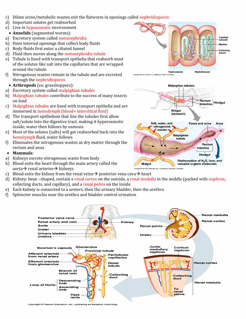
c) Dilute urine/metabolic wastes exit the flatworm in openings called nephridiopores d) Important solutes get reabsorbed e) Live in hypoosmotic environment Annelids (segmented worms):
a) Excretory system called metanephridia b) Have internal openings that collect body fluids c) Body fluids first enter a ciliated funnel d) Fluid then moves along the metanephridia tubule e) Tubule is lined with transport epithelia that reabsorb most
of the solutes like salt into the capillaries that are wrapped around the tubule
f) Nitrogenous wastes remain in the tubule and are excreted through the nephridiopores
Arthropods (ex: grasshoppers): a) Excretory system called malpighian tubules b) Malpighian tubules contribute to the success of many insects
on land c) Malpighian tubules are lined with transport epithelia and are
immersed in hemolymph (blood+ interstitial fluid) d) The transport epithelium that line the tubules first allow
salt/solute into the digestive tract, making it hyperosmotic inside; water then follows by osmosis
e) Most of the solutes (salts) will get reabsorbed back into the hemolymph fluid, water follows
f) Eliminates the nitrogenous wastes as dry matter through the rectum and anus
Mammals: a) Kidneys excrete nitrogenous waste from body b) Blood exits the heart through the main artery called the
aorta renal arteries kidneys c) Blood exits the kidney from the renal veins posterior vena cava heart d) Kidney: bean –shaped, contain a renal cortex on the outside, a renal medulla in the middle (packed with nephron,
collecting ducts, and capillary), and a renal pelvis on the inside e) Each kidney is connected to a ureters, then the urinary bladder, then the urethra f) Sphincter muscles near the urethra and bladder control urination
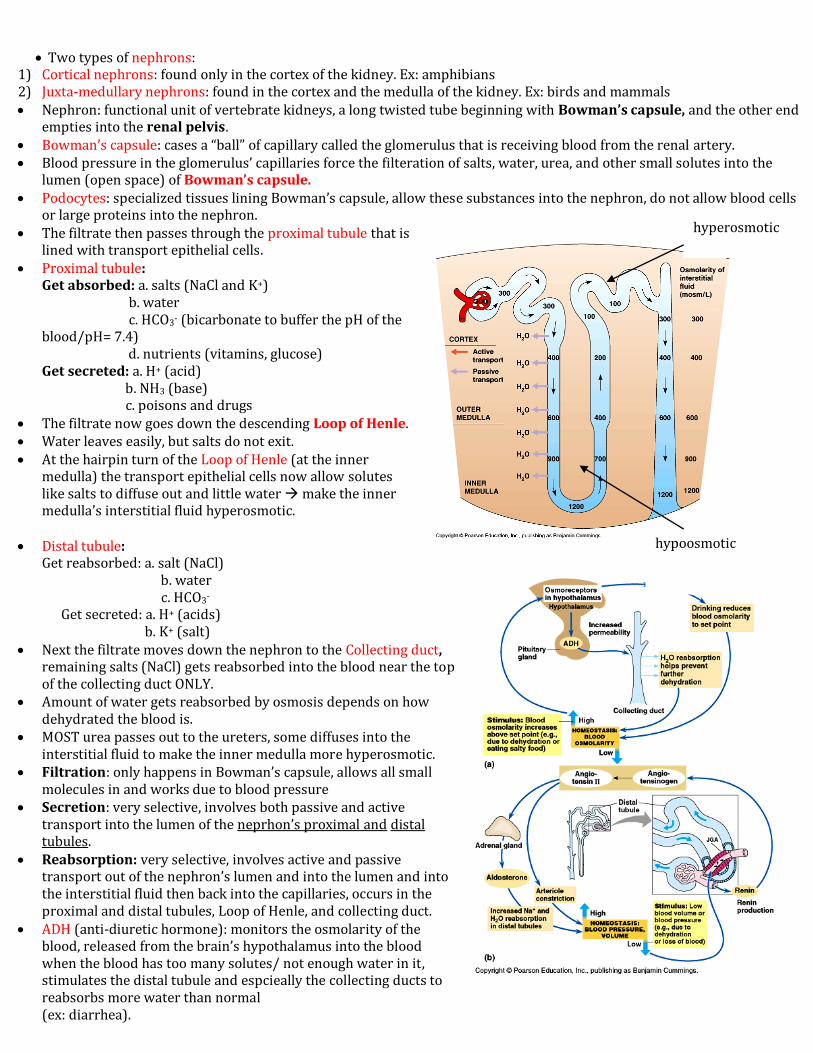
Two types of nephrons: 1) Cortical nephrons: found only in the cortex of the kidney. Ex: amphibians 2) Juxta-medullary nephrons: found in the cortex and the medulla of the kidney. Ex: birds and mammals Nephron: functional unit of vertebrate kidneys, a long twisted tube beginning with Bowman’s capsule, and the other end
empties into the renal pelvis. Bowman’s capsule: cases a “ball” of capillary called the glomerulus that is receiving blood from the renal artery. Blood pressure in the glomerulus’ capillaries force the filteration of salts, water, urea, and other small solutes into the
lumen (open space) of Bowman’s capsule. Podocytes: specialized tissues lining Bowman’s capsule, allow these substances into the nephron, do not allow blood cells
or large proteins into the nephron. The filtrate then passes through the proximal tubule that is
lined with transport epithelial cells. Proximal tubule:
Get absorbed: a. salts (NaCl and K+) b. water c. HCO3- (bicarbonate to buffer the pH of the blood/pH= 7.4) d. nutrients (vitamins, glucose) Get secreted: a. H+ (acid) b. NH3 (base) c. poisons and drugs
The filtrate now goes down the descending Loop of Henle. Water leaves easily, but salts do not exit. At the hairpin turn of the Loop of Henle (at the inner
medulla) the transport epithelial cells now allow solutes like salts to diffuse out and little water make the inner medulla’s interstitial fluid hyperosmotic.
Distal tubule: Get reabsorbed: a. salt (NaCl) b. water c. HCO3- Get secreted: a. H+ (acids) b. K+ (salt)
Next the filtrate moves down the nephron to the Collecting duct, remaining salts (NaCl) gets reabsorbed into the blood near the top of the collecting duct ONLY.
Amount of water gets reabsorbed by osmosis depends on how dehydrated the blood is.
MOST urea passes out to the ureters, some diffuses into the interstitial fluid to make the inner medulla more hyperosmotic.
Filtration: only happens in Bowman’s capsule, allows all small molecules in and works due to blood pressure
Secretion: very selective, involves both passive and active transport into the lumen of the neprhon’s proximal and distal tubules.
Reabsorption: very selective, involves active and passive transport out of the nephron’s lumen and into the lumen and into the interstitial fluid then back into the capillaries, occurs in the proximal and distal tubules, Loop of Henle, and collecting duct.
ADH (anti-diuretic hormone): monitors the osmolarity of the blood, released from the brain’s hypothalamus into the blood when the blood has too many solutes/ not enough water in it, stimulates the distal tubule and espcieally the collecting ducts to reabsorbs more water than normal (ex: diarrhea).
hypoosmotic
hyperosmotic

Alcohol blocks ADH. Hormones can raise or lower blood pressure Low blood pressure results from: 1) dehydration 2) blood loss 3) reduced salt intake. Hormone can raise the blood pressure by constrict capillaries and stimulating the reabsorption first of salts in the distal
tubule and then water enters by osmosis into the blood. Juxtamedullary nephrons are found only in birds and mammals because these land animals need to conserve water and
have concentrated hyperosmotic urine compared to animals in water. Desert birds and mammals have longer loops of Henle to absorb more water. Reptile urine is isosmotic; they do not have a loop of Henle, only have cortical nephrons. Amphibian urine is very dilute/ hypoosmotic because part of their life is in water, also have only cortical nephrons. The type of nitrogenous waste produced by an organism is linked to its habitat. Nitrogenous wastes is the result of the breakdown of protein and nucleic acid (DNA/RNA) during metabolism 3 types of nitrogenous waste:
1) Ammonia: most aquatic animals, very toxic, but it gets diluted by the water 2) Urea: mammals, most amphibians, and sharks, combining NH3 +one CO2, requires ATP, less toxic than ammonia 3) Uric acid: insects, birds, and reptiles, made by combining NH3+ many CO2’s, tends to be paste-like because it has very
little water in it, requires energy to form this even less toxic waste. Endotherms= warm-blooded: getting body heat from its own metabolism. Ex: mammals, birds, some fish, and many
insects. Ectotherm= cold-blooded: absorbing heat from its surroundings. Ex: reptiles, amphibians, most fish, and most
invertebrates. Endotherms:
I. Advantages: temperatures fluctuate more on land allows endotherm land animals to maintain a constant body temperature; they are more active than ectotherms.
II. Disadvantages: need to consume a lot more energy than ectotherms, it is energetically expensive to maintain constant internal temperatures.
Land animals maintain a relatively constant body temperature by: 1) physiologically or 2) behavior adjustments. Thermoregulation: maintaining a constant internal temperature 3 things do thermoregulate:
1. Adjust the rate of heat exchange between the animal and its surroundings. Reduce heat loss with body insulation. Ex: hair, fur,
feathers, and/or fat Change blood flow to the surface of the skin.
a) Vasodilate= increase blood flow by increase the diameter of the blood vessels closer to the surface of the skin so that heat can easily escape and cool an animal off.
b) Vasoconstrict= decrease blood flow by decrease the diameter of superficial blood vessels.
Counter-current heat exchanger: controls heat loss in marine mammals and birds from their extremities (limbs). Blood is cold in the extremities. Counter-current heat exchanger passed warm heat in the arteries to the cold veins before the blood goes back the body’s core/ the heart.
Temperatures: controlled through interactions with the environment.
a) Conduction: direct transfer of heat between the environment and the body. Ex: animal sits in cold water to cool off.
b) Convection: transfer of heat by the movement of air or water across the surface of the body. Ex: cool breeze contributes to heat loss.
c) Radiation: emission of electromagnetic waves from all warm objects like an animal’s body or the sun. d) Evaporation: loss of heat from the surface of a liquid, in the form of a gas, makes the animal’s body much
cooler. If there is low humidity in the air/dry air, then water will evaporate from a body (high concentration to low) through breathing and from the skin to cool the animal. It is increased by panting, bathing, and sweating.
2. Behavioral responses Relocate to thermoregulate. Ex: birds migrate during winter.
3. Changing the rate of metabolic heat production to thermoregulate. Applies ONLY to endotherms (birds, mammals). Ex: the body automatically doubles the metabolic heat
production inside the animal when exposed to cold. Some animals have a hormone that can trigger internal heat production called nonshivering thermogenesis.
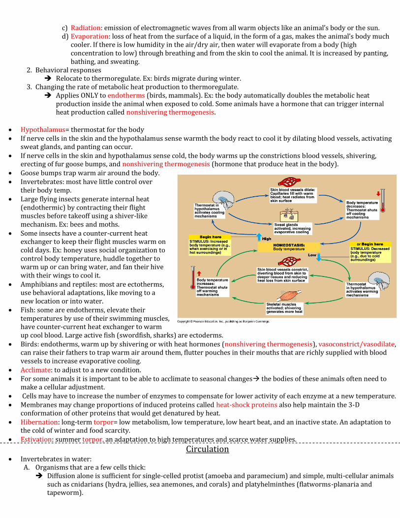
Hypothalamus= thermostat for the body If nerve cells in the skin and the hypothalamus sense warmth the body react to cool it by dilating blood vessels, activating
sweat glands, and panting can occur. If nerve cells in the skin and hypothalamus sense cold, the body warms up the constrictions blood vessels, shivering,
erecting of fur goose bumps, and nonshivering thermogenesis (hormone that produce heat in the body). Goose bumps trap warm air around the body. Invertebrates: most have little control over
their body temp. Large flying insects generate internal heat
(endothermic) by contracting their flight muscles before takeoff using a shiver-like mechanism. Ex: bees and moths.
Some insects have a counter-current heat exchanger to keep their flight muscles warm on cold days. Ex: honey uses social organization to control body temperature, huddle together to warm up or can bring water, and fan their hive with their wings to cool it.
Amphibians and reptiles: most are ectotherms, use behavioral adaptations, like moving to a new location or into water.
Fish: some are endotherms, elevate their temperatures by use of their swimming muscles, have counter-current heat exchanger to warm up cool blood. Large active fish (swordfish, sharks) are ectoderms.
Birds: endotherms, warm up by shivering or with heat hormones (nonshivering thermogenesis), vasoconstrict/vasodilate, can raise their fathers to trap warm air around them, flutter pouches in their mouths that are richly supplied with blood vessels to increase evaporative cooling.
Acclimate: to adjust to a new condition. For some animals it is important to be able to acclimate to seasonal changes the bodies of these animals often need to
make a cellular adjustment. Cells may have to increase the number of enzymes to compensate for lower activity of each enzyme at a new temperature. Membranes may change proportions of induced proteins called heat-shock proteins also help maintain the 3-D
conformation of other proteins that would get denatured by heat. Hibernation: long-term torpor= low metabolism, low temperature, low heart beat, and an inactive state. An adaptation to
the cold of winter and food scarcity. Estivation: summer torpor, an adaptation to high temperatures and scarce water supplies.
Circulation Invertebrates in water:
A. Organisms that are a few cells thick: Diffusion alone is sufficient for single-celled protist (amoeba and paramecium) and simple, multi-cellular animals
such as cnidarians (hydra, jellies, sea anemones, and corals) and platyhelminthes (flatworms-planaria and tapeworm).

They have a gastrovascular cavity: a central cavity used for digestion/ distribution/ circulation of substances throughout the body. It is continuous with the outside water.
B. Organisms that are several cell layers thick: A gastrovascular cavity is inadequate for invertebrate
organisms with many cell layers such as mollusks (clams, snails, octopi, and squid).
Invertebrates on land: Arthropods (insects) and annelids (earthworms) also do not have a gastrovascular cavity.
Circulatory systems: 1. Open circulatory system: blood is not in vessels system. Ex: arthropods and most
mollusks (clams, snails). Hemolymph: interstitial fluids and blood Sinuses: cavities/ organs located between organs.
2. Closed circulatory system: blood inside vessels. Ex: all vertebrates, some invertebrates: some mollusks (octopi,
squid) and annelids. The heart:
*1. Right atrium
*2. Tricuspid A-V valve
*3. Right ventricle 11
*4. Pulmonary semi-lunar valve 5
*5. Pulmonary Arteries 5
6. Pulmonary veins 13
7. Left atrium 7 6
8. Bicuspid/Mitral A-V valve 1 10
9. Left Ventricle 8
10. Aortic Semi-Lunar valve
11. Aorta 2
*12. Inferior/Posterior Vena Cava 3 9
*13. Superior/Anterior Vena Cava
*=deoxygenated blood 12
6

1) Heart: Atria: on top
a) Left atrium- oxy b) Right atrium- deoxy c) Left ventricle- oxy
(thickest muscle) d) Right ventricle- deoxy
Ventricles: on bottom 2) Vessels:
Arteries: away from heart *Main Artery: aorta
Arterioles: go into organs Capillaries: gas exchange
because they are thin and porous
Venules: “downstream” end to a capillary bed
Veins: towards the heart *Main vein: vena cava
Arteries and veins are distinguished by their direction and NOT what they contain!
3) Blood: pH 7.4, 4-6 liters Red Blood Cell/ Erythrocytes: no nuclei, no mitochondria (anaerobic),
do fermentation for energy so don’t consume the oxygen they are carrying. Blood is mostly this, made of hemoglobin (Fe binds to O2)
Plasma: fluids of blood, 90% water, salts, proteins (antibodies), nutrients (glucose).
White blood cells/ leukocytes: 5 different types, spend most of their time in lymphatic/ immune system.
Platelets: fragments of cells no nuclei, 1st at the scene with blood clotting.
Fish: 2 chambered hearts breathe through gills, 1 atrium and 1 ventricle. Amphibians: 3-chambered heart, breathe through lungs and skins, 2 atria, 1 ventricle, due to one ventricle blood mixes.
Pulmocutaneous Circuit: leads to lungs and skin Systemic Circuit: leads to the systems of the body Double Circulation: blood is pumped a 2nd time after it comes back from the lungs
Reptiles: 3-chambered heart, breathe through lungs, 2 atria, 1 ventricle, blood mixes but less because of a ridge in the ventricle, double circulation occurs.
Pulmonary Circuit: leads to lung Mammals and birds: 4-chambered heart, breathe through lungs, 2 atria, 2 ventricles, have a pulmonary circuit and
double circulation, no mixing of blood in the ventricles, use the most oxygen than others since they are endotherms.

Cardiac cycle= one complete sequence of the heart filling and pumping.
Systole: heart contracts (squeeze) ~110-120 Diastole: heart relaxes ~70-80
#1) The “relaxes” and fills with blood. Atrial diastolic and Ventricular diastolic Semi-lunar valves close and blood hits the closed valves (the
“dub” sound). #2) The heart’s atria “squeeze out the blood” and become empty.
Atrial systolic and Ventricular diastolic #3)The heart’s ventricle “squeeze out the blood” forcing it into the lungs and to the rest of the body.
Atrial diastolic and Ventricular systolic. A-V valves close and blood hits the closed valves (the “lub”
sound). Heart murmur: when the valves don’t close completely All of the heart’s muscle fibers are electrically linked, through the use of Ca++ions.
#1. An impulse starts in the heart’s SA node (sinoatrial)/pacemaker spreads across the 2 atria, causing them to contract in unison. #2. The impulse ten reaches the AV node (atriaventricular) between the right atrium and the right ventricle, den a delay occurs for 1 second to empty the atria completely. #3. Tissue between both the ventricles called bundle of His relays the impulse to branches in the ventricles called the purkinje fibers: causes the ventricles to contract in unison. These impulses can be detected by use of an electrocardiogram (ECG or EKG). SA nodes: start this whole process. Hormone controls the heart’s rhythm. Ex: epinephrine/ adrenalin which is used for the “fight or flight” response. Temperature controls the heart’s rhythm. Blood pressure: greater in artery than in veins, lowest in the capillaries. Blood is able to return to the heart via the veins as a result of:
A) Veins being squeezed whenever we move our skeletal muscles B) Smooth muscle contractions that line the vessels C) One-way valves that prevent blood from going the wrong direction D) Breathing keeps the exchange and process going.
“Pins and needles” in your extremities/ your foot fall asleep: blood pools in your foot/ not returning to the heart.
Capillaries in the 1) brain 2) heart 3) kidneys 4) liver are usually filled completely with blood; other locations are not always “full”.
Smooth muscle: involuntary and controls blood flow to capillary beds. Precapillary sphincters: rings of smooth muscle at the entrance to capillary beds that
change blood flow.

Small molecules (salt, water, sugars, urea, oxygen, and CO2) flow between the capillaries and interstitial fluid. There is some leakage of proteins. Most of these molecules get reabsorbed either in the venules or in the lymphatic system. Lymph capillaries help to return proteins and other solutes to the blood. Lymph: fluid inside the lymphatic system is similar to interstitial fluid. Lymph nodes: found along the lymph capillaries, honeycombed in shape, filter the lymph, and filled with white blood cells
that fight off infection. When an individual is sick, doctors check the patient’s lymph nodes because when sick, WBC’s multiply, and the lymph
nodes swell/become tender. Red bone marrow creates blood, found mostly in ribs, pelvis, vertebrae, and breast bone. Cells are called multipotent stem
cells. Multipotent: have the ability to differentiate into all other blood cell types. Totipoten stem cells: all cell types Pluripotent stem cells: most cell types Erythropoietin: a hormone released by kidneys when tissues are not receiving enough oxygen. It stimulates red bone
marrow to make more red blood cells. Cancer leukemia affects stem cells, creates abnormal white blood cells and crowds out red blood cells. Treatment: destroy
the patient’s own bone marrow and give them healthy new stem cells. Scrape your skin platelets (the plug) fibrogen (inactive protein) fibrin (active protein) Hemophilia: the ‘bleeder’s disease’ is a defect in any stem of the clotting process. Cardiovascular diseases lead to 1) heart attack 2) stroke
Causes include: a) Thrombus: blood clot in the absence of injury b) Embolus: blood clot that is transported and stops moving when it reaches a narrow path. c) Plaques: disease is called atherosclerosis d) Hardened plaque: if the plaque hardens, due to deposits of Ca++ ions. The hardening of the arteries is called
arteriosclerosis e) Hypertension: high blood pressure, it can be altered by diets (salt intake), exercise, medication, avoiding animal
fats, and quit smoking f) LDL (low density lipoproteins): “bad cholesterol”. Ex: saturated fats. LDL’s leave cholesterol on arteries/plaque g) HDL (high density lipoproteins): reverse cholesterol. Unsaturated fats: oils, salmon/trout, avocado
TPA (tissue plasminogen activator): dissolves blood clots/ makes blood thinner.
Respiration In water:
Gas exchange occurs across the entire surface of protists and simple water creatures (cnidarians, platyhelminthes) because they are all small, thin and some are flat in shape.
Gills are used for gas exchange in water in mollusks (clams), echinoderms (sea stars), crustaceans (crayfish), and fish. Gills have large surface area due to all the folding. Advantage of being in water: respiratory surfaces are always
moist. In order for diffusion of gasses to occur, the respiratory surfaces must be moist.
Disadvantage of being in water: little dissolved oxygen in the water in contrast to air (21%). To solve: many organisms with gills ventilate a current of water over their gills brings a fresh supply of oxygen in and expels out CO2.
Fish do a Counter-Current Exchange that increases gas exchange, means that blood in the capillaries near the gills flows opposite to the flow of water favors the transfer of oxygen from the water into the blood.
Gills are unsuitable structures on land because A) evaporation of water from the gills would occur B) the gills would stick together.
On land:
Annelids (earthworm) use their entire skin for gas exchange, must live in moist places to permit gas exchange with their capillaries located just below the surface of their skin. Disadvantage: evaporation of water to the air. This problem gets solved by putting the respiratory surfaces inside the body.
Spiracles: air enters insects through openings on their bodies. Air through the tracheal system: air tubes throughout the body of the insect, these tubes touch nearly every cell of the
body. Circulatory system must deliver the oxygen from the lungs to
the rest of the body. Lungs: found in all land vertebrates, spiders and terrestrial
snails. Birds: have a very efficient means of gas exchange, have both
air sacs and lung. When inhaling, both the air sacs inflate; and when exhaling, both deflate. The bottom air sac empties into the lungs, so the lungs are always full of oxygen and allows for flight. Air only goes in one direction.
Texture of lungs is spongy, always moist, have a huge surface area.
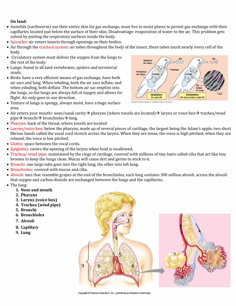
Air enters your mouth+ nose/nasal cavity pharynx (where tonsils are located) larynx or voice box trachea/wind pipe bronchi bronchioles lung.
Pharynx: back of the throat, where tonsils are located Larynx/voice box: below the pharynx, made up of several pieces of cartilage, the largest being the Adam’s apple, two short
fibrous bands called the vocal cord stretch across the larynx. When they are tense, the voice is high pitched; when they are relaxed, the voice is low pitched.
Glottis: space between the vocal cords. Epiglottis: covers the opening of the larynx when food is swallowed. Trachea/ wind pipe: maintained by the rings of cartilage, covered with millions of tiny hairs called cilia that act like tiny
brooms to keep the lungs clean. Mucus will cause dirt and germs to stick to it. Bronchi: one large tube goes into the right lung, the other into left lung. Bronchioles: covered with mucus and cilia. Alveoli: sacs that resemble grapes at the end of the bronchioles, each lung contains 300 million alveoli, across the alveoli
that oxygen and carbon dioxide are exchanged between the lungs and the capillaries. The lung:
1. Nose and mouth 2. Pharynx 3. Larynx (voice box) 4. Trachea (wind pipe) 5. Bronchi 6. Bronchioles
7. Alveoli 2 1
8. Capillary 3 9. Lung
4
5 5 7
9 6 6
8
Vertebrates ventilate the lungs (push air across them) by breathing. The lungs of mammals pull air down like a suction pump by using negative pressure breathing. When you inhale, the volume of the air in the lungs , the diaphragm pulls When you exhale, the volume of the air in the lungs , the diaphragm goes Tidal volume: volume of air an animal inhales and exhales with each normal breath. Vital capacity: most air you can get out of your lungs if you exhale as hard as you can. Residual volume: remaining air in the lungs that cannot be blown out no matter how forcefully you blow out Medulla oblongata and pons: regions of the brain that automatically control breathing, set breathing rhythm and monitor
the oxygen, CO2,/ pH of the blood Hemoglobin: a protein that carry oxygen, has 4 proteins subunits each bound with a metal Fe/heme group that gives
vertebrates red colored blood, only in vertebrates. Hemocyanin: found in invertebrates, carry oxygen, uses the metal copper to bind the oxygen and gives the blood bluish-
green colored. Blood’s buffering system: CO2+ H20 H2CO3 HCO3-+ H+ CO2 is formed as a waste product of cells, transformed into HCO3- (bicarbonate) to buffer the blood (pH=7.4) Hemoglobin binds most of the H+ ions so that the blood doesn’t become too acidic. When the blood reaches the lungs, the entire process is reversed, and carbon dioxide is expelled out the lungs. Deep-diving animals (seal, dolphin) require a lot of oxygen due to extended periods of time under the water. They have lot
of blood, 24 liters, and they have a huge spleen to help store the blood; they have myoglobin that stores oxygen. Deep-sea animals conserve oxygen by lowering their heart rate so the demand for oxygen decreases, and then route blood
primarily to their brain, eyes, adrenal glands, and, if pregnant, to the placenta.
Lymphatic System Immune system: body wide network of cells and organs that have evolved to defend the body against attacks by foreign
invaders. The targets of the immune defenses are infectious organisms. Ex: viruses, bacteria, parasites, fungi, and some protists. First line of defense:
1) Skin: tough and impermeable when intact. 2) Sweat: gives skin a pH of 3-5 3) Mucus membranes: found along the respirator and digestive, and urinary tracts.
Second line of defense: 1) White blood cells/leukocytes: 5 types, four are nonspecific, one is specific.
a) Neutrophils: 1st WBC’s to go to the site of an injury to ingest an invader, engulf a foreign invader by phagocytosis before they themselves self-destruct, short-lived
b) Monocytes: 2nd WBC’s to go to the site of an injury to ingest an invader, utilize phagocytosis and fuse with a lysosome to digest a foreign organism, long-lived. Ex: macrophage/”big eater”
c) Eosinophils: unlike neutrophils and monoctyes, fight off parasites by discharging enzymes into a parasite to digest it
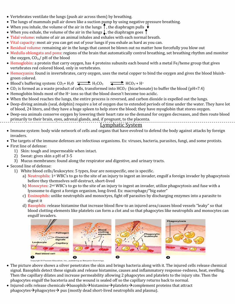
d) Basophils: release histamine that increase blood flow to an injured area/causes blood vessels “leaky” so that blood clotting elements like platelets can form a clot and so that phagocytes like neutrophils and monocytes can engulf invaders.
The picture above shows a silver penetrates the skin and brings bacteria along with it. The injured cells release chemical
signal. Basophils detect these signals and release histamine, causes and inflammatory response-redness, heat, swelling. Then the capillary dilates and increase permeability allowing 2 phagocytes and platelets to the injury site. Then the phagocytes engulf the baceteria and the wound is sealed off so the capillary returns back to normal.
Injured cells release chemicalsbasophilshistamineplateletscomplement proteins that attract phagocytesphagocytes pus (mostly dead short-lived neutrophils and plasma).

2) Natural killer cells: another non-specific second line of defense but not WBC’s, work on virus infected cells and abnormal cells (tumor) and cause them to lyse by attacking their cell membranes.
3) Body’s thermostat: moderate fever helps to destroy some invading microbes and increases repair of tissues/reactions in the body.
4) Anti-microbial proteins: 3 types:
a. Tears: have an enzyme/protein called lysozyme that breaks apart the cell wall of bacteria b. Complement proteins: 20 proteins that attach to a target invader to attract phagocytotic WBC’s (neutrophils and
monocytes) c. Interferons: when a cell is infected with viruses, the infected cell secretes interferon proteins to neighboring
body cells which alert and prepare the body cells to resist the invader, important in stopping the spread of cold and influenza viruses.
Third line of defense: 1) Lymphocytes: specific defenses attack selected targets and are the last line of defense, 5th type of WBC
2 types: a. B-cells: formed and finished in red bone marrow B-cells make Y-shaped proteins called antibodies that they
then secrete to specific targets. b. T-cells: formed in red bone marrow, finished in thymus gland, do not secrete antibodies, have receptors on
them to recognize specific targets. Antigen: a foreign that elicits a specific response. Specific because antigen has unique polypeptide and/or polysaccharide
on its surface The immune system is equipped to respond to million or possibly billions of different specific antigens. The lymphatic/immune system is made up of lymph vessels which return lymph fluid and proteins to the circulatory
system. Other organs of the immune system include the: tonsils, appendix, and spleen. Every body cell in us carries unique surface molecules (proteins and sugars) that are recognized as “self” and are not
attacked by the immune system. Epitopes: a distinctive marker carry by foreign molecules/antigens, it protrudes from their surfaces. The immune system is able to recognize millions to billions of distinctive non-self molecules and to respond by producing
molecules such as antibodies that can match up with and counteract non-self molecules. Steps to how both a non-specific and specific immune response can happen:
1. The antigen (ex: virus) enters the body through the nasal cavities notifies the non-specific WBC’s neutrophils and monocytes (ex: macrophage) to come and engulf the viruses by phagocytosis
2. Macrophage breaks off a fragment from the virus then displays it on its own surface and emits a chemical signal called interleukin-1. IL-1 increases the body’s temperature and attract/activate a specific helper T-cell. The helper T-cell has a receptor on its surface that will bind only with the displayed virus fragment/it is very specific. The activated helper T cell is just a helper, can’t destroy the antigen.
3. After binding to the displayed fragment, the helper T emits out a chemical signal called interleukin-2 to alert the “troops”.
4. IL-2 can causes 3 things to occur depend on what type of antigen enters: a) IL-2 causes more specific helper T cells to arrive, then clones both short-lived effector helper T cells and long-
lived memory helper T cells. This occurs in both a humoral response (humors=blood+ lymph) and in a cell-mediated response.
b) With a humoral response, IL-2 notifies the B-cells and being to clone specific antibodies. There are short-lived effector/plasma B-cells and long-lived memory B-cells. The effector/plasma B-cells are able to secrete antibodies into the bloodstream. The antibodies “tag” the antigens so they are easier for the macrophages to locate them and antibodies prevent the antigens from infecting other cells by blocking their surface proteins/carbohydrates. Plasma cells are able to release ~2000 antibodies/second for 4-5 days.
c) Cell-mediated response: IL-2 notifies the killer T-cells/cytotoxic T-cells that arrive at the body cells. Killer T cells are able to latch onto the infected body cell, caner/tumor, or a transplanted cell (organ donation) and secrete an enzyme called perforin, which pokes holes in the cell membrane, and then fluids rush in causing the cell to lyse. There are both effector killer T-cells and memory killer T-cells.
Immune response is turned off after an infection ends by suppressor T-cells. Primary immune response: first time the body gets exposed to an antigen (10-17 days), the body creates thousands of
effector and memory cells. Secondary immune response: 2nd time the body
gets exposed to that same antigen (2-7 days). Antibodies bind to the epitopes on antigen and
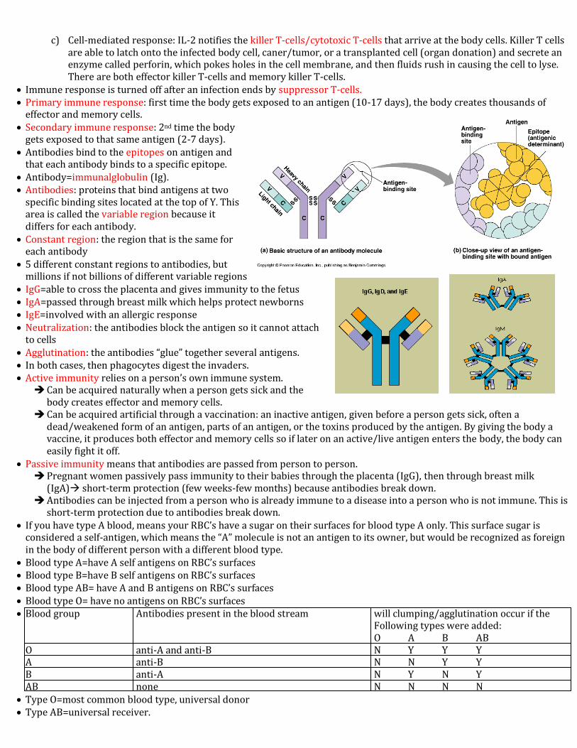
that each antibody binds to a specific epitope. Antibody=immunalglobulin (Ig). Antibodies: proteins that bind antigens at two
specific binding sites located at the top of Y. This area is called the variable region because it differs for each antibody.
Constant region: the region that is the same for each antibody
5 different constant regions to antibodies, but millions if not billions of different variable regions
IgG=able to cross the placenta and gives immunity to the fetus IgA=passed through breast milk which helps protect newborns IgE=involved with an allergic response Neutralization: the antibodies block the antigen so it cannot attach
to cells Agglutination: the antibodies “glue” together several antigens. In both cases, then phagocytes digest the invaders. Active immunity relies on a person’s own immune system. Can be acquired naturally when a person gets sick and the
body creates effector and memory cells. Can be acquired artificial through a vaccination: an inactive antigen, given before a person gets sick, often a
dead/weakened form of an antigen, parts of an antigen, or the toxins produced by the antigen. By giving the body a vaccine, it produces both effector and memory cells so if later on an active/live antigen enters the body, the body can easily fight it off.
Passive immunity means that antibodies are passed from person to person. Pregnant women passively pass immunity to their babies through the placenta (IgG), then through breast milk
(IgA) short-term protection (few weeks-few months) because antibodies break down. Antibodies can be injected from a person who is already immune to a disease into a person who is not immune. This is
short-term protection due to antibodies break down. If you have type A blood, means your RBC’s have a sugar on their surfaces for blood type A only. This surface sugar is
considered a self-antigen, which means the “A” molecule is not an antigen to its owner, but would be recognized as foreign in the body of different person with a different blood type.
Blood type A=have A self antigens on RBC’s surfaces Blood type B=have B self antigens on RBC’s surfaces Blood type AB= have A and B antigens on RBC’s surfaces Blood type O= have no antigens on RBC’s surfaces Blood group Antibodies present in the blood stream will clumping/agglutination occur if the
Following types were added: O A B AB O anti-A and anti-B N Y Y Y A anti-B N N Y Y B anti-A N Y N Y AB none N N N N
Type O=most common blood type, universal donor Type AB=universal receiver.

Rh factor (protein) present on the surface of some RBC’s. If have this antigen on RBC’S= Rh+. If do not have this antigen on RBC’s= Rh-, named after the rhesus monkey where it was
first discovered. If mother is Rh- and her developing baby is Rh+ (from dad), this wouldn’t be an issue because blood doesn’t cross the
placenta. However, sometimes during delivery blood leaks across and triggers the mom’s immune system to make antibodies against the Rh factor. The first baby is fine and healthy. If the 2nd developing baby is Rh+, mom’s immune system will think an invader is in the body and attack against the fetus/cause it to naturally abort by sending out anti-Rh antibodies.
Nowadays this situation is under control because doctors inject Rh- women with anti-Rh antibodies (rhogan) soon after delivering the first Rh+ baby only short term because antibodies break down.
When it comes to organ or tissue transplants it is difficult to prevent the body from attacking the new organ. To increase the chances of it not being rejected, the transplant should be as close of a match as possible. Drugs are also used that suppressor the immune system or parts of the immune system (like helper T-cells) to increase the chances of the body accepting the transplant.
Allergies are hyper sensitive response to environmental antigens called allergens. Ex: allergic to pollen, the body’s plasma cells release pollen antibodies (IgE) into the bloodstream where they attach to a cell called the mast cell. Pollen respiratory tract and attaches to the antibodies located on the mast cells. Like basophils, mast cells are also full of the inflammatory agent histamine. The bound allergens trigger the release of histamine into the bloodstream and the typical allergies symptoms occur-watery eyes and nose, swollen air passages/difficulty breathing, and sneezing.
Autoimmune disease: the immune system attacks self molecules. Ex: 1) Type I diabetes (the pancreas is attacked/stops making insulin) 2) Lupus (the body attacks its own DNA) 3) Multiple sclerosis (the body attacks nerves of the central nervous system) 4) Rheumatoid arthritis (the body attacks joints and cartilage).
Immunodeficiency diseases: immune system is not properly combating antigens. Ex: AIDS, caused by the retrovirus HIV.
HIV: mostly targets the helper T-cells, especially the surface molecule on them called CD4.
There are two major strains of HIV: a) HIV-1 b) HIV-2
HIV: a retrovirus because it has RNA not DNA, enters the body through blood or semen an attaches itself mostly to helper T-cells, injects it RNA into the host helper T-cell, RNA then gets transcribed into DNA with the help of reverse trascrpitase, newly formed DNA then becomes a part of the host DNA, remains hidden inside the host’s DNA possibly for years, during this time, new viruses bud off from the host cell and go make copies of themselves in other host cells.
HIV is so hard to combat is because each time the virus does replication inside a host cell, mutations occur because going from RNA DNA is backwards so no DNA polymerase to proofread.
When HIV does bud off from a helper T-cells, it takes part of the host cell’s plasma membrane.
Most people infected with HIV die from opportunistic infections like the flu because these infections take advantage of a weakened immune system.
HIV considered full blown AIDS=when the helper T-cells drop below 50 cells per mm2
AZT blocks reverse transcriptase (20-50 pills/day).