the journal of biological chemistry printed in u.s.a. · spectropolarimetry-a perkin-elmer model...
TRANSCRIPT

THE JOURNAL OF BIOLOGICAL CHEMISTRY Vol.243, No. 21, Issue of November 10, pp. 5663-5669, 1966
Printed in U.S.A.
Denaturation of Globular Proteins
I. THE INTERACTION OF UREA AND THIOUREA WITH BOVINE PLASMA ALBUMIN*
(Received for publication, May 16, 1968)
JULIUS A. GORDON AND JOHN R. WARREN~
From the Department of Pathology, University of Colorado School of Medicine, Denver, Colorado 80220
SUMMARY
The techniques of equilibrium dialysis and ultrafiltration were utilized to study the interaction of urea, thiourea, methylurea, tetramethylurea, acetamide, ethylene glycol, and dextrose with bovine plasma albumin in water. It was found that the addition of anhydrous protein to water con- taining a nondenaturant (dextrose or tetramethylurea) resulted in an increase of reagent concentration in the protein- free filtrates or dialysates. This finding is interpreted as a highly selective interaction of water with bovine plasma albumin in the presence of nondenaturants; from this finding, the hydration of bovine plasma albumin in these solvents is calculated to be between 20% to 30% (grams of HOH per g of protein). The increase in concentration of acetamide or ethylene glycol (weak denaturants) in filtrates from solutions containing bovine plasma albumin was found to be dependent on reagent concentration, indicating weak interaction of these denaturants with bovine plasma albumin. In contrast, the concentration of urea or thiourea in filtrates from aqueous solutions decreases following the addition of bovine plasma albumin to strong denaturing solutions of these amides. It is deduced that approximately 1 molecule of urea or thiourea interacts per 3 or 4 amino acid residues of each fully denatured albumin molecule. The extent of interaction of small molecules with albumin, in every case, parallels the ability of the molecules to denature albumin.
Although the mechanism remains uncertain, the treatment of most proteins with concentrated urea solutions produces pro-
found changes in protein structure. It is now clear that t,he early suggestion relating the denaturing ability of urea to its structural potential for disrupting monofuncOiona1 hydrogen bonds in aqueous solution (1) is open to serious question (2-4). Waugh called attention to the possible formation of stable com-
* This study was supported by Grant GM-11345 from the Na- tional Institutes of Health.
$ Trainee, United States Public Health Service (Grant GM-977).
plexes between nonpolar amino acid residues and urea in aqueou solutions (5). Subsequently, hydrophobic groups, pure hydro- carbons, and nonpolar amino acids have been found to be more soluble in urea than in pure water and it has been suggested, therefore, that urea denatures proteins by weakening the forces between nonpolar amino acid residues (4-6) or through a change in the structure of water (7-12). That urea probably denatures bovine plasma albumin by specific interaction with the protein has been emphasized by Gordon and Jencks (13). More recently, Robinson and Jencks observed a negative free energy of transfer from water to concentrated urea solutions for acetyl- tetraglycine ethyl ester, a molecule composed primarily of pep- tide groups (14). On the basis of the solubility behavior of this protein model, these authors suggest the formation of bifunctional hydrogen bonds between urea and peptide groups during the urea denaturation of bovine plasma albumin.
It is still undecided whether disruption of the molecular structure of water, complex formation with nonpolar amino acid side chains, formation of bifunctional hydrogen bonds with peptide groups, or a combination of these mechanisms is the mechanism b.y which urea denatures proteins. The latter two mechanisms suggest an interaction (or interactions) between protein and urea. The interaction of urea with proteins and poly- peptides has been previously studied, but largely in a qualitative, indirect, and noncomparative manner (15-20) .I The results, both quantitatively and qualitatively, are conflicting. Thus the present study was undertaken to detect binding,2 if any, of urea, thiourea, methylurea, acetamide, ethylene glycol, and dextrose to the globular protein, bovine plasma albumin. It was ob- served that approximately 1 urea or thiourea molecule appears to interact per 3 or 4 peptide bonds or amino acid residues of the denatured protein. These results support a mechanism (or mechanisms) of urea denaturation that involves some sort of interaction of urea with the protein or with its immediate envi- ronment. This conclusion is strengthened by the demonstration of a positive correlation of denaturing ability of urea and related
1 DOTY, P., AND KATZ, S., Abstracts of the American Chemical Societu Meeting. Chicaao, Sedember. 1960. v. 14C.
- I “ I . I _
2 The term “binding ” is used in its broadest sense, and inter- changeably with the term “interaction,” to mean those molecules of solvent which are in the domain of the protein. No conclusion as to the presence of specific sites of binding is intended.
by guest on August 21, 2019
http://ww
w.jbc.org/
Dow
nloaded from

5664
TABLE I used in all the centrifugation experiments (Fig. 1). Imperme- Experimental variables having no effect on results ability of the membranes to albumin was established in each
A. Materials and methods experiment by obtaining filtrates of pure water from aqueous
1. Type of membrane and relative flow rate solutions of 4% albumin after centrifugation at 3000 rpm and 20”
Visking 1 for 60 min. The membranes were then conditioned with ali-
LSG-60 10 quots of the appropriate solvent at 3000 rpm until equilibrated.
Amicon UM-1 100 Equilibration was ensured by showing that the refractive index
2. Thickness of membrane (e.g. use of two membranes) of the filtrate differed from that of the fluid retained in the cell
3. Alterations in flow rate of ultrafiltrates” or speed of agi- with protein-free solvent by less than 0.00004 unit. Anhydrous tation in equilibrium dialysis albumin was then added to three 20-ml aliquots of solvent to
R. Albumin preparations give 4% solutions and placed in three of the cells; the fourth cell 1. Use of different commercial lots of albumin contained solvent only. The cells were centrifuged for a time 2. Defatted albumin sufficient to collect 0.1 to 0.2 ml of filtrate (usually 3000 rpm for 3. Extensive dialysis of albumin prior to use 60 min). A series of four or five successive filtrates was ob-
Q Above a minimum flow rat.e, which was dependent on the tained from each cell, for a total loss in volume of the retained
type of membrane. fluid of 1 ml or less. If the refractive index of the filtrate from the protein-free solvent varied from that of the original solution
amides with the extent of interaction of these molecules with by a margin exceeding the experimental error for the deter-
bovine albumin. mination of the refractive index (0.00004), filtrates from all four
A preliminary report has been published? cells were discarded. Such deviation usually indicated solvent evaporation due to poor sealing of the collection chamber.
EXPERIMENTAL PROCEDURE Ultrafiltration experiments with the use of positive pressure
Materials were also carried out in a cell containing approximately 40 ml of solution. Pressure in the cell was maintained by nitrogen gas
Bovine plasma albumin (crystalline; Armour Pharmaceutical Company, Kankakee, Illinois; Lots X-69508, X-69805, A-69908,
at 50 pounds pressure. Amicon UM-1 membranes were used
and A-70011) was desiccated under vacuum at 4” for at least 12 exclusively and pre-equilibrated with solvent. Dried albumin
Resid- was then dissolved in a 40-ml aliquot of solvent,, and five to seven
hours and stored in tightly sealed containers prior to use. The flow rates ual moisture (grams of HOH per g of protein) never exceeded
filtrates were analyzed over a 2- to 3-hour period. were such that 0.2 ml of filtrate could be obtained every 10 min.
3 % as determined by weight loss of an aliquot following exposure to 100” for 12 hours. Initially, the commercial albumin was
Intermittent pressure was used, and the total loss in volume
prepared by dialysis against 0.1 M NaCl, dialysis-against distilled of the retained fluid averaged less than 3 ml.
water, and then lyophilization to dryness. This step was Equilibrium Dialysis-Small, cylindrical, stainless steel cells
with Teflon chambers were manufactured at this institut.ion dispensed with when many parallel experiments showed that the commercial preparation contained no dialyzable contaminants detectable by precision refractometry.
Defatted albumin was prepared by the procedures of Williams and Foster (21) or Chen (22).
Reagent grade urea (Mallinckrodt) was purified by two re- crystallizations from warm 75% ethanol and was dried by desiccation. All other chemicals were reagent grade and were used without further purification. The solutions were prepared in deionized and twice distilled water.
Membranes-Three basically different membranes were uti- lized in our studies. Some properties of Visking (Union Carbide Corporation), LSG-60 (Schleicher and Schuell) and Amicon UM-1 (Amicon Corporation) membranes are listed in Table I. Initially, the membranes were processed by extensive washing in distilled water and soaking in the appropriate solvent for 2 to 3 hours. In later experiments, the membranes were soaked in distilled water until used and then conditioned in the apparatus as described below.
VIEW A (TYPICAL)
Methods
Ultrafiltration-Companion experiments were performed with the use of either pressure or centrifugation as the driving force. Four two-chamber stainless steel tubes (International Centrifuge Company), the chambers of which were separated by a mem- ULTRAFILTRATION EQUILIBRIUM
brane (usually LSG60 or Amicon UM-l), were simultaneously DIALYSIS
FIG. 1. Apparatus for ultrafiltration or equilibrium dialysis. 3 GORDON, J. A., AND WARREN, J. R., Abstracts of the Second Stippled area.s indicate the two-component solvent. l , domain
International Biophysics Congress, Vienna, 1966, p. 95. of the added anhydrous protein.
Denaturation of Globular Proteins. I Vol. 243, No. 21
by guest on August 21, 2019
http://ww
w.jbc.org/
Dow
nloaded from

Issue of November 10, 1968 J. A. Gordon and J. R. Warren 5665
(Fig. 1). The chambers were faced with cut Visking dialysis tubing which had been washed in distilled water. Four to seven of these cells, each containing 0.2 to 0.3 ml of solvent, were placed in 40 ml of the same solvent and vigorously agitated on an orbital shaker at room temperature for 120 min. Equilibra- tion between the cells and external solution was experimentally verified by removing one cell and comparing its refractive index with that of the external solution. Following this, albumin, generally to make a 4% solution, was dissolved in the external solvent and agitation was continued for 2 hours. At intervals in the third hour, successive chambers were then removed and the contents were quickly transferred to the refractometer by syringe for analysis. It was experimentally shown by simple dilution that changes in external urea concentration (twice that ever seen in the presence of albumin) were reflected inside the cells within 30 min.
Analysis-The refractive index increment for aqueous solu- tions of urea, thiourea, methylurea, tetramethylurea, ethylene glycol, and dextrose was obtained by determining the refractive indices of each reagent over the entire range of concentra- tion.
Refractive indices relative to air at the sodium D-line were determined to an accuracy of ~0.00002 on a Bellingham and Stanley Abbe-60 high accuracy refractometer which was thermo- stated to 20” + 0.05”. The refractometer was calibrated daily against the known refractive index of distilled water, and inter- mittently with acetone. Standard curves for each reagent were constructed; they allowed determination of the concentration of filtrate or dialysates of unknown molarity with no greater error than +0.004 M, depending on the refractive index increment of the particular solvent.
Spectropolarimetry-A Perkin-Elmer model 141 polarimeter modified with a monochromator and xenon light source was used to obtain data from 300 to 600 mp. The data were ob- tained from 1 To solutions of albumin in the appropriate solvent at 30”. The refractive index correction at 589 mp was used over the entire spectral range to calculate the reduced mean residue rotation, [m’]:& and &he Moffitt-Yang parameters, a0 and b. (23).
RESULTS
The conventional arrangements used in equilibrium dialysis were unsuited for detecting small changes in concentrated solu- tions of denaturant. The ratio of protein-containing solvent to protein-free solvent must be large in order that very small changes in free solvent concentration may be directly reflected in the dialysates. The technique of equilibrium dialysis used here reverses the usual design by isolating small volumes of solvent as protein-free dialysates from the large volume of solvent containing added protein (Fig. 1). Ultrafiltration pro- vided a companion means for following changes in the concen- tration of bulk solvent as the volume of the retained material remained relatively unchanged during the course of the experi- ment (Fig. 1) ; the total loss in the retained volumes was kept to less than 5 %.
Even so, precision refractometry had to be used to detect changes of concentration with sufficient accuracy. This tech- nique of measurement necessitated carrying out the experi- ments in the absence of buffer or supporting electrolyte so that the changes in the refractive index would reflect only the ratio of the two dialyzable components. An increase or
TABLE II Interaction of water or reage mt with native albumin
1. Added albumin
g/100 wild
4
4 (heat-de- natured)’
4
-
2. Reagent
Dextrose
I’etramethyl urea
Ethylene glycol
Qcetamide
-
3
-
Concen~ tration
M
1.660
1.660
0.830
1.500
8.000 5.000 4.400
8.000 5.000 4.800
4.000
Change in molarity’ ,
AM 3~ 0.004 g/g protein $0.023 +0.019 0.29
+0.016 0.26
+O.Oll 0.32
+0.015 0.25
+0.027 $0.028 0.09 58 +0.021 0.10 33
$0.038 0.20 0
+0.023 0.08 +0.031 $0.029 0.15
$0.013 0.13 +0.026 0.18
67
17 16
5 -
0 Average of at least four separate experiments, each with no fewer than four successive filtrates or dialysates.
b Calculated on the basis that the second component, i.e. the reagent, was excluded from the neighborhood of the protein.
c Calculated on the basis of hydrated protein (0.20 g of water
per g of protein). d Grams of anhydrous protein added to 100 ml of solvent. e Only partially soluble.
decrease in the refractive index of filtrate (e.g. dialysate or ultrafiltrate) compared to the solvent before the addition of protein was accepted as significant only when (a) the changes exceeded the experimental error (&0.00004), (5) the increase or decrease was seen in over 80% of the filtrates or dialysates taken from the same solution, or (c) the average change could be verified by one or more additional experiments.
A further consideration was the role the membrane might play in the experimental results (24). The results to be re- ported were uninfluenced by the type of membrane (Table I), the thickness of the membrane, the speed of centrifugation, the filtration pressure, or the time interval between protein addition and first dialysate in each experimental method. In the absence of protein, the solvents always penetrated the membrane without any change in the refractive index.
After the addition of albumin to aqueous 1.660 M dextrose, the concentration of the average filtrate increased to 1.683 M
and that of the dialysates to 1.679 M (Table II). The absolute increase in the concentration of dialysate decreased when the concentration of the retained fluid was 0.830 M. In similar fashion, 1.515 M was the filtrate concentration from a solution of 1.500 M tetramethylurea to which 4% albumin had been added. If dextrose or tetramethylurea had remained evenly distributed throughout the original solvent water after the addition of protein, then a change in concentration would not be expected in the filtrate. The increases in concentration found in the filtrates are generally explained by the removal of solvent water by the selective interaction of water with the added (pre- viously desiccated) albumin in the presence of these nondena-
by guest on August 21, 2019
http://ww
w.jbc.org/
Dow
nloaded from

5666 Denaturation of Globular Proteins. I Vol. 243, No. 21
turants. The calculation of grams of water bound per g of pro- tein, h, from these experiments (Column 6 in Table II) is made as follows (25). Let CT be the concentration of reagent in the solvent before the addition of protein, P be the gram concen- tration of added protein per ml of solvent, and C, the concen- tration of reagent found in the filtrate or dialysate after addition of protein to the retained fluid. Then, h is given by
This treatment assumes that the reagent (in this case, dextrose or tetramethylurea) interacts little or not at all with albumin. This assumption makes the values listed in Table II estimations of minimum hydration, and these values should be recognized for their pragmatic worth. The generally accepted values for the hydration of albumin from other methods lie somewhere above 0.2 g of water per g of protein (26).
The results obtained with ethylene glycol and acetamide are somewhat different. We found that when the concentration of these reagents was increased the increase in reagent concentra- tion seen in the dialysates and filtrates was proportionately smaller. Assuming no interaction of reagent with albumin, the bound water apparently drops at higher concentrations of ethylene glycol or acetamide. We think that a more likely interpretation is that the hydration remains constant (see “Discussion”) at all concentrations of reagent and that these reagents undergo concentration-dependent interactions with albumin. The interaction of reagent may be determined quantitatively as follows. Let M, be the molar concentration of the original solvent adjusted for the binding of 0.20 g of water per g of added protein, PM the molar concentration of protein, and M, the concentration of solvent found in the filtrate. Then
MT - n/r, s=-
PM (2)
where S represents the moles of reagent interacting with 1 mole of hydrated protein (Column 7 in Table II). For example, in 5 M acetamide solution, the results are that each protein molecule interacts with 17 acetamide molecules, even when the concentra- tion of protein is halved. In 8 M acetamide or ethylene glycol, around 65 molecules interact; these concentrated solutions are known to have only slight denaturing ability (13, 27, 28).
Filtrates from strong denaturing solutions of urea show a loss of urea following the addition of albumin to the solutions (Table III). A decrease of 0.022 M was seen in filtrates from 8 &I urea solutions for every gram of protein added to 100 ml of solvent. From Equation 2, 1 albumin molecule interacts with 153 molecules of urea at this concentration. Dropping the urea concentration leads to a fall in the binding ratio. About 86 molecules of urea interact with albumin in 5 M urea. In 1.5 M thiourea solutions, the ratio is approximately 87. Both 5 M
urea and 1.5 M thiourea show equivalent denaturing ability by the criteria of change in optical rotation (Fig. 2). The poor solubility of thiourea at 20” unfortunately prevented experi- mentation at higher concentrations. We can conclude, though, that thiourea shows the greatest affinity toward albumin of all the reagents studied.
Attainable methylurea concentrations do allow us to make comparison with the full range of urea concentrations. It is
TABLE III Interaction of denaturants with albumin
Added albumin
gllO0 mlb
4 2 4
4 4
4
4 8
16
4 4 4
Den&want
Ureac
Methylmead
rhioureac
COlICen- tration
Filtrate Dialysate
-
I
-
tatio, bound de;,“tu;;;t
protein5
M
8.000
8.000
7.000 5.000
2.000
AM zk 0.004
-0.088
-0.044
-0.054
-0.010
-0.094
-0.076
-0.049 -0.021
moles/mole
153 147
127 86 18
7.000 -0.038 65 4.000 -0.033 57 4.000 -0.053 46 4.000 -0.106 45
1.500 1.250 0.500
-0.052
-0.019
-0.052 87 -0.038 63 -0.015 29
Change in molarity
a Adjusted for hydration of albumin (20%). b Grams of anhydrous protein added to 100 ml of solvent. c Values are the average of at least four separate experiments,
each with no fewer than four successive filtrates or dialysates. d Single experiments with a series of no fewer than four suc-
cessive filtrates or dialysates.
l *
A
. .
AM
I I I I I
20 60 100 140 180
Moles Reagent /Mole of Added Albumin
FIG. 2. Correlation of the extent of denaturation measured by optical rotation, [m’], and binding of reagent spread over the total protein. l , urea; A, thiourea; A, methylurea; Q, acetamide.
known that 7 M methylurea solutions show less denaturing ability toward bovine plasma albumin than 5 M urea (13). As shown in Table III, fewer molecules of methylurea interact with albumin under these conditions. In addition, we found that changing the concentration of protein k-fold in the presence of constant methylurea did not significantly change the number of methylurea molecules bound per molecule of protein.
Throughout the course of these experiments we were con-
cerned with the effect of albumin preparations on the results (Table I). We found that various commercial lots of albumin could not be distinguished by their ability to interact with urea. We found identical results with extensively dialyzed prepara.-
by guest on August 21, 2019
http://ww
w.jbc.org/
Dow
nloaded from

Issue of November 10, 1968 J. A. Gordon and J. R. Warren 6667
tions of crystalline protein, defatted albumin (21, 22), and even noncrystalline Fraction V.
DISCUSSION
Definitions and Methodology-Advantages and disadvantages to the use of various concentration scales depend on the charac- ter of the experiment (29). We made use of the molarity (volume molality) scale in the following fashion. To a given volume of diffusible components of known molarity, e.g. urea and water, a weight of protein was added. The concentrat,ion of the dialysate or filtrate was then compared to the concentra- tion of the original solvent. Alternatively, we could have used the weight molality or mole fraction scale to describe the same experiment. It is sometimes stated that the molal scale is intrinsically more fundamental than the molar scale of concen- tration. It is true that the molal scale usually provides a means of calculating mole fractions and thereby permits an easier association with certain physical laws. But there is no smooth and simple relationship between any concentration scale and the energy relationships that determine the many properties of the chemical system, especially in nonideal solutions. Therefore, we elected to express the results of these experiments in the units customarily used in biochemistry (30). Nevertheless, the choice of concentration units can lead to quantitatively different ratios of binding,2 and any direct comparisons made must all be of one kind of unit (29).
We would also note some of our reasons behind the avoidance of the adjective “preferential” to describe the interaction of one or another diffusible components with albumin. Use of the adjective includes at least three hidden elements: (a) the de- pendence of the absolute values of the preferential binding to a macromolecule, and especially hydration, on the experimental technique (31-34), (b) the dependence of the binding ratios on the concentration units used, as noted above (29) and (c) the connotation that preferential binding of one component auto- matically always leads to partial or complete exclusion of other solution components from the surface of the protein. If the areas of binding do not overlap, the preferential binding of water to a macromolecule need not invariably lead to “exclusion” of a second solvent component. The total concentration of this second component in the protein neighborhood may even be less than in the solvent bulk, and therefore be nonpreferentially bound, yet its concentration may be much higher in the selective areas of interaction.
Protein-Solvent Interaction--il large volume of experimental evidence on the hydration of albumin and other proteins has been gathered on anhydrous crystalline proteins and native proteins in solution (31, 33-36). The amount of water thought to be bound to solubiliaed albumin varies somewhat with the experimental technique, but reliable values are seen most con- sistently from a low of 0.2 to a high of 0.4 g of water per g of protein, dry weight (26,34). With the techniques of equilibrium dialysis and ultrafiltration used here, an increase of the non- aqueous component in the filtrate is a sensitive reflection of water of hydration when there is no binding of the nonaqueous component to the protein. Thus it was not surprising to find that approximately 0.25 g of water per g of protein became un- available to dextrose and tetramethylurea after the addition of dry protein. Many such nonelectrolytes have proven under somewhat similar circumstances, but at low concentrations, to measure bound water (25, 32).
Available experimental data on the hydration of denatured albumin and other denatured proteins is somewhat scarce. Some investigators find that upon denaturation there is an increase of protein hydration, and some find that the denatured molecule binds water in amounts equivalent to the native protein (34, 36-38). Our experiments with heat-denatured albumin in dextrose solutions agree with the latter conclusion. This point has bearing on the quantitative interpretation of the results. With acetamide and ethylene glycol, at low reagent concentra- tion, we found a protein hydration of 0.2 to 0.3 g of water per g of native protein. With increasing acetamide concentration, protein hydration apparently underwent progressive diminution. At 8 M acetamide, for example, it would seem that the bound water falls to 0.08 g of water per g of protein. If protein hydra- tion is progressively less at increasing concentrations of acetamide or glycol, then the absence of a concentration change in filtrates from 2 M urea-4.0% albumin solutions would also imply absence of hydration of native albumin in the 2 M urea solvent. But in these solvents the protein remains native (13) and its hy- drophilic surfaces continue to be exposed to the solvent. A more likely interpretation is that native albumin remains hydrated in 2 M urea, 8 M acetamide, and 8 M ethylene glycol, and that the apparent fall or absence of hydration represents a progressive interaction of acetamide, ethylene glycol, or urea molecules with nearly normally hydrated albumin. ‘l’he experimental tech- niques used do not reveal whether acetamide, glycol, or urea binds to native albumin by exclusion of water molecules from the protein solvation shell or by inclusion into a shell without displacement of water molecules (29, 39). However, since about 700 molecules of water would be contained in the solvation shell of each albumin molecule at 20 y. hydration, the displacement of up to 60 to 80 water molecules by 60 acetamide, glycol, or urea molecules would only diminish protein hydration to around
18%. ‘This is within the range of experimental error observed in our techniques. On the other hand, the binding of acetamide need not displace water at all, if the hydration does not form a complete solvation envelope.
Filtrates from solutions containing denaturing concentrations of urea, thiourea, or methylurea showed a concentration decrease which was proportional to the amount of added protein. On the basis of available information, we have assumed that the hydration of denatured albumin remains near the hydration of native albumin measured by the techniques used here (34, 36). An increase or decrease of albumin hydration following denatura- tion would alter the extent of interaction only quantitatively. The validity of assuming little change in hydration is strength- ened by the positive correlation we observed between the num- ber of small molecules which bind to each plasma albumin molecule and the ability of the small molecule to denature the protein (Table II). Alkyl substitution of urea is known to reduce the denaturing effectiveness of the compound toward plasma albumin (13). Thus, it is significant that alkylation of urea also reduces the ability of the substituted urea molecule to bind to albumin when compared to that of urea. Urea and thiourea, both strong denaturants (13), show greater affinity for tne plasma albumin molecule. The ability of thiourea to bind to albumin at molar concentrations much lower than those concentrations giving comparable binding in urea solvents is consistent with the relatively greater denaturing effectiveness of thiourea (13). The correlation with denaturing effectiveness is graphically illustrated in Fig. 2.
by guest on August 21, 2019
http://ww
w.jbc.org/
Dow
nloaded from
5668 Denaturation of Globular Proteins. I Vol. 243, No. 21
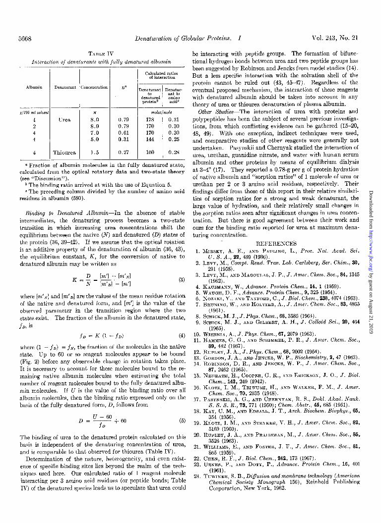
TABLE IV
Interaction of denaturants with fully denatured albumin
Albumin Denaturant Concentration
g/100 ml solvent M 4 8.0 2 8.0
Thiourea
7.0 5.0
1.5
0.79 0.79
0.61 0.31
0.27 160 0.28
Calculated ratios of interaction
Denaturant Denatur-
de&red ant to amino
protein6 acidC
moles/mole
178 0.31 170 0.30 170 0.30 144 0.25
a Fraction of albumin molecules in the fully denatured state, calculated from the optical rotatory data and two-state theory (see “Discussion”).
b The binding ratio arrived at with the use of Equation 5. c The preceding column divided by the number of amino acid
residues in albumin (580).
Binding to Denatured Albumin-In the absence of stable intermediates, the denaturing process becomes a two-state transition in which increasing urea concentrations shift the equilibrium between the native (N) and denatured (D) states of the protein (36, 3942). I f we assume that the optical rotation is an additive property of the denaturation of albumin (36, 43), the equilibrium constant, K, for the conversion of native to denatured albumin may be written as
K = _D = [m’l - [m’d N Im’d - [m’l
where [m’N] and [m’e] are the values of the mean residue rotation of the native and denatured form, and [m’] is the value of the observed parameter in the transition region where the two states exist. The fraction of the albumin in the denatured state, J~D, is
fD = K (1 - fD) (4)
where (1 - fD) = f~, the fraction of the molecules in the native state. Up to 60 or so reagent molecules appear to be bound (Fig. 2) before any observable change in rotation takes place. It is necessary to account for these molecules bound to the re- maining native albumin molecules when estimating the total number of reagent molecules bound to the fully denatured albu- min molecules. If U is the value of the binding ratio over all albumin molecules, then the binding ratio expressed only on the basis of the fully denatured form, D, follows from
U - 60 D=-----
fD + 60
The binding of urea to the denatured protein calculated on this basis is independent of the denaturing concentration of urea, and is comparable to that observed for thiourea (Table IV).
20. RUPLEY, J. A., AND PRAISSMAN, M., J. Amer. Chem. Sot., 86, 3526 (1963).
21. WILLIAMS, E., AND FOSTER, J. F., J. Amer. Chem. Sot., 81,
Determination of the nature, heterogeneity, and even exist- ence of specific binding sites lies beyond the realm of the tech- niques used here. Our calculated ratio of 1 reagent molecule interacting per 3 amino acid residues (or peptide bonds; Table
865 (1959). 22. &EN, R. F., J. Biol. Chem., 242, 173 (1967). 23. URNES, P., AND DOTY, P., Advance. Protein Chem., 16, 401
(1961). 24. TUWINER, S. B., Difusion and membrane technology (American
Chemical Society Monograph 156)) Reinhold Publishing IV) of the denatured species leads us to speculate that urea could Corporation, New York, 1962.
be interacting with peptide groups. The formation of bifunc- tional hydrogen bonds between urea and two peptide groups has been suggested by Robinson and Jencks from model studies (14). But a less specific interaction with the solvation shell of the protein cannot be ruled out (43, 4547). Regardless of the eventual proposed mechanism, the interaction of these reagents with denatured albumin should be taken into account in any theory of urea or thiourea denaturation of plasma albumin.
Other Studies-The interaction of urea with proteins and polypeptides has been the subject of several previous investiga- tions, from which conflicting evidence can be gathered (15-20, 48, 49). With one exception, indirect techniques were used, and comparative studies of other reagents were generally not undertaken. Pasynskii and Chernyak studied the interaction of urea, urethan, guanidine nitrate, and water with human serum albumin and other proteins by means of equilibrium dialysis at 34’ (17). They reported a 0.78-g per g of protein hydration of native albumin and “sorption ratios” of 1 molecule of urea or urethan per 2 or 3 amino acid residues, respectively. Their findings differ from those of this report in their relative similari- ties of sorption ratios for a strong and weak denaturant, the large value of hydration, and their relatively small changes in the sorption ratios seen after significant changes in urea concen- tration. But there is good agreement between their work and ours for the binding ratio reported for urea at, maximum dena- turing concentration.
REFERENCES 1. MIRSKY, A. E., AND PAULINO, L., Proc. Nat. Acad. Sci.
U. S. A., 22, 439 (1936). 2. LEVY, M., Compt. Rend. Trav. Lab. Curlsberg, Ser. Chim., 30,
291 (1958). 3. LEVY, M., AND MACIOULAS, J. P., J. Amer. Chem. Sot., 64,1345
(1962). 4. KAUZMANN, W., Advance. Protein Chem., 14, 1 (1959). 5. WAUGH, D. F., Advance. Protein Chem., 9,325 (1954). 6. NOZAKI, Y., AND TANFORD, C., J. Biol. Chem., 238,4074 (1963). 7. BRUNING, W., AND HOLTZER, A., J. Amer. Chem. Sot., 83,4865
(1961). 8. SCHICK, M. J., J. Phys. Chem., 66,3585 (1964). 9. SCHICK, M. J., AND GILBERT, A. H., J. Colloid Sci., 20, 464
(1965). 10. WISHNIA, A., J. Phys. Chem., 67, 2079 (1963). 11. HAMMES, G. G., AND SCHIMMEL, P. R., J. Amer. Chem. Sot.,
89, 442 (1967). 12. RUPLEY, J. A., J. Phys. Chem., 68, 2002 (1964). 13. GORDON, J. A., AND JENCKS, W. P., Biochemistry, 2, 47 (1963). 14. ROBINSON. D. R., AND JENCKS, W. P., J. Amer. Chem. Xoc.,
87, 2462 ‘(1965). 15. NEURATH, H., COOPER, G. R., AND ERICKSON, J. O., J. Biol.
Chem., 142, 249 (1942). 16. KLOTZ, I. M., TRIWUSH, H., AND WALKER, F. M., J. Amer.
Chem. Sot., 70, 2935 (1948). 17. PASYNSKII, A. G., AND CHERNYAK, R. S., Dokl. Akad. Nauk.
S. S. S. R., 73, 771 (1950); Chem. Abstr., 46, 685 (1951). 18. KAY, C. M., AND EDSALL, J. T., Arch. Biochem. Biophys., 66,
354 (1956). 19. K~OTZ, I. M., AND STRYKER, V. H., J. Amer. Chem. Sot., 82,
5169 (1960).
by guest on August 21, 2019
http://ww
w.jbc.org/
Dow
nloaded from

Issue of November 10, 1968 J. A. Gordon and J. R. Warren 5669
25. GREENBERG, D. M., AND GREENBERG, M. M., J. Gen. Physiol., 16, 559 (1932).
26. FISHER, H. F., Biochim. Biophys. Acta, 109, 544 (1965). 27. KAY, C. M., GREEN, W. A., AND OIKAWA, K., Arch. Biochem.
Biophys., 108, 89 (1964). 28. SAGE, H. J., AND SINGER, S. J., Biochemistry, 1, 305-(1962). 29. CASSASSA, E. F., AND EISENBERG, H., Advance. Protein Chem.,
19, 287 (1964). 30. CLARK, W. M., Topics of physical chemistry, The Williams and
Wilkins Company, Baltimore, 1952. 31. EDSALL, J. T., AND WYMAN, J., Biophysical chemistry, Vol. 1,
Academic Press, New York, 1958. 32. GORTNER, R. A., Annu. Rev. Biochem., 1,21 (1932). 33. LAUFFER, M. A., Biochemistry, 3,731 (1964). 34. PRIVALOV, P. L., AND MREVLISHVILI, G. M., Bio$zika, 12, 22
(1967); Chem. Abstr., 66,91597b (1967). 35. COX, D. J., AND SCHUMAKER, V. N., J. Amer. Chem. Sot., 83,
2433 (1961). 36. JOLY, M., A physico-chemical approach to the denaturation of
proteins, Academic Press, New York, 1965.
37. HAUROWITZ, F., The chemistry and biology of proteins, Aca- demic Press, New York, 1950, p. 32.
38. LOUGHLIN, W. J., Biochem. J., 26, 1557 (1932). 39. TIMASHEFF, S. N., AND INAUE, H., Biochemistry, 7, 2501 (1968). 40. HERMANS, J., AND SCHERAGA, H. A., J. Amer. Chem. Sot., 83,
3283 (1961). 41. BRANDTS, J. F., J. Amer. Chem. Sot., 86,4302 (1964). 42. POLAND, D. C., AND SCHERAGA, H. A., Biopolymers, 3, 401
(1965). 43. TANFORD, C., J. Amer. Chem. Sot., 86, 2050 (1964). 44. HARRINGTON, W. F., AND SCHELLMAN, J. A., Compt. Rend.
Trav. Lab. Car&berg, Ser. Chim., 30, 21 (1956). 45. WETLAUFER, D. G., MALIK, S. K., STOLLER, L., AND COFFIN.
R. L., J. Amer. Chem. Soi., 86, 508 (1964): 46. RICHARDS, F. M., Annu. Rev. Biochem., 32. 269 (1963). 47. WETLAUF~R, D. ‘B., AND LOVRIEN, R.‘, J: Biol: Chem., 239,
596 (1964). 48. HERMANS, J., JR., J. Amer. Chem. Sot., 88,2418 (1966). 49. BRESSLER, S. E., Biokhimiya, 14, 180 (1949); Chem. Abstr.,
43, 6264 (1949).
by guest on August 21, 2019
http://ww
w.jbc.org/
Dow
nloaded from

Julius A. Gordon and John R. WarrenTHIOUREA WITH BOVINE PLASMA ALBUMIN
Denaturation of Globular Proteins: I. THE INTERACTION OF UREA AND
1968, 243:5663-5669.J. Biol. Chem.
http://www.jbc.org/content/243/21/5663Access the most updated version of this article at
Alerts:
When a correction for this article is posted•
When this article is cited•
to choose from all of JBC's e-mail alertsClick here
http://www.jbc.org/content/243/21/5663.full.html#ref-list-1
This article cites 0 references, 0 of which can be accessed free at
by guest on August 21, 2019
http://ww
w.jbc.org/
Dow
nloaded from