the journal of biological chemistry vol. … cellular hypertrophy. angiotensin ii (ang ii)1 elicits...
TRANSCRIPT
A Novel Angiotensin II Type 1 Receptor-associated Protein InducesCellular Hypertrophy in Rat Vascular Smooth Muscle and RenalProximal Tubular Cells*
Received for publication, February 11, 2004, and in revised form, February 18, 2004Published, JBC Papers in Press, February 24, 2004, DOI 10.1074/jbc.M401544200
Deng-Fu Guo‡§, Valerie Tardif‡, Karin Ghelima‡, John S. D. Chan‡, Julie R. Ingelfinger¶,XiangMei Chen�, and Isabelle Chenier‡From the ‡Department of Medicine, University of Montreal and Research Center, CHUM-Hotel-Dieu Hospital,Montreal, Quebec H2W 1T8, Canada, ¶Massachusetts General Hospital, Pediatric Nephrology Unit, Boston,Massachusetts 02114-3117, and �Department of Nephrology, General Hospital of the People Liberty Army,100853 Beijing, China
Angiotensin II stimulates cellular hypertrophy in cul-tured vascular smooth muscle and renal proximal tubu-lar cells. This effect is believed to be one of earliestmorphological changes of heart and renal failure. How-ever, the precise molecular mechanism involved in an-giotensin II-induced hypertrophy is poorly understood.In the present study we report the isolation of a novelangiotensin II type 1 receptor-associated protein. It en-codes a 531-amino acid protein. Its mRNA is detected inall human tissues examined but highly expressed in thehuman kidney, pancreas, heart, and human embryonickidney cells as well as rat vascular smooth muscle andrenal proximal tubular cells. Protein synthesis and rel-ative cell size analyzed by flow cytometry studies indi-cate that overexpression of the novel angiotensin II type1 receptor-associated protein induces cellular hypertro-phy in cultured rat vascular smooth muscle and renalproximal tubular cells. In contrast, the hypertrophiceffects was reversed in renal proximal tubular cell linesexpressing the novel gene in the antisense orientationand its dominant negative mutant, which lacks the last101 amino acids in its carboxyl-terminal tail. The hyper-trophic effects are at least in part mediated via proteinkinase B activation or cyclin-dependent kinase inhibi-tor, p27kip1 protein expression level in vascular smoothmuscle, and renal proximal tubular cells. Moreover, an-giotensin II could not stimulate cellular hypertrophy inrenal proximal tubular cells expressing the novel genein the antisense orientation and its mutant. These find-ings may provide new molecular mechanisms to under-stand hypertrophic agents such as angiotensin II-in-duced cellular hypertrophy.
Angiotensin II (Ang II)1 elicits a wide range of physiologicalresponses in a variety of cell types. This octapeptide hormone
plays an important role in the regulation of cardiovascular andrenal functions via diverse mechanisms (1), which include ar-teriolar vasoconstriction, stimulation of aldosterone produc-tion, and electrolyte homeostasis (2). Although its vasoactiveeffects were initially considered to be a unique feature formaintaining blood pressure, recent studies have demonstratedthat Ang II exerts many other important actions on the cardio-vascular and renal systems. For example, Ang II stimulates thegrowth of diverse cell types, such as vascular smooth musclecells (VSMC), cardiac myocytes, and proximal tubular cells(3–6), and it increases the expression of enzymes that producemediators of inflammation (7, 8). These observations suggestthat Ang II may play an important role in various cardiovas-cular and renal diseases associated with abnormal cell growth(cellular hypertrophy or cell proliferation) and inflammation,such as hypertension, congestive heart failure, atherosclerosis,postangioplastic restenosis, and renal failure. Clinical trialsand animal studies demonstrated that angiotensin-convertingenzyme inhibitors and Ang II receptor blockers prevented vas-cular, left ventricular, and renal proximal tubular cellular hy-pertrophy (9–13), supporting the importance of Ang II in thepathogenesis of cardiovascular and renal diseases. In vitro,Ang II has also been shown to stimulate the growth of VSMCand renal proximal tubular cells (hypertrophic effect) as well ascardiac myocytes and fibroblasts (hyperplastic action) (14–17).
The physiological actions of Ang II are mediated by two majorsubtypes of G-protein-coupled receptors termed AT1 and AT2
(18–22). Most known actions of Ang II are believed to be medi-ated through AT1 receptors, whereas the function of AT2 recep-tors may be the opposite of AT1 receptors (23–26). Like mostG-protein-coupled receptors, the AT1 receptor exerts its functionthrough its carboxyl-terminal domain in addition to its intracel-lular cytoplasmic loops. The carboxyl-terminal domain of the AT1
receptor has been reported to directly interact with several down-stream effectors (10–16). Mutagenesis of this region has shownthat discrete amino acids are required for receptor desensitiza-tion and internalization (27–34). As for many G-protein-coupledreceptors, the carboxyl-terminal domain of the AT1 receptor pre-sumably interacts with G-protein-coupled receptor kinases andarrestins, causing the functional desensitization of the receptor(31–33). Two novel proteins, ATRAP and ARAP1, have recentlybeen isolated by us and another using the yeast two-hybrid
* This work was supported by Canadian Institutes of Health Re-search Grant MT-14726, the Heart and Stroke Foundation of Canada,the Canadian Foundation for Innovation (to D.-F. G.), the Kidney Foun-dation of Canada (to J. S. D. C.), and Creative Research Group Fund30121005 from National Foundation Committee of Natural Science ofthe People’s Republic of China (to X. M. C.). The costs of publication ofthis article were defrayed in part by the payment of page charges. Thisarticle must therefore be hereby marked “advertisement” in accordancewith 18 U.S.C. Section 1734 solely to indicate this fact.
§ Supported by a scholarship from the Heart and Stroke Foundationof Canada. To whom correspondence should be addressed: Dept. ofMedicine, University of Montreal and Research Center, CHUM-Hotel-Dieu Hospital, 3850, St.-Urbain, Montreal, Quebec H2W 1T8, Canada.Fax: 514-412-7204; E-mail: [email protected].
1 The abbreviations used are: Ang II, angiotensin II; VSMC, vascular
smooth muscle cell; GEF, GDP/GTP exchange factor; GLP, GEF-likeprotein; IRPTC, immortalized renal proximal tubular cell; GFP, greenfluorescence protein; PBS, phosphate-buffered saline; DPI, diphenyle-neiodonium chloride; PKB, protein kinase B; PI3 kinase, phosphatidy-linositol 3-kinase; kb, kilobase(s); cdk, cyclin-dependent kinase; HEKcells, human embryonic kidney cells.
THE JOURNAL OF BIOLOGICAL CHEMISTRY Vol. 279, No. 20, Issue of May 14, pp. 21109–21120, 2004© 2004 by The American Society for Biochemistry and Molecular Biology, Inc. Printed in U.S.A.
This paper is available on line at http://www.jbc.org 21109
by guest on July 12, 2018http://w
ww
.jbc.org/D
ownloaded from

system (35–37). ATRAP is a small protein that interacts specifi-cally with the carboxyl-terminal domain of the AT1 receptor (35).Overexpression of ATRAP causes a marked inhibition of the AT1
receptor-mediated activation of phospholipase C. Further studiesalso demonstrate that ATRAP overexpression enhances AT1 re-ceptor internalization and inhibits cell growth in VSMCs (36). Onthe other hand, ARAP1 is another protein that was found tointeract with the carboxyl-terminal domain of the AT1 receptor(37). Characterization of ARAP1 in HEK-293 cells has revealedthat ARAP1 binds and promotes the AT1 receptor to the plasmamembrane, indicating its role in the receptor recycling pathway.
The Ang II-induced hypertrophic effect is associated withincreased expression of the proto-oncogenes c-fos, c-myc, andc-jun and autocrine growth factors PDGF-AA and bFGF andactivation of extracellular signal-regulated kinase 1/2 mitogen-activated protein kinases in cultured VSMCs (38, 39). Despitethe activation of these proto-oncogenes and mitogenes, Ang IIdoes not induce hyperplasia but instead causes hypertrophy inconfluent quiescent VSMCs in serum-free medium. Recentstudies on the molecular mechanism(s) of Ang II-inducedVSMC and mesangial cell hypertrophy showed they are medi-ated at least partially at the level of the molecular machineryregulating cell-cycle progression (40), protein kinase B (PKB)(41, 42), and NADH/NADPH oxidase pathways (43). However,the precise molecular mechanisms of cellular hypertrophy re-main poorly understood. In the present study we describe theidentification of a novel GDP/GTP exchange factor (GEF)-likeprotein, GLP, and the characterization of the GLP gene on cellgrowth in cultured VSMCs and immortalized renal proximaltubular cells (IRPTCs). We report here that GLP induces cel-lular hypertrophy in both VSMC- and IRPTC-overexpressingGLP genes.
EXPERIMENTAL PROCEDURES
Materials—Phospho-specific (Ser-473) antibody against Akt2 andantibody against Akt2 were purchased from Cell Signaling TechnologyInc., Beverly, MA. p27kip1 antibody was from Transduction LaboratoriesInc., Mississauga, Ontario, Canada. Antibody against �-actin, Na3VO4,aprotinin, leupeptin, phenylmethylsulfonyl fluoride, pepstatin, and di-phenyleneiodonium (DPI) were purchased from Sigma. 9E10 mono-clonal antibody against Myc epitope tag and fetal bovine serum werefrom Invitrogen. LY294002 was obtained from Calbiochem.
Yeast Two-hybrid Screening—The yeast two-hybrid system was car-ried out essentially as described previously (44). Briefly, the bait plas-mid was pBTM116, expressing the carboxyl-terminal 64-amino acidresidues (295–359) of rat AT1A receptor in-frame with the lex A DNAbinding domain (pBTM116-AT1A). The L40 yeast strain was trans-formed with pBTM116-AT1A and subsequently with a pVP16 10.5-daymouse embryo cDNA library.
Plasmids—To overexpress the GLP gene in VSMCs and IRPTCs,human GLP cDNA was subcloned into a retroviral vector, pLNCX (45,46), and pcDNA3, respectively. To facilitate detection by Western blotanalysis, GLP was tagged with an Myc epitope tag (MGREFEQKLI-SEEDLL) followed by the GLP sequence (pcDNA3-Myc-GLP). A domi-nant negative mutant of GLP, lacking the last 101 amino acids, wascreated with an addition of a stop codon at position of 429(pcDNA3-Myc-GLP-M1). GLP in antisense orientation was also pro-duced (pcDNA3-GLP-AS) to reverse the effects observed in GLP over-expression in IRPTCs. For cellular localization experiments, GLP wasexpressed as an enhanced green fluorescence protein (GFP) fusionprotein in pcDNA3 vector (pcDNA3-GFP-GLP). To produce polyclonalantibodies against GLP, a DNA fragment of human GLP gene, whichconsists of the carboxyl-terminal tail from 430 to 531 amino acids, wasinfused into pGEX-5X-1 vector (Amersham Biosciences) as glutathioneS-transferase fusion protein. The epitope tags were integrated into thecDNAs encoding human GLP by oligonucleotide-directed mutagenesiswith polymerase chain reaction methods, and sequences of the recom-binant DNAs were confirmed by automatic sequencer.
Cells—VSMCs were prepared from 6-week male Brown Norway rats.IRPTCs (cell line #93-p2-1), produced in the laboratory of Dr. Julie R.Ingelfinger as passage 11–18, were used in the present studies. Thesecells express the mRNA of renin angiotensin system components in-
cluding angiotensinogen, renin, angiotensin-converting enzyme, andAT1 and AT2 receptors (47, 48). HEK-293 cells were obtained fromATCC. VSMCs, HEK-293, and IRPTCs were grown in low glucoseDulbecco’s modified Eagle’s medium with 10% fetal bovine serum and5% fetal bovine serum at 37 °C under 5% CO2. To establish a VSMC linepermanently overexpressing GLP gene, a retrovirus-based system wasused. Briefly, the pLNCX-GLP plasmid DNA was transfected into�CRIP-packaging cells with G418 (400 �g/ml) selection. Recombinantviruses obtained from the packaging cells were then used to infectVSMCs. After G418 (400 �g/ml) selection, positive VSMC lines weredetermined by Northern blot analysis. To establish the IRPTC stablyoverexpressing GLP gene in the sense and antisense orientation,pcDNA3-Myc-GLP and pcDNA3-GLP-AS and its mutant M1(pcDNA3-Myc-GLLP-M1) were transfected into the cells with the elec-troporation method (Bio-Rad). Briefly, 4 �g of DNA was added into 400�l of 107 cells/ml in PBS buffer at 400 V and 500 microfarads fortransfection. Positive cell lines after 2 weeks of G418 (600 �g/ml)selection were determined with either Western blot analysis (for GLPand GLP-M1) or a specific reverse transcription (RT)-PCR (for GLP-AS)as described below. To determine the cellular localization of GLP gene,an HEK-293 cell line stably expressing pcDNA3-GFP-GLP was used.
Northern Blot Analysis—Total RNA was extracted from differenthuman tissues and cell lines by a modified version of the guanidiniumthiocyanate procedure. Poly(A)� mRNAs were then extracted with anOligotex mRNA kit (Qiagen). Two �g of poly(A)� mRNA were resolvedby electrophoresis on a 1.2% agarose gel containing 1.8% formaldehyde,transferred to a Hybond-N membrane, fixed, and hybridized with a32P-labeled probe. Full-length human GLP cDNA was used as a probe.After exposition, the membrane was stripped and rehybridized with aglyceraldehyde-3-phosphate dehydrogenase probe.
Microscopy Analysis—HEK-293 cells expressing pcDNA3-GFP-GLPwere grown on glass coverslips and fixed in 4% paraformaldehyde. Thecells were visualized and photographed under a Zeiss microscope withEastman Kodak Co. film.
[3H]Thymidine Incorporation—To determine cell proliferation, DNAsynthesis was measured as described previously (49). Briefly, 50–80%confluent cells were washed with PBS and incubated in serum-freemedium for 48 h for VSMCs and 24 h for IRPTCs, respectively. Fourhours before harvesting the cells, 0.25 �Ci/ml [3H]thymidine was addedinto the medium. Cells were washed twice with PBS and fixed withethanol/acetic acid (3:1, v/v), and the acid-insoluble material was pre-cipitated with ice-cold perchloric acid. Then the DNA was extracted in1 ml of 0.5 N perchloric acid at 80 °C. Supernatants were transferred toplastic tubes and counted in a scintillation counter. The experimentswere performed in triplicate and repeated at least three times.
[3H]Leucine Incorporation—To determine cellular hypertrophy,[3H]leucine incorporation was measured. Cells were grown in 24-wellplates made quiescent in serum-free medium for 48 h for VSMCs or 24 hfor IRPTCs. Cells were pulsed with 0.5 �Ci/ml [3H]leucine for 24 h, thenwashed 3 times with PBS. Cellular proteins were precipitated for 30min at 4 °C with trichloroacetic acid, and lipid fractions were solubi-lized by washing twice with ice-cold ethanol. Precipitated proteins wereresuspended in 0.25 N NaOH, transferred to plastic tubes, and countedin a scintillation counter. The experiments were performed in triplicateand repeated at least three times.
Flow Cytometry Analysis—For measurement of forward-angle lightscatter, cells were harvested by trypsinization, fixed with 75% metha-nol, washed, and incubated with 100 �g/ml RNase and 10 �g/ml pro-pidium iodide in PBS for 1 h at 37 °C. Samples were analyzed usingstandard methods on a FACScan flow cytometer (BD Biosciences). Datawere computer analyzed with Cell Quest Pro software (BD Biosciences).
Western Blotting Analysis—Cells were rinsed with PBS and lysed inice-cold radioimmune precipitation assay buffer (50 mM Tris, pH 7.4,100 mM NaCl, 20 mM NaF, 5 mM EDTA, 1% Triton, 1 mM sodiumorthovanadate, 10 �g/ml phenylmethylsulfonyl fluoride, and 10 �g/mlpepstatin and leupeptin). Lysates were centrifuged at 4 °C for 10 min ina microcentrifuge at maximum speed, and the supernatant was used forWestern blot analysis. Thirty micrograms of protein were separated ondenaturing sodium dodecyl sulfate, 10% polyacrylamide gel electro-phoresis, and proteins were transferred to a nitrocellulose membrane(Hybond-C; Amersham Biosciences). Phosphorylation of Akt2/PKB wasdetected by Western blotting with a phospho-specific antibody, andprotein was normalized with total Akt2. p27kip1 was detected with aspecific antibody, and protein was normalized with �-actin expression.To determine positive IRPTC lines expressing pcDNA-Myc-GLP and itsmutant M1 (pcDNA3-Myc-GLP-M1), 9E10 monoclonal antibody, whichrecognizes Myc epitope tag, was used for Western blot detection. Todetect endogenous GLP protein expression, rabbit polyclonal antibodies
A Novel Ang II Type 1 Protein Induces Cellular Hypertrophy21110
by guest on July 12, 2018http://w
ww
.jbc.org/D
ownloaded from
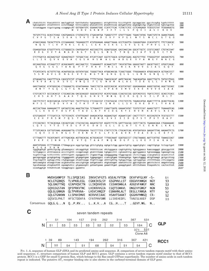
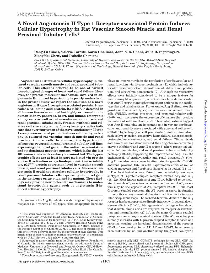
FIG. 1. A, sequence of human GLP cDNA and its predicted amino acid sequence. B, comparison of seven tandem repeats motif with their aminoacid sequences. C, structure comparison of human GLP and RCC1 genes. GLP contains a seven tandem repeats motif similar to that of RCC1protein. RCC1 is a GEF for small G-protein Ran, which belongs to the Ras small GTPase superfamily. The number of amino acids in each tandemrepeat is indicated. The putative AT1 receptor binding site is also shown in the carboxyl-terminal domain of GLP gene.
A Novel Ang II Type 1 Protein Induces Cellular Hypertrophy 21111
by guest on July 12, 2018http://w
ww
.jbc.org/D
ownloaded from
against glutathione S-transferase-GLP fusion protein, which consists of101 amino acid residues in the carboxyl-terminal tail of GLP, wereproduced. Proteins were visualized by enhanced chemiluminescencedetection system (Amersham Biosciences). The relative densities ofAkt2/PKB, p27kip1, and �-actin bands were determined with a comput-erized laser densitometer. We analyzed the data quantitatively byapplying a commercially available system (Kodak 1D image analysissoftware).
Cellular Protein Content—To assess the cellular protein content, thecells were rendered quiescent for 48 h and then harvested with 0.05%EDTA. The number of cells per well was counted and lysed in 100 �l of2 M NaOH, and cellular protein content was determined by proteinassay kit (Bio-Rad). Bovine serum albumin was used as a standard.Cellular protein content is an indicator of cellular hypertrophy, asshown by Mackovic-Basic et al. (50).
GLP mRNA Expression—To determine GLP mRNA expression, aspecific RT-PCR was performed using the primer sets 5�-AGTCATGG-TGGATGTCGGAAA-3� (sense) and 5�-ATGTGAGCCACAAGCTACT-TC-3� (antisense), corresponding to the nucleotide sequences of �4 to�17 and �393 to �414 of human GLP cDNA, respectively. Total RNAswere extracted with TRIzol reagent, (Invitrogen) according to the pro-tocol of the supplier and quantified by absorbance at 260 nm, and wereused in RT-PCR to quantify the amount of GLP mRNA expressed inIRPTCs. PCR reactions were performed for 5 min at 95 °C initialdenaturing followed by 32 cycles of 95 °C for 30 s, 54 °C for 45 s, 72 °Cfor 30 s, and 72 °C for 5 min in the thermal cycler (MJ Research Inc.).The GLP mRNA expression was normalized with �-actin mRNA expres-sion with the primer sets of 5�-ATGCCATCCTGCGTCTGGACCT-GGC-3� (sense) and 5�-AGCATTTGCGGTGCACGATGGAGGG-3� (an-tisense), corresponding to the nucleotide sequences of �155 to 139 ofexon 3 and nucleotide sequences of �155 to �139 of exon 5 of the rat�-actin gene, respectively (51).
Statistical Analysis—Three to thirteen separate experiments wereperformed, and each treatment group was assayed in triplicate unlessindicated. The data were analyzed with the Student’s t test or analysisvariance. A probability level of p � 0.05 was considered as statisticallysignificant.
RESULTS
Identification of GLP Gene—To identify proteins that inter-act with the carboxyl-terminal domain of the AT1A receptor, ayeast two-hybrid screen was performed in a system that in-cluded the AT1A (residues 295–359) fused to the lex A DNAbinding domain and a mouse embryo cDNA library (37) fused tothe VP16 transactivation domain. One positive clone, A8,which contained 162-amino acid residues of mouse GLP (resi-dues 371–533), was isolated. Clone A8 was subsequently re-tested in the yeast two-hybrid assay with the truncated AT1A
receptor (residues 295–318). Although the entire AT1A carboxylterminus domain (residues 295–359) interacted with mouseclone A8, the truncated fragment AT1A-T318, consisting ofresidues 295–318, failed to do so. The result indicates that theregion between residues 319 and 359 of AT1A is required forinteraction with the GLP gene in the yeast two-hybrid system.A total of 23 clones were subsequently isolated from a �gt11human VSMC primary cDNA library employing mouse cloneA8 as a probe. A 2.6-kb clone has a putative ATG start codon atposition 228 and contains an open reading frame of 1593 bpencoding a hydrophilic protein of 531 amino acid residues witha calculated molecular mass of 58,564 daltons (Fig. 1A). Themobility of the in vitro translation product was in agreementwith the molecular mass predicted for the 2.6-kb clone (datanot shown). An amino acid sequence data base search found asimilar human cDNA from GenBankTM (accession numbersAF334406, AJ319660, AK096654, AL833821, and NM_018191).There are three amino acid differences between the 2.6-kb cloneand the published amino acid sequence from GenBankTM; valine239 is changed to phenylalanine, tyrosine 242 to asparagine, andthreonine 245 to asparagine. Analysis of the 2.6-kb clone aminoacid sequence revealed that it contains a motif of seven tandemrepeats similar to that of RCC1 protein. RCC1 has been identifiedas a GEF for Ran small G-protein (52–57). Fig. 1B shows the amino
acid comparison between the seven tandem repeats of GLP protein.We believe that the 2.6-kb clone is a novel GEF-like protein andputatively named the 2.6-kb clone as GLP. Comparison of GLP andRCC1 protein revealed that the GLP protein has an additionalcarboxyl tail (amino acid residues 368–531) in addition to the seventandem repeat motif (Fig. 1C). We found that the amino acidresidues between 371 and 531 of the GLP protein are important forthe interaction with the carboxyl-terminal domain of the AT1 re-ceptor. This may explain why GLP protein has an additional car-boxyl tail, whereas RCC1 protein does not.
Tissue and Cell Distribution of GLP Gene—Northern blotanalysis showed that GLP mRNA is expressed in the humanheart, brain, lung, liver, kidney, pancreas, skeletal muscle, andplacenta but is particularly abundant in the kidney, pancreas,and heart (Fig. 2A). GLP mRNA was detected in cultured ratVSMCs and IRPTCs and human HEK-293 cells (Fig. 2B). Onlya single GLP transcript of about 4.0 kb was found in the humantissues and cell lines. The 4.0-kb size is in agreement with the
FIG. 2. GLP mRNA expression in different human tissues (A) anddifferent cell types (B). GLP protein expression in different cell types(C). GAPDH, glyceraldehyde-3-phosphate dehydrogenase.
A Novel Ang II Type 1 Protein Induces Cellular Hypertrophy21112
by guest on July 12, 2018http://w
ww
.jbc.org/D
ownloaded from
human cDNA published in GenBankTM. This may explain whythe 2.6-kb clone lacks both a polyadenylation signal AAUAAAsequence as well as a poly(A) tail in the 3�-untranslated termi-nus. GLP protein expression was also detected in rat VSMCsand IRPTCs and human HEK-293 cells analyzed by Westernblotting with polyclonal antibody against the last 101 aminoacids of GLP (Fig. 2C). The size of bands detected by Westernblot analysis is about 58 kDa, which is similar to the molecularmass predicted from its protein sequence.
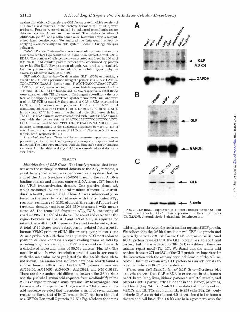
Cellular Localization of GLP Gene—To determine the intra-cellular localization of GLP gene, HEK-293 cells stably express-ing pcDNA3-GFP-GLP were used. As shown in Fig. 3, GFP-GLP protein was mainly localized in small vacuoles in thecytoplasm and also clearly on the plasma membrane but not inthe nucleus.
Hypertrophic Effects of GLP Gene in VSMCs—Although sev-eral groups have reported the GLP sequence to the Gen-BankTM, little is known about the function of this novel gene.To determine the biological roles of the GLP gene, VSMCsstably expressing pLNCX-GLP (GLP) and empty vector,pLNCX (control), were established. The morphology of VSMCsexpressing pLNCX or pLNCX-GLP after viral infection was notchanged, but VSMCs expressing pLNCX-GLP (Fig. 3C) seem tobe larger than control cells expressing pLNCX alone (Fig. 3B).Confluent, quiescent VSMCs were used to examine whetherGLP regulates cell growth. Fig. 4A shows that [3H]thymidineincorporation, an indicator of new DNA synthesis, decreased by
32% in VSMCs overexpressing the GLP gene compared withthe control cells. However, [3H]leucine incorporation, a meas-ure of protein synthesis and also an indicator of cellular hyper-trophy, significantly increased by 238% in VSMCs overexpress-ing the GLP gene when compared with the control cells (Fig.4B). The hypertrophic effects of the GLP gene in VSMCs werefurther demonstrated by a rightward shift of the forward-anglelight scatter, a relative measure of cell size on flow cytometry(data not shown). The relative cell size, as arbitrary units,increased by 178% in VSMCs overexpressing the GLP genecompared with the control cells (Fig. 4C). In parallel experi-ments, Ang II increased in relative cell size by only 112% inVSMCs stimulated with 100 nM Ang II for 48 h (data notshown). As a third hypertrophic marker, total protein contentwas measured. Fig. 4D shows that total protein content was2.2-fold higher in VSMCs overexpressing GLP than in controlcells. These results demonstrate that overexpression of GLPgene induces VSMC hypertrophy.
Effect of the GLP Gene on Phosphorylation of Akt2 inVSMCs—Activation of Akt2/PKB has been shown to play animportant role on cellular hypertrophy in VSMCs and mesang-ial cells. To explore the signaling pathways involved in GLP-induced VSMC hypertrophy, phosphorylation of Akt2/PKB wasdetermined by Western blot analysis. As shown in Fig. 5A,Akt2/PKB was significantly more activated in VSMCs overex-pressing the GLP gene than in the control cells. Activation ofAkt2/PKB increased by 2.2-fold in VSMCs overexpressing the
FIG. 3. Intracellular localization ofGFP-GLP in HEK-293 cells expressing apcDNA3-GFP-GLP fusion protein ob-served with a fluorescence microscope (A).GLP overexpression induces bigger cellsize in VSMCs visualized with a light mi-croscope. Shown are VSMCs infected withpLNCX (B) and pLNCX-GLP (C); magni-fication, �200.
A Novel Ang II Type 1 Protein Induces Cellular Hypertrophy 21113
by guest on July 12, 2018http://w
ww
.jbc.org/D
ownloaded from
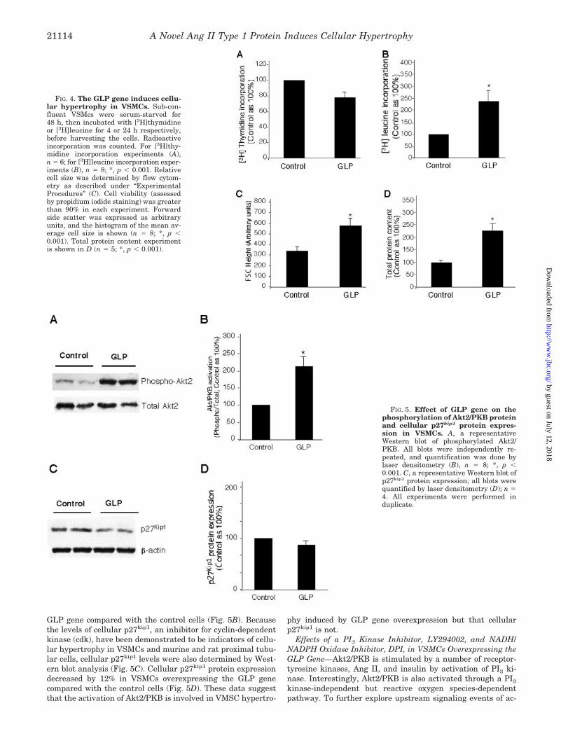
GLP gene compared with the control cells (Fig. 5B). Becausethe levels of cellular p27kip1, an inhibitor for cyclin-dependentkinase (cdk), have been demonstrated to be indicators of cellu-lar hypertrophy in VSMCs and murine and rat proximal tubu-lar cells, cellular p27kip1 levels were also determined by West-ern blot analysis (Fig. 5C). Cellular p27kip1 protein expressiondecreased by 12% in VSMCs overexpressing the GLP genecompared with the control cells (Fig. 5D). These data suggestthat the activation of Akt2/PKB is involved in VMSC hypertro-
phy induced by GLP gene overexpression but that cellularp27kip1 is not.
Effects of a PI3 Kinase Inhibitor, LY294002, and NADH/NADPH Oxidase Inhibitor, DPI, in VSMCs Overexpressing theGLP Gene—Akt2/PKB is stimulated by a number of receptor-tyrosine kinases, Ang II, and insulin by activation of PI3 ki-nase. Interestingly, Akt2/PKB is also activated through a PI3
kinase-independent but reactive oxygen species-dependentpathway. To further explore upstream signaling events of ac-
FIG. 5. Effect of GLP gene on thephosphorylation of Akt2/PKB proteinand cellular p27kip1 protein expres-sion in VSMCs. A, a representativeWestern blot of phosphorylated Akt2/PKB. All blots were independently re-peated, and quantification was done bylaser densitometry (B), n � 8; *, p �0.001. C, a representative Western blot ofp27kip1 protein expression; all blots werequantified by laser densitometry (D); n �4. All experiments were performed induplicate.
FIG. 4. The GLP gene induces cellu-lar hypertrophy in VSMCs. Sub-con-fluent VSMcs were serum-starved for48 h, then incubated with [3H]thymidineor [3H]leucine for 4 or 24 h respectively,before harvesting the cells. Radioactiveincorporation was counted. For [3H]thy-midine incorporation experiments (A),n � 6; for [3H]leucine incorporation exper-iments (B), n � 8; *, p � 0.001. Relativecell size was determined by flow cytom-etry as described under “ExperimentalProcedures” (C). Cell viability (assessedby propidium iodide staining) was greaterthan 90% in each experiment. Forwardside scatter was expressed as arbitraryunits, and the histogram of the mean av-erage cell size is shown (n � 8; *, p �0.001). Total protein content experimentis shown in D (n � 5; *, p � 0.001).
A Novel Ang II Type 1 Protein Induces Cellular Hypertrophy21114
by guest on July 12, 2018http://w
ww
.jbc.org/D
ownloaded from
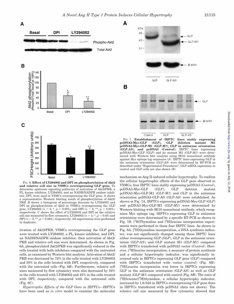
tivation of Akt2/PKB, VSMCs overexpressing the GLP genewere treated with LY294002, a PI3 kinase inhibitor, and DPI,an NADH/NADPH oxidase inhibitor, then activation of Akt2/PKB and relative cell size were determined. As shown in Fig.6A, phosphorylated Akt2/PKB was significantly reduced in thecells treated with both inhibitors compared with the untreatedcells, as examined by Western blot analysis. Activation of Akt2/PKB was decreased by 72% in the cells treated with LY294002and 50% in the cells treated with DPI, respectively, comparedwith the untreated cells (Fig. 6B). Furthermore, relative cellsizes measured by flow cytometry were also decreased by 16%in the cells treated with LY294002 and 45% in the cells treatedwith DPI, respectively, compared with the untreated cells(Fig. 6C).
Hypertrophic Effects of the GLP Gene in IRPTCs—IRPTCshave been used as in vitro model to examine the molecular
mechanism on Ang II-induced cellular hypertrophy. To confirmthe cellular hypertrophic effects of the GLP gene observed inVSMCs, four IRPTC lines stably expressing pcDNA3 (Control),pcDNA3-Myc-GLP (GLP), GLP deletion mutantpcDNA3-Myc-GLP-M1 (GLP-M1) and GLP in the antisenseorientation pcDNA3-GLP-AS (GLP-AS) were established. Asshown in Fig. 7A, IRPTCs expressing pcDNA3-Myc-GLP (GLP)and pcDNA3-Myc-GLP-M1 (GLP-M1) were determined byWestern blotting with 9E10 monoclonal antibody, which recog-nizes Myc epitope tag. IRPTCs expressing GLP in antisenseorientation were determined by a specific RT-PCR as shown inFig. 7B. [3H]Thymidine and [3H]leucine incorporation experi-ments were performed in these four IRPTC lines. As shown inFig. 8A, [3H]thymidine incorporation, a DNA synthesis indica-tor, was not significantly changed among these IRPTC lineseither overexpressing GLP (GLP), GLP in the antisense orien-tation (GLP-AS), and GLP mutant M1 (GLP-M1) comparedwith IRPTCs transfected with pcDNA3 vector (Control). How-ever, [3H]leucine incorporation, a measure of protein synthesisand a cellular hypertrophy indicator, was significantly in-creased only in IRPTCs expressing GLP gene (GLP) comparedwith IRPTCs transfected with vector (control) (Fig. 8B).[3H]Leucine incorporation was lower in IRPTCs expressingGLP in the antisense orientation (GLP-AS) as well as GLPmutant (GLP-M1) compared with control (Fig. 8B). The ratio of[3H]leucine/[3H]thymidine, a cellular hypertrophy indicator,increased by 1.6-fold in IRPTCs overexpressing GLP gene thanin IRPTCs transfected with pcDNA3 (data not shown). Therelative cell size measured by flow cytometry showed that
FIG. 6. Effect of LY294002 and DPI on phosphorylation of Akt2and relative cell size in VSMCs overexpressing GLP gene. Todetermine upstream signaling pathways of activation of Akt2/PKB, aPI3 kinase inhibitor, LY294002, and an NADH/NADPH oxidase inhib-itor, DPI, were used in VSMCs overexpressing the GLP gene. A showsa representative Western blotting result of phosphorylation of Akt2/PKB. B shows a histogram of percentage decrease by LY294002 andDPI on phosphorylation of Akt2 in VSMCs overexpressing the GLPgene, LY294002 (n � 3; *, p � 0.001), and DPI (n � 3; **, p � 0.001),respectively. C shows the effect of LY294002 and DPI on the relativecell size measured by flow cytometry, LY294002 (n � 3; *, p � 0.05) andDPI (n � 3; **, p � 0.001), respectively. All experiments were performedin duplicate.
FIG. 7. Establishment of IRPTC lines stably expressingpcDNA3-Myc-GLP (GLP), GLP deletion mutant M1pcDNA3-Myc-GLP-M1 (GLP-M1), GLP in antisense orientation(GLP-AS), and pcDNA3 (Control). IRPTC lines expressingpcDNA3-Myc-GLP (GLP) and its mutant M1 (GLP-M1) were deter-mined with Western blot analysis using 9E10 monoclonal antibodyagainst Myc epitope tag sequences (A). IRPTC lines expressing GLP inthe antisense orientation (GLP-AS) were determined by RT-PCR asdescribed under “Experimental Procedures”; GLP mRNA expression incontrol and GLP cells are also shown (B).
A Novel Ang II Type 1 Protein Induces Cellular Hypertrophy 21115
by guest on July 12, 2018http://w
ww
.jbc.org/D
ownloaded from
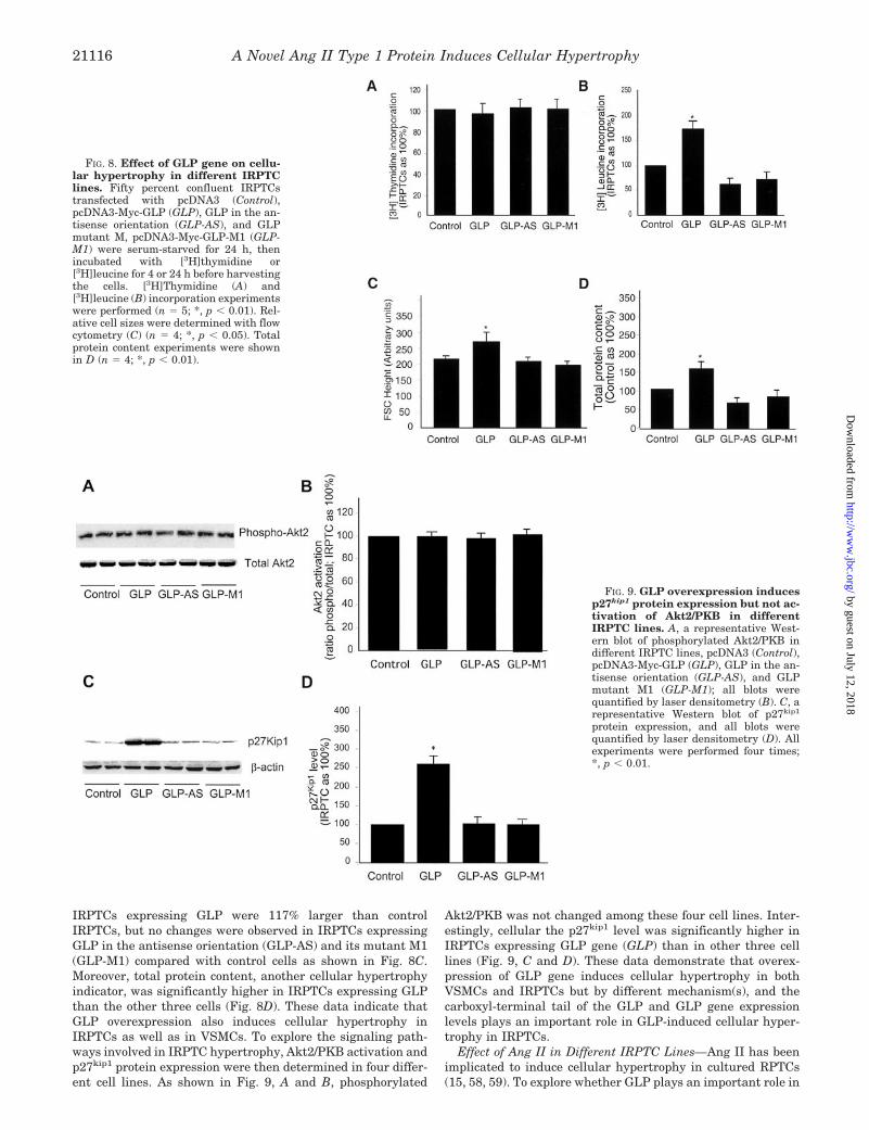
IRPTCs expressing GLP were 117% larger than controlIRPTCs, but no changes were observed in IRPTCs expressingGLP in the antisense orientation (GLP-AS) and its mutant M1(GLP-M1) compared with control cells as shown in Fig. 8C.Moreover, total protein content, another cellular hypertrophyindicator, was significantly higher in IRPTCs expressing GLPthan the other three cells (Fig. 8D). These data indicate thatGLP overexpression also induces cellular hypertrophy inIRPTCs as well as in VSMCs. To explore the signaling path-ways involved in IRPTC hypertrophy, Akt2/PKB activation andp27kip1 protein expression were then determined in four differ-ent cell lines. As shown in Fig. 9, A and B, phosphorylated
Akt2/PKB was not changed among these four cell lines. Inter-estingly, cellular the p27kip1 level was significantly higher inIRPTCs expressing GLP gene (GLP) than in other three celllines (Fig. 9, C and D). These data demonstrate that overex-pression of GLP gene induces cellular hypertrophy in bothVSMCs and IRPTCs but by different mechanism(s), and thecarboxyl-terminal tail of the GLP and GLP gene expressionlevels plays an important role in GLP-induced cellular hyper-trophy in IRPTCs.
Effect of Ang II in Different IRPTC Lines—Ang II has beenimplicated to induce cellular hypertrophy in cultured RPTCs(15, 58, 59). To explore whether GLP plays an important role in
FIG. 8. Effect of GLP gene on cellu-lar hypertrophy in different IRPTClines. Fifty percent confluent IRPTCstransfected with pcDNA3 (Control),pcDNA3-Myc-GLP (GLP), GLP in the an-tisense orientation (GLP-AS), and GLPmutant M, pcDNA3-Myc-GLP-M1 (GLP-M1) were serum-starved for 24 h, thenincubated with [3H]thymidine or[3H]leucine for 4 or 24 h before harvestingthe cells. [3H]Thymidine (A) and[3H]leucine (B) incorporation experimentswere performed (n � 5; *, p � 0.01). Rel-ative cell sizes were determined with flowcytometry (C) (n � 4; *, p � 0.05). Totalprotein content experiments were shownin D (n � 4; *, p � 0.01).
FIG. 9. GLP overexpression inducesp27kip1 protein expression but not ac-tivation of Akt2/PKB in differentIRPTC lines. A, a representative West-ern blot of phosphorylated Akt2/PKB indifferent IRPTC lines, pcDNA3 (Control),pcDNA3-Myc-GLP (GLP), GLP in the an-tisense orientation (GLP-AS), and GLPmutant M1 (GLP-M1); all blots werequantified by laser densitometry (B). C, arepresentative Western blot of p27kip1
protein expression, and all blots werequantified by laser densitometry (D). Allexperiments were performed four times;*, p � 0.01.
A Novel Ang II Type 1 Protein Induces Cellular Hypertrophy21116
by guest on July 12, 2018http://w
ww
.jbc.org/D
ownloaded from
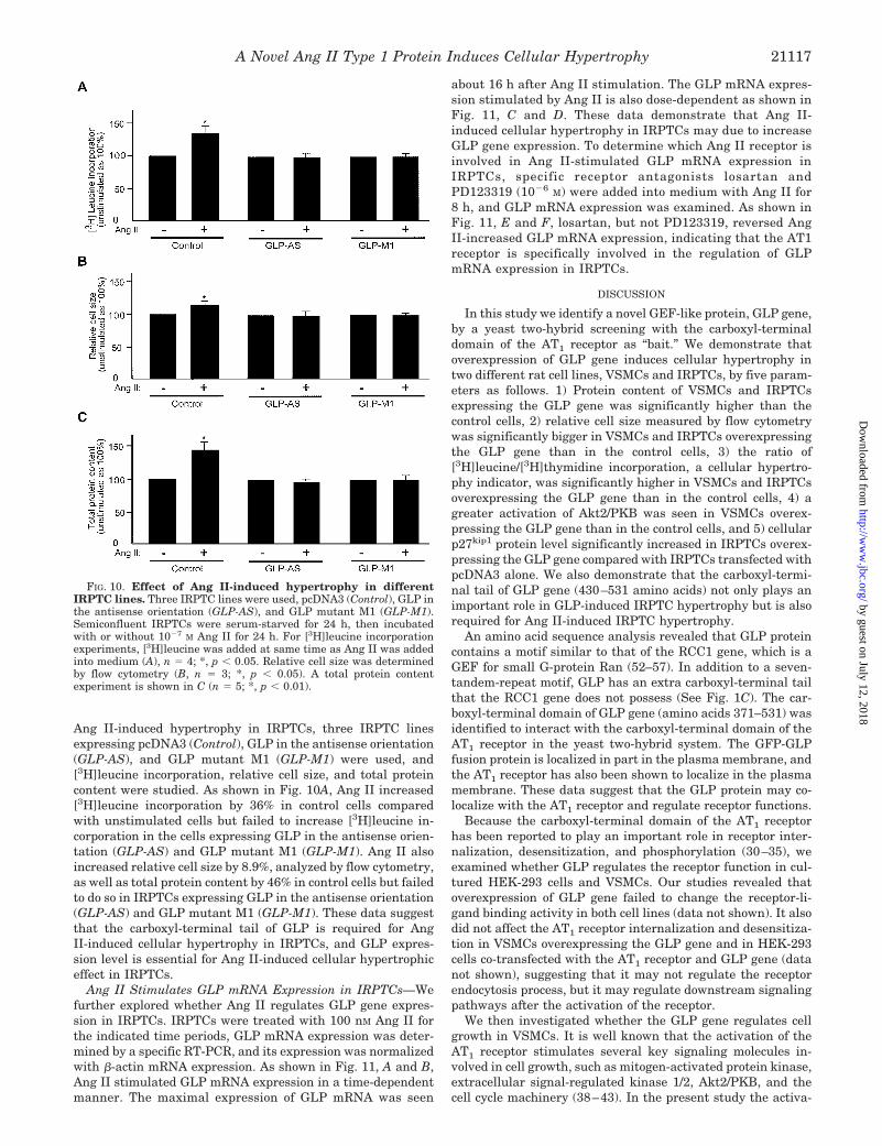
Ang II-induced hypertrophy in IRPTCs, three IRPTC linesexpressing pcDNA3 (Control), GLP in the antisense orientation(GLP-AS), and GLP mutant M1 (GLP-M1) were used, and[3H]leucine incorporation, relative cell size, and total proteincontent were studied. As shown in Fig. 10A, Ang II increased[3H]leucine incorporation by 36% in control cells comparedwith unstimulated cells but failed to increase [3H]leucine in-corporation in the cells expressing GLP in the antisense orien-tation (GLP-AS) and GLP mutant M1 (GLP-M1). Ang II alsoincreased relative cell size by 8.9%, analyzed by flow cytometry,as well as total protein content by 46% in control cells but failedto do so in IRPTCs expressing GLP in the antisense orientation(GLP-AS) and GLP mutant M1 (GLP-M1). These data suggestthat the carboxyl-terminal tail of GLP is required for AngII-induced cellular hypertrophy in IRPTCs, and GLP expres-sion level is essential for Ang II-induced cellular hypertrophiceffect in IRPTCs.
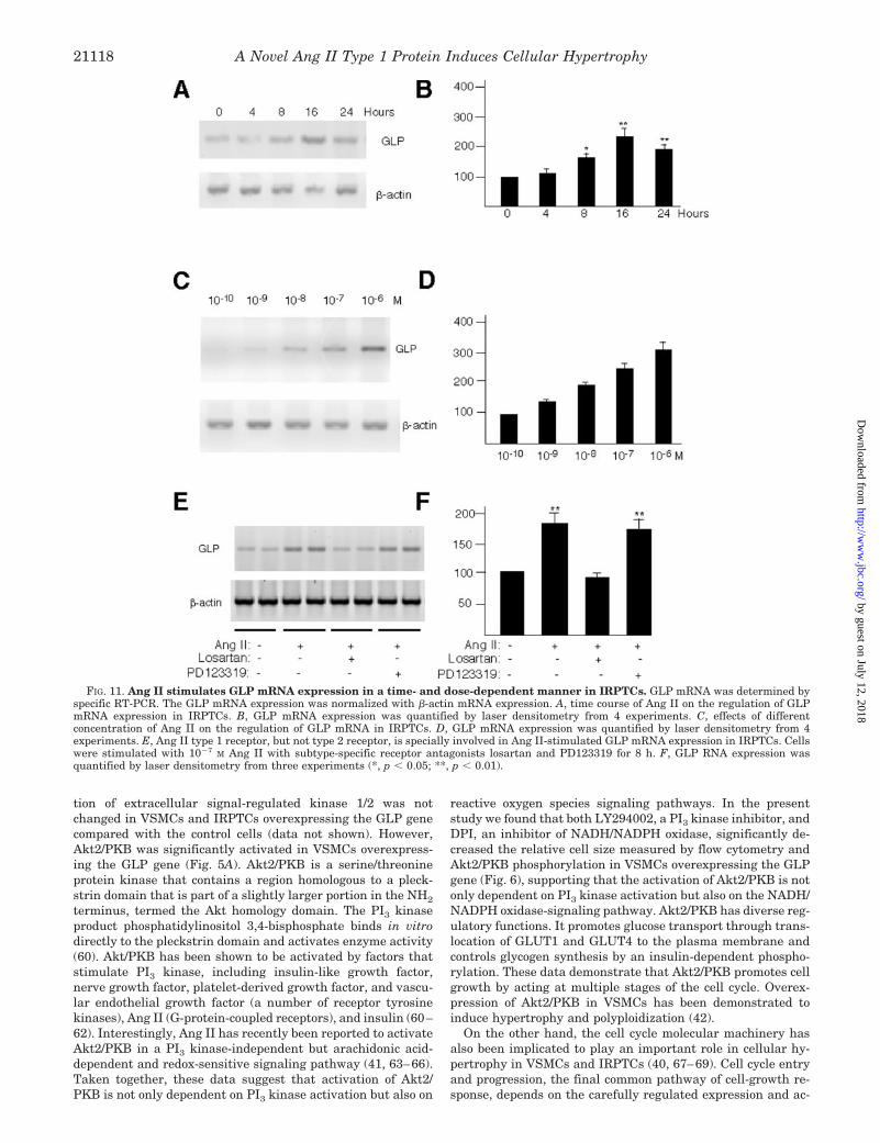
Ang II Stimulates GLP mRNA Expression in IRPTCs—Wefurther explored whether Ang II regulates GLP gene expres-sion in IRPTCs. IRPTCs were treated with 100 nM Ang II forthe indicated time periods, GLP mRNA expression was deter-mined by a specific RT-PCR, and its expression was normalizedwith �-actin mRNA expression. As shown in Fig. 11, A and B,Ang II stimulated GLP mRNA expression in a time-dependentmanner. The maximal expression of GLP mRNA was seen
about 16 h after Ang II stimulation. The GLP mRNA expres-sion stimulated by Ang II is also dose-dependent as shown inFig. 11, C and D. These data demonstrate that Ang II-induced cellular hypertrophy in IRPTCs may due to increaseGLP gene expression. To determine which Ang II receptor isinvolved in Ang II-stimulated GLP mRNA expression inIRPTCs, specific receptor antagonists losartan andPD123319 (10�6 M) were added into medium with Ang II for8 h, and GLP mRNA expression was examined. As shown inFig. 11, E and F, losartan, but not PD123319, reversed AngII-increased GLP mRNA expression, indicating that the AT1receptor is specifically involved in the regulation of GLPmRNA expression in IRPTCs.
DISCUSSION
In this study we identify a novel GEF-like protein, GLP gene,by a yeast two-hybrid screening with the carboxyl-terminaldomain of the AT1 receptor as “bait.” We demonstrate thatoverexpression of GLP gene induces cellular hypertrophy intwo different rat cell lines, VSMCs and IRPTCs, by five param-eters as follows. 1) Protein content of VSMCs and IRPTCsexpressing the GLP gene was significantly higher than thecontrol cells, 2) relative cell size measured by flow cytometrywas significantly bigger in VSMCs and IRPTCs overexpressingthe GLP gene than in the control cells, 3) the ratio of[3H]leucine/[3H]thymidine incorporation, a cellular hypertro-phy indicator, was significantly higher in VSMCs and IRPTCsoverexpressing the GLP gene than in the control cells, 4) agreater activation of Akt2/PKB was seen in VSMCs overex-pressing the GLP gene than in the control cells, and 5) cellularp27kip1 protein level significantly increased in IRPTCs overex-pressing the GLP gene compared with IRPTCs transfected withpcDNA3 alone. We also demonstrate that the carboxyl-termi-nal tail of GLP gene (430–531 amino acids) not only plays animportant role in GLP-induced IRPTC hypertrophy but is alsorequired for Ang II-induced IRPTC hypertrophy.
An amino acid sequence analysis revealed that GLP proteincontains a motif similar to that of the RCC1 gene, which is aGEF for small G-protein Ran (52–57). In addition to a seven-tandem-repeat motif, GLP has an extra carboxyl-terminal tailthat the RCC1 gene does not possess (See Fig. 1C). The car-boxyl-terminal domain of GLP gene (amino acids 371–531) wasidentified to interact with the carboxyl-terminal domain of theAT1 receptor in the yeast two-hybrid system. The GFP-GLPfusion protein is localized in part in the plasma membrane, andthe AT1 receptor has also been shown to localize in the plasmamembrane. These data suggest that the GLP protein may co-localize with the AT1 receptor and regulate receptor functions.
Because the carboxyl-terminal domain of the AT1 receptorhas been reported to play an important role in receptor inter-nalization, desensitization, and phosphorylation (30–35), weexamined whether GLP regulates the receptor function in cul-tured HEK-293 cells and VSMCs. Our studies revealed thatoverexpression of GLP gene failed to change the receptor-li-gand binding activity in both cell lines (data not shown). It alsodid not affect the AT1 receptor internalization and desensitiza-tion in VSMCs overexpressing the GLP gene and in HEK-293cells co-transfected with the AT1 receptor and GLP gene (datanot shown), suggesting that it may not regulate the receptorendocytosis process, but it may regulate downstream signalingpathways after the activation of the receptor.
We then investigated whether the GLP gene regulates cellgrowth in VSMCs. It is well known that the activation of theAT1 receptor stimulates several key signaling molecules in-volved in cell growth, such as mitogen-activated protein kinase,extracellular signal-regulated kinase 1/2, Akt2/PKB, and thecell cycle machinery (38–43). In the present study the activa-
FIG. 10. Effect of Ang II-induced hypertrophy in differentIRPTC lines. Three IRPTC lines were used, pcDNA3 (Control), GLP inthe antisense orientation (GLP-AS), and GLP mutant M1 (GLP-M1).Semiconfluent IRPTCs were serum-starved for 24 h, then incubatedwith or without 10�7 M Ang II for 24 h. For [3H]leucine incorporationexperiments, [3H]leucine was added at same time as Ang II was addedinto medium (A), n � 4; *, p � 0.05. Relative cell size was determinedby flow cytometry (B, n � 3; *, p � 0.05). A total protein contentexperiment is shown in C (n � 5; *, p � 0.01).
A Novel Ang II Type 1 Protein Induces Cellular Hypertrophy 21117
by guest on July 12, 2018http://w
ww
.jbc.org/D
ownloaded from
tion of extracellular signal-regulated kinase 1/2 was notchanged in VSMCs and IRPTCs overexpressing the GLP genecompared with the control cells (data not shown). However,Akt2/PKB was significantly activated in VSMCs overexpress-ing the GLP gene (Fig. 5A). Akt2/PKB is a serine/threonineprotein kinase that contains a region homologous to a pleck-strin domain that is part of a slightly larger portion in the NH2
terminus, termed the Akt homology domain. The PI3 kinaseproduct phosphatidylinositol 3,4-bisphosphate binds in vitrodirectly to the pleckstrin domain and activates enzyme activity(60). Akt/PKB has been shown to be activated by factors thatstimulate PI3 kinase, including insulin-like growth factor,nerve growth factor, platelet-derived growth factor, and vascu-lar endothelial growth factor (a number of receptor tyrosinekinases), Ang II (G-protein-coupled receptors), and insulin (60–62). Interestingly, Ang II has recently been reported to activateAkt2/PKB in a PI3 kinase-independent but arachidonic acid-dependent and redox-sensitive signaling pathway (41, 63–66).Taken together, these data suggest that activation of Akt2/PKB is not only dependent on PI3 kinase activation but also on
reactive oxygen species signaling pathways. In the presentstudy we found that both LY294002, a PI3 kinase inhibitor, andDPI, an inhibitor of NADH/NADPH oxidase, significantly de-creased the relative cell size measured by flow cytometry andAkt2/PKB phosphorylation in VSMCs overexpressing the GLPgene (Fig. 6), supporting that the activation of Akt2/PKB is notonly dependent on PI3 kinase activation but also on the NADH/NADPH oxidase-signaling pathway. Akt2/PKB has diverse reg-ulatory functions. It promotes glucose transport through trans-location of GLUT1 and GLUT4 to the plasma membrane andcontrols glycogen synthesis by an insulin-dependent phospho-rylation. These data demonstrate that Akt2/PKB promotes cellgrowth by acting at multiple stages of the cell cycle. Overex-pression of Akt2/PKB in VSMCs has been demonstrated toinduce hypertrophy and polyploidization (42).
On the other hand, the cell cycle molecular machinery hasalso been implicated to play an important role in cellular hy-pertrophy in VSMCs and IRPTCs (40, 67–69). Cell cycle entryand progression, the final common pathway of cell-growth re-sponse, depends on the carefully regulated expression and ac-
FIG. 11. Ang II stimulates GLP mRNA expression in a time- and dose-dependent manner in IRPTCs. GLP mRNA was determined byspecific RT-PCR. The GLP mRNA expression was normalized with �-actin mRNA expression. A, time course of Ang II on the regulation of GLPmRNA expression in IRPTCs. B, GLP mRNA expression was quantified by laser densitometry from 4 experiments. C, effects of differentconcentration of Ang II on the regulation of GLP mRNA in IRPTCs. D, GLP mRNA expression was quantified by laser densitometry from 4experiments. E, Ang II type 1 receptor, but not type 2 receptor, is specially involved in Ang II-stimulated GLP mRNA expression in IRPTCs. Cellswere stimulated with 10�7 M Ang II with subtype-specific receptor antagonists losartan and PD123319 for 8 h. F, GLP RNA expression wasquantified by laser densitometry from three experiments (*, p � 0.05; **, p � 0.01).
A Novel Ang II Type 1 Protein Induces Cellular Hypertrophy21118
by guest on July 12, 2018http://w
ww
.jbc.org/D
ownloaded from
tivation of certain proteins, termed cyclin-dependent kinases(ckd), and their regulatory subunits, the cyclins (40, 70). Forthe G1 phase, cyclin D, cyclin E, and cyclin A play an importantrole (71). Cyclin D complexes with cdk4/cdk6 and regulatesG1-phase progression, cyclin E/cdk2 or cyclin A/cdk2 is essen-tial for the G1/S transition, and cyclin A/cdk1 or cyclin B/cdk1or 2 initiates mitosis (72). Cdks are activated through phospho-rylation and dephosphorylation at specific sites, and this acti-vation is controlled by cdk inhibitors that bind to and inhibitthe activation of the cdk-cyclin complex. In cellular hypertro-phy, cell cycle entry takes place, but the progression throughthe cell cycle toward DNA synthesis and mitosis is blocked.Among cdk inhibitors, p27kip1 has been shown to play an im-portant role in cell cycle regulation. High levels of p27kip1
present in quiescent (G0) cells have been shown to decline uponmitogen induction (73). This decrease in p27kip1 appears to becritical in enabling the cells to enter the cell cycle. Cellularp27kip1 levels have been shown as an indicator of cellularhypertrophy in VSMCs (40) and murine and rat proximal tu-bular cells (63–65). Furthermore, p27kip1 regulates growth ar-rest in response to transforming growth factor, rapamycin, andcontact inhibition. Thus, studies of p27kip1 expression com-bined with assessment of relative cell size by flow cytometryand [3H]leucine incorporation will give a better assessment ofthe hypertrophic effect induced by GLP gene. Interestingly,cellular p27kip1 level was not changed between VSMCs overex-pressing the GLP gene and the control cells, suggesting thatthe hypertrophic effect of GLP gene is independent of p27kip1
expression in VSMCs. However, cellular p27kip1 expressionsignificantly increased in the IRPTC-overexpressing GLP gene,indicating that, unlike in VSMCs, the hypertrophic effect ofGLP gene is dependent on cell cycle regulation in IRPTCs.IRPTCs were chosen as a second in vitro model to investigatethe hypertrophic effect of the GLP gene in addition to VSMCs.The experiments are designed to eliminate the possibility ofside effects due to retroviral proteins, which were used to infectVSMCs in order to deliver the plasmid DNA into the cells.Although the relative cell size of the control cells (pLNCX)measured by flow cytometry was the same as that of uninfectedVSMCs (data not shown), the hypertrophy observed could notbe completely ruled out as a rival protein’s side effect. Theresults obtained from RPTCs overexpressing the GLP genewith increased [3H]leucine incorporation combined with ahigher p27kip1 expression clearly demonstrated that the cellu-lar hypertrophic effect was dependent on GLP gene overexpres-sion. Because the mRNA components of the renin-angiotensinsystem including angiotensinogen, rennin, angiotensin-con-verting enzyme, and AT1 receptor are all expressed in murine(rat and mouse) proximal tubular cell lines, IRPTCs can beused to further investigate the molecular mechanisms of AngII- or high glucose-induced hypertrophy. In the present study,we demonstrated that Ang II, as a hypertrophic agent, indeedsignificantly stimulates GLP mRNA expression in rat IRPTCs.Similar results were observed in rat VSMCs and kidney me-sangial cells. These data indicate that GLP gene expressionmay be important in Ang II-induced cellular hypertrophy incultured cells.
In summary, we have identified a novel GEF-like protein,GLP gene, that induces cellular hypertrophy in culturedVSMCs and IRPTCs. The signaling pathways involved in thecellular hypertrophic effect of GLP gene are different in thesetwo cell types. In VSMCs, the activation of Akt2/PKB plays animportant role, whereas in IRPTCs, cellular p27kip1 expressionplays an important role. The activation of Akt2/PKB in VSMCsseems to be PI3 kinase- and reactive oxygen species- depend-ent. Furthermore, the carboxyl-terminal tail of GLP plays an
important role in GLP- as well as Ang II-induced cellularhypertrophy in IRPTCs.
REFERENCES
1. Peach, M. J. (1977) Physiol. Rev. 57, 313–3702. Owens, G. K. (1989) Am. J. Physiol. 257, H1755–H17653. Phillips, M. I. (1987) Annu. Rev. Physiol. 49, 413–4354. Pratt, R. E., Dzau, V. J. (1993) in Cellular and Molecular Biology of the
Renin-Angiotensin System (Raizada, M. K., Phillips, M. I., and Sumners, C.,eds) pp. 471–483, CRC Press, Boca Raton, FL
5. Metsarinne, K. P., Stoll, M., Falkenhahn, M., Gohlke, P., and Unger, T. (1994)in Angiotensin Receptors (Saavedra, J. M., ed) pp. 235–253, Kluwer Aca-demic Publisher, Dordrecht, The Netherlands
6. Berk, B. C., Vekshtein. V., Gordon, H. M., and Tsuda, T. (1989) Hypertension13, 305–314
7. Geisterfer, A. A., Peach. M. J., and Owens, G. K. (1988) Circ. Res. 62, 749–7568. Schlondorff, D., DeCandido, S., and Satriano. J. A. (1987) Am. J. Physiol. 253,
C113–C1209. Griendling, K. K., Minieri, C. A., Ollerenshaw, J. D., and Alexander, R. W.
(1994) Circ. Res. 74, 1141–114810. Wang, D. H., and Prewitt, R. L. (1990) Hypertension 15, 68–7711. Morton, J. J., Beattie, E. C., and MacPherson, F. (1992) J. Vasc. Res. 29,
264–26912. Pfeffer, J. M., Pfeffer, M. A., Mirsky, I., and Braunwald, E. (1982) Proc. Natl.
Acad. Sci. U. S. A. 79, 3310–331413. Baker, K. M., Chernin, M. I., Wixson, S. K., and Aceto, J. F. (1990) Am. J.
Physiol. 259, H324–H33214. Mizuno, K., Tani, M., and Hashimoto, S. (1992) Life Sci. 51, 367–37415. Wolf, G., and Neilson, E. G. (1990) Am. J. Physiol. 259, F768–F77716. Sadoshima, J., and Izumo, S. (1993) Circ. Res. 73, 413–42317. Schorb, W., Booz, G. W., Dostal, D. E., Conrad, K. M., Chang, K. C., and Baker,
K. M. (1993) Circ. Res. 72, 1245–125418. Timmermans, P. B. M. W. M., Wong, P. C., Chiu, A. T., Herblin, W. F.,
Benfield, P., Carini, D. J., Lee, R. J., Wexler, R. R., Saye, J. M., and Smith,R. D. (1993) Pharmacol. Rev. 45, 205–251
19. Sasaki, K., Yamano, Y., Bardhan, S., Iwai, N., Murray, J. J., Hasegawa, M.,Matsuda, Y., and Inagami, T. (1991) Nature 351, 230–232
20. Murphy, T. J., Alexander, R. W., Griendling, K. K., Runge, M. S., andBernstein, K. E. (1991) Nature 351, 232–233
21. Mukoyama, M., Nakajima, M., Horiuchi, M., Sasamura, H., Pratt, R. E., andDzau, V. J. (1993) J. Biol. Chem. 268, 24539–24542
22. Kambayashi, Y., Bardhan, S., Takahashi, K., Tsuzuki, S., Inui, H., Hamakubo,T., and Inagami, T. (1993) J. Biol. Chem. 268, 24543–24546
23. Hughes, A. D. (1998) J. Hum. Hypertens. 12, 275–28124. Ribeiro, A. B., Kohlmann, O., Jr., Saragoca, M. A., Marson, O., and Ramos,
O. L. (1985) Drugs 30, 6–1225. Dinh, D. T., Frauman, A. G., Johnston, C. I., and Fabiani, M. E. (2001) Clin.
Sci. (Lond.) 100, 481–49226. Zhang, J., and Pratt, R. E. (1996) J. Biol. Chem. 271, 15026–1503327. Ali, M. S., Sayeski, P. P., Dirksen, L. B., Hayser, D. J., Marrero, M. B., and
Bernstein, K. E. (1997) J. Biol. Chem. 272, 23382–2338828. Ju, H., Venema, V. J., Marrero, M. B., and Venema, R. C. (1998) J. Biol. Chem.
273, 24025–2402929. Sano, T., Ohyama, K., Yamano, Y., Nakagomi, Y., Nakazama, S., Kikyo, M.,
Shirai, H., Blank, J. S., Exton, J. H., and Inagami, T. (1997) J. Biol. Chem.272, 23631–23636
30. Conchon, S., Peltier, N., Corvol, P., and Clauser, E. (1998) Am. J. Physiol. 274,E336–E345
31. Tang, H., Guo, D. F., Porter, J. P., Wanaka, Y., and Inagami. T. (1998) Circ.Res. 82, 523–531
32. Thomas, W. G., Thekkumkara, T. J., Motel, T. J., and Baker, K. M. (1995)J. Biol. Chem. 270, 207–213
33. Hunyady, L., Bor, M., Balla, T., and Catt, K. J. (1994) J. Biol. Chem. 269,31378–31382
34. Oppermann, M., Freedman, N. J., Alexandre, R. W., and Lefkowitz, R. J.(1996) J. Biol. Chem. 271, 13266–13272
35. Daviet, L., Lehtonen, J. Y. A., Tamura, K., Griese, D. P., Masatsugu, H., andDzau, V. J. (1999) J. Biol. Chem. 274, 17058–17062
36. Cui, T., Nakagami, H., Iwai, M., Takeda, Y., Shiuchi, T., Tamura, K., Daviet,L, and Horiuchi, M. (2000) J. Biol. Chem. 279, 938–941
37. Guo, D. F., Chenier, I., Tardif, V., Orlov, S. N., and Inagami, T. (2003)Biochem. Biophys. Res. Commun. 310, 1254–1265
38. Naftilan, A. J., Pratt, R. E., and Dzau, V. J. (1989) J. Clin. Invest. 83,1419–1424
39. Naftilan, A. J., Gilliland, G. K., Eldridge, C. S., and Kraft, A. S. (1990) Mol.Cell. Biol. 10, 5536–5540
40. Braun-Dullaeus, R. C., Mann, M. J., Ziegler, A., von der Leyen, H. E., andDzau, V. J. (1999) J. Clin. Invest. 104, 815–823
41. Gorin, Y., Kim, N. H., Feliers, D., Bhandari, B., Choudhury, G. G., andAbboud, H. E. (2001) FASEB J. 15, 1909–1920
42. Hixon, M. L., Muro-Cacho, C., Wagner, M., Obejero-Paz, C., Millie, E., Fujio,Y., Kureishi, Y., Hassold, T., Walsh, K., and Gualberto, A. (2000) J. Clin.Invest. 106, 1011–1020
43. Zafari, A. M., Ushio-Fukai, M., Akers, M., Yin, Q., Shah, A., Harrison, D. G.,Taylor, W. R., and Griendling, K. K. (1998) Hypertension 32, 488–495
44. Xu, Y. R., Guo. D. F., Mavidson, M., Inagami, T., and Garpenter, G. (1997)J. Biol. Chem. 272, 13463–13466
45. Miller, A. D., and Rosman, G. J. (1989) Biotechniques 7, 980–99046. Danos, O., and Mulligan, R. C. (1988) Proc. Natl. Acad. Sci. U. S. A. 98,
6460–646447. Ingelfinger, J. R., Zuo, W. M., Fon, E. A., Ellison, K. E., and Dzau, V. J. (1990)
J. Clin. Invest. 85, 417–423
A Novel Ang II Type 1 Protein Induces Cellular Hypertrophy 21119
by guest on July 12, 2018http://w
ww
.jbc.org/D
ownloaded from
48. Tang, S. S., Jung, F. F., Diamant, D., Brown, D., Bachinsky, D., Hellman, P.,and Ingelfinger, J. R. (1995) Am. J. Physiol. 268, F435–F436
49. Solban, N., Jia, H. P., Richard, S., Tremblay, S., Devlin, A., Peng, J., Gossard,F., Guo, D. F., Morel, G., Hamet, P., Lewanczuk, R., and Tremblay, J. (2000)J. Biol. Chem. 275, 32234–32243
50. Mackovic-Basic, M. Fine, L. G. Normal, J. T., Cargoe, E. J., and Kurtz, I. (1992)J. Am. Soc. Nephrol. 3, 1124–1130
51. Nudel, U., Zakut, R., Shani, M., Neuman, S., Levy, Z., and Yaffe, D. (1983)Nucleic Acids Res. 11, 1759–1771
52. Ohtsubo, M., Kai, R., Furuno, N., Sekiguchi, T., Sekiguchi, M., Hayashida, H.,Kuma, K., Miyata, T., Fukushige, S., Murotsu, T., Matsubara, K., andNishimoto, T. (1987) Genes Dev. 1, 585–593
53. Saitoh, H., and Dasso, M. (1995) J. Biol. Chem. 270, 10658–1066354. Cheng, Y., Dahlberg, J. E., and Lund, E. (1995) Science 267, 1807–181055. Bischoff, F. R., and Ponstingl, H. (1995) Methods Enzymol. 257, 135–14456. Tachibana, T., Imamoto, N., Seino, H., Nishimoto, Y., and Yoneda, Y. (1994)
J. Biol. Chem. 269, 24542–2454557. Carazo-Salas, R. E., Guarguaglini, G., Gruss, O. J., Segref, A., Karsenti, E.,
and Mattaj, I. W. (1999) Nature 400, 178–18158. Wolf, G., Neilson, E. G., Goldfarb, S., and Ziyadeh, F. N. (1991) Biochem.
Biophys. Res. Commun. 179, 902–909
59. Wolf, G., Killen, P. D., and Neison, E. G. (1991) Cell Regul. 2, 219–22760. Hemmings, B. A. (1997) Science 275, 628–63061. Fujio, Y., and Walsh, K. (1999) J. Biol. Chem. 274, 16349–1635462. Kohn, A. D., Summers, S. A., Birnbaum, M. J., and Roth, R. A. (1996) J. Biol.
Chem. 271, 31372–3137863. Downward, J. (1998) Curr. Opin. Cell Biol. 10, 262–26764. Saward, L., and Zahradka, P. (1997) Circ. Res. 81, 249–25765. Takahashi, T., Taniguchi, T., Konishi, H., Kikkawa, U., Ishikawa, Y., and
Yokoyama, M. (1999) Am. J. Physiol. Heart Circ. Physiol. 276, 1927–193466. Ushio-Fukai, M., Alexander, R. W., Akers, M., Yin, Q., Fujio, Y., Walsh, K., and
Griendling, K. K. (1999) J. Biol. Chem. 274, 22699–2270467. Hannken, T., Schroeder, R., Stahl, R. A., and Wolf, G. (1998) Kidney Int. 54,
1923–193368. Wolf, G. (1999) Kidney Int. 54, 1262–126569. Zhang, S. L., Chen, X., Wei, C. C., Filep, J. G., Tang, S. S., Ingelfinger, J. R.,
and Chan, J. S. D. (2002) Endocrinology 143, 4627–463570. Chellappan, S. P., Giordano, A., and Fisher, P. B. (1998) Circulation 98, 82–8971. Sherr, C. J. (1995) Proc. Assoc. Am. Physicians 107, 181–18672. King, R. W., Jackson, P. K., and Kirschner, M. W. (1994) Cell 79, 563–57173. Sherr, C. J., and Roberts, J. M. (1995) Genes Dev. 9, 1149–1163
A Novel Ang II Type 1 Protein Induces Cellular Hypertrophy21120
by guest on July 12, 2018http://w
ww
.jbc.org/D
ownloaded from

XiangMei Chen and Isabelle ChenierDeng-Fu Guo, Valerie Tardif, Karin Ghelima, John S. D. Chan, Julie R. Ingelfinger,
Hypertrophy in Rat Vascular Smooth Muscle and Renal Proximal Tubular CellsA Novel Angiotensin II Type 1 Receptor-associated Protein Induces Cellular
doi: 10.1074/jbc.M401544200 originally published online February 24, 20042004, 279:21109-21120.J. Biol. Chem.
10.1074/jbc.M401544200Access the most updated version of this article at doi:
Alerts:
When a correction for this article is posted•
When this article is cited•
to choose from all of JBC's e-mail alertsClick here
http://www.jbc.org/content/279/20/21109.full.html#ref-list-1
This article cites 62 references, 35 of which can be accessed free at
by guest on July 12, 2018http://w
ww
.jbc.org/D
ownloaded from