the lantibiotic nai-107 efficiently rescues drosophila melanogaster
TRANSCRIPT
1
The lantibiotic NAI-107 efficiently rescues Drosophila 1
melanogaster from infection with methicillin-resistant 2
Staphylococcus aureus USA300 3
4
5
Thomas T. Thomsen1, Biljana Mojsoska2, João C. S. Cruz3, Stefano Donadio3,4, Håvard Jenssen2, 6
Anders Løbner-Olesen1*, Kim Rewitz1*. 7
8
1Department of Biology, University of Copenhagen, Denmark 9
2Department of Science, Systems and Models, Roskilde University, Denmark 10
3Ktedogen, Milano, Italy 11
4Naicons Srl, Milano, Italy 12
13
*Correspondence: [email protected], Kim.Rewitz @bio.ku.dk 14
Key words: Antibacterial peptides; Drug screening; Antibiotics; Infection model 15
16
AAC Accepted Manuscript Posted Online 5 July 2016Antimicrob. Agents Chemother. doi:10.1128/AAC.02965-15Copyright © 2016 Thomsen et al.This is an open-access article distributed under the terms of the Creative Commons Attribution 4.0 International license.
on February 14, 2018 by guest
http://aac.asm.org/
Dow
nloaded from
2
ABSTRACT 17
We used the fruit fly Drosophila melanogaster as a cost-effective in vivo model to evaluate the efficacy 18
of novel antibacterial peptides and peptoids for treatment of methicillin-resistant staphylococcus 19
aureus (MRSA) infections. A panel of peptides with known antibacterial activity in vitro and/or in vivo 20
was tested in Drosophila. Although most peptides and peptoids that were effective in vitro failed to 21
rescue lethal effects of S. aureus infections in vivo, we found that the two lantibiotics, nisin and NAI-22
107 rescued adult flies from fatal infections. Furthermore, NAI-107 rescued mortality of infection with 23
the MRSA strain USA300 with equivalent efficacy to vancomycin, a widely applied antibiotic for the 24
treatment of serious MRSA infections. These results establish Drosophila as a useful model for in vivo 25
drug evaluation of antibacterial peptides. 26
27
28
on February 14, 2018 by guest
http://aac.asm.org/
Dow
nloaded from
3
INTRODUCTION 29
Since the golden era of antibiotic drug development during the 1940-1960`s, development and 30
spread of multidrug resistance have become a huge burden to societies. Today resistance to almost all 31
known antibiotics has emerged with the sequential introduction of new or improved antibiotics in the 32
clinical and agricultural setting (1, 2). Therefore, continued development of new or improved 33
antibiotics is of great importance to human health. However, new antibiotics are lacking and few are 34
under development for treatment of multidrug resistant (MDR) infectious bacteria, as drug 35
development is costly and success from in vitro discovery to application in clinical settings is limited. 36
Bacterial infections with MRSA (Methicillin Resistant Staphylococcus aureus) are no longer 37
sporadic in distribution and prevalence (3, 4). MRSA strains are associated with both community (CA-38
MRSA) and hospital (HA-MRSA) acquired infections, with the highly β-lactam resistant USA300 CA-39
MRSA clone accounting for up to 80% of all MRSA infections in the USA (5). High level β-lactam 40
resistance is due to acquisition of SCCmec elements (Staphylococcal Cassette Chromosome) including 41
the mecA gene that encodes an alternative version of the penicillin binding protein (PBP2A), which is 42
inducible (6, 7) and has a lowered affinity for β-lactam antibiotics (8). Often SCCmec elements are 43
associated with carriage of resistance genes to other antibiotics including aminoglycoside modifying 44
enzymes such as acetyltransferase, adenylyltransferase or phosphotransferase (9). Due to this 45
resistance, MRSA treatment often includes glycopeptide antibiotics such as vancomycin or 46
oxazolidinones such as linezolid. However, failure with vancomycin treatment has been reported in 47
vancomycin-intermediate S. aureus (VISA) (10) and vancomycin resistant S. aureus (VRSA) (11) 48
strains. On the other hand, linezolid resistance is rare (12), but has been observed associated with 49
mutations in the ribosomal DNA encoding the 23S RNA or through carriage of a Cfr rRNA 50
on February 14, 2018 by guest
http://aac.asm.org/
Dow
nloaded from
4
methyltransferase gene (13, 14). Furthermore, resistance to the last resort antibiotic daptomycin has 51
been reported (15, 16). Given the increasing frequency of resistance to these antibiotics, it is important 52
to develop improved or novel therapeutics, and to consider new strategies to contain the spread of the 53
growing resistance problem. 54
Peptide-based antibiotics have been proposed as the next generation of antibacterial compounds 55
because of their wide spread distribution in nature as part of innate immunity. These molecules are 56
often amphipathic and interact with the bacterial membrane to disrupt its function. The cationic peptide 57
colistin, a bacteriocin currently used for treatment of highly resistant Gram-negative infections, is part 58
of the polymyxins that are derived from natural producers such as Paenibacillus polymyxa (17). 59
Another bacteriocin, nisin, has been used in the food industry for decades against harmful bacteria such 60
as S. aureus, Listeria monocytogenes and Clostridium botulinum (18). Nisin belongs to a subgroup 61
known as lantibiotics, named so, for containing uncommon amino acids such as lanthionine, 62
methyllanthionine, didehydroalanine or didehydroaminobutyric acid (19). Nisin has been described to 63
disrupt membrane integrity through a dual mode of action with inhibition of cell wall synthesis by 64
binding to the cell wall precursor lipid-II and subsequent pore formation (20-22), although new 65
evidence points towards a more complex mechanism that includes aggregation of lipid-II (23). Peptides 66
may be used directly as antibacterials or could pose as templates for development of small molecule 67
mimetics such as peptoids, which can accommodate improvements to toxicity and are intrinsically less 68
prone to degradation by proteases (24). 69
The gap from in vitro drug screening to the large scale efficacy testing necessary for clinical 70
development is hampered by the expensive, labor-intensive and highly regulated mammalian infection 71
models. It is therefore of interest to develop improved cost-effective methods with high predictive 72
on February 14, 2018 by guest
http://aac.asm.org/
Dow
nloaded from
5
value for screening of antibacterial compounds before these are tested in mammalian models. Although 73
the fruit fly Drosophila has been used in drug discovery (25, 26), its application for screening of 74
antibacterial compounds has been limited (27-29). Drosophila is a powerful genetic model for studying 75
disease mechanisms and during the past decades it has been used extensively in elucidating the 76
mechanisms of innate immunity, leading to the discovery of the conserved role of the Toll receptors 77
(30) and the immune deficiency (IMD) pathway (31). Studies of innate immunity in Drosophila have 78
sprouted development of various methods for infecting flies with important human pathogens (28, 32-79
34). Here, we evaluate the therapeutic potential of antibacterial peptides and peptoids in vivo by testing 80
efficacy in a Drosophila model infected with S. aureus 8325-4 (35) or MRSA USA300 (36). Tests 81
were performed with a range of different peptides including the lantibiotics nisin-A (nisin) (37) and 82
NAI-107 (38, 39), a compound that is currently undergoing preclinical studies. Lantibiotics are usually 83
produced by Gram-positive bacteria and characterized as ribosomally synthesized peptides containing 84
post-translational introduced ring structures, introduced through the thioether containing lanthionine 85
and methyllanthionine residue (40). Furthermore, a panel of synthesized amphipathic cationic peptides 86
previously shown to have good in vitro and/or in vivo efficacy were tested: GN-2, GN-4 (41, 42), 87
HHC-9 (43), HHC-36 (44) and peptoids: GN-2 Npm9, GN-2 Ntrp5-8, GN-4 (45). We found that NAI-88
107 rescued an otherwise lethal infection with MRSA USA300 with an efficacy equivalent to 89
vancomycin in Drosophila. Our findings also show that treatment with nisin extends life expectancy in 90
animals infected with MRSA USA300, while the majority of the peptides and peptoids tested showed 91
no protection from infection or had detrimental effects on the survival of host. 92
93
94
on February 14, 2018 by guest
http://aac.asm.org/
Dow
nloaded from

6
MATERIALS AND METHODS 95
Bacteria and growth media 96
The S. aureus strains 8325-4 (35) and USA300 (36) were used as indicated in the individual 97
experiments. Bacterial cultures were grown in cation adjusted Müller Hinton Broth (MHB-II) at the 98
indicated temperature. 99
100
Growth rate and determination of bacterial load 101
The growth rate of the S. aureus was examined at 37°C in vitro, to determine the growth period 102
required for obtaining balanced cultures, here defined as cultures grown exponentially for no less than 103
6 generations. Prior to injection of bacteria into the fly in vivo model, the inoculum was prepared as 104
balanced cultures grown at 37°C. Since flies used for in vivo infections were kept at 29°C, the bacterial 105
in vitro generation time was also determined at this temperature. In vitro growth rates were defined in 106
MHB-II, by optical density measurements at 600 nanometers (OD600) at 10 minute intervals. 107
Furthermore, we determined the in vivo generation time by colony forming units (CFU) per animal, by 108
counting of CFU at various time points by homogenizing flies infected with bacteria and plating on 109
Mannitol Salt Agar (MSA). This was performed in triplicate experiments; 3 individual flies were 110
crushed in phosphate buffered saline (PBS) and 10x dilution series were prepared, from which 10 µl 111
was spot plated on MSA in triplicates. The mean value of each experiment was determined as CFU Fly-112
1 and plotted. However, for determination of bacterial titers post treatment, four replicative experiments 113
each containing 40 flies were performed, and at each time point 8 live individual flies were 114
homogenized. This homogenate was diluted in 1 ml PBS and dilutions were plated as 100 µl on MSA 115
on February 14, 2018 by guest
http://aac.asm.org/
Dow
nloaded from

7
for time points 0, 3, 12 and 24 hours. It should be noted that the concentration of the drug inside the fly 116
was based on multiples of the minimum inhibitory concentration (MIC) in a volume of 0.5 µl fly liquid 117
content (described in the next section). Therefore, it was not necessary to further wash the homogenate 118
as the drug inside the fly can be estimated to be diluted approximately 1:2000. Finally, initial trials 119
showed that washing of homogenate provided lower CFU titers compared to non-washed samples. It 120
should also be noted that drug treatment was performed at time point 3 hours post infection, and that 121
CFU Fly-1 for this time point was conducted prior to treatment. 122
123
Minimum inhibitory concentration (MIC) 124
MIC of all tested compounds were performed according to protocols using the micro-broth 125
dilution methodology (46) with minor modification. S. aureus was grown in 10 ml MHB-II overnight 126
at 37°C with shaking, then diluted 1:100 in fresh MHB-II and grown to OD600 = 0.2-0.4. Cultures were 127
then diluted 1:10 and grown to OD600 = 0.2-0.4. These steps were performed to ensure balanced 128
exponentially growing cultures as explained. Finally, cultures were diluted to 1 x 10⁶ CFU ml-1, and 129
further diluted 1:1 in microtiter plates in MHB-II with drug, leading to a final inoculum of 5 x 10⁵ 130
CFU ml-1. MIC`s were determined in triplicate, if more than one value was found, the highest was set 131
as the MIC to be conservative. 132
133
Injection assay 134
on February 14, 2018 by guest
http://aac.asm.org/
Dow
nloaded from
8
Injection assays were performed as previously described (33) using a nanoject-II microinjecter, 135
but with minor modification in preparation of bacterial inoculum to obtain balanced cultures as 136
explained. Flies were reared on standard bloomington formulation at 25°C under a 12:12 light:dark 137
cycle and constant humidity. Adult Oregon R male flies 4-7 days old were used for all injection 138
experiments. Initial experiments with strain 8325-4 were performed in duplicate with groups of 25-30 139
animals in each experiment. For USA300 experiments were performed in triplicates, except for the 140
nisin experiment which was only done in dublicate. Inoculum was prepared by resuspending cells in 10 141
mM MgSO4 vehicle (VEH) to an OD600 = 0.06 and kept on ice, giving an inoculum dose of 100-450 142
CFU (8325-4) and 200-700 CFU (USA300) in the flies after injecting 18.4 nl. Bacterial and VEH 143
injections were administered in the soft tissue surrounding the front legs, drug treatment was 144
administered in the lower thorax 3 hours post infection. After injection of bacteria, flies were kept at 145
29°C and followed for 48-96 hours to determine mortality. Drug delivery was performed at the 146
concentrations indicated for individual experiments. Flies which died within 3 hours post injections, 147
were considered to have died from handling and disregarded. It is important to note that when drug 148
concentrations were calculated, we performed a rough approximation of the fluid content of a fly. Fly 149
fluid content was measured by drying out 10 groups of 50 flies and comparing dry weight to wet 150
weight. This resulted in an average fluid content of 0.58 µl per adult male fly. For simplicity and 151
because we assumed that the compounds would not distribute to all fluids we used 0.5 µl fluid as our 152
measure for calculating drug concentrations in the flies. Further we assumed rapid distribution of the 153
compound in the open circulatory system of Drosophila and a slow clearance of the compounds by 154
Malpighian tubules. Therefore, drug concentrations are given as the highest concentration obtained in 155
multiples of the MIC. 156
on February 14, 2018 by guest
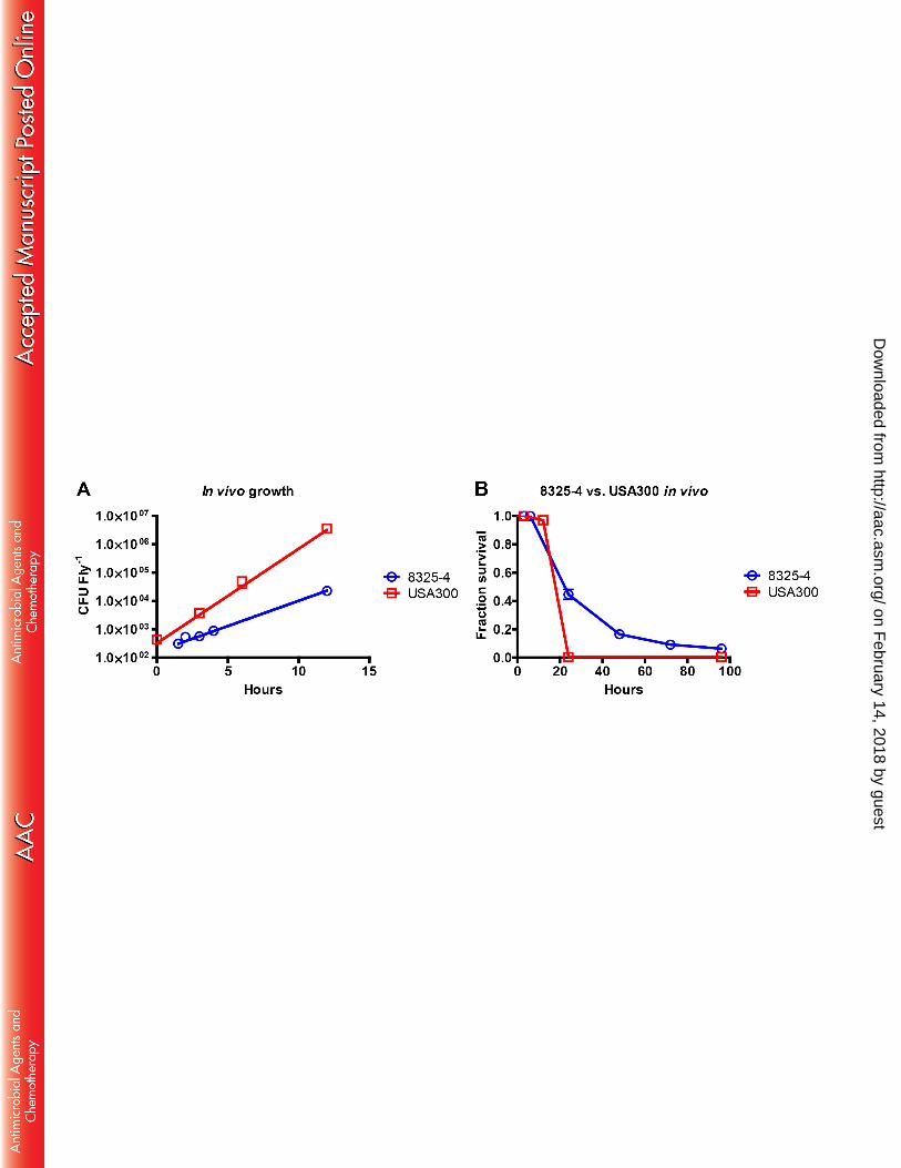
http://aac.asm.org/
Dow
nloaded from
9
Statistics and graphical plots 157
Plotting of data was performed using GraphPad Prism 5. All in vivo survival plots were 158
performed using Kaplan-Meier analysis on pooled data for repetitive experiments. Statistical analysis 159
was carried out with log-rank (Mantel-Cox) test for comparison of survival curves. Experiments with p 160
< 0.05 were considered significant and are stated in the results section. Statistics were performed on the 161
CFU per fly counts of the different treatment groups using one-way ANOVA (Kruskal-Wallis) with 162
Dunn’s multiple comparison test for comparison of individual groups. Relevant statistical results are 163
stated in the text in the results section. 164
165
RNA preparation and quantitative PCR 166
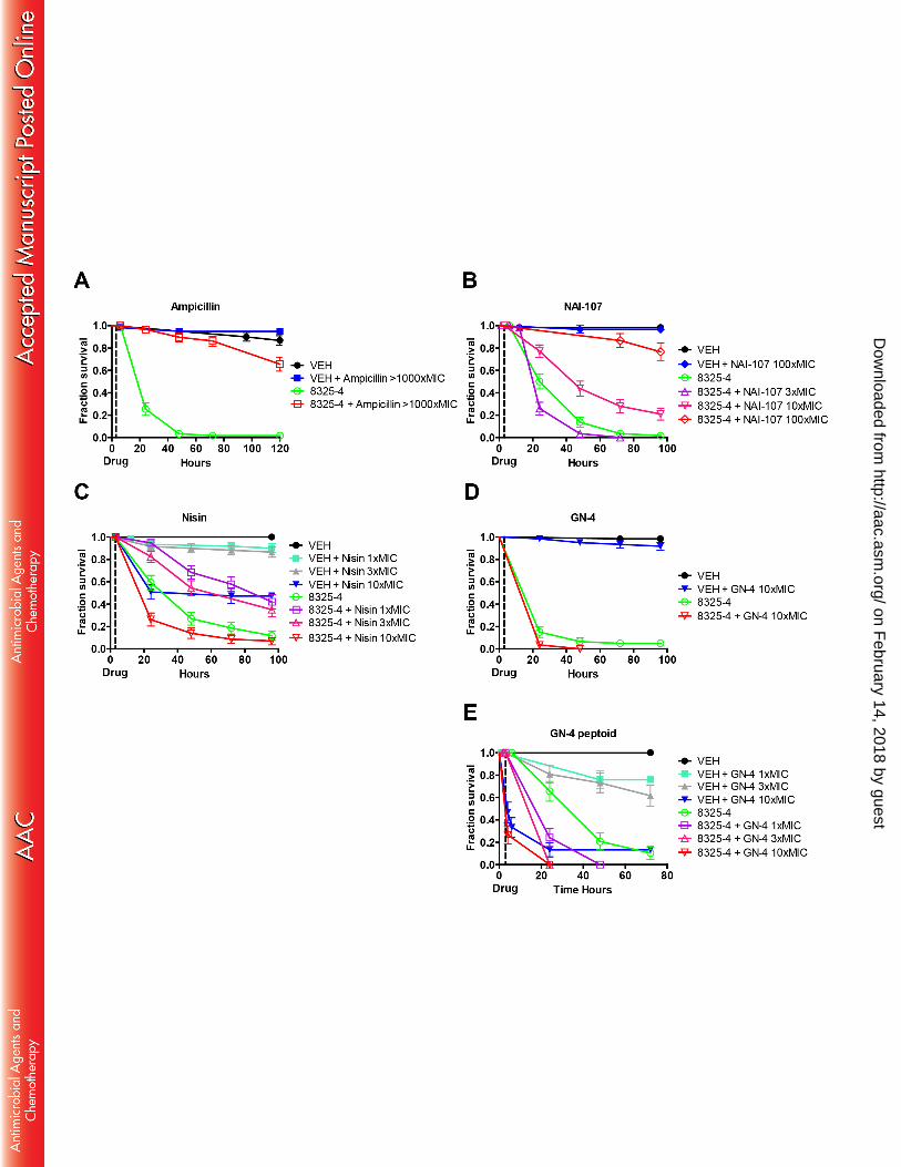
Isolation of total RNA for quantitative PCR (qPCR) was prepared by the use of RNeasy Mini 167
kit (Qiagen) according to manufacturer’s instructions. Biological samples were collected as 10 adult 168
male flies pooled for each replicate and time point. To reduce contamination with genomic DNA, all 169
samples were treated on-column with DNase. Total RNA concentrations were measures on a Qubit™ 170
3.0 fluorometer and equivalent amounts of total RNA were used for cDNA synthesis for each sample. 171
cDNA synthesis was performed using the SuperScript III First-Strand Synthesis kit (Invitrogen) 172
according to the manufacturer’s instructions. qPCR was performed on a Mx3000P qPCR system 173
(Agilent Technologies) using the following program: 95°C for 10 minutes, followed by 45 cycles of 174
95°C for 15 seconds, 60°C for 15 seconds and 72°C for 15 seconds. Dissociation curve analysis was 175
applied to all reactions. Primers are described in table S1 in the supplemental material. We used Rpl23 176
as housekeeping gene to normalize expression as previously described (47). 177
on February 14, 2018 by guest
http://aac.asm.org/
Dow
nloaded from

10
Compounds 178
Ampicillin sodium salt 99% (ROTH Art-Nr: K029.2 EG-Nr: 2007081) was used as control for 179
efficacy in in vitro and in vivo experiments. Vancomycin was acquired from Hospira as vancomycin 180
hydrocloride for intravenous treatment (#467918E01). The peptides GN-2, GN-4, HHC-9 and HHC-36 181
(all amidated in C-terminus), nisin-A (nisin) and peptoids were above 95% purity and synthesized 182
and/or purified by Håvard Jenssen, Roskilde University, Denmark. NAI-107 is a complex of congeners 183
produced by Microbispora sp. 107891 and was prepared as previously described (48). The distribution 184
of congeners for the batch used in the current study was as follows: A1+A2 = 80.8%, F1+F2 = 9.4%, 185
B1+B2 = 4%. 186
187
RESULTS 188
Determination of the growth of S. aureus in vitro and in vivo in a Drosophila infection model 189
We determined the growth rate of the S. aureus strains 8325-4 and MRSA USA300 in MHB-II 190
media at 29°C, because all successive in vivo experiments were performed at this temperature. Strain 191
8325-4 had a generation time of 57 minutes, while USA300 had a generation time of 44 minutes (data 192
not shown). The in vivo growth rate of the same strains was determined by injection of bacteria into the 193
flies at time zero and samples were collected between time 0-3, 4-6 and 12 hours post infection. 194
USA300 had a generation time of 54 minutes, whereas 8325-4 had a generation time of 104 minutes in 195
vivo (Fig 1A). Drosophila infected with USA300 died rapidly with no surviving flies 24 hours post 196
infection (Fig 1B). Flies infected with approximately the same number of 8325-4 lived significantly 197
on February 14, 2018 by guest
http://aac.asm.org/
Dow
nloaded from
11
longer. We suggest that these differences in viability reflect the difference in in vivo growth rates of 198
USA300 and 8325-4 bacteria. 199
200
Minimum inhibitory concentrations of antibacterial peptides and peptoids 201
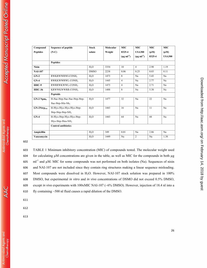
We determined the MIC for peptides and peptoids with the two strains (Table 1). The MIC 202
values for S. aureus 8325-4 of amphipathic cationic peptides GN-2, GN-4, HHC-9 and HHC-36 and 203
the lantibiotic nisin were in the range of 4-10 µg ml-1, while those of GN-2 and GN-4 peptoids were 204
higher (16-64 µg ml-1). On the other hand, the MIC of NAI-107 against strain 8325-4 was only 0.06 µg 205
ml-1, showing that NAI-107 is highly efficient in inhibiting in vitro growth of S. aureus. The MIC of 206
NAI-107 for S. aureus 8325-4 was comparable to ampicillin (0.01 µg ml-1). For S. aureus USA300, the 207
MIC was 0.25 µg ml-1 for NAI-107 and 2 µg ml-1 for vancomycin. 208
209
Identification of nisin and NAI-107 as efficacious treatment for systemic S. aureus infections in a 210
Drosophila in vivo model 211
To evaluate the therapeutic potential of the peptides and peptoids, we determined their ability to 212
rescue flies with an otherwise lethal systemic S. aureus 8325-4 infection. In order to establish the 213
appropriate dosages, we made the following reasoning: Because insects are known to have an open 214
circulatory system, we assumed that the administered compound would be rapidly and uniformly 215
distributed in the hemolymph of the fly. The volume of the fly hemolymph was estimated to be 0.5 µl 216
(see Materials and Methods) and we assumed that clearance was slow. Under these assumptions, the 217
on February 14, 2018 by guest
http://aac.asm.org/
Dow
nloaded from

12
highest concentration achieved for each compound can be expressed in multiples of the MIC. For 218
example, 1xMIC nisin [10µg ml-1] is equivalent to injection of 2.5 mg nisin kg-1 fly, and these 219
calculations can be found in Table 2 for all drugs. Ampicillin was chosen as control, as β-lactams in 220
general are considered non-toxic to the host and can be administered in high concentrations, in our case 221
>1000xMIC. Ampicillin efficiently promoted survival of 8325-4 infected flies (p < 0.001; Fig. 2A) 222
over a 70 hour period and with no lethal effects to control animals (p = 0.15); here defined as no 223
difference in survival when comparing flies injected with VEH to those injected with both VEH and 224
drug. 225
The two lantibiotics NAI-107 and nisin showed good efficacy by effectively rescuing or 226
delaying mortality of infected flies over a 96 hours period (Fig. 2B and C). Treatment with 3xMIC of 227
NAI-107 did not show positive effect on the survival of 8325-4 infected flies. However, treatment with 228
10xMIC of NAI-107 rescued around 20-30% of flies (p < 0.001). NAI-107 treatment of control animals 229
with 3xMIC and 10xMIC had no negative effect on the survival (data not shown; p = 0.62). We 230
therefore tested NAI-107 at 100xMIC, and at this concentration NAI-107 treatment rescued more than 231
70% (p < 0.0001) of the infected flies, again without negative effects on survival of control animals (p 232
= 0.62; Fig. 2B). Compared to NAI-107, nisin showed a difference in both efficacy and lethality to 233
control animals. While 1xMIC nisin delayed bacterial killing of flies (p < 0.001), it produced signs of 234
lethal side effects in uninfected control animals injected with 1xMIC nisin compared to VEH injected 235
control animals (p = 0.018; Fig. 2C). Higher concentrations of 3xMIC nisin also rescued a considerable 236
fraction of infected animals (p = 0.0002), but showed pronounced detrimental effects on the survival of 237
control animals (p = 0.006). These adverse effects were exacerbated when using 10xMIC nisin, which 238
on February 14, 2018 by guest
http://aac.asm.org/
Dow
nloaded from
13
resulted in the killing of 50% of control animals injected with nisin alone (p < 0.0001) and also resulted 239
in increased mortality of infected flies (Fig. 2C). Therefore, nisin was not tested at 100xMIC. 240
In contrast to NAI-107 and nisin, the GN-4 peptide, which possesses good in vitro efficacy 241
against S. aureus [Table 1 and (41)], did not rescue infected flies at 1x and 3xMIC (p > 0.05; data not 242
shown). When applied at 10xMIC, GN-4 showed no detrimental effects on survival of flies (Fig. 2D). 243
However, the results indicate that administration of this peptide to animals infected with bacteria may 244
reduce the survival because a higher number of the animals treated with the peptide after infection died, 245
although this was not statistically significant. The GN-4 peptoid showed pronounced negative effects 246
on animal survival even at 1xMIC (Fig. 2E) and was therefore not subjected to further testing. The GN-247
2 peptide had similar effect to that of GN-4 peptide and the two GN-2 peptoids clearly showed adverse 248
effects on survival in both control and infected animals (see Fig. S1 in the supplemental material). 249
Injection of HHC-9 and HHC-36 peptides in the absence of infection caused no obvious detrimental 250
effects on survival. However, treatment with these peptides did not rescue infected flies, but caused a 251
moderate decrease in survival of infected animals that may indicate detrimental effects of peptides, 252
although the results are somewhat ambiguous. 253
We also noted an adverse behavioral response that could be indicating neurotoxicity in flies 254
injected with high concentrations of nisin, GN-2 and GN-4 along with the peptoids, but not with NAI-255
107. Animals reacted to injection with these compounds by being partially paralyzed for up to 10 hours 256
post injection (data not shown). This paralysis was not manifested as complete immobilization but as 257
uncoordinated movements and an inability to walk or fly. 258
259
on February 14, 2018 by guest
http://aac.asm.org/
Dow
nloaded from

14
Treatment with nisin and NAI-107 reduces the immune response of S. aureus infected Drosophila 260
To further test drug efficacy of the two lantibiotics nisin and NAI-107 in vivo, we examined the 261
immune response of both treated and non-treated infected animals. We rationalized that infected 262
animals treated with these compounds would mount less of an immune response provided that bacterial 263
proliferation in the host was inhibited by the compounds. To test this we used flies infected with S. 264
aureus strain 8325-4. We administrated NAI-107 at 100xMIC, while nisin due to its detrimental side-265
effect at high concentrations was only injected corresponding to 3xMIC. Treatment of infected animals 266
with ampicillin (>1000xMIC) was included for comparison with an efficacious compound. Samples in 267
triplicate were taken 6 and 12 hours post infection and uninfected flies served as controls. As a measure 268
of immune response we analyzed expression of Drosomycin (Drs), Cecropin A1 (CecA1) and Attacin-B 269
(AttB) immunity genes, which have all been implicated in the immune response of Drosophila to 270
infection by Gram-positive bacteria (49, 50). In general we observed that animals that received any 271
form of treatment had elevated transcription of immune response genes (Fig. 3), which is most likely 272
because any injection into the animal, damages the tissue thereby elevating the immune response. 273
Moreover, it is highly plausible that injection of any protein-like structure will elicit some degree of 274
immune response. Another general observation was a higher expression level of immune responsive 275
genes in infected untreated animals compared to animals treated with nisin and NAI-107. 276
The response of the three immune pathway genes differed. Expression of Drs increased 30-180-277
fold within 6 hours post infection and remained at that level 12 hours post infection (Fig 3A). 278
Treatment with NAI-107 and nisin decreased Drs expression approximately 10-fold relative to non-279
treated infected flies 12 hours post infection. Expression of CecA1 followed the same pattern as 280
observed for Drs except that maximal induction was only around 20-fold (Fig. 3B). The AttB 281
on February 14, 2018 by guest
http://aac.asm.org/
Dow
nloaded from
15
expression level was different, since gene expression was increased considerably in all flies injected 282
with peptides and irrespective of a concurrent S. aureus infection (Fig. 3C). Because injection with 283
VEH did not result in the same fold increase of AttB induction, we propose that the AttB gene is 284
initially induced by either the pathogen or the administered peptides. The S. aureus infection further 285
increased AttB expression more than 1000-fold relative to the control 12 hours post infection. 286
Concurrent administration of nisin or NAI-107 reduced expression to the level observed for the 287
peptides alone or even below. Some compounds, including nisin, have previously been associated with 288
immunomodulatory actions in mice (51). Consistent with this, our results indicate a moderate elevation 289
in the expression of Drs, CecA1 and AttB in flies injected with nisin compared to VEH injected control 290
flies. However, whether this is due to true immunomodulatory action or because of the adverse side 291
effects of nisin is unclear. 292
293
NAI-107 efficiently rescues flies from infection with MRSA USA300 294
We proceeded to evaluate in vivo efficacy of lantibiotics relative to vancomycin in Drosophila 295
infected with USA300. Flies were treated with nisin at 1xMIC, 3xMIC and 10xMIC. Although nisin 296
did not rescue flies over the duration of the experiment, it did delay mortality by doubling the mean 297
survival time (p < 0.0001) at all concentrations tested (Fig. 4A). However, mortality was increased in 298
the control group injected with 10xMIC relative to the VEH injected control (p = 0.0008). A single 299
dose of 100xMIC NAI-107 rescued 50-60% of USA300-infected animals over a 96 hours period (p < 300
0.0001), equivalent to the survival found for vancomycin treatment of animals with 10xMIC (p = 0.94; 301
Fig. 4B). Positive effects on the survival of USA300 infected animals, were also found at dosages of 302
on February 14, 2018 by guest
http://aac.asm.org/
Dow
nloaded from

16
NAI-107 as low as 3xMIC (p < 0.0001). Similar to NAI-107, vancomycin showed no adverse effect on 303
survival of control animals at the concentrations tested here. The effect on survival of infected animals 304
was further corroborated by the finding that in vivo treatment with vancomycin, nisin or NAI-107 305
clearly disrupts the proliferation of bacteria inside the animals (Fig. 4C). By 24 hours the bacterial load 306
in animals treated with NAI-107 dropped below the count seen after 3 hours, at the time which the 307
animals were treated. Our data also indicate that by 24 hours NAI-107 is slightly more effective than 308
vancomycin when comparing median values, although this is not statistically significant. However, 309
treatment with NAI-107 or vancomycin significantly reduced bacterial load in flies compared to nisin 310
after 24 hours (p < 0.0001). Taken toghether these results demonstrate that NAI-107 delay killing of 311
Drosophila by systemic USA300 infections with an efficiency similar to vancomycin and with no 312
changes to survival of control animals. This highlights the potential of NAI-107 as a candidate for 313
systemically administered application. 314
315
DISCUSSION 316
We have used Drosophila as an infection model for testing the efficacy and adverse effects of 317
peptides and peptoids. We examined several cationic antibacterial peptides and peptoids previously 318
reported to have either in vitro or in vivo efficacy against S. aureus. Furthermore, the two lantibiotics 319
nisin and NAI-107 were included. We found that both lantibiotics, can delay or even rescue lethal 320
injections with wild type S. aureus 8325-4 isolate, but more importantly also the MRSA USA300 321
isolate. 322
on February 14, 2018 by guest
http://aac.asm.org/
Dow
nloaded from
17
We generally demonstrate that most of the amphipatic peptides and peptoids previously tested 323
in vitro and/or in vivo against both Gram-negative and Gram-positive bacteria (41) had no positive 324
effect on the survival of S. aureus infected flies. These peptides are generally believed to work through 325
electrostatic interactions with the negatively charged bacterial membrane (41, 42, 52, 53) and 326
consequently pore formation thereby disrupting the integrety of the bacterial membrane(s). This 327
mechanism of action should exclude interaction with the more zwitterionic membrane of eukaryotic 328
cells (52). The peptoids, however, proved most detrimental to the flies, but the effect might simply be 329
explained by the molecules high MIC whereby they had to be injected at high concentrations to reach 330
the same integer of MIC as the corresponding peptide. Previous studies have described that the 331
peptoids are hemolytic and cytotoxic in vitro at concentrations ranging from 100-170 µg ml-1 (45), yet 332
our findings indicate that these compounds may have adverse effects in vivo even at lower 333
concentrations [1xMIC (64 µg ml-1) of GN-4 peptoid and 3xMIC (48 µg ml-1) of GN-2 Ntrp5-8 334
peptoid]. For the GN peptides, previous studies have shown that they are cytotoxic at levels of 335
approximately 40 µg ml-1 (42, 45) largely consistent with most of our findings when comparing 336
injected concentrations with the in vitro data. Although our data cannot exclude the possibility that the 337
GN peptides and peptoids may be effective in mammalian models, the Drosophila in vivo data 338
presented here does not support their use for whole-animal infections. 339
The cationic amphipatic HHC-9 and HHC-36 peptides had either no or a marked negative effect 340
on the survival of infected flies. This contrasts previous data in which HHC-36 was found to have low 341
in vivo efficacy against S. aureus in a well-established mouse intra-peritoneal (IP) model (44). 342
However, Cherkasov et al. (44) only tested cytotoxicity of HHC-36 by hemolysis assay, which makes it 343
difficult to compare with our in vivo data. Furthermore, we observed that nisin reduces animal survival 344
on February 14, 2018 by guest
http://aac.asm.org/
Dow
nloaded from
18
even at relatively low concentrations in our model. Previous in vivo findings from rats that utilized 345
administration through oral dosing did not observe adverse effects of nisin (54). However, this might 346
be explained by the fact that our study utilizes injection into the circulatory system of whole animals, 347
while rats were exposed through oral administration, which inevitably changes the bioavailability and 348
potential adverse effects of a compound (55). Nisin has also been shown to be degraded by proteases 349
through the digestive system (56). Perhaps nisin, because of its poor bioavailability and fast 350
degradation (57), could be modified chemically to address these issues (19, 58), and in this context it 351
would be of importance to know more about its potential adverse effects. 352
Although we expected the HHC compounds and nisin to be able to clear or delay infection in 353
Drosophila, our results indicate that their injection into circulation at high concentrations has negative 354
effects on flies. The number of in vivo experiments performed previously for the analysis of systemic 355
administration of HHC peptides and nisin, are limited, and Drosophila has not been established as a 356
directly comparable model of infection to other mammalian systems. Therefore it is difficult to explain 357
the observed differences. However, we believe that there is one important difference that should be 358
considered between the in vivo studies performed for these peptides in mice and our work on 359
Drosophila. In Drosophila bacteria and peptides are delivered systemically into circulation, while both 360
are injected into the body cavity in the IP mouse model previously used to test the HHC peptides in 361
vivo (44). It is not clear whether the peptides and bacteria enter circulation in the IP mouse model, 362
which may minimize adverse effects of these compounds and therefore not be predictive of negative 363
effects in the whole animal. We argue that injection of peptides and bacteria into the open circulatory 364
system of a fly provides access to more diverse tissues, which may be an advantage when it comes to 365
identify compounds with minimum toxicity and high efficacy during early phases of development. The 366
on February 14, 2018 by guest
http://aac.asm.org/
Dow
nloaded from

19
open circulatory system of the fly could also make the fly model hypersensitive to adverse effects and 367
explain the differences observed. We contemplate that hypersensitivity in the Drosophila model might 368
actually be beneficial in early stage development when it comes to indentify candidates with good 369
efficacy and lower risk of toxicity that will succeed during later stages of development. Late stage 370
failure in clinical trials has been reported as a problem to development of peptide antibiotics previously 371
(59). Although the potential for systemic application of nisin and most of the other molecules seems 372
limited based on our findings, our data also reinforces the notion that nisin and these other molecules 373
may have other therapeutic applications in clinical settings. These antibacterials may be further 374
developed and optimized into topical usage, as is the case for the systemically toxic peptide antibiotic 375
bacitracin, which has been highly successful in topical ointments (60, 61). However, it is important to 376
note that adverse effects observed in Drosophila should not necessarily be considered as a definite 377
rejection of compounds, since these results may be used for further structure relationship studies and 378
development of better compounds. Therefore, further studies are needed to address the intricate 379
interactions of nisin with eukaryotic cell systems, especially since our data indicate possible adverse 380
effects to the nervous system. Although the bacterial targets of nisin has been characterized (22, 62, 381
63), the interplay of nisin with other molecules of eukaryotic cells remain poorly understood. 382
To the best of our knowledge Drosophila has not previously been used for testing of 383
antibacterial peptide efficacy and toxicity. Drosophila does not allow for high throughput screening of 384
large drug libraries by injection, as this procedure is relatively labor intensive compared to drug 385
screening methodologies developed in the worm Caenorhabditis elegans (64). However, in contrast to 386
C. elegans, our Drosophila model has the advantage that compounds that may be degraded during oral 387
uptake can be injected into circulation, which makes it suitable for testing of lead compounds. 388
on February 14, 2018 by guest
http://aac.asm.org/
Dow
nloaded from

20
Therefore, Drosophila could prove important as an initial whole-animal model for identifying lead 389
compounds with high efficacy and low toxicity, as classical toxicity screens usually involve hemolysis 390
and metabolic cell-based assays that do not recapitulate the complexity of a whole animal system. To 391
determine the usefulness of the Drosophila model of infection and whether it is hypersensitive it will 392
be important to determine the comparability to mammalian models and to develop strategies to measure 393
actual drug concentrations in the fly hemolymph (blood). Based on our data presented here and the fact 394
that Drosophila has proved to be an useful model for identifying other drugs, including anti-cancer 395
therapeutics that are now used used in the clinic (25, 65, 66), we believe that it has the potential be an 396
important model for antibacterial drug testing. 397
Additionally, our data enforces the notion that the lantibiotics remain of greatest interest for 398
development of new therapeutics. Nisin as one of the best studied lantibiotics (19) has recently gained 399
new interest as a therapeutic since it was proven effective against MRSA [(67, 68) and present study], 400
but may require further toxicological studies. The newly discovered lantibiotic NAI-107 is currently 401
undergoing preclinical studies, and it has already proven effective in vivo against MDR S. aureus (39, 402
69). NAI-107 delayed death due to infection at doses around 10xMIC in Drosophila. Higher doses of 403
NAI-107, resulted in remarkable in vivo efficacy with no adverse effects. This is consistent with 404
previous findings that the effects of NAI-107 is concentration dependent (69). Nisin was clearly less 405
potent than NAI-107 in vivo, although they both bind to lipid-II (70) and rapidly kill bacteria. 406
In conclusion, we provide evidence for the use of Drosophila as a model for in vivo efficacy 407
testing of antimicrobial peptides. Between the compounds tested by us, the lantibiotic NAI-107 was 408
superior to nisin, but equivalent to vancomycin. Our data clearly show that infected flies can be rescued 409
by treatment with certain antibacterial peptides. Importantly, the antibiotics (ampicillin and 410
on February 14, 2018 by guest
http://aac.asm.org/
Dow
nloaded from
21
vancomycin) that are efficacious in the clinic are also efficacious in our model and do not produce any 411
signs of adverse health effects. Furthermore, NAI-107 which shows efficacy in both IP and intra-venus 412
mammalian infection models (39, 69), shows high efficacy without adverse effects in our model. These 413
results highlight that the Drosophila model is useful for evaulating whole-animal efficacy of 414
antibacterials. The Drosophila model presented here provides a cost-effective whole-animal system for 415
development of lead antibacterial compounds with lower toxicity and better efficacy. 416
417
ACKNOWLGEDMENTS 418
A.L.O. is part of the Center for Bacterial Stress Response and Persistence (BASP) funded by a 419
grant from the Danish National Research Foundation (DNRF120). This work was supported by the 420
Danish Council for Independent Research, Technology and Production Sciences (FTP) grant 11-421
106387 to A.L.O. The research was also partially supported by the European Community's Seventh 422
Framework Programme (FP7/2007-2013) under grant agreement N°289285 held by S.D. and partially 423
funded by The Federation of European Microbiological Societies under grant agreement IT-424
SIMGBM2014-1. J.C.S.C. was supported by grant agreement N°289285 and IT-SIMGBM2014-1 held 425
by J.C.S.C. 426
427
REFERENCES 428
1. Clatworthy AE, Pierson E, Hung DT. 2007. Targeting virulence: a new paradigm for antimicrobial 429 therapy. Nat Chem Biol 3:541-548. 430
2. Barton MD. 2014. Impact of antibiotic use in the swine industry. Curr Opin Microbiol 19c:9-15. 431
on February 14, 2018 by guest
http://aac.asm.org/
Dow
nloaded from
22
3. Kang CI, Song JH. 2013. Antimicrobial resistance in Asia: current epidemiology and clinical implications. 432 Infect Chemother 45:22-31. 433
4. WHO. 2014. Antimicrobial resistance: global report on surveillance 2014.257. 434 5. Liu C, Graber CJ, Karr M, Diep BA, Basuino L, Schwartz BS, Enright MC, O'Hanlon SJ, Thomas JC, 435
Perdreau-Remington F, Gordon S, Gunthorpe H, Jacobs R, Jensen P, Leoung G, Rumack JS, Chambers 436 HF. 2008. A population-based study of the incidence and molecular epidemiology of methicillin-437 resistant Staphylococcus aureus disease in San Francisco, 2004-2005. Clin Infect Dis 46:1637-1646. 438
6. Hiramatsu K, Asada K, Suzuki E, Okonogi K, Yokota T. 1992. Molecular cloning and nucleotide 439 sequence determination of the regulator region of mecA gene in methicillin-resistant Staphylococcus 440 aureus (MRSA). FEBS Lett 298:133-136. 441
7. Katayama Y, Ito T, Hiramatsu K. 2000. A new class of genetic element, staphylococcus cassette 442 chromosome mec, encodes methicillin resistance in Staphylococcus aureus. Antimicrob Agents 443 Chemother 44:1549-1555. 444
8. Hartman BJ, Tomasz A. 1984. Low-affinity penicillin-binding protein associated with beta-lactam 445 resistance in Staphylococcus aureus. J Bacteriol 158:513-516. 446
9. Schmitz FJ, Fluit AC, Gondolf M, Beyrau R, Lindenlauf E, Verhoef J, Heinz HP, Jones ME. 1999. The 447 prevalence of aminoglycoside resistance and corresponding resistance genes in clinical isolates of 448 staphylococci from 19 European hospitals. J Antimicrob Chemother 43:253-259. 449
10. Hiramatsu K, Hanaki H, Ino T, Yabuta K, Oguri T, Tenover FC. 1997. Methicillin-resistant 450 Staphylococcus aureus clinical strain with reduced vancomycin susceptibility. J Antimicrob Chemother 451 40:135-136. 452
11. Hanaki H, Labischinski H, Inaba Y, Hiramatsu K. 1998. [Increase of non-amidated muropeptides in the 453 cell wall of vancomycin-resistant Staphylococcus aureus (VRSA) strain Mu50]. Jpn J Antibiot 51:272-454 280. 455
12. Gu B, Kelesidis T, Tsiodras S, Hindler J, Humphries RM. 2013. The emerging problem of linezolid-456 resistant Staphylococcus. J Antimicrob Chemother 68:4-11. 457
13. Fines M, Leclercq R. 2000. Activity of linezolid against Gram-positive cocci possessing genes conferring 458 resistance to protein synthesis inhibitors. J Antimicrob Chemother 45:797-802. 459
14. Long KS, Poehlsgaard J, Kehrenberg C, Schwarz S, Vester B. 2006. The Cfr rRNA methyltransferase 460 confers resistance to Phenicols, Lincosamides, Oxazolidinones, Pleuromutilins, and Streptogramin A 461 antibiotics. Antimicrob Agents Chemother 50:2500-2505. 462
15. Cui L, Tominaga E, Neoh HM, Hiramatsu K. 2006. Correlation between Reduced Daptomycin 463 Susceptibility and Vancomycin Resistance in Vancomycin-Intermediate Staphylococcus aureus. 464 Antimicrob Agents Chemother 50:1079-1082. 465
16. Mishra NN, Bayer AS, Weidenmaier C, Grau T, Wanner S, Stefani S, Cafiso V, Bertuccio T, Yeaman 466 MR, Nast CC, Yang SJ. 2014. Phenotypic and genotypic characterization of daptomycin-resistant 467 methicillin-resistant Staphylococcus aureus strains: relative roles of mprF and dlt operons. PLoS One 468 9:e107426. 469
17. Biswas S, Brunel JM, Dubus JC, Reynaud-Gaubert M, Rolain JM. 2012. Colistin: an update on the 470 antibiotic of the 21st century. Expert Rev Anti Infect Ther 10:917-934. 471
18. Cotter PD, Hill C, Ross RP. 2005. Bacteriocins: developing innate immunity for food. Nat Rev Microbiol 472 3:777-788. 473
19. Willey JM, van der Donk WA. 2007. Lantibiotics: peptides of diverse structure and function. Annu Rev 474 Microbiol 61:477-501. 475
on February 14, 2018 by guest
http://aac.asm.org/
Dow
nloaded from

23
20. van Kraaij C, Breukink E, Noordermeer MA, Demel RA, Siezen RJ, Kuipers OP, de Kruijff B. 1998. Pore 476 formation by nisin involves translocation of its C-terminal part across the membrane. Biochemistry 477 37:16033-16040. 478
21. Wiedemann I, Breukink E, van Kraaij C, Kuipers OP, Bierbaum G, de Kruijff B, Sahl HG. 2001. Specific 479 binding of nisin to the peptidoglycan precursor lipid II combines pore formation and inhibition of cell 480 wall biosynthesis for potent antibiotic activity. J Biol Chem 276:1772-1779. 481
22. Breukink E, de Kruijff B. 2006. Lipid II as a target for antibiotics. Nat Rev Drug Discov 5:321-332. 482 23. Scherer KM, Spille JH, Sahl HG, Grein F, Kubitscheck U. 2015. The lantibiotic nisin induces lipid II 483
aggregation, causing membrane instability and vesicle budding. Biophys J 108:1114-1124. 484 24. Tan NC, Yu P, Kwon YU, Kodadek T. 2008. High-throughput evaluation of relative cell permeability 485
between peptoids and peptides. Bioorg Med Chem 16:5853-5861. 486 25. Dar AC, Das TK, Shokat KM, Cagan RL. 2012. Chemical genetic discovery of targets and anti-targets for 487
cancer polypharmacology. Nature 486:80-84. 488 26. Willoughby LF, Schlosser T, Manning SA, Parisot JP, Street IP, Richardson HE, Humbert PO, Brumby 489
AM. 2013. An in vivo large-scale chemical screening platform using Drosophila for anti-cancer drug 490 discovery. Dis Model Mech 6:521-529. 491
27. Chamilos G, Samonis G, Kontoyiannis DP. 2011. Drosophila melanogaster as a model host for the 492 study of microbial pathogenicity and the discovery of novel antimicrobial compounds. CurrPharmDes 493 17:1246-1253. 494
28. Ben-Ami R, Watson CC, Lewis RE, Albert ND, Arias CA, Raad, II, Kontoyiannis DP. 2013. Drosophila 495 melanogaster as a model to explore the effects of methicillin-resistant Staphylococcus aureus strain 496 type on virulence and response to linezolid treatment. Microb Pathog 55:16-20. 497
29. Tzelepis I, Kapsetaki S-E, Panayidou S, Apidianakis Y. 2013. Drosophila melanogaster: a first step and a 498 stepping-stone to anti-infectives. Current Opinion in Pharmacology 13:763-768. 499
30. Lemaitre B, Nicolas E, Michaut L, Reichhart JM, Hoffmann JA. 1996. The dorsoventral regulatory gene 500 cassette spatzle/Toll/cactus controls the potent antifungal response in Drosophila adults. Cell 86:973-501 983. 502
31. Lemaitre B, Kromer-Metzger E, Michaut L, Nicolas E, Meister M, Georgel P, Reichhart JM, Hoffmann 503 JA. 1995. A recessive mutation, immune deficiency (imd), defines two distinct control pathways in the 504 Drosophila host defense. Proceedings of the National Academy of Sciences of the United States of 505 America 92:9465-9469. 506
32. Dionne MS, Ghori N, Schneider DS. 2003. Drosophila melanogaster is a genetically tractable model 507 host for Mycobacterium marinum. InfectImmun 71:3540-3550. 508
33. Apidianakis Y, Rahme LG. 2009. Drosophila melanogaster as a model host for studying Pseudomonas 509 aeruginosa infection. NatProtoc 4:1285-1294. 510
34. Atilano ML, Yates J, Glittenberg M, Filipe SR, Ligoxygakis P. 2011. Wall teichoic acids of 511 Staphylococcus aureus limit recognition by the drosophila peptidoglycan recognition protein-SA to 512 promote pathogenicity. PLoS Pathog 7:e1002421. 513
35. Novick R. 1967. Properties of a cryptic high-frequency transducing phage in Staphylococcus aureus. 514 Virology 33:155-166. 515
36. McDougal LK, Steward CD, Killgore GE, Chaitram JM, McAllister SK, Tenover FC. 2003. Pulsed-field gel 516 electrophoresis typing of oxacillin-resistant Staphylococcus aureus isolates from the United States: 517 establishing a national database. J Clin Microbiol 41:5113-5120. 518
37. Mattick AT, Hirsch A. 1947. Further observations on an inhibitory substance (nisin) from lactic 519 streptococci. Lancet 2:5-8. 520
on February 14, 2018 by guest
http://aac.asm.org/
Dow
nloaded from
24
38. Castiglione F, Cavaletti L, Losi D, Lazzarini A, Carrano L, Feroggio M, Ciciliato I, Corti E, Candiani G, 521 Marinelli F, Selva E. 2007. A novel lantibiotic acting on bacterial cell wall synthesis produced by the 522 uncommon actinomycete Planomonospora sp. Biochemistry 46:5884-5895. 523
39. Jabes D, Brunati C, Candiani G, Riva S, Romano G, Donadio S. 2011. Efficacy of the new lantibiotic NAI-524 107 in experimental infections induced by multidrug-resistant Gram-positive pathogens. Antimicrob 525 Agents Chemother 55:1671-1676. 526
40. Ross AC, Vederas JC. 2011. Fundamental functionality: recent developments in understanding the 527 structure-activity relationships of lantibiotic peptides. J Antibiot (Tokyo) 64:27-34. 528
41. Fjell CD, Jenssen H, Cheung WA, Hancock REW, Cherkasov A. 2011. Optimization of Antibacterial 529 Peptides by Genetic Algorithms and Cheminformatics. Chemical Biology & Drug Design 77:48-56. 530
42. Troels Godballe BM, Hanne M. Nielsen, Håvard Jenssen. 2015. Antimicrobial activity of GN peptides 531 and their mode of action Biopolymers doi:BIP-PEP-2015-00052.R1. 532
43. Fjell CD, Jenssen H, Hilpert K, Cheung WA, Pante N, Hancock RE, Cherkasov A. 2009. Identification of 533 novel antibacterial peptides by chemoinformatics and machine learning. J Med Chem 52:2006-2015. 534
44. Cherkasov A, Hilpert K, Jenssen H, Fjell CD, Waldbrook M, Mullaly SC, Volkmer R, Hancock RE. 2009. 535 Use of artificial intelligence in the design of small peptide antibiotics effective against a broad spectrum 536 of highly antibiotic-resistant superbugs. ACS Chem Biol 4:65-74. 537
45. Mojsoska B, Zuckermann RN, Jenssen H. 2015. Structure-Activity Relationship Study of Novel Peptoids 538 That Mimic the Structure of Antimicrobial Peptides. Antimicrob Agents Chemother 59:4112-4120. 539
46. Wiegand I, Hilpert K, Hancock RE. 2008. Agar and broth dilution methods to determine the minimal 540 inhibitory concentration (MIC) of antimicrobial substances. Nat Protoc 3:163-175. 541
47. Danielsen ET, Moeller ME, Dorry E, Komura-Kawa T, Fujimoto Y, Troelsen JT, Herder R, O'Connor MB, 542 Niwa R, Rewitz KF. 2014. Transcriptional Control of Steroid Biosynthesis Genes in the Drosophila 543 Prothoracic Gland by Ventral Veins Lacking and Knirps. PLoS Genet 10:e1004343. 544
48. Maffioli SI, Iorio M, Sosio M, Monciardini P, Gaspari E, Donadio S. 2014. Characterization of the 545 congeners in the lantibiotic NAI-107 complex. J Nat Prod 77:79-84. 546
49. Wu K, Conly J, Surette M, Sibley C, Elsayed S, Zhang K. 2012. Assessment of virulence diversity of 547 methicillin-resistant Staphylococcus aureus strains with a Drosophila melanogaster infection model. 548 BMC Microbiol 12:274. 549
50. Gordon MD, Ayres JS, Schneider DS, Nusse R. 2008. Pathogenesis of listeria-infected Drosophila wntD 550 mutants is associated with elevated levels of the novel immunity gene edin. PLoS Pathog 4:e1000111. 551
51. Kindrachuk J, Jenssen H, Elliott M, Nijnik A, Magrangeas-Janot L, Pasupuleti M, Thorson L, Ma S, 552 Easton DM, Bains M, Finlay B, Breukink EJ, Georg-Sahl H, Hancock RE. 2013. Manipulation of innate 553 immunity by a bacterial secreted peptide: lantibiotic nisin Z is selectively immunomodulatory. Innate 554 Immun 19:315-327. 555
52. Alberto Oddo TT, Susanne Kjelstrup, Ciara Gorey, Henrik Franzyk, Niels Frimodt-Møller, Anders 556 Løbner-Olesen, and Paul Hansen. 2015. An all-D amphipathic undecapeptide shows promising activity 557 against colistin-resistant strains of Acinetobacter baumannii and a dual mode of action. AAC01966-558 15R1. 559
53. Jenssen H, Hamill P, Hancock RE. 2006. Peptide antimicrobial agents. Clin Microbiol Rev 19:491-511. 560 54. Reddy KV, Gupta SM, Aranha CC. 2011. Effect of antimicrobial Peptide, nisin, on the reproductive 561
functions of rats. ISRN Vet Sci 2011:828736. 562 55. Padovan J, Ralic J, Letfus V, Milic A, Bencetic Mihaljevic V. 2012. Investigating the barriers to 563
bioavailability of macrolide antibiotics in the rat. Eur J Drug Metab Pharmacokinet 37:163-171. 564 56. Delves-Broughton J, Blackburn P, Evans RJ, Hugenholtz J. 1996. Applications of the bacteriocin, nisin. 565
Antonie Van Leeuwenhoek 69:193-202. 566
on February 14, 2018 by guest
http://aac.asm.org/
Dow
nloaded from
25
57. Brand AM, de Kwaadsteniet M, Dicks LM. 2010. The ability of nisin F to control Staphylococcus aureus 567 infection in the peritoneal cavity, as studied in mice. Lett Appl Microbiol 51:645-649. 568
58. Cotter PD, Hill C, Ross RP. 2005. Bacterial lantibiotics: strategies to improve therapeutic potential. Curr 569 Protein Pept Sci 6:61-75. 570
59. Fox JL. 2013. Antimicrobial peptides stage a comeback. Nat Biotechnol 31:379-382. 571 60. Johnson BA, Anker H, Meleney FL. 1945. BACITRACIN: A NEW ANTIBIOTIC PRODUCED BY A MEMBER 572
OF THE B. SUBTILIS GROUP. Science 102:376-377. 573 61. Spann CT, Taylor SC, Weinberg JM. 2004. Topical antimicrobial agents in dermatology. Dis Mon 574
50:407-421. 575 62. Breukink E, Wiedemann I, van Kraaij C, Kuipers OP, Sahl HG, de Kruijff B. 1999. Use of the cell wall 576
precursor lipid II by a pore-forming peptide antibiotic. Science 286:2361-2364. 577 63. Scherer K, Wiedemann I, Ciobanasu C, Sahl HG, Kubitscheck U. 2013. Aggregates of nisin with various 578
bactoprenol-containing cell wall precursors differ in size and membrane permeation capacity. Biochim 579 Biophys Acta 1828:2628-2636. 580
64. Moy TI, Ball AR, Anklesaria Z, Casadei G, Lewis K, Ausubel FM. 2006. Identification of novel 581 antimicrobials using a live-animal infection model. Proc Natl Acad Sci U S A 103:10414-10419. 582
65. Levine BD, Cagan RL. 2016. Drosophila Lung Cancer Models Identify Trametinib plus Statin as 583 Candidate Therapeutic. Cell Rep 14:1477-1487. 584
66. Vidal M, Wells S, Ryan A, Cagan R. 2005. ZD6474 suppresses oncogenic RET isoforms in a Drosophila 585 model for type 2 multiple endocrine neoplasia syndromes and papillary thyroid carcinoma. Cancer Res 586 65:3538-3541. 587
67. Piper C, Draper LA, Cotter PD, Ross RP, Hill C. 2009. A comparison of the activities of lacticin 3147 and 588 nisin against drug-resistant Staphylococcus aureus and Enterococcus species. J Antimicrob Chemother 589 64:546-551. 590
68. Dosler S, Gerceker AA. 2011. In vitro activities of nisin alone or in combination with vancomycin and 591 ciprofloxacin against methicillin-resistant and methicillin-susceptible Staphylococcus aureus strains. 592 Chemotherapy 57(6):511-516. 593
69. Lepak AJ, Marchillo K, Craig WA, Andes DR. 2015. In vivo pharmacokinetics and pharmacodynamics of 594 the lantibiotic NAI-107 in a neutropenic murine thigh infection model. Antimicrob Agents Chemother 595 59:1258-1264. 596
70. Munch D, Muller A, Schneider T, Kohl B, Wenzel M, Bandow JE, Maffioli S, Sosio M, Donadio S, 597 Wimmer R, Sahl HG. 2014. The lantibiotic NAI-107 binds to bactoprenol-bound cell wall precursors and 598 impairs membrane functions. J Biol Chem 289:12063-12076. 599
600
601
on February 14, 2018 by guest
http://aac.asm.org/
Dow
nloaded from
26
Compound Peptides
Sequence of peptide (N-C)
Stock solute
Molecular Weight
MIC 8325-4 (µg ml-1)
MIC USA300 (µg ml-1)
MIC (µM) 8325-4
MIC (µM) USA300
Peptides
Nisin H2O 3354 10 4 2.98 1.19
NAI-107 DMSO 2238 0.06 0.25 0.03 0.11
GN-2 RWKRWWRWI-CONH2 H2O 1473 8 Na 5.43 Na
GN-4 RWKKWWRWL-CONH2 H2O 1445 4 Na 2.77 Na
HHC-9 RWRRWKWWL-CONH2 H2O 1473 4 Na 2.71 Na
HHC-36 KRWWKWWRR-CONH2 H2O 1488 8 Na 5.38 Na
Peptoids
GN-2 Npm9 H-Nae-Ntrp-Nae-Nae-Ntrp-Ntrp-
Nae-Ntrp-Nile-Nh2
H2O 1477 32 Na 22 Na
GN-2Ntrp5-8 H-Nlys-Nlys-Nlys-Nlys-Ntrp-
Ntrp-Ntrp-Ntrp-NH2
H2O 1443 16 Na 11 Na
GN-4 H-Nlys-Ntrp-Nlys-Nlys-Ntrp-
Nlys-Ntrp-Nleu-NH2
H2O 1443 64 Na 44 Na
Control antibiotics
Ampicillin H2O 349 0.01 Na 2.86 Na
Vancomycin H2O 1449 Na 2 Na 1.38
602
TABLE 1 Minimum inhibitory concentration (MIC) of compounds tested. The molecular weight used 603
for calculating µM concentrations are given in the table, as well as MIC for the compounds in both µg 604
ml-1 and µM. MIC for some compounds was not performed on both isolates (Na). Sequences of nisin 605
and NAI-107 are not included since they contain ring structures making a linear sequence misleading. 606
Most compounds were dissolved in H2O. However, NAI-107 stock solution was prepared in 100% 607
DMSO, but experimental in vitro and in vivo concentrations of DSMO did not exceed 0.5% DMSO, 608
except in vivo experiments with 100xMIC NAI-107 (∼6% DMSO). However, injection of 18.4 nl into a 609
fly containing ~500 nl fluid causes a rapid dilution of the DMSO. 610
611
612
613
on February 14, 2018 by guest
http://aac.asm.org/
Dow
nloaded from

27
Target 1xMIC [µg µl-1] in fly
Amount injected for 1xMIC (µg)
Compound injected (mg kg Fly-1)
Compounds 8325-4 USA300 8325-4 USA300 8325-4 USA300
Peptides Nisin 0.01 0.004 0.005 0.002 6.25 2.5 NAI-107 0.00006 0.00025 0.00003 0.000125 0.04 0.16 GN-2 0.008 - 0.004 - 4 - GN-4 0.004 - 0.002 - 2.5 - HHC-9 0.004 - 0.002 - 2.5 -
HH-C36 0.008 - 0.004 - 5 -
Peptoids GN-2 Npm9 0.032 - 0.016 - 20 - GN-2 Ntrp5-8 0.016 - 0.008 - 10 -
GN-4 Peptoid 0.064 - 0.032 - 4 -
Control antibiotics Ampicillin 0.00001 - 0.000005 - 0.01 -
Vancomycin - 0.002 - 0.001 - 1.25
614
TABLE 2 Antibacterial peptide dosages. The calculated concentration of compound injected in mg kg-1 615
fly. All data presented are based on 1xMIC of the compounds. Fly weight was 0.8 mg and fluid content 616
of flies was estimated to 0.5 µl (see Material and Methods) 617
618
on February 14, 2018 by guest
http://aac.asm.org/
Dow
nloaded from

28
FIGURE LEGENDS 619
620
FIG 1 In vivo growth rate and killing of flies by the two bacterial isolates. (A) In vivo growth rate of 621
USA300 = 54 minutes and of 8325-4 = 104 minutes demonstrates difference in proliferation. Three 622
flies were homogenized and serial dilutions were made in PBS, before plating on S. aureus selective 623
MSA to determine CFU. (B) USA300 kills close to 100% of flies infected within 24 hours, while 624
isolate 8325-4 kills approximately 50% of flies within 24 hours of infection (p < 0.0001). Minor 625
differences were observed in starting inoculum (see Materials and Methods). Survival data are 626
compiled results from all in vivo kill-rate experiments presented in figures 2 and 4. 627
628 629 FIG 2 In vivo efficacy of compounds against S. aureus 8325-4 in a Drosophila whole-animal model. 630
(A-E) Graphs showing effect of different peptides and peptoids on survival of flies. Flies were counted 631
at time points 0, 3, 6, 12, 24, 48 – 120 hours. Flies were either injected with vehicle (VEH) or isolate 632
8325-4 at time 0 hours and the + indicates treatment at time point 3 hours (dotted line). Flies were 633
counted prior to injection with compound. Compound concentrations [C] are given as approximated 634
concentration in animals. 635
636 637
FIG 3 Induction of immune response genes. (A) Drosomycin (B) Cecropin A1 and (C) Attacin B 638
transcript levels determined by qPCR. A non-infected control was used as reference of normal 639
expression and the average expression of this group was set as 1. Flies infected with S. aureus 8325-4 640
were sampled for qPCR analysis 6 and 12 hours post infection. Drug treatment was performed at 3 641
on February 14, 2018 by guest
http://aac.asm.org/
Dow
nloaded from

29
hours and injection of vehicle (VEH) alone was used as control. Due the small sample size statistics 642
were not performed. Error bars indicate standard errors (n=3). 643
644
645 FIG 4 Efficacy of nisin and NAI-107 in vivo against S. aureus USA300. (A) Nisin prolonged the 646
lifespan of infected flies at all concentrations. (B) NAI-107 rescued 50-60% of flies at 100xMIC (p < 647
0.001) similar to 10xMIC vancomycin. Antibiotics were injected 3 hours post infection (dotted line). 648
(C) Bacterial titers in vivo in flies of the different treatment groups. Treatment with antibiotics was 649
given 3 hours post infection and bacterial titers at 3 hours were determined prior to treatment. 650
on February 14, 2018 by guest
http://aac.asm.org/
Dow
nloaded from