the latest version is at ... · degradation of read-through mutant proteins 2 background: 20...
TRANSCRIPT

Degradation of read-through mutant proteins
1
Degradation of stop codon read-through mutant proteins via the ubiquitin-proteasome system
causes hereditary disorders.
Norihito Shibata1, Nobumichi Ohoka1, Yusuke Sugaki2, Chiaki Onodera2, Mizuho Inoue3, Yoshiyuki
Sakuraba4, Daisuke Takakura5, Noritaka Hashii5, Nana Kawasaki5, Yoichi Gondo4, and Mikihiko Naito1
1Division of Molecular Target and Gene Therapy Products, National Institute of Health Sciences,
Setagaya-ku, Tokyo 158-8501, Japan 2Graduate School of Frontier Sciences, The University of Tokyo, Kashiwa, Chiba 277-8561, Japan 3Faculty of Pharmaceutical Sciences, The University of Tokyo, Bunkyo-ku, Tokyo 113-0033, Japan 4Mutagenesis and Genomics Team, RIKEN BioResource Center, Tsukuba, Ibaraki 305-0074, Japan 5Division of Biological Chemistry and Biologicals, National Institute of Health Sciences, Setagaya-ku,
Tokyo 158-8501, Japan
Running title: Degradation of read-through mutant proteins
To whom correspondence should be addressed: Mikihiko Naito. Division of Molecular Target and
Gene Therapy Products, National Institute of Health Sciences, 1-18-1 Kamiyoga, Setagaya-ku, Tokyo
158-8501, Japan. Tel.: +81-3-3700-9428, Fax: +81-3-3707-6950. E-mail: [email protected]
Keywords: ubiquitin, ubiquitylation, proteasome, apoptosis, mutagenesis, read-through mutation, cFLIP-
L, TRIM21, PNPO, HSD3B2
http://www.jbc.org/cgi/doi/10.1074/jbc.M115.670901The latest version is at JBC Papers in Press. Published on October 6, 2015 as Manuscript M115.670901
Copyright 2015 by The American Society for Biochemistry and Molecular Biology, Inc.
by guest on Decem
ber 23, 2019http://w
ww
.jbc.org/D
ownloaded from

Degradation of read-through mutant proteins
2
Background: 20 read-through mutations that
produce carboxy-terminally extended proteins are
related to human hereditary disorders.
Result: The carboxy-terminal extended proteins
of mouse cFLIP-L, human PNPO and HSD3B2
are ubiquitylated and degraded, involving an E3
ligase, TRIM21 for cFLIP-L and PNPO
degradation.
Conclusion: Read-through mutant cFLIP-L,
PNPO, and HSD3B2 are degraded by the
ubiquitin-proteasome system.
Significance: Degradation of read-through mutant
proteins may cause hereditary disorders.
Abstract
During translation, stop codon read-through
occasionally happens when the stop codon is
misread, skipped or mutated, resulting in the
production of aberrant proteins with carboxy-
terminal extension. These extended proteins are
potentially deleterious, but their regulation is
poorly understood. Here we show in vitro and
in vivo evidence that mouse cFLIP-L with a 46
amino acid extension encoded by a read-
through mutant gene is rapidly degraded by the
ubiquitin-proteasome system (UPS), causing
hepatocyte apoptosis during embryogenesis.
The extended peptide interacts with an E3
ubiquitin ligase, TRIM21, to induce
ubiquitylation of the mutant protein. In human,
20 read-through mutations are related to
hereditary disorders, and extended peptides
found in human PNPO and HSD3B2 similarly
destabilize these proteins, involving TRIM21
for PNPO degradation. Our findings indicate
that degradation of aberrant proteins with
carboxy-terminal extension encoded by read-
through mutant genes is a mechanism for loss-
of-function resulting in hereditary disorders.
Introduction
Translation from an mRNA to a protein is
normally terminated at stop codons (UAA, UAG
and UGA). However, reading through a stop
codon occasionally happens, the frequency of
which is affected by the nucleotides around the
stop codon (1,2). When read-through of a stop
codon occurs, the translation continues to the next
in-frame stop codon or to the poly (A) tail at the 3′
terminus of the mRNA, resulting in the production
of aberrant proteins with carboxy-terminal (C-
terminal) extension. Similar C-terminally extended
proteins are constitutively produced when a stop
codon is mutated to code for an amino acid; in
both cases this can lead to potentially deleterious
proteins.
Translation from mRNAs lacking a stop codon
(nonstop mRNA) is suppressed by several
mechanisms, including accelerated degradation of
nonstop mRNA and translational repression
triggered by stalled polysomes at around the poly
(A) tail (3-5). However, genome-wide
by guest on Decem
ber 23, 2019http://w
ww
.jbc.org/D
ownloaded from

Degradation of read-through mutant proteins
3
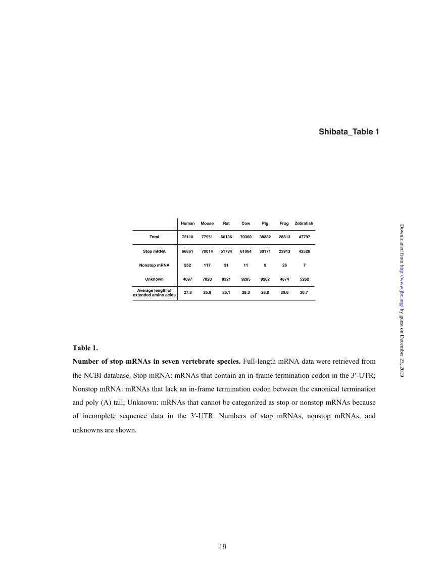
bioinformatics analysis of seven vertebrate animal
species indicated that a relatively small number of
mRNAs lacks a downstream stop codon, and that
most mRNAs contain additional in-frame stop
codons in the 3′-untranslated region (3′-UTR)
(Table 1). Therefore, even if a canonical stop
codon is mutated to code for an amino acid,
translation is terminated at a downstream stop
codon in many cases. Currently, more than 400
read-through single nucleotide polymorphisms
(SNPs) that result in encoding C-terminally
extended proteins terminated at a downstream stop
codon have been identified in humans
(Supplemental Dataset), and some of these SNPs
are related to hereditary disorders. However,
regulation of these C-terminally extended proteins
is poorly understood.
As a model system to study the regulation of the
read-through mutant genes, we found a read-
through mutation in the long form of the mouse
cellular FLICE-like apoptosis inhibitory protein
(cFLIP-L) gene that results in a mutant cFLIP-L
protein containing a C-terminal 46 amino acid
extension (cFLIP-L+46). Structurally, cFLIP-L
resembles caspase 8 but has no caspase activity
and, therefore, it inhibits apoptosis signaling
initiated by death receptor ligation (6). In this
study, we demonstrate that homozygous cFlip-
l+46 mutant mice die at around 13.5 days post-
coitum (dpc) and undergo extensive apoptosis in
the liver. Mechanistic analysis revealed that the
extended 46 amino acid peptide functions as a
degron to destabilize the cFLIP-L protein, and that
an E3 ubiquitin ligase, tripartite motif containing
21 (TRIM21), mediates the ubiquitylation of
cFLIP-L+46. We also show that C-terminally
extended pyridoxamine 5'-phosphate oxidase
(PNPO) and 3β-hydroxysteroid dehydrogenase
type II (HSD3B2) proteins, encoded by read-
through mutant genes found in patients of
congenital diseases, are degraded by the similar
mechanism. These findings indicate that
degradation of aberrant C-terminally extended
proteins that are encoded by genes with read-
through mutations is a mechanism for loss-of-
function and can lead to hereditary disorders.
Experimental Procedures
Reagents and plasmids
Tissue culture plastics were purchased from
Greiner Bio-One (Tokyo, Japan). Cycloheximide
(CHX) and anti-FLAG-agarose (M2) were from
Sigma-Aldrich (St. Louis, MO, USA). MG132
was from Peptide Institute (Osaka, Japan). Anti-
Myc-agarose (9E10) was from Santa Cruz. Cell
Counting Kit-8 was from Dojindo Laboratories
(Kumamoto, Japan). TNFα was provided by Prof.
Tsujimoto (Teikyo Heisei University, Japan).
cDNAs encoding mouse cFLIP-L, human PNPO
and HSD3B2 were amplified by PCR and cloned
into the p3xFLAG-CMV-10 (Sigma-Aldrich) or
pcDNA3 expression vectors (Invitrogen). cDNAs
encoding extended peptides were PCR amplified
by guest on Decem
ber 23, 2019http://w
ww
.jbc.org/D
ownloaded from

Degradation of read-through mutant proteins
4
and inserted into the pEGFP-C2 expression vector
(Clontech). pcDNA4/His/LacZ was purchased
from Invitrogen. A human TRIM21 expression
vector was provided by Prof. Hatakeyama
(Hokkaido University, Japan). To generate
siRNA-resistant TRIM21 expression vectors,
synonymous nucleotide substitutions were
introduced using PfuUltra High-Fidelity DNA
Polymerase (Agilent Technologies) according to
the manufacturer’s protocol.
Mice and histological analysis
Animal experiments were approved by the Animal
Experiment Committee of the National Institute of
Health Sciences, Japan. N-ethyl-N-nitrosourea
(ENU)-mutagenized cFlip-l+46 mutant mouse
lines were originally generated by the RIKEN
BioResource Center
(http://www.brc.riken.jp/lab/mutants/RGDMSavai
lability.htm), as previously described (7). cFlip
deficient mice were provided by Dr. Yeh (Amgen
Inc., USA) (8). All mice were housed under
specific pathogen-free conditions.
Embryos at E12.5 were fixed in 10%
neutral phosphate-buffered formalin, embedded in
paraffin, sectioned at 6 µm and stained with
hematoxylin-eosin. For TUNEL staining, sections
were stained using an In situ Apoptosis Detection
Kit (Takara Bio, Shiga, Japan).
Cell culture and transfection
Mouse embryonic fibroblasts (MEFs) and HeLa
cells were cultured in DMEM (Sigma-Aldrich)
supplemented with 10% fetal bovine serum
(Gibco), and 50 µg/ml kanamycin (Sigma-Aldrich).
Transient transfections were carried out using
Lipofectamine 2000 (Invitrogen) according to the
manufacturer’s instructions. siRNA transfections
were carried out using Lipofectamine RNAi/MAX
(Invitrogen) according to the manufacturer’s
instructions. Typically, cells were transfected and
48 h later the cells were treated with 10 µM
MG132 for 6 h. Target sequences for mouse cFlip-
l, human Trim21, and mouse Trim21 were as
follows (5′ to 3′): mouse cFlip-l,
GAATAGACTTGAACACAAA; human Trim21
#1, AAGCAGGAGTTGGCTGAGAAG; human
Trim21 #2, GCTCCCTCATCTACTCCTT; mouse
Trim21 #1, CCTAAACCCTCATCTCTTT; mouse
Trim21 #2, CCTGGACACGTTAGATATT.
Immunoblot analysis and
coimmunoprecipitation
For immunoblot analysis, cells were lysed in a
lysis buffer containing 0.1 M Tris/HCl (pH 7.5),
1% SDS, 10% glycerol. For
coimmunoprecipitation, cells were lysed in a lysis
buffer containing 0.02 M Tris/HCl (pH 7.5), 0.2%
NP-40, 0.15 M NaCl, 10% glycerol, and 1 x
complete protease inhibitor cocktail (Roche) and
the lysates were immunoprecipitated with
indicated antibodies. Lysates or
immunoprecipitates were separated by SDS-PAGE
by guest on Decem
ber 23, 2019http://w
ww
.jbc.org/D
ownloaded from

Degradation of read-through mutant proteins
5
and transferred to polyvinylidene difluoride
membranes. The following antibodies were used
to detect proteins; anti-cFLIP-L (Dave-2) (Enzo
Life Sciences, Plymouth Meeting, PA, USA), anti-
HSP90 (610419) (BD Transduction), anti-cleaved
Caspase-3 (9664), anti-β-Catenin (9562), and anti-
Myc (2276) (Cell Signaling), anti-human TRIM21
(12108-1-AP) (Proteintech Group Chicago, IL,
USA), anti-β-Tubulin (ab6046) (Abcam), anti-
FLAG (M2) (Sigma-Aldrich), anti-HA (3F10)
(Roche), anti-GAPDH (FL-335), anti-β-Actin (C-
2), anti-GFP (B-2), anti-His (H-3), anti-mouse
TRIM21 (M-20) (Santa Cruz).
Measurement of apoptosis by flow cytometer
Apoptosis was analyzed with an Annexin V-FITC
Apoptosis Detection Kit (BioVision, Milpitas, CA,
USA), as previously described(9). Briefly, after
treatment, cells were gently trypsinized and
washed with serum-containing medium. Cells
were collected by centrifugation, and additionally
washed with PBS, and resuspended in Binding
Buffer. The cells were stained with annexin V-
FITC and Propidium Iodide (PI) at room
temperature for 5 min in the dark, according to the
manufacturer’s instructions, and analyzed on a
FACScan flow cytometer (Becton Dickinson,
Braintree, MA, USA).
Ubiquitylation experiments
HeLa cells transfected with the indicated vectors
were treated with 10 µM MG132 and lysed in lysis
buffer containing 0.1 M Tris/HCl (pH 7.5), 1%
SDS, 10% glycerol. After heat-denaturation, the
lysates were diluted 10 times with 0.1 M Tris/HCl
(pH7.5). Proteins were immunoprecipitated with
anti-FLAG or anti-Myc agarose-conjugated beads,
and the immunoprecipitates were analyzed by
western blotting with anti-HA.
Identification of TRIM21 by liquid
chromatography/mass spectrometry (LC/MS)
Lysates from HeLa cells expressing GFP+46 or
GFP+22 were immunoprecipitated with antibody
against GFP. Following SDS-PAGE, gels were
stained with SYPRO Ruby Protein Gel Stain
(Lonza, Rockland, ME, USA), and protein bands
that specifically bind to the GFP+46 were excised
from the gel and subjected to in gel trypsin
digestion. The peptides extracted from the gel
pieces were analyzed by LC/MS/MS using an
Orbitrap Elite mass spectrometer (Thermo Fisher
Scientific, San Jose, CA, USA) connected to a
Paradigm MS4 HPLC system (Michrom
BioResources, Auburn, CA, USA). The analytical
column for LC/MS was a reversed-phase column
(L-column 2 ODS; 150 × 0.075 mm, 3 µm;
Chemicals Evaluation and Research Institute,
Tokyo, Japan). The mobile phase was 0.1% formic
acid containing 2% acetonitrile and 0.1% formic
acid containing 90% acetonitrile (A buffer). The
peptides were eluted at a flow rate of 300 nl/min
with a gradient of 2 to 65% of A buffer over 50
min. MS/MS conditions were as follows: an
by guest on Decem
ber 23, 2019http://w
ww
.jbc.org/D
ownloaded from

Degradation of read-through mutant proteins
6
electrospray voltage of 2.5 kV in positive ion
mode, a capillary temperature of 275°C, and a
collision energy of 35% for MS/MS. The spectra
data obtained by MS/MS were subjected to
database search analysis with the SEQUEST
algorithm (Proteome Discoverer 1.4, Thermo
Fisher Scientific) using the UniProt database.
RNA isolation and quantitative PCR
Total RNA was prepared from MEFs with RNeasy
(Qiagen). First strand cDNA was synthesized from
1 µg total RNA with an oligo-dT primer using the
SuperScript First-Strand Synthesis System
(Invitrogen). Quantitative real-time PCR was
performed with an ABI Prism 7300 sequence
detection system using TaqMan real-time PCR
Master Mix (Applied Biosystems, Foster City,
CA) with TaqMan probes/primers for mouse
cFlip-l and 18S ribosome RNA (Applied
Biosystems). The relative amounts of cFlip-l
mRNAs were calculated by using the comparative
Ct method. Mouse 18S ribosomal RNA was used
as an invariant control.
Bioinformatics analysis
Data for human, mice, rat, cow, pig, frog, and
zebrafish full-length mRNAs were retrieved from
the NCBI RefSeq database
(ftp://ftp.ncbi.nlm.nih.gov/refseq/) in January 2015.
Read-through SNP data and associated full-length
human mRNA data were retrieved from dbSNP
and RefSeq in the NCBI database
(http://www.ncbi.nlm.nih.gov/snp and
ftp://ftp.ncbi.nlm.nih.gov/refseq/) in March 2014.
Stop mRNAs that contain an in-frame termination
codon in the 3′-UTR were selected. 3′-UTR
sequences were translated in silico using a Perl
translate module.
Results
Homozygous cFlip-l read-through mutant mice
show extensive apoptosis in liver and die during
embryogenesis.
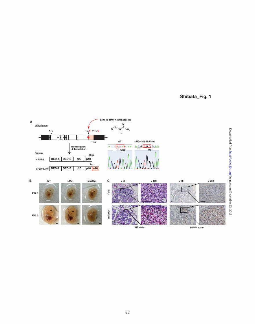
In a large-scale archive of ENU-mutagenized mice
(10), we found a read-through mutation (X482W)
in cFlip-l gene that results in encoding a mutant
cFLIP-L protein containing a C-terminal 46 amino
acid extension (cFLIP-L+46) (Fig. 1A). To
understand how the extended protein is regulated
and the phenotypic consequences of the read-
through mutation, we analyzed the mutant mice
and the cFLIP-L+46 protein.
Heterozygous cFlip-l+46 mutant (+/Mut) mice
developed normally and are fertile, but
homozygous mutant (Mut/Mut) mice died at
around E13.5 (Fig. 1B and Table 2A). To exclude
a possibility that abnormalities found in the mutant
(Mut/Mut) mice are due to ENU-generated
mutation of another, unknown gene that happened
to be homozygous in the cFlip-l+46 Mut/Mut
mice, we crossed heterozygous cFlip-l+46 mutant
(+/Mut) mice onto heterozygous cFlip deficient
(+/-) mice. The resulting transheterozygote (-/Mut)
by guest on Decem
ber 23, 2019http://w
ww
.jbc.org/D
ownloaded from

Degradation of read-through mutant proteins
7
mice died at around E11.5 (Table 2B), which is
slightly later than the embryonic lethality observed
in cFlip null (-/-) mice (Table 2C). Since any
mutation except for cFlip-l is heterozygous in the
transheterozygotes and gives no phenotypic
changes, the lethality observed in the
transheterozygote embryos is due to the
complication in the cFlip-l gene. Accordingly,
these results indicate that the homozygous read-
through mutation of cFlip-l gene is embryonic
lethal. Histological analysis of cFlip-l+46
Mut/Mut embryos at E12.5 showed hemorrhage
with many condensed nuclei and TUNEL-positive
cells in the liver (Fig. 1C). Thus, homozygous
cFlip-l read-through mutant (Mut/Mut) embryos
survived beyond E10.5 when cFlip null mice die
because of impaired yolk sac vasculature
development (8,11), but eventually died at around
E13.5, probably because of extensive apoptosis in
the liver. Because the extended cFLIP-L+46
protein is destabilized (described later), these
results suggest that a tiny amount of cFLIP-L+46
protein enables the embryos to survive beyond
E10.5, but that it is not sufficient to suppress
hepatocyte apoptosis at a later embryonic stage.
Reduction of cFLIP-L+46 protein levels results
in higher sensitivity to TNFα-induced apoptosis.
Next, we analyzed the behavior of the cFLIP-L+46
protein. Levels of cFLIP-L+46 protein were
dramatically reduced in the Mut/Mut whole
embryo (Fig. 2A) and liver (Fig. 2B) at E12.5. In
the liver, the active form of caspase 3 was
observed (Fig. 2B), which is in good agreement
with the observed hepatocyte apoptosis (Fig. 1C).
Reduced cFLIP-L+46 protein levels were similarly
observed in Mut/Mut MEFs (Fig. 2C). Consistent
with the reduced levels of cFLIP-L+46 protein, the
Mut/Mut MEFs were highly sensitive to TNFα-
induced apoptosis (Fig. 2D) as were wild-type
MEFs depleted of cFLIP-L using siRNA (Fig. 2E).
To examine whether the residual cFLIP-L+46
can inhibit apoptosis signaling in Mut/Mut MEFs,
we down-regulated cFLIP-L+46 in Mut/Mut
MEFs using siRNA. As shown in Figure 3A and
3B, depletion of the residual cFLIP-L+46 protein
significantly sensitized the cells to TNFα-induced
apoptosis. Conversely, exogenous expression of
cFLIP-L+46 protein in Mut/Mut MEFs suppressed
TNFα-induced caspase activation (Fig. 3C) and
increase of Annexin V positive apoptotic cells (Fig.
3D). These results indicate that cFLIP-L+46
protein has an activity to inhibit apoptosis, but that
the amount of cFLIP-L+46 protein is severely
reduced, resulting in higher sensitivity to apoptosis
in the Mut/Mut MEFs and hepatocytes.
C-terminally extended cFLIP-L+46 protein is
rapidly degraded by UPS.
We next investigated why levels of cFLIP-L+46
protein are severely reduced. In MEFs, the
turnover of cFLIP-L+46 protein was much faster
than that of wild-type cFLIP-L protein. When the
MEFs were treated with CHX, cFLIP-L+46
by guest on Decem
ber 23, 2019http://w
ww
.jbc.org/D
ownloaded from

Degradation of read-through mutant proteins
8
protein disappeared within 1 h whereas wild-type
cFLIP-L protein was retained for over 12 h (Fig.
4A). Similarly, cFLIP-L+46 exogenously
expressed in HeLa cells turned over more rapidly
than wild-type cFLIP-L (Fig. 4B). Treatment with
a proteasome inhibitor, MG132, increased the
accumulation of cFLIP-L+46 in Mut/Mut MEFs
(Fig. 4C). In addition, ubiquitylation of cFLIP-
L+46 exogenously expressed in HeLa cells was
highly enhanced compared with that of wild-type
cFLIP-L (Fig. 4D). On the other hand, levels of
cFlip-l+46 mRNA in Mut/Mut MEFs were not
reduced compared with cFlip-l mRNA in wild-
type (WT) MEFs (Fig. 4E). These results
collectively indicate that the expression of cFLIP-
L+46 protein is severely reduced because of the
rapid degradation by the UPS.
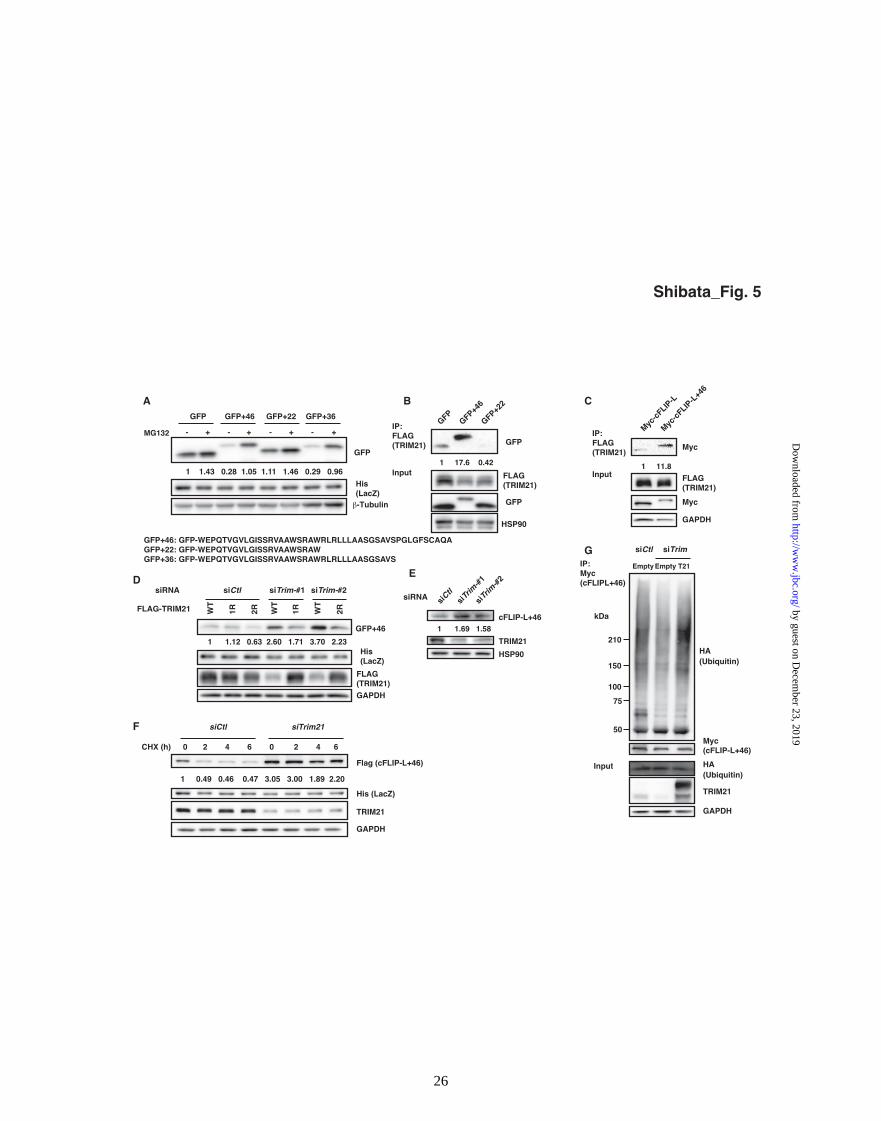
TRIM21 is responsible for ubiquitylation of
cFLIPL+46 protein.
To examine if the extended peptide can promote
proteasomal degradation when grafted onto a
heterologous protein, we genetically fused the
extended peptide to the C-terminus of GFP and
expressed it in HeLa cells. GFP+46 levels were
much lower than those of GFP, and this difference
was reduced by proteasome inhibitor MG132 (Fig.
5A), indicating that the extended peptide functions
as a degradation signal. Trimming 10 C-terminal
amino acids from the extended peptide to form
GFP+36 did not affect the degradation signal
activity, while deletion of 24 C-terminal amino
acids to form GFP+22 abolished the activity (Fig.
5A).
Since the extended peptide does not contain a
lysine residue that can be ubiquitylated, we
reasoned that the peptide functions as a degron
that interacts with a ubiquitin ligase to destabilize
cFLIP-L protein. To identify the ubiquitin ligase
involved in the destabilization of cFLIP-L+46
protein, we searched for proteins that interact with
GFP+46 but not with GFP+22 by liquid
chromatography-mass spectrometry, and identified
TRIM21, a RING-finger E3 ubiquitin ligase
involved in innate and acquired immunity (12-14).
Figure 5B shows that GFP+46, but not GFP+22,
co-precipitates TRIM21 protein in the cells. In
addition, cFLIP-L+46 strongly, but cFLIP-L
minimally, co-precipitates TRIM21 in the cells
(Fig. 5C). These results confirmed the selective
interaction of TRIM21 with the extended peptide
harboring the degron activity.
We next investigated whether TRIM21 is
required for the degradation of GFP+46 and
cFLIP-L+46 proteins. siRNA-mediated down-
regulation of human TRIM21 in HeLa cells
increased the levels of GFP+46, which was
suppressed by restoring TRIM21 levels by co-
transfecting an siRNA-resistant FLAG-tagged
human TRIM21 construct (Fig. 5D). In addition,
knockdown of mouse Trim21 in cFlip-l+46
Mut/Mut MEFs significantly increased the level of
cFLIP-L+46 (Fig. 5E). The rapid protein turnover
of cFLIP-L+46 exogenously expressed in HeLa
by guest on Decem
ber 23, 2019http://w
ww
.jbc.org/D
ownloaded from

Degradation of read-through mutant proteins
9
cells was suppressed by knockdown of human
Trim21 (Fig. 5F). Furthermore, the ubiquitylation
of cFLIP-L+46 protein was suppressed in cells
depleted of TRIM21, and restored by co-
transfection of the siRNA-resistant Trim21 gene
(Fig. 5G). These results indicate that TRIM21 is a
ubiquitin ligase responsible for the destabilization
of cFLIP-L+46 and GFP+46 proteins.
Read-through PNPO and HSD3B2 mutant
proteins are degraded through the UPS.
In human, more than 400 read-through SNPs result
in encoding C-terminally extended proteins that
terminate at downstream in-frame stop codons
(Supplemental Dataset). Among them, 20 are
related to hereditary disorders (Table 3). We
hypothesized that some of the read-through mutant
proteins could be regulated by a mechanism
similar to that which degrades cFLIP-L+46 protein.
To test this hypothesis, we examined in HeLa cells
the expression of GFP proteins fused to the
extended peptides encoded by the 20 genes
involved with hereditary disorders. We found that
the extended peptides from PNPO (28 amino
acids) and HSD3B2 (95 amino acids) destabilized
the GFP protein, which was abrogated by MG132
(Fig. 6A).
Consistent with this, levels of PNPO+28
expressed in HeLa cells were suppressed
compared with wild-type PNPO, and this
suppression was abrogated by MG132 (Fig. 6B,
left panels). PNPO+28 protein turned over more
rapidly than wild-type PNPO (Fig. 6C, left panels),
and PNPO+28 was more heavily ubiquitylated
compared with wild-type PNPO (Fig. 6D, left
panels). Similar results were obtained with
HSD3B2+95 and the corresponding wild-type
HSD3B2 proteins (Fig. 6B-D, right panels). These
results indicate that C-terminal extension of PNPO
and HSD3B2 proteins caused by read-through
mutations results in protein instability through the
UPS.
Finally, we examined whether TRIM21 is
involved in the destabilization of the extended
PNPO+28 and HSD3B2+95 proteins. siRNA-
mediated knockdown of human TRIM21 increased
the levels of PNPO+28, but not of HSD3B2+95
protein (Fig. 6E). Furthermore, the rapid turnover
of PNPO+28 protein was suppressed by
knockdown of human Trim21 (Fig. 6F), and
TRIM21 strongly interacted with PNPO+28 in the
cells (Fig. 6G). These results suggest that TRIM21
plays a role in the ubiquitylation of PNPO+28, but
that another E3 ligase is responsible for the
ubiquitylation of HSD3B2+95 protein.
Discussion
Currently, 20 stop codon read-through mutations
are related to hereditary disorders in human,
however, it is not understood how the C-terminally
extended read-through mutant proteins are
regulated. In this study, we analyzed a read-
through mutant of mouse cFlip-l as a model
by guest on Decem
ber 23, 2019http://w
ww
.jbc.org/D
ownloaded from

Degradation of read-through mutant proteins
10
system and showed that the homozygous read-
through mutant embryos undergo extensive
apoptosis in liver. The C-terminally extended
peptide interacts with a ubiquitin ligase TRIM21,
and promotes ubiquitylation and proteasomal
degradation of cFLIP-L+46 protein. We also
showed that the C-terminally extended proteins of
human PNPO and HSD3B2 encoded by read-
through mutant genes found in patients are
degraded by the similar mechanism involving
TRIM21 for PNPO degradation.
PNPO plays an essential role in brain
metabolism, catalyzing the conversion of
pyridoxine 5'-phosphate and pyridoxamine 5'-
phosphate to pyridoxal 5'-phosphate, a
metabolically active form of vitamin B6 (15).
Patients with homozygous read-through mutation
in Pnpo gene (X262Q) suffer from neonatal
epileptic encephalopathy (16), and show no PNPO
activity. The null PNPO activity can be explained
by degradation of the extended PNPO+28 protein
via the UPS. Since TRIM21 plays an important
role in the degradation of the extended PNPO+28
protein (Fig. 6E and F), inhibition and/or
suppression of TRIM21 could be a strategy to
restore PNPO protein levels in these cases.
HSD3B2 catalyzes the oxidation and
isomerization of Δ5-3β-hydroxysteroid precursors
into Δ4-ketosteroids, and is essential for the
formation of progesterone, a precursor for all
classes of steroid hormones. Deficiency in the
activity of HSD3B2 in the adrenal cortex causes
congenital adrenal hyperplasia (CAH) (17). A
read-through mutation in Hsd3b2 (X373C) was
found in patients with CAH, resulting in dramatic
reduction of HSD3B2 protein levels and activity
(18). It is likely that the mutant protein is degraded
by the UPS, although the E3 ligase involved is
currently unknown.
Among 20 read-through mutant proteins that are
related to hereditary disorders in human, C-
terminally extended PNPO+28 and HSD3B2+95
proteins are degraded by the UPS, as well as
mouse cFLIP-L+46 protein. Interestingly, the
extended peptides of PNPO+28, HSD3B2+95, and
cFLIP-L+46 share the Leu-Xaa-Xaa-Leu-Leu
(LxxLL) sequence, whereas other extended
peptides encoded by the mutant genes in Table 3
do not have the sequence. The LxxLL sequence is
well known as a protein-recognition motif widely
used in transcriptional regulation (19). To
investigate whether the LxxLL sequence is
involved in the degradation of C-terminally
extended proteins, we generated point or deletion
mutants of the sequence in cFLIP-L+46, but the
mutations did not affect the protein degradation of
cFLIP-L+46 (data not shown). Therefore, the
LxxLL sequence in the C-terminally extended
proteins is not likely to play an important role in
regulating the protein stability.
The 3′-UTR is known to regulate the stability of
mRNAs and the translation(20), but is rarely
considered to encode functional protein sequence.
However, in some cases, functional peptides are
by guest on Decem
ber 23, 2019http://w
ww
.jbc.org/D
ownloaded from

Degradation of read-through mutant proteins
11
encoded in the 3′-UTR, downstream of the
canonical termination codon and they can
influence the function of the mother protein.
Examples include an isoform of vascular
endothelial growth factor A (VEGF-Ax) and a
read-through mutant of CRYM. VEGF-Ax with a
C-terminal 22 amino acid extension is generated
by a programmed translational read-through
mechanism depending on heterogeneous nuclear
ribonucleoprotein A2/B1. The extended peptide
converts a proangiogenic VEGF-A into an anti-
angiogenic VEGF-Ax protein (21). A read-through
mutation in the Crym gene is found in patients
with non-syndromic deafness, and subcellular
localization of CRYM protein is altered by the C-
terminal extension (22). In addition to these
observations, our study provides evidence for the
destabilization of proteins by C-terminally
extended peptides encoded in the 3′-UTRs of
human Pnpo and Hsd3b2, which result in
congenital diseases, and in mouse cFlip-l, which
causes embryonic lethality. Since there are more
read-through mutations known to be involved in
human hereditary disorders (Table 3), further
study will clarify the function of the extended
peptides and how the C-terminally extended
proteins are regulated.
Acknowledgements
The authors thank Drs. T. Hattori and K. Okuhira for helpful discussions. We thank Dr. W-C Yeh for
kindly providing cFlip deficient mice, Prof. Hatakeyama for FLAG-TRIM21 plasmid, and Prof.
Tsujimoto for TNFα. Histological analysis was carried out by Genostaff Co., Ltd (Tokyo, Japan). This
work was supported by JSPS KAKENHI (No. 25112521 to M.N., No. 26860050 to N.S., and No.
25241016 to Y.G.) and by Health and Labor Sciences Research Grants (No. 26401201 to N.K.), Japan.
Conflict of interests
The authors declare that they have no conflicts of interest with the contents of this article.
Author contributions
N.S. and M.N. designed the experiments and wrote the manuscript. Yu.S. performed bioinformatics
analysis. N.S., N.O., Yu.S., C.O., M.I. and M.N. performed the experiments. Yo.S. and Y.G. generated
cFlip-l+46 mutant mice. D.T., N.H. and N.K. performed LC/MS experiments. M.N. supervised all
research. All authors approved the final version of the manuscript.
by guest on Decem
ber 23, 2019http://w
ww
.jbc.org/D
ownloaded from

Degradation of read-through mutant proteins
12
References
1. McCaughan, K. K., Brown, C. M., Dalphin, M. E., Berry, M. J., and Tate, W. P. (1995)
Translational termination efficiency in mammals is influenced by the base following the stop
codon. Proc. Natl. Acad. Sci. USA 92, 5431-5435
2. Cassan, M., and Rousset, J. P. (2001) UAG readthrough in mammalian cells: effect of upstream
and downstream stop codon contexts reveal different signals. BMC Mol. Biol. 2, 3
3. Ito-Harashima, S., Kuroha, K., Tatematsu, T., and Inada, T. (2007) Translation of the poly(A) tail
plays crucial roles in nonstop mRNA surveillance via translation repression and protein
destabilization by proteasome in yeast. Genes Dev. 21, 519-524
4. Tsuboi, T., Kuroha, K., Kudo, K., Makino, S., Inoue, E., Kashima, I., and Inada, T. (2012)
Dom34:hbs1 plays a general role in quality-control systems by dissociation of a stalled ribosome
at the 3' end of aberrant mRNA. Mol. Cell 46, 518-529
5. Brandman, O., Stewart-Ornstein, J., Wong, D., Larson, A., Williams, C. C., Li, G. W., Zhou, S.,
King, D., Shen, P. S., Weibezahn, J., Dunn, J. G., Rouskin, S., Inada, T., Frost, A., and Weissman,
J. S. (2012) A ribosome-bound quality control complex triggers degradation of nascent peptides
and signals translation stress. Cell 151, 1042-1054
6. Irmler, M., Thome, M., Hahne, M., Schneider, P., Hofmann, K., Steiner, V., Bodmer, J. L.,
Schroter, M., Burns, K., Mattmann, C., Rimoldi, D., French, L. E., and Tschopp, J. (1997)
Inhibition of death receptor signals by cellular FLIP. Nature 388, 190-195
7. Inoue, M., Sakuraba, Y., Motegi, H., Kubota, N., Toki, H., Matsui, J., Toyoda, Y., Miwa, I.,
Terauchi, Y., Kadowaki, T., Shigeyama, Y., Kasuga, M., Adachi, T., Fujimoto, N., Matsumoto,
R., Tsuchihashi, K., Kagami, T., Inoue, A., Kaneda, H., Ishijima, J., Masuya, H., Suzuki, T.,
Wakana, S., Gondo, Y., Minowa, O., Shiroishi, T., and Noda, T. (2004) A series of maturity onset
diabetes of the young, type 2 (MODY2) mouse models generated by a large-scale ENU
mutagenesis program. Hum. Mol. Genet. 13, 1147-1157
8. Yeh, W. C., Itie, A., Elia, A. J., Ng, M., Shu, H. B., Wakeham, A., Mirtsos, C., Suzuki, N.,
Bonnard, M., Goeddel, D. V., and Mak, T. W. (2000) Requirement for Casper (c-FLIP) in
regulation of death receptor-induced apoptosis and embryonic development. Immunity 12, 633-
642
by guest on Decem
ber 23, 2019http://w
ww
.jbc.org/D
ownloaded from

Degradation of read-through mutant proteins
13
9. Ohoka, N., Nagai, K., Hattori, T., Okuhira, K., Shibata, N., Cho, N., and Naito, M. (2014) Cancer
cell death induced by novel small molecules degrading the TACC3 protein via the ubiquitin-
proteasome pathway. Cell Death Dis. 5, e1513
10. Sakuraba, Y., Sezutsu, H., Takahasi, K. R., Tsuchihashi, K., Ichikawa, R., Fujimoto, N., Kaneko,
S., Nakai, Y., Uchiyama, M., Goda, N., Motoi, R., Ikeda, A., Karashima, Y., Inoue, M., Kaneda,
H., Masuya, H., Minowa, O., Noguchi, H., Toyoda, A., Sakaki, Y., Wakana, S., Noda, T.,
Shiroishi, T., and Gondo, Y. (2005) Molecular characterization of ENU mouse mutagenesis and
archives. Biochem. Biophys. Res. Commun. 336, 609-616
11. Sakamaki, K., Inoue, T., Asano, M., Sudo, K., Kazama, H., Sakagami, J., Sakata, S., Ozaki, M.,
Nakamura, S., Toyokuni, S., Osumi, N., Iwakura, Y., and Yonehara, S. (2002) Ex vivo whole-
embryo culture of caspase-8-deficient embryos normalize their aberrant phenotypes in the
developing neural tube and heart. Cell Death Differ. 9, 1196-1206
12. McEwan, W. A., Tam, J. C., Watkinson, R. E., Bidgood, S. R., Mallery, D. L., and James, L. C.
(2013) Intracellular antibody-bound pathogens stimulate immune signaling via the Fc receptor
TRIM21. Nat. Immunol. 14, 327-336
13. Oke, V., and Wahren-Herlenius, M. (2012) The immunobiology of Ro52 (TRIM21) in
autoimmunity: a critical review. J. Autoimmun. 39, 77-82
14. Zhang, Z., Bao, M., Lu, N., Weng, L., Yuan, B., and Liu, Y. J. (2013) The E3 ubiquitin ligase
TRIM21 negatively regulates the innate immune response to intracellular double-stranded DNA.
Nat. Immunol. 14, 172-178
15. Ngo, E. O., LePage, G. R., Thanassi, J. W., Meisler, N., and Nutter, L. M. (1998) Absence of
pyridoxine-5'-phosphate oxidase (PNPO) activity in neoplastic cells: isolation, characterization,
and expression of PNPO cDNA. Biochemistry 37, 7741-7748
16. Mills, P. B., Surtees, R. A., Champion, M. P., Beesley, C. E., Dalton, N., Scambler, P. J., Heales,
S. J., Briddon, A., Scheimberg, I., Hoffmann, G. F., Zschocke, J., and Clayton, P. T. (2005)
Neonatal epileptic encephalopathy caused by mutations in the PNPO gene encoding
pyridox(am)ine 5'-phosphate oxidase. Hum. Mol. Genet. 14, 1077-1086
17. Pang, S. (2001) Congenital adrenal hyperplasia owing to 3 beta-hydroxysteroid dehydrogenase
deficiency. Endocrinol Metab. Clin. North Am. 30, 81-99, vi-vii
18. Pang, S., Wang, W., Rich, B., David, R., Chang, Y. T., Carbunaru, G., Myers, S. E., Howie, A. F.,
Smillie, K. J., and Mason, J. I. (2002) A novel nonstop mutation in the stop codon and a novel
missense mutation in the type II 3beta-hydroxysteroid dehydrogenase (3beta-HSD) gene causing,
by guest on Decem
ber 23, 2019http://w
ww
.jbc.org/D
ownloaded from

Degradation of read-through mutant proteins
14
respectively, nonclassic and classic 3beta-HSD deficiency congenital adrenal hyperplasia. J. Clin.
Endocrinol. Metab. 87, 2556-2563
19. Plevin, M. J., Mills, M. M., and Ikura, M. (2005) The LxxLL motif: a multifunctional binding
sequence in transcriptional regulation. Trends Biochem. Sci. 30, 66-69
20. Conne, B., Stutz, A., and Vassalli, J. D. (2000) The 3' untranslated region of messenger RNA: A
molecular 'hotspot' for pathology? Nat. Med. 6, 637-641
21. Eswarappa, S. M., Potdar, A. A., Koch, W. J., Fan, Y., Vasu, K., Lindner, D., Willard, B.,
Graham, L. M., DiCorleto, P. E., and Fox, P. L. (2014) Programmed Translational Readthrough
Generates Antiangiogenic VEGF-Ax. Cell 157, 1605-1618
22. Abe, S., Katagiri, T., Saito-Hisaminato, A., Usami, S., Inoue, Y., Tsunoda, T., and Nakamura, Y.
(2003) Identification of CRYM as a candidate responsible for nonsyndromic deafness, through
cDNA microarray analysis of human cochlear and vestibular tissues. Am. J. Hum. Genet. 72, 73-
82
by guest on Decem
ber 23, 2019http://w
ww
.jbc.org/D
ownloaded from

Degradation of read-through mutant proteins
15
Footnotes
Abbreviations: UPS, ubiquitin-proteasome system; C-terminal, carboxy-terminal; 3′-UTR, 3′-
untranslated region; SNPs, single nucleotide polymorphisms; cFLIP-L, long form of cellular FLICE-like
apoptosis inhibitory protein; dpc, days post-coitum; TRIM2, tripartite motif containing 21; PNPO,
pyridoxamine 5'-phosphate oxidase; HSD3B, 3β-hydroxysteroid dehydrogenase type II; CHX,
cycloheximide; ENU, N-ethyl-N-nitrosourea; MEFs, mouse embryonic fibroblasts; LC/MS, liquid
chromatography/mass spectrometry; CAH, congenital adrenal hyperplasia; VEGF-Ax, vascular
endothelial growth factor A
by guest on Decem
ber 23, 2019http://w
ww
.jbc.org/D
ownloaded from
Degradation of read-through mutant proteins
16
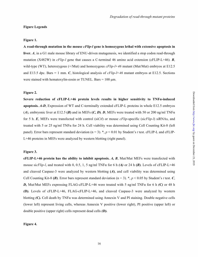
Figure Legends
Figure 1.
A read-through mutation in the mouse cFlip-l gene is homozygous lethal with extensive apoptosis in
liver. A, in a G1 male mouse library of ENU-driven mutagenesis, we identified a stop codon read-through
mutation (X482W) in cFlip-l gene that causes a C-terminal 46 amino acid extension (cFLIP-L+46). B,
wild-type (WT), heterozygous (+/Mut) and homozygous cFlip-l+46 mutant (Mut/Mut) embryos at E12.5
and E13.5 dpc. Bars = 1 mm. C, histological analysis of cFlip-l+46 mutant embryos at E12.5. Sections
were stained with hematoxylin-eosin or TUNEL. Bars = 100 µm.
Figure 2.
Severe reduction of cFLIP-L+46 protein levels results in higher sensitivity to TNFα-induced
apoptosis. A-D, Expression of WT and C-terminally extended cFLIP-L proteins in whole E12.5 embryos
(A), embryonic liver at E12.5 (B) and in MEFs (C, D). D, MEFs were treated with 50 or 200 ng/ml TNFα
for 5 h. E, MEFs were transfected with control (siCtl) or mouse cFlip-specific (sicFlip-l) siRNAs, and
treated with 5 or 25 ng/ml TNFα for 24 h. Cell viability was determined using Cell Counting Kit-8 (left
panel). Error bars represent standard deviation (n = 3). *, p < 0.01 by Student’s t test. cFLIP-L and cFLIP-
L+46 proteins in MEFs were analyzed by western blotting (right panel).
Figure 3.
cFLIP-L+46 protein has the ability to inhibit apoptosis. A, B, Mut/Mut MEFs were transfected with
mouse sicFlip-l, and treated with 0, 0.5, 1, 5 ng/ml TNFα for 6 h (A) or 24 h (B). Levels of cFLIP-L+46
and cleaved Caspase-3 were analyzed by western blotting (A), and cell viability was determined using
Cell Counting Kit-8 (B). Error bars represent standard deviation (n = 3). *, p < 0.05 by Student’s t test. C,
D, Mut/Mut MEFs expressing FLAG-cFLIP-L+46 were treated with 5 ng/ml TNFα for 6 h (C) or 48 h
(D). Levels of cFLIP-L+46, FLAG-cFLIP-L+46, and cleaved Caspase-3 were analyzed by western
blotting (C). Cell death by TNFα was determined using Annexin V and PI staining. Double negative cells
(lower left) represent living cells, whereas Annexin V positive (lower right), PI positive (upper left) or
double positive (upper right) cells represent dead cells (D).
Figure 4.
by guest on Decem
ber 23, 2019http://w
ww
.jbc.org/D
ownloaded from

Degradation of read-through mutant proteins
17
The extended peptide facilitates degradation of cFLIP-L+46 through the UPS. A, B, Turnover of
cFLIP-L or cFLIP-L+46 proteins in MEFs (A), and HeLa cells constitutively expressing wild-type
FLAG-cFLIP-L or extended FLAG-cFLIP-L+46 proteins (B). MEFs and HeLa cells were treated with 30
µg/ml CHX for the indicated times. C, Proteasome inhibitor increases the level of cFLIP-L+46 protein in
Mut/Mut MEFs. MEFs were treated with 10 µM MG132 for 6 h. D, Enhanced ubiquitylation of cFLIP-
L+46 protein. HeLa cells were transfected with FLAG-tagged cFLIP-L or cFLIP-L+46, and HA-tagged
ubiquitin (HA-Ub) for 48 h, and treated with 10 µM MG132 for 6 h. Ubiquitylation was analyzed as
described in Materials and Methods. E, Levels of cFlip-l mRNA in MEFs were measured by quantitative
PCR and normalized against 18S ribosomal RNA expression. Error bars represent standard deviation (n =
4).
Figure 5.
TRIM21 mediates the ubiquitylation and degradation of cFLIPL+46 protein. A, Expression of GFP
fused to the extended peptide of cFLIP-L. HeLa cells were transfected with indicated GFP-fusions and
His-LacZ as an internal control for transfection efficiency for 48 h, and then cells were treated with 10
µM MG132 for 6 h. Numbers below the panel represent relative expression levels normalized against His-
LacZ expression. B, C, TRIM21 interacts with the extended peptide of mouse cFLIP-L. HeLa cells were
transfected with FLAG-tagged TRIM21 and indicated GFP-fusions (B), or transfected with FLAG-tagged
TRIM21 together with Myc-tagged cFLIP-L or cFLIP-L+46 (C), and were treated with 10 µM MG132
for 6 h. Lysates were immunoprecipitated with anti-FLAG antibody, and the immunoprecipitates were
analyzed by immunoblotting with an antibody against GFP (B) or Myc (C). Numbers below the panel
represent relative amounts in the immunoprecipitates normalized against FLAG-TRIM21 expression
together with indicated GFP-fusions (B), or with cFLIP-L or cFLIP-L+46 (C) expression in lysates. D,
TRIM21 mediates the down-regulation of GFP+46 protein. HeLa cells were transfected with GFP+46 and
His-LacZ together with indicated siRNAs and siRNA-sensitive (WT) or siRNA-resistant mutants (1R,
2R) of FLAG-tagged TRIM21. Numbers below the panel represent relative expression levels normalized
against His-LacZ expression. E, Depletion of TRIM21 increases cFLIP-L+46 protein levels in Mut/Mut
MEFs. Numbers below the panel represent relative intensity of cFLIP-L+46 protein normalized against
HSP90 expression. F, HeLa cells were transfected with FLAG-cFLIP-L+46 and His-LacZ as an internal
control for transfection efficiency together with siRNAs against Trim21, and were treated with 10 µg/ml
CHX for the indicated times. Numbers below the panel represent relative expression levels normalized
against His-LacZ expression. G, Ubiquitylation of cFLIP-L+46 protein by TRIM21. HeLa cells were
by guest on Decem
ber 23, 2019http://w
ww
.jbc.org/D
ownloaded from

Degradation of read-through mutant proteins
18
transfected with Myc-cFLIP-L+46, HA-Ub, siRNA against Trim21, and a siRNA-resistant Flag-TRIM21
(T21), and treated with 10 µM MG132 for 2 h. Ubiquitylation was analyzed as described in Materials and
Methods.
Figure 6.
Read-through PNPO and HSD3B2 mutant proteins are degraded through the UPS. A, Wild-type
GFP or GFP fused to the extended peptides of PNPO (GFP+28) or HSD3B2 (GFP+95) were transfected
into HeLa cells, and the cells were treated with 10 µM MG132 for 6 h. B, C, Myc-tagged wild-type
(PNPO or HSD3B2) or read-through mutant (PNPO+28 or HSD3B2+95) genes were transfected into
HeLa cells, and the cells were treated with 10 µM MG132 for 6 h (B) or were treated with 10 µg/ml CHX
for the indicated times (C). D, Enhanced ubiquitylation of the read-through mutant proteins. HeLa cells
were transfected with Myc-tagged wild-type or read-through mutant genes together with HA-tagged
ubiquitin (HA-Ub) and 48 h later were treated with 10 µM MG132 for 6 h. Ubiquitylation was analyzed
as described in Materials and Methods. E, Effect of Trim21-knockdown on PNPO+28 and HSD3B2+95
levels. HeLa cells were transfected with Myc-PNPO+28 or Myc-HSD3B2+95 together with siRNAs
against Trim21. F. Effect of Trim21-knockdown on PNPO+28 protein stability. HeLa cells were
transfected with Myc-PNPO+28 together with siRNAs against Trim21, and were treated with 10 µg/ml
CHX for the indicated times. The cells (A-C, E, F) were also co-transfected with His-LacZ and the
expression was measured as an internal control for transfection efficiency. Numbers below the panels
represent relative expression levels normalized against His-LacZ expression. G, TRIM21 interacts with
the extended peptide of human PNPO. HeLa cells were transfected with FLAG-tagged TRIM21 together
with Myc-tagged PNPO or PNPO+28, and were treated with 10 µM MG132 for 6 h. Lysates were
immunoprecipitated with anti-FLAG antibody, and the immunoprecipitates were analyzed by
immunoblotting with an antibody against Myc. Numbers below the panel represent relative amounts in
the immunoprecipitates normalized against FLAG-TRIM21 expression together with PNPO or PNPO+28
expression in lysates.
by guest on Decem
ber 23, 2019http://w
ww
.jbc.org/D
ownloaded from
Shibata_Table 1
Stop mRNA
Average length ofextended amino acids
66861
27.8
70014
25.9
51784
25.1
61064
28.3
23913
20.6
42528
20.7
Total
Human
72110
Mouse
77951
Rat
60136
Cow
70360
Frog
28813
Zebrafish
47797
Pig
38382
30171
28.0
Nonstop mRNA
Unknown
552
4697
117
7820
31
8321
11
9285
9
8202
26
4874
7
5262
Table 1.
Number of stop mRNAs in seven vertebrate species. Full-length mRNA data were retrieved from
the NCBI database. Stop mRNA: mRNAs that contain an in-frame termination codon in the 3 -UTR;
Nonstop mRNA: mRNAs that lack an in-frame termination codon between the canonical termination
and poly (A) tail; Unknown: mRNAs that cannot be categorized as stop or nonstop mRNAs because
of incomplete sequence data in the 3 -UTR. Numbers of stop mRNAs, nonstop mRNAs, and
unknowns are shown.
19
by guest on Decem
ber 23, 2019http://w
ww
.jbc.org/D
ownloaded from

C
A
B
WT +/Mut Mut/Mut TotalE11.5 3 5 2 10E12.5 10 7 7 24E13.5 7 16 4 (2) 29E14.5 15 30 1 (5) 51E15.5 3 9 0 (4) 16
+/+ +/- -/-5 (5)2 (10)0 (2)
TotalE10.5 15 19(2) 46E11.5 12 21 35E12.5 2 8 12
36 (2) 10 (1) 4413 3 (7) 416 0 (4) 16
+/+ +/- +/Mut TotalE9.5 1 2 4 10E10.5 16 9E11.5 14 14E12.5 1 5
-/Mut
Shibata_Table 2
Table 2.
Viability of cFlip-l read-through mutant and cFlip deficient embryos. Embryos were obtained at
the indicated gestation periods after crossing heterozygous cFlip-l+46 mutant (+/Mut) mice (A),
intercrossing heterozygous cFlip deficient (+/-) mice with heterozygous cFlip-l+46 mutant (+/Mut)
mice (B) or crossing heterozygous cFlip deficient (+/-) mice (C). Numbers of living embryos are
shown. Numbers in parenthesis represent dead embryos.
20
by guest on Decem
ber 23, 2019http://w
ww
.jbc.org/D
ownloaded from

Shibata_Table 3
CRYM 104894509 123740 Deafness, Autosomal Dominat Nonsyndromic 5DBT 121965000 248610 Maple Syrup Urine Disease, Intermediate, Type II 7
ITM2B 104894417 603904 Dementia, Familial British 11SH2D1A 111033625 300490 Lymphoproliferative syndrome, X-linked, 1 12
PAX6 121907922 607108 Aniridia 14MOCS2 121908609 603708 Molybdenum cofactor deficiency B 18CTSK 74315301 601105 Pycnodysostosis 19FKRP 104894682 606596 Muscular dystrophy-dystroglycanopathy (limb-girdle), type C, 5 21
RUNX2 104893994 600211 Cleidocranial Dysplasia 23
PNPO 104894631 603287 Pyridoxamine 5'-phosphate oxidase deficiency 28HBA2 41321345 141850 Hemoglobin H disease, nondeletional 31SHOX 137852559 312865 Leri-Weill Dyschondrosteosis 48NHP2 121908091 606470 Dyskeratosis congenita, autosomal recessive 2 51FHL1 122459148 300163 Emery-Dreifuss muscular dystrophy 6, X-linked 52
RAD50 121912629 604040 Nijmegen Breakage Syndrome-like Disorder 66FOXF1 121909337 601089 Alveolar capillary dysplasia with misalignment of pulmonary veins 72
HSD3B2 80358218 201810 3-beta-hydroxysteroid dehydrogenase, type II, deficiency 95FGFR3 121913101 134934 THANATOPHORIC DYSPLASIA, TYPE I 101CLCF1 137853935 610313 Cold-induced sweating syndrome 1 170
Length
Ectodermal Dysplasia, Anhidrotic, With immunodeficiency,Osteopetrosis, and Lymphedema
Symbol dbSNP ID OMIM Disease
27300301137853321IKBKG
Table 3.
Read-through SNPs related to human hereditary disorders. Column headings are: Symbol, Gene
Symbol; dbSNP ID, SNP Database Identifier; OMIM, Online Mendelian Inheritance in Man
Identifier; Disease, Description of genetic disease caused by read-through SNPs; Length, Length of
extended peptides encoded by read-through mutant genes.
21
by guest on Decem
ber 23, 2019http://w
ww
.jbc.org/D
ownloaded from
WT cFlip-l+46 Mut/Mut
Stop Trp
DED-A DED-B p20 p10
DED-A DED-B p20 p10 +46
Stop
Trp
cFLIP-L
cFLIP-L+46
ATG TGA TGG
TGA
N NH2
NO
O
ENU (N-ethyl-N-nitrosourea)
cFlip-l gene
Protein
Transcription & Translation
A
CB
E12.5
E13.5
WT +/Mut Mut/Mut
+/M
utM
ut/M
ut
HE stain TUNEL stain
x 200x 50 x 400 x 50
Shibata_Fig. 1
22
by guest on Decem
ber 23, 2019http://w
ww
.jbc.org/D
ownloaded from

Shibata_Fig. 2
cFLIP-L+46cFLIP-L
WT +/Mut Mut/Mut
HSP90
E12.5 whole body
HSP90
MEFs
cFLIP-L+46cFLIP-L
WT +/Mut Mut/Mut
Cel
l via
bilit
y (%
of c
ontr
ol)
WT Mut/Mut
GAPDH
WT Mut/Mut
MEFs
cFLIP-L+46cFLIP-L
0
20
40
60
80
100
120
0 5 25 0 5 25 0 5 25TNFα (ng/ml):
siCtl sicFlip-l siCtl
siCtl
sicFlip
-l
siCtl
* * * *
β-Actin
Cleaved Caspase-3
E12.5 Liver
cFLIP-L+46cFLIP-L
WT Mut/Mut
GAPDH
TNFα(ng/ml) 0 50 200 0 50 200
Mut/Mut
MEFs
cFLIP-L+46cFLIP-L
WTD
B
Cleaved Caspase-3
A C
E
23
by guest on Decem
ber 23, 2019http://w
ww
.jbc.org/D
ownloaded from

1.87% 1.92%
92.91% 3.30%
0.98% 1.49%
93.49% 3.64%
1.36% 2.88%
90.89% 4.87%
0.98% 5.19%
86.02% 7.81%
1.23% 1.54%
91.79% 5.44%
0.21% 0.76%
92.72% 6.31%
Shibata_Fig. 3
GAPDH
Cleaved Caspase-3
TNFα
Empty FLAG-cFLIP-L+46
55 kDa
55 kDa
WT Mut/MutC
FLAG-cFLIP-L+46cFLIP-L+46cFLIP-LFLAG-cFLIP-L+46
cFLIP-L+46 (Long exposure)
GAPDH
sicFlip-lsiCtl
0 0.5 1 5 0 0.5 1 5
Cel
l via
bilit
y (%
of c
ontr
ol)
0
20
40
60
80
100
120
0 0.5 1 5TNFα (ng/ml):
Mut/Mut MEFs
TNFα(ng/ml)
Cleaved Caspase-3
A
BMut/Mut MEFs
siCtlsicFlip-l
* *
*
cFLIP-L+46 (Normal exposure)
- + - + - +
Annexin V-FITC
Prop
idiu
m Io
dide
WT Mut/Mut
Empty FLAG-cFLIP-L+46 TNFα
D
-
+
24
by guest on Decem
ber 23, 2019http://w
ww
.jbc.org/D
ownloaded from

Shibata_Fig. 4
E
cFLIP-L+46(Normal exposure)
cFLIP-L
HSP90
HSP90
0 1 3 6 9 12CHX(h)
A
HA (Ubiquitin)
175
80
58
46
30
GAPDH
Input
IP: FLAG
Empty cFLIP-L cFLIP-L+46
HA-Ub
FLAG-cFLIP-L
- + - + - +
kDa
HA (Ubiquitin)
cFLIP-L+46cFLIP-L
CMG132
β-Catenin
GAPDH
WT Mut/MutSA2 SA4 SA9
cFLIP-L+46cFLIP-L
cFLIP-L+46(Long exposure)
0 1 3 6 9 12CHX(h)
WT MEFs
Mut/Mut MEFs
CHX(h) 0 1 3 6 9
FLAG (cFLIP-L+46)
FLAG (cFLIP-L)
HSP90
HSP90
FLAG-cFLIP-L
FLAG-cFLIP-L+46
B
0
1
2
WT Mut/Mut+/Mut
Rel
ativ
e am
ount
of
cFl
ip-l
mR
NA
D
CHX(h) 0 1 3 6 9
- + - + - + - +
25
by guest on Decem
ber 23, 2019http://w
ww
.jbc.org/D
ownloaded from
Shibata_Fig. 5
MG132
1 1.43 0.28 1.05 1.11 1.46 0.29 0.96
GFP
His (LacZ)β-Tubulin
GFP GFP+46
- + - + - + - +
GFP+22 GFP+36
HSP90
GFP
FLAG(TRIM21)
GFP
Input
IP:FLAG(TRIM21)
GFPGFP+4
6
GFP+22
GFP+46: GFP-WEPQTVGVLGISSRVAAWSRAWRLRLLLAASGSAVSPGLGFSCAQAGFP+22: GFP-WEPQTVGVLGISSRVAAWSRAWGFP+36: GFP-WEPQTVGVLGISSRVAAWSRAWRLRLLLAASGSAVS
GFP+46
GAPDH
siCtl siTrim-#1 siTrim-#2
WT
1R 2R WT
1R WT
2R
siRNA
FLAG-TRIM21
1 1.12 0.63 2.60 1.71 3.70 2.23
D
FLAG(TRIM21)
His (LacZ)
E
siCtl
siTrim
-#1
siTrim
-#2
siRNA
HSP90TRIM21
cFLIP-L+461 1.69 1.58
G
HA (Ubiquitin)
GAPDH
Myc(cFLIP-L+46)
TRIM21
Input
IP:Myc(cFLIPL+46)
210
150
10075
kDa
siCtl siTrim
Empty Empty T21
50
A B
HA (Ubiquitin)
Myc
GAPDH
Myc
Myc-cF
LIP-L
Myc-cF
LIP-L+46
Input
IP:FLAG(TRIM21)
Flag (cFLIP-L+46)
His (LacZ)
TRIM21
GAPDH
siCtl siTrim21
C
F
1 0.49 0.46 0.47 3.05 3.00 1.89 2.20
0 2 4 6 0 2 4 6CHX (h)
FLAG(TRIM21)
1 17.6 0.42 1 11.8
26
by guest on Decem
ber 23, 2019http://w
ww
.jbc.org/D
ownloaded from

Shibata_ Fig. 6
A
C
D
His (LacZ)
β-Actin
- + - +MG132
GFPGFP+28(PNPO)
1 1.27 0.43 0.85GFP
GFP
β-Tubulin
MG132
GFPGFP+95
(HSD3B2)
1 1.13 0.49 1.04His (LacZ)
His (LacZ)
Myc (PNPO)
PNPO
PNPO+28
CHX (h) 0 2 4 6
GAPDH
Myc (PNPO+28)
GAPDH
CHX (h) 0 2 4 6
1 0.97 0.79 0.54
1 0.64 0.46 0.27
His (LacZ)
HSD3B2
HSD3B2+95
Myc (HSD3B2)
GAPDH
CHX (h) 0 2 4 6
CHX (h) 0 2 4 6
1 0.45 0.45 0.47
Myc (HSD3B2+95)
GAPDH
1 0.91 0.87 1.04His (LacZ)
HSP90
175
80
58
46
Empty PNPO PNPO+28HA-Ub
Myc-PNPO
Input
IP:Myc
kDa
GAPDH
175
80
Empty HSD3B2HSD3B2
+95HA-Ub
Myc-HSD3B2
Input
kDa
5846
46
30
30
HA (Ubiquitin)
HA (Ubiquitin)
PNPO+28PNPO
HA (Ubiquitin)
HA (Ubiquitin)
HSD3B2+95HSD3B2
IP:Myc
B
E
β-Tubulin
MG132
1 0.77 0.25 0.45
PNPO PNPO+28
GAPDH
1 1.40 0.26 1.05
MG132
HSD3B2 HSD3B2+95
PNPO+28PNPO
HSD3B2+95HSD3B2
His (LacZ) His (LacZ)
Myc (HSD3B2+95)
siRNA
MG132
TRIM21GAPDH
1 3.12 0.69 0.79 His (LacZ)
Myc (PNPO+28)
TRIM21
GAPDH
1 4.12 3.28 3.68His (LacZ)
siRNA
MG132
siCtl siTrim
-#1
siTrim
-#2
siCtl siTrim
-#1
siTrim
-#2
His (LacZ)
- + - +
- + - + - + - +
- + - + - + - + - + - +
- + - - - + - -
Myc (PNPO+28)
His (LacZ)
TRIM21
GAPDH
F
1 0.68 0.76 0.50 5.80 4.63 6.75 4.39
siCtl siTrim21
0 2 4 6 0 2 4 6CHX (h) Myc
HSP90
Myc
Myc-PNPO
Myc-PNPO+2
8
Input
IP:FLAG(TRIM21)
G
FLAG(TRIM21)
1 7.3
27
by guest on Decem
ber 23, 2019http://w
ww
.jbc.org/D
ownloaded from

and Mikihiko NaitoYoshiyuki Sakuraba, Daisuke Takakura, Noritaka Hashii, Nana Kawasaki, Yoichi Gondo
Norihito Shibata, Nobumichi Ohoka, Yusuke Sugaki, Chiaki Onodera, Mizuho Inoue,system causes hereditary disorders.
Degradation of stop codon read-through mutant proteins via the ubiquitin-proteasome
published online October 6, 2015J. Biol. Chem.
10.1074/jbc.M115.670901Access the most updated version of this article at doi:
Alerts:
When a correction for this article is posted•
When this article is cited•
to choose from all of JBC's e-mail alertsClick here
Supplemental material:
http://www.jbc.org/content/suppl/2015/10/06/M115.670901.DC1
by guest on Decem
ber 23, 2019http://w
ww
.jbc.org/D
ownloaded from