the latest version is at factor signaling is essential for ... · 1 forkhead box f2 regulation of...
TRANSCRIPT
1
Forkhead box F2 Regulation of Platelet-Derived Growth Factor and myocardin/Serum Response
Factor Signaling is Essential for Intestinal Development
Craig Bolte1#
, Xiaomeng Ren1, Tatiana Tomley
1, Vladimir Ustiyan
1, Arun Pradhan
1, April Hoggatt
2,
Tanya V. Kalin1, B. Paul Herring
2 and Vladimir V. Kalinichenko
1#
1Department of Pediatrics, Perinatal Institute, Cincinnati Children’s Research Foundation, Cincinnati,
OH, 45229 USA.
2Department of Cellular and Integrative Physiology, Indiana University School of Medicine, Indianapolis,
IN, 46202 USA.
Running title: Foxf2 is Essential for Intestinal Development
#Correspondence to: Dr. Vladimir V. Kalinichenko ([email protected]) or Dr. Craig
Bolte ([email protected]), 1Department of Pediatrics, Perinatal Institute, Cincinnati Children’s
Research Foundation, 3333 Burnet Ave., MLC 7009, Cincinnati, OH 45229.
Keywords: Foxf2; gut development; smooth muscle hyperplasia; myocardin/SRF___________________
Background: Transcriptional regulation of smooth
muscle cells is an understudied component of
intestinal development and physiology.
Results: Foxf2 deletion from smooth muscle
causes intestinal malformations and colon
remodeling.
Conclusion: Foxf2 regulation of PDGF and
myocardin/SRF signaling is essential for intestinal
development and homeostasis.
Significance: Better understanding transcriptional
mechanisms regulating postnatal intestine
development and homeostasis may provide
therapeutic approaches for congenital and acquired
gastrointestinal diseases.
ABSTRACT
Alterations in Forkhead box F2 gene expression
have been reported in numerous pathologies
and Foxf2-/-
mice are perinatal lethal with
multiple malformations; however, molecular
mechanisms pertaining to Foxf2 signaling are
severely lacking. In the present study, Foxf2
requirements in murine smooth muscle cells
were examined using a conditional knockout
approach. We generated novel Foxf2-floxed
mice, which we bred to smMHC-Cre-eGFP mice
to generate a mouse line with Foxf2 deleted
specifically from smooth muscle. These mice
exhibited growth retardation due to reduced
intestinal length as well as inflammation and
remodeling of the small intestine. Colons of
Tg(smMHC-Cre-eGFP+/-
);Foxf2-/-
mice had
expansion of the myenteric nerve plexus and
increased proliferation of smooth muscle cells
leading to thickening of the longitudinal smooth
muscle layer. Foxf2-deficiency in colonic smooth
muscle was associated with increased expression
of Foxf1, PDGFa, PDGFb, PDGFRα and
myocardin. FOXF2 bound to promoter regions
of these genes indicating direct transcriptional
regulation. Foxf2 repressed Foxf1 promoter
activity in co-transfection experiments. We also
show that knockdown of Foxf2 in colonic
smooth muscle cells in vitro and in transgenic
mice increased myocardin/serum response
factor signaling and increased expression of
contractile proteins. Foxf2 attenuated
myocardin/serum response factor signaling in
smooth muscle cells through direct binding to
the N-terminal region of myocardin. Our results
indicate that Foxf2 signaling in smooth muscle
cells is essential for intestinal development and
serum response factor signaling.____
http://www.jbc.org/cgi/doi/10.1074/jbc.M114.609487The latest version is at JBC Papers in Press. Published on January 28, 2015 as Manuscript M114.609487
Copyright 2015 by The American Society for Biochemistry and Molecular Biology, Inc.
2
The mammalian gut is derived from the
endodermal and mesodermal germ layers, which
give rise to gut epithelium, mesenchyme and
smooth muscle (1). The enteric nervous system is
derived from invading cells from the neural crest
(1). Digestive diseases affect 60 to 70 million
Americans resulting in 13.5 million
hospitalizations annually (2). Digestive diseases
run a gamut from viral infections to irritable bowel
syndrome and hemorrhoids, and inflict all tissue
layers (2). To this point, studies of gut
development and diseases have primarily focused
on the epithelial layer due to its obvious
importance in digestion and nutrient absorption.
However, smooth muscle is also of critical
importance to gut anatomy and physiology,
maintaining shape and allowing for motility of
luminal content. Despite the great breadth of
knowledge pertaining to vascular smooth muscle
development and pathology, surprisingly little is
known about molecular and transcriptional
mechanisms critical for development of visceral
smooth muscle lining the gut.
Development of visceral smooth muscle requires
controlled cross-talk between epithelial, neuronal
and mesenchymal cell layers (3-5). The muscularis
of the gastrointestinal (GI) tract contains two
distinct smooth muscle layers separated by the
myenteric plexus; an inner circular layer and an
outer longitudinal layer (1,6,7). The muscularis
mucosa is also of mesenchymal origin and
demonstrates features similar to smooth muscle
(1). Numerous signaling pathways have been
identified that play important roles in regulating
visceral smooth muscle development during
embryogenesis, including the TGF-β and bone
morphogenetic protein (BMP) pathways (5,8,9),
Wnt signaling (9,10), the Hedgehog pathway
(3,4,9) and a number of growth factor-mediated
pathways (i.e. PDGF, fibroblast growth factor
(FGF) and insulin-like growth factor (IGF))
(5,7,11,12). However, knowledge pertaining to the
regulation of postnatal visceral smooth muscle
development remains scarce.
The forkhead box (Fox) family of transcription
factors has been shown to mediate a wide variety
of cellular activities including embryonic and
postnatal cell growth, tissue repair after injury, cell
migration and tumor formation (13). The Foxf
subgroup contains two members, Foxf1 and Foxf2
(1,14). Lessons from knockout mice indicate that
both Foxf members are critical for embryonic
development as Foxf1-/-
mice die in utero (15,16)
and Foxf2-/-
as well as compound heterozygotic
(Foxf1+/-
/Foxf2+/-
) mice die shortly after birth (17).
Several Fox proteins, including Foxo4, Foxq1 and
Foxf1, have been shown to be critical for smooth
muscle cell development via regulation of the
myocardin/serum response factor (SRF) axis (18-
20). Using in situ hybridization, embryonic Foxf2
expression has been found in the mesenchyme of
the oral cavity, limb buds, genitalia, CNS, eyes,
lung, prostate, ear and placenta as well as the
lamina propria region and smooth muscle of the
developing GI tract (21,22). During embryonic
development, Hedgehog signaling from the
epithelium induces Foxf2 expression (17);
however, postnatal Foxf2 expression and signaling
remain poorly characterized. Studies have shown
alterations in Foxf2 expression in prostate cancer
(23) and that Foxf2 decreases the size and
frequency of colonic polyps in colon adenoma
(24). Foxf2 has been further shown to mediate
cardiac metabolism (25) and a W174R amino acid
substitution has been shown to cause anterior
segment mesenchymal dysgenesis in the eye
(Foxf2W174R
) (26). However, molecular
mechanisms for Foxf2 signaling in smooth muscle
cells have not been previously elucidated due to
lack of mouse models allowing conditional
inactivation of Foxf2.
In the present study, we generated a novel Foxf2-
floxed (Foxf2fl) mouse line to investigate the
importance of Foxf2 in smooth muscle cells. By
breeding these mice with smooth muscle myosin
heavy chain-Cre mice (smMHC-Cre-eGFP) (27),
we were able to efficiently delete Foxf2 from
mature smooth muscle in the gut. Tg(smMHC-Cre-
eGFP+/-
);Foxf2-/-
mice were viable and fertile.
Juvenile mice showed no difference in size or
morphology; however, adult Tg(smMHC-Cre-
eGFP+/-
);Foxf2-/-
mice were significantly smaller
in stature and had a significant decrease in the
length of the GI tract (both large and small
intestine). Adult mice had focal inflammatory
regions in the small intestine and a significant
increase in thickness of the smooth muscle and
3
enteric neuron layers in the colon. We used colon
smooth muscle from Tg(smMHC-Cre-eGFP+/-
);Foxf2-/-
mice and cultured Foxf2-/-
smooth muscle
cells to demonstrate that Foxf2 regulates the PDGF
pathway and physically interacts with myocardin
to inhibit SRF signaling in visceral smooth muscle
cells.
EXPERIMENTAL PROCEDURES
Generation of Foxf2-floxed mice and deletion of
Foxf2 from smooth muscle myocytes. A LoxP site
was inserted into the Foxf2 promoter via Foxf2-
targeting vector and PGK-gb2 LoxP/FRT-flanked
Neomycin (neo) cassette was placed into the first
intron (Figure 1D). Electroporation of mouse ES
cells (C57Bl/6 x 129/SVEV) with the Foxf2fl-
targeting vector, following neo (G418) selection,
was performed at the inGenious Targeting
Laboratory (Stony Brook, NY). PCR analysis with
multiple primer sets identified ES cells with the
appropriate Foxf2fl-targeted locus. Foxf2
fl ES cells
were subsequently used to generate chimeric mice
by injection into mouse blastocysts. Mice
containing the Foxf2fl-targeted allele were
determined by PCR amplification with primers
flanking the LoxP sequence located in the Foxf2
promoter (P1 and P2) (Table 1) and primers
located in the 3’ region of the Foxf2fl allele (P3 and
P4). Chimeric mice were bred with C57Bl/6 mice
in the animal facility of Cincinnati Children’s
Research Foundation to produce Foxf2fl/+
mice.
The neo cassette was deleted by breeding Foxf2fl/+
mice with ACT-FLP1 mice (Jackson Lab) (Figure
1D). The loss of neo in Foxf2fl/+
mice was
confirmed by PCR using P5 and P6 primers (Table
1) and sequencing of the Foxf2 locus using mouse
tail DNA. Foxf2fl/+
mice were backcrossed to
generate viable Foxf2fl/fl
mice that were bred into
the C57Bl/6 background for ten generations.
Deletion of the Foxf2fl alleles from visceral smooth
muscle was accomplished through breeding with
smooth muscle myosin heavy chain-Cre-eGFP
(smMHC-Cre-eGFP) transgenic mice (C57Bl/6)
(27). Animal studies were approved by the Animal
Care and Use Committee of Cincinnati Children’s
Research Foundation.
Tissue collection. Intestinal tracts were collected
from Foxf2fl/fl
and Tg(smMHC-Cre-eGFP+/-
);Foxf2-
/- mice and placed in ice cold, calcium-free, sterile
PBS. Colons and small intestines were separated at
this time. Luminal contents were manually forced
out by gentle manipulation with forceps. Tissues
destined for immunohistochemistry were placed in
4% paraformaldehyde at this time. Tissues for
whole organ RNA were placed in RNA-STAT-60.
To enrich the smooth muscle layer of the colon,
colons were split along their length and laid flat
with the smooth muscle side down. A scalpel was
then used to scrape away the epithelium and other
layers leaving behind a smooth muscle enriched
segment that was then placed in RNA-STAT-60
for RNA isolation or used for enzymatic digestion
to prepare a single cell suspension as previously
described (28).
Immunohistochemistry. To characterize FOXF2
expression throughout development of the GI tract,
intestinal tissues were collected from wildtype
mice at embryonic day 14.5 (E14.5), E17.5,
postnatal day 1 (P1) as well as Tg(smMHC-Cre-
eGFP+/-
);Foxf2-/-
and Foxf2fl/fl
mice at, E16.5,
E18.5, P12 and in the adult (8-12 weeks).
Intestines and abdomens were fixed in 4%
paraformaldehyde overnight and embedded into
paraffin blocks. Paraffin sections of 5 µm were
immunostained with antibodies against FOXF2
(1:1000; Santa Cruz Biotech), CGRP (1:4000;
Sigma), PDGFRα (1:200; Santa Cruz Biotech), α-
smooth muscle actin (αSMA; 1:10,000; Sigma-
Aldrich), γSMA (1:1000; 7 Hills Biotech),
PECAM-1 (1:500; Pharminogen), FOXA2 (1:300;
7 Hills Biotech), FOXF1 (1:1000;(29)), pan-
CYTOKERATIN (1:500; Sigma), Ki-67 (1:10,000;
Dako) or Cyclin D1 (1:250; AbCam). Antibody-
antigen complexes were detected using
biotinylated secondary antibody followed by
avidin-HRP complex and DAB substrate (Vector
Labs, Burlingame, CA) as previously described
(30-34). Sections were counterstained with nuclear
fast red (Vector Labs). Intestinal sections were also
stained with hematoxylin and eosin (H&E) to
evaluate morphology, Masson’s Trichrome to
detect fibrosis or wheat-germ agglutinin (WGA;
Sigma) to measure myocyte size. Size of myocytes
was measured from WGA-stained slides as
previously described (35,36). Slides were
photographed using a Zeiss Axioplan2 microscope
and Axiovision Rel 4.8 software.
4
To detect Cre-dependent reporter activity,
smMHC-Cre-eGFP mice were crossed with the
Cre-dependent reporter strain, mT/mG
(B6.129(Cg)-Gt(ROSA)26Sortm4(ACTB-
tdTomato,-EGFP)Luo/J (Jackson Lab). Tissues
were harvested from adult double heterozygous
mice, fixed in 4% paraformaldehyde and frozen in
OCT tissue freezing media (Tissue-Tech). Frozen
sections were washed in 100mM Tris pH7.6,
150mM NaCl, stained with Hoechst, mounted in
Prolong Gold (Invitrogen) and visualized by
confocal microscopy (Olympus Fluoview
FV1000). Under these conditions cytoplasmic
eGFP encoded by the smMHC-Cre-eGFP
transgene is washed out of the tissue sections.
Quantitative real-time RT-PCR (qRT-PCR). Whole
organ RNA was prepared from small intestine and
P12 colon. Smooth muscle enriched RNA was
prepared as described (28) from adult colon of
individual Tg(smMHC-Cre-eGFP+/-
);Foxf2-/-
and
control Foxf2fl/fl
mice using RNA-STAT-60 (Tel-
Test “B” Inc. Friendswood, TX). cDNA was
generated using the Applied Biosystems High
Capacity cDNA Reverse Transcription kit
(Applied Biosystems, Foster City, CA). Evaluation
of expression levels of specific genes was
performed by qRT-PCR using inventoried Taqman
probes (Table 2) and the StepOnePlus Real-Time
PCR system (Applied Biosystems, Foster City,
CA) as previously described (37-42).
Chromatin immunoprecipitation (ChIP) assays.
Stably transfected mouse rhabdomyosarcoma cells
with a dual His/Flag-tagged Foxf2 construct were
generated and used for ChIP analysis. The
pMIEG3 retroviral vector was used for dual-tagged
protein expression in mammalian cells and has
been described previously (43). In order to
generate a double-tagged construct, Foxf2 ORF
was PCR-amplified using high-fidelity pfx
Polymerase with N-terminal Flag and C-terminal
(His)6 affinity epitopes according to
manufacturer’s protocol (Invitrogen). Nuclear
extracts from transfected mouse
rhabdomyosarcoma cells were cross-linked by
addition of formaldehyde, sonicated and used for
immunoprecipitation with anti-6x HIS tag rabbit
polyclonal antibodies (Abcam) as described
previously (44-48). DNA fragments were
approximately 500 base pairs as verified by
agarose gel. Reverse cross-linked ChIP DNA
samples were subjected to PCR, using
oligonucleotides specific to promoter regions of
mouse PDGFa, PDGFb, PDGFRα, Foxf1 and
myocardin genes (Table 3). Potential FOXF2
binding sites were identified using the MacVector
program and the previously published FOXF2
consensus binding sequence TA(G)TTTA(G)T
(44). DNA sites with the highest homology to the
published FOXF2 consensus binding sequence
were investigated via ChIP. DNA binding was
normalized to control ChIP DNA samples, which
were immunoprecipitated using control rabbit IgG.
Luciferase and mammalian two-hybrid assays.
Foxf2 was amplified by RT-PCR from mRNA
isolated from mouse intestine. The encoded protein
of 446 amino acids is identical to that encoded by
NM_010225.2. A Foxf1 mammalian expression
construct was amplified by PCR from an HFH8
clone obtained from Robert Costa (44). This
resulted in expression of a FOXF1 protein of 353
amino acids identical to that encoded by
NM_010426.1. Fox expression plasmids were
transfected together with SRF expression plasmid,
telokin promoter luciferase reporter gene (-256 to
+147) and TK-renilla luciferase internal control
into 10T1/2 cells and luciferase assays performed
as described previously (46). The Foxf1luciferase
reporter (-5.3 kb Foxf1 + 3’ RE) and Foxf2
luciferase reporter (6 repeats of Foxf2 binding
sequence) genes were transfected into U2OS cells
and luciferase assays performed as previously
described (46,49). Mammalian two hybrid assays
utilizing SRF fused to the GAL4 DNA binding
domain and myocardin fused to the GAL4
activation domain were performed as described
previously (50).
GST-pulldown assays. GST-SRF and GST-
myocardin bacterial expression plasmids were
described previously (50-53). GST-NT MRTFA
(encoding amino acids 1-628) and GST-CT
MRTFA (encoding amino acids 618-929) were
generated by PCR amplification of MRTFA
fragments from the MRTFA mammalian
expression vector (52). All expression constructs
were confirmed by DNA sequencing. Full length
and fragments of Foxf2 were PCR amplified and
5
cloned into pET vectors for expression in bacteria.
GST-pulldown assays were performed as described
previously (53).
Statistical analysis. Student’s T-test was used to
determine statistical significance. P values <0.05
were considered significant. Values for all
measurements were expressed as mean ± standard
error of mean (SEM).
RESULTS
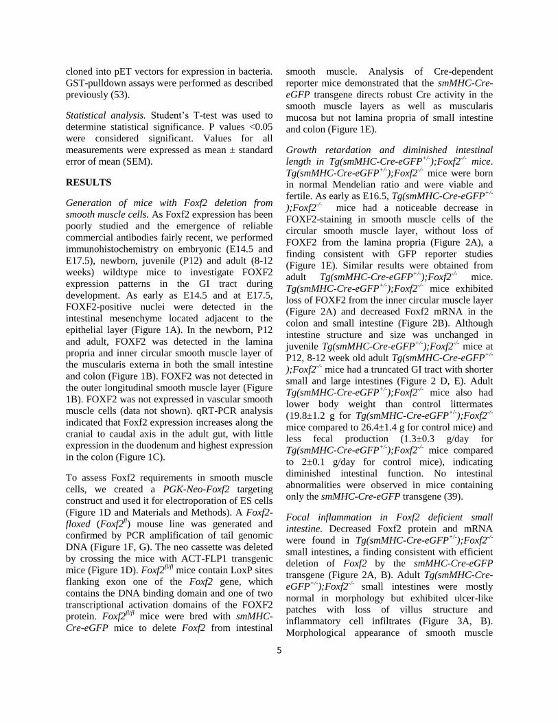
Generation of mice with Foxf2 deletion from
smooth muscle cells. As Foxf2 expression has been
poorly studied and the emergence of reliable
commercial antibodies fairly recent, we performed
immunohistochemistry on embryonic (E14.5 and
E17.5), newborn, juvenile (P12) and adult (8-12
weeks) wildtype mice to investigate FOXF2
expression patterns in the GI tract during
development. As early as E14.5 and at E17.5,
FOXF2-positive nuclei were detected in the
intestinal mesenchyme located adjacent to the
epithelial layer (Figure 1A). In the newborn, P12
and adult, FOXF2 was detected in the lamina
propria and inner circular smooth muscle layer of
the muscularis externa in both the small intestine
and colon (Figure 1B). FOXF2 was not detected in
the outer longitudinal smooth muscle layer (Figure
1B). FOXF2 was not expressed in vascular smooth
muscle cells (data not shown). qRT-PCR analysis
indicated that Foxf2 expression increases along the
cranial to caudal axis in the adult gut, with little
expression in the duodenum and highest expression
in the colon (Figure 1C).
To assess Foxf2 requirements in smooth muscle
cells, we created a PGK-Neo-Foxf2 targeting
construct and used it for electroporation of ES cells
(Figure 1D and Materials and Methods). A Foxf2-
floxed (Foxf2fl) mouse line was generated and
confirmed by PCR amplification of tail genomic
DNA (Figure 1F, G). The neo cassette was deleted
by crossing the mice with ACT-FLP1 transgenic
mice (Figure 1D). Foxf2fl/fl
mice contain LoxP sites
flanking exon one of the Foxf2 gene, which
contains the DNA binding domain and one of two
transcriptional activation domains of the FOXF2
protein. Foxf2fl/fl
mice were bred with smMHC-
Cre-eGFP mice to delete Foxf2 from intestinal
smooth muscle. Analysis of Cre-dependent
reporter mice demonstrated that the smMHC-Cre-
eGFP transgene directs robust Cre activity in the
smooth muscle layers as well as muscularis
mucosa but not lamina propria of small intestine
and colon (Figure 1E).
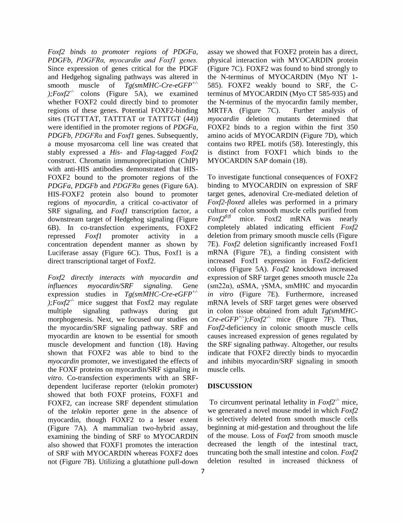
Growth retardation and diminished intestinal
length in Tg(smMHC-Cre-eGFP+/-
);Foxf2-/-
mice.
Tg(smMHC-Cre-eGFP+/-
);Foxf2-/-
mice were born
in normal Mendelian ratio and were viable and
fertile. As early as E16.5, Tg(smMHC-Cre-eGFP+/-
);Foxf2-/-
mice had a noticeable decrease in
FOXF2-staining in smooth muscle cells of the
circular smooth muscle layer, without loss of
FOXF2 from the lamina propria (Figure 2A), a
finding consistent with GFP reporter studies
(Figure 1E). Similar results were obtained from
adult Tg(smMHC-Cre-eGFP+/-
);Foxf2-/-
mice.
Tg(smMHC-Cre-eGFP+/-
);Foxf2-/-
mice exhibited
loss of FOXF2 from the inner circular muscle layer
(Figure 2A) and decreased Foxf2 mRNA in the
colon and small intestine (Figure 2B). Although
intestine structure and size was unchanged in
juvenile Tg(smMHC-Cre-eGFP+/-
);Foxf2-/-
mice at
P12, 8-12 week old adult Tg(smMHC-Cre-eGFP+/-
);Foxf2-/-
mice had a truncated GI tract with shorter
small and large intestines (Figure 2 D, E). Adult
Tg(smMHC-Cre-eGFP+/-
);Foxf2-/-
mice also had
lower body weight than control littermates
(19.8±1.2 g for Tg(smMHC-Cre-eGFP+/-
);Foxf2-/-
mice compared to 26.4±1.4 g for control mice) and
less fecal production (1.3±0.3 g/day for
Tg(smMHC-Cre-eGFP+/-
);Foxf2-/-
mice compared
to 2±0.1 g/day for control mice), indicating
diminished intestinal function. No intestinal
abnormalities were observed in mice containing
only the smMHC-Cre-eGFP transgene (39).
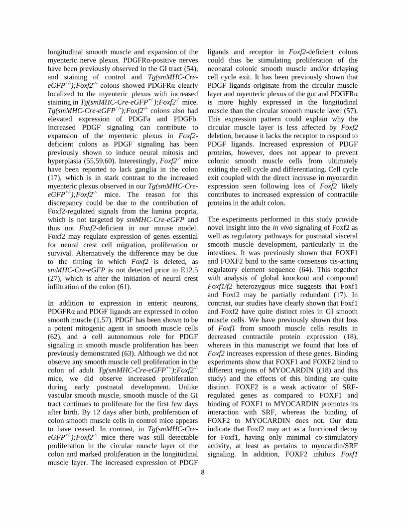
Focal inflammation in Foxf2 deficient small
intestine. Decreased Foxf2 protein and mRNA
were found in Tg(smMHC-Cre-eGFP+/-
);Foxf2-/-
small intestines, a finding consistent with efficient
deletion of Foxf2 by the smMHC-Cre-eGFP
transgene (Figure 2A, B). Adult Tg(smMHC-Cre-
eGFP+/-
);Foxf2-/-
small intestines were mostly
normal in morphology but exhibited ulcer-like
patches with loss of villus structure and
inflammatory cell infiltrates (Figure 3A, B).
Morphological appearance of smooth muscle

6
layers was normal (Figure 3A). Interestingly, the
small intestine of juvenile (P12) and embryonic
(E17.5) Tg(smMHC-Cre-eGFP+/-
);Foxf2-/-
mice
were morphologically normal (Figure 3C and data
not shown), indicating that Foxf2 is important for
maintaining intestinal structure in adults.
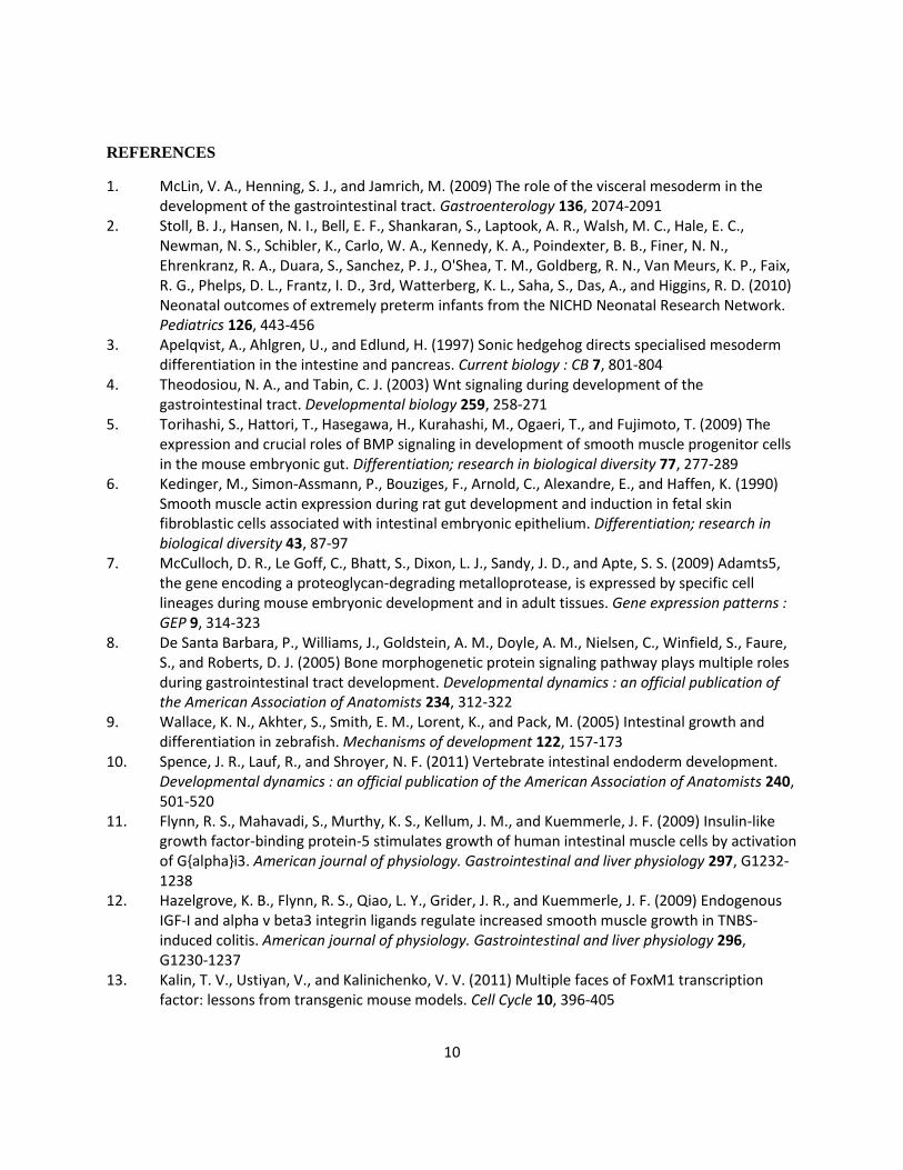
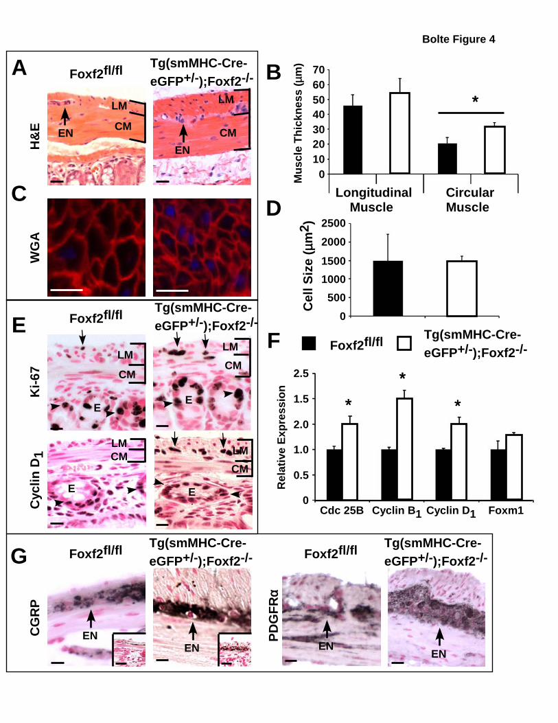
Increased smooth muscle cell proliferation in
Foxf2 deficient colons. The outer longitudinal
smooth muscle layer was significantly thicker in
adult Tg(smMHC-Cre-eGFP+/-
);Foxf2-/-
colons
compared to control littermates (Figure 4A, B and
2C). Thicker muscle in the colon of Tg(smMHC-
Cre-eGFP+/-
);Foxf2-/-
mice was not due to
pronounced hypertrophy as there was no detectable
difference in myocyte size between control and
Tg(smMHC-Cre-eGFP+/-
);Foxf2-/-
mice (Figure
4C, D). This would suggest that increased muscle
thickness was due to hyperplasia. Although no
myocyte proliferation was observed in adult
colons, neonatal P12 Tg(smMHC-Cre-eGFP+/-
);Foxf2-/-
mice exhibited increased Ki-67 and
cyclin D1 staining in myocytes from the outer
longitudinal smooth muscle layer (Figure 4E).
qRT-PCR analysis confirmed increased expression
of the cell cycle regulators Cdc 25B, cyclin B1 and
cyclin D1 in P12 Tg(smMHC-Cre-eGFP+/-
);Foxf2-/-
mice compared to age-matched controls (Figure
4F). Therefore, increased smooth muscle thickness
in Tg(smMHC-Cre-eGFP+/-
);Foxf2-/-
colons likely
results from increased or prolonged myocyte
proliferation during postnatal development and not
from hypertrophy of individual myocytes.
Expansion of the myenteric plexus in Tg(smMHC-
Cre-eGFP+/-
);Foxf2-/-
colons. Tg(smMHC-Cre-
eGFP+/-
);Foxf2-/-
mice also had an expanded
myenteric nerve plexus. Immunohistochemical
staining using the calcitonin-related gene protein
(CGRP), which marks serotonergic neurons,
showed a clearly enlarged network of enteric
neurons in adult Tg(smMHC-Cre-eGFP+/-
);Foxf2-/-
mice (Figure 4G). Greater CGRP staining of the
myenteric plexus was also observed at P12, prior
to thickening of the longitudinal muscle layer
(Figure 4G inset). Expansion of the myenteric
plexus in Tg(smMHC-Cre-eGFP+/-
);Foxf2-/-
mice
was confirmed by immunostaining with antibodies
against the platelet-derived growth factor receptor
alpha (PDGFRα) (Figure 4G) which is highly
expressed in neurons (54-56) and weakly
expressed in smooth muscle cells (57). Consistent
with increased PDGFRα staining, qRT-PCR
analysis showed increased expression of PDGFRα,
as well as its ligands PDGFa and PDGFb, in
Tg(smMHC-Cre-eGFP+/-
);Foxf2-/-
colons (Figure
5A).
Altered expression of genes critical for gut
morphogenesis in Tg(smMHC-Cre-eGFP+/-
);Foxf2-/-
colons. To determine molecular
mechanisms underlying increased proliferation in
Foxf2-deficient colons, we examined expression of
genes critical for the Hedgehog pathway, which
has been shown to be important for proliferation of
smooth muscle cells and their mesenchymal
precursors (3,4,9). Indian Hedgehog (IHH) mRNA
was increased in adult Tg(smMHC-Cre-eGFP+/-
);Foxf2-/-
colons, but was not changed at P12
(Figure 5B, data not shown). In agreement with
increased Hedgehog signaling there was increased
mRNA expression of Hedgehog target genes
Foxf1, Gli1, Gli2 and the Hedgehog receptor
Patched in smooth muscle-enriched tissue from
Tg(smMHC-Cre-eGFP+/-
);Foxf2-/-
mice (Figure
5A). Considerably more FOXF1-positive myocytes
were observed in the colon of Tg(smMHC-Cre-
eGFP+/-
);Foxf2-/-
compared to control mice (Figure
5C), further indicating increased Hedgehog
signaling. qRT-PCR analysis showed elevated
mRNA levels of Wnt5a, BMP4 and myocardin
(Figure 5A), factors critical for gut development.
Despite significant alterations in smooth muscle
regulatory genes in Tg(smMHC-Cre-eGFP+/-
);Foxf2-/-
mice, muscle markers αSMA and γSMA
were unaltered following Foxf2 deletion (Figure
5C), indicating that Foxf2-deficient smooth muscle
cells are fully differentiated. No fibrosis was
detected in Tg(smMHC-Cre-eGFP+/-
);Foxf2-/-
mice
as visualized by Masson’s trichrome stain (Figure
5C). In addition, there was no observed difference
in vascularization of the muscle layers of the colon
as indicated by PECAM-1 immunostaining (Figure
5D). Furthermore, neither FOXA2 nor
CYTOKERATIN immunostaining demonstrated
any difference in the structure of colon epithelium
(Figure 5D). Altogether, our data indicate that
Foxf2 deletion from smooth muscle cells alters
expression of genes critical for intestinal
morphogenesis.
7
Foxf2 binds to promoter regions of PDGFa,
PDGFb, PDGFRα, myocardin and Foxf1 genes.
Since expression of genes critical for the PDGF
and Hedgehog signaling pathways was altered in
smooth muscle of Tg(smMHC-Cre-eGFP+/-
);Foxf2-/-
colons (Figure 5A), we examined
whether FOXF2 could directly bind to promoter
regions of these genes. Potential FOXF2-binding
sites (TGTTTAT, TATTTAT or TATTTGT (44))
were identified in the promoter regions of PDGFa,
PDGFb, PDGFRα and Foxf1 genes. Subsequently,
a mouse myosarcoma cell line was created that
stably expressed a His- and Flag-tagged Foxf2
construct. Chromatin immunoprecipitation (ChIP)
with anti-HIS antibodies demonstrated that HIS-
FOXF2 bound to the promoter regions of the
PDGFa, PDGFb and PDGFRα genes (Figure 6A).
HIS-FOXF2 protein also bound to promoter
regions of myocardin, a critical co-activator of
SRF signaling, and Foxf1 transcription factor, a
downstream target of Hedgehog signaling (Figure
6B). In co-transfection experiments, FOXF2
repressed Foxf1 promoter activity in a
concentration dependent manner as shown by
Luciferase assay (Figure 6C). Thus, Foxf1 is a
direct transcriptional target of Foxf2.
Foxf2 directly interacts with myocardin and
influences myocardin/SRF signaling. Gene
expression studies in Tg(smMHC-Cre-eGFP+/-
);Foxf2-/-
mice suggest that Foxf2 may regulate
multiple signaling pathways during gut
morphogenesis. Next, we focused our studies on
the myocardin/SRF signaling pathway. SRF and
myocardin are known to be essential for smooth
muscle development and function (18). Having
shown that FOXF2 was able to bind to the
myocardin promoter, we investigated the effects of
the FOXF proteins on myocardin/SRF signaling in
vitro. Co-transfection experiments with an SRF-
dependent luciferase reporter (telokin promoter)
showed that both FOXF proteins, FOXF1 and
FOXF2, can increase SRF dependent stimulation
of the telokin reporter gene in the absence of
myocardin, though FOXF2 to a lesser extent
(Figure 7A). A mammalian two-hybrid assay,
examining the binding of SRF to MYOCARDIN
also showed that FOXF1 promotes the interaction
of SRF with MYOCARDIN whereas FOXF2 does
not (Figure 7B). Utilizing a glutathione pull-down
assay we showed that FOXF2 protein has a direct,
physical interaction with MYOCARDIN protein
(Figure 7C). FOXF2 was found to bind strongly to
the N-terminus of MYOCARDIN (Myo NT 1-
585). FOXF2 weakly bound to SRF, the C-
terminus of MYOCARDIN (Myo CT 585-935) and
the N-terminus of the myocardin family member,
MRTFA (Figure 7C). Further analysis of
myocardin deletion mutants determined that
FOXF2 binds to a region within the first 350
amino acids of MYOCARDIN (Figure 7D), which
contains two RPEL motifs (58). Interestingly, this
is distinct from FOXF1 which binds to the
MYOCARDIN SAP domain (18).
To investigate functional consequences of FOXF2
binding to MYOCARDIN on expression of SRF
target genes, adenoviral Cre-mediated deletion of
Foxf2-floxed alleles was performed in a primary
culture of colon smooth muscle cells purified from
Foxf2fl/fl
mice. Foxf2 mRNA was nearly
completely ablated indicating efficient Foxf2
deletion from primary smooth muscle cells (Figure
7E). Foxf2 deletion significantly increased Foxf1
mRNA (Figure 7E), a finding consistent with
increased Foxf1 expression in Foxf2-deficient
colons (Figure 5A). Foxf2 knockdown increased
expression of SRF target genes smooth muscle 22α
(sm22α), αSMA, γSMA, smMHC and myocardin
in vitro (Figure 7E). Furthermore, increased
mRNA levels of SRF target genes were observed
in colon tissue obtained from adult Tg(smMHC-
Cre-eGFP+/-
);Foxf2-/-
mice (Figure 7F). Thus,
Foxf2-deficiency in colonic smooth muscle cells
causes increased expression of genes regulated by
the SRF signaling pathway. Altogether, our results
indicate that FOXF2 directly binds to myocardin
and inhibits myocardin/SRF signaling in smooth
muscle cells.
DISCUSSION
To circumvent perinatal lethality in Foxf2-/-
mice,
we generated a novel mouse model in which Foxf2
is selectively deleted from smooth muscle cells
beginning at mid-gestation and throughout the life
of the mouse. Loss of Foxf2 from smooth muscle
decreased the length of the intestinal tract,
truncating both the small intestine and colon. Foxf2
deletion resulted in increased thickness of
8
longitudinal smooth muscle and expansion of the
myenteric nerve plexus. PDGFRα-positive nerves
have been previously observed in the GI tract (54),
and staining of control and Tg(smMHC-Cre-
eGFP+/-
);Foxf2-/-
colons showed PDGFRα clearly
localized to the myenteric plexus with increased
staining in Tg(smMHC-Cre-eGFP+/-
);Foxf2-/-
mice.
Tg(smMHC-Cre-eGFP+/-
);Foxf2-/-
colons also had
elevated expression of PDGFa and PDGFb.
Increased PDGF signaling can contribute to
expansion of the myenteric plexus in Foxf2-
deficient colons as PDGF signaling has been
previously shown to induce neural mitosis and
hyperplasia (55,59,60). Interestingly, Foxf2-/-
mice
have been reported to lack ganglia in the colon
(17), which is in stark contrast to the increased
myenteric plexus observed in our Tg(smMHC-Cre-
eGFP+/-
);Foxf2-/-
mice. The reason for this
discrepancy could be due to the contribution of
Foxf2-regulated signals from the lamina propria,
which is not targeted by smMHC-Cre-eGFP and
thus not Foxf2-deficient in our mouse model.
Foxf2 may regulate expression of genes essential
for neural crest cell migration, proliferation or
survival. Alternatively the difference may be due
to the timing in which Foxf2 is deleted, as
smMHC-Cre-eGFP is not detected prior to E12.5
(27), which is after the initiation of neural crest
infiltration of the colon (61).
In addition to expression in enteric neurons,
PDGFRα and PDGF ligands are expressed in colon
smooth muscle (1,57). PDGF has been shown to be
a potent mitogenic agent in smooth muscle cells
(62), and a cell autonomous role for PDGF
signaling in smooth muscle proliferation has been
previously demonstrated (63). Although we did not
observe any smooth muscle cell proliferation in the
colon of adult Tg(smMHC-Cre-eGFP+/-
);Foxf2-/-
mice, we did observe increased proliferation
during early postnatal development. Unlike
vascular smooth muscle, smooth muscle of the GI
tract continues to proliferate for the first few days
after birth. By 12 days after birth, proliferation of
colon smooth muscle cells in control mice appears
to have ceased. In contrast, in Tg(smMHC-Cre-
eGFP+/-
);Foxf2-/-
mice there was still detectable
proliferation in the circular muscle layer of the
colon and marked proliferation in the longitudinal
muscle layer. The increased expression of PDGF
ligands and receptor in Foxf2-deficient colons
could thus be stimulating proliferation of the
neonatal colonic smooth muscle and/or delaying
cell cycle exit. It has been previously shown that
PDGF ligands originate from the circular muscle
layer and myenteric plexus of the gut and PDGFRα
is more highly expressed in the longitudinal
muscle than the circular smooth muscle layer (57).
This expression pattern could explain why the
circular muscle layer is less affected by Foxf2
deletion, because it lacks the receptor to respond to
PDGF ligands. Increased expression of PDGF
proteins, however, does not appear to prevent
colonic smooth muscle cells from ultimately
exiting the cell cycle and differentiating. Cell cycle
exit coupled with the direct increase in myocardin
expression seen following loss of Foxf2 likely
contributes to increased expression of contractile
proteins in the adult colon.
The experiments performed in this study provide
novel insight into the in vivo signaling of Foxf2 as
well as regulatory pathways for postnatal visceral
smooth muscle development, particularly in the
intestines. It was previously shown that FOXF1
and FOXF2 bind to the same consensus cis-acting
regulatory element sequence (64). This together
with analysis of global knockout and compound
Foxf1/f2 heterozygous mice suggests that Foxf1
and Foxf2 may be partially redundant (17). In
contrast, our studies have clearly shown that Foxf1
and Foxf2 have quite distinct roles in GI smooth
muscle cells. We have previously shown that loss
of Foxf1 from smooth muscle cells results in
decreased contractile protein expression (18),
whereas in this manuscript we found that loss of
Foxf2 increases expression of these genes. Binding
experiments show that FOXF1 and FOXF2 bind to
different regions of MYOCARDIN ((18) and this
study) and the effects of this binding are quite
distinct. FOXF2 is a weak activator of SRF-
regulated genes as compared to FOXF1 and
binding of FOXF1 to MYOCARDIN promotes its
interaction with SRF, whereas the binding of
FOXF2 to MYOCARDIN does not. Our data
indicate that Foxf2 may act as a functional decoy
for Foxf1, having only minimal co-stimulatory
activity, at least as pertains to myocardin/SRF
signaling. In addition, FOXF2 inhibits Foxf1

9
promoter activity, which can further exacerbate
diminished myocardin/SRF signaling.
In addition to regulating the activity of
MYOCARDIN/SRF complexes through protein-
protein interactions, FOXF2 also directly affects
the expression of myocardin through binding to a
forkhead site in the myocardin promoter. ChIP
analysis showed that FOXF2 can bind to the
promoter region of myocardin, implicating Foxf2
in transcriptional regulation of the myocardin gene.
Our data indicates that Foxf2 influences GI tract
development via regulation of PDGF, SRF and
Hedgehog pathways, all of which have been shown
to be critical for GI tract development and
physiology. Foxf2 may directly or indirectly
regulate expression of genes critical for all three
pathways and through this regulation mediate
postnatal intestinal development and maintenance.
In summary, we generated a novel mouse line
containing Foxf2-floxed alleles and used it to
create smooth muscle-specific Foxf2 knockout
mice. Utilizing this model we demonstrated a
distinct phenotype for smooth muscle Foxf2
deletion compared to Foxf2-/-
mice. We also
showed that FOXF2 bound to the promoter region
of the myocardin gene as well as several members
of the PDGF and Hedgehog signaling pathways.
FOXF2 directly binds to and inhibits Foxf1
promoter activity and regulates SRF signaling
through a protein-protein interaction with
MYOCARDIN. Via its capacity to regulate PDGF,
Hedgehog and SRF signaling, Foxf2 is a critical
regulator of GI tract development and
maintenance.
10
REFERENCES
1. McLin, V. A., Henning, S. J., and Jamrich, M. (2009) The role of the visceral mesoderm in the development of the gastrointestinal tract. Gastroenterology 136, 2074-2091
2. Stoll, B. J., Hansen, N. I., Bell, E. F., Shankaran, S., Laptook, A. R., Walsh, M. C., Hale, E. C., Newman, N. S., Schibler, K., Carlo, W. A., Kennedy, K. A., Poindexter, B. B., Finer, N. N., Ehrenkranz, R. A., Duara, S., Sanchez, P. J., O'Shea, T. M., Goldberg, R. N., Van Meurs, K. P., Faix, R. G., Phelps, D. L., Frantz, I. D., 3rd, Watterberg, K. L., Saha, S., Das, A., and Higgins, R. D. (2010) Neonatal outcomes of extremely preterm infants from the NICHD Neonatal Research Network. Pediatrics 126, 443-456
3. Apelqvist, A., Ahlgren, U., and Edlund, H. (1997) Sonic hedgehog directs specialised mesoderm differentiation in the intestine and pancreas. Current biology : CB 7, 801-804
4. Theodosiou, N. A., and Tabin, C. J. (2003) Wnt signaling during development of the gastrointestinal tract. Developmental biology 259, 258-271
5. Torihashi, S., Hattori, T., Hasegawa, H., Kurahashi, M., Ogaeri, T., and Fujimoto, T. (2009) The expression and crucial roles of BMP signaling in development of smooth muscle progenitor cells in the mouse embryonic gut. Differentiation; research in biological diversity 77, 277-289
6. Kedinger, M., Simon-Assmann, P., Bouziges, F., Arnold, C., Alexandre, E., and Haffen, K. (1990) Smooth muscle actin expression during rat gut development and induction in fetal skin fibroblastic cells associated with intestinal embryonic epithelium. Differentiation; research in biological diversity 43, 87-97
7. McCulloch, D. R., Le Goff, C., Bhatt, S., Dixon, L. J., Sandy, J. D., and Apte, S. S. (2009) Adamts5, the gene encoding a proteoglycan-degrading metalloprotease, is expressed by specific cell lineages during mouse embryonic development and in adult tissues. Gene expression patterns : GEP 9, 314-323
8. De Santa Barbara, P., Williams, J., Goldstein, A. M., Doyle, A. M., Nielsen, C., Winfield, S., Faure, S., and Roberts, D. J. (2005) Bone morphogenetic protein signaling pathway plays multiple roles during gastrointestinal tract development. Developmental dynamics : an official publication of the American Association of Anatomists 234, 312-322
9. Wallace, K. N., Akhter, S., Smith, E. M., Lorent, K., and Pack, M. (2005) Intestinal growth and differentiation in zebrafish. Mechanisms of development 122, 157-173
10. Spence, J. R., Lauf, R., and Shroyer, N. F. (2011) Vertebrate intestinal endoderm development. Developmental dynamics : an official publication of the American Association of Anatomists 240, 501-520
11. Flynn, R. S., Mahavadi, S., Murthy, K. S., Kellum, J. M., and Kuemmerle, J. F. (2009) Insulin-like growth factor-binding protein-5 stimulates growth of human intestinal muscle cells by activation of G{alpha}i3. American journal of physiology. Gastrointestinal and liver physiology 297, G1232-1238
12. Hazelgrove, K. B., Flynn, R. S., Qiao, L. Y., Grider, J. R., and Kuemmerle, J. F. (2009) Endogenous IGF-I and alpha v beta3 integrin ligands regulate increased smooth muscle growth in TNBS-induced colitis. American journal of physiology. Gastrointestinal and liver physiology 296, G1230-1237
13. Kalin, T. V., Ustiyan, V., and Kalinichenko, V. V. (2011) Multiple faces of FoxM1 transcription factor: lessons from transgenic mouse models. Cell Cycle 10, 396-405

11
14. Ormestad, M., Astorga, J., and Carlsson, P. (2004) Differences in the embryonic expression patterns of mouse Foxf1 and -2 match their distinct mutant phenotypes. Developmental dynamics : an official publication of the American Association of Anatomists 229, 328-333
15. Kalinichenko, V. V., Lim, L., Stolz, D. B., Shin, B., Rausa, F. M., Clark, J., Whitsett, J. A., Watkins, S. C., and Costa, R. H. (2001) Defects in pulmonary vasculature and perinatal lung hemorrhage in mice heterozygous null for the Forkhead Box f1 transcription factor. Developmental biology 235, 489-506
16. Mahlapuu, M., Ormestad, M., Enerback, S., and Carlsson, P. (2001) The forkhead transcription factor Foxf1 is required for differentiation of extra-embryonic and lateral plate mesoderm. Development 128, 155-166
17. Ormestad, M., Astorga, J., Landgren, H., Wang, T., Johansson, B. R., Miura, N., and Carlsson, P. (2006) Foxf1 and Foxf2 control murine gut development by limiting mesenchymal Wnt signaling and promoting extracellular matrix production. Development 133, 833-843
18. Hoggatt, A. M., Kim, J. R., Ustiyan, V., Ren, X., Kalin, T. V., Kalinichenko, V. V., and Herring, B. P. (2013) The transcription factor Foxf1 binds to serum response factor and myocardin to regulate gene transcription in visceral smooth muscle cells. The Journal of biological chemistry
19. Hoggatt, A. M., Kriegel, A. M., Smith, A. F., and Herring, B. P. (2000) Hepatocyte nuclear factor-3 homologue 1 (HFH-1) represses transcription of smooth muscle-specific genes. The Journal of biological chemistry 275, 31162-31170
20. Liu, Z. P., Wang, Z., Yanagisawa, H., and Olson, E. N. (2005) Phenotypic modulation of smooth muscle cells through interaction of Foxo4 and myocardin. Developmental cell 9, 261-270
21. Aitola, M., Carlsson, P., Mahlapuu, M., Enerback, S., and Pelto-Huikko, M. (2000) Forkhead transcription factor FoxF2 is expressed in mesodermal tissues involved in epithelio-mesenchymal interactions. Developmental dynamics : an official publication of the American Association of Anatomists 218, 136-149
22. McLin, V. A., Shah, R., Desai, N. P., and Jamrich, M. (2010) Identification and gastrointestinal expression of Xenopus laevis FoxF2. The International journal of developmental biology 54, 919-924
23. van der Heul-Nieuwenhuijsen, L., Dits, N., Van Ijcken, W., de Lange, D., and Jenster, G. (2009) The FOXF2 pathway in the human prostate stroma. The Prostate 69, 1538-1547
24. Nik, A. M., Reyahi, A., Ponten, F., and Carlsson, P. (2013) Foxf2 in intestinal fibroblasts reduces numbers of Lgr5(+) stem cells and adenoma formation by inhibiting Wnt signaling. Gastroenterology 144, 1001-1011
25. Philip-Couderc, P., Tavares, N. I., Roatti, A., Lerch, R., Montessuit, C., and Baertschi, A. J. (2008) Forkhead transcription factors coordinate expression of myocardial KATP channel subunits and energy metabolism. Circulation research 102, e20-35
26. McKeone, R., Vieira, H., Gregory-Evans, K., Gregory-Evans, C. Y., and Denny, P. (2011) Foxf2: a novel locus for anterior segment dysgenesis adjacent to the Foxc1 gene. PloS one 6, e25489
27. Xin, H. B., Deng, K. Y., Rishniw, M., Ji, G., and Kotlikoff, M. I. (2002) Smooth muscle expression of Cre recombinase and eGFP in transgenic mice. Physiological genomics 10, 211-215
28. Hoggatt, A. M., Kim, J. R., Ustiyan, V., Ren, X., Kalin, T. V., Kalinichenko, V. V., and Herring, B. P. (2013) The transcription factor Foxf1 binds to serum response factor and myocardin to regulate gene transcription in visceral smooth muscle cells. The Journal of biological chemistry 288, 28477-28487
29. Malin, D., Kim, I. M., Boetticher, E., Kalin, T. V., Ramakrishna, S., Meliton, L., Ustiyan, V., Zhu, X., and Kalinichenko, V. V. (2007) Forkhead box F1 is essential for migration of mesenchymal cells and directly induces integrin-beta3 expression. Molecular and cellular biology 27, 2486-2498

12
30. Kalin, T. V., Wang, I. C., Meliton, L., Zhang, Y., Wert, S. E., Ren, X., Snyder, J., Bell, S. M., Graf, L., Jr., Whitsett, J. A., and Kalinichenko, V. V. (2008) Forkhead Box m1 transcription factor is required for perinatal lung function. Proceedings of the National Academy of Sciences of the United States of America 105, 19330-19335
31. Kim, I. M., Ramakrishna, S., Gusarova, G. A., Yoder, H. M., Costa, R. H., and Kalinichenko, V. V. (2005) The forkhead box m1 transcription factor is essential for embryonic development of pulmonary vasculature. The Journal of biological chemistry 280, 22278-22286
32. Kim, I. M., Zhou, Y., Ramakrishna, S., Hughes, D. E., Solway, J., Costa, R. H., and Kalinichenko, V. V. (2005) Functional characterization of evolutionarily conserved DNA regions in forkhead box f1 gene locus. The Journal of biological chemistry 280, 37908-37916
33. Ramakrishna, S., Kim, I. M., Petrovic, V., Malin, D., Wang, I. C., Kalin, T. V., Meliton, L., Zhao, Y. Y., Ackerson, T., Qin, Y., Malik, A. B., Costa, R. H., and Kalinichenko, V. V. (2007) Myocardium defects and ventricular hypoplasia in mice homozygous null for the Forkhead Box M1 transcription factor. Developmental dynamics : an official publication of the American Association of Anatomists 236, 1000-1013
34. Balli, D., Zhang, Y., Snyder, J., Kalinichenko, V. V., and Kalin, T. V. (2011) Endothelial cell-specific deletion of transcription factor FoxM1 increases urethane-induced lung carcinogenesis. Cancer research 71, 40-50
35. Bolte, C., Newman, G., and Schultz Jel, J. (2009) Hypertensive state, independent of hypertrophy, exhibits an attenuated decrease in systolic function on cardiac kappa-opioid receptor stimulation. American journal of physiology. Heart and circulatory physiology 296, H967-975
36. Bolte, C., Zhang, Y., Wang, I. C., Kalin, T. V., Molkentin, J. D., and Kalinichenko, V. V. (2011) Expression of Foxm1 transcription factor in cardiomyocytes is required for myocardial development. PloS one 6, e22217
37. Ren, X., Shah, T. A., Ustiyan, V., Zhang, Y., Shinn, J., Chen, G., Whitsett, J. A., Kalin, T. V., and Kalinichenko, V. V. (2013) FOXM1 promotes allergen-induced goblet cell metaplasia and pulmonary inflammation. Molecular and cellular biology 33, 371-386
38. Ren, X., Zhang, Y., Snyder, J., Cross, E. R., Shah, T. A., Kalin, T. V., and Kalinichenko, V. V. (2010) Forkhead box M1 transcription factor is required for macrophage recruitment during liver repair. Molecular and cellular biology 30, 5381-5393
39. Ustiyan, V., Wang, I. C., Ren, X., Zhang, Y., Snyder, J., Xu, Y., Wert, S. E., Lessard, J. L., Kalin, T. V., and Kalinichenko, V. V. (2009) Forkhead box M1 transcriptional factor is required for smooth muscle cells during embryonic development of blood vessels and esophagus. Developmental biology 336, 266-279
40. Wang, I. C., Meliton, L., Ren, X., Zhang, Y., Balli, D., Snyder, J., Whitsett, J. A., Kalinichenko, V. V., and Kalin, T. V. (2009) Deletion of Forkhead Box M1 transcription factor from respiratory epithelial cells inhibits pulmonary tumorigenesis. PloS one 4, e6609
41. Balli, D., Ustiyan, V., Zhang, Y., Wang, I. C., Masino, A. J., Ren, X., Whitsett, J. A., Kalinichenko, V. V., and Kalin, T. V. (2013) Foxm1 transcription factor is required for lung fibrosis and epithelial-to-mesenchymal transition. The EMBO journal 32, 231-244
42. Cheng, X. H., Black, M., Ustiyan, V., Le, T., Fulford, L., Sridharan, A., Medvedovic, M., Kalinichenko, V. V., Whitsett, J. A., and Kalin, T. V. (2014) SPDEF inhibits prostate carcinogenesis by disrupting a positive feedback loop in regulation of the Foxm1 oncogene. PLoS genetics 10, e1004656
43. Singh, T. R., Saro, D., Ali, A. M., Zheng, X. F., Du, C. H., Killen, M. W., Sachpatzidis, A., Wahengbam, K., Pierce, A. J., Xiong, Y., Sung, P., and Meetei, A. R. (2010) MHF1-MHF2, a
13
histone-fold-containing protein complex, participates in the Fanconi anemia pathway via FANCM. Molecular cell 37, 879-886
44. Peterson, R. S., Lim, L., Ye, H., Zhou, H., Overdier, D. G., and Costa, R. H. (1997) The winged helix transcriptional activator HFH-8 is expressed in the mesoderm of the primitive streak stage of mouse embryos and its cellular derivatives. Mechanisms of development 69, 53-69
45. Wang, I. C., Snyder, J., Zhang, Y., Lander, J., Nakafuku, Y., Lin, J., Chen, G., Kalin, T. V., Whitsett, J. A., and Kalinichenko, V. V. (2012) Foxm1 mediates cross talk between Kras/mitogen-activated protein kinase and canonical Wnt pathways during development of respiratory epithelium. Molecular and cellular biology 32, 3838-3850
46. Zhou, J., Hoggatt, A. M., and Herring, B. P. (2004) Activation of the smooth muscle-specific telokin gene by thyrotroph embryonic factor (TEF). The Journal of biological chemistry 279, 15929-15937
47. Ren, X., Ustiyan, V., Pradhan, A., Cai, Y., Havrilak, J. A., Bolte, C. S., Shannon, J. M., Kalin, T. V., and Kalinichenko, V. V. (2014) FOXF1 transcription factor is required for formation of embryonic vasculature by regulating VEGF signaling in endothelial cells. Circulation research 115, 709-720
48. Wang, I. C., Ustiyan, V., Zhang, Y., Cai, Y., Kalin, T. V., and Kalinichenko, V. V. (2014) Foxm1 transcription factor is required for the initiation of lung tumorigenesis by oncogenic Kras(G12D.). Oncogene 33, 5391-5396
49. Cai, Y., Balli, D., Ustiyan, V., Fulford, L., Hiller, A., Misetic, V., Zhang, Y., Paluch, A. M., Waltz, S. E., Kasper, S., and Kalin, T. V. (2013) Foxm1 expression in prostate epithelial cells is essential for prostate carcinogenesis. The Journal of biological chemistry 288, 22527-22541
50. Zhou, J., Hu, G., and Herring, B. P. (2005) Smooth muscle-specific genes are differentially sensitive to inhibition by Elk-1. Molecular and cellular biology 25, 9874-9885
51. Herring, B. P., Kriegel, A. M., and Hoggatt, A. M. (2001) Identification of Barx2b, a serum response factor-associated homeodomain protein. The Journal of biological chemistry 276, 14482-14489
52. Zhang, M., Fang, H., Zhou, J., and Herring, B. P. (2007) A novel role of Brg1 in the regulation of SRF/MRTFA-dependent smooth muscle-specific gene expression. The Journal of biological chemistry 282, 25708-25716
53. Zhou, J., Zhang, M., Fang, H., El-Mounayri, O., Rodenberg, J. M., Imbalzano, A. N., and Herring, B. P. (2009) The SWI/SNF chromatin remodeling complex regulates myocardin-induced smooth muscle-specific gene expression. Arteriosclerosis, thrombosis, and vascular biology 29, 921-928
54. Blair, P. J., Bayguinov, Y., Sanders, K. M., and Ward, S. M. (2012) Relationship between enteric neurons and interstitial cells in the primate gastrointestinal tract. Neurogastroenterology and motility : the official journal of the European Gastrointestinal Motility Society 24, e437-449
55. Eccleston, P. A., Funa, K., and Heldin, C. H. (1993) Expression of platelet-derived growth factor (PDGF) and PDGF alpha- and beta-receptors in the peripheral nervous system: an analysis of sciatic nerve and dorsal root ganglia. Developmental biology 155, 459-470
56. Lobsiger, C. S., Schweitzer, B., Taylor, V., and Suter, U. (2000) Platelet-derived growth factor-BB supports the survival of cultured rat Schwann cell precursors in synergy with neurotrophin-3. Glia 30, 290-300
57. Kurahashi, M., Niwa, Y., Cheng, J., Ohsaki, Y., Fujita, A., Goto, H., Fujimoto, T., and Torihashi, S. (2008) Platelet-derived growth factor signals play critical roles in differentiation of longitudinal smooth muscle cells in mouse embryonic gut. Neurogastroenterology and motility : the official journal of the European Gastrointestinal Motility Society 20, 521-531
14
58. Guettler, S., Vartiainen, M. K., Miralles, F., Larijani, B., and Treisman, R. (2008) RPEL motifs link the serum response factor cofactor MAL but not myocardin to Rho signaling via actin binding. Molecular and cellular biology 28, 732-742
59. Fruttiger, M., Calver, A. R., and Richardson, W. D. (2000) Platelet-derived growth factor is constitutively secreted from neuronal cell bodies but not from axons. Current biology : CB 10, 1283-1286
60. Reneker, L. W., and Overbeek, P. A. (1996) Lens-specific expression of PDGF-A in transgenic mice results in retinal astrocytic hamartomas. Investigative ophthalmology & visual science 37, 2455-2466
61. Young, H. M., Turner, K. N., and Bergner, A. J. (2005) The location and phenotype of proliferating neural-crest-derived cells in the developing mouse gut. Cell and tissue research 320, 1-9
62. Stanzel, R. D., Lourenssen, S., Nair, D. G., and Blennerhassett, M. G. (2010) Mitogenic factors promoting intestinal smooth muscle cell proliferation. American journal of physiology. Cell physiology 299, C805-817
63. Graham, M. F., Willey, A., Adams, J., Yager, D., and Diegelmann, R. F. (1996) Interleukin 1 beta down-regulates collagen and augments collagenase expression in human intestinal smooth muscle cells. Gastroenterology 110, 344-350
64. Hellqvist, M., Mahlapuu, M., Samuelsson, L., Enerback, S., and Carlsson, P. (1996) Differential activation of lung-specific genes by two forkhead proteins, FREAC-1 and FREAC-2. The Journal of biological chemistry 271, 4482-4490
15
Acknowledgements
We would like to thank Yufang Zhang for excellent technical assistance.
FOOTNOTES
*These studies were supported by NIH grants HL84151 (V.V.K.), HL123490 (V.V.K.), CA142724
(T.V.K.) and DK061130 (B.P.H.).
1To whom correspondence should be addressed: Dr. Vladimir V. Kalinichenko
([email protected]) or Dr. Craig Bolte ([email protected]), Department of
Pediatrics, Perinatal Institute, Cincinnati Children’s Research Foundation, 3333 Burnet Ave., MLC 7009,
Cincinnati, OH 45229.
2Department of Cellular and Integrative Physiology, Indiana University School of Medicine, Indianapolis,
IN, 46202 USA.
3Abbreviations used: GI, gastrointestianal; Fox, Forkhead box; smMHC, smooth muscle myosin heavy
chain; P, postnatal day; E, embryonic day; ChIP, chromatin immunoprecipitation; qRT-PCR, quantitative
real-time reverse transcription polymerase chain reaction; CGRP, calcitonin gene-related peptide; PDGF,
platelet-derived growth factor.
FIGURE LEGENDS
Figure 1. Deletion of Foxf2 from GI smooth muscle. Foxf2 is expressed in the mouse GI tract from
embryonic time points through adulthood. (A-B) Immunostaining with anti-FOXF2 antibodies showed
restriction of FOXF2 to mesenchymal layers of the developing GI tract at E14.5 and E17.5. Postnatally,
Foxf2 was expressed in lamina propria [L] and circular muscle layer [C] but was absent from the
longitudinal muscle layer [LM] and epithelium [E]. (C) qRT-PCR analysis of adult whole organ mRNA
showed that Foxf2 is more highly expressed in the jejunum and ileum than the duodenum of the small
intestine and more highly expressed in the colon than small intestine. Expression levels were normalized
to β-actin (n=3). (D) Schematic shows Foxf2-floxed targeting construct. LoxP sites (solid triangles) were
inserted to flank the first exon [E1] of the Foxf2 gene. Locations of primers [P] and sizes of PCR products
are indicated. Primers used are listed in Table 1. Foxf2fl/fl
mice were bred with ACT-FLP1 mice to remove
neo and later with smMHC-Cre-eGFP mice to delete Foxf2 from smooth muscle cells. (E) Visualization
of immunofluorescence in frozen-fixed cross sections of adult ileum and colon from smMHC-Cre-eGFP
mice crossed with the Cre-dependent mT/mG reporter strain. Membrane bound eGFP is detected in the
smooth muscle layers as well as the muscularis mucosa, but not lamina propria. meGFP is expressed
stronger in the muscularis mucosa of colon compared to ileum. Abbreviations: CM, circular muscle. LM,
longitudinal muscle. MP, myenteric nerve plexus. MM, muscularis mucosa. SMF, submucosal
fibroblasts. EP, epithelium. (F-G) PCR analysis of mouse tail DNA shows the presence of neo cassette,
Cre transgene, as well as wildtype and Foxf2-floxed alleles. Scale bar = 10 μm.
Figure 2. Decreased Foxf2 and length of intestinal tract in Tg(smMHC-Cre-eGFP+/-
);Foxf2-/-
mice.
(A-B) FOXF2 staining and Foxf2 mRNA were significantly decreased in Tg(smMHC-Cre-eGFP+/-
);Foxf2-/-
embryonic intestines at E16.5 as well as adult small intestine and colon smooth muscle.
Expression levels were analyzed by qRT-PCR and normalized to β-actin (n=4). *p<0.05 versus control.
(C) Representative images of H&E stained adult small intestine and colon from Foxf2fl/fl
and Tg(smMHC-
Cre-eGFP+/-
);Foxf2-/-
mice are shown. (D) Gross view of intestines from adult Tg(smMHC-Cre-eGFP+/-
);Foxf2-/-
and control Foxf2fl/fl
mice indicated shorter intestinal tract in Foxf2-deficient mice. (E)
16
Measurements of intestinal tract confirmed it was significantly shorter in Tg(smMHC-Cre-eGFP+/-
);Foxf2-/-
mice due to decreased length of small and large intestines. (n=4). Scale bar = 10 μm in A, 100
μm in C.
Figure 3. Focal inflammation in Tg(smMHC-Cre-eGFP+/-
);Foxf2-/-
small intestine. (A) Adult
Tg(smMHC-Cre-eGFP+/-
);Foxf2-/-
small intestines had mostly normal morphology with focal regions of
inflammatory infiltration leading to disruption of villus structure, though muscle morphology remained
normal. (B) CYTOKERATIN (CK) staining shows disruption of villus epithelium in Tg(smMHC-Cre-
eGFP+/-
);Foxf2-/-
small intestines. (C) H&E staining of control and Foxf2-deficient small intestines
showed normal morphology at P12. Scale bar = 10 μm.
Figure 4. Increased muscle thickness and expanded myenteric plexus in Tg(smMHC-Cre-eGFP+/-
);Foxf2-/-
colons. (A) H&E staining indicated increased muscle thickness in adult Tg(smMHC-Cre-
eGFP+/-
);Foxf2-/-
colons. Enteric neurons [EN] are shown with arrows, whereas longitudinal [LM] and
circular [CM] smooth muscle layers are indicated by brackets. (B) Measurement of muscle thickness was
performed in 12 random colon sections with n=4 mice per group. The longitudinal muscle layer was
significantly thicker (*p<0.05 versus control) in Tg(smMHC-Cre-eGFP+/-
);Foxf2-/-
colons. (C) Wheat
germ agglutinin (WGA) staining and (D) measurement of cell size in adult mice eliminated hypertrophy
as the cause of muscle thickening. (E) Immunostaining with Ki-67 and cyclin D1 showed increased
myocyte proliferation (arrows) in Tg(smMHC-Cre-eGFP+/-
);Foxf2-/-
colons at P12. Longitudinal and
circular smooth muscle layers are shown with brackets while epithelial layer is indicated as E
(arrowheads show epithelial proliferation). (F) mRNA levels of Cdc 25B, cyclin B1 and cyclin D1 were
increased but Foxm1 mRNA was unaltered in P12 Tg(smMHC-Cre-eGFP+/-
);Foxf2-/-
colons. Expression
levels were determined by qRT-PCR and normalized to β-actin mRNA (n=4). (G) CGRP and PDGFRα
immunostaining showed expansion of myenteric plexus (arrows) in adult Tg(smMHC-Cre-eGFP+/-
);Foxf2-/-
colons. Insets show expanded CGRP staining in P12 Tg(smMHC-Cre-eGFP+/-
);Foxf2-/-
colons.
Scale bar = 10 μm.
Figure 5. Regulation of gene transcription by Foxf2. (A) Increased expression of Foxf1, Wnt, BMP, as
well as members of the PDGF, Hedgehog and SRF pathways in smooth muscle isolated from adult
Tg(smMHC-Cre-eGFP+/-
);Foxf2-/-
colons as determined by qRT-PCR. (B) Indian Hedgehog expression
was increased in whole colon RNA from adult Tg(smMHC-Cre-eGFP+/-
);Foxf2-/-
mice. Epidermal growth
factor receptor (EGFR) expression was unaltered. Expression levels were normalized to β-actin (n=4).
*p<0.05 versus control. (C) FOXF1-positive myocytes were more abundant in Tg(smMHC-Cre-eGFP+/-
);Foxf2-/-
smooth muscle than control myocytes as shown by immunostaining of the adult colon. Despite
prolonged proliferation and altered gene expression, muscle markers αSMA and γSMA were observed in
Tg(smMHC-Cre-eGFP+/-
);Foxf2-/-
colon. Fibrosis was not observed in adult Tg(smMHC-Cre-eGFP+/-
);Foxf2-/-
colons as determined by Masson’s Trichrome staining of intestinal paraffin sections. (D)
Vasculature was unaltered in adult Tg(smMHC-Cre-eGFP+/-
);Foxf2-/-
colons as visualized by PECAM-1
staining. Epithelial staining for FOXA2 and CYTOKERATIN was normal in adult colons. Scale bar = 10
μm.
Figure 6. FOXF2 binds to PDGF, myocardin and Foxf1 promoter regions. (A-B) FOXF2 binds to the
promoter regions of PDGFa, PDGFb, PDGFRα, myocardin and Foxf1 genes as determined by ChIP
analysis. ChIP was performed using a rhabdomyosarcoma cell line which stably expressed His- and Flag-
tagged FOXF2 protein. Anti-His antibodies or control IgG were used for ChIP. (n=3) Negative control for
ChIP included a DNA region 1.7 kb 3’ to the Foxf1 regulatory element (Foxf1 3’ Region). Anti-Flag
antibody was used for ChIP of Foxf1 to verify results obtained with anti-His antibody. (C) Luciferase
reporter gene assays show that FOXF2 inhibits transcriptional activity of the Foxf1 promoter (-5.3 kb
17
Foxf1 + 3’ RE) in a dose-dependent manner (left panel). Conversely FOXF2 activated a reporter gene
driven by multiple copies of a FOXF2 binding site (right panel). (n=3)
Figure 7. FOXF2 regulates myocardin/SRF signaling in smooth muscle cells. (A) FOXF1 and, to a
lesser extent, FOXF2 stimulate a SRF-dependent telokin reporter in the absence of myocardin. SRF,
Foxf1 and Foxf2 expression plasmids were co-transfected with the SRF-dependent telokin reporter gene
into 10TI1/2 cells. (B) Mammalian two-hybrid assay examining the binding between SRF and
MYOCARDIN in the presence or absence of FOXF1 or FOXF2. In these assays SRF was fused to the
GAL4 DNA binding domain [BD] and myocardin to the GAL4 activation domain [AD]. Interaction
between SRF-BD and MYOCARDIN-AD results in activation of a GAL4-dependent reporter gene
(SRF/Myo+vector compared to SRF/pACT+vector: pACT- empty GAL4 activation domain vector).
FOXF1 promotes MYOCARDIN binding to SRF while FOXF2 has no effect on MYOCARDIN/SRF
binding. (C) GST-pull down assays in which bacterial expressed FOXF2 was incubated with SRF,
MYOCARDIN or MRTFA-GST fusion proteins. Following extensive washing, glutathione bound fusion
proteins were analyzed by Western blot (upper panel). Input GST-fusion proteins visualized by Ponceau
staining are shown in the lower panel. FOXF2 protein physically binds to SRF, the N-terminal half of
MYOCARDIN (Myo NT 1-585) more weakly to the C-terminal half of MYOCARDIN (Myo CT 585-
935) and weakly with the N-terminal half of MRTFA but not the C-terminal half of MRTFA. (D) Upper
panel, schematic representation of MYOCARDIN structural domains. Middle panel, GST-pulldown
assays using various fragments of MYOCARDIN fused to GST. The GST-fusion protein bound FOXF2
is shown in the middle panel and GST-fusion protein inputs in the lower panel. FOXF2 specifically bound
to the first 220 amino acids of the MYOCARDIN protein, which contains RPEL motifs. Weak binding
was also observed to amino acids 221-350 of MYOCARDIN. (E) Adenoviral Cre-mediated deletion of
Foxf2fl alleles from primary colon smooth muscle cells increased mRNA levels of Foxf1 as well as
expression of myocardin/SRF target genes SM22α, αSMA, γSMA, smMHC and myocardin. mRNA
expression levels were quantitated by qRT-PCR and normalized to β-actin (n=6). *p<0.05 versus control.
(F) mRNA levels of myocardin/SRF target genes SM22α, αSMA, γSMA and smMHC were increased in
adult Tg(smMHC-Cre-eGFP+/-
);Foxf2-/-
mouse colons (n=4).
18
Table1. Foxf2-floxed allele mouse primers
Primer name Sequence
P1 TGGAGGAGTGTTCTCGAATGGAG
P2 CACTGGACGCCCTTGAGCTG
P3 CCAGAGGCCACTTGTGTAGC
P4 AAAGCTTGTGTCTAGCGGTTGTCCAC
P5 AGCATGTCTTCCTACTCGTTGGAG
P6 GCTCCTGCCGAGAAAGTATCC
Primer sets in table were used for genotyping from mouse tail DNA.
19
Table 2. Taqman Probes
Gene name Assay number
acta2 Mm00725412_s1
actb Mm00607939_s1
actg2 Mm00656102_m1
bmp4 Mm01321704_m1
ccnb1 Mm00838401_g1
ccnd1 Mm00432359_m1
cdc25b Mm00499136_m1
Foxf1 Mm00487497_m1
Foxf2 Mm00515793_m1
Foxm1 Mm00514924_m1
gli1 Mm01160468_g1
gli2 Mm01293116_m1
myh11 Mm00443013_m1
myocd Mm00455051_m1
pdgfa Mm01205760_m1
pdgfb Mm01298578_m1
pdgfra Mm01211694_m1
ptch1 Mm00436026_m1
sox9 Mm00448840_m1
srf Mm00491032_m1
tagln Mm00441661_g1
wnt5a Mm00437347_m1
Table lists inventoried Taqman probes used for qRT-PCR analysis of gene expression.

20
Table 3. ChIP Primers
Gene (Promoter
region) Forward primer (5’-3’) Reverse primer (5’-3’)
Foxf1 + Enhancer
(248/255) CCCCACCCCTAACGGATTATTTG TCCCCTGCTTCTAAAAACTGTCCC
Foxf13’Region
(Negative
Control)
ACCACCATCTCCATCACCAGCC CCTCCCGTCCAGAATGTCAAGTC
Myocardin
(-4532/-4525 & -
4523/-4516)
CCTGGGACCACACAAACAAC AGGCTCCGTCTCAGCGTG
PDGFa
(-6421/-6414) CCCATCATCCTCTCCTCTTTGC GGTAGCCTTTAGAAATGTGTGCCAG
PDGFb
(-1435/-1428) TAGATGAGTTCTGGGACTGGACT AGACATAACCGGAGGAGAAGAAG
PDGFRα
(-531/-524) TGTGCCTCCCAATGGATGAAATC CCTCTGTGAGACGGAAGCCTGC
Primer sets in table were used in ChIP analysis of Foxf2 binding to gene promoter regions (listed in
parenthesis).

Smal
lIn
test
ine
Bolte Figure 1
A Intestine Newborn P12 Adult
E14.
5E1
7.5
Col
on
6
0
1
2
3
4
5
Duo
denu
m
Ileum
Jeju
num
Col
onNeo
Foxf
2fl/fl
Neo
+/-
Foxf
2fl/fl
Neo
-/-
Tg(s
mM
HC
-Cre
-eG
FP+/
- );Fo
xf2-/-
Foxf
2fl/fl
Tg(smMHC-Cre-eGFP)Internal control
Foxf2 floxed alleleWT Foxf2 allele
Tg(s
mM
HC
-Cre
-eG
FP+/
- );Fo
xf2+/
-
B C
D E
F G
E M LE LMC C C
CCMEEL
LM LM
LMLM C LM
M
E E
M
LMCM
L
LMCM
L
LM
CM
L
Tg(smMHC-Cre-eGFP+/-);Foxf2-/-
Foxf2fl/fl
Foxf2fl/flTg(smMHC-Cre-eGFP+/-);Foxf2-/-
Embr
yoni
cIn
test
ine
Smal
lIn
test
ine
Col
on
AAdduulltt
Smal
lIn
test
ine
Col
on
A B
C
D
Bolte Figure 2
E
M
EE1166..55Foxf2fl/fl Tg(smMHC-Cre-
eGFP+/-);Foxf2-/-
SmallIntestine
Colon
**
Foxf2
Rel
ativ
eEx
pres
sion
00.20.40.60.81.01.21.4
SmallIntestine
Colon
*
*Leng
th(c
m)
05
101520253035404550
E Foxf2fl/flTg(smMHC-Cre-eGFP+/-);Foxf2-/-
LMCM
L

Foxf2fl/fl Tg(smMHC-Cre-eGFP+/-);Foxf2-/-A
B CFoxf2fl/fl Foxf2fl/flTg(smMHC-Cre-eGFP+/-);Foxf2-/-
Tg(smMHC-Cre-eGFP+/-);Foxf2-/-
Bolte Figure 3
CK CK
LM
CMEN
LM
CMEN
LongitudinalMuscle
CircularMuscle
Mus
cle
Thic
knes
s(µ
m)
010203040506070
∗∗
2500200015001000
5000
Cel
lSiz
e(µµ
m2 )
CM
LM
E
LMCM
E
CMLM
E E
CMLM
0
0.5
1.0
2.0
1.5
2.5
Cdc 25B Cyclin B1 Cyclin D1 Foxm1
∗∗∗∗
∗∗
Rel
ativ
eEx
pres
sion
ENEN EN EN
Bolte Figure 4
A B
C D
E F
G
Foxf2fl/fl Tg(smMHC-Cre-eGFP+/-);Foxf2-/-
H&
EW
GA
Ki-6
7C
yclin
D1
CG
RP
PDG
FRαα
Foxf2fl/fl Tg(smMHC-Cre-eGFP+/-);Foxf2-/-
Foxf2fl/fl Tg(smMHC-Cre-eGFP+/-);Foxf2-/-
Foxf2fl/fl Tg(smMHC-Cre-eGFP+/-);Foxf2-/- Foxf2fl/fl Tg(smMHC-Cre-
eGFP+/-);Foxf2-/-

Foxf1BMP4
Wnt5aPDGFa
PDGFbPDGFRα
Gli1Gli2
PTCHMyocardin
SRF
Rel
ativ
eEx
pres
sion
00.51.01.52.02.53.03.54.04.5
*
* * * **
*
* **
IHH EGFR
3.0
2.5
2.0
1.5
1.0
0.5
0
*
A B
C
D
Foxf2fl/fl Tg(smMHC-Cre-eGFP+/-);Foxf2-/-
Foxf1 ααSMA γγSMA Masson’s
PECAM Foxa2 Cytokeratin
Foxf
2fl/f
lTg
(sm
MH
C-C
re-
eGFP
+/- );
Foxf
2-/-
Bolte Figure 5Fo
xf2f
l/fl
Tg(s
mM
HC
-Cre
-eG
FP+/
- );Fo
xf2-
/-

TotalInput
His-Foxf2
IgG
PDGFa
TotalInput
His-Foxf2
IgG
PDGFb
TotalInput
His-Foxf2
IgG
PDGFRαα
TotalInput
His-Foxf2
IgG
Myocardin
His antibody Flag antibody
Foxf1 3' Region(negative control)
His antibody
%
100
75
50
25
0
Foxf1 Promoter-LUCC
MV-em
pty CMV-Foxf2
100ng
200ng
500ng
*
Fold
Cha
nge
876
54
32
10 C
MV-em
pty
CM
V-Foxf2
*
Foxf1 Enhancer
A
B
C
Bolte Figure 6
6x(Foxf2)-LUC
TotalInput
His-Foxf2
IgG
Flag-Foxf2
TotalInput
IgG
TotalInput
His-Foxf2
IgG

Foxf2+SRFFoxf1+SRF
SRFFoxf2Foxf1
Empty0 1 2 3 4 5 6
******
********
Fold Stimulation
SRF/Myo+Foxf2
SRF/Myo+Foxf1
SRF/Myo+vector
SRF/pACT+vector ****
**
0 1 2 3 4 5Fold Stimulation
Inpu
t
GST
SRF
Myo
NT
1-58
5M
yoC
T58
5-93
5
MRT
FAN
TM
RTFA
CT
Foxf2
GSTFusionProteins
25
375075
100150
RPEL ++ Q SAP CC TAD
1 220 380 474 585 731 935
Inpu
tG
ST1-
585
1-22
0
351-
474
474-
581
Foxf2
GST-Myo
150755037
2522
1-35
0
Rel
ativ
eEx
pres
sion
Foxf
2
Foxf
1
SM22
α
αSM
A
γSM
A
smM
HC
Myo
cd
0
0.5
1.0
1.5
2.0
2.5
*
**
* * * *
0
0.5
1.0
1.5
2.0
2.5
Rel
ativ
eEx
pres
sion
SM22
α
αSM
A
γSM
A
smM
HC
* * * *
A B
C D
E F
Bolte Figure 7
YFP Cre Foxf2fl/fl Tg(smMHC-Cre-eGFP+/-);Foxf2-/-