the legacy of drosophila imaginal discs - eth z · a desire to elu- cidate the mysteries ... (cohen...
TRANSCRIPT

REVIEW
The legacy of Drosophila imaginal discs
Jorge V. Beira1 & Renato Paro1,2
Received: 18 December 2015 /Revised: 12 April 2016 /Accepted: 21 April 2016# The Author(s) 2016. This article is published with open access at Springerlink.com
Abstract The study of Drosophila imaginal discs has con-tributed to a number of discoveries in developmental and cel-lular biology. In addition to the elucidation of the role of tissuecompartments and organ-specific master regulator genes dur-ing development, imaginal discs have also become wellestablished as models for studying cellular interactions andcomplex genetic pathways. Here, we review key discoveriesresulting from investigations of these epithelial precursor or-gans , ranging f rom cel l fa te de termina t ion andtransdetermination to tissue patterning. Furthermore, the de-sign of increasingly sophisticated genetic tools over the lastdecades has added value to the use of imaginal discs as modelsystems. As a result of tissue-specific genetic screens, severalcomponents of developmentally regulated signaling pathwayswere identified and epistasis revealed the levels at which theyfunction. Discs have been widely used to assess cellular inter-actions in their natural tissue context, contributing to a betterunderstanding of growth regulation, tissue regeneration, andcancer. With the continuous implementation of novel tools,imaginal discs retain significant potential as model systemsto address emerging questions in biology and medicine.
Keywords Drosophila . Imaginal discs . Patterning .
Regeneration . Tumormodel
Introduction
The initial discovery and molecular characterization of numer-ous gene products, known to play key roles in human physi-ology and medicine, were first described in fruit flies. Indeed,detailed genetic analyses in Drosophila revealed when andwhere many genes are required for developmental and cellularprocesses, and thus furthered our understanding of conservedmolecular mechanisms. The amenability ofDrosophila to car-ry out genetic screens, given the short generation time andconsiderable number of progeny, remains one of the mainadvantages of this model organism. While many genes havebeen identified through embryonic mutant screens, the char-acterization of critical gene function at different developmen-tal stages has substantially benefitted from studies in imaginaldiscs. These structures are epithelial tissues that develop dur-ing the earlier stages of the life cycle in holometabolous in-sects and ultimately give rise to major adult body parts such aseyes, wings, legs, or genitalia.
Imaginal discs have provided a useful platform for study-ing fundamental aspects of biology, mainly due to their acces-sibility and the development of ingenious methods to manip-ulate the genetic content of cell populations within discs.Critically, the shared similarities of disc cells with the epithe-lial cells that protect most human organs highlights the rele-vance of discoveries made with imaginal discs for biomedicalresearch (Jennings 2011; Wangler et al. 2015). The use ofimaginal discs as experimental systems overcomes the limita-tions of lethal embryonic mutations, because patches of mu-tant tissue can be generated and analyzed at later developmen-tal stages. Indeed, significant technical advances have contrib-uted to these achievements, mainly due to targeted or tissue-specific expression of a construct of interest (with theGAL4/UAS system) and its possible combination with thegeneration of homozygous mutant clones within a wild-type
* Renato [email protected]
1 Department of Biosystems Science and Engineering, ETH Zürich,Mattenstrasse 26, 4058 Basel, Switzerland
2 Faculty of Science, University of Basel, Klingelbergstrasse 50,4056 Basel, Switzerland
ChromosomaDOI 10.1007/s00412-016-0595-4

tissue (with Flp/FRT) (Brand and Perrimon 1993; Chou andPerrimon 1992, 1996; Golic and Lindquist 1989).
This review is part of a series celebrating the work ofWalter Gehring. He was first exposed to the biology ofimaginal discs as a graduate student with Ernst Hadorn inZurich, where he studied the capabilities of antennal imag-inal discs to change fate (transdetermine). A desire to elu-cidate the mysteries of antenna-to-leg reprogramming ob-served in these early disc transplantation experiments andhis concomitant identification of a Drosophila homeoticgene phenocopying this particular transformation providedthe driving force behind his outstanding research career.This eventually led to the molecular characterization ofthe Antennapedia gene and the groundbreaking discoveryof the homeobox, providing the basis for a new concept indevelopmental biology and highlighting extraordinaryevolutionary conservation at the molecular level.Throughout his career, Walter Gehring treasured the ad-vantages of imaginal discs for studying developmental pro-cesses and his legendary skills in genetically manipulatingor transplanting discs inspired many scholars to continuethe use of this exceptional experimental paradigm.
Here, we review a number of key discoveries made possi-ble through experiments in imaginal discs as well as describecurrent topics and active research areas that benefit from discsas experimental systems. We bring together a wide range offundamental concepts and discoveries, which are usually keptdisconnected in topic-centered reviews, to ultimately show-case the importance of imaginal discs. We aim to provide anintegrated perspective that connects key discoveries, such ascell determination, transdetermination, the homeobox, and thegenetic control of development. We build upon these topics tofurther connect conserved signaling cascades functioning dur-ing normal development and regeneration to how their derail-ment permits tumor initiation. We also provide a succinctoverview of genetic methods and clever tools available fordisc manipulation, which have recently permitted sophisticat-ed experiments for elucidating basic concepts of tissue regen-eration and cancer. Discs serve as a canvas to experimentallyaddress how cells and tissues respond to gain or loss of spe-cific gene functions. The potential of using imaginal discs toaid future discoveries remains unchallenged, thus promisingfurther contributions in cell and developmental biology.
The development of imaginal discs and theirembryonic origin
In order to fully appreciate the contributions of imaginal discsand their lasting impact on modern biology, it is important tofirst provide some background about their development fromembryonic tissues. During development, many organisms firstdevelop miniature versions of their adult body structures,
which eventually increase in size. However, Drosophila un-dergo substantial morphological changes during the life cycle,forming precursor structures during earlier molting stages thatwill not simply grow but be substantially transformed duringmetamorphosis. Adult flies have well-developed appendages(eyes, wings, legs, halteres, and genitals), while larvae requireless complex structures for simple behaviors such as feedingor foraging. The structures that will give rise to external ap-pendages in the adult remain protected within the larva. Theseprecursor structures are referred to as Bimaginal,^ as they willgive rise to the adult body structures known as the Bimago.^Imaginal structures are not limited to the epidermal sac-likecell clusters known as discs but also include histoblast nests,which will form the abdominal epidermis, and other smallgroups of cells of the gut or salivary glands (Cohen 1993;Lawrence 1992).
In total, there are 19 discs in the larva, with nine bilateralpairs that will form epidermal structures, and a genital medialdisc (Held 2005) (Fig. 1). Labial and clypeolabral discs willform the mouthparts. Eye-antennal discs will give rise to thecompound eye and the antenna, and are in close contact withthe mouth hooks and the optic lobes of the central nervoussystem. The three pairs of leg disc primordia arise in the em-bryo from the ventral ectoderm, one pair per thoracic segment(first-leg discs in T1 and second- and third-leg discs in T2 andT3, respectively) (Cohen 1993). Each thoracic segment alsoproduces a pair of dorsal imaginal discs, namely humeral (ordorsal prothoracic), wing, and haltere discs. Finally, the gen-italia arise from a medial disc spanning abdominal segmentsA8 to A10 and are sexually dimorphic, thus having distinctmorphology and growth in males and females (Chen andBaker 1997; Estrada et al. 2003; Held 2005; Sánchez andGuerrero 2001).
Each imaginal disc arises from a cluster of few cells in theembryo, and the morphology matures during larval stages.The embryonic epidermis is formed by epithelial cells, whichhave a characteristic apico-basolateral architecture that is keyfor their function. The apical domains of disc cells face thelumen, and cell-cell adhesion enables epithelia to serve as aprotective barrier. Disc experiments have formed our under-standing of the protective barrier function of the epithelia,since they surround most organs across species. Imaginaldiscs become sac-like structures upon invagination from theembryonic ectoderm. Discs have an outer layer, the peripodialmembrane, with squamous cells that provide little contribu-tion to the cuticular structures in the adult (Haynie and Bryant1986; Held 2005). The disc proper is formed by a single co-lumnar epithelial layer, which confers advantages for imagingand analysis of structural changes in tissue and cell shapeupon manipulations. Most discs also contain someadepithelial cells (mesodermal myoblasts), as well as trachealcells and a few neurons that all reside between the epitheliumand the basal lamina. The genital disc is the only case where
Chromosoma

cells from the mesoderm are recruited into the epithelium(Held 2005). In a newly hatched first instar larva (about 24 hafter egg laying, AEL), the larger discs (wing, leg, and eye-antennal) contain about 20–70 cells (Madhavan andSchneiderman 1977). By mid-to-late first instar, disc cells re-sume mitosis and continue dividing exponentially during sec-ond and third instar stages (Cohen 1993; Nöthiger 1972).Notably, a considerable number of cells appear during thethird instar, with cell number doubling about every 10 h.Prior to pupariation, each disc contains from 10,000 to 50,000 cells (Johnston et al. 1999; Morata and Ripoll 1975).
Mature discs undergo a major morphogenetic eventduring metamorphosis, as they evert through their stalkin a process that is triggered by the ecdysone hormonalcascade (Fristrom and Fristrom 1993; Poodry 1980).Recent imaging and culturing methods have opened the
path to document this process using live imaging of fluo-rescently labeled portions of the wing disc. The dynamicsof such morphogenetic movements have been describedand include a 90° folding of the disc followed by a rap-prochement with the pupal epidermis and finally the dis-integration of the peripodial membrane by apoptosis(Aldaz et al. 2010). Whole-tissue imaging further refinedthis process by following groups of fluorescently markedcells through the epidermis of the developing larva,avoiding the need for dissection (Kanca et al. 2014).This brief overview of imaginal disc development setsthe basis for the main purpose of revisiting key discover-ies with discs that had a lasting impact in modern biologyand across species. For a more comprehensive overviewof imaginal disc development, see for example (Cohen1993; Held 2005).
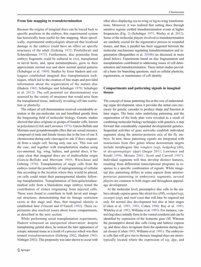
Fig. 1 Imaginal discs, their embryonic primordia, and adult cuticularproducts. The location of imaginal tissue primordia is represented at thecellular blastoderm stage (top), with corresponding numbering in larval(middle) and adult (bottom) stages. Axes orientation is indicated by theperpendicular arrows (A anterior, P posterior, D dorsal, V ventral). T1 toT3 represent thoracic segments, and A1 to A8 correspond to abdominalsegments. The epidermis of adult structures like the head, thorax, andappendages come from 9 pairs of bilateral discs (here, only one of each
pair is shown in the larva), and genitals derive from a middle disc (19discs in total): 1, clypeolabral; 2, eye-antennal; 3, labial; 4, humeral (orprothoracic); 5, first leg; 6, second leg; 7, third leg; 8, wing; 9, haltere; 10,genital. Note that some portions of the head and thorax, including thenotum, also originate from imaginal discs. For instance, the wing discscontribute both to the wings and the notum in the adult fly, which is notrepresented here for simplicity. Parts of the figure were inspired by (Held2005), where additional details are described
Chromosoma
From fate mapping to transdetermination
Because the origins of imaginal discs can be traced back tospecific positions in the embryo, this experimental systemhas historically been useful for fate mapping. More specif-ically, experimental embryologists observed that localizeddamage to the embryo could have an effect on specificstructures of the adult (Gehring 1972; Postlethwait andSchneiderman 1973). Furthermore, disc primordia fromembryo fragments could be cultured in vivo, transplantedto larval hosts, and, upon metamorphosis, grow to theirapproximate normal size and show normal spatial patterns(Schubiger et al. 1969). Studies by Ernst Hadorn and col-leagues established imaginal disc transplantation tech-niques, which led to the creation of fate maps and providedinformation about the organization of the mature disc(Hadorn 1965; Schubiger and Schubiger 1978; Schubigeret al. 2012). The cell potential (or determination) wasassessed by the variety of structures that would arise fromthe transplanted tissue, indirectly revealing cell fate restric-tion or plasticity.
The subject of cell determination received considerable at-tention in the pre-molecular era and benefitted greatly fromthe burgeoning field of molecular biology. Genetic studiesshowed that discs originate as groups of founder cells, knownas polyclones (Crick and Lawrence 1975). Garcia-Bellido andMerriam used gynandromorphs (flies that are sexual mosaics,composed of male and female tissues due to the loss of one Xchromosome during early cleavages) to ask if discs originat-ed from a single cell, having only one sex. This was notthe case, and together with transplantation studies usingeye-antennal, leg, wing, haltere, and genital discs, it be-came clear that imaginal discs are of multicellular origin(Garcia-Bellido and Merriam 1969; Wieschaus andGehring 1976). Transplantation of single cells from theembryo tested the possibility of reprogramming of cellularfate according to the location where they would be placed,yet cells could retain their parasegmental identity follow-ing transplantation. Transplantation of beta-galactosidase-marked cells from a blastoderm stage embryo tested thecontribution of clones originating from injected cells.These were found to contribute to both larval and imagi-nal structures, demonstrating that no lineage restrictionexists at this stage and, thus, that imaginal identity isestablished later (Vincent and O’Farrell 1992). These ex-periments also resolved issues about tissue compartments,as described in the next section.
While performing serial transplantation experiments,Hadorn witnessed an unexpected observation: despitetransplanting genital discs, he noticed the later appearance ofectopic antennal tissue as a result of a process which was thentermed transdetermination (Gehring 2002; Hadorn 1965;Nöthiger 2002). This propensity was later shown to occur with
other discs displaying eye-to-wing or leg-to-wing transforma-tions. Moreover, it was realized that cutting discs throughsensitive regions yielded transdetermination events at higherfrequencies (Fig. 2) (Schubiger 1971; Worley et al. 2012).Some of the molecular players involved in transdeterminationare similarly crucial for the regenerative process in woundedtissues, and thus, a parallel has been suggested between themolecular mechanisms regulating transdetermination and re-generation (Bergantiños et al. 2010b) (as discussed in moredetail below). Experiments based on disc fragmentation andtransplantation contributed to addressing issues of cell deter-mination and transdetermination. These initial studies provid-ed a basis for branching questions, such as cellular plasticity,regeneration, or maintenance of cell identity.
Compartments and patterning signals in imaginaltissues
The concept of tissue patterning lies at the core of understand-ing organ development, since it provides the initial cues nec-essary for genetic cascades to produce shape and function atlater stages. The basic rules underlying patterning and theorganization of the body plan were revealed as a result ofcombining molecular biology techniques with genetics, a stepforward that considerably expanded developmental genetics.Sequential activities of gene networks establish individualsegments along the anterior-posterior axis of the fly em-bryo. In turn, these patterning signals are integrated withinstructions from Hox genes whose downstream targetsinclude morphogens like wingless (wg), hedgehog (hh),or decapentaplegic (dpp) (Jaeger 2011; Lawrence andStruhl 1996; Morata 2001; Scott and Carroll 1987).Individual segments will thus develop distinct features,resulting from differential transcriptional programs in re-sponse to a specific combination of signals. While imagi-nal disc patterning differs in some aspects from anterior-posterior patterning in embryonic segments, severalplayers are common to both stages and throughout append-age development.
At the molecular level, presumptive disc cells in the em-bryo already express genes like distal-less (Dll), vestigial (vg),escargot (esg), and snail (sna) that will later be required notonly for normal disc development but also at later stages(Cohen et al. 1993, 1991; Cohen 1990; Ray et al. 1991;Whiteley et al. 1992; Williams et al. 1991). For instance, ven-tral (leg) discs initially form in the ventral ectoderm and can beidentified by expression of the homeotic gene Dll. Whereasthe presumptive dorsal disc cells (wing and haltere) expressvg, and these discs invaginate from the epidermis during dor-sal closure (Cohen 1993; Williams et al. 1991). The embryon-ic cells that will give rise to thoracic imaginal discs are stereo-typically located where the expression of wg, dpp, and
Chromosoma

engrailed intersects (Cohen et al. 1993). The expression ofdpp in the embryo is perpendicular to that of the segmentpolarity genes wg and engrailed (en), a feature that is alsoobserved, for example, in the wing disc. Engrailed isexpressed in the posterior region of both embryonic segmentsand imaginal discs, where it not only functions as a selectorgene but also defines tissue compartmentalization, a conceptfurther examined below. It became clear that anterior-posterior(A-P) boundaries in imaginal discs reflect earlier lineage re-striction and are initially established as parasegment bound-aries in the embryo (Dahmann and Basler 1999; Martinez-Arias and Lawrence 1985).
The discovery of tissue compartments was revealed byclonal analysis, which also proved very fruitful for fate map-ping and lineage tracing. Compartments are the result of alineage restriction between the anterior and posterior portionsof the wing disc (Garcia-Bellido et al. 1973; Morata andLawrence 1975). Such restriction occurs early in theDrosophila embryo, where all segments contain two separatelineages, anterior and posterior, which are segregated andform a stable boundary throughout development. For instance,when clones are genetically induced, they can arise in eithercompartment, but they do not cross from one to the other. Theconcept of tissue compartments had broader implications forunderstanding development in other organisms, since theywere later found to similarly exist in the developing chickenhindbrain and the mammalian brain (Dahmann and Basler1999; Fraser et al. 1990; Ingham and Arias 1992; Levittet al. 1997).
The molecular mechanisms underlying allocation of cellfates among anterior and posterior identities were found torequire a key gene, engrailed (Morata and Lawrence1975). Clonal experiments uncovered a role for en in theselection of posterior identity. Clones lacking en in theposterior compartment develop into anterior structuresand no longer respect the boundary, while mutant clonesarising in the anterior compartment develop normally.Furthermore, an ectopic boundary formed in the P com-partment at the interface between wild-type posterior cellsand engrailed mutant clones, which behave as anteriorcells (Lawrence and Struhl 1996; Morata and Lawrence1975). These results from clonal analyses, together withthe observation that wings of adult flies lacking en exhibittransformation to anterior structures, clarified the role of enas a selector gene. In addition to the A-P compartmentboundary, a second tissue division appears during larvalstages, at the dorsal-ventral (D-V) border. In the case ofthis orthogonal subdivision, the selector gene apterous wasidentified to establish the differences in dorsal-ventralidentities and also to play a role in wing growth and pat-terning (Diaz-Benjumea 1993).
The wing imaginal disc proved ideal to uncover the molec-ular relationships between patterning regulators, as withinthis, tissue morphogens play a role in both the A-P and D-Vaxes, which intersect perpendicularly in the pouch region. Thesame cascades also function in other discs, as illustrated inFig. 3. Engrailed inhibits Hedgehog signaling in the posteriorcompartment, but Hh secretion permits short-range signaling
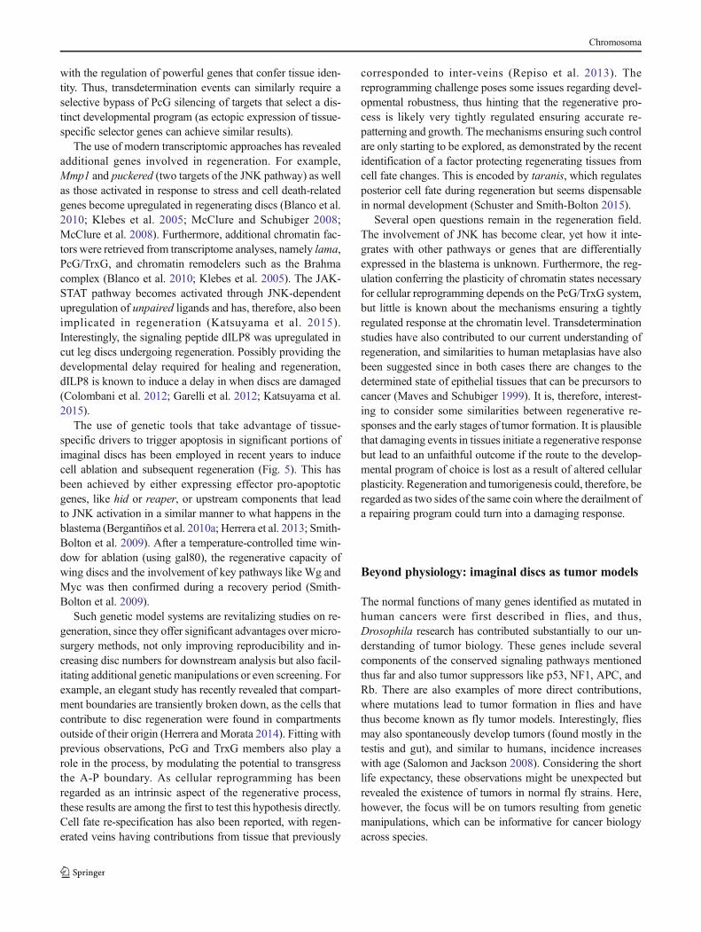
Fig. 2 An overview of transdetermination events, resulting both fromdisc fragmentation and through genetic manipulations. Classicaltransdetermination events occur with some probability upon discfragmentation and culture through transplantation, some with higherfrequencies like leg-to-wing (see main text). Ectopic expression of wg(wingless) also results in transdetermination in situ, similarly to whatseems to occur at Bweak points,^ where endogenous high levels of Wg
and Dpp have been linked with switches in cell fates. Geneticmanipulations also recapitulate transdetermination of some fly tissuesinto distinct ones (arrows connecting the blue circles), specifically bygain (+) or loss (−) of homeotic or selector gene expression (Ubx,Ultrabithorax; Antp, Antennapedia; pb, proboscipedia; Scr, Sex combsreduced; ey, eyeless; Dll, distal-less; vg, vestigial). The figure wasinspired by Wei et al. (2000) and McClure and Schubiger (2007)
Chromosoma

and thus regulates key genes like wg (encoding a Wnt familymember) or dpp (a homolog of the TGFβ family of growthfactors) in neighboring cells such as to activate the expressionof dpp in a stripe next to the A-P boundary. The identificationof several components of these signaling cascades revealedconserved signal transduction pathways, having an impacton physiological and cellular functions across species. Foran overview on the patterning roles of these three pathwaysand their morphogen properties, see Tabata and Takei (2004).
Importantly, regulation of compartment boundaries islinked to the establishment of novel proximal-distal signaling
axes in insect appendages, leading to the activation of proxi-mal selector genes that confer appendage identity, such asvestigial and scalloped in the wing. The pioneering researchconnecting compartment boundaries to the proximal-distalpatterning axis in fly imaginal discs has illuminated importantaspects of vertebrate limb development as many basic rulesoperating in this system are shared in distantly related organ-isms (Brook et al. 1996; Dahmann et al. 2011). For a compre-hensive description of the molecular determinants involved inappendage development, see for example Mann and Morata(2000) and Morata (2001).
Fig. 3 Expression domains of key signaling and patterning pathways inwing, eye-antennal, and leg discs. Imaginal discs become subdivided andpatterned during development, under the concerted action of signalingpathways and morphogens. Four key signals are representedindividually in the wing disc (top). Engrailed (en) is expressed in allcells in the posterior (P) compartment, conferring posterior identity andthus establishing the anterior-posterior (A-P) boundary. En directsexpression of the secreted short-range signaling molecule, Hedgehog(Hh), which can cross the A-P boundary and induce expression ofDecapentaplegic (Dpp). Dpp is expressed along the A-P boundary, and
its secretion permits long-range signaling to direct patterning of a widerdisc region. Wingless (Wg) is produced at the dorsal-ventral (D-V)boundary, a signal that is also key for wing development (see maintext). The signaling domains are represented all together in the bottomcentral wing disc, and their roles are also similarly conserved in othertissues, like the eye-antennal (bottom left) and leg (bottom right) discs. Asimilar color coding is used in all discs, which are orientedwith anterior tothe left and dorsal up. Expression patterns at other developmental stagesare described in Held (2005)
Chromosoma

The functional relevance of tissue compartments becamemore apparent with the finding that the domain of action ofsome homeotic genes was also restricted. Homeotic transfor-mations refer to alterations of specific body segments or struc-tures and are the phenotypic manifestation of mutations inhomeotic genes. For example, a spontaneous homeotic muta-tion led to transformation of the anterior portion of the thirdthoracic segment (aT3) into the anterior portion of the second(aT2) and was named bithorax (bx, later found to be an alleleof Ubx). Another mutation with a complementary effect wasalso isolated, postbithorax (pbx), where the posterior part ofT3 (pT3) is transformed into pT2. The combination of bx andpbx mutations by meiotic recombination enabled Ed Lewis toproduce the famous four-winged fly, where a second pair ofwings develops instead of halteres, showcasing a functionalconsequence of mutations in the bithorax complex (BX-C)(Lewis 1978). For a comprehensive discussion of the bithoraxcomplex, see another review in this series (Maeda and Karch2015). Another homeotic transformation that was identified,among several, was Antennapediawhere legs develop insteadof antennal structures, thus naming a second Hox gene com-plex, ANT-C (Schneuwly and Gehring 1985).
The realization that many genes implicated in developmentand cell fate decisions contained a homeobox established ashared principle for regulation relying on transcriptionalchanges. More explicitly, the combinatorial activities of ho-meobox genes result in distinct cell fates in specific tissuesand organs (Struhl 1982). The strict regulation of Hox geneexpression in specific segments or compartments proved to beat the core of a faithful developmental program, as evidencedby homeotic phenotypes resulting from their dysregulation.The maintenance of ON or OFF states of homeobox genesin specific expression patterns was found to depend onTrithorax (TrxG) and Polycomb group (PcG) genes, respec-tively (Ringrose and Paro 2007). Although initially studied fortheir embryonic phenotype, their function is also crucial forcellular memory in imaginal discs, as early studies showed forsome genes that are regulated by PcG/TrxG, like en, hh, orwg(Ingham 1983; Maurange and Paro 2002; Paro and Hogness1991; Randsholt et al. 2000). Molecular analyses made clearthat PcG and TrxG proteins form multimeric complexes in-volved in epigenetic regulation, especially as they containenzymatic activities responsible for catalyzing histone modi-fications, H3K27me3 by E(z) or H3K4me3 by Trx (Beisel andParo 2011; Byrd and Shearn 2003; Czermin et al. 2002;Müller et al. 2002). PcG/TrxG target several hundred genes(most of which are developmental regulators) and play a glob-al role in chromatin regulation and genome architecture that isconserved in many species (Boyer et al. 2006; Schwartz et al.2006; Sexton et al. 2012; Tolhuis et al. 2006). Thus, the Hoxgene clusters were used as a starting point to identify theunderlying regulatory mechanisms, resulting in far broaderimplications for global genome function and chromatin
biology, a field that is currently sprouting aided by newmethodologies.
Studies on organ growth and shape, cellularinteractions, and signaling cascades
Alongside the identification of spontaneousmutations in somehomeotic genes, screens for mutations affecting specific imag-inal discs uncovered several classes of phenotypes, many af-fecting organ growth or shape (disc undergrowth, overgrowth,or even hyperplastic growth) (Cohen 1993; Shearn and Garen1974). However, in general, these approaches only identifiedmutations causing local effects or those not leading to prema-ture lethality because observing the phenotype required sur-vival beyond embryonic stages. To overcome this limitation ofwhole organism mutations, successful genetic screens com-bined mutagenesis (with chemical mutagens, like ethylmethanesulfonate (EMS), or P-element-mediated mutagene-sis) with genetic mosaic-inducing techniques, such as Flp/FRT-mediated mitotic recombination. Such strategies permit-ted the characterization of gene function in restricted tissuepatches (Chou and Perrimon 1996; Spradling and Rubin1982; St Johnston 2002) (Fig. 4). Furthermore, clonal analysisenabled discrimination between cell-autonomous and non-cell-autonomous effects to further clarify the importance ofcellular interactions between mutant tissue and the surround-ing Bwild-type^ tissue.
Imaginal discs have also been fundamental for characteriz-ing the role of conserved signaling pathways in developingtissues. The amenability for epistasis experiments revealed thefunction of several pathway members in vivo and at whatcascade levels they work. In addition to the main patterningsignals mentioned before (Wg, Dpp, and Hh), fly research hasalso been at the root of discoveries spanning many membersof conserved signaling cascades such as Notch, EGFR (epi-dermal growth factor receptor), MAPK (mitogen-activatedprotein kinase), JAK-STAT (Janus kinase and signaling trans-ducer and activator of transcription), JNK (Jun N-terminalKinase), among others.
Defective eye specification revealed roles for transcriptionfactors like eyeless, twin of eyeless (toy), eyes absent (eya),eyegone, sine oculis, and also Notch and EGFR signalingacting upstream of this transcriptional network (Kumar 2001).Interestingly, many members of this gene network responsiblefor the eye-antennal transcriptional program contain a homeo-box, which is a recurring feature of transcription factors actingduring development. Enhancer and suppressor screens re-vealed a number of components acting within the same path-ways. For example, epistasis experiments clarified the role ofspitz as a ligand that can bind to the Egf receptor (torpedo),leading to the activation of downstream kinases encoded byras and raf and culminating in transcriptional regulation by
Chromosoma

Pointed (Kumar 2001; St Johnston 2002). Notch signaling canbe triggered by ligands, such as Serrate and Delta, binding tothe receptor (Notch) which, after cleavage of the intracellulardomain by gamma-secretase, can partner with Su(Hw) (sup-pressor of hairy wing) to modulate transcriptional activation.For a more complete summary of Notch signaling, seeGuruharsha et al. (2012). Importantly, bristle specificationand imaginal disc experiments uncovered three Notch activi-ties that established paradigms in neural development acrossspecies: cell fate assignment, boundary formation, and lateralinhibition (Bray 1998; Gómez-Skarmeta et al. 2003).
The global signaling rules employed by cells in a tissue arekey to understanding how cells communicate with their im-mediate neighbors or even how distant intercellular interac-tions play a role in normal physiology. The in vivo function ofa considerable number of newly identified signaling compo-nents was achieved through clonal or genetic experiments inDrosophila (which goes beyond the scope of this review)(Hynes et al. 2013; Jenny and Basler 2015; Kumar 2001).One of the central questions regarding the integration of dif-ferent signaling instructions through cellular interactions con-cerns how growth control is achieved. Growth regulation hasattracted considerable attention in the last decades and thusdeserves some mention, as several genetic screens have iden-tified regulatory genes.
Wg and Dpp are instrumental in wing disc patterning andgrowth, and have been classified as morphogens since bothproteins can spread further away from the cells where therespective genes are expressed (Vincent and Briscoe 2001).It has been proposed that morphogen gradients are responsiblefor instructing growth and patterning of the wing disc and mayhave similar functions in vertebrates (Ashe and Briscoe 2006;Tabata 2001). For example, the Dpp gradient could lead toactivation of distinct targets depending on the distance fromthe source (the A-P boundary), as suggested by the nestedexpression patterns of its target genes. Three well-characterized targets are spalt major (salm), optomotor blind(omb), and brinker (brk). While salm expression is centeredaround the source, the domain of omb expression is broaderthan salm, and brk is expressed at higher levels at the periph-ery, forming an opposing gradient to dpp (Affolter and Basler2007; Campbell and Tomlinson 1999; Nellen et al. 1996).Moreover, ectopic dpp-expressing clones could produce du-plications of wing veins with correctly patterned territories(Capdevila and Guerrero 1994; Zecca et al. 1995). Similarly,the evidence for the existence of a Wg gradient was also sup-ported by nested expression domains of targets describedalong the D-V boundary, e.g., senseless (sens, a high-leveltarget), distal-less (Dll, with a more extended range), and vg(a low-level target expressed in most of the prospective wing)(Neumann and Cohen 1997; Zecca et al. 1996). However,recent evidence challenges the requirement of a Wg gradientsince flies carrying solely an engineered version of wg that is
unable to spread develop wings with nearly the right size andwithout apparent morphological defects (Alexandre et al.2014). It has been suggested that cells expressing wg at earlierstages maintain expression of target genes due to a cellularmemory mechanism, even after the inducing signal is absent.Regarding Dpp, the role of this morphogen in tissue patterningand growth has attracted considerable attention, and aconcentration-dependent response has been put forward toaccount for differences in size by affecting cell proliferation(Vuilleumier et al. 2010; Wartlick et al. 2011). Two recentstudies, taking advantage of endogenous genome editingmethods, suggest that the Dpp gradient is required for pattern-ing but not essential for cell proliferation in lateral portions ofthe wing disc, thus contributing to growth mostly in the pouchregion (Akiyama and Gibson 2015; Harmansa et al. 2015). Allthese reports showcase a topic of intense research that also has
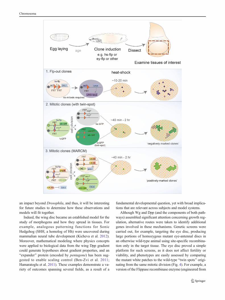
�Fig. 4 Overview of clone-generating techniques. Three broadly usedtechniques relying on the Flp/FRT system, where the source of Flippasecan be chosen depending on experimental design, e.g., commonly usingthe heat shock promoter with temporal control or tissue-restrictedpromoters (ey, Ubx). Top: after egg laying for a defined period andaging larvae to the desired stage, mosaics are induced upon Flpexpression and, by dissecting the tissues of interest, the effect of geneticmanipulations is analyzed by comparing clones with neighboring wild-type tissue. (1) Flp-out clones do not rely on mitosis, since Flp mediatesrecombination of two FRT (Flippase-Recognition Targets) elements withthe same orientation located in close proximity in one chromosome.Usually, a constitutive promoter (actin, tubulin, etc.) is not active in theabsence of Flp due to an element flanked by FRTsites (e.g. stop, or yellowmarker). Excision of the flanked element upon Flp-dependentrecombination enables the promoter to activate a downstream gene onlywithin the clone (for example, a marker such as lacZ, Gal4 (that couldactivate a UAS-lacZ), or other alternatives). (2) Mitotic clones require celldivision, where somatic recombination of chromosomes in heterozygouscells provides the opportunity for Flp-mediated recombination. In a givenparental cell (heterozygous for a mutation of interest), recombinationbetween matching FRT elements located in homologous chromosomalarms produces two daughter cells: one carrying two copies of the wild-type chromosome and another homozygous for the desired mutation. Thefollowing cell divisions will therefore generate two cell populations,hence a clone and its wild-type counterpart. A constitutive marker isoften used (e.g., a ubiquitous GFP construct inserted in the wild-typechromosome), and thus the homozygous mutant clone can be identifiedby the absence of the marker (negatively marked), while the wild-typeBtwin^ resulting from the same cell division (hence called twin spot) canalso be visualized as it harbors two copies of the marker (2xGFP). (3) Analternative method was developed to enable clones to be markedBpositively,^ i.e., labeling the tissue harboring the desired geneticmanipulation with GFP. This option is provided by MARCM (MosaicAnalysis with a Repressive Cell Marker), where the constitutiveexpression of a repressive component (Gal80) on the homologouschromosome prevents Gal4 from activating downstream genes ormarkers (usually UAS-GFP). Upon Flp expression, recombinationproduces a homozygous mutant cell that lacks Gal80 while the othercontains a pair of FRT chromosomes carrying Gal80 (and no mutation).In the mutant clone, the absence of Gal80 permits Gal4 to activate theUAS-GFP marker (and also permits further flexibility as other UAStransgenes can be expressed specifically within clones). In this case, thewild-type tissue remains unlabeled and only mutant clones are visuallydetected
b
Chromosoma
an impact beyond Drosophila, and thus, it will be interestingfor future studies to determine how these observations andmodels will fit together.
Indeed, the wing disc became an established model for thestudy of morphogens and how they spread in tissues. Forexample, analogous patterning functions for SonicHedgehog (SHH, a homolog of Hh) were uncovered duringmammalian neural tube development (Kicheva et al. 2012).Moreover, mathematical modeling where physics conceptswere applied to biological data from the wing Dpp gradientcould generate hypotheses about gradient properties, and anBexpander^ protein (encoded by pentagone) has been sug-gested to enable scaling control (Ben-Zvi et al. 2011;Hamaratoglu et al. 2011). These examples demonstrate a va-riety of outcomes spanning several fields, as a result of a
fundamental developmental question, yet with broad implica-tions that are relevant across subjects and model systems.
Although Wg and Dpp (and the components of both path-ways) assembled significant attention concerning growth reg-ulation, alternative routes were taken to identify additionalgenes involved in these mechanisms. Genetic screens werecarried out, for example, targeting the eye disc, producinglarge portions of homozygous mutant eye-antennal discs inan otherwise wild-type animal using site-specific recombina-tion only in the target tissue. The eye disc proved a simpleplatform for such screens, as it does not affect fertility orviability, and phenotypes are easily assessed by comparingthe mutant white patches to the wild-type Btwin spots^ origi-nating from the same mitotic division (Fig. 4). For example, aversion of the Flippase recombinase enzyme (engineered from
Chromosoma

yeast) only active in the eye-antennal disc (eyFlp) was used toinduce recombination of FRT (Flippase-recognition target)sites, producing homozygousmutant tissue upon cell division.Screens were thus carried out using this approach with a widerange of mutations and led to the characterization of genesrequired for normal tissue architecture and growth.Examples of hits include the tumor suppressor kinases warts,salvador, and hippo, the latter giving its name to a novelpathway that can restrict cell proliferation and promote apo-ptosis (Tapon et al. 2002; Wu et al. 2003; Xu et al. 1995).Other initial screens for abnormal eye development revealedregulators of cell proliferation, like archipelago (throughcyclin E) and homologs of the tuberous sclerosis complex(Tsc1/2). Likewise, cytoskeletal or nuclear componentsinfluencing cell affinity, adhesion, and eye developmentwere also identified (Janody et al. 2004, 2003; Moberg etal. 2001; Tapon et al. 2001).
Sequencing of the fly genome and the discovery of RNAinterference (RNAi) led to the generation of genome-widetransgenic RNAi libraries under UAS control, offering an un-precedented potential to precisely carry out selective reversegenetics using tissue-specific Gal4 drivers (Adams et al. 2000;Dietzl et al. 2007). These tools prompted the design of manygenetic screens, either in the eye (with GMR-gal4) or wingcompartments (en-gal4 or hh-gal4) where the effect of targetgene knockdown could be directly compared to an internalcontrol (e.g., the anterior compartment) where gene functionremained intact. Such RNAi-based screens expanded ourknowledge about further members of previously mentionedpathways (like Wg, Dpp, Notch, Hippo, etc.) and also fine-tuned our understanding of additional processes. For instance,screens uncovered regulators of apoptosis that could rescuesmall-eye phenotypes induced by downstream pro-apoptoticinducers (e.g., GMR>hid) and compensatory proliferation(Fan and Bergmann 2008; Herz et al. 2006; Mummery-Widmer et al. 2009; Saj et al. 2010; Thompson andCohen 2006).
The previous examples of hits resulting from screens andclonal analyses in their complex tissue environments highlightthe power of Drosophila discs as platforms to identify andcharacterize conserved factors and associated pathways, oftenfor the first time. The foundation of detailed knowledge gath-ered on the basic biology of imaginal discs places us in anexciting position to further exploit these model systems toaddress medically relevant topics where developmental statesare challenged, like regeneration and tumor initiation.
From disc transplantation to tissue regeneration
The process of tissue and organ regeneration has attractedcuriosity for many decades. Classical examples of remarkableregenerative capacity come from amphibians, which are able
to replace large and complex structures like the tail or limbs(Tanaka and Reddien 2011). The potential to build up on thebasic knowledge underlying regenerative mechanisms forpossible exploitation into medical applications has captivatedmany. Despite this profound interest, research using these or-ganisms has been limited because few exploratory tools areavailable. Conversely, Drosophila stands at an interestingpoint for regenerative studies, since it is less complex thanamphibians and humans and a wide palette of experimentaltools is available.
The regenerative capacity of fly imaginal discs was noticedseveral decades ago alongside the transplantation experimentsmentioned previously (Bryant 1971, 1975; Hadorn 1965).Upon disc fragmentation, for example, along the D-V axis ofthe leg disc, the anterior portion can regenerate while the pos-terior half undergoes duplication where a mirror image of thetissue arises, instead of forming the missing part. Interestingly,the mechanism for this difference still remains a mystery(Bryant 1971; Schubiger 1971). The cells around the cutedges form a blastema containing cells that are able to dividemore frequently than in other regions. These fast-proliferatingcells are mainly responsible for disc regeneration, althoughcell death also occurs (Fig. 5) (Abbott et al. 1981; Kiehleand Schubiger 1985). The determination state of the discseems to be maintained in blastema cells, yet when fragmen-tation occurs along defined Bweak points,^ for example,where dpp and wg overlap, transdetermination events occurwith some frequency (Johnston and Schubiger 1996; Mavesand Schubiger 1995, 1999). Regeneration studies using discf r agmen ta t i on have , t he r e fo r e , been t i ed wi thtransdetermination (Worley et al. 2012).
Themolecular events required for disc regeneration follow-ing fragmentation revealed a role for Wg, as its ectopic ex-pression frequently promoted leg-to-wing transdetermination(Johnston and Schubiger 1996). The molecular basis fortransdetermination relies at least in part on interactions be-tween Wg and Dpp signaling, as the activation of vg down-stream ofWg signaling resulted in the formation of more wingtissue from the dorsal region (where dpp is higher) (Mavesand Schubiger 1995, 1998). The function of these pathways inblastema cells may facilitate cellular plasticity underlying cellfate re-assignment during regeneration, and parallels withstem cell-like potency have been put forward (McClure andSchubiger 2007; Wei et al. 2000).
The micro-surgery techniques employed to study regener-ation, for example, by dissecting the anterior quarter of the legdisc, trigger a wounding response at the early stages of blas-tema formation. Thus, the early regenerative response utilizessimilar molecular routes as wound healing, involving the ac-tivation of stress signaling like JNK (Jun N-terminal kinase)(Bosch et al. 2005; Mattila et al. 2005). JNK is crucial atseveral stages of the regenerative response by directing cyto-skeletal rearrangements to bridge the gap between wounded
Chromosoma

tissues (similar to its function during embryonic dorsal closure(Martin and Parkhurst 2004)), by promoting cell death aroundthe cut edges, and finally by modulating chromatin regulators.The latter function was uncovered as a result of inducing leg-to-wing transdetermination through ectopic wg expressionusing the flp-out clonal technique (Struhl and Basler 1993).
This leads to vg expression in leg discs, which in turn results intransdetermination to wing tissue, of which frequency is in-creased upon JNK-mediated overcoming of PcG silencing(Lee et al. 2005). Since Polycomb group (PcG) proteins si-lence many developmental and signaling-related genes, theinvolvement of PcG/TrxG links regenerative mechanisms
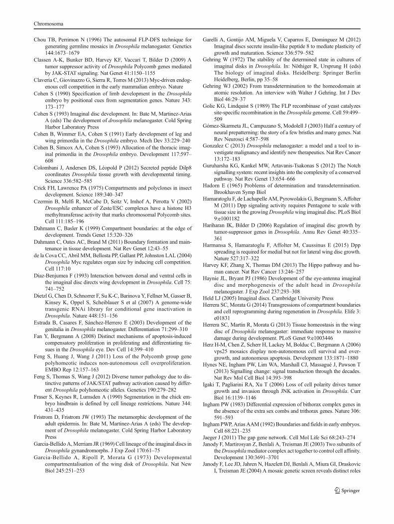
Fig. 5 Regeneration in Drosophila imaginal discs. Imaginal discs areable to regenerate upon micro-surgery and culture throughtransplantation to host flies. The leg disc has routinely been used infragmentation experiments (top), often with a standard cut thatseparates the Banterior one quarter^ from the Bposterior three quarters.^The larger fragment tends to result in tissue duplication, while the smallerfragment regenerates the remaining tissue with the correct pattern andfunction. The cut elicits a wounding-like response, followed by a phaseof regenerative growth, for which chromatin remodeling is importantduring cellular reprogramming that can produce extra cells whilemaintaining the correct pattern and cellular identities. Recentlydeveloped tools (bottom) allow genetic-induced tissue ablation andregeneration within the same organism (overcoming the limitations ofablation and transplantation). Localized tissue ablation, for example, ofthe pouch region of the wing disc, can be spatially and temporally
controlled. Different systems have been described (see main text), butgenerally, the design involves the following: a region-specific Gal4 isnormally inhibited by ubiquitous Gal80 expression, which can beovercome by means of a temperature shift (with a temperature-sensitiveform of Gal80 that is inactive at 29 °C or higher), which duration can betitrated to achieve specific conditions. During the period when Gal80 isinactive, Gal4 can activate a downstream UAS target that triggersapoptosis (e.g., using rpr, hid, egr, etc.). After an acute or controlledBdamage-induction^ phase, larvae are returned to a temperature thatyields a functional Gal80 and thus inactivates the damage trigger,allowing tissues to regenerate during a Brecovery^ phase. Many of thehallmarks are conserved between the classical ablation experiments andthe genetic-ablation systems, and the recent use of the latter promiseconsiderable advantages that improve reproducibility and larger-scaleexperiments
Chromosoma
with the regulation of powerful genes that confer tissue iden-tity. Thus, transdetermination events can similarly require aselective bypass of PcG silencing of targets that select a dis-tinct developmental program (as ectopic expression of tissue-specific selector genes can achieve similar results).
The use of modern transcriptomic approaches has revealedadditional genes involved in regeneration. For example,Mmp1 and puckered (two targets of the JNK pathway) as wellas those activated in response to stress and cell death-relatedgenes become upregulated in regenerating discs (Blanco et al.2010; Klebes et al. 2005; McClure and Schubiger 2008;McClure et al. 2008). Furthermore, additional chromatin fac-tors were retrieved from transcriptome analyses, namely lama,PcG/TrxG, and chromatin remodelers such as the Brahmacomplex (Blanco et al. 2010; Klebes et al. 2005). The JAK-STAT pathway becomes activated through JNK-dependentupregulation of unpaired ligands and has, therefore, also beenimplicated in regeneration (Katsuyama et al. 2015).Interestingly, the signaling peptide dILP8 was upregulated incut leg discs undergoing regeneration. Possibly providing thedevelopmental delay required for healing and regeneration,dILP8 is known to induce a delay in when discs are damaged(Colombani et al. 2012; Garelli et al. 2012; Katsuyama et al.2015).
The use of genetic tools that take advantage of tissue-specific drivers to trigger apoptosis in significant portions ofimaginal discs has been employed in recent years to inducecell ablation and subsequent regeneration (Fig. 5). This hasbeen achieved by either expressing effector pro-apoptoticgenes, like hid or reaper, or upstream components that leadto JNK activation in a similar manner to what happens in theblastema (Bergantiños et al. 2010a; Herrera et al. 2013; Smith-Bolton et al. 2009). After a temperature-controlled time win-dow for ablation (using gal80), the regenerative capacity ofwing discs and the involvement of key pathways like Wg andMyc was then confirmed during a recovery period (Smith-Bolton et al. 2009).
Such genetic model systems are revitalizing studies on re-generation, since they offer significant advantages over micro-surgery methods, not only improving reproducibility and in-creasing disc numbers for downstream analysis but also facil-itating additional genetic manipulations or even screening. Forexample, an elegant study has recently revealed that compart-ment boundaries are transiently broken down, as the cells thatcontribute to disc regeneration were found in compartmentsoutside of their origin (Herrera and Morata 2014). Fitting withprevious observations, PcG and TrxG members also play arole in the process, by modulating the potential to transgressthe A-P boundary. As cellular reprogramming has beenregarded as an intrinsic aspect of the regenerative process,these results are among the first to test this hypothesis directly.Cell fate re-specification has also been reported, with regen-erated veins having contributions from tissue that previously
corresponded to inter-veins (Repiso et al. 2013). Thereprogramming challenge poses some issues regarding devel-opmental robustness, thus hinting that the regenerative pro-cess is likely very tightly regulated ensuring accurate re-patterning and growth. The mechanisms ensuring such controlare only starting to be explored, as demonstrated by the recentidentification of a factor protecting regenerating tissues fromcell fate changes. This is encoded by taranis, which regulatesposterior cell fate during regeneration but seems dispensablein normal development (Schuster and Smith-Bolton 2015).
Several open questions remain in the regeneration field.The involvement of JNK has become clear, yet how it inte-grates with other pathways or genes that are differentiallyexpressed in the blastema is unknown. Furthermore, the reg-ulation conferring the plasticity of chromatin states necessaryfor cellular reprogramming depends on the PcG/TrxG system,but little is known about the mechanisms ensuring a tightlyregulated response at the chromatin level. Transdeterminationstudies have also contributed to our current understanding ofregeneration, and similarities to human metaplasias have alsobeen suggested since in both cases there are changes to thedetermined state of epithelial tissues that can be precursors tocancer (Maves and Schubiger 1999). It is, therefore, interest-ing to consider some similarities between regenerative re-sponses and the early stages of tumor formation. It is plausiblethat damaging events in tissues initiate a regenerative responsebut lead to an unfaithful outcome if the route to the develop-mental program of choice is lost as a result of altered cellularplasticity. Regeneration and tumorigenesis could, therefore, beregarded as two sides of the same coin where the derailment ofa repairing program could turn into a damaging response.
Beyond physiology: imaginal discs as tumor models
The normal functions of many genes identified as mutated inhuman cancers were first described in flies, and thus,Drosophila research has contributed substantially to our un-derstanding of tumor biology. These genes include severalcomponents of the conserved signaling pathways mentionedthus far and also tumor suppressors like p53, NF1, APC, andRb. There are also examples of more direct contributions,where mutations lead to tumor formation in flies and havethus become known as fly tumor models. Interestingly, fliesmay also spontaneously develop tumors (found mostly in thetestis and gut), and similar to humans, incidence increaseswith age (Salomon and Jackson 2008). Considering the shortlife expectancy, these observations might be unexpected butrevealed the existence of tumors in normal fly strains. Here,however, the focus will be on tumors resulting from geneticmanipulations, which can be informative for cancer biologyacross species.
Chromosoma

It has become clear that many human tumors result fromone or more clonal events where cells lose their normal func-tion and start overproliferating, often compromising the inter-actions with neighboring cells or the microenvironment. Theinitial steps that abnormal cells require to compromise homeo-stasis in their native tissue are poorly understood, since theseearly stages are not clinically accessible. Fortunately,Drosophila offers a significant advantage in this regard, wherethe behavior of only a few cells can be followed after inducinga trigger. In fact, the phenomenon of cell competition, whichinvolves differential cell behavior within a tissue, has beenobserved and well documented for more than four decadesin flies. The discovery of cell competition in Drosophilaestablished a remarkable new concept to describe the survivaland proliferation of cells with higher fitness at the expense ofweaker neighbors. It was first observed in mosaics where het-erozygous Minute (M) cells are eliminated when surroundedby wild-type cells (Morata and Ripoll 1975). M encodes aribosomal component and M homozygous mutant cells arelethal, but heterozygous animals are viable and cell competi-tion only occurs when clones of cells with different fitnessarise in close proximity (Johnston 2009; Martín et al. 2009;Morata and Ripoll 1975). Additional stimuli can also triggercell competition, such as different doses of myc, yorkie, andothers. Furthermore, cells seem to assess their fitness in acontext-dependent manner (Vincent et al. 2013). For example,cells carrying only one copy of myc (myc/+) are outcompetedby the surrounding wild-type cells. However, cells that carryan extra copy of myc (myc(+)) can also outcompete wild-typecells, having become known as Bsupercompetitors^ (de laCova et al. 2004; Moreno and Basler 2004). Interestingly,the involvement of myc in cell competition has recently beendescribed in the early mouse embryo as well, suggesting con-served features in mammals (Clavería et al. 2013). It is impor-tant to note that cell competition has direct implications innormal development, namely in growth regulation and main-tenance of tissue homeostasis. Despite being a general phe-nomenon occurring in multicellular organisms, this processparallels several features that are required for cancer forma-tion, where weaker cells are actively eliminated in response tosignals originating from fast-growing cells that can take overmore space to proliferate (Wagstaff et al. 2013).
InDrosophila, tumors have generally been categorized intohyperplastic or neoplastic (Bilder 2004; Gonzalez 2013).While imaginal discs carrying hyperplastic tumors show ex-tensive overproliferation, they maintain relatively normal tis-sue organization with cells in a monolayer and can often dif-ferentiate into adult tissues. On the other hand, neoplastictumors are more aggressive and the overproliferating cellslose their epithelial architecture. Forming multi-layers, thesecells are able to grow uncontrollably and have limited differ-entiation capacity. The isolation of the first mutation causingtumorigenesis, lethal giant larvae (lgl), dates back to Bridges,
but its malignant properties (cells growing rapidly andinvasively, killing their host) were only reported in the1960s. Mutations in two other genes, discs large (dlg) andscribble (scrib), were later identified to also lead to neoplastictumors (Bilder and Perrimon 2000; Stewart et al. 1972). Thethree genes share phenotypic similarities where homozygousmutant larvae survive but keep growing as L3 for several dayswithout reaching the pupal stage. Larvae size increases dra-matically resembling Bgiant larvae,^ and finally will kill thehost. These genes have become known as neoplastic tumorsuppressor genes (nTSG).
Hyperplastic mutations exhibit uncontrolled proliferation,yet tissue architecture and differentiation capacity are stillmaintained. These phenotypes can be caused by inactivationof genes regulating cell growth, proliferation, or cell death,including many of the genes previously mentioned to regulategrowth in a normal context, such as salvador,warts, hippo, fat(an atypical cadherin), expanded, Tsc1/2, PTEN, or Csk (fordetails, see (Brumby and Richardson 2005; Hariharan andBilder 2006; Harvey et al. 2013; Vidal and Cagan 2006)).More invasive tumors, in the neoplastic category, display cellsthat form rounded cyst-like patches, having lost the ability tomaintain an epithelial monolayer crucial for tissue architectureand being unable to differentiate (Fig. 6). A common denom-inator was identified among lgl, dlg, and scrib in that theproteins they encode interact with the cytoskeleton and definethe apico-basal polarity that underlies epithelial cell function(Bilder et al. 2000). Therefore, cell polarity and architecturelie at the core of healthy physiology in epithelia, the tissuetype where many human cancers also arise.
Identification of additional neoplastic tumor suppressorgenes was facilitated by clonal analysis, as these mutationsare lethal at earlier stages and larvae do not survive as longas for lgl/dlg/scrib. Such nTSGs include avalanche, Rab5,tsg101, and vps25 which play roles in endocytosis (Lu andBilder 2005; Moberg et al. 2005; Thompson et al. 2005;Vaccari and Bilder 2005; Wucherpfennig et al. 2003). In thesemutants, inactivation of components of the endocytic machin-ery or ESCRTcomplexes compromise trafficking between dif-ferent cellular compartments. There is a shared link betweenthe endocytic and polarity-associated mutants, since failures inendocytosis lead to inappropriate accumulation of apical andbasal proteins or signaling receptor molecules, which canmimic phenotypes of polarity nTSGs (Bilder 2004).
Although animals that are entirely mutant for nTSGs dis-play giant larvae phenotypes, it became clear that scribmutantclones could not outcompete their wild-type neighbor cells.Instead, these clones are progressively eliminated in a JNK-dependent manner but can still become tumorigenic if JNKsignaling is blocked (Brumby and Richardson 2003). Clonalstudies, therefore, uncovered a cooperative effect between dis-ruption of epithelial integrity and oncogenes, such as activatedforms of Ras (rasV12/act) and also Notchact. Although rasact
Chromosoma

clones alone do not lead to neoplastic tumors (they result inovergrowth), combination with scrib clones leads to neoplas-tic phenotypes (Brumby and Richardson 2003). These obser-vations prompted MARCM genetic screens in the eye disc toidentify other factors involved in oncogenic cooperativity(both with rasact and Notchact). Examples include cytoskeletalcomponents like rho/rhoGEF or the transcription factorabrupt (Brumby et al. 2011; Turkel et al. 2013). These tumors
also display a metastatic behavior, as some cells can invadethe ventral nerve cord and distant portions of the central ner-vous system (Pagliarini and Xu 2003).
A common denominator of neoplastic tumors is ectopicstress signaling through JNK activation. Constitutive Ras sig-naling allows cells to survive by evading JNK-mediated apo-ptosis (Brumby and Richardson 2003). The invasiveness ofscrib/rasact clones depends on transcriptional activation of
Fig. 6 Tumor models in Drosophila epithelial tissues. Powerful geneticscreens revealed mutations in genes involved in tumorigenesis, namelysome classes affecting the development of imaginal discs. Classicalmutations like lgl (lethal giant larvae) displayed abnormal discdevelopment where all tissues of homozygous individuals were affected(top). As larval development progresses, it becomes clear that theepithelial structure of disc tissues is aberrant with architectural defectsleading to hyperplastic or neoplastic growth. Additional genes werelater found to display similar phenotypes and share molecular functionsas components of the apico-basal polarity machinery. The combination ofmosaic analysis with gain- or loss-of-function experiments providedalternative means to fine-tune screening approaches. Clonal analysis(bottom) enables mutant patches to grow surrounded by wild-type
tissue, and thus, interactions between tumor and neighboring normaltissue can occur. These approaches revealed, in some instances, thatcooperative interactions between conserved oncogenes and polaritydeterminants enhanced a tumorigenic phenotype in comparison to theeffect of each component individually. On the other hand, mutations inepigenetic components (like ph, a Polycomb Group member) displaystrong neoplastic phenotypes in clones that also display abnormalepithelial architecture. Furthermore, the involvement of key signalingpathways is a common feature among fly epithelial tumors, suggestingthat signaling events and potentially the interactions between healthy andtumor cells are likely to play a role in the disruptive events leading totumor formation
Chromosoma

mmp1 in a JNK-dependent manner, and changes in E-cadherinexpression were also reported. These findings evoke a possibleparallel with evidence from human tumors, which frequentlyhave E-cadherin mutations, and the phenotypes of cancer tissueare reminiscent of alterations known as the epithelial-mesenchymal transition (EMT) (Harvey et al. 2013; Igaki etal. 2006; Uhlirova and Bohmann 2006). Interestingly, neigh-boring scrib/rasact clones can actively cooperate and metasta-size, by forming interclonal tumors that seem to rely on loss ofscribmostly during the early steps, as in later stages clones aremostly composed of rasV12-expressing cells (Wu et al. 2010).JAK-STATactivity has also been implied as an oncogenic driv-er in ras-mediated tumors. Overall, several aspects of thesetumors exhibit features reminiscent of cell competition, hintingthat it will be interesting to further investigate the parallelsbetween competitive interactions in regulating normal growthor upon losing the ability to control it, leading to tumors(Levayer et al. 2015; Vincent et al. 2013).
Disruption of key chromatin regulators has also beenshown to lead to tumorigenesis. Loss of polyhomeotic (ph),a component of the Polycomb Repressive Complex 1 (PRC1),in eye disc clones results in massive overgrowth of eye andantennal tissue (Classen et al. 2009; Martinez et al. 2009).Clones induced by the MARCM technique (Fig. 4) display aseverely disorganized architecture, loss of polarity, and somemetastatic properties as well as aberrant signaling activities(Fig. 6). These tumors are dependent on JAK-STAT andNotch signaling activities, and have also been reported to leadto massive overproliferation in an RNAi screen for Notchregulators in the wing disc (Saj et al. 2010). Blocking thesepathways ameliorates tumor burden, suggesting that the de-railment of signaling pathways underlies a significant effecton the tumor phenotype (Feng et al. 2011, 2012). An overex-pression screen for modifiers of Notchact tumors uncovered ahit, named eyeful, whichmapped within two genes (pipsqueakand longitudinals lacking) that produce transcription fac-tors involved in the recruitment of Polycomb and chro-matin complexes to promoter regions (Vallejo andGutierrez-Aviño 2006).
Globally, fly tumor models display several phenotypes thatparallel human cancer hallmarks, such as the ability to sustainoverproliferation and growth, the capacity to evade apoptoticsignals, the inability to respond to anti-proliferative signals,and the capability to metastasize to sites distant from theirorigin (Brumby and Richardson 2005; Gonzalez 2013). Theuse of transplantation techniques, pioneered several decadesago, became again instrumental to demonstrate the unlimitedgrowth and survival of fly tumors in hosts, analogous to rou-tine allograft experiments in mice (Rossi and Gonzalez 2015).It was demonstrated, for example, that ph tumor tissue doesnot accumulate genome instability even after prolonged culti-vation. This suggests that an epigenetic derailment of geneexpression control is sufficient to maintain neoplastic growth
(Sievers et al. 2014). There are, however, important limita-tions in that certain facets of human tumorigenesis are miss-ing, since flies have a more rudimentary immune system andlack a closed circulatory system. These aspects restrict somedirect comparisons with human cancer since angiogenesis anddedicated tumor immunological responses would not be easilymodeled in flies. Nonetheless, the great advantages of fly re-search remain at the forefront in identifying the basic mole-cules that may have conserved roles which would otherwisebe very difficult to find directly in mammals.
Namely,Drosophila’s ability to contribute to the early stepsof tumorigenesis has great potential, not only from the exam-ples described here but also from emerging knowledgeresulting from this very active research field. Many initialcancer triggers remain a mystery, which could be revealedfrom studies concerning key aspects of the observed fly tumorphenotypes. Moreover, it will be interesting to further explorethe links between disrupted polarity, a common feature, aswell as ectopic activation of several signaling cascades. Animportant open question entails how such pathways may in-fluence the regulation of gene expression through transcrip-tion factors or chromatin regulators that exert global switches.These can, in a controlled fashion (like the regenerative pro-cess), result in faithful tissue organization, but when impaired,they may redefine cellular identities in a pathological manner.
Conclusions and perspectives
Imaginal discs are the Swiss army knife in Drosophila re-search, serving as versatile experimental systems to addressa variety of scientific topics in cell and developmental biology.Here, we have covered a broad range of topics where the useof imaginal discs supported discoveries in cell and develop-mental biology, revealing conserved pathways and mecha-nisms. The use of these epithelial precursor organs has en-abled functional studies in the context of a developing organ-ism, benefitting from the implementation of genetic tools thatconsiderably contributed to elucidating conserved molecularmechanisms.
With the emergence of new methods and technologies, thepotential of imaginal discs as experimental systems remainsunchallenged. The combination of established sophisticatedtools with modern techniques promises to yield further contri-butions in developmental and cell biology. Research topics atthe interface of physics and biology, like tissue mechanics, arehaving considerable progress in recent years by using imagi-nal discs to examine forces and cell interactions in their nativetissues of origin. The improvement of long-term in vitro discculture methods promises to bring many benefits to this field,and combination with other techniques such as live imaging,optogenetics, genome engineering (e.g., CRISPR/Cas9coupled to homologous recombination), or tissue-specific
Chromosoma

protein-targeting nanobodies is expected to enable novel andcross-disciplinary discoveries that are yet unforeseen.
Despite the minute size of imaginal structures within thesmall fly, their contribution to answering fundamental ques-tions has been immense, leading to a far-reaching insightacross species. The conservation of many fly genes, proteins,and processes, together with developmental concepts com-mon to all organisms, will provide profound implications fora better understanding of human development, physiology,and biomedicine going beyond measure.
Acknowledgments We thank P. Gaspar and S. Geisler for the discus-sions and comments on the manuscript and three anonymous reviewersfor the suggestions leading to a significant improvement of the manu-script. We apologize to the many authors, whose papers we were not ableto cite due to space restrictions.
Compliance with ethical standards This study was funded by grantsfrom the Swiss National Science Foundation (grant numbers SNF31003A_143922), Epigenesys, and the ETH Zurich. J.V.B. is supportedby an EMBO long-term fellowship (ALTF1131-2014 and Marie CurieActions LTFCOFUND2013/ GA-2013-609409).
Conflict of interest The authors declare that they have no conflict ofinterest.
Ethical approval This article does not contain any studies with humanparticipants or animals performed by any of the authors.
Open Access This article is distributed under the terms of theCreative Commons Attribution 4.0 International License (http://creativecommons.org/licenses/by/4.0/), which permits unrestricted use,distribution, and reproduction in any medium, provided you give appro-priate credit to the original author(s) and the source, provide a link to theCreative Commons license, and indicate if changes were made.
References
Abbott LC, Karpen GH, Schubiger G (1981) Compartmental restrictionsand blastema formation during pattern regulation in Drosophilaimaginal leg discs. Dev Biol 87:64–75
AdamsMD, Celniker SE, Holt RA, Evans CA, Gocayne JD, AmanatidesPG, Scherer SE, Li PW, Hoskins RA, Galle RF et al (2000) Thegenome sequence of Drosophila melanogaster. Science 287:2185–2195
Affolter M, Basler K (2007) The Decapentaplegic morphogen gradient:from pattern formation to growth regulation. Nat Rev Genet 8:663–674
Akiyama T, Gibson MC (2015) Decapentaplegic and growth control inthe developing Drosophila wing. Nature 527:375–378
Aldaz S, Escudero LM, Freeman M (2010) Live imaging of Drosophilaimaginal disc development. Proc Natl Acad Sci U S A 107:14217–14222
Alexandre C, Baena-Lopez A, Vincent J-P (2014) Patterning and growthcontrol by membrane-tethered Wingless. Nature 505:180–185
Ashe HL, Briscoe J (2006) The interpretation of morphogen gradients.Development 133:385–394
Beisel C, Paro R (2011) Silencing chromatin: comparing modes andmechanisms. Nat Rev Genet 12:123–135
Ben-Zvi D, Pyrowolakis G, Barkai N, Shilo B-Z (2011) Expansion-repression mechanism for scaling the Dpp activation gradient inDrosophila wing imaginal discs. Curr Biol 21:1391–1396
Bergantiños C, Corominas M, Serras F (2010a) Cell death-induced re-generation in wing imaginal discs requires JNK signalling.Development 137:1169–1179
Bergantiños C, Vilana X, Corominas M, Serras F (2010b) Imaginal discs:renaissance of a model for regenerative biology. Bioessays 32:207–217
Bilder D (2004) Epithelial polarity and proliferation control: links fromthe Drosophila neoplastic tumor suppressors. Genes Dev 18:1909–1925
Bilder D, Perrimon N (2000) Localization of apical epithelial determi-nants by the basolateral PDZ protein Scribble. Nature 403:676–680
Bilder D, LiM, PerrimonN (2000) Cooperative regulation of cell polarityand growth byDrosophila tumor suppressors. Science 289:113–116
Blanco E, Ruiz-Romero M, Beltran S, Bosch M, Punset A, Serras F,CorominasM (2010) Gene expression following induction of regen-eration in Drosophila wing imaginal discs expression profile ofregenerating wing discs. BMC Dev Biol 10:94
Bosch M, Serras F, Martin-Blanco E, Baguñà J (2005) JNK signalingpathway required for wound healing in regenerating Drosophilawing imaginal discs. Dev Biol 280:73–86
Boyer LA, Plath K, Zeitlinger J, Brambrink T, Medeiros LA, Lee TI,Levine SS, Wernig M, Tajonar A, Ray MK et al (2006) Polycombcomplexes repress developmental regulators in murine embryonicstem cells. Nature 441:349–353
Brand AH, Perrimon N (1993) Targeted gene expression as a means ofaltering cell fates and generating dominant phenotypes.Development 118:401–415
Bray S (1998) Notch signalling in Drosophila: three ways to use a path-way. Semin Cell Dev Biol 9:591–597
BrookWJ, Diaz-Benjumea FJ, Cohen S (1996) Organizing spatial patternin limb development. Annu Rev Cell Dev Biol 12:161–180
Brumby AMA, Richardson HEH (2003) scribble mutants cooperate withoncogenic Ras or Notch to cause neoplastic overgrowth inDrosophila. EMBO J 22:5769–5779
Brumby AM, Richardson HE (2005) Using Drosophila melanogaster tomap human cancer pathways. Nat Rev Cancer 5:626–639
BrumbyAM, Goulding KR, Schlosser T, Loi S, Galea R, Khoo P, BoldenJE, Aigaki T, Humbert PO, Richardson HE (2011) Identification ofnovel ras-cooperating oncogenes in Drosophila melanogaster: aRhoGEF/Rho-Family/JNK pathway is a central driver of tumorigen-esis. Genetics 188:105–125
Bryant PJ (1971) Regeneration and duplication following operations insitu on the imaginal discs ofDrosophilamelanogaster. Dev Biol 26:637–651
Bryant PJ (1975) Regeneration and duplication in imaginal discs. CibaFound Symp 29:71–93
Byrd KN, Shearn A (2003) ASH1, a Drosophila trithorax group protein,is required for methylation of lysine 4 residues on histone H3. ProcNatl Acad Sci U S A 100:11535–11540
Campbell G, Tomlinson A (1999) Transducing the Dpp morphogen gra-dient in the wing of Drosophila: regulation of Dpp targets bybrinker. Cell 96:553–562
Capdevila J, Guerrero I (1994) Targeted expression of the signaling mol-ecule decapentaplegic induces pattern duplications and growth al-terations in Drosophila wings. EMBO J 13:4459–4468
Chen EH, Baker BS (1997) Compartmental organization of theDrosophila genital imaginal discs. Development 124:205–218
Chou TB, Perrimon N (1992) Use of a yeast site-specific recombinase toproduce female germline chimeras in Drosophila. Genetics 131:643–653
Chromosoma
Chou TB, Perrimon N (1996) The autosomal FLP-DFS technique forgenerating germline mosaics in Drosophila melanogaster. Genetics144:1673–1679
Classen A-K, Bunker BD, Harvey KF, Vaccari T, Bilder D (2009) Atumor suppressor activity of Drosophila Polycomb genes mediatedby JAK-STAT signaling. Nat Genet 41:1150–1155
Clavería C, Giovinazzo G, Sierra R, Torres M (2013) Myc-driven endog-enous cell competition in the early mammalian embryo. Nature
Cohen S (1990) Specification of limb development in the Drosophilaembryo by positional cues from segmentation genes. Nature 343:173–177
Cohen S (1993) Imaginal disc development. In: Bate M, Martinez-AriasA (eds) The development of drosophila melanogaster. Cold SpringHarbor Laboratory Press
Cohen B, Wimmer EA, Cohen S (1991) Early development of leg andwing primordia in the Drosophila embryo. Mech Dev 33:229–240
Cohen B, Simcox AA, Cohen S (1993) Allocation of the thoracic imag-inal primordia in the Drosophila embryo. Development 117:597–608
Colombani J, Andersen DS, Léopold P (2012) Secreted peptide Dilp8coordinates Drosophila tissue growth with developmental timing.Science 336:582–585
Crick FH, Lawrence PA (1975) Compartments and polyclones in insectdevelopment. Science 189:340–347
Czermin B, Melfi R, McCabe D, Seitz V, Imhof A, Pirrotta V (2002)Drosophila enhancer of Zeste/ESC complexes have a histone H3methyltransferase activity that marks chromosomal Polycomb sites.Cell 111:185–196
Dahmann C, Basler K (1999) Compartment boundaries: at the edge ofdevelopment. Trends Genet 15:320–326
Dahmann C, Oates AC, Brand M (2011) Boundary formation and main-tenance in tissue development. Nat Rev Genet 12:43–55
de la Cova CC, Abril MM, Bellosta PP, Gallant PP, Johnston LAL (2004)Drosophila Myc regulates organ size by inducing cell competition.Cell 117:10
Diaz-Benjumea F (1993) Interaction between dorsal and ventral cells inthe imaginal disc directs wing development in Drosophila. Cell 75:741–752
Dietzl G, Chen D, Schnorrer F, Su K-C, Barinova Y, FellnerM, Gasser B,Kinsey K, Oppel S, Scheiblauer S et al (2007) A genome-widetransgenic RNAi library for conditional gene inactivation inDrosophila. Nature 448:151–156
Estrada B, Casares F, Sánchez-Herrero E (2003) Development of thegenitalia in Drosophila melanogaster. Differentiation 71:299–310
Fan Y, Bergmann A (2008) Distinct mechanisms of apoptosis-inducedcompensatory proliferation in proliferating and differentiating tis-sues in the Drosophila eye. Dev Cell 14:399–410
Feng S, Huang J, Wang J (2011) Loss of the Polycomb group genepolyhomeotic induces non-autonomous cell overproliferation.EMBO Rep 12:157–163
Feng S, Thomas S, Wang J (2012) Diverse tumor pathology due to dis-tinctive patterns of JAK/STAT pathway activation caused by differ-ent Drosophila polyhomeotic alleles. Genetics 190:279–282
Fraser S, Keynes R, Lumsden A (1990) Segmentation in the chick em-bryo hindbrain is defined by cell lineage restrictions. Nature 344:431–435
Fristrom D, Fristrom JW (1993) The metamorphic development of theadult epidermis. In: Bate M, Martinez-Arias A (eds) The develop-ment of Drosophila melanogaster. Cold Spring Harbor LaboratoryPress
Garcia-BellidoA,Merriam JR (1969) Cell lineage of the imaginal discs inDrosophila gynandromorphs. J Exp Zool 170:61–75
Garcia-Bellido A, Ripoll P, Morata G (1973) Developmentalcompartmentalisation of the wing disk of Drosophila. Nat NewBiol 245:251–253
Garelli A, Gontijo AM, Miguela V, Caparros E, Dominguez M (2012)Imaginal discs secrete insulin-like peptide 8 to mediate plasticity ofgrowth and maturation. Science 336:579–582
Gehring W (1972) The stability of the determined state in cultures ofimaginal disks in Drosophila. In: Nöthiger R, Ursprung H (eds)The biology of imaginal disks. Heidelberg: Springer BerlinHeidelberg, Berlin, pp 35–58
Gehring WJ (2002) From transdetermination to the homeodomain atatomic resolution. An interview with Walter J Gehring. Int J DevBiol 46:29–37
Golic KG, Lindquist S (1989) The FLP recombinase of yeast catalyzessite-specific recombination in theDrosophila genome. Cell 59:499–509
Gómez-Skarmeta JL, Campuzano S, Modolell J (2003) Half a century ofneural prepatterning: the story of a few bristles and many genes. NatRev Neurosci 4:587–598
Gonzalez C (2013) Drosophila melanogaster: a model and a tool to in-vestigate malignancy and identify new therapeutics. Nat Rev Cancer13:172–183
Guruharsha KG, Kankel MW, Artavanis-Tsakonas S (2012) The Notchsignalling system: recent insights into the complexity of a conservedpathway. Nat Rev Genet 13:654–666
Hadorn E (1965) Problems of determination and transdetermination.Brookhaven Symp Biol
Hamaratoglu F, de Lachapelle AM, Pyrowolakis G, Bergmann S, AffolterM (2011) Dpp signaling activity requires Pentagone to scale withtissue size in the growingDrosophilawing imaginal disc. PLoSBiol9:e1001182
Hariharan IK, Bilder D (2006) Regulation of imaginal disc growth bytumor-suppressor genes in Drosophila. Annu Rev Genet 40:335–361
Harmansa S, Hamaratoglu F, Affolter M, Caussinus E (2015) Dppspreading is required for medial but not for lateral wing disc growth.Nature 527:317–322
Harvey KF, Zhang X, Thomas DM (2013) The Hippo pathway and hu-man cancer. Nat Rev Cancer 13:246–257
Haynie JL, Bryant PJ (1986) Development of the eye-antenna imaginaldisc and morphogenesis of the adult head in Drosophilamelanogaster. J Exp Zool 237:293–308
Held LJ (2005) Imaginal discs. Cambridge University PressHerrera SC, Morata G (2014) Transgressions of compartment boundaries
and cell reprogramming during regeneration in Drosophila. Elife 3:e01831
Herrera SC, Martín R, Morata G (2013) Tissue homeostasis in the wingdisc of Drosophila melanogaster: immediate response to massivedamage during development. PLoS Genet 9:e1003446
Herz H-M, Chen Z, Scherr H, Lackey M, Bolduc C, Bergmann A (2006)vps25 mosaics display non-autonomous cell survival and over-growth, and autonomous apoptosis. Development 133:1871–1880
Hynes NE, Ingham PW, Lim WA, Marshall CJ, Massagué J, Pawson T(2013) Signalling change: signal transduction through the decades.Nat Rev Mol Cell Biol 14:393–398
Igaki T, Pagliarini RA, Xu T (2006) Loss of cell polarity drives tumorgrowth and invasion through JNK activation in Drosophila. CurrBiol 16:1139–1146
Ingham PW (1983) Differential expression of bithorax complex genes inthe absence of the extra sex combs and trithorax genes. Nature 306:591–593
Ingham PWP, Arias AAM (1992) Boundaries and fields in early embryos.Cell 68:221–235
Jaeger J (2011) The gap gene network. Cell Mol Life Sci 68:243–274Janody F, Martirosyan Z, Benlali A, Treisman JE (2003) Two subunits of
theDrosophilamediator complex act together to control cell affinity.Development 130:3691–3701
Janody F, Lee JD, Jahren N, Hazelett DJ, Benlali A, Miura GI, DraskovicI, Treisman JE (2004) A mosaic genetic screen reveals distinct roles
Chromosoma

for trithorax and polycomb group genes inDrosophila eye develop-ment. Genetics 166:187–200
Jennings BH (2011) Drosophila—a versatile model in biology & medi-cine. Mater Today 14:190–195
Jenny FH, Basler K (2015) Powerful drosophila screens that paved thewingless pathway. Fly (Austin) 0
Johnston LA (2009) Competitive interactions between cells: death,growth, and geography. Science 324:1679–1682
Johnston LA, Schubiger G (1996) Ectopic expression of wingless inimaginal discs interferes with decapentaplegic expression and alterscell determination. Development 122:3519–3529
Johnston LA, Prober DA, Edgar BA, Eisenman RN, Gallant P (1999)Drosophilamyc regulates cellular growth during development. Cell98:779–790
Kanca O, Caussinus E, Denes AS, Percival-Smith A, Affolter M (2014)Raeppli: a whole-tissue labeling tool for live imaging ofDrosophiladevelopment. Development 141:472–480
Katsuyama T, Comoglio F, Seimiya M, Cabuy E, Paro R (2015) DuringDrosophila disc regeneration, JAK/STAT coordinates cell prolifera-tion with Dilp8-mediated developmental delay. Proc Natl Acad SciU S A 112:E2327–E2336
Kicheva A, CohenM, Briscoe J (2012) Developmental pattern formation:insights from physics and biology. Science 338:210–212
Kiehle CP, Schubiger G (1985) Cell proliferation changes during patternregulation in imaginal leg discs of Drosophila melanogaster. DevBiol 109:336–346
Klebes A, Sustar A, Kechris K, Li H, Schubiger G, Kornberg TB (2005)Regulation of cellular plasticity inDrosophila imaginal disc cells bythe Polycomb group, trithorax group and lama genes. Development132:3753–3765
Kumar JP (2001) Signalling pathways in Drosophila and vertebrate ret-inal development. Nat Rev Genet 2:846–857
Lawrence PA (1992) The making of a fly: the genetics of animal design.Blackwell Scientific
Lawrence PA, Struhl G (1996) Morphogens, compartments, and pattern:lessons from Drosophila? Cell 85:951–961
Lee N,Maurange C, Ringrose L, Paro R (2005) Suppression of Polycombgroup proteins by JNK signalling induces transdetermination inDrosophila imaginal discs. Nature 438:234–237
Levayer R, Hauert B, Moreno E (2015) Cell mixing induced by myc isrequired for competitive tissue invasion and destruction. Nature 524:476–480
Levitt P, Barbe MF, Eagleson KL (1997) Patterning and specification ofthe cerebral cortex. Annu Rev Neurosci 20:1–24
Lewis EB (1978) A gene complex controlling segmentation inDrosophila. Nature 276:565–570
Lu H, Bilder D (2005) Endocytic control of epithelial polarity and pro-liferation in Drosophila. Nat Cell Biol 7:1232–1239
Madhavan MM, Schneiderman HA (1977) Histological analysis of thedynamics of growth of imaginal discs and histoblast nests during thelarval development of Drosophila melanogaster. Wilhelm RouxArchiv 183:269–305
Maeda RK, Karch F (2015) The open for business model of the bithoraxcomplex in Drosophila. Chromosoma 124:1–15
Mann RS, Morata G (2000) The developmental and molecular biology ofgenes that subdivide the body of Drosophila. Annu Rev Cell DevBiol 16:243–271
Martin P, Parkhurst SM (2004) Parallels between tissue repair and em-bryo morphogenesis. Development 131:3021–3034
Martín FA, Herrera SC, Morata G (2009) Cell competition, growth andsize control in the Drosophila wing imaginal disc. Development136:3747–3756
Martinez A-M, Schuettengruber B, Sakr S, Janic A, Gonzalez C, CavalliG (2009) Polyhomeotic has a tumor suppressor activity mediated byrepression of Notch signaling. Nat Genet 41:1076–1082
Martinez-Arias A, Lawrence PA (1985) Parasegments and compartmentsin the Drosophila embryo. Nature 313:639–642
Mattila J, Omelyanchuk L, Kyttälä S, Turunen H, Nokkala S (2005) Roleof Jun N-terminal kinase (JNK) signaling in the wound healing andregeneration of aDrosophilamelanogaster wing imaginal disc. Int JDev Biol 49:391–399
Maurange C, Paro R (2002) A cellular memory module conveys epige-netic inheritance of hedgehog expression during Drosophila wingimaginal disc development. Genes Dev 16:2672–2683
Maves L, Schubiger G (1995) Wingless induces transdetermination indeveloping Drosophila imaginal discs. Development 121:1263–1272
Maves L, Schubiger G (1998) Amolecular basis for transdetermination inDrosophila imaginal discs: interactions between wingless anddecapentaplegic signaling. Development 125:115–124
Maves L, Schubiger G (1999) Cell determination and transdeterminationin Drosophila imaginal discs. Curr Top Dev Biol 43:115–151
McClure KD, Schubiger G (2007) Transdetermination:Drosophila imag-inal disc cells exhibit stem cell-like potency. Int J Biochem Cell Biol39:1105–1118
McClure KD, Schubiger G (2008) A screen for genes that function in legdisc regeneration in Drosophila melanogaster. Mech Dev 125:67–80
McClure KD, Sustar A, Schubiger G (2008) Three genes control thetiming, the site and the size of blastema formation in Drosophila.Dev Biol 319:68–77
Moberg KH, Bell DW, Wahrer DC, Haber DA, Hariharan IK (2001)Archipelago regulates Cyclin E levels in Drosophila and is mutatedin human cancer cell lines. Nature 413:311–316
Moberg KH, Schelble S, Burdick SK, Hariharan IK (2005) Mutations inerupted, theDrosophila ortholog ofmammalian tumor susceptibilitygene 101, elicit non-cell-autonomous overgrowth. Dev Cell 9:699–710
Morata G (2001) How Drosophila appendages develop. Nat Rev MolCell Biol 2:89–97
Morata G, Lawrence PA (1975) Control of compartment development bythe engrailed gene in Drosophila. Nature 255:614–617
Morata G, Ripoll P (1975) Minutes: mutants of drosophila autonomouslyaffecting cell division rate. Dev Biol 42:211–221
Moreno EE, Basler KK (2004) dMyc transforms cells into super-compet-itors. Cell 117:13
Müller J, Hart CM, Francis NJ, Vargas ML, Sengupta A, Wild B, MillerEL, O’Connor MB, Kingston RE, Simon JA (2002) Histone meth-yltransferase activity of a Drosophila Polycomb group repressorcomplex. Cell 111:197–208
Mummery-Widmer JL, Yamazaki M, Stoeger T, Novatchkova M,Bhalerao S, Chen D, Dietzl G, Dickson BJ, Knoblich JA (2009)Genome-wide analysis of Notch signalling in Drosophila by trans-genic RNAi. Nature 458:987–992
Nellen D, Burke R, Struhl G, Basler K (1996) Direct and long-rangeaction of a DPP morphogen gradient. Cell 85:357–368
Neumann CJ, Cohen S (1997) Long-range action of Wingless organizesthe dorsal-ventral axis of the Drosophila wing. Development 124:871–880
Nöthiger R (1972) The larval development of imaginal disks. In: NöthigerR, Ursprung H (eds) The biology of imaginal disks. Heidelberg:Springer Berlin Heidelberg, Berlin, pp 1–34
Nöthiger R (2002) Ernst Hadorn, a pioneer of developmental geneticsPagliarini RA, Xu T (2003) A genetic screen inDrosophila for metastatic
behavior. Science 302:1227–1231Paro R, Hogness DS (1991) The Polycomb protein shares a homologous
domain with a heterochromatin-associated protein of Drosophila.Proc Natl Acad Sci U S A 88:263–267
Poodry CA (1980) Imaginal discs: morphology and development.Genetics and Biology of Drosophila
Chromosoma

Postlethwait JH, Schneiderman HA (1973) Pattern formation in imaginaldiscs of Drosophila melanogaster after irradiation of embryos andyoung larvae. Dev Biol 32:345–360
Randsholt NB, Maschat F, Santamaria P (2000) polyhomeotic controlsengrailed expression and the hedgehog signaling pathway in imag-inal discs. Mech Dev 95:89–99
Ray RP, Arora K, Nüsslein-Volhard C, GelbartWM (1991) The control ofcell fate along the dorsal-ventral axis of the Drosophila embryo.Development 113:35–54
Repiso A, Bergantiños C, Serras F (2013) Cell fate respecification andcell division orientation drive intercalary regeneration inDrosophilawing discs. Development 140:3541–3551
Ringrose L, Paro R (2007) Polycomb/Trithorax response elements andepigenetic memory of cell identity. Development 134:223–232
Rossi F, Gonzalez C (2015) Studying tumor growth in Drosophila usingthe tissue allograft method. Nat Protoc 10:1525–1534
Saj A, Arziman Z, Stempfle D, van Belle W, Sauder U, Horn T,Dürrenberger M, Paro R, Boutros M, Merdes G (2010) A combinedex vivo and in vivo RNAi screen for Notch regulators in Drosophilareveals an extensive Notch interaction network. Dev Cell 18:862–876
Salomon RN, Jackson FR (2008) Tumors of testis and midgut in agingflies. Fly 2:265–268
Sánchez L, Guerrero I (2001) The development of theDrosophila genitaldisc. Bioessays 23:698–707
Schneuwly S, GehringWJ (1985) Homeotic transformation of thorax intohead: developmental analysis of a new Antennapedia allele inDrosophila melanogaster. Dev Biol 108:377–386
Schubiger G (1971) Regeneration, duplication and transdetermination infragments of the leg disc of Drosophila melanogaster. Dev Biol 26:277–295
Schubiger G, Schubiger M (1978) Distal transformation in Drosophilaleg imaginal disc fragments. Dev Biol 67:286–295
Schubiger G, Schubiger-Staub M, Hadorn E (1969) Mischungsversuchemit Keimteilen vonDrosophila melanogaster zur Ermittlung desDeterminationszustandes imaginaler Blasteme im Embryo.Wilhelm Roux Arch Entwickl Mech Org 163:33–39
Schubiger G, Schubiger M, Sustar A (2012) The three leg imaginal discsof Drosophila: BVive la différence^. Dev Biol 369:76–90
Schuster KJ, Smith-Bolton RK (2015) Taranis protects regenerating tis-sue from fate changes induced by the wound response inDrosophila. Dev Cell
Schwartz YB, Kahn TG, Nix DA, Li X-Y, Bourgon R, BigginM, PirrottaV (2006) Genome-wide analysis of Polycomb targets in Drosophilamelanogaster. Nat Genet 38:700–705
Scott MP, Carroll SB (1987) The segmentation and homeotic gene net-work in early Drosophila development. Cell 51:689–698
Sexton T, Yaffe E, Kenigsberg E, Bantignies F, Leblanc B, Hoichman M,Parrinello H, Tanay A, Cavalli G (2012) Three-dimensional foldingand functional organization principles of theDrosophila genome. Cell
Shearn A, Garen A (1974) Genetic control of imaginal disc developmentin Drosophila. Proc Natl Acad Sci U S A 71:1393–1397
Sievers C, Comoglio F, Seimiya M, Merdes G, Paro R (2014) A deter-ministic analysis of genome integrity during neoplastic growth inDrosophila. PLoS ONE 9:e87090
Smith-Bolton RK, Worley MI, Kanda H, Hariharan IK (2009)Regenerative growth in Drosophila imaginal discs is regulated byWingless and Myc. Dev Cell 16:797–809
Spradling AC, Rubin GM (1982) Transposition of cloned P elements intoDrosophila germ line chromosomes. Science 218:341–347
St Johnston D (2002) The art and design of genetic screens: Drosophilamelanogaster. Nat Rev Genet 3:176–188
Stewart M, Murphy C, Fristrom JW (1972) The recovery and preliminarycharacterization of X chromosome mutants affecting imaginal discsof Drosophila melanogaster. Dev Biol 27:71–83
Struhl G (1982) Genes controlling segmental specification in theDrosophila thorax. Proc Natl Acad Sci U S A 79:7380–7384
Struhl G, Basler K (1993) Organizing activity of wingless protein inDrosophila. Cell 72:527–540
Tabata T (2001) Genetics of morphogen gradients. Nat Rev Genet 2:620–630
Tabata T, Takei Y (2004)Morphogens, their identification and regulation.Development 131:703–712
Tanaka EM, Reddien PW (2011) The cellular basis for animal regenera-tion. Dev Cell 21:172–185
Tapon N, Ito N, Dickson BJ, Treisman JE, Hariharan IK (2001) TheDrosophila tuberous sclerosis complex gene homologs restrict cellgrowth and cell proliferation. Cell 105:345–355
Tapon N, Harvey KF, Bell DW, Wahrer DCR, Schiripo TA, Haber DA,Hariharan IK (2002) Salvador promotes both cell cycle exit andapoptosis in Drosophila and is mutated in human cancer cell lines.Cell 110:467–478
Thompson BJ, Cohen SM (2006) The Hippo pathway regulates the ban-tam microRNA to control cell proliferation and apoptosis inDrosophila. Cell 126:767–774
Thompson BJ, Mathieu J, Sung H-H, Loeser E, Rørth P, Cohen SM(2005) Tumor suppressor properties of the ESCRT-II complex com-ponent Vps25 in Drosophila. Dev Cell 9:711–720
Tolhuis B, Muijrers I, de Wit E, Teunissen H, Talhout W, van Steensel B,van LohuizenM (2006) Genome-wide profiling of PRC1 and PRC2Polycomb chromatin binding in Drosophila melanogaster. NatGenet 38:694–699
Turkel N, Sahota VK, Bolden JE, Goulding KR, Doggett K, WilloughbyLF, Blanco E, Martin-Blanco E, Corominas M, Ellul J et al (2013)The BTB-zinc finger transcription factor abrupt acts as an epithelialoncogene in Drosophila melanogaster through maintaining aprogenitor-like cell state. PLoS Genet 9:e1003627
Uhlirova M, Bohmann D (2006) JNK- and Fos-regulated Mmp1 expres-sion cooperates with Ras to induce invasive tumors in Drosophila.EMBO J 25:5294–5304
Vaccari T, Bilder D (2005) The Drosophila tumor suppressor vps25 pre-vents nonautonomous overproliferation by regulating Notch traf-ficking. Dev Cell 9:687–698
Vallejo DM, Gutierrez-Aviño FJ (2006) Epigenetic silencers and Notchcollaborate to promote malignant tumours by Rb silencing. Nature439:430–436
Vidal M, Cagan RL (2006) Drosophila models for cancer research. CurrOpin Genet Dev 16:10–16
Vincent JP, Briscoe J (2001) Morphogens. Curr Biol 11:R851–R854Vincent JP, O’Farrell PH (1992) The state of engrailed expression is not
clonally transmitted during early Drosophila development. Cell 68:923–931
Vincent J-P, Fletcher AG, Baena-Lopez LA (2013) Mechanisms and me-chanics of cell competition in epithelia. Nat Rev Mol Cell Biol 14:581–591
Vuilleumier R, Springhorn A, Patterson L, Koidl S, Hammerschmidt M,Affolter M, Pyrowolakis G (2010) Control of Dpp morphogen sig-nalling by a secreted feedback regulator. Nat Cell Biol 12:611–617
Wagstaff L, Kolahgar G, Piddini E (2013) Competitive cell interactions incancer: a cellular tug of war. Trends Cell Biol 23:160–167
Wangler MF, Yamamoto S, Bellen HJ (2015) Fruit flies in biomedicalresearch. Genetics 199:639–653
Wartlick O, Mumcu P, Jülicher F, González-Gaitán M (2011)Understanding morphogenetic growth control—lessons from flies.Nat Rev Mol Cell Biol 12:594–604
Wei G, Schubiger G, Harder F, Müller AM (2000) Stem cell plasticity inmammals and transdetermination in Drosophila: common themes?Stem Cells 18:409–414
Whiteley M, Noguchi PD, Sensabaugh SM, Odenwald WF, Kassis JA(1992) The Drosophila gene escargot encodes a zinc finger motiffound in snail-related genes. Mech Dev 36:117–127
Wieschaus E, GehringW (1976) Clonal analysis of primordial disc cells inthe early embryo of Drosophila melanogaster. Dev Biol 50:249–263
Chromosoma

Williams JA, Bell JB, Carroll SB (1991) Control ofDrosophila wing andhaltere development by the nuclear vestigial gene product. GenesDev 5:2481–2495
Worley MI, Setiawan L, Hariharan IK (2012) Regeneration andtransdetermination in Drosophila imaginal discs. Annu Rev Genet46:289–310
Wu S, Huang J, Dong J, Pan D (2003) hippo encodes a Ste-20 familyprotein kinase that restricts cell proliferation and promotes apoptosisin conjunction with salvador and warts. Cell 114:445–456
WuM, Pastor-Pareja JC, Xu T (2010) Interaction between Ras(V12) andscribbled clones induces tumour growth and invasion. Nature 463:545–548
Wucherpfennig T, Wilsch-Bräuninger M, González-Gaitán M (2003)Role of Drosophila Rab5 during endosomal trafficking at the syn-apse and evoked neurotransmitter release. J Cell Biol 161:609–624
Xu T, Wang W, Zhang S, Stewart RA, Yu W (1995) Identifyingtumor suppressors in genetic mosaics: the Drosophila latsgene encodes a putative protein kinase. Development 121:1053–1063
Zecca M, Basler K, Struhl G (1995) Sequential organizing activities ofengrailed, hedgehog and decapentaplegic in the Drosophila wing.Development 121:2265–2278
Zecca M, Basler K, Struhl G (1996) Direct and long-range action of awingless morphogen gradient. Cell 87:833–844
Chromosoma