the local and systemic angiogenic and immunological ... fileop vrijdag 11 maart 2005 om 13.45 uur in...
TRANSCRIPT

1
The local and systemic angiogenic and immunological responses to surgery
Francis P.K. Wu

2
Financial support for the publication of this thesis was kindly provided by: Jurriaanse Stichting, Eurotec BV, Pharmadeal BV, Glaxo Wellcome, Pfizer, Valeant, Novartis, Bipharma Cover Photo: from Victoria Peak tower, with views of Hong Kong, Kowloon skylines, and Victoria Harbour. Wu, Francis Po Keung The local and systemic angiogenic and immunological responses to surgery. Thesis, Vrije Universiteit Amsterdam. ISBN 90-9019023-6 Printed by Thela Thesis Amsterdam Copyright © 2005 by F.P.K. Wu, Amsterdam, The Netherlands

3
VRIJE UNIVERSITEIT
THE LOCAL AND SYSTEMIC ANGIOGENIC AND IMMUNOLOGICAL RESPONSES TO SURGERY
ACADEMISCH PROEFSCHRIFT
ter verkrijging van de graad van doctor aan de Vrije Universiteit Amsterdam, op gezag van de rector magnificus
prof.dr. T. Sminia, in het openbaar te verdedigen
ten overstaan van de promotiecommissie van de faculteit der Geneeskunde
op vrijdag 11 maart 2005 om 13.45 uur in de aula van de universiteit,
De Boelelaan 1105
door
Francis Po Keung Wu
geboren te Hong Kong

4
promotor: prof.dr. M.A. Cuesta copromotor: dr. K. Hoekman

5
Aan mijn ouders
6
Contents Chapter 1 General Introduction 7 Chapter 2 Systemic and Peritoneal Inflammatory Response 51
After Laparoscopic or Conventional Colon Resection
in Cancer Patients: a Prospective, Randomized Trial
Chapter 3 The effect of laparoscopic versus conventional Nissen 73
fundoplication on VEGF and endostatin levels in blood Chapter 4 VEGF and endostatin levels in wound fluid and plasma 89
after breast surgery
Chapter 5 The systemic and local angiogenic response after laparoscopic 101
or open colon resection in cancer patients: a prospective,
randomized trial
Chapter 6 The effects of surgery, with or without rhGM-CSF, on the 117
angiogenic profile of patients treated for colorectal carcinoma
Chapter 7 The effects of major liver resection, with or without recombinant 135
bactericidal/permeability-increasing protein (rBPI21), on the
angiogenic profile of patients with metastatic colorectal carcinoma
Chapter 8 General summary 155 Samenvatting 161
Dankwoord 167
Curriculum vitae 170

7
Chapter 1
General Introduction

8
Angiogenesis is the process in which new blood vessels are formed from pre-existing vessels.
The purpose of angiogenesis is to provide oxygen and nutrient and to remove waste products.
This process is essential in embryogenesis, and in healthy adults during the menstrual cycle
and healing wounds. Tumor growth beyond 2-3 mm and the development of mestastases are
also angiogenesis dependent.1-3 Angiogenesis is a complex and highly co-ordinated process
and thought to be regulated by an angiogenic balance, which essentially favors pro-angiogenic
growth factors (stimulators) over anti-angiogenic growth factors (inhibitors). Locally
activated stromal cells produce these angiogenic factors and recruited immune cells in wound
healing. During tumor growth, tumor cells are an additional and a continuous source of
angiogenic factor production. These angiogenic factors are also responsible for the
recruitment of circulating endothelial progenitors (CEPs) from the bone marrow, which
contribute to local angiogenesis in different amounts, depending on the demand and local
situation. An impressive number of pro- and anti-angiogenic factors have been identified so
far, which confirms the complexity of this process (Table 1). Besides angiogenic factors, a
variety of proteases are also involved in angiogenesis. Among them, the family of
metalloproteases (MMPs) and plasminogen activators (PAs) and their inhibitors are found to
be important players in angiogenesis. Proteases facilitate the invasion and migration of
endothelial cells (ECs), but by the very process of degradation of matrix proteins they may
also generate protein fragments which have anti-angiogenic potential (e.g. endostatin).
Finally, activated endothelial cells express a variety of adhesion proteins (integrins,
cadherins), that are essential for the communication of endothelial cells with extracellular
matrix proteins (ECMs). Together, this results in proliferation, migration and tube formation
of endothelial cells. Maturation of these newly formed vessels is achieved by recruitment of
smooth muscle cells (pericytes), which cover these vessels. In wound healing the acute
generation of cell damage and local hypoxia and acidity initiates the process of wound

9
healing. Although the understanding of this complex process is growing, the fundamental
aspects of wound healing have not been completely been characterized as yet.
Table 1 Pro-angiogenic factors Anti-angiogenic factors
Vascular endothelial growth factor (VEGF) Basic fibroblast growth factor (bFGF) Platelet-derived endothelial cell growth factor (PD-ECGF) Transforming growth factor (TGF-α & β) Epidermal growth factor (EGF) Insulin-like growth factor (IGF) Hepatocyte growth factor/ Scatter factor (HGF/SF) Granulocyte-macrophage colony growth factor (GM-CSF) Interleukins (IL-1, 4, 6, 8 & 15) Tumor Necrosis Factor-α (TNF-α) Angiogenin Angiotensin Angiotropin Fibrin Fibronectin Matrix metalloproteinases
Endostatin Collagen IV fragments Angiostatin Thrombospondins (TSP-1 & 2) Tumor Necrosis Factor-α (TNF-α) Interleukins (IL-10, 12) Interferon (IFN-α, β and γ) Plasminogen activator inhibitors (PAIs) Tissue inhibitors of MMP (TIMP-1/-2) Transforming growth factor (TGF-β) Platelet Factor-4 Thrombin Antithrombin Complex Soluble VEGF-receptor-1
10
THE THREE PHASES OF WOUND HEALING
The wound healing process proceeds in three phases that overlap in time.
These are: 1. Hemostasis and inflammation
2. Proliferation and granulation
3. Scar tissue remodeling.3
Angiogenesis is an essential component in phase 1 and 2.
Wound healing requires a complex control of biological events involving immunological and
cellular reaction cascades, angiogenesis, production of extracellular matrix (ECM) proteins
and cytokines.
Cytokines are a large group of non-enzymatic proteins that sophisticatedly co-ordinate
communications between target cells. Nearly all nucleated cells are capable of producing
these proteins in response to intrinsic (autoimmunity) or extrinsic (infection) responses or
wound healing. A common pathway in immune responses is the initial and immediate
production of interleukin-1 (IL-1) and tumor necrosis factor-α (TNF-α), followed after 6-12
hours by interleukin-6 (IL-6) and interleukin-8 (IL-8). The local release of these cytokines
will either directly or indirectly control the wound healing phases. It is the balance of various
cytokines that play a pivotal role in regulating the initiation, progression and completion of
wound healing.
ANGIOGENIC CYTOKINES
Cytokines that are associated with angiogenesis during wound healing are discussed here.
IL-1 and TNF-α
Both IL-1 and TNF-α level have been characterized by their quick systemic appearance and
disappearance after surgery, due to their half-life in the circulation, which is respectively 6
and 20 minutes.4,5 The primary sources of IL-1 after wounding are keratinocytes,
11
macrophages and endothelial cells, and TNF-α is also released by keratinocytes, neutrophils,
monocytes/macrophages and T-cells.6
Both IL-1 and TNF-α are locally and systemically active. Abundant production of pro-
inflammatory cytokines from the local site of injury can be manifested systemically as fever,
tachycardia, leucocytosis, and even shock and death.4
A tempered and localized cytokine production is beneficial in the postoperative course of
patients.
Local IL-1 and TNF-α expression in wounds plays a role in immune response activation, such
as neutrophil activation and proliferation of lymphocytes. In addition, these cytokines
stimulate wound healing indirectly via fibroblast, immune cell activation, enhanced matrix
turnover, and by their direct effects on endothelial cells.7-10 In vitro, in a dose dependent
manner, TNF-α administration to endothelial cells causes structural changes and inter-
nucleosome cleavage of DNA associated with apoptosis in a concentration and time
dependent manner.11 In vivo, TNF-α is a potent pro-angiogenic factor.9 TNF-α is significantly
related to the vascular density index.12 An in vitro study suggested that TNF-α stimulated
neutrophils resulting in increased release of intracellular stored vascular endothelial growth
factor (VEGF), which is considered as one of the most potent angiogenic growth factors.13
IL-6
IL-6 is a pleiotrophic cytokine that can be expressed by various cells. The main sources in
vivo are stimulated monocytes, fibroblasts, and endothelial cells.
IL-6 is involved locally and systemically in modulation of host immune defense, such as
activation of neutrophils and T-cells, and mediating the production of hepatic acute-phase
proteins such as C-reactive protein, serum amyloid A, fibrinogen, α1-antitripsin, haptoglobin.
In addition, IL 6 induces the expression of vascular endothelial growth factor in a variety of
cells.14-16

12
The role of IL-6 in wound healing is still under investigation. Lin et al. conducted a wound
study in IL-6-deficient mice and control mice. The IL-6-deficient mice had a delayed closure
of the wounded area, with attenuated leukocyte infiltration, re-epithelialization, angiogenesis,
and collagen deposition in contrast to the control mice. They suggested that IL-6 played a
crucial role in wound healing, probably by regulating leukocyte infiltration, angiogenesis, and
collagen accumulation.17
Postoperative circulatory IL-6 levels have been found to correlate with the magnitude of the
surgical trauma.16 Numerous studies have demonstrated convincingly lower systemic IL-6
levels after minimal invasive or laparoscopic cholecystectomy than the open procedure.18,19
In addition, IL-6 may be a predictor of postoperative complications.18 Pera et al. investigated
the influence of postoperative inflammatory responses on angiogenesis and tumor growth.
Mice with a coecum tumor were randomized into an open or laparoscopic cecectomy. They
suggested that the increased systemic levels of IL-6 and VEGF were associated with
increased angiogenesis and tumor growth after open procedure when compared to
laparoscopy. In addition, a positive correlation between IL-6 and VEGF postoperative serum
levels was found.20 Of special interest are the IL-6 levels in wound fluid and peritoneal fluid
after laparoscopic and conventional surgery, since the source for pro-inflammatory cytokines
may be derived from cells accumulated in the wounded area. This may provide insight in
wound healing caused by the two different surgical techniques. Several studies have
investigated the concentration of pro-inflammatory cytokines locally (wound fluid) and
systemically (blood) after laparotomy and mammoplasty and found that the cytokine levels in
the wound were markedly higher than in blood. This may indicate compartmentalization, a
local accumulation of these cytokines,21,22 which may be required for local host defense,
angiogenesis and wound healing.
13
IL-8
IL-8 is identified as a neutrophil and T-cell chemotactic factor.23,24 In addition, IL-8 is found
to be a pro-angiogenic factor, inducing proliferation and chemotaxis of human umbilical vein
endothelial cells.25 The fact that IL-8 does not bind directly to ECs suggests that the IL-8
angiogenic effect is indirect.26 IL-8 is produced by neutrophils, monocytes and T-cells.27 Non-
hematological cells can also generate IL-8, e.g. keratinocytes, fibroblasts and endothelial
cells, suggesting a role in wound healing.28 In vitro the effect of recombinant human IL-8
(rhIL-8) resulted in significant keratinocyte proliferation and in vivo enhanced re-
epithelialization was observed in topically applied IL-8 on human split skin grafts in an
experimental model.29 The postoperative IL-8 response is less frequently studied. Decker et
al. demonstrated significantly higher plasma IL-8 level after open cholecystectomy when
compared to minimal invasive surgery.30
ANGIOGENIC GROWTH FACTORS INVOLVED IN WOUND HEALING
A large number of pro-angiogenic and anti-angiogenic growth factors involved in
angiogenesis have been found. Under normal conditions there is a tight physiological control
of growth factors through a balance of pro-angiogenic and anti-angiogenic factors. Increased
pro-angiogenic factors over anti-angiogenic factors results in angiogenesis. The most
important pro- and anti-angiogenic factors, with emphasis on the ones, which have been
investigated by us, are discussed here.

14
PRO-ANGIOGENIC FACTORS
Vascular endothelial growth factor (VEGF)
VEGF is a strong endothelial cell specific mitogen. Five VEGF ligands have been discovered,
VEGF-A, VEGF-B, VEGF-C, VEGF-D, and VEGF-E. The most intensively explored is
VEGF-A, which includes six isoforms, which are produced due to alternative RNA splicing,
and contain 121, 145, 165, 189 and 206 amino acids.31 VEGF121 is completely soluble,
whereas VEGF189 and VEGF206 bind to heparinsulphate glycoproteins (HSGPs) in the
extracellular matrix (ECM) and are mainly responsible for VEGF gradients, which direct
endothelial cell migration to the sources of VEGF production.
Sofar three high affinity receptors have been identified VEGFR-1 or Fms-like tyrosine kinase
(Flt-1), and VEGFR-2 or kinase domain receptor (KDR/Flk-1) are mainly localized on
vascular endothelial cells. VEGFR-1 participates in cell migration, while VEGFR-2 is
responsible for mitogenic signaling and is considered to be the main regulator of tumor
angiogenesis. VEGF-C and VEGF-D are ligands for VEGFR-3, which is expressed on
lymphatic endothelial cells in adults. Neuropilin-1 (VEGF-4) binds selectively with VEGF165
and co-ordinates neuronal and vascular development.31-34

15
Figure 1. Receptors for VEGF and related ligands include: VEGF R1 (Flt-1), VEGF R2 (KDR/Flk-1), VEGF R3 (Flt-4), Neuropilin-1, and Neuropilin-2. The interaction of heparin-binding forms of VEGF with heparan sulfate may assist in presentation to VEGF receptors. Note: The figure is provided by R&D Systems, Vascular Endothelial Growth Factor (VEGF), in R&D Systems 2000 Catalog.
VEGF production is up-regulated by a wide array of factors, including hypoxia (by
stabilization of the transcription factor hypoxia inducible factor 1α (HIF-1α)), hypoglycemia,
mutations of oncogenes (Ras, Raf, Src) and suppressor genes (p53), cytokines such as IL-1,
IL-6, TNF-α, insulin-like growth factor (IGF-1), transforming growth factor (TGF-α and-β),
and basic fibroblast growth factor (bFGF). VEGF is released by a variety of hematological
cells, such as platelets, neutrophils, macrophages, T- and B-lymphocytes, as well as non-
hematological cells, such as keratinocytes, hepatocytes and almost every type of tumor
cells.31,35-41 Elevated tumor or circulating VEGF levels are predictive of poor survival for
many solid tumors, and are associated with enhanced microvessel density in tumor tissue.42-47
Raised VEGF levels in cancer patients are contributed to tumor cell harboring specific genetic
alterations leading to VEGF overproduction. Coagulation abnormalities and increased
platelets turnover are also frequently found in patients with cancer. Verheul et al.
16
demonstrated that the occurrence of platelet adhesion and activation within the tumor may be
induced by VEGF activated ECs. This implicates that intratumoral trapping of platelets may
be responsible for raised platelet turnover in cancer patients.48 The discussion whether plasma
or serum VEGF levels should be used is still ongoing. Lee et al. suggested that platelets
facilitate the process of tumor metastasis by forming aggregates with circulating tumor cells.
For this reason, they advocate the usage of serum VEGF in the diagnosis and follow-up of
cancer.49 Coagulation during the processing of serum induces platelet degranulation and
subsequent release of stored VEGF.48 The processing of plasma is not dealing with platelet
spillover of VEGF and may therefore represent the actual circulating VEGF concentration,
which is about four times lower than in serum.
Raised circulating VEGF levels have been identified after surgery50,51 which may be
generated by local platelet degranulation, recruited leucocytes that are involved in the repair
of injured tissue, by cells activated by hypoxia in devitalized tissue and by up-regulation of
IL-6 and several growth factors.16,40,41,52,53 When systemic VEGF levels is compared to local
VEGF values a large difference is found. It appears that the circulatory VEGF reflects only a
fragment of what is generated in wound fluid.54
This observation was supported by Hormbrey et al. who describe the VEGF production in
human surgical wounds and the systemic VEGF level changes in patients undergoing benign
breast or breast cancer surgery. They suggested that the small blood changes compared with
the abundant wound fluid VEGF levels show that there is a tissue barrier.55
The local VEGF production is thought to initiate wound angiogenesis, restore the route for
oxygen and nutrient delivery and removal of waste products. It induces vascular permeability
resulting in deposition of plasma proteins in the wounds or tumor environment. These
proteins (fibrin, e.g.) form a provisional matrix, which strongly stimulates the proliferation
and migration of endothelial cells. VEGF stimulates granulation tissue formation56 and has an
17
anti-apoptotic effect on EC,57 which may be crucial for survival of ECs during wound healing.
In addition, it may be speculated that local VEGF is a physiological immunosuppressive agent
in the injured area. Injured tissue contains devitalized tissue and exposed self-antigens that are
physiologically controlled by immunosuppressive factors present within the wound.58 VEGF
inhibits the maturation and activation of dendritic cells and may act as an immunosuppressive
agent that prevents a local autoimmune reaction. Cancer cells may use the
immunosuppressive capacity of VEGF for immunological tumor escape.59-61
In summary, the local generation of VEGF is of great importance for wound healing, however
VEGF may also have stimulating effects on tumor cells that are left or disseminated in a
wound.
Fibroblast growth factor (FGF)
Two forms of FGF, acidic FGF (aFGF or FGF-1) and basic FGF (bFGF or FGF-2) have been
identified. This review is restricted to bFGF.
Basic FGF is a growth factor present in normal tissues as well as in tumors. It is a heparin
binding polypeptide with several isoforms ranging from 18 to 24 kDa.62 Basic FGF induces
angiogenesis both in vitro and in vivo.63,64
Endothelial cells, granulocytes and platelets contain large amounts of this factor.62-68 Locally
generated bFGF is bound to FGF-receptors in the basement membrane and ECM, where it can
be released by ECM-degrading enzymes.69-70 Wounding as well as degradation of the ECM by
invasive tumors may release sequestered bFGF hereby stimulating wound healing, but also
tumor growth and angiogenesis.71,72 The prognostic impact of bFGF is still inconclusive.
Several authors demonstrated significant association between tumors expressing bFGF and
poor prognosis72-75 and others did not find such a correlation.76-78

18
ANTI-ANGIOGENIC FACTORS
Several endogenous angiogenic inhibitors have been identified. They may be down regulated
during tumor growth or overpowered by the abundance of pro-angiogenic factors. The
physiological function of angiogenic inhibitors in wound healing is unclear, but, as it is a
physiological process with an initiating and a stopping phase, one might expect that these
factors may decrease in the first and reappear in the end phase of wound healing.
Angiostatin
Angiostatin is a 38 kDa internal fragment of plasminogen. Enzymatic digestion of
plasminogen by matrix metalloproteinases (MMPs) and plasminogen activators (PAs), such as
tissue plasminogen activator (tPA) and urokinase plasminogen activator (uPA), lead to the
formation of angiostatin. MMP members such as stromelysin-1 (MMP-3), matrilysin (MMP-
7), gelatinase/typeIV collagenase (MMP-9) and macrophage-derived metalloelastase (MMP-
12) have been implicated in the in vitro production of angiostatin.79-83 Angiostatin containing
kringle 1-5, K1-4 or K1-3 have angiogenesis suppressing activity.84-86
O’Reilly et al. demonstrated in an experimental study that a primary tumor could suppress its
own micrometastases through production of angiostatin. Surgical excision of that primary
tumor removes the production source of angiostatin, which can result in permissive outgrowth
of previously dormant micrometastases.87
In our study patients undergoing colorectal carcinoma surgery two types of angiostatin
(kringle 1-3 and kringle 1-4) became visible postoperatively.88 The postoperative angiostatin
expression is interesting. An in vitro study has shown that upon stimulation with a pro-
inflammatory stimulus, human PMN release enzymatic activities that generate bioactive
angiostatin fragments from plasminogen.89

19
It is also conceivable that the same proteolytic enzymes, involved in early stages of wound
healing are involved in the conversion of plasminogen into the angiostatin isoforms.90
This role of postoperative circulatory angiostatin expression is unclear. It may prevent
postoperative vessel sprouting outside the wounded area, by functioning as a systemic
homeostatic control, counterbalancing spilled-over circulatory pro-angiogenic factors such as
VEGF.
Endostatin
Endostatin is produced from the C-terminal fragment of collagen XVIII by enzymatic
digestion. In vitro generation of endostatin by elastase and cathepsin L has been
demonstrated.91,92 Collagen XVIII has been shown to reside in basement membranes.
Hepatocytes are a major source of collagen XVIII and may contribute to circulating
endostatin levels.93,94 Circulating endostatin values are detectable in both healthy controls as
well in cancer patients.95 It is suggested that endostatin measured in the circulation of healthy
volunteers may serve as an angiogenic homeostatic surveillance, controlling undesired vessel
outgrowth. Endostatin may temper circulatory VEGF, also detectable in healthy individuals.
Hajitou et al. demonstrated in a mouse aortic ring model a down-regulation of VEGF mRNA
expression in endostatin-treated rings. A similar down-regulation of VEGF expression at both
mRNA and protein levels in tumor cells was also shown in in vivo cancer models after
treatment with endostatin and angiostatin.96 Treatment with endostatin decreased also the
levels of progenitors of endothelial cells in the circulation in vivo.97 Higher endostatin levels
have been detected in some cancer patients,98,99 suggesting that primary tumors generate
proteases able to properly cleave collagen XVIII present in the direct environment.100 It is
unclear whether tumor-derived endostatin, just as angiostatin, is effective in inhibiting its own
metastases. The systemic and local endostatin level after a surgical trauma and subsequent

20
wound healing is interesting. We observed a significant decrease of both systemic and local
endostatin levels in patients undergoing benign or cancer surgery.54,88,101 The precise
mechanism of the decrease of endostatin levels is unclear. It may be caused by the expression
of various proteolytic enzymes during wound healing resulting in the degradation of
endostatin. Alternatively, Wu et al. demonstrated in an in vitro study that ECs and pericytes
are able to generate endostatin, which decreased under hypoxic conditions. These results
suggest that the reduction of autocrine endostatin is an important aspect of hypoxia-driven
angiogenesis.102
Bloch at al. demonstrated that exogenously administered endostatin impaired blood vessel
maturation in mice with excisional wounds on their back.103
Treatment with endostatin has shown to inhibit endothelial cell migration in vitro, as well as
tumor growth in in vivo studies.104-107 The precise mechanisms of endostatins anti-angiogenic
activity are complex, with highly interactive angiogenic signaling network (Figure 2).108 It has
been shown that endostatin binds to α5β1 integrin resulting in inhibition of EC migration.109
Further binding to specific isoforms of tropomyocin in endothelial cells is suggested, leading
to a disruption of microfilament integrity and inhibition of cell motility, the promotion of
apoptosis by suppressing bcl-2 and the induction of endothelial cell cycle arrest by down-
regulating the cyclin D1 promoter in vitro.110-113 A recent study suggested in an in vitro study
that endostatin predominantly causes autophagic cell death in human endothelial cells through
an oxidative-independent pathway, which is regulated by serine and cysteine lysosomal
proteases.114
Together, the recent evidence concerning the mechanisms of action of endostatin suggests that
it brings about an orchestrated anti-angiogenic response by up-regulating a number of
essential angiogenesis inhibitors and down-regulating the expression of important
angiogenesis stimulators.108

21
Figure 2. Endostatin Signaling Network Integration of endostatin signaling network in endothelial cells with emphasis on the downregulation of proangiogenic pathways. Effects of endostatin include the RNA downregulation of key pathways involved in angiogenesis such as Ids, HIF1-α, Ephrins, NF-κB, AP-1 (and MAPK), Stats, Ets, and thrombinreceptors (the coagulation cascade) (orange ovals). The orange ovals represent major pathways that we focused on because of their ability to regulate several genes and their importance for elucidation of the network. Upstream and downstream of these key regulatory elements, endostatin downregulates a cascade of interdependent genes including genes of the VEGF family, Bcl-2, LDH-A, MMPs, TNF-α, COX-2, αVβ3 (blue ovals). In addition, endostatin also dephosphorylates many proteins involved in angiogenic cell signaling including Id1, JNK, NF-κB, or Bcl-2 (small circle P-) or phosphorylates proteins such as cyclin D (small circle P+). This network of inter-pathway communications shows that endostatin influences a large number of signaling pathways involved in angiogenesis. [Note the illustration and figure are adapted with permission from A. Abdollahi. Endostatin's Antiangiogenic Signaling Network. Mol Cell. 2004;13:649-63].

22
WOUND HEALING PROCESS
We mentioned three wound healing phases: hemostasis and inflammation, proliferation and
granulation, and scar formation, overlap in time. In these three phases different cells play an
eminent role.
PHASE 1: HEMOSTASIS AND INFLAMMATION
Platelets
Injury and the accompanying vessel rupture exposes subendothelial collagen to platelets,
resulting in platelet sequestration, degranulation and initiating the clotting cascade. Growth
factors such as platelet derived growth factor (PDGF), insulin-like growth factor-I (IGF- I),
transforming growth factor (TGF-β), bFGF and VEGF are stored within the α-granules of
platelets and are released upon platelet degranulation. Simultaneously, the clotting cascade is
initiated consisting of the intrinsic and the extrinsic system. The intrinsic factor is activated by
Hageman factor (factor XII) when contact is made between blood and exposed endothelial
cell surfaces. The extrinsic factor is initiated by exposure of tissue factor, which is released by
tissue damage. The two pathways converge into the final common pathway leading to the
formation of fibrin, anaphylatoxins, and the complement factors, C5a and C3a. The formed
plug consists of platelets trapped in fibrin fibers, which serve as a temporary cytokine
reservoir.115
This first wave of cytokines, consisting of growth and chemotactic factors, initiates the wound
healing process and recruitment of inflammatory cells.
The degranulation of platelets releasing a first wave of growth factors suggests that platelets
are important in the wound healing process. Szpaderska et al. however, could not confirm this
idea. They obtained full-thickness excisional dermal wounds from normal and
thrombocytopenic mice. The thrombocytopenic mice exhibited no delay in the reparative
23
aspects of wound healing and the rate of wound re-epithelialization, and collagen synthesis
and angiogenesis was nearly identical when compared to control mice. They suggest that
platelets do not significantly affect the proliferative aspects of repair, including wound
closure, angiogenesis, and collagen synthesis.116
Neutrophils
The neutrophils are the first nucleated cell to arrive in minutes to hours by leaving the
circulation via endothelial cell transmigration to the site of injury (Figure 3). The migration is
promoted by increased vascular permeability and the release of chemotactic substances such
as IL-1, IL-6, TGF-β, platelet factor 4 (PF-4) and complement factors. The neutrophils adhere
to the endothelium by selectins on the endothelial cell surface.117 Further migration into the
ECM occurs by expressing integrin receptors on the neutrophil cell surfaces.118
Activated neutrophils release free oxygen radicals and lysosomal enzyms, which cleanse the
injured area from foreign particles. Neutrophils have been mainly considered to be involved
in infection control and their contribution to the wound healing process is thought to be
minimal.119 McCourt et al. showed that human neutrophils activated by LPS and TNF-α
release VEGF, resulting in stimulation of endothelial cell proliferation and tube formation.13
In contrary, an in vitro study suggested that activated neutrophils by a pro-inflammatory
stimuli, generate bioactive angiostatin fragments from purified plasminogen.89
A recent study investigated the wound healing process in neutrophil-depleted mice. The
epidermal healing, measured by wound closure, proceeded significantly faster in neutropenic
than control mice. However, neutrophil depletion did not affect dermal healing, collagen
deposition and wound-breaking strength was significantly different between neutropenic and
control mice.120

24
In conclusion, neutrophils typically provide a first line defense against infections. In addition,
activated neutrophils may also produce a number of growth factors that appear to have
stimulatory or inhibitory effect on the angiogenic process.
Macrophages
Macrophages replace the neutrophils in the wound by the third or fourth day. When
monocytes leave the vascular system they will adhere to the extracellular matrix and undergo
metamorphosis into inflammatory or reparative macrophages. The differentiation of
macrophages is mediated by specific cytokines, such as granulocyte-macrophage colony-
stimulating factor (GM-CSF), TNF-α, and IL-4.121
Reduced vascular perfusion in tissues generates tissue ischemia and a marked reduction in
local levels of oxygen and glucose may stimulate macrophages to express pro-angiogenic
factors in wounds.122
Constant et al. investigated the effects of hypoxia, lactate on the expression of vascular
endothelial growth factor by cultured macrophages. A significantly increased level of VEGF
mRNA and VEGF protein in the conditioned media was found.123 The wounds of animals
depleted of macrophages healed poorly.124

25
Figure 3. Wound healing is a complex process encompassing a number of overlapping phases, including inflammation, epithelialisation, angiogenesis and matrix deposition. During inflammation, the formation of a blood clot re-establishes hemostasis and provides a provisional matrix for cell migration. Cytokines play an important role in the evolution of granulation tissue through recruitment of inflammatory leukocytes and stimulation of fibroblasts and epithelial cells. [Note the illustrations are provided courtesy of R&D Systems, Inc. Cytokine Bulletin, Winter 2001. The figure is adapted from Singer, A.J. and R.A.F Clark (1999) "Cutaneous Wound Healing" The New England Journal of Medicine 341:738-746].
Epithelialization
Within hours after wounding, the epithelial cells start loosening cell-cell and cell-matrix
contacts and migration occurs on the collagen-fibronectin wound surface. Local release of
growth factors, loss of contact inhibition and exposure to fibronectin stimulate migration and
proliferation until the epidermis reaches its appropriate thickness.

26
PHASE 2: PROLIFERATION AND GRANULATION
Granulation tissue developing from the connective tissue surrounding the damaged area is a
provisional matrix for inflammatory cells, ECs, fibroblasts and myofibroblasts. In the wound
platelets and fibroblasts synthesize this provisional matrix. The constituents of granulation
tissue include fibrin, fibronectin, and hyalounouric acid. VEGF is a key factor in this process,
highlighted by the fact that when neutralizing antibodies experimentally inactivate VEGF, a
near complete absence of granulation tissue was observed.56
Chemotactic factors and growth factors, such as TGF-β, which are derived from activated
macrophages and platelets in the wound, provide a cytokine concentration gradient that
coordinates the migration of endothelial cells and fibroblasts. Fibroblasts migrate into the
wound site from the surrounding mesodermal elements on the third day after wounding and
peak at day seven.3
An acellular collagenous matrix gradually replaces the provisional matrix and the production
stimulus stops by not well defined signals. Most probably the disappearance of activated cells,
the normalization of oxygen tension and the restoration of the angiogenic balance contribute
to these stopping signals.
TGF-β has been shown to be the most important growth factor for the differentiation of
fibroblasts to contractile wound myofibroblasts,125 which is required for wound closure.
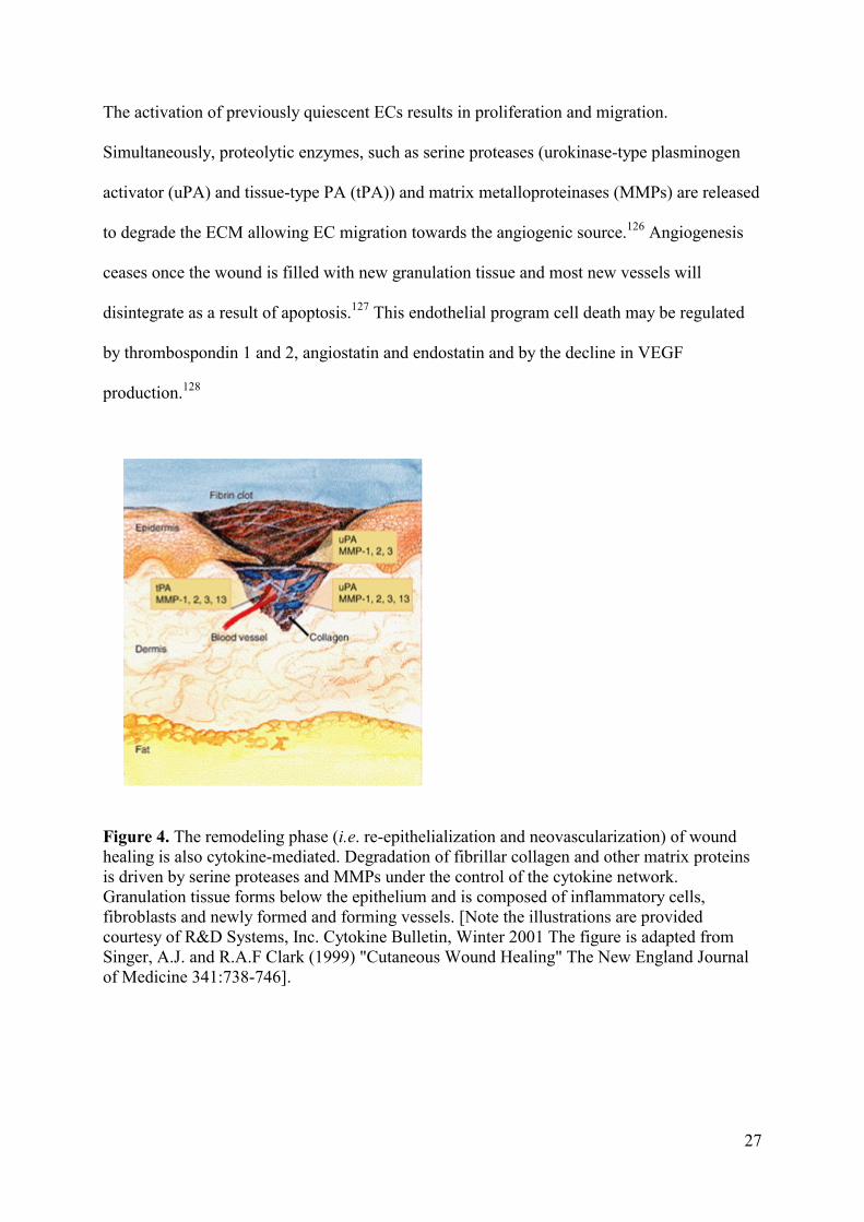
Angiogenesis
The series of events leading to new vessel growth is complex. Angiogenesis is already
initiated minutes after wounding and during wound healing phase tube formation is completed
(Figure 4). Local acidosis, hypoxia due to tissue and vessel destruction and local induction of
pro-angiogenic factors attribute to the initiation of angiogenesis.
27
The activation of previously quiescent ECs results in proliferation and migration.
Simultaneously, proteolytic enzymes, such as serine proteases (urokinase-type plasminogen
activator (uPA) and tissue-type PA (tPA)) and matrix metalloproteinases (MMPs) are released
to degrade the ECM allowing EC migration towards the angiogenic source.126 Angiogenesis
ceases once the wound is filled with new granulation tissue and most new vessels will
disintegrate as a result of apoptosis.127 This endothelial program cell death may be regulated
by thrombospondin 1 and 2, angiostatin and endostatin and by the decline in VEGF
production.128
Figure 4. The remodeling phase (i.e. re-epithelialization and neovascularization) of wound healing is also cytokine-mediated. Degradation of fibrillar collagen and other matrix proteins is driven by serine proteases and MMPs under the control of the cytokine network. Granulation tissue forms below the epithelium and is composed of inflammatory cells, fibroblasts and newly formed and forming vessels. [Note the illustrations are provided courtesy of R&D Systems, Inc. Cytokine Bulletin, Winter 2001 The figure is adapted from Singer, A.J. and R.A.F Clark (1999) "Cutaneous Wound Healing" The New England Journal of Medicine 341:738-746].
28
PHASE 3: SCAR TISSUE REMODDELING
Collagen remodeling to scar formation is a dynamic process. Clinically this is the most
important phase of healing since the rate, quality and quantity of matrix deposition determines
the strength of the scar. Despite the increase in wound strength, the healed wound is at 70%
maximum strength when compared to uninjured skin.
WOUND FLUID
Wound fluid is an exudate composed of cell lysate and products secreted by different cells.
Wound fluid is believed to reflect the local wound environment and represents the sum of all
local specific activities at the time of harvest. Various quantitative cytokine analysis studies in
wound fluid, peritoneal fluid and serum indicated that the local cytokine production is much
higher than found in the circulation.129-131 Functional analysis of wound fluid suggested that
wound fluid taken in the early healing stages increased the proliferation of fibroblasts and EC,
whereas fluid taken from later wound healing phases (day 15) decreased the proliferation of
these cells.132-134 Wound fluid stimulates the synthesis of collagen.133-134
Very limited clinical studies have focused on the pro-and anti-angiogenic balance in wound
fluid and in the circulation of patients, who underwent surgery because of cancer or other
reasons.
SURGERY AND WOUND HEALING
Tumor cells shedded in the circulation during oncologic surgery have been detected135,136 and
are of main concern. Dormant micrometastasis are biologically active with a rate of cell
proliferation equal to the rate of apoptosis, with no net growth of the metastasis as a result.137
The pro-angiogenic environment during wound healing may contribute to the genesis of
recurrent disease, locally and at distance.
29
In 1860 Virchow’s studies implied that inflammation during wound healing could function as
an initiator of tumor growth. This early concept has been confirmed by current experimental
studies that tumors specifically developed in injured tissue and as the wound healed their
ability to grow and implant decreased.138 Bogden et al. demonstrated that surgical wounding
of normal tissues significantly stimulated tumor growth at distance.139 Recently it was
demonstrated that pro-angiogenic factors which are generated during wound healing are also
involved in the outgrowth of tumors.140 An immediate increase of circulating VEGF in
patients after pulmonary metastasis resection was observed and it has been shown
experimentally that VEGF administration resulted in rapid outgrowth of micrometastases.
This outgrowth was abolished by an anti-angiogenesis treatment.51
In addition, an intact primary tumor can regulate growth of micrometastasis through
production of anti-angiogenic factors, notably angiostatin and endostatin. Surgical removal of
a primary tumor removes the sources of these inhibitors, and might allow growth of
previously dormant micrometastases.87 This observation has only been demonstrated in
animal models but may well exist in humans.
Angiogenesis blocking strategies make use of a wide array of direct, EC targeting, and
indirect, influencing the EC microenvironment, anti-angiogenesis agents. Various
experimental and clinical anti-angiogenic trials are focused on sustaining perpetual
micrometastases in dormancy.104,141-145 The use of postoperative anti-angiogenic agents in
surgical cancer patients is tempting in order to control the excessive production of pro-
angiogenic growth factors, which are generated as a physiological response in the early
postoperative period. However, these clinical studies are limited, mostly due to an
understandable fear of impaired angiogenesis during wound healing.
Roman et al. administered perioperatively the antiangiogenic agent SU5416, an inhibitor of
signaling via the VEGFR, in an experimental placebo controlled study in which he performed
30
a right pulmonary lobectomy and biopsies. Interestingly no gross effect on wound healing in
treatment groups was observed. In addition, no drug-related impairment of histologic healing
or decrease in wound tensile strength was demonstrated.146
Another experimental study evaluated the effect of postoperative continuous or discontinuous
angiostatin treatment on the healing of colonic anastomoses. They suggested that the
anastomotic healing was impaired when angiostatin was continuously administered, whereas
normal colonic healing was restored when the anti-angiogenic agent was preoperative
discontinued.147
In conclusion, anti-angiogenic therapy combined with surgical treatment may be a future
strategy for induction of remission by maintaining tumor cells dormant. However, the timing
of anti-angiogenic administration is essential, as wound healing (skin, bowel anastomoses,
and liver regeneration) may be impaired by anti-angiogenic agents.
SCOPE OF THE THESIS
This thesis is divided in three parts and addresses the peri-operative angiogenic balance
between stimulators and inhibitors of the wound healing process.
PART I
A common pathway in response to trauma is initiated first by interleukin-1 (IL-1) and tumor
necrosis factor-α (TNF-α), followed by interleukin-6 (IL-6) and interleukin-8 (IL-8). This
consequently recruits immune cells required for host defense and wound healing.
The aims of Chapter 2 were to study the generation of some important pro-inflammatory
cytokines in wound fluid and in the circulation after conventional and minimal invasive
surgery in patients with a primary colon carcinoma. In addition, the systemic immune
responses after both procedures are investigated.
31
PART II Cancers generate VEGF, which may accumulate in their direct environment and increase in
the circulation. Therefore, we first wanted to study the effect of surgery on the angiogenic
balance in non-cancerous patients in which minimal invasive versus conventional surgery was
performed.
The aim of Chapter 3 was to investigate the effect of the extent of operative trauma of
patients without cancer (laparoscopic versus conventional Nissen fundoplication) on the
angiogenic balance of VEGF and endostatin in plasma.
In Chapter 4 we examined the VEGF and endostatin profile in wound fluid and blood of
patients undergoing breast surgery. Two patient groups were compared, group I had a breast
carcinoma and group II were female-to-male transsexuals undergoing mastectomy.
In Chapter 5 local and systemic angiogenic changes of VEGF and endostatin in patients
undergoing laparoscopic or open surgery for colon cancer were investigated.
PART III Peri-operative immune therapy is a valuable option to avoid postoperative infections. Various
studies have investigated the immunomodulatory response of a wide array of cytokines. These
cytokines have anti- (interferon, e.g.) or pro-angiogenic potential, but the effect on
angiogenesis has not been studied extensively. RhGM-CSF has been widely used to stimulate
the immune system. In addition, it has been suggested that rhGM-CSF affects the process of
angiogenesis via multiple pathways. The aim of Chapter 6 was to investigate the effects of
surgery with or without rhGM-CSF on angiogenic parameters, notably VEGF, endostatin and
angiostatin, in patients with a colorectal carcinoma.
Perioperative recombinant bactericidal/permeability-increasing protein (rBPI21)
administration in patients undergoing liver surgery resulted in a reduced incidence of
postoperative infectious complications. Recently it was demonstrated that rBPI21 had also

32
anti-angiogenic capacity. Chapter 7 is a double blind randomized controlled study in which
patients with metastasized colorectal carcinoma were enrolled to investigate the effect of liver
surgery, with perioperative rBPI21 or placebo administration, on circulatory angiogenic
cytokines.
33
REFERENCES
1. Folkman J. Seminars in Medicine of the Beth Israel Hospital, Boston. Clinical
applications of research on angiogenesis. N Engl J Med. 1995;333:1757-63.
2. Tonnesen MG, Feng X, Clark RA. Angiogenesis in wound healing. J Investig
Dermatol Symp Proc. 2000;5:40-6. 3. Singer AJ, Clark RA. Cutaneous wound healing. N Engl J Med. 1999;341:738-
46.
4. Dinarello CA. Proinflammatory cytokines. Chest 2000;118:503-8.
5. Mayers I, Johnson D. The nonspecific inflammatory response to injury. Can J
Anaesth. 1998;45:871-9.
6. Helmy SA, Wahby MA, El-Nawaway M. The effect of anaesthesia and surgery on
plasma cytokine production. Anaesthesia 1999;54:733-8.
7. Raines EW, Dower SK, Ross R. Interleukin-1 mitogenic activity for fibroblasts
and smooth muscle cells is due to PDGF-AA. Science 1989 ;243:393-6.
8. Fan TP, Hu DE, Guard S, Gresham GA, Watling KJ. Stimulation of angiogenesis
by substance P and interleukin-1 in the rat and its inhibition by NK1 or
interleukin-1 receptor antagonists. Br J Pharmacol. 1993;110:43-9.
9. Frater-Schroder M, Risau W, Hallmann R, Gautschi P, Bohlen P. Tumor necrosis
factor type alpha, a potent inhibitor of endothelial cell growth in vitro, is
angiogenic in vivo. Proc Natl Acad Sci U S A 1987;84:5277-81.
10. Witte MB, Barbul A. General principles of wound healing. Surg Clin North Am.
1997;77:509-28.
11. Robaye B, Mosselmans R, Fiers W, Dumont JE, Galand P. Tumor necrosis
factor induces apoptosis (programmed cell death) in normal endothelial cells in
vitro. Am J Pathol. 1991;138:447-53.

34
12. Maas JW, Calhaz-Jorge C, ter Riet G, Dunselman GA, de Goeij AF,
Struijker-Boudier HA. Tumor necrosis factor-alpha but not interleukin-1 beta or
interleukin-8 concentrations correlate with angiogenic activity of peritoneal fluid
from patients with minimal to mild endometriosis. Fertil Steril. 2001;75:180-5.
13. McCourt M, Wang JH, Sookhai S, Redmond HP. Proinflammatory mediators
stimulate neutrophil-directed angiogenesis. Arch Surg. 1999;134:1325-31.
14. Gallucci RM, Sugawara T, Yucesoy B, et al. Interleukin-6 treatment augments
cutaneous wound healing in immunosuppressed mice. J Interferon Cytokine Res.
2001;21:603-9.
15. Biffl WL, Moore EE, Moore FA, Peterson VM. Interleukin-6 in the injured
patient. Marker of injury or mediator of inflammation? Ann Surg. 1996;224:647-
64.
16. Cohen T, Nahari D, Cerem LW, Neufeld G, Levi BZ. Interleukin 6 induces the
expression of vascular endothelial growth factor. J Biol Chem. 1996;271:736-41.
17. Lin ZQ, Kondo T, Ishida Y, Takayasu T, Mukaida N. Essential involvement of
IL-6 in the skin wound-healing process as evidenced by delayed wound healing
in IL-6-deficient mice. J Leukoc Biol. 2003;73:713-21.
18. Baigrie RJ, Lamont PM, Kwiatkowski D, Dallman MJ, Morris PJ. Systemic
cytokine response after major surgery. Br J Surg. 1992;79:757-60.
19. Sakamoto K, Arakawa H, Mita S, et al. Elevation of circulating interleukin 6 after
surgery: factors influencing the serum level. Cytokine 1994;6:181-6.
20. Pera M, Nelson H, Rajkumar SV, Young-Fadok TM, Burgart LJ. Influence of
postoperative acute-phase response on angiogenesis and tumor growth: open vs.
laparoscopic-assisted surgery in mice. J Gastrointest Surg. 2003;7:783-90.
35
21. Holzheimer RG, Steinmetz W. Local and systemic concentrations of pro- and
anti-inflammatory cytokines in human wounds. Eur J Med Res. 2000;5:347-55.
22. van Berge Henegouwen MI, van der Poll T, van Deventer SJ, Gouma DJ.
Peritoneal cytokine release after elective gastrointestinal surgery and
postoperative complications. Am J Surg. 1998;175:311-6.
23. Lin E, Calvano SE, Lowry SF. Inflammatory cytokines and cell response in
surgery. Surgery 2000;127:117-26.
24. Larsen CG, Anderson AO, Appella E, Oppenheim JJ, Matsushima K. The
neutrophil-activating protein (NAP-1) is also chemotactic for T lymphocytes.
Science 1989;243:1464-6.
25. Koch AE, Polverini PJ, Kunkel SL, et al. Interleukin-8 as a macrophage-derived
mediator of angiogenesis. Science 1992;258:1798-801.
26. Petzelbauer P, Watson CA, Pfau SE, Pober JS. IL-8 and angiogenesis: evidence
that human endothelial cells lack receptors and do not respond to IL-8 in vitro.
Cytokine 1995;7:267-72.
27. Oppenheim JJ, Zachariae CO, Mukaida N, Matsushima K. Properties of the novel
proinflammatory supergene "intercrine" cytokine family. Annu Rev Immunol.
1991;9:617-48.
28. Miller MD, Krangel MS. Biology and biochemistry of the chemokines: a family
of chemotactic and inflammatory cytokines. Crit Rev Immunol. 1992;12:17-46.
29. Rennekampff HO, Hansbrough JF, Kiessig V, Dore C, Sticherling M, Schroder
JM. Bioactive interleukin-8 is expressed in wounds and enhances wound healing.
J Surg Res. 2000;93:41-54.

36
30. Decker D, Lindemann C, Low A, Bidlingmaier F, Hirner A, von Ruecker A.
Changes in the cytokine concentration (Il-6, Il-8, Il-1ra) and their cellular
expression of membrane molecules (CD25, CD30, HLA-DR) after surgical
trauma. Zentralbl Chir. 1997;122:157-63.
31. Neufeld G, Cohen T, Gengrinovitch S, Poltorak Z. Vascular endothelial growth
factor (VEGF) and its receptors. FASEB J. 1999;13:9-22.
32. Stacker SA, Achen MG. The vascular endothelial growth factor family: signaling
for vascular development. Growth Factors 1999;17:1-11.
33. Waltenberger J, Claesson-Welsh L, Siegbahn A, Shibuya M, Heldin CH. Different
signal transduction properties of KDR and Flt1, two receptors for vascular
endothelial growth factor. J Biol Chem. 1994;269:26988-95.
34. Makinen T, Veikkola T, Mustjoki S, et al. Isolated lymphatic endothelial cells
transduce growth, survival and migratory signals via the VEGF-C/D receptor
VEGFR-3. EMBO J. 2001;20:4762-73.
35. Viac J, Palacio S, Schmitt D, Claudy A. Expression of vascular endothelial growth
factor in normal epidermis, epithelial tumors and cultured keratinocytes. Arch
Dermatol Res. 1997;289:158-63.
36. Frank S, Hubner G, Breier G, Longaker MT, Greenhalgh DG, Werner S.
Regulation of vascular endothelial growth factor expression in cultured
keratinocytes. Implications for normal and impaired wound healing. J Biol Chem.
1995;270:12607-13.
37. Brown LF, Yeo KT, Berse B, et al. Expression of vascular permeability factor
(vascular endothelial growth factor) by epidermal keratinocytes during wound
healing. J Exp Med. 1992;176:1375-9.
37
38. Shimizu H, Miyazaki M, Wakabayashi Y, et al. Vascular endothelial growth
factor secreted by replicating hepatocytes induces sinusoidal endothelial cell
proliferation during regeneration after partial hepatectomy in rats. J Hepatol.
2001;34:683-9.
39. Grugel S, Finkenzeller G, Weindel K, Barleon B, Marme D. Both v-Ha-Ras and
v-Raf stimulate expression of the vascular endothelial growth factor in NIH 3T3
cells. J Biol Chem. 1995;270:25915-9.
40. Salven P, Orpana A, Joensuu H. Leukocytes and platelets of patients with cancer
contain high levels of vascular endothelial growth factor. Clin Cancer Res. 1999;
5:487-91.
41. Yamamoto Y, Toi M, Kondo S, et al. Concentrations of vascular endothelial
growth factor in the sera of normal controls and cancer patients. Clin Cancer Res.
1996;2:821-6.
42. Sheng H, Aoe M, Doihara H, Andou A, Shimizu N. Prognostic value of vascular
endothelial growth factor expression in primary lung carcinoma. Acta Med
Okayama 2000;54:119-26.
43. Tae K, El-Naggar AK, Yoo E, et al. Expression of vascular endothelial growth
factor and microvessel density in head and neck tumorigenesis. Clin Cancer Res.
2000;6:2821-8.
44. Shen GH, Ghazizadeh M, Kawanami O, et al. Prognostic significance of vascular
endothelial growth factor expression in human ovarian carcinoma. Br J Cancer
2000;83:196-203.
45. Paradis V, Lagha NB, Zeimoura L, et al. Expression of vascular endothelial
growth factor in renal cell carcinomas. Virchows Arch. 2000;436:351-6.

38
46. Lee JC, Chow NH, Wang ST, Huang SM. Prognostic value of vascular endothelial
growth factor expression in colorectal cancer patients. Eur J Cancer 2000;
36:748-53.
47. Gasparini G, Toi M, Gion M, et al. Prognostic significance of vascular endothelial
growth factor protein in node-negative breast carcinoma. J Natl Cancer Inst.
1997;89:139-47.
48. Verheul HM, Jorna AS, Hoekman K, Broxterman HJ, Gebbink MF, Pinedo HM.
Vascular endothelial growth factor-stimulated endothelial cells promote adhesion
and activation of platelets. Blood 2000;96:4216-21.
49. Lee JK, Hong YJ, Han CJ, Hwang DY, Hong SI. Clinical usefulness of serum and
plasma vascular endothelial growth factor in cancer patients: which is the optimal
specimen? Int J Oncol. 2000;17:149-52.
50. Bondestam J, Salven P, Jaaskela-Saari H, et al. Major surgery increases serum
levels of vascular endothelial growth factor only temporarily. Am J Surg.
2000;179:57-9.
51. Maniwa Y, Okada M, Ishii N, Kiyooka K. Vascular endothelial growth factor
increased by pulmonary surgery accelerates the growth of micrometastases in
metastatic lung cancer. Chest 1998;114:1668-75.
52. Salgado R, Vermeulen PB, Benoy I, et al. Platelet number and interleukin-6
correlate with VEGF but not with bFGF serum levels of advanced cancer patients.
Br J Cancer 1999;80:892-7.
53. Howdieshell TR, Riegner C, Gupta V, et al. Normoxic wound fluid contains high
levels of vascular endothelial growth factor. Ann Surg. 1998;228:707-15.

39
54. Wu FP, Hoekman K, Meijer S, Cuesta MA. Lessons from studying angiognesis
locally versus at distance: VEGF and endostatin levels in wound fluid and plasma
after breast surgery. Angiogenesis 2003;6:255 -7.
55. Hormbrey E, Han C, Roberts A, McGrouther DA, Harris AL. The relationship of
human wound vascular endothelial growth factor (VEGF) after breast cancer
surgery to circulating VEGF and angiogenesis. Clin Cancer Res. 2003;9:4332-9.
56. Howdieshell TR, Callaway D, Webb WL, et al. Antibody neutralization of
vascular endothelial growth factor inhibits wound granulation tissue formation. J
Surg Res. 2001;96:173-82.
57. Gerber HP, McMurtrey A, Kowalski J, et al. Vascular endothelial growth factor
regulates endothelial cell survival through the phosphatidylinositol 3'-kinase/Akt
signal transduction pathway. Requirement for Flk-1/KDR activation. J Biol
Chem. 1998;273:30336-43.
58. Lazarou SA, Barbul A, Wasserkrug HL, Efron G. The wound is a possible source
of posttraumatic immunosuppression. Arch Surg. 1989;124:1429-31.
59. Ohm JE, Carbone DP. VEGF as a mediator of tumor-associated
immunodeficiency. Immunol Res. 2001;23:263-72.
60. Gabrilovich DI, Chen HL, Girgis KR, et al. Production of vascular endothelial
growth factor by human tumors inhibits the functional maturation of dendritic
cells. Nat Med. 1996;2:1096-103.
61. Price DJ, Miralem T, Jiang S, Steinberg R, Avraham H. Role of vascular
endothelial growth factor in the stimulation of cellular invasion and signaling of
breast cancer cells. Cell Growth Differ. 2001;12:129-35.
62. Florkiewicz RZ, Baird A, Gonzalez AM. Multiple forms of bFGF: differential
nuclear and cell surface localization. Growth Factors 1991;4:265-75.

40
63. Bastaki M, Nelli EE, Dell'Era P, et al. Basic fibroblast growth factor-induced
angiogenic phenotype in mouse endothelium. A study of aortic and microvascular
endothelial cell lines. Arterioscler Thromb Vasc Biol. 1997;17:454-64.
64. Basilico C, Moscatelli D.The FGF family of growth factors and oncogenes. Adv
Cancer Res. 1992;59:115-65.
65. Baird A, Klagsbrun M. The fibroblast growth factor family. Cancer Cells
1991;3:239-43.
66. Cordon-Cardo C, Vlodavsky I, Haimovitz-Friedman A, Hicklin D, Fuks Z.
Expression of basic fibroblast growth factor in normal human tissues. Lab Invest.
1990;63:832-40.
67. Schweigerer L, Neufeld G, Friedman J, Abraham JA, Fiddes JC, Gospodarowicz D.
Capillary endothelial cells express basic fibroblast growth factor, a mitogen that
promotes their own growth. Nature 1987;325:257-9.
68. Brunner G, Nguyen H, Gabrilove J, Rifkin DB, Wilson EL. Basic fibroblast
growth factor expression in human bone marrow and peripheral blood cells.
Blood 1993;81:631-8.
69. D'Amore PA. Modes of FGF release in vivo and in vitro. Cancer Metastasis Rev.
1990;9:227-38.
70. Folkman J, Klagsbrun M, Sasse J, Wadzinski M, Ingber D, Vlodavsky I. A
heparin-binding angiogenic protein--basic fibroblast growth factor--is stored
within basement membrane. Am J Pathol. 1988;130:393-400.
71. Nissen NN, Polverini PJ, Gamelli RL, DiPietro LA. Basic fibroblast growth factor
mediates angiogenic activity in early surgical wounds. Surgery 1996;119:457-65.

41
72. Vlodavsky I, Korner G, Ishai-Michaeli R, Bashkin P, Bar-Shavit R, Fuks Z.
Extracellular matrix-resident growth factors and enzymes: possible involvement in
tumor metastasis and angiogenesis. Cancer Metastasis Rev. 1990;9:203-26.
73. Takanami I, Tanaka F, Hashizume T, et al. The basic fibroblast growth factor and
its receptor in pulmonary adenocarcinomas: an investigation of their expression as
prognostic markers. Eur J Cancer 1996;32A:1504-9.
74. Ueki T, Koji T, Tamiya S, Nakane PK, Tsuneyoshi M. Expression of basic
fibroblast growth factor and fibroblast growth factor receptor in advanced gastric
carcinoma. J Pathol. 1995;177:353-61.
75. Kurimoto M, Endo S, Hirashima Y, Nishijima M, Takaku A.Elevated plasma
basic fibroblast growth factor in brain tumor patients. Neurol Med Chir (Tokyo).
1996;36:865-8.
76. Brattstrom D, Bergqvist M, Larsson A, et al. Basic fibroblast growth factor and
vascular endothelial growth factor in sera from non-small cell lung cancer
patients. Anticancer Res. 1998;18:1123-7.
77. Landriscina M, Cassano A, Ratto C, et al. Quantitative analysis of basic fibroblast
growth factor and vascular endothelial growth factor in human colorectal cancer. Br J
Cancer 1998;78:765-70.
78. Volm M, Koomagi R, Mattern J, Stammler G. Prognostic value of basic fibroblast
growth factor and its receptor (FGFR-1) in patients with non-small cell lung
carcinomas. Eur J Cancer 1997;33:691-3.
79. Westphal JR, Van 't Hullenaar R, Geurts-Moespot A, et al. Angiostatin generation
by human tumor cell lines: involvement of plasminogen activators. Int J Cancer
2000;86:760-7.
42
80. Gately S, Twardowski P, Stack MS, et al. The mechanism of cancer-mediated
conversion of plasminogen to the angiogenesis inhibitor angiostatin. Proc Natl Acad Sci U
S A 1997;94:10868-72.
81. Patterson BC, Sang QA.Angiostatin-converting enzyme activities of human
matrilysin (MMP-7) and gelatinase B/type IV collagenase (MMP-9). J Biol Chem.
1997;272:28823-5.
82. Lijnen HR, Ugwu F, Bini A, Collen D.Generation of an angiostatin-like fragment
from plasminogen by stromelysin-1 (MMP-3). Biochemistry 1998;37:4699-702.
83. Dong Z, Kumar R, Yang X, Fidler IJ. Macrophage-derived metalloelastase is
responsible for the generation of angiostatin in Lewis lung carcinoma. Cell 1997;
88:801-10.
84. MacDonald NJ, Murad AC, Fogler WE, Lu Y, Sim BK. The tumor-suppressing
activity of angiostatin protein resides within kringles 1 to 3. Biochem Biophys Res
Commun. 1999;264:469-77.
85. Cao R, Wu HL, Veitonmaki N, et al. Suppression of angiogenesis and tumor
growth by the inhibitor K1-5 generated by plasmin-mediated proteolysis. Proc
Natl Acad Sci U S A 1999;96:5728-33.
86. Morikawa W, Yamamoto K, Ishikawa S, et al. Angiostatin generation by
cathepsin D secreted by human prostate carcinoma cells. J Biol Chem. 2000;275:38912-
20.
87. O'Reilly MS, Holmgren L, Shing Y, et al. Angiostatin: a novel angiogenesis
inhibitor that mediates the suppression of metastases by a Lewis lung carcinoma. Cell
1994;79:315-28.

43
88. Wu FP, Westphal JR, Hoekman K, et al. The effects of surgery, with or without
rhGM-CSF, on the angiogenic profile of patients treated for colorectal carcinoma.
Cytokine 2004;25:68-72.
89. Scapini P, Nesi L, Morini M, et al. Generation of biologically active angiostatin
kringle 1-3 by activated human neutrophils. J Immunol. 2002;168:5798-804.
90. Arumugam S, Jang YC, Chen-Jensen C, Gibran NS, Isik FF. Temporal activity of
plasminogen activators and matrix metalloproteinases during cutaneous wound
repair. Surgery 1999;125:587-93.
91. Wen W, Moses MA, Wiederschain D, Arbiser JL, Folkman J. The generation of
endostatin is mediated by elastase. Cancer Res. 1999;59:6052-6.
92. Felbor U, Dreier L, Bryant RA, Ploegh HL, Olsen BR, Mothes W. Secreted
cathepsin L generates endostatin from collagen XVIII. EMBO J. 2000;19:1187-
94.
93. Muragaki Y, Timmons S, Griffith CM, et al. Mouse Col18a1 is expressed in a
tissue-specific manner as three alternative variants and is localized in basement
membrane zones. Proc Natl Acad Sci U S A 1995;92:8763-7.
94. Schuppan D, Cramer T, Bauer M, Strefeld T, Hahn EG, Herbst H. Hepatocytes as
a source of collagen type XVIII endostatin. Lancet 1998;352:879-80.
95. Kuroi K, Tanaka C, Toi M. Circulating levels of endostatin in cancer patients.
Oncol Rep. 2001;8:405-9.
96. Hajitou A, Grignet C, Devy L, et al. The antitumoral effect of endostatin and
angiostatin is associated with a down-regulation of vascular endothelial growth
factor expression in tumor cells. FASEB J. 2002;16:1802-4.

44
97. Capillo M, Mancuso P, Gobbi A, et al. Continuous infusion of endostatin inhibits
differentiation, mobilization, and clonogenic potential of endothelial cell
progenitors. Clin Cancer Res. 2003;9:377-82.
98. Suzuki M, Iizasa T, Ko E, et al. Serum endostatin correlates with progression and
prognosis of non-small cell lung cancer. Lung Cancer 2002;35:29-34.
99. Feldman AL, Pak H, Yang JC, Alexander HR Jr, Libutti SK. Serum endostatin
levels are elevated in patients with soft tissue sarcoma. Cancer 2001;91:1525-9.
100. Hansma AHG, van Hensbergen Y, Kuenen BC, et al. A patient with a VEGF and
endostatin producing gastrointestinal autonomic nerve tumor (GANT). J Clin
Pathology 2004;57:536-8. 101. Wu FP, Boelens PG, van Leeuwen PA, et al. The effects of major liver resection,
with or without recombinant bactericidal/permeability-increasing
protein(rBPI21), on the angiogenic profile of patients with metastatic colorectal
carcinoma. J Surg Oncol. 2003;84:137-42.
102. Wu P, Yonekura H, Li H, et al. Hypoxia down-regulates endostatin production
by human microvascular endothelial cells and pericytes. Biochem Biophys Res
Commun. 2001;288:1149-54.
103. Bloch W, Huggel K, Sasaki T, et al. The angiogenesis inhibitor endostatin
impairs blood vessel maturation during wound healing. FASEB J. 2000;
14:2373-6.
104. O'Reilly MS, Boehm T, Shing Y, et al. Endostatin: an endogenous inhibitor of
angiogenesis and tumor growth. Cell 1997;88:277-85.
105. Feldman AL, Alexander HR, Hewitt SM, et al. Effect of retroviral endostatin gene
transfer on subcutaneous and intraperitoneal growth of murine tumors. J Natl Cancer
Inst. 2001;93:1014-20.
45
106. Yokoyama Y, Green JE, Sukhatme VP, Ramakrishnan S. Effect of endostatin on
spontaneous tumorigenesis of mammary adenocarcinoma in a transgenic mouse
model. Cancer Res. 2000;60:4362-5.
107. Yamaguchi N, Anand-Apte B, Lee M, et al. Endostatin inhibits VEGF-induced
endothelial cell migration and tumor growth independently of zinc binding.
EMBO J. 1999;18:4414-23.
108. Abdollahi A, Hahnfeldt P, Maercker C, et al. Endostatin's Antiangiogenic
Signaling Network. Mol Cell. 2004;13:649-63.
109. Sudhakar A, Sugimoto H, Yang C, Lively J, Zeisberg M, Kalluri R. Human
tumstatin and human endostatin exhibit distinct antiangiogenic activities
mediated by alpha v beta 3 and alpha 5 beta 1 integrins. Proc Natl Acad Sci
U S A 2003;100:4766-71.
110. Dhanabal M, Ramchandran R, Waterman MJ, et al. Endostatin induces
endothelial cell apoptosis. J Biol Chem.1999;274:11721-6.
111. Hanai Ji J, Dhanabal M, Karumanchi SA, et al. Endostatin causes G1 arrest of
endothelial cells through inhibition of cyclin D1. J Biol Chem. 2002;277:16464-9.
112. MacDonald NJ, Shivers WY, Narum DL, et al. Endostatin binds tropomyosin. A
potential modulator of the antitumor activity of endostatin. J Biol Chem.
2001;276:25190-6.
113. Dixelius J, Larsson H, Sasaki T, et al. Endostatin-induced tyrosine kinase
signaling through the Shb adaptor protein regulates endothelial cell apoptosis.
Blood 2000;95:3403-11.
114. Chau YP, Lin SY, Chen JH, Tai MH. Endostatin induces autophagic cell death in
EAhy926 human endothelial cells. Histol Histopathol. 2003;18:715-26.

46
115. Weltermann A, Wolzt M, Petersmann K, et al. Large amounts of vascular
endothelial growth factor at the site of hemostatic plug formation in vivo.
Arterioscler Thromb Vasc Biol. 1999;19:1757-60.
116. Szpaderska AM, Egozi EI, Gamelli RL, DiPietro LA. The effect of
thrombocytopenia on dermal wound healing. J Invest Dermatol. 2003;120:1130-
7.
117. Subramaniam M, Saffaripour S, Van De Water L, et al. Role of endothelial
selectins in wound repair. Am J Pathol. 1997;150:1701-9.
118. van den Berg JM, Mul FP, Schippers E, Weening JJ, Roos D, Kuijpers TW.
Beta1 integrin activation on human neutrophils promotes beta2 integrin-mediated
adhesion to fibronectin. Eur J Immunol. 2001;31:276-84.
119. Brown EJ. Phagocytosis. Bioessays 1995;17:109-17.
120. Dovi JV, He LK, DiPietro LA. Accelerated wound closure in neutrophil-depleted
mice. J Leukoc Biol. 2003;73:448-55.
121. Leibovich SJ, Polverini PJ, Shepard HM, Wiseman DM, Shively V, Nuseir N.
Macrophage-induced angiogenesis is mediated by tumour necrosis factor-alpha.
Nature 1987;329:630-2.
122. Crowther M, Brown NJ, Bishop ET, Lewis CE. Microenvironmental influence
on macrophage regulation of angiogenesis in wounds and malignant tumors. J
Leukoc Biol. 2001;70:478-90.
123. Constant JS, Feng JJ, Zabel DD, et al. Lactate elicits vascular endothelial growth
factor from macrophages: a possible alternative to hypoxia. Wound Repair Regen.
2000;8:353-60.
124. Leibovich SJ , Ross R. The role of the macrophage in wound repair. A study
with hydrocortisone and antimacrophage serum. Am. J.Pathol. 1975;78:71-100.

47
125. Abe R, Donnelly SC, Peng T, Bucala R, Metz CN. Peripheral blood fibrocytes:
differentiation pathway and migration to wound sites. J Immunol. 2001;166:7556-62.
126. Pepper MS. Role of the matrix metalloproteinase and plasminogen activator
plasmin systems in angiogenesis. Arterioscler Thromb Vasc Biol. 2001;21:1104-
17.
127. Ilan N, Mahooti S, Madri JA. Distinct signal transduction pathways are utilized
during the tube formation and survival phases of in vitro angiogenesis. J Cell Sci.
1998;11:3621-31.
128. Folkman J. Angiogenesis and angiogenesis inhibition: an overview. EXS.
1997;79:1-8.
129. Tsukada K, Katoh H, Shiojima M, Suzuki T, Takenoshita S, Nagamachi Y.
Concentrations of cytokines in peritoneal fluid after abdominal surgery. Eur J
Surg. 1993;159:475-9.
130. Holzheimer RG, Steinmetz W. Local and systemic concentrations of pro- and
anti-inflammatory cytokines in human wounds. Eur J Med Res. 2000;5:347-55.
131. Tokunaga A, Onda M, Fujita I, et al. Sequential changes in the cell mediators of
peritoneal and wound fluids after surgery. Surg Today 1993;23:841-4.
132. Nissen NN, Polverini PJ, Koch AE, Volin MV, Gamelli RL, DiPietro LA.
Vascular endothelial growth factor mediates angiogenic activity during the
proliferative phase of wound healing. Am J Pathol. 1998;152:1445-52.
133. Regan MC, Kirk SJ, Wasserkrug HL, Barbul A. The wound environment as a
regulator of fibroblast phenotype. J Surg Res. 1991;50:442-8.
134. Pricolo VE, Caldwell MD, Mastrofrancesco B, Mills CD. Modulatory activities
of wound fluid on fibroblast proliferation and collagen synthesis. J Surg Res.
1990;48:534-8.

48
135. Choy A, McCulloch P. Induction of tumour cell shedding into effluent venous
blood breast cancer surgery. Br J Cancer 1996;73:79-82.
136. Weitz J, Kienle P, Lacroix J, et al. Dissemination of tumor cells in patients
undergoing surgery for colorectal cancer. Clin Cancer Res. 1998;4:343-8.
137. Holmgren L, O'Reilly MS, Folkman J. Dormancy of micrometastases: balanced
proliferation and apoptosis in the presence of angiogenesis suppression. Nat
Med. 1995;1:149-53.
138. Murthy SM, Goldschmidt RA, Rao LN, Ammirati M, Buchmann T, Scanlon EF.
The influence of surgical trauma on experimental metastasis. Cancer
1989;64:2035-44.
139. Bogden AE, Moreau JP, Eden PA. Proliferative response of human and animal
tumours to surgical wounding of normal tissues: onset, duration and inhibition.
Br J Cancer 1997;75:1021-7.
140. Hofer SO, Molema G, Hermens RA, Wanebo HJ, Reichner JS, Hoekstra HJ. The
effect of surgical wounding on tumour development. Eur J Surg Oncol.
1999;25:231-43.
141. White CW, Sondheimer HM, Crouch EC, Wilson H, Fan LL. Treatment of
pulmonary hemangiomatosis with recombinant interferon alfa-2a. N Engl J Med.
1989;320:1197-200.
142. Shaheen RM, Ahmad SA, Liu W, et al. Inhibited growth of colon cancer
carcinomatosis by antibodies to vascular endothelial and epidermal growth factor
receptors. Br J Cancer 2001;85:584-9.
143. Ingber D, Fujita T, Kishimoto S, et al. Synthetic analogues of fumagillin that
inhibit angiogenesis and suppress tumour growth. Nature 1990;348:555-7.
49
144. Soh EY, Eigelberger MS, Kim KJ, et al. Neutralizing vascular endothelial
growth factor activity inhibits thyroid cancer growth in vivo. Surgery
2000;128:1059-65.
145. Okamoto K, Oshika Y, Fukushima Y, et al. Inhibition of liver metastasis of
colon cancer by in vivo administration of anti-vascular endothelial growth factor
antibody. Oncol Rep. 1999;6:553-6.
146. Roman CD, Choy H, Nanney L, et al. Vascular endothelial growth factor-
mediated angiogenesis inhibition and postoperative wound healing in rats. J Surg
Res. 2002;105:43-7.
147. te Velde EA, Voest EE, van Gorp JM, et al. Adverse effects of the
antiangiogenic agent angiostatin on the healing of experimental colonic
anastomoses. Ann Surg Oncol. 2002;9:303-9.

50

51
Chapter 2
Systemic and Peritoneal Inflammatory Response After Laparoscopic
or Conventional Colon Resection in Cancer Patients: a Prospective,
Randomized Trial
F.P.K. Wu1, C. Sietses1, B.M.E. von Blomberg2, P.A.M. van Leeuwen1,
S. Meijer1, M.A. Cuesta1
1Department of Surgery, 2Department of Pathology and Immunology, VU
University Medical Center, Amsterdam, The Netherlands.
Diseases of the Colon & Rectum 2003;46:147-55
52
ABSTRACT Purpose: This study is to evaluate differences in both the peritoneal and systemic
immune response following laparoscopic and conventional surgical approaches.
Methods: Patients with a primary carcinoma were prospectively randomized to
curative laparoscopic (n = 12) or conventional (n = 14) colon resection. Pro-inflammatory
cytokines interleukin-6 (IL-6), interleukin-8 (IL-8) and tumor necrosis factor-α (TNF-α) were
measured in the peritoneal drain fluid and in the serum. C-reactive protein (CRP) and
leucocyte counts as well as the differences in leucocyte subpopulations and expression of
human leucocyte antigen-DR (HLA-DR) on monocytes were measured perioperatively.
Results: Significantly higher pro-inflammatory cytokine levels are found in the
peritoneal drain fluid than in the circulation after both procedures. Serum IL-6 and IL-8 levels
were significantly lower 2 hours after laparoscopic surgery compared to the conventional
procedure. Postoperative cellular immune counts and HLA-DR expression normalized earlier
after the laparoscopic approach.
Conclusions: The systemic pro-inflammatory concentrations after both surgical
approaches represent only a small fragment of what is generated in the peritoneal drain fluid.
Even if the immediate pro-inflammatory cytokines in the serum are significantly lower in the
laparoscopic group, the same cytokines locally produced showed no differences, suggesting
that both intra-abdominal approaches are equally traumatic. No differences in cellular
response between the two groups were observed.

53
INTRODUCTION
Surgery, whether conventional (CO) or laparoscopic (LP), is a controlled trauma with
immunologic consequences. The extent and duration of the postoperative immune suppression
depends on the magnitude and type of the intraoperative injury.1 Postoperative immune
suppression may have considerable consequences as it has been related to infectious
complications and the development of tumor metastases in animal studies.2,3 Some clinical
research has focused on the prevention or reversal of this immune suppressive state, by
modulating the operative trauma or by administration of different growth factors, in order to
reduce postoperative morbidity and gain a better prognosis.
In comparing laparoscopic versus conventional surgery, significantly better protection of the
systemic immune system was shown with laparoscopic cholecystectomy and Nissen
fundoplication than with the conventional approach.4-6 The differences between laparoscopic
and conventional colectomy are less convincing. Laparoscopic resection of colorectal cancer
has not gained universal acceptance, because of the fear of port-site metastasis and the fact
that the immunologic advantage of laparoscopy remains controversial in clinical trials and
limited prospective, randomized trials.7-12 To understand the differences between these two
approaches, systemic but also locally inflammatory and immunologic parameters will add
information important to the final clinical outcome.
Circulating pro-inflammatory cytokines such as interleukin-6 (IL-6) and tumor necrosis
factor-α (TNF-α) have been related to the extent and severity of the surgical procedure.13 The
main source for these circulating pro-inflammatory cytokines is largely derived from the
operative area. Systemic as well as local measurement in peritoneal wound fluid (PDF) of
these cytokines may provide insight into the differences of operative trauma here considered.
To our knowledge, this has not yet been evaluated in this context.
54
To assess the preservation of the postoperative immunologic defenses, the expression of
human leukocyte antigen-DR (HLA-DR) on monocytes proved to be a reliable parameter,14
since HLA-DR molecules are a prerequisite for effective antigen presentation and play an
important role in the immune response.14
In this prospective, randomized study, the systemic and local acute inflammatory responses as
well as the immunologic consequences of both surgical procedures have been evaluated. The
primary endpoints of the study were to demonstrate differences in both local and systemic
immune parameters by evaluating pro-inflammatory cytokines (IL-6, interleukin-8 (IL-8),
TNF-α, and C-reactive protein (CRP)), leukocyte counts, and the differences in leukocyte
subpopulations and HLA-DR expression after laparoscopic and conventional colon resection
for cancer.

55
STUDY DESIGN
Twenty-six patients were enrolled as part of the international multi-center COLOR (colon
cancer laparoscopic or open resection) trial. In this prospective, randomized study, patients
were randomly allocated a computer-generated number, which assigned them to undergo
either a laparoscopic, or conventional curative colon carcinoma resection. The Ethics
Committee of the VU Medical Center, Amsterdam approved this protocol. Informed consent
was obtained from all patients. The inclusion and exclusion criteria are according COLOR
trial.
Peripheral heparinized plasma and serum plain tube samples (7 ml Vacutainer Systems
Europe, Becton Dickinson, from Meylan Cedex France and Plymouth UK, respectively) were
collected preoperatively (baseline), 2 hours, 1 day and 4 days after surgery. A low vacuum
abdovac drainage system (Astra, Rijswijk, the Netherlands) was left at the resection site for
peritoneal fluid drainage. Twenty-four-hour peritoneal fluid production was collected on days
1 and 4. Serum and wound fluid IL-6, IL-8 and TNF-α samples were obtained by
centrifugation for 10 minutes at 3,000 rpm at 40C. All samples were stored in aliquots at
–800C until tested.
Phenotyping of Immune Cells
The following immune parameters were determined in fresh (<6h) heparinized venous blood
after erythrocyte lysis and paraformaldehyde fixation of whole blood samples (Q-prep, the
Coulter Corporation, Miami, FL, USA): the absolute numbers of leucocytes, total lymphocyte
count, the counts of monocytes (CD14+), T-helper lymphocytes (CD4+), T-suppressor
lymphocytes (CD8+), B-lymphocytes (CD19+), natural killer (NK) cells (CD3neg,
CD16/56pos) and the counts of NK-like T-cells (CD3neg, CD16/56pos). In addition, HLA-
DR expression on CD14+ cells was evaluated by FACS analysis (FACStar Plus, Becton
56
Dickinson, San Jose, CA, USA) and expressed as the ratio of fluorescence intensity (mean
fluorescence and peak channel) with and without anti-HLA-DR-FITC. All monoclonal
antibodies were purchased from Becton Dickinson, San Jose USA and used for overnight
staining (each cell pellet with 20 :l 10-fold diluted Moabs or 30-fold diluted anti-HLA-DR, at
4oC in the dark).
Interleukin-6, and -8 and Tumor Necrosis Factor-αααα
IL-6, IL-8 and TNF-α concentrations in serum and PDF were measured using commercially
available enzyme-linked immunosorbent assay kit (Pelikin compact human IL-6, IL-8 and
TNF-α ELISA kits, CLB, Amsterdam, the Netherlands).
C-Reactive protein (CRP)
Plasma CRP levels were measured by the immunoturbidimetric method, using the
BM/Hitachi 705 (Boeringher, Mannheim, Germany).
STATISTICAL ANALYSIS
The results are reported as mean ± standard error of the mean (SEM). The “Statistical
Package for the Social Sciences” (SPSS 7.5 tm) was used to analyze the data. Overall
differences between groups were analyzed by means of a two-way analysis of variance, and if
a significant overall difference between groups was found, the two-sample Mann-Whitney U
test was used. The Wilcoxon Signed Ranks Test for two related samples analyzed differences
within groups. Significance was accepted at a two-tailed P<0.05.

57
RESULTS
The demographic data are shown in Table 1.
The average hospital stay for the laparoscopic group was shorter than in the conventional
group, P=0.02. On the first postoperative day after conventional sigmoidectomy, one patient
required relaparotomy because of hemorrhaging. One patient who had a laparoscopic right
colectomy had a wound abscess that resolved after draining.
Laparoscopy Conventional No. Patients 12 14
Sex (M/F) 2/10 8/6
Age (yr) 66 ± 3 69 ± 2
Astler-Coller (A:B:C) Males Females Hemicolectomy right Sigmoid resection
0:2:0 0:0:10 6 6
0:7:1 0:3:3 8 6
Operative time (minutes)
176 ± 15 143 ± 9
Hospital stay (days)
9 ± 0.5# 12 ± 1
Table 1: Demographic data
Mean ± SEM
# P < 0.05, laparoscopy versus conventional
58
Pro-inflammatory cytokines
Serum IL-6, TNF-α, IL-8 and CRP plasma levels were measured in 12 laparoscopic and 14
conventional patients. In addition, the local production of IL-6, IL-8 and TNF-α was
evaluated in wound fluid. Tables 2 & 3 summarizes PDF and serum levels of respectively
IL-6 and IL-8 matched for five laparoscopic and eleven conventional patients.
Circulating IL-6 and IL-8 are depicted in figures 1 and 2, respectively. In the conventional
group, the serum IL-6 level was highest at two hours postoperatively (P=0.01) and was still
elevated at day +1 (P=0.001), as compared to baseline values. In the laparoscopic group,
increased IL-6 levels were found 2 hours postoperatively (P=0.03) and were highest at day +1
(P=0.01) as compared to baseline values. Between the groups, IL-6 levels were lower in the
laparoscopic group than in the conventional procedure (P=0.04) two hours after surgery.
The IL-6 levels were much higher in the PDF than in the serum. Remarkably, serum levels of
IL-6 dropped at day +4 despite a continuing increase in local production. In the laparoscopic
group, PDF IL-6 concentrations were not significantly lower than in the conventionally
operated patients at day +1 and day +4, respectively P=0.257 and P=0.610. In addition, serum
IL-6 levels did not differ between the two groups at these time points.
All TNF-α levels in both groups were below the detectable level, < 25 pg/ml in the serum and
< 12 pg/ml in the PDF at all time-intervals.
Similar to IL-6 levels, IL-8 serum levels were significantly lower in the laparoscopic group
2 hours after operating (P = 0.03), whereas no differences were found at day +1 and day +4.
Local production, expressed as the PDF IL-8 concentration, was much higher than serum IL-8
levels. As shown in Table 3, no differences were found in local IL-8 production between
laparoscopic and conventional surgery, although PDF levels P=0.04 tended to be higher in the
laparoscopic group at day +4.

59
Interleukin-6 serum
020406080
100120140160180200220
Preop 2 Hrs Day +1 Day +4
Time
pg/m
l LPCO
#
*
*
* *
Figure 1: IL-6 levels in mean ± SEM * P < 0.05, postoperative values compared to baseline levels
# P < 0.05, laparoscopy versus conventional
Interleukin-8 serum
020406080
100120140160180200
Preop 2 Hrs Day +1 Day +4
Time
pg/m
l LPCO
#
Figure 2: IL-8 levels in mean ± SEM # P < 0.05, laparoscopy versus conventional

60
Interleukin-6
Preoperative Day +1 Day +4
Laparoscopy Serum PDF
2.4 ± 0.3
83 ± 7¶ 82.400 ± 28.553
6 ± 1¶ 65.600 ± 17.583
Conventional Serum PDF
6.4 ± 2.6
105 ± 33¶ 139.909 ± 34.026
17 ± 6¶ 80.272 ± 28.277
Table 2: Peritoneal Drain Fluid (PDF)
IL-6 levels in pg/ml
¶ P < 0.05, serum versus PDF
Interleukin-8
Preoperative Day +1 Day +4
Laparoscopy Serum PDF
135 ± 56
67 ± 27¶ 4.209 ± 1.127
134 ± 59¶ 17.972 ± 2.385#
Conventional Serum PDF
126 ± 41
121 ± 40¶ 7.312 ± 1.511
90 ± 30¶ 8.547 ± 2.457
Table 3: Peritoneal Drain Fluid (PDF)
IL-8 levels in pg/ml
¶ P < 0.05, serum versus PDF
# P < 0.05, laparoscopy versus conventional

61
The CRP plasma levels are depicted in Figure 3. Both the laparoscopic and conventional
procedure resulted in increased plasma CRP levels, both with a significant peak increase on
day +1 (LP, P=0.003, CO, P=0.001) and on day +4 (LP, P=0.003, CO P=0.001) when
compared to baseline values. No differences between the two procedures were observed.
CRP
020406080
100120140160
Preop 2 Hrs Day +1 Day +4
time
mg/
ml
LPCO
**
* *
Figure 3: CRP levels in mean ± SEM
* P < 0.05, postoperative values compared to baseline levels
Phenotyping of Immune Cells
To assess the cellular inflammatory response of all the twenty-six patients, WBC, lymphocyte
subpopulations, NK-Cells and CD14 counts were measured and depicted in Table 4. WBC
counts increased on day +1, (LP, P=0.04, CO, P=0.001) in both groups. On day +4, WBC
counts normalized after laparoscopy, whereas counts remained elevated in the conventional
group (P=0.02).
Total lymphocyte counts dropped on day +1 in both groups, as compared to preoperative
values (LP, P=0.007, CO, P=0.006). Again, normalization had begun on day +4 in the
laparoscopic group, whereas total lymphocyte numbers remained low in the conventional
group (P=0.003) when compared to preoperative levels. The ratio of the lymphocyte

62
subpopulations expressing CD4 and CD8 was not significantly changed in either the
laparoscopic or the conventional group, depicted in Figure 4.
After 2 hours, an initial selective increase of NK-cell (CD3neg, CD16/56pos) counts in both
groups was observed. On day +1, a transient decrease in NK counts the laparoscopic group
(P=0.01) was observed when compared to preoperative values. In the conventional group,
NK-cell counts still showed a decrease on day +4 (P=0.02), whereas NK cells had already
normalized by this time in the laparoscopic group.
Increased CD14 counts were already observed 2 hours after laparoscopy and peaked on
day +1 (P=0.01); however, in the conventional group, no significant CD14 changes were
observed. CD19 counts decreased (P=0.02) in the control group only, observed 2 hours after
the procedure.
None of the immune cell parameters showed significant differences between the laparoscopic
and conventional procedures.
CD4/CD8 ratio
020406080
100120140
Preop 2 Hrs Day +1 Day +4
Time
%
LPCO
Figure 4: CD4/CD8 ratio in percentage of preoperative value
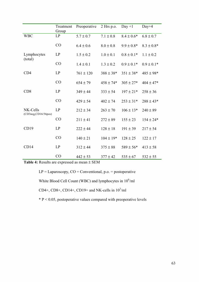
63
Treatment Group
Preoperative 2 Hrs p.o. Day +1 Day+4 WBC LP
CO
5.7 ± 0.7 6.4 ± 0.6
7.1 ± 0.8 8.0 ± 0.8
8.4 ± 0.6* 9.9 ± 0.8*
6.8 ± 0.7 8.3 ± 0.8*
Lymphocytes (total)
LP CO
1.5 ± 0.2 1.4 ± 0.1
1.0 ± 0.1 1.3 ± 0.2
0.8 ± 0.1* 0.9 ± 0.1*
1.1 ± 0.2 0.9 ± 0.1*
CD4 LP CO
761 ± 120 654 ± 79
388 ± 38* 458 ± 74*
351 ± 38* 305 ± 27*
485 ± 98* 404 ± 47*
CD8 LP CO
349 ± 44 429 ± 54
333 ± 54 402 ± 74
197 ± 21* 253 ± 31*
258 ± 36 288 ± 43*
NK-Cells (CD3neg,CD16/56pos)
LP CO
212 ± 34 211 ± 41
263 ± 70 272 ± 89
106 ± 13* 155 ± 23
240 ± 89 154 ± 24*
CD19 LP CO
222 ± 44 140 ± 21
128 ± 18 104 ± 19*
191 ± 39 128 ± 25
217 ± 54 122 ± 17
CD14 LP CO
312 ± 44 442 ± 53
375 ± 88 377 ± 42
589 ± 56* 535 ± 67
413 ± 58 532 ± 55
Table 4: Results are expressed as mean ± SEM
LP = Laparoscopy, CO = Conventional, p.o. = postoperative
White Blood Cell Count (WBC) and lymphocytes in 106/ml
CD4+, CD8+, CD14+, CD19+ and NK-cells in 103/ml
* P < 0.05, postoperative values compared with preoperative levels
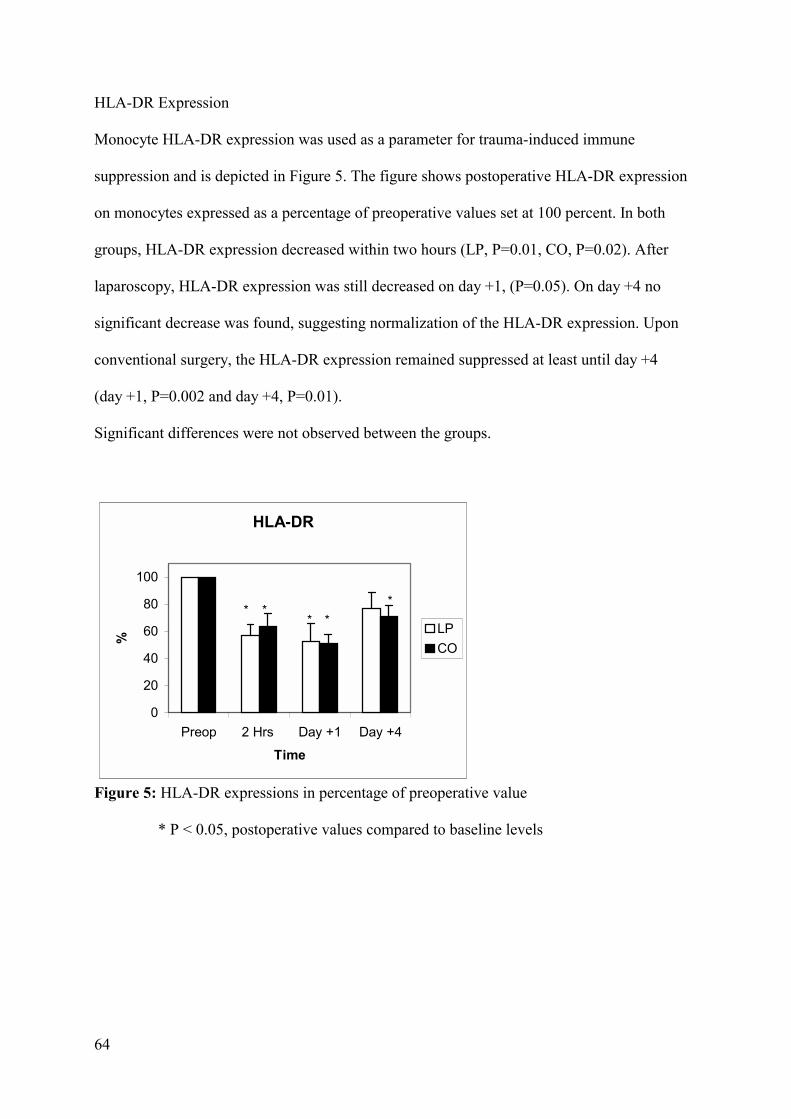
64
HLA-DR Expression
Monocyte HLA-DR expression was used as a parameter for trauma-induced immune
suppression and is depicted in Figure 5. The figure shows postoperative HLA-DR expression
on monocytes expressed as a percentage of preoperative values set at 100 percent. In both
groups, HLA-DR expression decreased within two hours (LP, P=0.01, CO, P=0.02). After
laparoscopy, HLA-DR expression was still decreased on day +1, (P=0.05). On day +4 no
significant decrease was found, suggesting normalization of the HLA-DR expression. Upon
conventional surgery, the HLA-DR expression remained suppressed at least until day +4
(day +1, P=0.002 and day +4, P=0.01).
Significant differences were not observed between the groups.
HLA-DR
0
20
40
60
80
100
Preop 2 Hrs Day +1 Day +4
Time
%
LPCO
* ** *
*
Figure 5: HLA-DR expressions in percentage of preoperative value
* P < 0.05, postoperative values compared to baseline levels
65
DISCUSSION
In this study, the patients who underwent laparoscopic colon carcinoma resection enjoyed a
shorter hospitalization time than patients who underwent conventional surgery. This
observation is in accordance with other laparoscopic vs open colorectal resection studies.15-17
Even if the clinical advantages of laparoscopic approach are clear in procedures such as
cholecystectomy or Nissen fundoplication, in which a grade of immunologic preservation is
recorded, in more advanced laparoscopic procedures such as colon resection, these advantages
become less apparent and sometimes conflictive.
Extensive randomized studies are ongoing in order to demonstrate whether laparoscopic colon
resection for cancer is better or worse than or equal in advantages to the conventional
approach and to give answers concerning cancer survival. Along with this question, the study
of the biologic consequences will offer more insight in the possible advantages of these two
different procedures.
The production of the pro-inflammatory cytokines IL-6, TNF-α, and CRP are accurate
markers of the overall acute-phase response and cytokine levels in the circulation are used to
monitor the impact of surgical trauma.18 In this study, the serum IL-6 levels taken two hours
after surgery were significantly lower after laparoscopic surgery than after conventional
surgery and therefore considered less traumatic.
Localized productions of pro-inflammatory cytokines in the PDF after surgical trauma have
not been well characterized. The inflammatory cascade is initiated by IL-1 and TNF-α,
followed by IL-619 and IL-8. Peritoneal mesothelial cells, fibroblasts, macrophages and
leucocytes are the probable sources of local cytokine.20,21 Several studies have reported
significant postoperative TNF-α levels measured in wound and peritoneal fluids; however,
perioperative circulatory TNF-α changes were undetectable.12,22,23 In this study, serum and
PDF TNF-α levels were undetectable in both groups. This can be explained by the production
66
of soluble TNF receptors I & II that are shed in the circulation in response to the same stimuli
that are known to induce TNF-α. This may compete with other cellular receptors for the
binding of free TNF-α.24
Serum IL-6 is significantly lower two hours after laparoscopic surgery, suggesting that the
laparoscopic approach is initially less traumatic than conventional surgery. In the following
days a diminished IL-6 difference between the two groups is in concordance to a previous
study of ours.25
In both groups, the IL-6 levels in PDF were about a thousand-fold higher than the serum
levels. This supports the idea that IL-6 is generated locally and is compartmentalized in
response to trauma. Interestingly, no differences were found in PDF on day +1 and +4
between the two groups. This suggests that the local dissected area may be equal after
laparocopic and conventional colon surgery. The difference between IL-6 levels in the PDF
and serum observed at day +4 is remarkable. IL-6 levels in PDF were still highly elevated
whereas the IL-6 levels in serum nearly reached preoperative values at day +4. This
observation suggests that circulatory IL-6 is a pivotal factor for the liver to generate CRP; the
increased local IL-6 may play a role in local immune cell recruitment and wound healing.
IL-8 is a potent chemoattractant for neutrophil and lymphocyte as well as an angiogenic
growth factor promoting wound healing.26,27 A couple of observations regarding IL-8 levels
can be made from this study. Firstly, local IL-8 expression is much higher than levels
measured systemically. Finding higher IL-6 and IL-8 expression in the PDF supports the
above-mentioned idea of compartmentalization of cytokines within the abdominal cavity. This
suggests that IL-6 and IL-8 measured systemically represent only a small fragment of what is
generated locally. The precise mechanism of compartmentalization is unclear. It is
conceivable that high local cytokines production and/or the disrupted lymph drainage system
and vascular continuity, due to surgical trauma, may prevent transfer into the circulation. The

67
localized production stimulates the influx of immune cells,28,29 which in turn express
cytokines and growth factors beneficial for wound healing and host defense. Secondly, IL-8
PDF levels increase in the postoperative period. The prolonged and increased IL-8 expression
in the PDF suggests additional cell sources such as endothelial cells and fibroblasts30
contributing to the wound healing process. Finally, the significantly lower systemic IL-8
level, two hours postoperatively and higher IL-8 expression in the PDF at day +4 after
laparoscopic surgery compared to the conventional approach is unclear. It is tempting to
speculate on correlations with pneumoperitoneum, or longer surgical injury and exposure to
anesthesia.
It is unclear to what extent the peritoneal fluid drain contributes to the cytokine production;
however, it is unlikely that the drain itself causes the thousand folds increase.
CRP is a regulatory component of the innate immune system and participates in tissue repair
and regeneration as well.31 In this study, CRP levels were significantly elevated in the
postoperative days following laparoscopic and open colon resection; however, no differences
between the groups were found. This was in contrast to our previous study that showed
significantly lower CRP levels after laparoscopic Nissen fundoplication compared to the
conventional procedure observed at day +1.6 An explanation for this difference is probably
due to trauma size, since Nissen fundoplication is considered less traumatic than colectomy.
Both surgical procedures affect the systemic cellular immune numbers and usually a
postoperative rise in WBC and a decrease in total lymphocytes are found.3,10 In this study,
normalization in WBC and total lymphocyte levels was achieved earlier after laparoscopy
than conventional surgery.
Remarkably, the postoperative CD4/CD8 ratio did not change significantly in either group.
Often the proportion of CD4 lymphocyte decrease is greater than that of CD8 lymphocytes
and it is suggested that the magnitude of the CD4/CD8 ratio drop is related to the size of the
68
surgical trauma.3 The observation of increased CD8 and reduced CD4 activity was more
pronounced after conventional cholecystectomy when compared to the laparoscopic
approach.32
HLA-DR expression on monocytes was measured to assess whether the reduced inflammatory
response would be associated with a preservation of postoperative presenting antigen capacity
of the monocytes. It has been demonstrated that reduced HLA-DR expression and slow
restoration to baseline values after surgery are associated with an increased incidence of
infection.33,34 We have shown previously that HLA-DR expression was better preserved after
laparoscopic cholecystectomy than the conventional approach.35 And in a previous
laparoscopic versus conventional study of Nissen fundoplication, we observed that after
postoperative HLA-DR decrease in both groups, HLA-DR restoration occurred earlier (within
one day) in the laparoscopic group.
In the present study on colectomy, HLA-DR restoration was observed in the laparoscopic
group on day +4. The degree of operative trauma may determine the rate of HLA-DR
restoration, since colectomy is considered major trauma and Nissen fundoplication moderate.
Between the groups we did not find statistical differences in HLA-DR expression at the fourth
postoperative day, as in the Ordeman study.10
In conclusion, in clinical terms, the patients who underwent laparoscopic surgery enjoyed a
shorter hospitalization. Immunologically, large amounts of pro-inflammatory cytokines are
produced in the peritoneal drain fluid after both procedures and the systemic responses after
both surgical approaches are a small reflection of these local events. In the early postoperative
period the pro-inflammatory cytokines levels generated locally suggests that both approaches
is equally traumatic. No differences in cellular response between the two groups were
observed.
69
REFERENCES
1. Allendorf JD, Bessler M, Whelan RL, et al. Postoperative immune function varies
inversely with the degree of surgical trauma in a murine model. Surg Endosc. 1997;11:427-
30.
2. Allendorf JD, Bessler M, Horvath KD, Marvin MR, Laird DA, Whelan RL.
Increased tumor establishment and growth after open vs laparoscopic surgery in mice
may be related to differences in postoperative T-cell function. Surg Endosc. 1999;13:233-5.
3. Lennard TW, Shenton BK, Borzotta A, et al. The influence of surgical operations on
components of the human immune system. Br J Surg. 1985;72:771-6.
4. Karayiannakis AJ, Makri GG, Mantzioka A, Karousos D, Karatzas G. Systemic stress
response after laparoscopic or open cholecystectomy: a randomized trial. Br J Surg.
1997;84:467-71.
5. Roumen RM, van Meurs PA, Kuypers HH, Kraak WA, Sauerwein RW. Serum
interleukin-6 and C reactive protein responses in patients after laparoscopic or conventional
cholecystectomy. Eur J Surg. 1992;158:541-4.
6. Sietses C, Wiezer MJ, Eijsbouts QA, et al. A prospective randomized study of the systemic
immune response after laparoscopic and conventional Nissen fundoplication. Surgery
1999;126:5-9.
7. Hewitt PM, Ip SM, Kwok SP, et al. Laparoscopic-assisted vs. open surgery for colorectal
cancer: comparative study of immune effects. Dis Colon Rectum 1998;41:901-9.
8. Santoro E, Carlini M, Carboni F, Feroce A. Colorectal carcinoma: laparoscopic versus
traditional open surgery. A clinical trial. Hepatogastroenterology 1999;46:900-4.
9. Schwenk W, Jacobi C, Mansmann U, Bohm B, Muller JM. Inflammatory response after
laparoscopic and conventional colorectal resections - results of a prospective randomized
trial. Langenbecks Arch Surg. 2000;385:2-9.

70
10. Ordemann J, Jacobi CA, Schwenk W, Stösslein, Müller JM. Cellular and humoral
inflammatory response after laparoscopic and conventional colorectal resections. Results
of a prospective randomized trial. Surg Endosc. 2001;15:600-8.
11. Stage JG, Schulze S, Moller P, et al. Prospective randomized study of laparoscopic versus
open colonic resection for adenocarcinoma. Br J Surg. 1997;84:391-6.
12. Leung KL, Lai PB, Ho RL, et al. Systemic cytokine response after laparoscopic-assisted
resection of rectosigmoid carcinoma: a prospective randomized trial. Ann Surg.
2000;231:506-11.
13. Lin E, Calvano SE, Lowry SF. Inflammatory cytokines and cell response in surgery.
Surgery 2000;127:117-26.
14. Johnston RB. Current concepts: immunology. Monocytes and macrophages. N Engl J
Med. 1988;318:747-52.
15. Psaila J, Bulley SH, Ewings P, Sheffield JP, Kennedy RH. Outcome following
laparoscopic resection for colorectal cancer. Br J Surg. 1998;85:662-4.
16. Lacy AM, Garcia-Valdecasas JC, Pique JM, et al. Short-term outcome analysis of a
randomized study comparing laparoscopic vs open colectomy for colon cancer.
Surg Endosc. 1995;9:1101-5.
17. Delgado S, Lacy AM, Filella X, et al. Acute phase response in laparoscopic and open
colectomy in colon cancer: randomized study. Dis Colon Rectum 2001;44:638-46.
18. Gabay C, Kushner I. Acute-phase proteins and other systemic responses to inflammation.
N Engl J Med. 1999;340:448-54.
19. Kondo T, Ohshima T. The dynamics of inflammatory cytokines in the healing process of
mouse skin wound: a preliminary study for possible wound age determination. Int J Legal
Med. 1996;108:231-6.

71
20. Kinnaert P, De Wilde JP, Bournonville B, Husson C, Salmon I. Direct activation of
human peritoneal mesothelial cells by heat-killed microorganisms. Ann Surg.
1996;224:749-54.
21. Heel KA, Hall JC. Peritoneal defences and peritoneum-associated lymphoid tissue.
Br J Surg. 1996;83:1031-6.
22. Tokunaga A, Onda M, Fujita I, et al. Sequential changes in the cell mediators of
peritoneal and wound fluids after surgery. Surg Today 1993;23:841-4.
23. Wiik H, Karttunen R, Haukipuro K, Syrjala H. Maximal local and minimal systemic
cytokine response to colorectal surgery: the influence of perioperative filgrastim.
Cytokine 2001;14:188-92.
24. Van Zee KJ, Kohno T, Fischer E, Rock CS, Moldawer LL, Lowry SF. Tumor necrosis
factor soluble receptors circulate during experimental and clinical inflammation and can
protect against excessive tumor necrosis factor alpha in vitro and in vivo. Proc Natl Acad
Sci U S A 1992;89:4845-9.
25. Sietses C, Havenith CE, Eijsbouts QA, et al.Laparoscopic surgery preserves monocyte
mediated tumor cell killing in contrast to the conventional approach. Surg Endosc.
2000;14:456-60.
26. Schall TJ, Bacon K, Toy KJ, Goeddel DV. Selective attraction of monocytes and T
lymphocytes of the memory phenotype by cytokine RANTES. Nature 1990;347:669-71.
27. Koch AE, Polverini PJ, Kunkel SL, et al. Interleukin-8 as a macrophage-derived mediator
of angiogenesis. Science 1992;258:1798-801.
28. Rennekampff HO, Hansbrough JF, Kiessig V, Dore C, Sticherling M, Schroder JM.
Bioactive interleukin-8 is expressed in wounds and enhances wound healing. J Surg Res.
2000;93:41-54.

72
29. Schein M, Wittmann DH, Holzheimer R, Condon RE. Hypothesis: compartmentalization
of cytokines in intraabdominal infection. Surgery 1996;119:694-700.
30. Sticherling M, Hetzel F, Schroder JM, Christophers E. Time- and stimulus-dependent
secretion of NAP-1/IL-8 by human fibroblasts and endothelial cells. J Invest Dermatol.
1993;101:573-6.
31. Steel DM, Whitehead AS. The major acute phase reactants: C-reactive protein, serum
amyloid P component and serum amyloid A protein. Immunol Today 1994;15:81-8.
32. Decker D, Schondorf M, Bidlingmaier F, Hirner A, von Ruecker AA. Surgical stress
induces a shift in the type-1/type-2 T-helper cell balance, suggesting down-regulation of
cell-mediated and up-regulation of antibody-mediated immunity commensurate to the
trauma. Surgery 1996;119:316-25.
33. Wakefield CH, Carey PD, Foulds S, Monson JR, Guillou PJ. Changes in major
histocompatibility complex class II expression in monocytes and T cells of patients
developing infection after surgery. Br J Surg. 1993;80:205-9.
34. Cheadle WG, Hershman MJ, Wellhausen SR, Polk HC Jr. HLA-DR antigen expression on
peripheral blood monocytes correlates with surgical infection. Am J Surg. 1991;161:639-
45.
35. Kloosterman T, von Blomberg BM, Borgstein P, Cuesta MA, Scheper RJ, Meijer S.
Unimpaired immune functions after laparoscopic cholecystectomy. Surgery
1994;115:424-8.
73
Chapter 3
The effect of laparoscopic versus conventional Nissen fundoplication
on VEGF and endostatin levels in plasma
F.P.K. Wu1, F.J. Berends1, C. Sietses1, K. Hoekman2, A.H.G. Hansma2,
A. Jorna2, S. Meijer1, M.A Cuesta1
1Department of Surgery, Surgical Gastro-Intestinal & Oncology, 2Department of
Medical Oncology, VU University Medical Center, Amsterdam, The
Netherlands.
International Journal of Surgical Investigation 2002;3:499-504

74
ABSTRACT
Background: Vascular endothelial growth factor (VEGF) and endostatin are
respectively known as an important endogenous stimulator or inhibitor of angiogenesis.
Angiogenesis is essential for wound healing.
The aim of this study was to study the effect of the extent of operative trauma (laparoscopic
versus conventional Nissen fundoplication) on the angiogenic balance of VEGF and
endostatin in plasma.
Methods: Sixteen patients with gastro-esophageal reflux disease were randomly
assigned to undergo either a laparoscopic or a conventional procedure. Postoperative
inflammatory response was assessed by measuring white blood cells (WBC) and C-reactive
protein (CRP). Plasma VEGF and endostatin levels were monitored by an enzyme-linked
immunosorbent assay.
Results: CRP levels one day after surgery were significant lower after laparoscopy
compared to conventional surgery (P=0.01). WBC and endostatin were not different in both
groups. The VEGF level was decreased two hours after the operation in the laparoscopic
group (P=0.04) and returned to preoperative values 24 hours later (P=0.03). In the
conventional group, VEGF levels increased four days after surgery (P=0.01).
Conclusion: The extent of the operative trauma influences the VEGF response to
surgery. No significant change in endostatin levels was seen.
75
INTRODUCTION
The formation of new vessel from pre-existing capillary vessels is called angiogenesis.
Angiogenesis plays an important role in a variety of processes such as wound healing and
tumor growth.1 Under physiological conditions, adult endothelial cells (ECs) are quiescent
and do not proliferate. In response to various stimuli such as trauma and hypoxia, quiescent
ECs become activated.
Vascular endothelial growth factor (VEGF) is considered the most potent inducer of
angiogenesis, since it stimulates EC proliferation, migration and tube formation.2
Furthermore, the importance of VEGF enhances vascular permeability which induces local
effusion of various plasma proteins and consequently migration of endothelial cells into the
injured area.3 Studies have indicated that VEGF is involved early in the wound healing
process.
In response to trauma, platelets aggregate and release growth factors, such as platelet derived
growth factor (PDGF), platelet derived-endothelial cell growth factor (PD-ECGF) and VEGF
which are contained within the α-granules of platelets.4,5
Subsequently, peripheral white blood cells (WBC), such as granulocytes, B and T-
lymphocytes and monocytes will shortly appear in the injured area. Each type of cells are also
able to release VEGF from their secretory granular compartment.6,7,8 In addition, pro-
inflammatory cytokines, such as interleukin-6 (IL-6), interleukin-1β (IL-1β) and tumor
necrosis factor-α (TNF-α) are released locally following trauma. All these cytokines are able
to induce VEGF expression.9-11
Devitalized peripheral tissue is exposed to hypoxia. Tissue pO2 is usually low in the centre of
the wound, increasing gradually as the wound heals. Under hypoxic in vitro conditions, not
only host-derived cells but also tumor cells express VEGF.12,13,14
In contrast to VEGF, endostatin is a potent endogenous anti-angiogenic cytokine.
76
Endostatin specifically inhibits EC proliferation, migration and tube formation in vitro.
Furthermore it has the ability to suppress tumor growth.15
All together, we suppose that the cellular import and cytokines generated locally will result in
a net balance of pro-angiogenic over anti-angiogenic stimuli, which promotes wound healing.
It is well accepted that laparoscopic surgery reduces the extent of the surgical trauma in
comparison to the conventional technique. It can therefore be speculated that a minor trauma
will result in a slighter release of angiogenic factors.
The aim of this randomized study was to compare the influence of laparoscopic and
conventional Nissen fundoplication on the production of VEGF, endostatin, and the
generation of CRP and white blood cells (WBC), representing the inflammatory response.
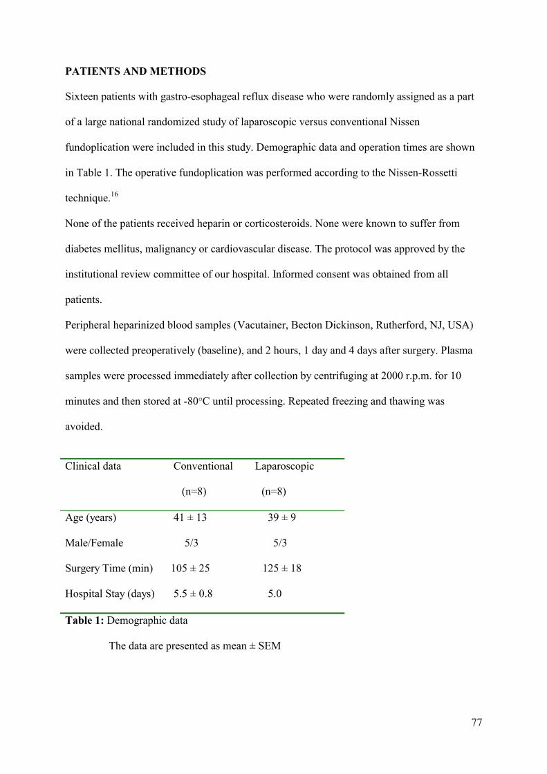
77
PATIENTS AND METHODS
Sixteen patients with gastro-esophageal reflux disease who were randomly assigned as a part
of a large national randomized study of laparoscopic versus conventional Nissen
fundoplication were included in this study. Demographic data and operation times are shown
in Table 1. The operative fundoplication was performed according to the Nissen-Rossetti
technique.16
None of the patients received heparin or corticosteroids. None were known to suffer from
diabetes mellitus, malignancy or cardiovascular disease. The protocol was approved by the
institutional review committee of our hospital. Informed consent was obtained from all
patients.
Peripheral heparinized blood samples (Vacutainer, Becton Dickinson, Rutherford, NJ, USA)
were collected preoperatively (baseline), and 2 hours, 1 day and 4 days after surgery. Plasma
samples were processed immediately after collection by centrifuging at 2000 r.p.m. for 10
minutes and then stored at -80°C until processing. Repeated freezing and thawing was
avoided.
Clinical data Conventional Laparoscopic
(n=8) (n=8)
Age (years)
Male/Female
Surgery Time (min)
Hospital Stay (days)
41 ± 13 39 ± 9
5/3 5/3
105 ± 25 125 ± 18
5.5 ± 0.8 5.0
Table 1: Demographic data
The data are presented as mean ± SEM

78
VEGF
Plasma VEGF165 levels were measured using a commercially available enzyme-linked
immunosorbent assay (ELISA) kit (R & D system Minneapolis, MN). Optical density was
measured at 450 nm using a Spectra Fluor, Tecan plate reader, according to the
manufacturer’s recommended protocol.
Endostatin
Endostatin was measured using a commercial available enzyme-linked immunosorbent assay
(ELISA kit, Cytimmune, College Park, Maryland, U.S.A.). The procedure was according to
the manufacturer’s recommended protocol.
Pheriperal white blood cell count (WBC)
WBC was measured using a Coulter blood analyzer (Coulter, Bastingstoke, England).
C-reactive protein (CRP)
CRP plasma levels were measured by the immunoturbidimetric method, using the BM/Hitachi
705 (Boeringer, Mannheim, Germany).

79
STATISTICAL ANALYSIS
The results are reported as mean ± SEM. The “Statistical Package for the Social Sciences”
(SPSS 7.5 tm) was used to analyze the data. Differences within groups were analyzed by the
Wilcoxon Signed Ranks Test for two related samples. Differences between the groups were
analyzed by means of a two-way analysis of variance. When significant differences between
the two groups were found, the two-sample Mann-Whitney U test was used. Significance was
accepted at a two-tailed P<0.05.
RESULTS
None of the patients suffered any major complications or needed postoperative blood
transfusions; one patient in the conventional group had a wound abcess that resolved after
draining. VEGF, endostatin levels are shown in Table 2, WBC counts and CRP levels are
shown in Figure 1 and 2, respectively.
Groups Preop. 2 Hours Day 1 Day 4
VEGF CO
LP
60 ± 23
78 ± 29
79 ± 37
26 ± 17*
53 ± 16
79 ± 22#
102±21*
55 ± 22
Endostatin
CO
LP
21 ± 4.7
17 ± 2.2
21 ± 3.7
18 ± 1.5
16 ± 3.0
16 ± 2.9
21 ± 3.7
20 ± 2.5
Table 2: Results Mean ± SEM
CO = Conventional, LP = Laparoscopic
VEGF in pg/ml, Endostatin in ng/ml
* P < 0.05, postoperative value compared to preoperative values
# P < 0.05, day +1 value compared to 2 hours after the operation
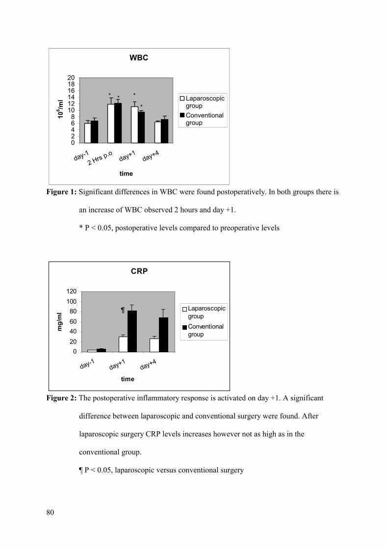
80
WBC
02468
101214161820
day-12 Hrs p.o
day+1day+4
time
106 /m
l LaparoscopicgroupConventionalgroup
* * *
*
Figure 1: Significant differences in WBC were found postoperatively. In both groups there is
an increase of WBC observed 2 hours and day +1.
* P < 0.05, postoperative levels compared to preoperative levels
CRP
020406080
100120
day-1day+1
day+4
time
mg/
ml Laparoscopic
groupConventionalgroup
Figure 2: The postoperative inflammatory response is activated on day +1. A significant
difference between laparoscopic and conventional surgery were found. After
laparoscopic surgery CRP levels increases however not as high as in the
conventional group.
¶ P < 0.05, laparoscopic versus conventional surgery
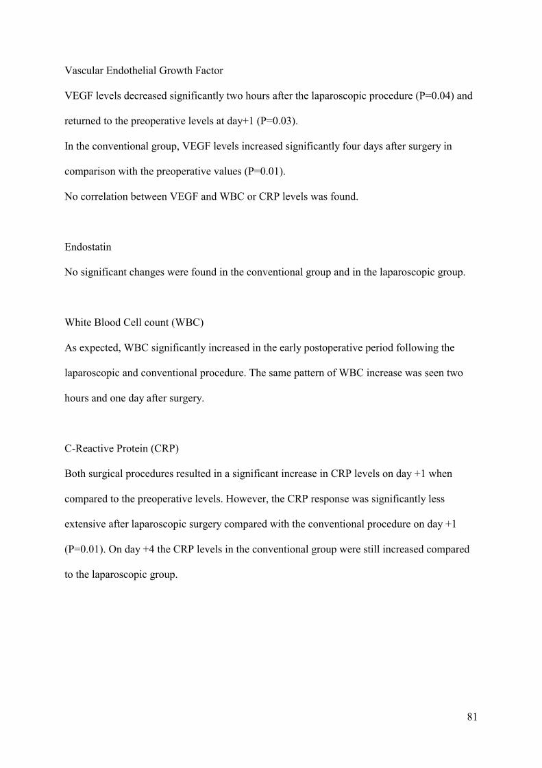
81
Vascular Endothelial Growth Factor
VEGF levels decreased significantly two hours after the laparoscopic procedure (P=0.04) and
returned to the preoperative levels at day+1 (P=0.03).
In the conventional group, VEGF levels increased significantly four days after surgery in
comparison with the preoperative values (P=0.01).
No correlation between VEGF and WBC or CRP levels was found.
Endostatin
No significant changes were found in the conventional group and in the laparoscopic group.
White Blood Cell count (WBC)
As expected, WBC significantly increased in the early postoperative period following the
laparoscopic and conventional procedure. The same pattern of WBC increase was seen two
hours and one day after surgery.
C-Reactive Protein (CRP)
Both surgical procedures resulted in a significant increase in CRP levels on day +1 when
compared to the preoperative levels. However, the CRP response was significantly less
extensive after laparoscopic surgery compared with the conventional procedure on day +1
(P=0.01). On day +4 the CRP levels in the conventional group were still increased compared
to the laparoscopic group.

82
DISCUSSION
The goal of this study was to evaluate the effect of a minor and major trauma on circulatory
VEGF and endostatin levels of patients with a benign disease. The knowledge about the effect
of a surgical trauma and subsequent wound healing on the local and systemic angiogenic
balance is limited.
The healing of the wound is a complex process and has three consecutive phases, first
hemostasis and inflammation (days 0-3 after injury), second the proliferative phase leading to
re-epithelialization and granulation formation (days 3-14 after injury) and finally scar tissue
remodelling (days 7-30 after injury).17,18
The points at which VEGF and endostatin were assessed (preoperative, 2 hours postoperation,
day +1 and day +4) allowed us insight in these circulatory angiogenic factor levels in the first
two phases of wound healing.
A Nissen fundoplication procedure induces an inflammatory response, which was monitored
by means of plasma CRP and WBC. In both groups, WBC increased equally postoperatively.
The increase in CRP was more pronounced in the patients undergoing the conventional
procedure, as could be expected after major surgery.19,20
With respect to VEGF, two significant observations could be made. Firstly, in the
laparoscopic group there was a significant decrease in VEGF level two hours after operation,
returning to preoperative values on day +1. Secondly, a significant increase in VEGF level
was found at day +4 after conventional Nissen procedure. The first observation may be
correlated to insufflation of CO2 for establishing a pneumoperitoneum. Taura et al.
demonstrated that CO2 insufflation at 15 mm Hg caused increased plasma lactic acid which
reached its highest value 60 minutes after the operation.21 Others have confirmed that
increased intra-abdominal pressure due to CO2 insufflation resulted in higher PaCO2 in the
postoperative period after laparoscopic approach.22,23 The decrease of VEGF two hours after

83
the laparoscopic operation may be explained through direct effects of CO2 due to insufflation.
The increase of VEGF to preoperative level on day +1 may be due to the postoperative effect
of acidosis on endothelial cells. D’Arcangelo et al. demonstrated in vitro that hypercarbic
acidosis will induces VEGF expression in bovine aortic endothelial cells (BAECs).24
The significant increase of VEGF at day +4 only found in the conventional group may be
related to the extent of operative trauma. A recent study has shown a transient postoperative
increase in serum VEGF levels on the first and third day after major vascular surgery in
contrast to minor changes after less extensive surgery.25
VEGF is also involved in malignancy.26 Different tumors are able to produce VEGF and a
relationship has been suggested between tumor progression and VEGF levels.27 Maniwa et al.
showed that in patients with lung cancer, the increase of VEGF levels after radical surgery
may have a detrimental effect on outgrowth of micrometastases.28
In contrast with the pro-angiogenic factor VEGF, endostatin is considered an important anti-
angiogenic factor. Endostatin is produced by enzymatic digestion of a protein precursor,
collagen XVIII, and has been shown to reside from basement membranes, vessel walls and
mostly in the liver.29,30 The mechanisms of endostatin generation in vivo are unknown.
Finding endostatin levels in the circulation is interesting and suggests that it may serve as an
homeostatic angiogenic control. Theoretically, a net balance of pro-angiogenic over anti-
angiogenic factors, would switch in the on position, hereby facilitating wound healing.
Nevertheless, in this study the level of endostatin was not influenced by the two surgical
techniques.
In conclusion, the levels of VEGF, WBC and CRP change after surgery and the amount of
surgical trauma influences the degree of these postoperative changes. The unexpected
decrease of VEGF levels immediately after laparoscopic surgery along with unchanged
endostatin levels awaits confirmation by additional studies.

84
REFERENCES
1. Brown LF, Yeo KT, Berse B, Senger DR, Dvorak HF, van de Water L.
Expression of vascular permeability factor (vascular endothelial growth factor)
by epidermal keratinocytes during wound healing. J Exp Med. 1992;176:1375-9.
2. Neufeld G, Cohen T, Gengrinovitch S, Poltorak Z Vascular endothelial
growth factor (VEGF) and its receptors. FASEB J. 1999;13:9-22.
3. Esser S, Wolburg K, Wolburg H, Breier G, Kurzchalia T, Risau W. Vascular
endothelial growth factor induces endothelial fenestrations in vitro. J Cell Biol.
1998;140:947-59.
4. Bussolino F, Albini A, Camussi G, et al. Role of soluble mediators in angiogenesis.
Eur J Cancer 1996;32A:2401-12.
5. Bennet NT, Schultz GS. Growth factors and wound healing: biochemical
properties of growth factors and their receptors. Am J Surg. 1993;165:728-37.
6. Verheul HMW, Hoekman K, Luykx-de Bakker S, et al. Platelets: transporter of vascular
endothelial growth factor. Clin Cancer Res. 1997;3:2187-90.
7. Salven P, Orpana A, Joensuu H. Leucocytes and platelets of patients with
cancer contain high levels of vascular endothelial growth factor. Clin Cancer
Res. 1999;5:487-91.
8. Freeman MR, Schneck FX, Gagnon ML, et al. Peripheral blood T lymphocytes and B
lymphocytes infiltrating human cancers express vascular endothelial growth factor: a
potential role for T cells in angiogenesis. Cancer Res. 1995;55:4140-5.
9. Cohen T, Nahari D, Cerem-Weiss L, Neufeld G, Levi B. Interleukin-6 induces
expression of vascular endothelial growth factor. J Biol Chem. 1996;271:736-41.

85
10. Li J, Perella MA, Tsai JC, et al. Induction of vascular endothelial growth factor
gene expression by interleukin-1 beta in rat aortic smooth muscle cells.
J Biol Chem. 1995;270:308-12.
11. Ryuto M, Ono M, Izumi H, et al. Induction of vascular endothelial growth factor by
tumour necrosis factor alpha in human glioma cells-possible roles of SP-1. J Biol Chem.
1996;271:28220-8.
12. Chang N, Goodson WH, Gottrup F, Hunt TK. Direct measurement of wound
and tissue oxygen tension in postoperative patients. Ann Surg. 1983;197:470-8.
13. Kuwabara K, Ogawa S, Matsumoto M, et al. Hypoxia-mediated induction of acidic/basic
fibroblast growth factor and platelet-derived growth factor in mononuclear phagocytes
stimulates growth of hypoxic endothelial cells. Proc Natl Acad Sci. 1995;92:4606-10.
14. Shweiki D, Itin A, Soffer D, Keshet E. Vascular endothelial growth factor
induced by hypoxia may mediate hypoxia-initiated angiogenesis. Nature 1992;359:843-5.
15. O'Reilly MS, Boehm T, Shing Y, et al. Endostatin: an endogenous inhibitor of
angiogenesis and tumor growth. Cell 1997;88:277-85.
16. Dallemagne B, Weerts JM, Jehaes C, Markiewicz S, Lombard R. Techniques
and results of endoscopic fundoplication. Endosc Surg & All Techn. 1993;1:72-5.
17. Lynch SE. Interactions of growth factors in tissue repair. Prog Clin Biol Res.
1991;365:341-457.
18. Moulin V. Growth factors in skin wound healing. Eur J Cell Biol. 1995;68:1-7.
19. Sietses C, Wiezer MJ, Eijsbouts QA, et al. A prospective randomized study of the
systemic immune response after laparoscopic and conventional Nissen fundoplication.
Surgery 1999;126:5-9.
86
20. Zieren J, Jacobi CA, Wenger FA, Volk HD, Muller JM. Fundoplication: a
model for immunologic aspects of laparoscopic and conventional surgery.
J Laparoendosc Adv Surg Tech A. 2000;10:35-40.
21. Taura P, Lopez A, Lacy AM, et al. Prolonged pneumoperitoneum at 15 mm Hg
causes lactic acidosis. Surg Endosc. 1998;12:198-201.
22. Iwasaka H, Miyakawa H, Yamamoto H, Kitano T, Taniguchi K, Honda N.
Respiratory mechanics and arterial blood gases during and after laparoscopic
cholecystectomy. Can J Anaesth. 1996;43:129-33.
23. Koivusalo AM, Kellokumpu I, Ristkari S, Lindgren L. Splanchnic and renal
deterioration during and after laparoscopic cholecystectomy: a comparison of
the carbon dioxide pneumoperitoneum and the abdominal wall lift method.
Anesth Analg. 1997;85:886-91.
24. D’Arcangelo D, Facchiano F, Barlucchi LM, et al. Acidosis inhibits endothelial cell
apoptosis and function and induces basic fibroblast growth factor and vascular endothelial
growth factor expression. Circ Res. 2000;86:312-8.
25. Bondestam J, Salven P, Jääskelä-Saari H, et al. Major surgery increases serum level of
vascular endothelial growth factor only temporarily. Am J Surg. 2000;179:57-9.
26. Hyodo I, Doi T, Endo H, et al. Clinical significance of plasma vascular endothelial growth
factor in gastrointestinal cancer. Eur J Cancer 1998;34:2041-5.
27. Kumar H, Heer K, Lee PW. Preoperative serum vascular endothelial growth factor can
predict stage in colorectal cancer. Clin Cancer Res. 1998;4:1279-85.
28. Maniwa Y, Okada M, Ishii N, Kiyooka K. Vascular endothelial growth factor
increased by pulmonary surgery accelerates the growth of micrometastases
in metastatic lung cancer. Chest 1998;114:1668-75.
87
29. Muragaki Y, Timmons S, Griffith CM, et al. Mouse Col18a1 is expressed in a tissue-
specific manner as three alternative variants and is localized in basement membrane
zones. Proc Natl Acad Sci U S A 1995;92:8763-7.
30. Schuppan D, Cramer T, Bauer M, Strefeld T, Hahn EG, Herbst H.
Hepatocytes as a source of collagen type XVIII endostatin. Lancet 1998;352:879-80.

88
89
Chapter 4
VEGF and endostatin levels in wound fluid and plasma after breast surgery
F.P.K. Wu1, K. Hoekman2, S. Meijer1, M.A Cuesta1
1Department of Surgery, 2Department of Medical Oncology, VU University
Medical Center, Amsterdam, The Netherlands.
Angiogenesis 2004;6:255-7

90
ABSTRACT
Background and Objectives: Angiogenesis is essential for wound repair after
surgical trauma. Vascular endothelial growth factor (VEGF) and endostatin are endogenous
angiogenic factors involved in the initiation and completion of angiogenesis. The aim of this
study was to examine the local and systemic VEGF and endostatin profiles in patients
undergoing surgery for benign and malignant breast processes.
Methods: A total of sixteen patients with or without cancer underwent breast surgery.
Group I: eight patients with primary breast cancer underwent a simple or radical mastectomy
according to Madden including dissection of axillary lymph nodes. Group II: eight healthy
female-to-male transsexuals underwent subcutaneous mastectomy. VEGF and endostatin
levels in plasma and wound fluid were determined.
Results: In both groups VEGF levels in wound fluid were significantly higher
compared to postoperative plasma levels, whereas wound fluid endostatin levels were lower
than plasma levels and decreased progressively after surgery. In both groups plasma VEGF
and endostatin levels did not change significantly.
Conclusions: The local VEGF increase and endostatin decrease observed immediately
after surgery appears to be a physiological response to operative trauma, which can be studied
more profoundly in locally generated fluid than in blood. This process did not seem to be
influenced by the type of process (cancerous or non-cancerous) involved in the surgical
intervention.

91
INTRODUCTION Angiogenesis in wound repair is essential to restore the delivery of oxygen and nutrients and
the removal of waste products from the injured area. Numerous cells and cytokines are
involved during the three phases of wound healing and the balance of stimulatory or
inhibitory cytokines will determine each phase of the wound healing process.1,2 VEGF is a
cytokine that is considered the most potent inducer of angiogenesis, since it specifically
stimulates endothelial cell (EC) proliferation, migration and tube formation.3 Furthermore,
VEGF enhances vascular permeability, which induces local effusion of various plasma
proteins, facilitates the migration of endothelial cells into the injured area4 and stimulate
granular tissue formation.5 Moreover, in oncology, tumors produce constitutively VEGF as a
consequence of their aberrant genetic profile and because of hypoxia inside tumor tissue. The
VEGF level is found to correlate with tumor progression.6,7
Endostatin, the C-terminal fragment of collagen XVIII, is a potent angiogenesis inhibitor.
Hepatocytes are a major source of collagen XVIII and may contribute to endostatin levels in
the circulation.8 Endostatin specifically inhibits EC migration in vitro9 and potently inhibits
tumor growth in various animal models.10
In the present study we examined the VEGF and endostatin profile in wound fluid and plasma
of patients undergoing curative breast cancer surgery and female-to-male mastectomy. The
main interest was to compare the angiogenic profile in wound fluid with the pattern in the
circulation.

92
PATIENTS AND METHODS
The ethical committee of the Vrije Universiteit Medisch Centrum (VUMC) approved the
protocol. Informed consent was obtained from all patients.
Group I consisted of eight patients with primary breast ductal carcinomas. A simple
mastectomy was carried out on four patients and a drain was left in the resected area. Four
other patients underwent a radical mastectomy according to Madden.
Group II consisted of eight healthy female-to-male transsexuals who underwent a mastectomy
by a transareolar approach to achieve a male chest figure.
The mastectomies in this group were performed on both breasts and a drain was placed at
each side. The wound fluid was collected from the side with the highest wound fluid
production. Group II received an androgen supplement (Sustanon 250 mg i.m., Organon,
The Netherlands) twice a month as part of the sex reassignment treatment.
EDTA blood samples were collected preoperatively (baseline), and 1 day and 4 days after
surgery. Twenty-four hours of wound fluid production was collected on postoperative days 1
and 4. Plasma and wound fluid samples were centrifuged at 2000 rpm for 10 minutes and then
stored at -80°C. Freezing and thawing was avoided.
Detection of VEGF and endostatin
VEGF165 and endostatin levels in plasma and wound fluid were measured using a
commercially available enzyme-linked immunosorbent assay (ELISA) kit (R & D system
Minneapolis, USA). Both intra- and inter-assay variance was less than 10%. The minimum
detectable VEGF level is 9.0 pg/ml and endostatin level is 1.95 ng/ml. The local VEGF and
endostatin concentration may be affected by a dilution effect of wound fluid production in
both groups. Therefore the total VEGF and endostatin production was calculated by

93
multiplying the concentration by volume for each individual patient, followed by calculating
the mean of the levels.
STATISTICAL ANALYSIS
The results are reported as mean ± standard error of the mean (SEM). Overall differences
between groups were analyzed by means of a two-way analysis of variance, and if a
significant overall difference between groups was found, the two-sample Mann-Whitney U
test was used. The Wilcoxon Signed Ranks Test for two related samples analyzed differences
within groups. Significance was accepted at a two-tailed P < 0.05.
RESULTS
The mean age of the patients included was 51.6 ± 4.9 years for group I and 36.6 ± 4.9 for
group II, P=0.04. The average operating time was 128.8 ± 10.9 minutes for group I and 199.3
± 17.5 minutes for group II, P=0.01. None of the patients experienced any postoperative
complication.
The wound fluid production within group I, obtained from the simple mastectomies, (day 1:
70.0 ± 35.6 ml and day 4: 38.8 ± 20.2 ml) and radical Madden mastectomies, (day 1: 90.0 ±
25.8 ml and day 4: 63.8 ± 53.1 ml) was not significantly different.
Four days after surgery the wound fluid production was higher in group I, P=0.001 when
compared to group II, displayed in Figure 1.

94
Wound Fluid Production
020406080
100
day +1 day +4
ml Group I
Group II
#
*
Figure 1: # P < 0.05, differences between the groups
* P < 0.05, day +4 compared to day +1
In both groups, the mean VEGF level in wound fluid was much higher compared to plasma
(Table 1). In group I, an eight-fold VEGF increase in wound fluid, P=0.004, compared with
plasma level was found one day after surgery. After four days, this difference increased to
twenty-four-fold, P=0.018.
In group II, the wound fluid VEGF level increased by eighteen-fold one day after surgery,
P=0.001 and thirty-two-fold four days after surgery, P = 0.001, compared to the plasma level
of the corresponding day. In both groups, the VEGF concentration in wound fluid was higher
at day four compared to day one after surgery, P=0.031. The VEGF levels in plasma and
wound fluid were not significantly different between both groups.
In group I, endostatin wound fluid levels were lower than plasma one day, P=0.03, and four
days, P=0.004 after surgery. The wound fluid endostatin level decreased after four days
compared to day one after surgery, P=0.03. In group II, the endostatin wound fluid level was
lower than the plasma level, four days after surgery, P=0.004. In both groups the wound
endostatin levels decreased significantly from day one to four days after surgery.
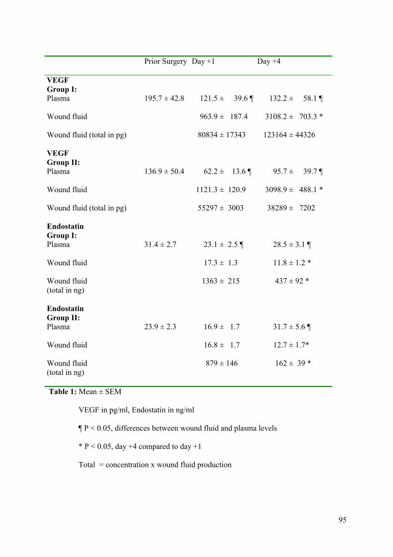
95
Prior Surgery Day +1 Day +4
VEGF Group I: Plasma Wound fluid Wound fluid (total in pg)
195.7 ± 42.8
121.5 ± 39.6 ¶ 963.9 ± 187.4 80834 ± 17343
132.2 ± 58.1 ¶ 3108.2 ± 703.3 * 123164 ± 44326
VEGF Group II: Plasma Wound fluid Wound fluid (total in pg)
136.9 ± 50.4
62.2 ± 13.6 ¶ 1121.3 ± 120.9 55297 ± 3003
95.7 ± 39.7 ¶ 3098.9 ± 488.1 * 38289 ± 7202
Endostatin Group I: Plasma Wound fluid Wound fluid (total in ng)
31.4 ± 2.7
23.1 ± 2.5 ¶ 17.3 ± 1.3 1363 ± 215
28.5 ± 3.1 ¶ 11.8 ± 1.2 * 437 ± 92 *
Endostatin Group II: Plasma Wound fluid Wound fluid (total in ng)
23.9 ± 2.3
16.9 ± 1.7 16.8 ± 1.7 879 ± 146
31.7 ± 5.6 ¶ 12.7 ± 1.7* 162 ± 39 *
Table 1: Mean ± SEM
VEGF in pg/ml, Endostatin in ng/ml
¶ P < 0.05, differences between wound fluid and plasma levels
* P < 0.05, day +4 compared to day +1
Total = concentration x wound fluid production
96
DISCUSSION
Angiogenesis plays an important role in the process of wound healing. Insight into the
mechanisms of angiogenesis is increasing, but little is known about how local angiogenesis
initiated in the wound influences the systemic angiogenic parameters after operative trauma.
We evaluated the profiles of VEGF and endostatin in wound fluid and plasma from patients
operated for breast cancer or transsexual patients undergoing a double mastectomy.
Wound fluid production in the group with cancer was significantly higher four days after
surgery. The fact that their wounded area is larger may explain this.
In this study, the plasma VEGF level was not significantly different between the two groups.
It is unknown whether testosterone treatment in women (group II) has an effect on VEGF
production.
The systemic VEGF levels after breast surgery did not change significantly, however, the
large difference between VEGF in plasma and in wound fluid observed postoperatively in
both groups, is remarkable. It appears that the concurrently measured plasma VEGF reflects
only a fragment of what is generated locally. Local VEGF generation after a surgical
procedure is probably due to instant platelet degranulation, production by recruited immune
cells and indirectly by the expression of pro-inflammatory cytokines and local hypoxia due to
devitalized tissue.11-13 In this study it was unlikely that residual tumor was another source of
VEGF, since the surgical margins were free of tumor.
The role of a high VEGF level in the wound may have two functions, one to initiate
angiogenesis and another to temper the local immune response in the injured area. The
devitalized tissue and exposed self-antigens in a wound contain an intense immunologic
stimulus. VEGF may act as an immunosuppressive agent14,15 that prevents a local autoimmune
reaction.

97
In advanced cancer patients, the VEGF level increase may have negative consequences. A
recent in vitro study showed that breast cancer cells stimulated by VEGF had an increased
invasive character.16 In addition, a VEGF increase induced by surgery resulted in a rapid
outgrowth of micrometastases, which could be abolished by an angiogenesis inhibitor.17
Endostatin levels were also investigated in plasma and wound fluid. Preoperative plasma
endostatin levels were not significantly different between the groups and did not change
significantly after surgery. However, in contrast to VEGF, endostatin levels decreased in
wound fluid in both groups. The mechanism of the decrease of local endostatin levels is
unclear. It may be the effect of the expression of various proteolytic enzymes1,18 during
wound healing that degrade endostatin. Anyhow, the decrease of endostatin in the early phase
of wound healing seems to be a physiological response to injury. Together, the strong increase
in local VEGF and decrease in endostatin supports the idea of a physiological tissue
angiogenic switch in the first phase of wound healing. These changes were comparable in
both groups, which confirm the idea that wound healing after radical surgery for normal and
malignant indications develops through similar physiological mechanisms.
In conclusion, the pattern of change in VEGF and endostatin levels in wound fluid was
similar in both patient groups. Wound fluid was much more informative than blood, which
emphasizes wound fluid as a “conditioned medium” of this physiological process. In addition,
this study showed that angiogenesis is started immediately after surgery. This process did not
seem to be influenced by the type of process, cancerous or non-cancerous) involved in the
surgical intervention.

98
REFERENCES
1. Singer AJ, Clark RA. Cutaneous wound healing. N Engl J Med. 1999;341:738-
46.
2. Lynch SE. Interactions of growth factors in tissue repair. Prog Clin Biol Res.
1991;365:341-57.
3. Neufeld G, Cohen T, Gengrinovitch S, Poltorak Z. Vascular endothelial growth
factor (VEGF) and its receptors. FASEB J. 1999;13:9-22.
4. Esser S, Wolburg K, Wolburg H, et al. Vascular endothelial growth factor
induces endothelial fenestrations in vitro. J Cell Biol. 1998;140:947-59.
5. Howdieshell TR, Callaway D, Webb WL, et al. Antibody neutralization of
vascular endothelial growth factor inhibits wound granulation tissue formation.
J Surg Res. 2001;96:173-82.
6. Shweiki D, Neeman M, Itin A et al. Induction of vascular endothelial growth
factor expression by hypoxia and by glucose deficiency in multicell spheroids:
Implications for tumor angiogenesis. Proc Natl Acad Sci USA 1995;92:768-72.
7. Gasparini G. Prognostic value of vascular endothelial growth factor in breast
cancer. Oncologist 2000;5:37-44.
8. Schuppan D, Cramer T, Bauer M, et al. Hepatocytes as a source of collagen
type XVIII endostatin. Lancet 1998;352:879-80.
9. Dhanabal M, Ramchandran R, Waterman MJ, et al. Endostatin induces
endothelial cell apoptosis. J Biol Chem. 1999;274:11721-6.
10. Yoon SS, Eto H, Lin CM, et al. Mouse endostatin inhibits the formation of lung
and liver metastases. Cancer Res. 1999;59:6251-6.
11. Cohen T, Nahari D, Cerem LW, et al. Interleukin 6 induces the expression of
vascular endothelial growth factor. J Biol Chem. 1996;271:736-41.
99
12. Salven P, Orpana A, Joensuu H. Leukocytes and platelets of patients with cancer
contain high levels of vascular endothelial growth factor. Clin Cancer Res.
1999;5:487-91.
13. Elson DA, Ryan HE, Snow JW, et al. Coordinate up-regulation of hypoxia inducible
factor (HIF)-1alpha and HIF-1 target genes during multi-stage epidermal carcinogenesis
and wound healing. Cancer Res. 2000;60:6189-95.
14. Ohm JE, Carbone DP. VEGF as a mediator of tumor-associated immunodeficiency.
Immunol Res. 2001;23:263-72.
15. Gabrilovich DI, Chen HL, Girgis KR, et al. Production of vascular endothelial growth
factor by human tumors inhibits the functional maturation of dendritic cells. Nat Med.
1996;2:1096-103.
16. Price DJ, Miralem T, Jiang S, et al. Role of vascular endothelial growth factor
in the stimulation of cellular invasion and signaling of breast cancer cells.
Cell Growth Differ. 2001;12:129-35.
17. Maniwa Y, Okada M, Ishii N, Kiyooka K. Vascular endothelial growth factor
increased by pulmonary surgery accelerates the growth of micrometastases in
metastatic lung cancer. Chest 1998;114:1668-75.
18. Arumugum S, Jang YC, Chen-Jensen C, et al. Temporal activity of plasminogen
activators and matrix metalloproteinases during cutaneous wound repair. Surgery
1999;125:587-93.

100
101
Chapter 5
The systemic and local angiogenic response after laparoscopic or open
colon resection in cancer patients: a prospective, randomized trial
F.P.K. Wu1, K. Hoekman2, C. Sietses1, B.M.E. von Blomberg3, S. Meijer1,
H.J. Bonjer4, M.A. Cuesta1
1Department of Surgery, 2Department of Medical Oncology and 3Department of
Pathology and Immunology, VU University Medical Center, Amsterdam and
44Department of Surgery, Dijkzigt Ziekenhuis, Rotterdam, The Netherlands.
Diseases of the Colon & Rectum 2004;47:1670-4

102
ABSTRACT
Background and Objectives: Angiogenesis is essential for wound healing. Vascular
endothelial growth factor and endostatin are both endogenous angiogenic factors, thought to
be involved in the initiation, respectively termination of angiogenesis. The aim of this study
was to assess the local and systemic angiogenic profile in patients undergoing laparoscopic or
open surgery for colon cancer.
Methods: Patients with a primary colon carcinoma were prospectively randomized to
curative laparoscopic (n = 12) or conventional (n = 14) resection. Vascular endothelial growth
factor and endostatin levels in serum and wound fluid were investigated.
Results: In both groups vascular endothelial growth factor levels in wound fluid were
significantly higher compared to postoperative serum levels, whereas endostatin levels in
wound fluid were lower than serum levels and decreased progressively after surgery. The
vascular endothelial growth factor levels in wound fluid measured at day 4 were significantly
higher in the laparoscopy group compared with the laparotomy patients.
Conclusions: Wound healing is associated with a strong local increase in pro-
angiogenic factors and a decrease of anti-angiogenic factors. The investigation of locally
produced factors offered greater insight in the process of angiogenesis during wound healing
than could be acquired from the circulation.

103
INTRODUCTION
Angiogenesis, the formation of new vessels from pre-existing post-capillary vessels, plays an
important role in physiological processes such as healing of wounds and in pathological
processes, such as tumor growth and metastases.
Angiogenesis in wound healing is necessary to restore the delivery of oxygen and nutrients
and the removal of waste products from the injured area. The healing of the wound is a multi-
factorial and complex process involving a cascade of events that overlap in time.1,2 Numerous
cytokines are involved in each phase of wound repair. Among the cytokines involved in
wound healing, Vascular endothelial growth factor (VEGF) is considered the most potent
inducer of angiogenesis, since it specifically stimulates endothelial cell (EC) proliferation,
migration and tube formation.3 Furthermore, VEGF enhances vascular permeability, which
induces local effusion of plasma proteins, facilitating the migration of endothelial cells into
the injured area.4 Multiple cells found in wounds express VEGF,3 whereas endothelial cells
are unique in the expression of VEGF receptors, which suggests that VEGF is necessary for
wound healing and granulation tissue formation.5 In addition, tumor cells produce VEGF and
the tissue VEGF level is correlated with increased vessel density and tumor progression.6
Endostatin, the C-terminal fragment of collagen XVIII, is a potent angiogenesis inhibitor.
Hepatocytes are a major source of collagen XVIII and may contribute to endostatin levels in
the circulation.7 In addition, endostatin levels are elevated in some cancer patients, suggesting
that cancer cells are able to generate endostatin.8 Endostatin specifically inhibits EC migration
in vitro9 and potently inhibits tumor growth in various animal models.10
In summary, in the first phase of wound healing a number of activated cells produce proteins
locally resulting in a transient, positive balance of angiogenic stimulators over angiogenic
inhibitors, which promotes wound healing.
104
Laparoscopic surgery reduces the extent of the surgical trauma in comparison to the
conventional technique. It may therefore be expected that minimal invasive surgery will result
in less release of angiogenic factors. In the present study we examined the VEGF and
endostatin profile in wound fluid and serum of patients undergoing laparoscopic and
conventional colon cancer resection.

105
STUDY DESIGN
Twenty-six patients were enrolled as part of the international multi-center COLOR (colon
cancer laparoscopic or open resection) trial. In this prospective randomized study, patients
were randomly allocated a computer-generated number, which assigned them to undergo
either a laparoscopic, or conventional curative colon carcinoma resection. The Ethics
Committee of the VU Medical Center, Amsterdam approved this protocol. Informed consent
was obtained from all patients. Serum plain tube samples (7 ml Vacutainer Systems) were
collected preoperatively (baseline), 2 hours, 1 day and 4 days after surgery. A low vacuum
abdovac drainage system (Astra, Rijswijk, the Netherlands) was left at the resection site for
peritoneal fluid drainage. Twenty-four-hour peritoneal fluid production was collected on day
1 and 4. Serum and wound fluid samples were obtained by centrifugation for 10 minutes at
3,000 rpm and 40C. All samples were stored in aliquots at –800C until tested.
Measurement of VEGF and endostatin
VEGF165 and endostatin levels in serum and wound fluid were measured using a
commercially available enzyme-linked immunosorbent assay (ELISA) kit (R&D system
Minneapolis, USA), according to the manufacturer’s protocol. Both intra- and inter-assay
variance was less than 10%.
106
STATISTICAL ANALYSIS
The results are reported as mean ± standard error of the mean (SEM). The “Statistical
Package for the Social Sciences” (SPSS 7.5 tm) was used to analyze the data. Overall
differences between groups were analyzed by means of a two-way analysis of variance, and
significance tested by the two-sample Mann-Whitney U test. The Wilcoxon Signed Ranks
Test for two related samples analyzed differences within groups. Significance was accepted at
a two-tailed P < 0.05.
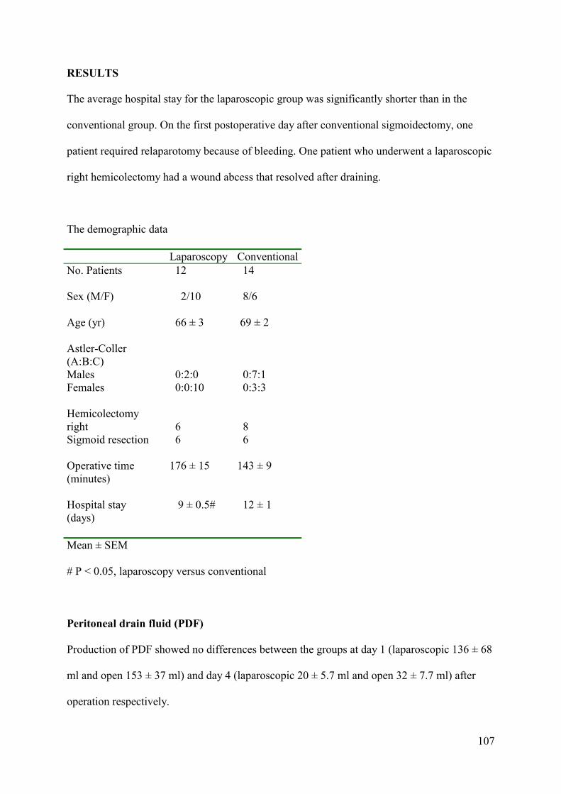
107
RESULTS
The average hospital stay for the laparoscopic group was significantly shorter than in the
conventional group. On the first postoperative day after conventional sigmoidectomy, one
patient required relaparotomy because of bleeding. One patient who underwent a laparoscopic
right hemicolectomy had a wound abcess that resolved after draining.
The demographic data
Laparoscopy Conventional No. Patients
12 14
Sex (M/F)
2/10 8/6
Age (yr)
66 ± 3 69 ± 2
Astler-Coller (A:B:C) Males Females Hemicolectomy right Sigmoid resection
0:2:0 0:0:10 6 6
0:7:1 0:3:3 8 6
Operative time (minutes)
176 ± 15 143 ± 9
Hospital stay (days)
9 ± 0.5# 12 ± 1
Mean ± SEM
# P < 0.05, laparoscopy versus conventional
Peritoneal drain fluid (PDF)
Production of PDF showed no differences between the groups at day 1 (laparoscopic 136 ± 68
ml and open 153 ± 37 ml) and day 4 (laparoscopic 20 ± 5.7 ml and open 32 ± 7.7 ml) after
operation respectively.

108
Angiogenic factors
VEGF and endostatin serum levels were measured in 12 laparoscopic and 14 conventionally
treated patients. In addition, local production of both factors was evaluated in wound fluid
from 5 laparoscopic and 11 conventional-treatment patients.
In both groups, VEGF levels in serum increased about 2-fold at day 4 compared with baseline
values (P =0.001), but with no significant difference between these two groups (Figure 1). In
the laparoscopic group, VEGF concentration in PDF was three-fold higher at day 1 compared
with serum level at the same day. At day 4 this difference increased to seven-fold (Figure 2).
The VEGF increase in PDF was significant at day 4 compared with day 1 (P=0.02). In the
conventional group, wound fluid VEGF level was about three-fold higher than serum at day 1
and at day 4 after surgery (P=0.01). The VEGF levels in PDF of the laparoscopic group at day
4 were significantly higher than in PDF from the conventional group (P=0.02).
Serum VEGF
0200400600800
10001200
Preop 2 Hrs Day +1 Day +4
Time
pg/m
l LPCO
* *
Figure 1: VEGF level in serum
* P < 0.05, day +4 compared to preoperative value
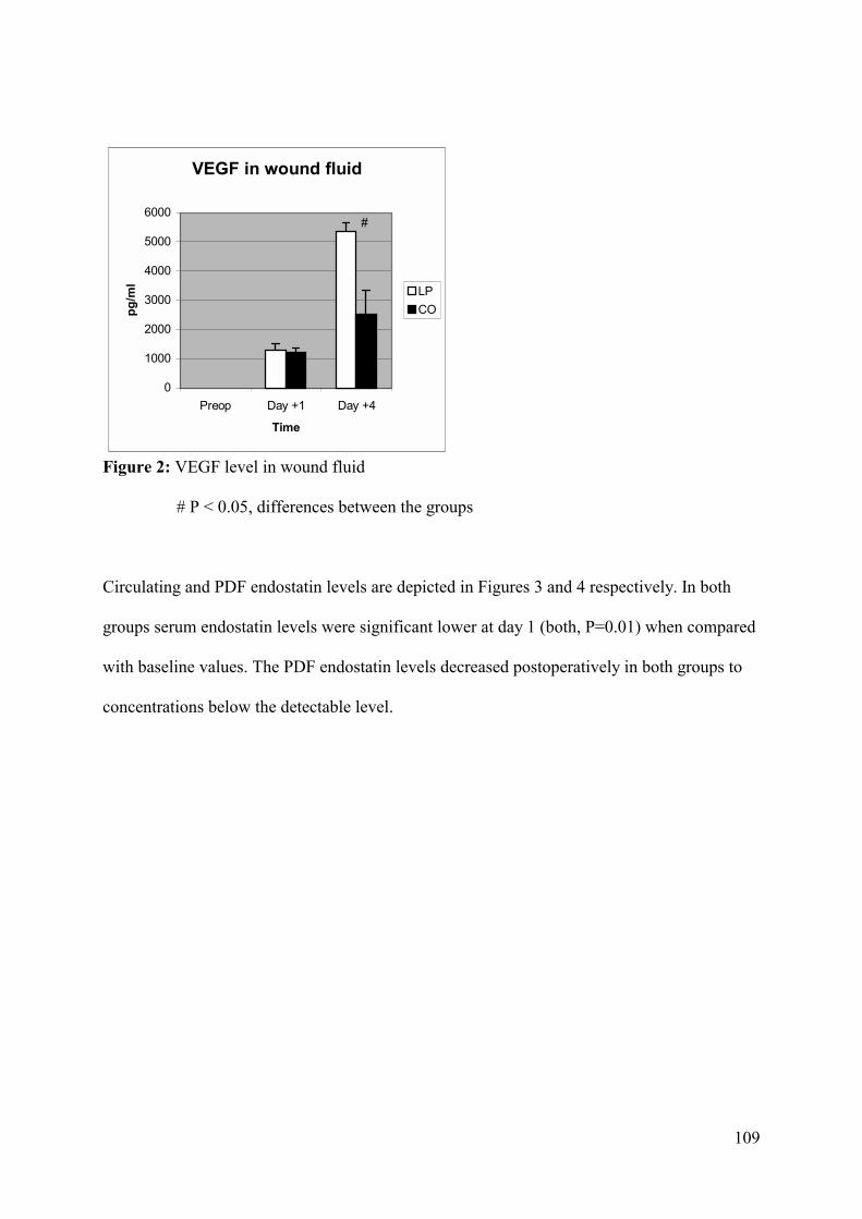
109
VEGF in wound fluid
0
1000
2000
3000
4000
5000
6000
Preop Day +1 Day +4
Time
pg/m
l LPCO
#
Figure 2: VEGF level in wound fluid
# P < 0.05, differences between the groups
Circulating and PDF endostatin levels are depicted in Figures 3 and 4 respectively. In both
groups serum endostatin levels were significant lower at day 1 (both, P=0.01) when compared
with baseline values. The PDF endostatin levels decreased postoperatively in both groups to
concentrations below the detectable level.

110
Figure 3: Endostatin level in serum
* P < 0.05, day +4 compared to preoperative value
Figure 4: Endostatin level in wound fluid. N.D.= not detectable
Serum Endostatin
05
101520253035
Preop 2 Hrs Day +1 Day +4
Time
ng/m
l LPCO
* *
E n d o s ta tin in w o u n d f lu id
02468
1 01 21 41 61 8
P reop D ay + 1 D ay + 4
T im e
ng/m
l L PC O
N .D .

111
DISCUSSION
Clinical and immunologic advantages of laparoscopic approach are clear in procedures such
as cholecystectomy or Nissen fundoplication.11,12 In more extented laparoscopic procedures
such as colon resection for cancer these advantages are less apparent.13 Long term results of a
randomized study of Lacy et al. has shown a significant longer survival of stage III colon
cancer of patients who underwent laparoscopic surgery when compared to the conventional
approach.14
We investigated the peritoneal and systemic immune response following laparoscopic and
conventional surgical approaches in patients with colorectal carcinoma. We found that large
amounts of pro-inflammatory cytokines were produced in the wound fluid after both
procedures while the systemic responses after both surgical approaches were only a marginal
reflection of these local events. These findings suggest that the surgical wound creates a
separate compartment from the systemic circulation in terms of inflammatory response.13
We studied two important regulators of angiogenesis at a systemic and a local level after
surgery. The data showed increased systemic VEGF levels postoperatively after both
conventional and laparoscopic surgery, and significant differences were not seen between
both approaches. VEGF levels measured in peritoneal drain fluid were much higher when
compared to the systemic values. Local VEGF generation after a surgical procedure is
probably due to instant platelet degranulation, production by recruited immune cells and
indirectly by the expression of pro-inflammatory cytokines and local hypoxia found in
devitalized tissue.15-17 It was unlikely that residual tumor was another source of VEGF, since
the surgical margins were free of tumor. The role of a high VEGF level in the wound may
have two functions, one to initiate angiogenesis and another to temper the local immune
response in the injured area. The devitalized tissue and exposed self-antigens in a wound
contain an intense immunologic stimulus. VEGF may act as an immunosuppressive agent,18,19

112
that prevents a local autoimmune reaction. The significantly higher increase of local VEGF at
day 4 in the laparoscopy group versus the laparotomy group is interesting. This may be
explained by the postoperative effect of local acidosis, due to CO2 insufflation, on endothelial
cells. D’Arcangelo et al. demonstrated in vitro that hypercarbic acidosis will induces VEGF
expression in bovine aortic endothelial cells (BAECs).20,21
In contrast to VEGF, the systemic and, more profoundly, the local endostatin levels decreased
during the postoperative period. The mechanism of the decrease of local endostatin levels is
speculative. It may be due to the release of various proteolytic enzymes during wound healing
that degrade endostatin.1,22 The local decrease of endostatin in the early phase of wound
healing may be seen as a physiological response to injury.
In conclusion, the strong increase of local VEGF, and the disappearance of endostatin
highlights the concept of a physiological angiogenic switch in the first phase of wound
healing. As was shown for immunological parameters,13 angiogenic factors detected in wound
fluid were found at remarkable higher levels when compared to serum after colon surgery.
The mechanisms of wound healing can be better studied locally than in the circulation.
113
REFERENCES
1. Singer AJ, Clark RA. Cutaneous wound healing. N Engl J Med. 1999;341:738-
46.
2. Lynch SE. Interactions of growth factors in tissue repair. Prog Clin Biol Res.
1991;365:341-57.
3. Neufeld G, Cohen T, Gengrinovitch S, Poltorak Z. Vascular endothelial growth
factor (VEGF) and its receptors. FASEB J. 1999;13:9-22.
4. Esser S, Wolburg K, Wolburg H, et al. Vascular endothelial growth factor
induces endothelial fenestrations in vitro. J Cell Biol. 1998;140:947-59.
5. Howdieshell TR, Callaway D, Webb WL, et al. Antibody neutralization of
vascular endothelial growth factor inhibits wound granulation tissue formation.
J Surg Res. 2001;96:173-82.
6. Werther K, Christensen IJ, Brunner N, Nielsen HJ. Soluble vascular endothelial
growth factor levels in patients with primary colorectal carcinoma. The Danish
RANX05 Colorectal Cancer Study Group. Eur J Surg Oncol. 2000;26:657-62.
7. Schuppan D, Cramer T, Bauer M, et al. Hepatocytes as a source of collagen
type XVIII endostatin. Lancet 1998;352:879-80.
8. Kuroi K, Tanaka C, Toi M. Circulating levels of endostatin in cancer patients.
Oncol Rep. 2001;8:405-9.
9. Dhanabal M, Ramchandran R, Waterman MJ, et al. Endostatin induces
endothelial cell apoptosis. J Biol Chem. 1999;274:11721-6.
10. Yoon SS, Eto H, Lin CM, et al. Mouse endostatin inhibits the formation of lung
and liver metastases. Cancer Res. 1999;59:6251-6.
114
11. Kloosterman T, von Blomberg BM, Borgstein P, Cuesta MA, Scheper RJ,
Meijer S. Unimpaired immune functions after laparoscopic cholecystectomy.
Surgery 1994;115:424-8.
12. Sietses C, Wiezer MJ, Eijsbouts QA, et al. A prospective randomized study of the
systemic immune response after laparoscopic and conventional Nissen
fundoplication. Surgery 1999;126:5-9.
13. Wu FP, Sietses C, von Blomberg BM, van leeuwen PA, Meijer S, Cuesta MA.
Systemic and peritoneal Inflammatory response after laparoscopic or
conventional colon resection in cancer patients. Dis Colon rectum 2003;46:
147-55.
14. Lacy AM, Garcia Valdecasas JC, Delgado S, et al. Laparoscopic assisted
colectomy versus open colectomy for treatment of non-metastatic colon cancer: a
randomized trial. Lancet 2002;359:2224-9.
15. Cohen T, Nahari D, Cerem LW, et al. Interleukin 6 induces the expression of
vascular endothelial growth factor. J Biol Chem. 1996;271:736-41.
16. Salven P, Orpana A, Joensuu H. Leukocytes and platelets of patients with cancer
contain high levels of vascular endothelial growth factor. Clin Cancer Res.
1999;5:487-91.
17. Elson DA, Ryan HE, Snow JW, et al. Coordinate up-regulation of hypoxia
inducible factor (HIF)-1alpha and HIF-1 target genes during multi-stage epidermal
carcinogenesis and wound healing. Cancer Res. 2000;60:6189-95.
18. Ohm JE, Carbone DP. VEGF as a mediator of tumor-associated immunodeficiency.
Immunol Res. 2001;23:263-72.
115
19. Gabrilovich DI, Chen HL, Girgis KR, et al. Production of vascular
endothelial growth factor by human tumors inhibits the functional maturation
of dendritic cells. Nat Med. 1996;2:1096-103.
20. Taura P, Lopez A, Lacy AM, et al. Prolonged pneumoperitoneum at 15 mm Hg
causes lactic acidosis. Surg Endosc. 1998;12:198-201.
21. D’Arcangelo D, Facchiano F, Barlucchi LM, et al. Acidosis inhibits endothelial
cell apoptosis and function and induces basic fibroblast growth factor and vascular
endothelial growth factor expression. Circ Res. 2000;86:312-8.
22. Arumugum S, Jang YC, Chen-Jensen C, et al. Temporal activity of
plasminogen activators and matrix metalloproteinases during cutaneous wound
repair. Surgery 1999;125:587-93.

116

117
Chapter 6
The effects of surgery, with or without rhGM-CSF, on the angiogenic
profile of patients treated for colorectal carcinoma
F.P.K. Wu1, J.R. Westphal2, K. Hoekman 3, A.K. Mels1, M.G. Statius Muller1,
R.W. de Waal2, C. Sietses1, R.H.J. Beelen4, P.A.M. van Leeuwen1, S. Meijer1,
M.A. Cuesta1
1Department of Surgery, VU University Medical Center, Amsterdam,
2Department of Pathology, University Medical Center, Nijmegen, 3Department
of Medical Oncology, VU University Medical Center, Amsterdam, 4Department
of Cell Biology and Immunology, VU University Medical Center, Amsterdam,
The Netherlands.
Cytokine 2004;25:68-72
118
ABSTRACT
Wound healing is a process with immunological and angiogenic aspects. RhGM-CSF is
known to stimulate the immune system and angiogenesis via multiple pathways. In this study
we investigated the combined effects of surgery, with or without rhGM-CSF, on angiogenic
parameters in patients with a colorectal carcinoma. In this phase II randomized, placebo-
controlled trial, sixteen patients were assigned to perioperative rhGM-CSF (2.8 µg/kg body
weight) treatment or saline. Patients received subcutaneous injections from three days before
surgery until four days after. IL-6, VEGF, endostatin and angiostatin levels were measured
perioperatively. RhGM-CSF enhanced the production of IL-6 and VEGF, but had no effect on
the anti-angiogenic agents endostatin and angiostatin. Surgery induced a transient decrease of
endostatin. Two types of angiostatin (kringle 1-3 and kringle 1-4) became visible
postoperatively. We conclude that this study demonstrated the immediate initiation of
angiogenesis postoperatively, reflected by the increase of VEGF and a transient decrease of
endostatin, followed by the appearance of two angiostatin bands, which confirms
physiological wound healing in these cancer patients.

119
INTRODUCTION Radical surgery is the initial treatment for patients with primary colorectal carcinoma.
However, major surgery induces immune suppression and tumor resection may induce
shedding of tumor cells into the circulation.1 The transient impairment of the host defense
may be associated with bacterial infections and outgrowth of previous dormant
micrometastases. RhGM-CSF has been widely used to enhance the number and the activity of
granulocytes and monocytes, thereby augmenting host defences.2
Apart from immunological activity, GM-CSF also stimulates migration of endothelial cells
(ECs) and the formation of new blood vessels from pre-existing vessels, a process known as
angiogenesis.3,4 This process is tightly regulated by pro-angiogenic and anti-angiogenic
factors. Among them, vascular endothelial growth factor (VEGF) is considered the most
potent and specific pro-angiogenic factor.5 VEGF production by cells involved in wound
healing is partly constitutive, and partly up-regulated by a wide array of external factors, e.g.
hypoxia, interleukin-6 (IL-6) and GM-CSF.6,7 In addition, peripheral blood cells such as
platelets and leucocytes contain VEGF, which is released locally after trauma.8
Theoretically, GM-CSF might also inhibit angiogenesis by activation of macrophages to
produce matrix metalloproteinase-12 (MMP-12). MMP-12 is capable to cleave angiostatin
from plasminogen.9 Angiostatin inhibits endothelial cell proliferation in vitro10 and systemic
administration of angiostatin in a murine model induced regression in breast, prostate and
colon cancers.11 MMP-12 produced by macrophages may also be involved in the generation
of endostatin.12 Endostatin induces apoptosis in ECs in vitro13 and potently inhibits tumor
growth in animal models.14
In short, GM-CSF is pro-angiogenic by stimulating EC directly and enhancing VEGF
production. However, GM-CSF may also have anti-angiogenic effects since activated
macrophages produce enzymes, which may generate angiostatin and endostatin.
120
In this randomized study we describe the angiogenic consequences of surgery, with or without
perioperative rhGM-CSF administration, in patients with a colorectal carcinoma. The immune
stimulating effects of perioperative rhGM-CSF have been published elsewhere.2
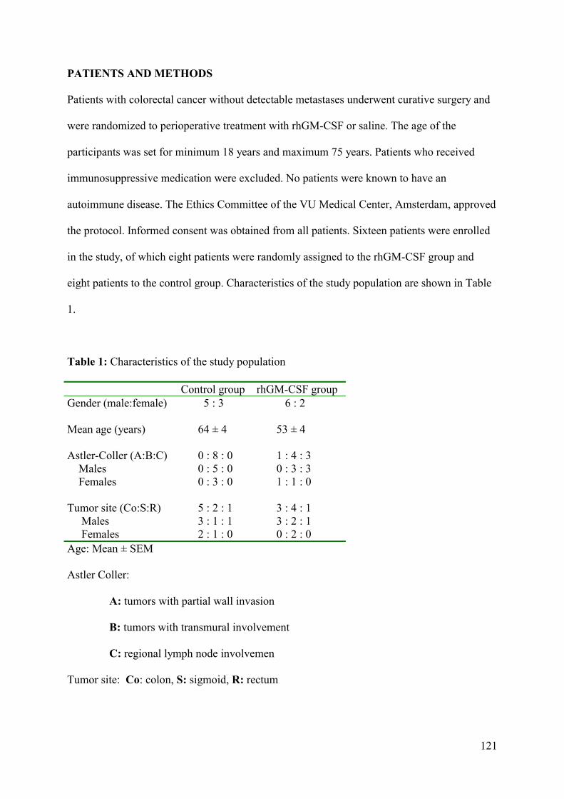
121
PATIENTS AND METHODS
Patients with colorectal cancer without detectable metastases underwent curative surgery and
were randomized to perioperative treatment with rhGM-CSF or saline. The age of the
participants was set for minimum 18 years and maximum 75 years. Patients who received
immunosuppressive medication were excluded. No patients were known to have an
autoimmune disease. The Ethics Committee of the VU Medical Center, Amsterdam, approved
the protocol. Informed consent was obtained from all patients. Sixteen patients were enrolled
in the study, of which eight patients were randomly assigned to the rhGM-CSF group and
eight patients to the control group. Characteristics of the study population are shown in Table
1.
Table 1: Characteristics of the study population Control group rhGM-CSF group Gender (male:female) Mean age (years) Astler-Coller (A:B:C) Males Females Tumor site (Co:S:R) Males Females
5 : 3 64 ± 4 0 : 8 : 0 0 : 5 : 0 0 : 3 : 0 5 : 2 : 1 3 : 1 : 1 2 : 1 : 0
6 : 2 53 ± 4 1 : 4 : 3 0 : 3 : 3 1 : 1 : 0 3 : 4 : 1 3 : 2 : 1 0 : 2 : 0
Age: Mean ± SEM
Astler Coller:
A: tumors with partial wall invasion
B: tumors with transmural involvement
C: regional lymph node involvemen
Tumor site: Co: colon, S: sigmoid, R: rectum

122
Treatment schedule
All patients were operated through a median laparotomy on day 0. Both groups received daily
a subcutaneous injection from 3 days (day –3) before, until 4 days
(day +4) after the operation. The patients received either 1 ml of saline or 2.8 µg/kg rhGM-
CSF dissolved in 1 ml 0.9% NaCl (Leucomax®, Schering-Plough, Maarssen, The
Netherlands).
Samples
Heparinized blood samples were collected on day –4, day 0 (prior to surgery), and day +1 and
day +6 after the operation. The cytokine samples were centrifuged at 2000 rpm for 10 minutes
and then stored at -800C.
VEGF
VEGF concentrations were determined in plasma using an enzyme-linked immunosorbent
assay (ELISA) kit from R & D system, Minneapolis, U.S.A..
The intra- and inter-assay variance was less than 10%. The range of detection of VEGF was
9.0 - 200 pg/ml.
IL-6
Plasma IL-6 was measured using an ELISA from Pelikine, CLB, Amsterdam, The
Netherlands. Normal values are < 10 pg/ml.

123
Endostatin
Endostatin was measured using an ELISA from Cytimmune, College Park, Maryland, U.S.A..
The intra- and inter-assay variance was less than 10%. The range of detection of endostatin
was 1.95 - 500 ng/ml.
Angiostatin
Angiostatin was purified by lysine-sepharose affinity chromatography. Briefly, lysine-
sepharose slurry, prepared according to the manufacturer’s instructions (Pharmacia, Uppsala,
Sweden) was added to plasma samples and incubated under continuous agitation for 15 hours
at 4°C. After two washes with 50 mM phosphate buffer pH 6.0, bound material was eluted
with 0.2M ε-amino-capronic acid. Aliquots of 15 µl of eluate (equivalent to 60 µl of plasma)
plus 15 μl of sample buffer were run on a 10% polyacrylamide gel under non-reducing
conditions. After electrophoresis, samples were electroblotted onto nitrocellulose membrane,
blocked, and incubated O/N with affinity-purified rabbit anti-plasminogen kringle 1-3
antibody (DAKO, Glostrup, Denmark). This antibody recognizes plasminogen as well as both
angiostatin moieties. After washing, blots were incubated for two hours with swine anti-rabbit
peroxidase-conjugated secondary antibody (Dako), washed again, and developed by
chemiluminescence according to the manufacturer’s protocol (Boehringer Mannheim).
STATISTICAL ANALYSIS
Results are reported as mean ± SEM. The “Statistical Package for the Social Sciences” (SPSS
7.5tm) was used to analyze data. Differences within groups were analyzed by the Wilcoxon
Signed Rank Test for two related samples. Differences between the groups at different time
points were analyzed by means of the two-sample Mann-Whitney test. Significance was
accepted at a two-tailed P < 0.05.

124
RESULTS
VEGF, IL-6 and endostatin
The results are shown in Table 2. RhGM-CSF treatment before surgery had no significant
effect on these three parameters. One day after surgery VEGF levels were increased in both
groups, however more pronounced in the rhGM-CSF group (P = 0.02). In the control group
VEGF levels were increased on day +6, (P = 0.01).
IL-6 levels were increased in both groups on day +1 (control, P = 0.01), but in the rhGM-CSF
group the increase was more pronounced (P = 0.02).
A significant 3 to 4-fold decrease in the endostatin level was found on day +1 in both groups.
Treatment
Group
Day - 4 Day 0 Day +1 Day +6
VEGF
rhGM-CSF Control
118 ± 45
88 ± 28
218 ± 73
56 ± 12
637 ± 59 * # 160 ± 55
166 ± 19
245 ± 91 *
IL-6 rhGM-CSF Control
11.8 ±16.3 6.1 ± 0.4
8.8 ± 7.9 6.0 ± 0.0
116 ± 38.4 * 41.1± 10.2 *
7.0 ± 2.83 8.1 ± 5.7
Endostatin
rhGM-CSF Control
20 ± 2 23 ± 2
17 ± 5 18 ± 3
4 ± 1 * 5 ± 1 *
17 ± 2 16 ± 3
Table 2: Angiogenic growth factors
Results: Mean ± SEM
VEGF and IL-6 in pg/ml, endostatin in ng/ml
* P < 0.05, postoperative value compared to day -4
# P < 0.05, rhGM-CSF versus control group
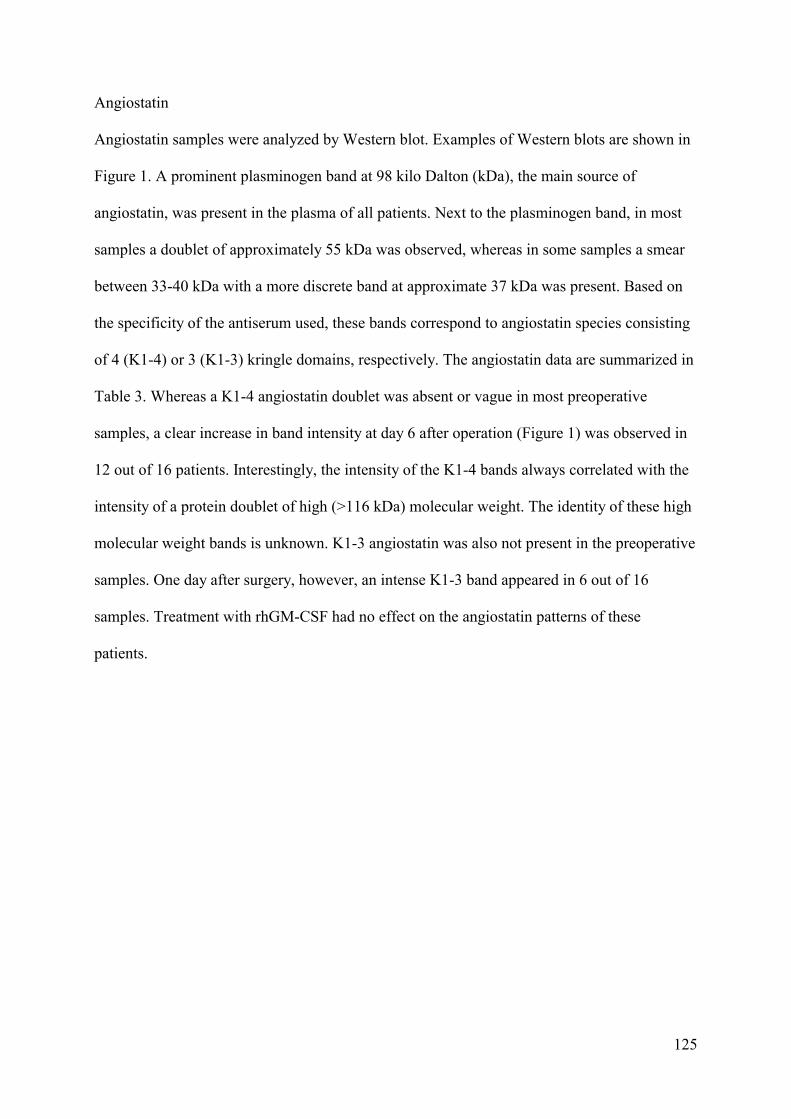
125
Angiostatin
Angiostatin samples were analyzed by Western blot. Examples of Western blots are shown in
Figure 1. A prominent plasminogen band at 98 kilo Dalton (kDa), the main source of
angiostatin, was present in the plasma of all patients. Next to the plasminogen band, in most
samples a doublet of approximately 55 kDa was observed, whereas in some samples a smear
between 33-40 kDa with a more discrete band at approximate 37 kDa was present. Based on
the specificity of the antiserum used, these bands correspond to angiostatin species consisting
of 4 (K1-4) or 3 (K1-3) kringle domains, respectively. The angiostatin data are summarized in
Table 3. Whereas a K1-4 angiostatin doublet was absent or vague in most preoperative
samples, a clear increase in band intensity at day 6 after operation (Figure 1) was observed in
12 out of 16 patients. Interestingly, the intensity of the K1-4 bands always correlated with the
intensity of a protein doublet of high (>116 kDa) molecular weight. The identity of these high
molecular weight bands is unknown. K1-3 angiostatin was also not present in the preoperative
samples. One day after surgery, however, an intense K1-3 band appeared in 6 out of 16
samples. Treatment with rhGM-CSF had no effect on the angiostatin patterns of these
patients.
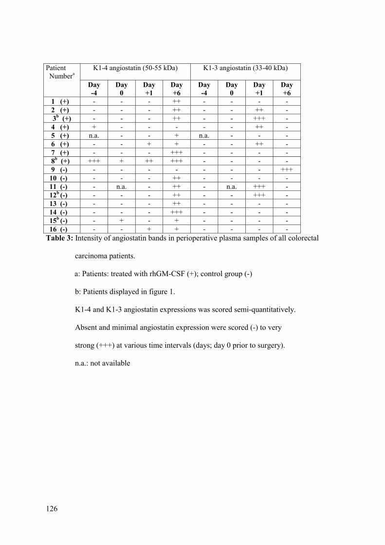
126
K1-4 angiostatin (50-55 kDa)
K1-3 angiostatin (33-40 kDa) Patient
Numbera
Day -4
Day 0
Day +1
Day +6
Day -4
Day 0
Day +1
Day +6
1 (+) - - - ++ - - - - 2 (+) - - - ++ - - ++ -
3b (+) - - - ++ - - +++ - 4 (+) + - - - - - ++ - 5 (+) n.a. - - + n.a. - - - 6 (+) - - + + - - ++ - 7 (+) - - - +++ - - - - 8b (+) +++ + ++ +++ - - - - 9 (-) - - - - - - - +++ 10 (-) - - - ++ - - - - 11 (-) - n.a. - ++ - n.a. +++ - 12b (-) - - - ++ - - +++ - 13 (-) - - - ++ - - - - 14 (-) - - - +++ - - - - 15b (-) - + - + - - - - 16 (-) - - + + - - - - Table 3: Intensity of angiostatin bands in perioperative plasma samples of all colorectal
carcinoma patients.
a: Patients: treated with rhGM-CSF (+); control group (-)
b: Patients displayed in figure 1.
K1-4 and K1-3 angiostatin expressions was scored semi-quantitatively.
Absent and minimal angiostatin expression were scored (-) to very
strong (+++) at various time intervals (days; day 0 prior to surgery).
n.a.: not available

127

128
DISCUSSION
Following radical resection for colorectal carcinoma a number of these patients will develop
metastatic disease in the liver,15 which largely determines the overall survival of these
patients. Liver metastases may have been already there in an occult form or induced by
shedding of cancer cells into the circulation as a result of surgical intervention, and this
outgrowth may be stimulated by the effect of postoperative immunosuppression, the
angiogenic response as part of wound healing, or the combination of these effects.1,16
Perioperative rhGM-CSF has been successfully used in order to enhance the immune status of
patients with colorectal carcinoma.2
In the present study we investigated the effects of surgery, with or without perioperative
rhGM-CSF, on VEGF and IL-6 plasma levels. In both groups VEGF increased after surgery,
which is in accordance to other observations.17 Tissue hypoxia is the most probable
explanation for the postoperative VEGF increase. A surgical procedure inevitably causes
vascular disruption with subsequent reduction of tissue oxygen tension. The observed steep
and transient increase of VEGF and IL-6 level in the rhGM-CSF group was remarkable. Two
effects may explain the elevated postoperative levels of VEGF induced by rhGM-CSF. First,
the surgical trauma itself results in local platelet degranulation and leucocyte sequestration,
with local VEGF release. The enhanced number and state of activation of leucocytes induced
by rhGM-CSF might have been responsible for the increased VEGF generation. Second,
rhGM-CSF induces secondary cytokines, which may affect the VEGF levels.6,18 The elevated
VEGF level found in the rhGM-CSF group would be expected to have a positive effect on
wound healing. On the other hand, elevated VEGF levels may have a stimulatory effect on the
development of micrometastases, demonstrated in an experimental study.19 The involvement
of VEGF has been further demonstrated in a recent study, where anti-VEGF had a beneficial
effect in treatment of metastatic colon cancer.20

129
Furthermore, we investigated whether surgery and rhGM-CSF had an effect on endostatin and
angiostatin levels. These are two endogenous anti-angiogenic factors produced by enzymatic
digestion of a protein precursor, collagen XVIII and plasminogen, respectively. Collagen
XVIII has been shown to reside in tissues such as basement membranes, and in the liver.21-23
In vitro generation of endostatin by elastase and cathepsin L has been demonstrated,12,23 but
the mechanisms of endostatin generation in vivo are largely unknown. A recent study
suggested that hepatocytes are the major contributors of circulating endostatin. In addition,
colon carcinoma may also produce proteases able to properly cleave collagen XVIII present in
the direct environment.24
In our study, both groups showed a sharp decrease of endostatin immediately after surgery.
Various postoperative effects may explain the endostatin decrease. It may concern an effect
on the production or clearance of endostatin. Since the half-life of endostatin is less than one
hour, an acute reduction of the production of endostatin by hepatocytes as a negative acute
phase reaction on stress, or the excision of the tumor could explain an immediate decrease
after surgery.
Angiostatin is generated on the endothelial surface from circulating plasminogen.
Angiostatin bands appeared in the postoperative period. RhGM-CSF treatment did not affect
the fluctuation of angiostatin levels, as these were similar in both the treated and the control
group. K1-4 angiostatin became manifest in almost all patients 6 days after surgery, whereas
the K1-3 form increased transiently in 6/16 of the patients. Both isoforms may function as a
stopping signal for angiogenesis. Cellular migration during early wound healing is dependent
on proteolytic enzymes such as matrix metalloproteinases (MMPs) and plasminogen
activators.25 It is conceivable that the same proteolytic enzymes, involved in early stages of
wound healing may be responsible for the conversion of plasminogen into the angiostatin
isoforms at a latter stage.

130
In conclusion, we have demonstrated that rhGM-CSF enhanced IL-6 and VEGF. RhGM-CSF
had no effect on the production of the anti-angiogenic factors endostatin and angiostatin.
However, surgery induced a sharp, but transient decrease of endostatin and a significant
increase of angiostatin. The immediate generation of VEGF and decrease of endostatin,
followed by the generation of both angiogenesis inhibitors at a later stage of wound healing
may reflect a physiological tendency to achieve an angiogenic balance, which is characterized
by initiating and stopping angiogenic signals.
131
REFERENCES 1. Weitz J, Kienle P, Lacroix J, et al. Dissemination of tumor cells in patients undergoing
surgery for colorectal cancer. Clin Cancer Res. 1998;4:343-8.
2. Mels AK, Muller MG, Van Leeuwen PA, et al. Immune-stimulating effects of low-dose
perioperative recombinant granulocyte-macrophage colony-stimulating factor in patients
operated on for primary colorectal carcinoma. Br J Surg. 2001;88:539-44.
3. Bussolino F, Ziche M, Wang JM, Alessi D, et al. In vitro and in vivo activation of
endothelial cells by colony-stimulating factors. J Clin Invest. 1991;87:986-95.
4. Valdembri D, Serini G, Vacca A, Ribatti D, Bussolino F. In vivo activation of
JAK2/STAT-3 pathway during angiogenesis induced by GM-CSF. FASEB J.
2002;16:225-7.
5. Neufeld G, Cohen T, Gengrinovitch S, Poltorak Z.Vascular endothelial growth factor
(VEGF) and its receptors. FASEB J. 1999;13:9-22.
6. Cohen T, Nahari D, Cerem LW, Neufeld G, Levi BZ. Interleukin 6 induces the expression
of vascular endothelial growth factor. J Biol Chem. 1996;271:736-41.
7. Horiuchi T, Weller PF. Expression of vascular endothelial growth factor by human
eosinophils: upregulation by granulocyte macrophage colony-stimulating factor and
interleukin-5. Am J Respir Cell Mol Biol. 1997;17:70-7.
8. Salven P, Orpana A, Joensuu H. Leukocytes and platelets of patients with cancer contain
high levels of vascular endothelial growth factor. Clin Cancer Res. 1999;15:487-91.
9. Dong Z, Yoneda J, Kumar R, Fidler IJ. Angiostatin-mediated suppression of cancer
metastases by primary neoplasms engineered to produce granulocyte/macrophage colony-
stimulating factor. J Exp Med. 1998;188:755-63.
10. Moser TL, Stack MS, Asplin I, et al. Angiostatin binds ATP synthase on the surface of
human endothelial cells. Proc Natl Acad Sci U S A 1999;96:2811-6.

132
11. O’Reilly MS, Holmgren L, Chen C, Folkman J. Angiostatin induces and sustains
dormancy of human primary tumors in mice. Nat Med. 1996;2:689-92.
12. Wen W, Moses MA, Wiederschain D, Arbiser JL, Folkman J. The generation of
endostatin is mediated by elastase. Cancer Res. 1999;59:6052-6.
13. Dhanabal M, Ramchandran R, Waterman MJ, et al. Endostatin induces endothelial cell
apoptosis. J Biol Chem. 1999;274:11721-6.
14. Yoon SS, Eto H, Lin CM, et al. Mouse endostatin inhibits the formation of lung and liver
metastases. Cancer Res. 1999;59:6251-6.
15. Butler J, Attiyeh FF, Daly JM. Hepatic resection for metastases of the colon and rectum.
Surg Gynecol Obstet. 1986;162:109-13.
16. Hofer SO, Molema G, Hermens RA, Wanebo HJ, Reichner JS, Hoekstra HJ. The effect of
surgical wounding on tumour development. Eur J Surg Oncol. 1999;25:231-43.
17. Bondestam J, Salven P, Jaaskela-Saari H, Ikonen T, Lepantalo M, Mattila S, Joensuu H.
Major surgery increases serum levels of vascular endothelial growth factor only
temporarily. Am J Surg. 2000;179:57-9.
18. Bergamini A, Bolacchi F, Bongiovanni B, et al. Granulocyte-macrophage colony-
stimulating factor regulates cytokine production in cultured macrophages through CD14-
dependent and -independent mechanisms. Immunology 2000;101:254-61.
19. Maniwa Y, Okada M, Ishii N, Kiyooka K. Vascular endothelial growth factor increased
by pulmonary surgery accelerates the growth of micrometastases in metastatic lung
cancer. Chest 1998;114:1668-75.
20. Hurwitz H. Oral presentation ASCO 2003.
21. Muragaki Y, Timmons S, Griffith CM, et al. Mouse Col18a1 is expressed in a tissue-
specific manner as three alternative variants and is localized in basement membrane
zones. Proc Natl Acad Sci U S A 1995;92:8763-7.
133
22. Schuppan D, Cramer T, Bauer M, Strefeld T, Hahn EG, Herbst H. Hepatocytes as a source
of collagen type XVIII endostatin. Lancet 1998;352:879-80.
23. Felbor U, Dreier L, Bryant RA, Ploegh HL, Olsen BR, Mothes W. Secreted cathepsin L
generates endostatin from collagen XVIII. EMBO J. 2000;19:1187-94.
24. Bjornland K, Buo L, Scott H, Konttinen Y, Johansen HT, Aasen AO. Polymorphonuclear
elastase in human colorectal carcinoma. Int J Oncol. 1998;12:535-40.
25. Arumugam S, Jang YC, Chen-Jensen C, Gibran NS, Isik FF. Temporal activity of
plasminogen activators and matrix metalloproteinases during cutaneous wound repair.
Surgery 1999;125:587-93.

134
135
Chapter 7
The effects of major liver resection, with or without recombinant
bactericidal/permeability-increasing protein (rBPI21), on the
angiogenic profile of patients with metastatic colorectal carcinoma
F.P.K. Wu1, P.G. Boelens1, P.A.M. van Leeuwen1, K. Hoekman2, A.H.G.
Hansma2, M.J. Wiezer1, C. Meijer1, S. Meijer1, M. Scotté3, M.A. Cuesta1
1Department of Surgery, 2Department of Medical Oncology, VU University
Medical Center, Amsterdam, The Netherlands, 3Department of Surgery, Charles
Nicolle Hospital, Rouen, France.
Journal of Surgical Oncology 2003;84:137-42

136
ABSTRACT
Background: Surgery induces a process of wound healing, which has immunological
and angiogenic aspects. Bactericidal/Permeability-Increasing protein (BPI) is found in
azurophilic granules of human neutrophils, which is bactericidal and neutralizes
lipopolysaccharide (LPS). This may reduce postoperative infectious complications. In
addition, BPI has been shown to be an inhibitor of angiogenesis.
Methods: A total of 18 patients with metastasized colorectal carcinoma to the liver
were double blind randomized. The levels of the pro-angiogenic factors interleukin-6 (IL-6)
and vascular endothelial growth factor (VEGF) and the angiogenesis inhibitor endostatin were
investigated after liver surgery with perioperative administration of either rBPI21 or placebo.
Results: The highest IL-6 levels were found during the first 24 hours and reached
peak levels already at 2 hours postoperatively in both groups. In both groups VEGF levels
decreased sharply in the postoperative hours, returning to baseline levels in the days
afterwards. In both groups, an immediate decrease in endostatin levels was observed which
remained significantly low. RBPI21 transiently influenced IL-6 and VEGF.
Conclusions: RBPI21 only marginally affected IL-6 and VEGF levels. Surgery per se
induced an immediate immune response (IL-6) and an immediate angiogenic response,
reflected in an initial VEGF decrease and a longer lasting decrease of endostatin. These
findings demonstrate the dynamics of tissue responses in the first phase of wound healing.
137
INTRODUCTION
Following radical resection for colorectal cancer, still a large number of patients will develop
liver metastases,1 for which major liver resection is a viable option.2,3 Partial liver resection is
a major trauma, and is associated with considerable morbidity such as liver failure,
disseminated intravascular coagulation (DIC) and systemic infections.4,5 We demonstrated
earlier that continuous perioperative administration of recombinant bactericidal/permeability-
increasing protein (rBPI21) administration in patients undergoing partial liver resection
resulted in a reduced incidence of postoperative infectious complications.6 BPI is normally
found in azurophilic granules of human neutrophils and has bactericidal properties against
gram-negative bacteria and neutralizes lipopolysaccharide (LPS)-mediated effects.7,8 After
liver surgery, the remnant liver has the ability to regenerate to full size. An important factor in
the restoration of injured tissue, such as liver regeneration and wound healing is angiogenesis,
the formation of new blood vessels. Angiogenesis is regulated by a number of specific
stimulators and inhibitors. A recent study showed that BPI also functions as an angiogenic
inhibitor.9 Therefore we were interested in the effects of rBPI21 on angiogenic parameters
after liver surgery. We measured interleukin-6 (IL-6) and VEGF, since both are potent
angiogenic stimulators and known to be involved in liver regeneration, and endostatin, an
endogenous angiogenesis inhibitor.
IL-6 is a multifunctional cytokine, which induces VEGF production10 and is of fundamental
importance in liver regeneration. A study by Cressman et al, showed that hepatectomy in IL-6
-/- mice resulted in postoperative liver failure and that IL-6 administration before the
hepatectomy normalized hepatocyte proliferation postoperatively.11
VEGF is a potent inducer of angiogenesis, since it stimulates endothelial cell (EC)
proliferation, migration and vessel formation.12 An increase in VEGF after surgical
procedures has been observed13,14 and may be directly caused by platelet degranulation or
138
leucocytes which are involved in the repair of injured tissue, or indirectly by hypoxia in
devitalized tissue and by up-regulation of interleukin-6 (IL-6).10, 13-16 It has been demonstrated
in an experimental study that hepatocytes produce VEGF, whereas sinusoidal endothelial cells
express the VEGF receptors, flt-1 and KDR/flk-1, suggesting that VEGF may contribute to
liver regeneration.17 Taniguchi et al. showed that postoperative VEGF expression was
associated with hepatocyte proliferation and anti-VEGF inhibited hepatocyte proliferative
activity after partial hepatectomy in rats.18
Endostatin is generated from the C-terminal fragment of collagen XVIII.19 Endostatin
specifically inhibits EC migration in vitro and potently inhibits tumor growth in animal
models.20 Endostatin can be generated by elastase and cathepsin L in vitro.21,22 The
mechanism of endostatin generation in vivo is largely unknown. Collagen XVIII has been
shown to reside in basement membranes and vessel walls.23 A study suggested that
hepatocytes are a major source of collagen XVIII and may contribute to endostatin levels in
the circulation.24
Recombinant BPI (rBPI21) is a 21 kDa amino-terminal protein derived from the 55 kDa
human BPI55. RBPI21 has the same bactericidal properties as the endogenous BPI.25,26 The
main object of this study was to determine the effects of liver surgery on the angiogenic
profile in the circulation. In addition, the immunologic and angiogenic effects of rBPI21, given
in a perioperative infusion were studied.
139
MATERIALS AND METHODS
Patients. This pilot study was part of a phase II, double-blind, placebo-controlled trial in
patients with metastasized colon carcinoma undergoing a partial hepatectomy (PH). The
protocol was approved by the Food and Drug Administration, by the Council for Medical
Research of the Netherlands Organization for Scientific Research, and by the institutional
review boards or ethical review committees of each participating institution. Informed consent
was obtained from each patient before entry to this study.
Study design. A patient’s eligibility for study entry was based on the predefined criteria
listed in Table 1. The patients were assigned to perioperative treatment with rBPI21 (8
mg/kg/48hr) or placebo by a 48-hour continuous intravenous infusion starting one hour before
liver resection. The rBPI21 dose used in this study was based on a previous pharmacokinetic
study.27 Patients were randomly assigned to treatment (rBPI21 or placebo) by the central
pharmacist at the VU Medical Center. A computer-generated randomization schedule at a 1:1
ratio was used. Patients, investigators and clinical monitors remained blinded to individual
treatment until all data were evaluated. Test articles (rBPI21 and placebo) were supplied by Xoma
US (LLC, Berkeley, California) as a clear, colorless, sterile non-pyrogenic solution in 10 ml
glass vials, containing either 2 mg/ml rBPI21 or 0.2 mg/ml human serum albumin (placebo) in 5
mM sodium citrate/0.15 M sodium chloride buffer, pH 5.0. The rBPI21 solution also contained
0.2 % poloxamer 188 and 0.002 % polysorbate 80. Until use, the rBPI21 and placebo solutions
were stored at 2-8 ºC. Before the start of the actual liver resection and study-drug infusion,
manual exploration and intra-operative ultrasonography were performed to confirm the
preoperative findings of tumor size and localization. Only when a resection was estimated to be
≥ 3 liver segments, patients were included and treated with either rBPI21 or placebo in an equal
volume by 48-hours intravenous infusion. Starting after laparotomy at about 1 hour prior to the
actual resection of liver tissue, the study medication was administered into a central vein as the
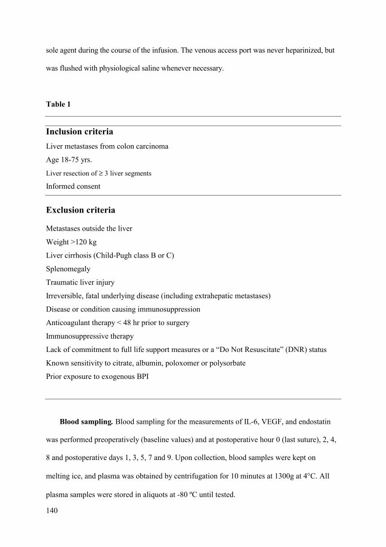
140
sole agent during the course of the infusion. The venous access port was never heparinized, but
was flushed with physiological saline whenever necessary.
Table 1
Inclusion criteria Liver metastases from colon carcinoma
Age 18-75 yrs.
Liver resection of ≥ 3 liver segments
Informed consent
Exclusion criteria Metastases outside the liver
Weight >120 kg
Liver cirrhosis (Child-Pugh class B or C)
Splenomegaly
Traumatic liver injury
Irreversible, fatal underlying disease (including extrahepatic metastases)
Disease or condition causing immunosuppression
Anticoagulant therapy < 48 hr prior to surgery
Immunosuppressive therapy
Lack of commitment to full life support measures or a “Do Not Resuscitate” (DNR) status
Known sensitivity to citrate, albumin, poloxomer or polysorbate
Prior exposure to exogenous BPI
Blood sampling. Blood sampling for the measurements of IL-6, VEGF, and endostatin
was performed preoperatively (baseline values) and at postoperative hour 0 (last suture), 2, 4,
8 and postoperative days 1, 3, 5, 7 and 9. Upon collection, blood samples were kept on
melting ice, and plasma was obtained by centrifugation for 10 minutes at 1300g at 4°C. All
plasma samples were stored in aliquots at -80 ºC until tested.

141
Assessment of total white blood cells (WBC) and platelets
WBCs including cell differentiation were assessed (Coulter JS, Coulter Electronics, Luton,
U.K.). Platelet counts were determined using an automated blood coulter counter. Blood
sampling for measurements of WBC and platelets was performed pre- and postoperatively at
days 1 and 7.
Assessment of plasma IL-6
The plasma IL-6 concentration was measured using a commercially available enzyme-linked
immunosorbent assay (ELISA) kit (Pelikine compact human IL-6 ELISA kit, CLB,
Amsterdam, the Netherlands). Normal values are < 10 pg/ml
Assessment of plasma VEGF
VEGF165 concentrations were determined using an ELISA kit (R & D system, Minneapolis,
USA) according to the manufacturer’s guidelines. The intra- and inter-assay variance was less
than 10%. The reliable minimum detectable VEGF level is 9.0 pg/ml.
Assesment of plasma endostatin
Endostatin was measured using a commercially available enzyme-linked immunosorbent
assay kit (ELISA kit, Cytimmune, College Park, Maryland, USA) according to the
manufacturer’s guidelines. The intra- and inter-assay variance was less than 10%. The reliable
minimum detectable endostatin level is 1.95 ng/ml.
142
STATISTICAL ANALYSIS
The results are reported as means and standard errors of the mean (SEM). The “Statistical
Package for the Social Sciences” (SPSS 7.5 tm) was used to analyze the data. Overall
differences between groups were analyzed by means of a one-way analysis of variance, and if
a significant overall difference between groups was found, the two-sample Mann-Whitney U
test was used. The Wilcoxon Signed Ranks Test for two related samples analyzed differences
within groups. Significance was accepted at a two-tailed P < 0.05.
143
RESULTS
Hemihepatectomy was performed because of metastases of colorectal carcinoma. Of the 31
patients, 8 were not treated because the tumor was either not resectable (5 patients) or could
be removed by a small liver resection (3 patients). Five patients were not included due to
exclusion criteria. A total of 18 patients were eligible for this study: 9 (6 male/ 3 female)
controls and 9 (7 male/2 female) rBPI21.
Vascular inflow occlusion by the intermittent Pringle maneuver was used when necessary to
reduce blood loss during the transection fase. In the control group, 6 required no vascular
occlusion; the other 3 hemihepatectomy had a mean inflow occlusion time of 79 ± 24
minutes. In the rBPI21 group, no vascular occlusion was required in 5 patients; the other 4
liver resection patients had a mean occlusion time of 76 ± 38 minutes. In the control group,
the mean age was 62.1 ± 2.3 years; the mean operative time was 316.1 ± 26.8 minutes, the
mean liver resection was 4.4 ± 0.3 segments, perioperative fluid intake 6977.8 ± 561.2 ml,
perioperative blood loss 2694.4 ± 343.2 ml and the mean hospital stay 25.2 ± 5.1 days. In the
rBPI21, the mean age was 59.4 ± 2.8 years, the mean operative time 317.7 ± 29.8 minutes, the
mean liver resection was 4.2 ± 0.2 segments, perioperative fluid intake 7744.4 ± 849.9 ml,
perioperative blood loss 3025.0 ± 569.8 ml and the mean hospital stay 19.7 ± 3.1 days. No
significant differences were found between the 2 groups. The administration of rBPI21 was
not associated with any side effect.
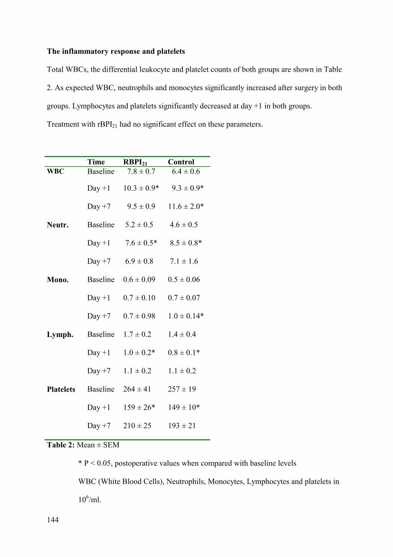
144
The inflammatory response and platelets
Total WBCs, the differential leukocyte and platelet counts of both groups are shown in Table
2. As expected WBC, neutrophils and monocytes significantly increased after surgery in both
groups. Lymphocytes and platelets significantly decreased at day +1 in both groups.
Treatment with rBPI21 had no significant effect on these parameters.
Time RBPI21 Control WBC Baseline 7.8 ± 0.7 6.4 ± 0.6
Day +1
10.3 ± 0.9* 9.3 ± 0.9*
Day +7 9.5 ± 0.9 11.6 ± 2.0*
Neutr.
Baseline 5.2 ± 0.5 4.6 ± 0.5
Day +1 7.6 ± 0.5* 8.5 ± 0.8*
Day +7 6.9 ± 0.8 7.1 ± 1.6
Mono.
Baseline 0.6 ± 0.09 0.5 ± 0.06
Day +1 0.7 ± 0.10 0.7 ± 0.07
Day +7 0.7 ± 0.98 1.0 ± 0.14*
Lymph.
Baseline 1.7 ± 0.2 1.4 ± 0.4
Day +1 1.0 ± 0.2* 0.8 ± 0.1*
Day +7 1.1 ± 0.2 1.1 ± 0.2
Platelets
Baseline 264 ± 41 257 ± 19
Day +1 159 ± 26* 149 ± 10*
Day +7 210 ± 25 193 ± 21
Table 2: Mean ± SEM
* P < 0.05, postoperative values when compared with baseline levels
WBC (White Blood Cells), Neutrophils, Monocytes, Lymphocytes and platelets in
106/ml.
145
Cytokines (Interleukin-6, VEGF, endostatin)
The interleukin-6, VEGF and endostatin concentrations are shown in Table 3 and 4.
Interleukin-6
Plasma IL-6 levels were in the normal range preoperatively, but significantly increased in
both groups in all the postoperative periods. In both groups the highest IL-6 levels were found
during the first 24 hours and reached peak levels at 2 hours postoperatively. After day +1, the
IL-6 levels decreased, though levels remained significantly elevated till day +9 in both
groups. The IL-6 levels in the rBPI21 group were less increased in all postoperative time
intervals. A difference between the two groups was found 2 hours after surgery, P=0.03.
VEGF
In both groups VEGF levels decreased significantly in the postoperative hours, whereas in the
control group the VEGF levels even dropped under the minimum detectable levels. At day +1
the VEGF levels increased in both groups. The VEGF level in the rBPI21 group was
significantly higher than the control group, P=0.01, at day +1.
Endostatin
In both groups, a three to four-fold decrease in endostatin levels was immediately observed,
which remained significantly low up to day +3. In the control group the endostatin levels were
still low at day +5, (P=0.04) and +7, (P=0.05) when compared to baseline value.

146
Time rBPI21 Control
IL-6 Baseline 2.8 ± 1.9 4.2 ± 2.1
0 Hr 174.9 ± 48.8* 252.8 ± 78.4*
2 Hr 211.7 ± 52.6* # 527.7 ± 184* #
4 Hr 181.1 ± 44.8* 486.9 ± 221*
8 Hr 165.5 ± 36.1* 355.8 ± 161*
Day +1 168.1 ± 48.9* 133.4 ± 31.3*
Day +3 45.3 ± 14.0* 55.9 ± 17.7*
Day +5 38.4 ± 13.0* 70.8 ± 29.2*
Day +7 25.1 ± 4.9* 48.0 ± 10.8*
Day +9 20.2 ± 4.4* 43.9 ± 13.7*
VEGF Baseline 24.3 ± 3.9 35.2 ± 5.5
0 Hr
17.1 ± 3.8* N.D.
2 Hr
10.0 ± 4.1* N.D.
4 Hr
14.0 ± 4.9* N.D.
8 Hr
9.4 ± 2.1 N.D.
Day +1 38.4 ± 6.6# 15.6 ± 3.1*#
Day +3 29.8 ± 5.0 19.9 ± 4.8
Day +5 24.5 ± 4.5 34.4 ± 10.3
Day +7 33.3 ± 7.7 27.1 ± 7.9
Day +9 22.9 ± 4.5 24.0 ± 5.1
Table 3: Mean ± SEM
* P < 0.05, postoperative values when compared with baseline levels
# P < 0.05, significant difference between the groups
N.D. = not detectable, VEGF and IL-6 in pg/ml
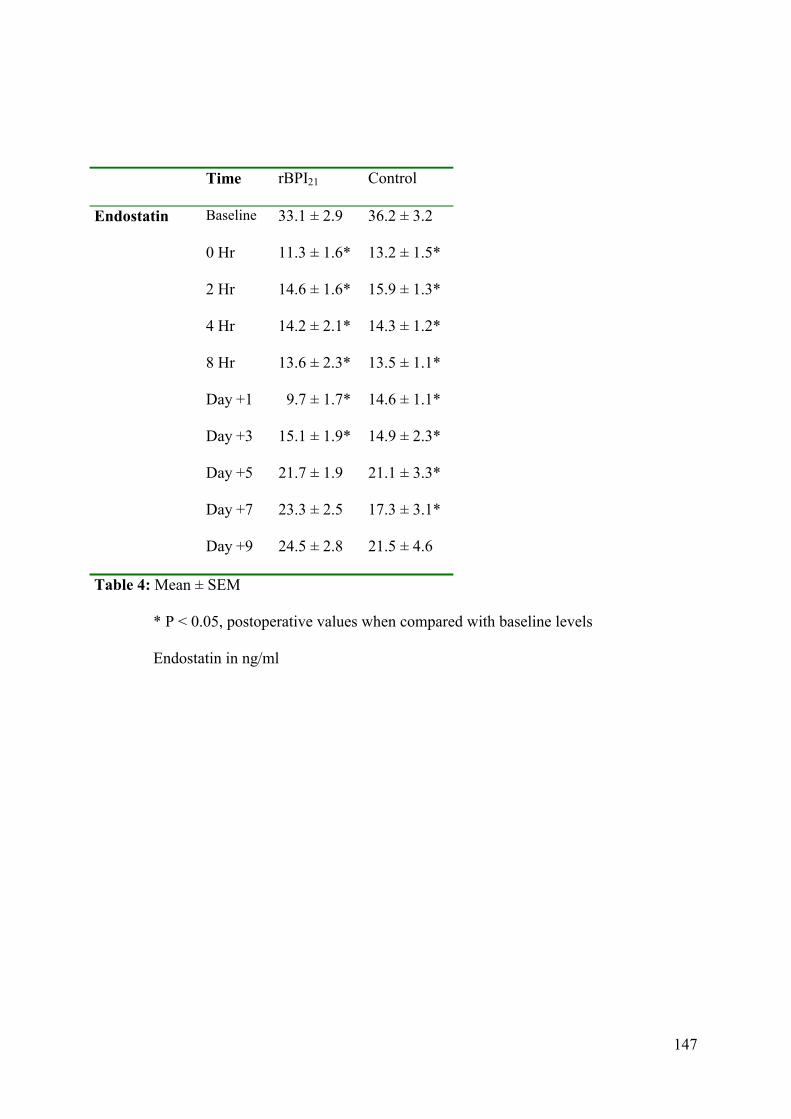
147
Time rBPI21 Control
Endostatin Baseline 33.1 ± 2.9 36.2 ± 3.2
0 Hr
11.3 ± 1.6* 13.2 ± 1.5*
2 Hr
14.6 ± 1.6* 15.9 ± 1.3*
4 Hr
14.2 ± 2.1* 14.3 ± 1.2*
8 Hr
13.6 ± 2.3* 13.5 ± 1.1*
Day +1 9.7 ± 1.7* 14.6 ± 1.1*
Day +3 15.1 ± 1.9* 14.9 ± 2.3*
Day +5 21.7 ± 1.9 21.1 ± 3.3*
Day +7 23.3 ± 2.5 17.3 ± 3.1*
Day +9 24.5 ± 2.8 21.5 ± 4.6
Table 4: Mean ± SEM
* P < 0.05, postoperative values when compared with baseline levels
Endostatin in ng/ml
148
DISCUSSION
In this study, we measured the inflammatory response reflected by the immune cells and IL-6,
and the angiogenic response reflected by VEGF and endostatin after partial liver surgery with
or without perioperative 48-hour continuous rBPI21 administration.
The postoperative increase of total WBC counts was similar in both groups, which is in
accordance with other observations.28 Both groups had elevated postoperative IL-6
concentrations; however, IL-6 levels in the control group were higher than in the rBPI21
group, suggesting that rBPI21 depresses the IL-6 production. This effect may be explained by
the LPS-neutralizing capacity of rBPI21. This observation is in accordance with the study by
Meszaros et al. who demonstrated that the release of pro-inflammatory cytokines IL-1 beta,
IL-6, IL-8 and tumor necrosis factor, induced by E.coli endotoxin, was tempered by
administrating rBPI23.26
Interestingly, IL-6 levels increased and VEGF levels decreased in the postoperative hours in
both groups. This seems paradoxical since IL-6 can induce VEGF10 and un-physiological as
well, since VEGF is needed for the initiation of angiogenesis after surgery. The precise
mechanism for this observation is unclear, but the effect may be explained by experimental
studies that showed up-regulation of the high affinity VEGF receptors, flt-1 and KDR/flk-1 by
the endothelium following partial hepatectomy.17,29 In the immediate postoperative period,
circulatory VEGF and additional VEGF released locally from platelets and leucocytes may be
scavenged by the up-regulated VEGF receptors, which is needed for an immediate initiation
of the angiogenic process. The data suggest that the remnant liver endothelium acts as a
sponge extracting all available VEGF necessary for liver regeneration. Most studies observe
an increase of circulating VEGF after surgery but studied this at later time points.14,30 We
demonstrate that profound VEGF physiological dynamics already start in the first
postoperative hours.

149
After 24-hour continuous rBPI21 administration, VEGF levels are significantly higher when
compared to the control group on day +1. This observation may be explained by the
competition of rBPI21 with locally generated VEGF on heparin binding sites in the
extracellular matrix (ECM), resulting in less storage of VEGF during rBPI21 infusion.9,12 This
may alter a VEGF concentration gradient that is required for guided EC migration, confirming
the idea of anti-angiogenic effects of rBPI21.
The VEGF increase in the later postoperative period may be produced by leucocytes, which
play an important role in wound healing, as well as hypoxic cells in devitalized tissue and the
increased IL-6 that can induce VEGF expression.10,16
Endostatin is an endogenous anti-angiogenic factor. Theoretically, colorectal carcinoma
metastasized to the liver can generate endostatin by producing proteases able to cleave
collagen XVIII in the direct environment.31,32 In this study, high preoperative endostatin
levels were found in cancer patients. This confirms the hypothesis that colon carcinoma
metastasized to the liver may generate endostatin. Both groups showed a significant
endostatin decrease immediately after surgery. Multiple factors may be responsible for this
effect. Removal of the tumor plus a substantial part of the liver means a reduction of
endostatin production. Surgical trauma with subsequent release of various proteolytic
enzymes involved in healing of the wound may degrade endostatin. Finally, the effect of
anesthesia on the liver may inhibit endostatin production. Since the half-life of endostatin is
less than one hour (Entremed Endostatin manual book), these effects together easily explain
the immediate decrease after surgery. The postoperative endostatin decrease further
substantiates the idea that a tumor may produce angiogenic inhibitors, suppressing their
metastases at distance.33
In this study, rBPI21 only transiently influenced IL-6 and VEGF production. The most striking
finding was that surgery per se induced an immediate and impressive immune response (IL-6)
150
together with an immediate angiogenic response, reflected in a paradoxal VEGF decrease and
a longer lasting decrease of endostatin. These immediate responses, even the initial, paradoxal
decrease of VEGF, have a clear physiological meaning and emphasize the importance of
studying the dynamics of immunological and angiogenic responses in the early phase of
wound healing.

151
REFERENCES
1. Butler J, Attiyeh FF, Daly JM. Hepatic resection for metastases of the colon and
rectum. Surg Gynecol Obstet. 1986;162:109-13.
2. Nagasue N, Kohno H, Chang YC, et al. Liver resection for hepatocellular
carcinoma. Results of 229 consecutive patients during 11 years. Ann Surg.
1993;217:375-84.
3. Finch MD, Crosbie JL, Currie E, Garden OJ. An 8 year experience of hepatic
resection: indications and outcome. Br J Surg. 1998;85:315-9.
4. Shirabe K, Shimada M, Gion T, et al. Postoperative liver failure after major
hepatic resection for hepatocellular carcinoma in the modern era with special
reference to remnant liver volume. J Am Coll Surg. 1999;188:304-9.
5. Pace RF, Blenkharn JI, Edwards WJ, et al. Intra-abdominal sepsis after hepatic
resection. Ann Surg. 1989;209:302-6.
6. Wiezer MJ, Boelens PG, Vuylsteke RJ, et al. Perioperative treatment with
bactericidal/permeability-increasing protein (rBPI21) in major liver surgery: a
concise summary. Ann Acad Med Singapore 2001;30:226-33.
7. Marra MN, Wilde CG, Griffith JE, et al. Bactericidal permeability-increasing
protein has endotoxin-neutralizing activity. J Immunol. 1990;144:662-6.
8. Weiss J, Elsbach P, Shu C, et al. Human bactericidal/permeability-increasing
protein and a recombinant NH2-terminal fragment cause killing of serum-
resistant gram-negative bacteria in whole blood and inhibit tumor necrosis factor
release induced by the bacteria. J Clin Invest. 1992;90:1122-30.
9. van der Schaft DW, Toebes EA, Haseman JR, et al. Bactericidal/permeability-increasing
protein (BPI) inhibits angiogenesis via induction of apoptosis in vascular endothelial cells.
Blood 2000;96:176-81.
152
10. Cohen T, Nahari D, Cerem LW, et al. Interleukin 6 induces the expression of vascular
endothelial growth factor. J Biol Chem. 1996;271:736-41.
11. Cressman DE, Greenbaum LE, DeAngelis RA, et al. Liver failure and defective
hepatocyte regeneration in interleukin-6 deficient mice. Science 1996;274:1379-
83.
12. Neufeld G, Cohen T, Gengrinovitch S, Poltorak Z. Vascular endothelial growth
factor (VEGF) and its receptors. FASEB J. 1999;13:9-22.
13. Maniwa Y, Okada M, Ishii N, Kiyooka K. Vascular endothelial growth factor
increased by pulmonary surgery accelerates the growth of micrometastases in
metastatic lung cancer. Chest 1998;114:1668-75.
14. Bondestam J, Salven P, Jaaskela-Saari H, et al. Major surgery increases serum
levels of vascular endothelial growth factor only temporarily. Am J Surg. 2000; 179:57-9.
15. Salven P, Orpana A, Joensuu H. Leukocytes and platelets of patients with cancer
contain high levels of vascular endothelial growth factor. Clin Cancer Res.
1999;5:487-91.
16. Mateo RB, Reichner JS, Albina JE. Interleukin-6 activity in wounds. Am J
Physiol. 1994;266:R1840-4.
17. Mochida S, Ishikawa K, Inao M, et al. Increased expressions of vascular
endothelial growth factor and its receptors, flt-1 and KDR/flk-1, in regenerating
rat liver. Biochem Biophys Res Commun. 1996;226:176-9.
18. Taniguchi E, Sakisaka S, Matsuo K, et al. Expression and role of vascular endothelial
growth factor in liver regeneration after partial hepatectomy in rats. J Histochem
Cytochem. 2001;49:121-30.
19. Dhanabal M, Ramchandran R, Waterman MJ, et al. Endostatin induces endothelial cell
apoptosis. J Biol Chem. 1999;274:11721-6.

153
20. Yoon SS, Eto H, Lin CM, et al. Mouse endostatin inhibits the formation of lung
and liver metastases. Cancer Res. 1999;59:6251-6.
21. Wen W, Moses MA, Wiederschain D, et al. The generation of endostatin
is mediated by elastase. Cancer Res. 1999;59:6052-6.
22. Felbor U, Dreier L, Bryant RA, et al. Secreted cathepsin L generates endostatin
from collagen XVIII. EMBO J. 2000;19:1187-94.
23. Muragaki Y, Timmons S, Griffith CM, et al. Mouse Col18a1 is expressed in a
tissue-specific manner as three alternative variants and is localized in basement
membrane zones. Proc Natl Acad Sci U S A 1995;92:8763-7.
24. Schuppan D, Cramer T, Bauer M, et al. Hepatocytes as a source of collagen
type XVIII endostatin. Lancet 1998;352:879-80.
25. Wiezer MJ, Meijer C, Sietses C, et al. Bactericidal/permeability-increasing protein
preserves leukocyte functions after major liver resection. Ann Surg. 2000;232:208-15.
26. Meszaros K, Parent JB, Gazzano-Santoro H, et al. A recombinant amino terminal
fragment of bactericidal/permeability-increasing protein inhibits the induction
of leukocyte responses by LPS. J Leukoc Biol. 1993;54:558-63.
27. Wiezer MJ, Langendoen SI, Meijer C, et al. Pharmacokinetics of a recombinant
amino terminal fragment of bactericidal/permeability-increasing protein (rBPI21) after
liver surgery in rats and humans. Shock 1998;10:161-6.
28. Boermeester MA, Houdijk AP, Meyer S, et al. Liver failure induces a systemic
inflammatory response. Prevention by recombinant N-terminal
bactericidal/permeability increasing protein. Am J Pathol. 1995;147:1428-40.
154
29. Yamane A, Seetharam L, Yamaguchi S, et al. A new communication system
between hepatocytes and sinusoidal endothelial cells in liver through vascular
endothelial growth factor and Flt tyrosine kinase receptor family (Flt-1 and
KDR/Flk-1). Oncogene 1994;9:2683-90.
30. Fujisaki K, Mitsuyama K, Toyonaga A, et al. Circulating vascular endothelial
growth factor in patients with colorectal cancer. Am J Gastroenterol.
1998;93:249-52.
31. Keppler D, Waridel P, Abrahamson M, et al. Latency of cathepsin B secreted
by human colon carcinoma cells is not linked to secretion of cystatin C and
is relieved by neutrophil elastase. Biochim Biophys Acta 1994;1226:117-25.
32. Bjornland K, Buo L, Scott H, et al. Polymorphonuclear elastase in human
colorectal carcinoma. Int J Oncol. 1998;12:535-40.
33. O'Reilly MS, Holmgren L, Shing Y, et al. Angiostatin: a novel angiogenesis
inhibitor that mediates the suppression of metastases by a Lewis lung carcinoma.
Cell 1994;79:315-28.

155
Chapter 8
GENERAL SUMMARY

156
Since the discovery of angiogenesis by Dr. Folkman in 1971 the explosion of publications
demonstrated the great importance biomedical researchers attached to this process for human
biology. However, the knowledge about the effect of a surgical trauma on the local and
systemic angiogenic balance is limited.
This thesis aimed to evaluate the angiogenic balance in blood and in local fluid in patients:
1. Undergoing elective benign or cancer surgery.
2. Undergoing minimal invasive or conventional surgery.
3. In whom the angiogenic dynamics were measured at early and later time-
intervals.
4. Who were perioperatively treated with or without immunomodulators.
This last issue was based on the fact that wounds generate immunologically active cytokines,
and that the addition of some specific immunological factors might increase (rhGM-CSF,
chapter 6) or inhibit (rBPI21, chapter 7) the immunological response to surgery. This might
have consequences for the angiogenic response since many immunological cytokines induce
also angiogenic effects.
In Chapter 2 patients with a primary carcinoma were prospectively randomized to a curative
laparoscopic or conventional colon resection. A difference in both the peritoneal and systemic
immune response following both surgical approaches was observed. We show evidently that
the plasma concentrations of pro-inflammatory cytokines after both surgical approaches
represent only a fraction of what is generated locally. Interestingly the appearance of pro-
inflammatory cytokines in the circulation is significantly lower in the laparoscopic group,
whereas the same cytokines locally produced showed no differences, confirming the idea that
both intra-abdominal approaches are different in their impact on the immunological system as
a whole. In addition, monocyte HLA-DR-expression was measured as a parameter for trauma-
induced immune suppression. A postoperative depression was observed in both groups, but in
157
the conventional group the HLA-DR-expression remained suppressed at least until four
postoperative days. The cellular response in terms of leucocytes was similar between the two
groups. Together, subtle differences in the immunological response to a major or minor
trauma were observed, which warrants further investigation.
In Chapter 3 we studied the effect of an operative trauma (minimal invasive versus
conventional Nissen fundoplication) on the circulatory angiogenic balance and the
inflammatory response. The VEGF plasma level was significantly decreased two hours after
the operation in the laparoscopic group and returned to preoperative values after 24 hours. In
the conventional group, the VEGF plasma level significantly increased four days after
surgery. The white blood cells and C-reactive protein increased rapidly, the latter more
intensively in the conventional group. The plasma endostatin concentration remained about
the same in both groups. Again, these findings suggest that the extent of a surgical
intervention has consequences for the angiogenic and immunological response.
In Chapter 4 we examined the VEGF and endostatin levels in wound fluid and plasma of
patients undergoing curative breast cancer surgery and female-to-male mastectomy. We
compared the angiogenic profile in wound fluid with the pattern in the circulation. In both
groups VEGF levels in wound fluid were much higher compared to postoperative plasma
levels, showing a similar trend as was found for pro-inflammatory cytokines in local fluid and
in plasma. The wound fluid endostatin concentration, however, was lower than in plasma and
decreased progressively after surgery. The local VEGF increase and endostatin decrease
observed immediately after surgery appears to be a physiological response to operative
trauma, since endostatin impairs blood vessel maturation during wound healing. This
‘angiogenic switch’ was not influenced by the type of process (malignant or benign) involved
in the surgical intervention.

158
Chapter 5 describes the local and systemic angiogenic profile in patients undergoing curative
laparoscopic or conventional surgery for colon cancer. Like in chapter 4 the VEGF levels in
wound fluid were significantly higher compared to postoperative circulatory levels, whereas
endostatin levels in wound fluid were lower than levels in blood, and decreased progressively
after surgery in both groups. Interestingly the VEGF levels in wound fluid measured at day 4
were significantly higher in the laparoscopic group compared with the conventional group.
This may be explained by the effect of local acidosis, due to CO2 insufflation, on endothelial
cells.
In the last two chapters the systemic angiogenic balance was evaluated in patients treated with
perioperative immunomodulatory agents. Recombinant human GM-CSF is known to
stimulate the immune system as well as angiogenesis via multiple pathways. In chapter 6 we
investigated the combined effects of curative surgery, with or without rhGM-CSF, on
angiogenic parameters in patients with a colorectal carcinoma. In this phase II randomized,
placebo-controlled trial, patients were assigned to perioperative rhGM-CSF (2.8 µg/kg body
weight) treatment or saline. RhGM-CSF enhanced the production of IL-6 and VEGF, but had
no effect on the anti-angiogenic agents endostatin and angiostatin. Surgery induced a transient
decrease of endostatin. After surgery two types of angiostatin (kringle 1-3 and kringle 1-4)
became visible in both groups. K1-4 angiostatin became manifest in almost all patients,
whereas the K1-3 form increased transiently in 6/16 of the patients. This may be explained by
the fact that proteolytic enzymes, which are part of the coagulation pathway and involved in
early stages of wound healing, are also active in the conversion of plasminogen into the
angiostatin. This study suggests an immediate (within 24 hours) initiation of angiogenesis
postoperatively, reflected by the circulatory increase of VEGF and a transient decrease of
endostatin, followed by the appearance of two angiostatin bands, which confirms
physiological wound healing in these cancer patients.

159
BPI is bactericidal and neutralizes lipopolysaccharide (LPS), which may reduce postoperative
infectious complications. In addition, BPI has been shown to be an inhibitor of angiogenesis.
In chapter 7 patients with metastasized colorectal carcinoma to the liver were double blind
randomized. The levels of the pro-angiogenic factors IL-6 and VEGF and the angiogenesis
inhibitor endostatin were investigated after liver surgery with a 48-hours during, perioperative
administration of either rBPI21 or placebo. The highest IL-6 levels were found during the first
24 hours and reached peak levels already at two hours postoperatively in both groups. In both
groups VEGF levels decreased sharply in the postoperative hours, returning to baseline levels
in the days afterwards. Also, in both groups, an immediate decrease in endostatin levels was
observed which remained significantly low. RBPI21 only marginally affected IL-6 and VEGF
levels. Because blood sampling was performed in the first hours after surgery, it became clear
that drastic wounding per se induced an immediate immune response (IL-6) and an immediate
angiogenic response, reflected in an initial VEGF decrease and a longer lasting decrease of
endostatin. This initial VEGF decrease could be explained by upregulation of the VEGF-
receptors as the very first response to wounding, increasing the uptake of available VEGF,
followed by an abundant increase in local VEGF production by almost all cells present in the
wound and, subsequently, an increase in blood VEGF concentrations.
In conclusion, a surgical trauma elicits a cascade of complex humoral and cellular changes,
which is intended to protect the host and promote wound healing. For a wound to heal a
temporary dysequilibrium between angiogenic stimulators and inhibitors occurs. In this thesis
we demonstrated that a surgical trauma and subsequent wound healing was associated with a
strong local increase in angiogenic stimulator (VEGF) and a decrease of angiogenic inhibitor
(endostatin), suggesting a sort of angiogenic switch. The fact that we included surgery for
patients with a benign or a cancerous process, and the usage of perioperative
immunomodulators did not heavily influence this physiological process. Due to the fact that

160
we studied locally generated fluid together with blood, we could show that both the
immunological and the angiogenic consequences of wounding were much more pronounced
in wound fluid than in the circulation. This suggests that investigation of wound fluid is much
more informative concerning the process of wound healing than the study of blood. Proteomic
studies of wound fluid versus blood might be of great interest. In addition, we demonstrated
that both the immunological response and the angiogenic response were initiated immediately
after wounding, and showed a pattern which is physiological. The inhibitor generated in tissue
(endostatin) declined progressively, while enzymatic responses to the formation of an intra-
endothelial clot induced two isoforms of a circulating angiogenesis inhibitor (angiostatin).
It may be of great interest to investigate the biological significance of the differences in
surgical interventions (minimal invasive or conventional, concerning different organs or areas
of the body, different forms of anaesthesia, different types and stages of cancer) for the
generation of local and circulatory angiogenic stimulators and inhibitors, and their
consequences for tumor biology. An increased local VEGF may support the proliferation of
residual tumor cells, and in addition with the immunosuppressive nature of VEGF, which
inhibits the maturation of dendritic cells, may ultimately result in permission of tumor cell
outgrowth, locally (graft metastases or local recurrence) or at distance. Anti-angiogenic
therapy during surgical interventions may have negative consequences for wound healing,
but, at the same time, may be beneficial for the tumor status of a cancer patient. Different
angiogenesis inhibitors will vary in their effects on wound healing and anti-tumor activity.
Timing of these inhibitors during surgery may be of importance. Together, it will be clear that
further studies are needed to elucidate the process of angiogenesis during wound healing and
the possible effects of angiogenic factors, generated by surgery, on tumor biology.

161
SAMENVATTING
162
Angiogenese is het proces van de aanmaak van nieuwe bloedvaten. Dit vindt met name plaats
in een embryo, maar ook tijdens de menstruatie en wondgenezing en bij ziektes, zoals kanker.
Angiogenese is ontdekt door Dr. Folkman in 1971 en daarna ontstond een explosie van
publicaties over dit proces. De kennis van het effect van chirurgisch trauma op angiogenese is
echter beperkt. Het doel van dit onderzoek was het angiogene evenwicht in bloed en in
wondvocht van patiënten te evalueren. De patiënten in de onderzoeksgroep voldeden aan èèn
van de volgende kenmerken:
1. Patiënt heeft goedaardige of kanker chirurgie ondergaan
2. Patiënt heeft minimaal invasieve of conventionele chirurgie ondergaan
3. Bij de patiënt is de angiogene dynamiek gemeten, direct na de ingreep en op latere
tijdstippen na de ingreep
4. Patiënt is perioperatief behandeld met of zonder immunomodulators.
De aandacht voor het verschil in perioperatieve behandeling is gebaseerd op het feit dat
wonden cytokinen genereren en dat de toevoeging van sommige specifieke immunologische
factoren de postoperatieve immunologische respons zou kunnen stimuleren (rhGM-CSF,
hoofdstuk 6) of remmen (rBPI21, hoofdstuk 7). Dit heeft mogelijk gevolgen voor de
angiogene respons, want het is bekend dat vele immunologische cytokinen ook angiogene
effecten hebben.
In hoofdstuk 2 werden patiënten met darmkanker zonder uitzaaiing prospectief
gerandomiseerd naar een laparoscopische of conventionele ingreep. Een verschil in zowel de
lokale (wondvocht) als systemische (plasma) immuunreactie die op beide chirurgische
benaderingen volgt, werd opgemerkt. Er wordt aangetoond dat de pro-inflammatoire cytokine
concentraties gemeten in plasma na beide chirurgische benaderingen slechts een fractie zijn
van wat er plaatselijk gegenereerd wordt. Opmerkelijk is dat de gemeten expressie van pro-
inflammatoire cytokinen in plasma beduidend lager was na een laparoscopische ingreep,

163
terwijl dezelfde expressie gemeten in wondvocht geen verschillen toonde. Hiermee bevestigen
we het idee dat beide intra-abdominale benaderingen verschillend zijn in hun uitwerking op
het immunologische systeem als geheel. Tevens werd HLA-DR-expressie op monocyten
gemeten, wat als een erkende parameter voor postoperatieve immuunsuppressie fungeert. Een
postoperatieve suppressie werd in beide groepen opgemerkt, maar in de conventionele groep
bleef de HLA-DR-expressie onderdrukt tot tenminste vier postoperatieve dagen. De
postoperatieve leukocytenreactie was gelijk in de twee groepen.
In hoofdstuk 3 bestudeerden wij het effect van een chirurgisch trauma (laparoscopische versus
conventionele Nissen fundoplication) op het angiogene evenwicht in de circulatie en op het
immuunsysteem. De VEGF concentratie in plasma was twee uur postoperatief beduidend
lager in de laparoscopische groep en keerde na 24 uur terug naar de preoperatieve waarde. In
de conventionele groep was de VEGF plasmaconcentratie na vier postoperatieve dagen
significant toegenomen. De concentratie witte bloedcellen en C-reactief proteïne stegen
postoperatief snel, waarbij C-reactief proteïne in de conventionele groep sneller toenam dan in
de laparoscopische groep. De endostatine concentratie in plasma bleef hetzelfde over de
gehele periode in beide groepen. Opnieuw heeft de grootte van het chirurgische trauma
gevolgen voor de angiogene en immunologische reacties.
In hoofdstuk 4 onderzochten wij VEGF- en endostatine concentraties in wondvocht en plasma
van patiënten met borstkanker en vrouw-naar-man mastectomie. Wij vergeleken het
angiogene profiel tussen waarden in wondvocht en in plasma. In beide groepen waren de
VEGF-concentraties in wondvocht veel hoger dan in plasma. Een soortgelijke trend werd ook
gevonden voor pro-inflammatoire cytokinen. De endostatine concentratie in wondvocht was
lager dan in plasma en nam progressief af. De locale VEGF-toename en endostatine afname
meteen na een chirurgisch ingreep, lijkt een fysiologisch reactie te zijn op een trauma. Deze
164
postoperatieve 'angiogenic switch' werd niet door het soort proces (maligne of benigne)
beïnvloed.
In hoofdstuk 5 onderzochten wij het lokale en systemisch angiogene profiel bij darmkanker
patiënten die een curatieve laparoscopische of conventionele ingreep ondergingen. Net als in
hoofdstuk 4 was de VEGF-concentratie in wondvocht beduidend hoger dan de postoperatieve
VEGF-concentratie in plasma, terwijl de endostatine waarden in wondvocht lager waren dan
waarden in plasma, en postoperatief progressief afnamen in beide groepen. Op de vierde
postoperatieve dag was de VEGF-concentratie in wondvocht hoger in de laparoscopische
groep dan in de conventionele groep. Dit zou verklaard kunnen worden door de gevolgen van
lokale acidose, tengevolge van CO2-insufflatie, op endotheel cellen.
In de laatste twee hoofdstukken evalueerden we de angiogene balans in het bloed van
patiënten die perioperatief behandeld werden met een immunomodulerend middel. Het is
bekend dat GM-CSF zowel het immuunsysteem als de angiogenese stimuleert.
In hoofdstuk 6 onderzochten wij de gecombineerde effecten van chirurgie en rhGM-CSF, op
angiogene parameters bij patiënten met darmkanker. In deze fase II ge-randomiseerde,
placebo-gecontroleerde studie werden patiënten perioperatief behandeld met rhGM-CSF (2,8
mg/kg lichaamsgewicht) of met fysiologisch zout. rhGM-CSF versterkte de productie van IL-
6 en VEGF, maar had geen invloed op endostatine en angiostatine. De chirurgische ingreep
induceerde een kortdurende endostatine afname. In beide groepen verschenen na de ingreep
twee soorten angiostatine (kringle 1-3 en kringle 1-4). K1-4 angiostatine werd manifest in
bijna alle patiënten en K1-3 angiostatine nam in 6 van de 16 patiënten toe. Een mogelijke
verklaring hiervoor is dat proteolytische enzymen, die in het vroege wondgenezingsstadium
actief worden, ook betrokken zijn in de conversie van plasminogeen naar angiostatine.
Samenvattend wordt de angiogenese in een vroeg postoperatief stadium geïnitieerd door
toename van VEGF en afname van een angiogenese-remmer (endostatine), gevolgd door de
165
verschijning van twee angiostatine banden. Dit patroon bevestigt dat het
wondgenezingsproces bij kankerpatiënten fysiologisch verloopt. Postoperatieve infectueuze
complicaties kunnen mogelijk worden verminderd door het bactericidal permeability
increasing protein (BPI), dat lipopolysaccharide (LPS) neutraliseert. Bovendien is aangetoond
dat BPI een anti-angiogene werking heeft.
In hoofdstuk 7 werden patiënten met levermetastasen bij darmkanker perioperatief behandeld
met rBPI21 of een placebo. De pro-angiogene factoren IL-6, VEGF en de anti-angiogene
factor endostatine werden gemeten. In beide groepen werden in de eerste 24 uur de hoogste
IL-6 concentraties gemeten met een piek twee uur na de ingreep. In beide groepen werd
tevens in de eerste postoperatieve uren een sterke VEGF-daling waargenomen en in de dagen
erna nam de VEGF-concentratie toe. De VEGF-afname zou mogelijk verklaard kunnen
worden door een acute expressie van VEGF-receptoren, waardoor circulerend VEGF wordt
weggevangen. De toename van VEGF daarna wordt geproduceerd door alle cellen die
betrokken zijn in het wondgenezingsproces. Recombinant BPI21 had marginale invloed op IL-
6 en VEGF. Ook in beide groepen werd een acute endostatine afname waargenomen.
Sammenvattend start een chirurgisch trauma een cascade van complexe humorale en
cellulaire veranderingen die zijn gastheer beschermen en de wondgenezing bevorderen. Bij
een wond zal een tijdelijke disbalans ontstaan tussen angiogene stimulatoren en remmers. In
dit proefschrift demonstreerden wij dat wondgenezing gepaard gaat met een sterke lokale pro-
angiogene (VEGF) toename en lokale afname van een anti-angiogene factor (endostatine),
wat lijkt op een lokale "angiogenic switch". Dit fenomeen treedt op na een chirurgische
ingreep bij zowel patiënten met als zonder kanker en wordt niet beïnvloed door het gebruik
van perioperatieve immunomodulatoren. Door het tegelijk bestuderen van wondvocht en
plasma toonden we aan dat zowel de immunologische als de angiogene reacties meer
uitgesproken zijn in wondvocht dan in de circulatie. Bestudering van het

166
wondgenezingsproces via het wondvocht levert kennelijk betere informatie op dan
bestudering van factoren in het bloed. Tevens demonstreerden wij dat zowel de
immunologische als de angiogene reacties onmiddellijk na een verwonding beginnen. Het is
interessant de biologische betekenis van lokale en circulerende angiogene factoren na
verschillende chirurgische interventies te onderzoeken. Bijvoorbeeld na een laparoscopische
of conventionele ingreep, in verschillende organen of gebieden van het lichaam, de invloeden
van anesthesie en bij verschillende typen en stadia van kanker. De consequenties voor de
tumorbiologie zijn hierbij bijzonder interessant. Het gebruik van anti-angiogene therapie
tijdens een chirurgische ingreep valt te overwegen. De combinatie van anti-angiogene
therapie en chirurgie zou negatieve gevolgen kunnen hebben voor de wondgenezing, maar
tegelijkertijd heilzaam kunnen zijn voor patiënten met kanker. De verschillende angiogenese
remmers zullen mogelijk in hun gevolgen voor wondgenezing en anti-tumor activiteit
variëren. Bij het gebruik van deze remmers tijdens chirurgie is optimale timing van groot
belang. Vanzelfsprekend zijn er meer studies nodig om het angiogenese proces tijdens
wondgenezing verder te doorgronden en de mogelijke biologische effecten van angiogene
factoren, die door een chirurgisch ingreep gegenereerd worden, op tumorbiologie in kaart te
brengen.

167
DANKWOORD
168
Graag wil ik de volgende mensen bedanken voor hun bijdrage aan dit proefschrift.
Professor dr. M.A. Cuesta, Miquel mijn promotor. Behalve een leermeester, culinair expert,
en vriend was je de initiator van dit onderzoek. Bedankt voor je aanstekelijke enthousiasme en
de vrijheid die je me voor het onderzoek gaf om mijn creativiteit te ontplooien. Ik heb veel
van je over "het leven" geleerd en ik hoop dat onze vriendschap lang blijft voortbestaan.
Dr. K. Hoekman, mijn co-promotor: Klaas, dank je voor je expertise, geduld en enorme
bijdrage aan dit proefschrift. Ik vond het heel prettig om ook onder jouw hoede gewerkt te
hebben. Je scherpzinnigheid en "to-the-point" houding leerde mij structuur en richting te
geven aan het uiteindelijke proefschrift.
Professor dr. S. Meijer, Sybren, de onmisbare vrijdagochtend researchbesprekingen waren
altijd stimulerende en inzichtgevende discussies. Jouw kritische commentaar gaf me altijd stof
tot nadenken.
Professor dr. P.A.M. van Leeuwen, Paul, hartelijk bedankt voor de extra steuntje in de rug.
Overige leden van de promotiecommissie: Prof. dr. J.D. Bos, Prof. dr. R.J. Scheper, Prof. dr.
J. Lankelma , dr. J.R.M. van der Sijp en Prof. dr. I.H.M. Borel-Rinkes wil ik bedanken voor
hun interesse die ze aan mijn proefschrift hebben geschonken.
Annemarie Hansma jouw magische 'ELISA' handjes zijn doorslaggevend geweest voor het tot
stand komen van dit proefschrift. Bedankt voor je positieve instelling, ookal zag ik het soms
niet zitten. Veel succes met jouw onderzoek, promotie en ongetwijfeld je opleiding.
Dr. Wim 'congres buddy' van de Broek, we hebben tijdens onze vele wetenschappelijke reizen
een hoop gelachen, en genoten van de verschillende trendy clubs. Veel succes als
laparoscopisch chirurg in het zuiden van het land.
Colin Sietses, bedankt voor de begeleiding van mijn eerste onderzoeksperiode en de
introductie van de heilzame werking van koffie.
Anna Landegent bedankt voor de secretariële hulp en luisterend oor.

169
Elly de Lange bedankt voor de statistische hulp. Alle medewerkers van de afdeling
celbiologie. Professor dr. R.H.J. Beelen, Rob, dank je voor de interesse en gastvrijheid.
Mary von Blomberg bedankt voor je enthousiasme en de samenwerking.
Alle medewerkers van de afdeling klinische immunologie en in het bijzonder Petra Scholten.
De hoogbouw "Research mates": Frank Termaat, Robert Nijveldt, Ronald Vuylsteke, Petra
Boelens, Taco Blokhuis, Michiel Siroen, Tammo de Vries Reilingh, Manuel Koppe. Bedankt
voor de vele gezellige tijden, interessante internetsites en niet-wetenschappelijk discussies.
Mijn studievrienden, sportbuddies, en collega's bedankt voor de vriendschap, steun en vooral
begrip. Eindelijk is het boekje klaar en vanaf nu gaan we nog vaker iets doen.
Peter Brockhoff, Norbert Gret, paranimfen, sinds de kleuterschool zijn we al vrienden en nog
steeds hebben we goede contacten. Ik wil jullie bedanken voor het bijstaan op mijn
promotiedag.
Mijn ouders, ik ben enorm dankbaar voor het vertrouwen, de oneindige steun en vrijheid die
ze mij gegeven hebben. Ik hoop dat jullie in goede gezondheid nog meer kunnen reizen.
Mijn broer, Paul, bedankt voor je computer expertise en het opsporen van"verdwenen" of per
ongeluk gewiste originele manuscripten.

170
CURRICULUM VITAE The author of this thesis was born on September 25th, 1971 in Kowloon, Hong Kong. He and
his parents came to Holland in 1972. In 1992 he graduated from high school (Keizer Karel
College in Amstelveen) and started medical school at the Vrije Universiteit in Amsterdam.
During his study he entered a six-month scientific internship program at Children’s Mercy
Hospital in Kansas City, U.S.A. in which a newly developed anti-tumour compound called
Bullatacin was investigated (head: dr. A.Leyva). He obtained his medical degree in 1999 and
hereafter he worked as a research fellow at the department of gastro-intestinal surgery (head:
Prof. dr. M.A, Cuesta) at the Vrije Universiteit Medical Centre in Amsterdam. In january
2002 he worked as a surgical resident at the Vrije Universiteit in Amsterdam. He switched to
dermatology and started his training at the Academic Medical Centre in Amsterdam (head:
Prof. dr. J.D. Bos) in 2004.