the main methods of radiotherapy
TRANSCRIPT
The department of radiology and radiation medicine

Historically, the three main divisions of
radiation therapy are external beam radiation
therapy (EBRT or XRT) or teletherapy,
brachytherapy or sealed source radiation
therapy, and systemic radioisotope therapy
or unsealed source radiotherapy. The
differences relate to the position of the
radiation source; external is outside the body,
brachytherapy uses sealed radioactive sources
placed precisely in the area under treatment,
and systemic radioisotopes are given by
infusion or oral ingestion.

Brachytherapy can use temporary or
permanent placement of radioactive sources.
The temporary sources are usually placed by
a technique called afterloading. In
afterloading a hollow tube or applicator is
placed surgically in the organ to be treated,
and the sources are loaded into the applicator
after the applicator is implanted. This
minimizes radiation exposure to health care
personnel.
Particle therapy is a special case of external
beam radiation therapy where the particles
are protons or heavier ions. Intraoperative
radiation therapy or IORT is a special type of
radiation therapy that is delivered
immediately after surgical removal of the
cancer. This method has been employed in
breast cancer (TARGeted Introperative
radiation therapy or TARGIT), brain tumors
and rectal cancers.
Conventional external beam radiation therapy
Conventional external beam radiationtherapy (2DXRT) is delivered via two-dimensional beams using kilovoltage therapyx-ray units or medical linear acceleratorswhich generate high energy x-rays.
2DXRT mainly consists of a single beam ofradiation delivered to the patient from severaldirections: often front or back, and bothsides.

Conventional refers to the way the treatment
is planned or simulated on a specially calibrated
diagnostic x-ray machine known as a simulator
because it recreates the linear accelerator actions (or
sometimes by eye), and to the usually well-
established arrangements of the radiation beams to
achieve a desiredplan. The aim of simulation is to
accurately target or localize the volume which is to
be treated. This technique is well established and is
generally quick and reliable. The worry is that some
high-dose treatments may be limited by the
radiation toxicity capacity of healthy tissues which
lay close to the target tumor volume.
An example of this problem is seen in radiationof the prostate gland, where the sensitivity ofthe adjacent rectum limited the dose whichcould be safely prescribed using 2DXRTplanning to such an extent that tumor controlmay not be easily achievable. Prior to theinvention of the CT, physicians and physicistshad limited knowledge about the true radiationdosage delivered to both cancerous andhealthy tissue. For this reason, 3-dimensionalconformal radiation therapy is becoming thestandard treatment for a number of tumor sites.More recently other forms of imaging are usedincluding MRI, PET, SPECT and Ultrasound.

Stereotactic radiation is a specialized type of
external beam radiation therapy. It uses
focused radiation beams targeting a well-
defined tumor using extremely detailed
imaging scans. Radiation oncologists perform
stereotactic treatments, often with the help of
a neurosurgeon for tumors in the brain or
spine.

There are two types of stereotactic radiation:
Stereotactic radiosurgery (SRS) is
when doctors use a single or several
stereotactic radiation treatments of the brain
or spine.
Stereotactic body radiation
therapy (SBRT) refers to one or several
stereotactic radiation treatments with the
body, such as the lungs.

Some doctors say an advantage to stereotactic
treatments is that they deliver the right
amount of radiation to the cancer in a shorter
amount of time than traditional treatments,
which can often take six to 11 weeks. Plus
treatments are given with extreme accuracy,
which should limit the effect of the radiation
on healthy tissues. One problem with
stereotactic treatments is that they are only
suitable for certain small tumors.
Stereotactic treatments can be confusing
because many hospitals call the treatments by
the name of the manufacturer rather than
calling it SRS or SBRT. Brand names for
these treatments include
Axesse, Cyberknife, Gamma Knife, Novalis,
Primatom, Synergy, X-
Knife, TomoTherapy, Trilogy and Truebeam.
This list changes as equipment manufacturers
continue to develop new, specialized
technologies to treat cancers.
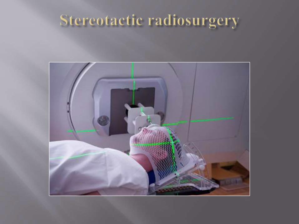

Despite its name, SRS is not a surgical
procedure.
The technique provides high-precision radiation
delivery to the tumor large dose by passing
healthy tissue located near. This distinguishes
SRH of standard radiation therapy.
Methods of three-dimensional visualization and localization, to determine the exact coordinates of the tumor or target organ.
Accessories for immobilization and careful positioning of the patient.
Clearly focused beams of gamma rays or X-rays that converge on the tumor or other pathological formation.
Methods of conducting radiotherapy under visual control, meaning the position of the tumor tracking during radiation cycle, thus increasing the accuracy and effectiveness of treatment

Typically, stereotactic radiosurgical intervention
carried out simultaneously. However, some experts
recommend several sessions of radiotherapy,
especially in larger tumors 3-4 cm in diameter.
This method of appointment 2-5 treatment sessions
is called fractionated stereotactic radiotherapy.
SRS and extracranial stereotactic intervention are an important alternative to open surgical procedures, especially for patients with contraindications. In addition, stereotactic intervention are indicated in tumors that are:
Located in hard areas for the surgeon.
Located next to vital organs.
Changes its position at physiological movements such as breathing


Radiosurgical procedures used in the following cases:
For the treatment of many brain tumors, including: benign and malignant tumors primary and metastatic lesions single and multiple tumors residual tumor foci after surgery intracranial lesions and tumors of the skull base and
orbit to treat arteriovenous malformations (AVM), which
represent an accumulation of modified or extended form blood vessels. AVM impair the normal blood flow to nerve tissue and prone to bleeding.
For the treatment of other neurological conditions and diseases
The treatment does not remove the tumor and only damages the DNA of tumor cells. As a result, cells lose their ability to reproduce. After the intervention radiosurgical tumor size reduced gradually over 1.5-2 years. This malignant and metastatic foci decreased even faster, sometimes for 2-3 months. If SRH is used for arteriovenous malformation, that for several years a gradual thickening of the vessel wall is observed and its lumen is complete close.

Gamma Knife is for radiation target organs used
192 or 201 clearly focused beam of gamma rays.
Gamma Knife treatment is perfect for small or
medium-sized intracranial injuries.

Linear accelerators - devices that are widely
distributed around the world and used to deliver
high-energy X-rays (photon beam). Suitable for
treating large tumor foci. The procedure can be
performed once or in several stages.

The realization stereotaxic surgery requires a
team approach. The group attending staff include:
oncologist, radiologist, medical physicist,
dosimetrist, radiologist / technician and nurse of
radiology department.

In most cases, after radiosurgical treatment return to normal
life is possible in 1-2 days.
Side effects are determined by the area of the body affected
by radiation. These include:
Hair loss in region of exposure
Ulceration of the mucous membrane of the mouth and
difficulty swallowing
Loss of appetite and indigestion
Diarrhea
Nausea and vomiting
Headaches
Disorder of urination

Radiosurgical treatment using the Gamma Knife system consists of four stages:
Setting retaining frame on the patient's head
Visualization of the tumor position
Treatment plan using a computer program
The procedure of radiation.
Stereotactic Radiosurgical procedure usually can be held ambulatory. However, it may be needed a short-term hospitalization.
About the supporting of the patient back home doctor must notify in advance.
For 12 hours before the procedure it is needed to stop taking food and liquids. It is also important to know your doctor about limitations in taking drugs.