the maize transcription factor myb-related protein-1 is a key
TRANSCRIPT

The Maize Transcription Factor Myb-Related Protein-1 Is aKey Regulator of the Differentiation of Transfer Cells C W
Elisa Gomez,a,1 Joaquın Royo,a,1 LuisM.Muniz,a Olivier Sellam,bWyatt Paul,b Denise Gerentes,b Cristina Barrero,a
Maribel Lopez,a Pascual Perez,b and Gregorio Huerosa,2
a Departamento de Biologıa Celular y Genetica, Universidad de Alcala, 28871 Alcala de Henares (Madrid), Spainb Biogemma SAS, Campus Universitaire des Cezeaux, 63170 Aubiere, France
Transfer cells are highly modified plant cells specialized in the transport of solutes. They differentiate at many plant
exchange surfaces, including phloem loading and unloading zones such as those present in the sink organs and seeds. In
maize (Zea mays) seeds, transfer cells are located at the base of the endosperm. It is currently unknown how apical-basal
polarity is established or why the peripheral cells at the base of the endosperm differentiate into transfer instead of aleurone
cells. Here, we show that in epidermal cells committed to develop into aleurone cells, the ectopic expression of the transfer
cell-specific transcriptional activator Myb-Related Protein-1 (MRP-1) is sufficient to temporarily transform them into
transfer cells. These transformed cells acquire distinct transfer cell features, such as cell wall ingrowths and an elongated
shape. In addition, they express a number of MRP-1 target genes presumably involved in defense. We also show that the
expression of MRP-1 is needed to maintain the transfer cell phenotype. Later in development, an observed reduction in the
ectopic expression of MRP-1 was followed by the reversion of the transformed cells, which then acquire aleurone cell
features.
INTRODUCTION
The regulation of solute and solvent fluxes across the plant body
and between source (e.g., leaves) and sink (e.g., seeds or fruits)
areas is a crucial process that influences plant growth and
development. The entry, branching, and exiting zones of the
vascular network are priority points where this regulation is
executed. Specialized cells differentiate to facilitate the loading/
unloading processes at these areas, and very frequently (albeit
not universally) these cells develop deep cell wall ingrowths
(CWIs), resulting in a remarkable increase in the membrane
surface area available for transport. The term transfer cell (TC)
(Pate and Gunning, 1972) has traditionally been used to describe
cells involved in transport that show these surfacemodifications.
Histological studies have identified modified TCs strategically
positioned at the interfaces where solute exchange takes place
(reviewed inOffler et al., 2002), such as the loading areas ofminor
leaf veins, areas surrounding the vascular bundle at stem nodes,
points of glandular secretion, and places of delivery of nutrients
at sink organs (i.e., at the base of flowers and fruits). Highly
differentiated TCs have also been found at the symplastic
discontinuities between individuals belonging to different gen-
erations (e.g., at the interface between gametophyte and spo-
rophyte generations [Ligrone and Gambardella, 1988] and at the
base of seeds [Kiesselbach, 1949; Cochrane and Duffus, 1980;
Offler and Patrick, 1993; Talbot et al., 2001]) and symbiotic
species (e.g., at mycorrhizal and rhizobium-root nodule inter-
faces [Gunning et al., 1974; Allaway et al., 1985]), and even at
points of plant–parasite interaction (e.g., in nematode infections
[Jones and Northcote, 1972]). This work makes use of the maize
(Zea mays) basal endosperm TC layer as a model for investigat-
ing the regulatory pathways that govern TC differentiation.
Seeds originate from a double fertilization event that gives rise
to the embryo and the endosperm (reviewed in Becraft, 2001;
Berger, 2003; Olsen, 2004). The endosperm develops from the
fertilized triploid central cell of the ovule, which undergoes
nuclear divisions without cytokinesis to produce a coenocyte
containing nuclei that are evenly distributed throughout the
peripheral cytoplasm. In cereals, periclinal cell division of the
first cellularized layer of the endosperm gives rise to two different
cell types: the aleurone and starchy endosperm initials (Becraft,
2001; Olsen, 2004). The aleurone layer serves as the epithelium
of the filial tissues, surrounding both the endosperm and embryo.
At the base of the maize endosperm, however, epithelial cells
differentiate to form the basal endosperm transfer cell layer
(BETL) instead of an aleurone layer, producing an interface
between the filial and maternal tissues of the seed and thus
facilitating nutrient uptake from the apoplastic space in the
placento-chalaza area. The differentiation and function of this
cell layer therefore critically influence the agronomically key
process of grain filling (Kiesselbach, 1949; Thompson et al.,
2001; Royo et al., 2007). The embryo-surrounding region (ESR),
located around and below the developing embryo, represents
a further expression domain within the endosperm (Opsahl-
Ferstad et al., 1997; Balandın et al., 2005).
1 These authors contributed equally to this work.2 Address correspondence to [email protected] author responsible for distribution of materials integral to thefindings presented in this article in accordance with the policy describedin the Instructions for Authors (www.plantcell.org) is: Gregorio Hueros([email protected]).CSome figures in this article are displayed in color online but in blackand white in the print edition.WOnline version contains Web-only data.www.plantcell.org/cgi/doi/10.1105/tpc.108.065409
The Plant Cell, Vol. 21: 2022–2035, July 2009, www.plantcell.org ã 2009 American Society of Plant Biologists
Dow
nloaded from https://academ
ic.oup.com/plcell/article/21/7/2022/6095443 by guest on 23 Septem
ber 2021
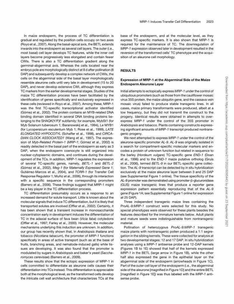
In maize endosperm, the process of TC differentiation is
gradual and regulated by the position cells occupy on two axes
(Royo et al., 2007). Along the basal-apical axis, the BETL extends
inwards into the endosperm as several cell layers. The outer (i.e.,
most basal) cell layer develops TC features, while the inner cell
layers become progressively less elongated and contain fewer
CWIs. There is also a TC differentiation gradient along the
germinal-abgerminal axis. Whereas the cells located near the
embryo pole aremorphologically distinct at 8 d after pollination (8
DAP) and subsequently develop a complex network of CWIs, the
cells on the abgerminal side of the basal layer morphologically
resemble aleurone cells until very late in development (15 to 20
DAP), and never develop extensive CWI, although they express
TCmarkers from the earlier developmental stages. Studies of the
maize TC differentiation process have been facilitated by the
identification of genes specifically and exclusively expressed in
these cells (reviewed in Royo et al., 2007). Among these, MRP-1
was the first TC-specific transcriptional activator identified
(Gomez et al., 2002). The protein contains an MYB-related DNA
binding domain identified in several DNA binding proteins be-
longing to the SHAQK(Y/F)F subfamily: for example, MybSt1 (for
Myb Solanum tuberosum 1; Baranowskij et al., 1994), Le MYB1
(for Lycopersicum esculentum Myb 1; Rose et al., 1999), LATE
ELONGATED HYPOCOTYL (Schaffer et al., 1998), and CIRCA-
DIAN CLOCK ASSOCIATED1 (Wang et al., 1997). The expres-
sion of Myb-Related Protein-1 (MRP-1; Gomez et al., 2002) is
readily detected in the basal part of the endosperm as early as 3
DAP, when the endosperm coenocyte is still organized into
nuclear-cytoplasmic domains, and continues during the devel-
opment of the TCs. In addition, MRP-1 regulates the expression
of several TC-specific genes, namely, BETL-1 and BETL-2
(Gomez et al., 2002), Meg-1 (for Maternally Expressed Gene 1;
Gutierrez-Marcos et al., 2004), and TCRR-1 (for Transfer Cell
Response Regulator 1; Muniz et al., 2006), through its interaction
with a specific sequence in the corresponding promoters
(Barrero et al., 2006). These findings suggest that MRP-1 might
be a key player in the TC differentiation process.
TC differentiation presumably occurs as a response to an
increased demand for solute transport. Little is known about the
molecular signals that induce TCdifferentiation, but it is likely that
transported solutes are involved (Offler et al., 2002). Certainly, it
has been shown that a transient increase in monosaccharide
concentration early in development induces the differentiation of
TC in the adaxial surface of fava bean (Vicia faba) cotyledons
(Offler et al., 1997; Farley et al., 2000). However, the molecular
mechanisms underlying this induction are unknown. In addition,
our group has recently shown that, in Arabidopsis thaliana and
tobacco (Nicotiana tabacum), the promoter of MRP-1 functions
specifically in areas of active transport (such as at the base of
fruits, branching areas, and nematode-induced galls) while tis-
sues are developing. It was also found that the promoter is
modulated by sugars in Arabidopsis and baker’s yeast (Saccha-
romyces cerevisiae) (Barrero et al., 2009).
These results show that the ectopic expression of MRP-1 in
cells committed to differentiate as aleurone cells causes their
differentiation into TCs instead. This differentiation is appreciable
both at themorphological level, as the transformed cells develop
the intricate cell wall architecture that characterizes TCs at the
base of the endosperm, and at the molecular level, as they
express TC-specific markers. It is also shown that MRP-1 is
required for the maintenance of TC. The downregulation of
MRP-1 expression observed later in development resulted in the
reversion of the transformed cells’ TC phenotype and the acqui-
sition of an aleurone cell morphology.
RESULTS
Expression of MRP-1 at the Abgerminal Side of the Maize
Endosperm Aleurone Layer
Initial attempts to ectopically expressMRP-1 under the control of
ubiquitous promoters (such as those from the cauliflower mosaic
virus 35S protein, the maize ubiquitin gene, and the cassava vein
mosaic virus) failed to produce stable transgenic lines. In all
cases, maize primary transformants were produced, albeit at a
low frequency, but they did not transmit the construct to the
progeny. Identical results were obtained in attempts to over-
express MRP-1 under the control of the 35S promoter in
Arabidopsis and tobacco. Plants containing constructs express-
ing significant amounts ofMRP-1 transcript produced nontrans-
genic progeny.
We next attempted to expressMRP-1 under the control of the
aleurone-specific promoter AL-9. AL-9 was originally isolated in
a search for compartment-specific molecular markers and en-
codes a protein of unknown function but related in sequence to
the barley (Hordeum vulgare) TC-specific gene END-1 (Doan
et al., 1996) and to the END-1 maize putative ortholog (Gruis
et al., 2006), termed BETL-9 in our BETL-specific gene collec-
tion. The AL-9 transcript can be detected by in situ hybridization
exclusively at the maize aleurone layer between 3 and 25 DAP
(see Supplemental Figure 1 online). The tissue specificity of the
AL-9 promoterwas demonstrated using promoter-b-glucuronidase
(GUS) maize transgenic lines that produce a reporter gene
expression pattern essentially reproducing that of the AL-9
gene (Figure 1A; see Supplemental Figure 1 online for expression
at 25 DAP).
Three independent transgenic maize lines containing the
ProAL-9:MRP-1 construct were selected for this study. No
special phenotypes were observed for these plants besides the
features described for the immature kernels below. Adult plants
and mature seeds were indistinguishable from nontransgenic
material.
Pollination of heterozygous ProAL-9:MRP-1 transgenic
maize plants with nontransgenic pollen produced a 1:1 segre-
gation in the sibling kernels. These were collected for analysis at
two developmental stages: 12 and 17 DAP. In situ hybridization
analyses using a MRP-1 antisense probe and 12-DAP kernels
(Figures 1B to 1E) showed that half of the kernels expressed
MRP-1 in the BETL (large arrow in Figure 1B), while the other
half also expressed the gene in the epithelial layer on the
abgerminal side of the endosperm (arrowheads in Figure 1C).
Part of the outer cell layer of the endosperm (i.e., the abgerminal
side of the aleurone [magnified in Figure 1D] and the entire BETL
[magnified in Figure 1E]) was thus labeled with the MRP-1 anti-
sense probe.
MRP-1 Induces Transfer Cell Differentiation 2023
Dow
nloaded from https://academ
ic.oup.com/plcell/article/21/7/2022/6095443 by guest on 23 Septem
ber 2021

The Expression of MRP-1 Induced the TC Differentiation
ProcessWhen Expressed in the Abgerminal Aleurone Layer
The expression of MRP-1 promoted a profound change in the
morphology of the cells otherwise committed to differentiate into
aleurone cells (Figure 2). At 12 DAP, wild-type seeds showed
multiple layers of small, cubic cells with no obvious asymmetric
features in either the upper or abgerminal endosperm (Figure 2A).
In the transgenic kernels, however, the cells on the abgerminal
side (Figure 2D) were seen to undergo a dramatic differentiation
process. These cells appeared elongated along the axis perpen-
dicular to the endosperm surface and started to accumulate newly
deposited cell wall material at their outer side (stained white-blue
with the cell wall stain Calcofluor white), resembling the CWIs that
densely fill themature TCat the baseof the endosperm (Figure 2J).
The inner cell layers immediately adjacent to the transdetermined
cells also elongated in the same direction as the external layer,
further reinforcing the similarity between this area and the BETL
(see Supplemental Figure 2 online for a diagram).
The cell morphology was then examined using semithin (0.5
mm) sections of LR white–embedded material at a 31000 mag-
nification. The very thin cell walls of the aleurone cells at the
abgerminal endosperm side (Figure 2B) contrasted with the thick
cell wall (stained pale blue with toluidine blue) developed by the
cells expressing MRP-1 (Figure 2E). The cell wall outgrowth
completely covered the outer side of the cells and about one-
third of their lateral sides. The density of CWI found at these cells
was much lower than that observed at the more differentiated
BETL cells (Figure 2K) but remarkably similar to the those
observed in the less developed TCs that can be observed at
the abgerminal side of the BETL (Figure 2L).
The cell wall structure was further studied by examining
ultrathin sections of the LR white–embedded material with
transmission electron microscopy (TEM). Abgerminal aleurone
cells (Figure 2C, equivalent area framed in red in Figure 2B)
possessed thin cell walls with no sign of wall outgrowth and very
few mitochondria. Cells expressing MRP-1 developed instead a
thick, massive cell wall from which small, limitedly branched
CWIs protruded into the cytoplasm (composite Figures 2F and
2G, equivalent area framed in 2E). The inner surface of the cell
wall thickenings was densely covered by mitochondria; these
mitochondria are also evident in the lower magnification image
(Figure 2E), but appeared now to be part of a complex synthetic
machinery (SM in Figures 2G to 2I) in an organelle-rich cytoplasm
also composed of reticulum, Golgi cisternae, and cytoskeletal
elements, resembling the situation previously described in
models for CWI formation (Offler et al., 2002). TEM analyses of
the CWIs developed at the BETL cells (Figures 2M and 2N)
revealed a higher structural organization than that observed in
the CWIs of transformed cells. Even in the higher-density areas
(Figure 2M) it was possible to distinguish each flange of CWI as
an individual entity emerging from an otherwise thin intercellular
cell wall. The relatively regular pattern of formation of individual
CWI was easily observed at the initial stages of cell wall growth
(Figure 2N). As compared with the situation in the BETL, the
transformed cells’ intercellular cell walls were thicker, probably
reflecting an initial disorganized growth preceding the formation
of CWIs. Occasionally, a very long, unbranched, flange-like CWI
formed in the transgenic abgerminal aleurone (Figure 2E, TEM
image in 2H). However, the CWIs generally had very limited
extension. Examination of the initial stages of cell wall growth at
the lateral sides of the cells (Figure 2I) showed that the massive
intercellular cell wall probably formed by coalescence of rather
amorphous CWIs.
The development of an ectopic endosperm transfer cell layer
(EETL) in the aleurone area expressing MRP-1 was also evident
Figure 1. Ectopic Expression of MRP-1.
(A) GUS staining of a ProAL9:GUS transgenic kernel at 11 DAP. The aleurone shows positive GUS staining.
(B) and (C) In situ hybridization experiments in 12-DAP nontransgenic (B) or ProAL9:MRP-1 transgenic (C) sibling kernels, using a MRP-1 antisense
riboprobe. The hybridization signal (white spots) was detected in the BETL in both cases (large arrows) and in one side of the aleurone in (C)
(arrowheads).
(D) and (E)Magnification of the areas boxed in (C), containing the abgerminal epithelial cells (D) or the BETL cells (E). The image in (D) has been rotated
908 clockwise.
Al, aleurone; Em, embryo; En endosperm; Pd, pedicel. Bars = 1 mm in (A) to (C) and 50 mm (D) and (E). (B) and (C) are dark-field micrographs; (D) and
(E) combine dark field and epifluorescence.
[See online article for color version of this figure.]
2024 The Plant Cell
Dow
nloaded from https://academ
ic.oup.com/plcell/article/21/7/2022/6095443 by guest on 23 Septem
ber 2021

Figure 2. The Ectopic Expression of MRP-1 Induces TC-Like Modifications.
(A) to (C) Images of the abgerminal side of nontransgenic endosperms at 12 (A) or 10 ([B] and [C]) DAP.
(D) to (I) Images of the abgerminal side of transgenic endosperms at 12 (D) or 10 DAP ([E] to [I]). (Enclosed in a common frame.)
(J) to (N) Images of the BETL at 12 (J) or 10 DAP ([K] to [N]).
(A), (D), and (J)Wax-embedded material sectioned at 8 m and stained with calcofluor white. CWIs appear light blue with calcofluor white staining (some
indicated by arrowheads in [D]). Transformed cells (those containing CWIs) appear in (D), elongated along the germinal-abgerminal axis; compare these
with the cubic cells at the aleurone layer (Al) or the rounded starchy endosperm cells (SE). The germinal-abgerminal (G-AB) or apico-basal (AP-B)
polarity is indicated for figures (A), (D), and (J). The image in (J) has been rotated 90˚ for comparison with the EETL. However, note that the images in (K)
and (L) are shown in a natural orientation, with the placento-chalaza at the bottom part of the micrograph.
(B), (E), (K), and (L) Semithin sections (0.5 m) of LR white–embedded material (10 DAP) stained with toluidine blue. Cell wall secondary growth is stained
pale blue in the transformed cells (E) and TCs ([K] and [L]).
(C), (F) to (I), (M), and (N) Ultrathin sections (0.05 m) of LR white–embedded material stained with lead nitrate and uranyl acetate. Images were taken
using TEM. The corresponding areas of the cells shown in these images are framed in red in (B), (E), and (L).
P, pericarp side; PCH, placento-chalaza; CW, cell wall; M, mitochondria, SM, synthesis machinery associated with the internal side of the CWI. Bars =
50m in (A), (D), and (J), 25 m in (B), (E), (K), and (L), and the indicated values in the TEM images. Materials analyzed in (A), (D), and (J)were derived from
the transgenic line EER-3b. Materials analyzed in (B), (C), (E) to (I), and (K) to (N) were derived from the transgenic line EER-2a.
MRP-1 Induces Transfer Cell Differentiation 2025
Dow
nloaded from https://academ
ic.oup.com/plcell/article/21/7/2022/6095443 by guest on 23 Septem
ber 2021
from the in situ hybridization and immunolocalization results
presented. The EETL images shown in this article were obtained
from the analyses of two different transgenic events and two
plant generations, and the EETL was always found on the
abgerminal side of the endosperm. No other aleurone cells
developed an obvious TC phenotype, although isolated cells at
different positions were found to express TC markers (see
below).
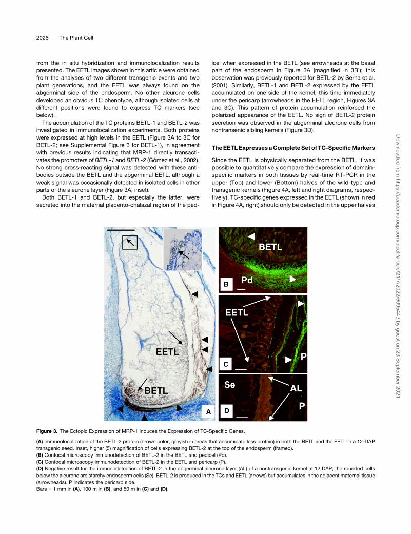
The accumulation of the TC proteins BETL-1 and BETL-2 was
investigated in immunolocalization experiments. Both proteins
were expressed at high levels in the EETL (Figure 3A to 3C for
BETL-2; see Supplemental Figure 3 for BETL-1), in agreement
with previous results indicating that MRP-1 directly transacti-
vates the promoters of BETL-1 and BETL-2 (Gomez et al., 2002).
No strong cross-reacting signal was detected with these anti-
bodies outside the BETL and the abgerminal EETL, although a
weak signal was occasionally detected in isolated cells in other
parts of the aleurone layer (Figure 3A, inset).
Both BETL-1 and BETL-2, but especially the latter, were
secreted into the maternal placento-chalazal region of the ped-
icel when expressed in the BETL (see arrowheads at the basal
part of the endosperm in Figure 3A [magnified in 3B]); this
observation was previously reported for BETL-2 by Serna et al.
(2001). Similarly, BETL-1 and BETL-2 expressed by the EETL
accumulated on one side of the kernel, this time immediately
under the pericarp (arrowheads in the EETL region, Figures 3A
and 3C). This pattern of protein accumulation reinforced the
polarized appearance of the EETL. No sign of BETL-2 protein
secretion was observed in the abgerminal aleurone cells from
nontransenic sibling kernels (Figure 3D).
TheEETLExpressesaCompleteSetof TC-SpecificMarkers
Since the EETL is physically separated from the BETL, it was
possible to quantitatively compare the expression of domain-
specific markers in both tissues by real-time RT-PCR in the
upper (Top) and lower (Bottom) halves of the wild-type and
transgenic kernels (Figure 4A, left and right diagrams, respec-
tively). TC-specific genes expressed in the EETL (shown in red
in Figure 4A, right) should only be detected in the upper halves
Figure 3. The Ectopic Expression of MRP-1 Induces the Expression of TC-Specific Genes.
(A) Immunolocalization of the BETL-2 protein (brown color, greyish in areas that accumulate less protein) in both the BETL and the EETL in a 12-DAP
transgenic seed. Inset, higher (5) magnification of cells expressing BETL-2 at the top of the endosperm (framed).
(B) Confocal microscopy immunodetection of BETL-2 in the BETL and pedicel (Pd).
(C) Confocal microscopy immunodetection of BETL-2 in the EETL and pericarp (P).
(D) Negative result for the immunodetection of BETL-2 in the abgerminal aleurone layer (AL) of a nontransgenic kernel at 12 DAP; the rounded cells
below the aleurone are starchy endosperm cells (Se). BETL-2 is produced in the TCs and EETL (arrows) but accumulates in the adjacent maternal tissue
(arrowheads). P indicates the pericarp side.
Bars = 1 mm in (A), 100 m in (B), and 50 m in (C) and (D).
2026 The Plant Cell
Dow
nloaded from https://academ
ic.oup.com/plcell/article/21/7/2022/6095443 by guest on 23 Septem
ber 2021

of the transgenic kernels. In addition to the expression of
MRP-1 itself, the expression of another six TC-specific genes,
BETL-1 (Hueros et al., 1995), BETL-2 (Hueros et al., 1999),
BETL-9 (a TC-specific homolog of End-1 [Doan et al., 1996;
Gruis et al., 2006]), BETL-10 (a defensin-related gene [G.
Hueros, unpublished data]), the response regulator TCRR-1
(Muniz et al., 2006), and the cell wall invertase INCW-2 (Cheng
et al., 1996), was examined. As expected, MRP-1 expression
(Figure 4B) was detected in the lower half of all the kernels
(containing the BETL) and also in the upper half of the trans-
genic kernels (although the expression level was nearly two
orders of magnitude lower than in the BETL). The five TC-
specific markers examined in this case were found to be
expressed in the upper part of the transgenic endosperm
(Figure 4B, red bars), although at a lower level than in the
lower, BETL-containing, halves (Figure 4B, blue bars). The
expression level in the upper halves of the nontransgenic
kernels (Figure 4B, striped red bars) was, for four of the
markers, between one and two orders of magnitude lower than
in the transgenic kernels. This weak signal might be due to
contamination of the top samples with basal endosperm TC
during the kernel dissection procedure, and this possibility
was strongly supported by examination of the expression of
the ESR marker ESR-6 (Balandın et al., 2005). ESR-6 expres-
sion was weakly detectable in the upper halves of nontrans-
genic kernels, at a comparable level to that of the four TC
markers detected in this material, indicating that these signals
might be derived from contaminating material introduced
during the dissection procedure. The expression of ESR-6 in
the upper part of the transgenic material was, however, below
the limit of detection, suggesting that this material lacked
BETL contamination and that the expression of TC markers
detected in this tissue was completely derived from the EETL.
Consistent with the in situ hybridization results indicating that
the EETL is not very large, all samples contained similar levels
of the aleurone-specific marker AL-9. Similar results were
obtained using a different transgenic line (Figure 4C). In this
case, the comparison of kernels derived from the self-cross of
a heterozygous plant, which expressed different levels of
MRP-1 in the upper part of the seed, showed a positive
correlation between the MRP-1 transcript level and that of the
six BETL markers in the upper halves of the kernels. Samples
with a lower ectopic MRP-1 expression (presumably hetero-
zygous, HET in Figure 4C) showed, for all markers examined, a
similar expression level to that of the transgenic kernels of the
line tested above (Figure 4B). Samples with a higher ectopic
MRP-1 expression (presumably homozygous, HOM in Figure
4C) showed a much higher expression of TC-specific markers
than the heterozygous kernels; in fact, the expression levels
were within the same order of magnitude as found in the BETL.
Analysis of the ESR marker, which showed an expression
value four orders of magnitude lower than that of the TC
markers in the homozygous samples, ruled out the possibility
of contamination of the upper halves of the transgenic kernels
with material from their lower halves. The relative expression
levels found in the upper and lower seed halves for the
aleurone marker AL-9 (Figures 4B and 4C) probably reflect
the position of the plane through which the seeds were cut.
MRP-1 Is Only Transiently Expressed at the Aleurone Layer
and the EETL Reverts to Normal Aleurone Morphology
during Development
The transformation of the abgerminal aleurone layer into a TC
layer through the expression of the ProAL-9:MRP-1 construct
requires amechanistic explanation. Since ProAL-9 is specifically
expressed in the aleurone layer, cells assuming a TC role ought
to progressively reduce the expression of the MRP-1 transcript
derived from the transgene. To test this hypothesis, the expres-
sion of AL-9 and BETL-9 (a TC-specific, AL-9–related sequence)
was examined by in situ hybridization using kernels at 12 DAP
with and without an EETL (Figure 5). The results showed that
while the EETL expressed high levels of the TC marker BETL-9
(Figure 5A) in a pattern similar to that observed in the BETL (see
higher magnifications of the EETL and BETL in Figures 5B and
5C, respectively), AL-9 was almost excluded from both areas
(Figure 5D). AL-9 homogeneously labeled the aleurone layer in
the developing kernels of nontransgenic siblings (Figure 5E).
The data shown in Figure 5 suggest that theMRP-1 expression
level detected in the abgerminal aleurone layer during the early
developmental stages (in the context of this article 10 to 12 DAP;
11 DAP in Figure 1) is the result of the equilibrium between forces
promoting aleurone and TC differentiation. The examination of
kernels at later developmental stages indicated that this equilib-
rium is always broken in favor of the aleurone cell fate. At 17 DAP,
although the MRP-1 transcript was detectable by in situ hybrid-
ization (see Supplemental Figure 4 online) in the abgerminal part
of transgenic kernels, the level of transcript accumulation was
greatly reduced. Furthermore, the morphology of the cells on the
abgerminal side of the endosperm took on an aleurone or starchy
endosperm appearance. In situ hybridization analysis using TC
markers and an aleurone marker (Figure 6) confirmed this trans-
formation. The BETL-9 TC marker was never detected at the
abgerminal side of the aleurone in kernels at 17 DAP (Figure 6A),
while it was readily detected in the BETL. However, the aleurone
marker AL-9 became detectable in all aleurone cells (Figure 6B).
Nonetheless, immunolocalization studies detected the presence
of BETL-2 peptides at 17 DAP in the apoplastic space between
the pericarp and the aleurone layer (Figure 6C) at the position
where the EETL was normally detected during earlier stages
(Figure 3). The endosperm cells positioned beside the apoplastic
deposits (labeled as aleurone-like [Al-L] in Figure 6) had the same
size and morphological features as those located at equivalent
positions in nontransgenic kernels of the same age (Figure 6D). In
some sections, however, patches of four to five cells with no
aleurone features (enlarged cells [EC] in Figure 6C) were identi-
fied beside the BETL-2 deposits. No comparable cells were
observed in preparations from nontransgenic maize kernels.
Compared with the aleurone cells (Figure 6E), the enlarged cells
contained a less dense cytoplasm and amuch larger cell volume.
Compared with the EETL cells, the enlarged cells extended in an
axis parallel to the endosperm surface, while the characteristic
elongation along a surface-orthogonal axis observed in the EETL
was not evident. Cell wall modifications were also not observed
in the EETL in azure-B stained sections produced from wax-
embeddedmaterial (Figure 6E). To obtain a better morphological
characterization, we analyzed LR white–embedded, 16-DAP
MRP-1 Induces Transfer Cell Differentiation 2027
Dow
nloaded from https://academ
ic.oup.com/plcell/article/21/7/2022/6095443 by guest on 23 Septem
ber 2021
Figure 4. Expression Analyses of BETL Markers at the EETL
(A)Diagram showing a sagittal section of a maize kernel at 15 DAP. En, starchy endosperm; Em, embryo; Al, aleurone; ESR, embryo surrounding region;
Pd, pedicel (maternal tissue).
(B) and (C) Real-time RT-PCR expression analyses ofMRP-1 and six TC-specific genes (BETL-1, BETL-2, BETL-9, BETL-10, TCRR-1, and INCW-2), an
embryo surrounding region marker (ESR-6), and the aleurone marker AL-9. Solid red and blue bars, upper (Top) and lower (Bottom) halves of a
transgenic seed, respectively (dissected as shown in [A]); striped red and blue bars, upper (Top) or lower (Bottom) halves of a seed not expressing (B) or
expressing low levels (C) ofMRP-1. Materials analyzed were derived from the transgenic lines EER-3b and EER-2a, respectively. Values are means + SD
of three technical replicates. Asterisks denote that the transcript level was below the technique sensitivity.
2028 The Plant Cell
Dow
nloaded from https://academ
ic.oup.com/plcell/article/21/7/2022/6095443 by guest on 23 Septem
ber 2021

endosperms from transgenic and nontransgenic kernels. At this
developmental stage, aleurone cells (Figure 6F) have a dense
cytoplasm containing numerous storage vesicles. TEM analysis
(Figure 6G) showed that the aleurone cell walls are thin and lack
any sign of secondary growth. The cytoplasm of the enlarged
cells found at the abgerminal aleurone (Figure 6H) appeared
more dense than that of the starchy endosperm cells but was
muchmore vacuolated than that of the aleurone cells (cf. starchy
endosperm and aleurone cells in Figure 6F). Themassive cell wall
outgrowth that characterizes the EETL cells was only found in a
single case (Figure 6H, arrow). More common was the presence
in these enlarged cells (Figure 6I) of a moderately thicker cell wall
at the pericarp-facing side, and TEM analysis revealed the
presence of minute CWIs protruding from this cell wall (Figure
6J, arrows). The internal cell layers showed almost no evidence
of cell wall secondary outgrowth (Figure 6K). The lateral sides of
the enlarged cells showed scarcely discrete masses of cell wall
material (Figure 6I, arrows) and short stretches of slightly thicker
cell wall (Figures 6I, CWT, and 6L).
The comparison of the abgerminal region of the endosperm
between 10 to 12 DAP and 16 to 17 DAP showed a dramatic
reduction in the number of cells displaying nonaleurone features
as well as in the TC-like morphology of these cells. To further
confirm this apparent reversion process, we studied the cell
morphology in mature kernels. Sixteen segregating mature ker-
nels were imbibed for 72 h, the embryo axis was used for
genotyping, and the endosperm and scutellum were fixed and
embedded in wax. The central part of the seeds (;3 mm thick)
was studied by taking 8-mm-thick sections every 150 mm.
Analyses of the BETL, abgerminal aleurone, and upper aleurone
regions (see Supplemental Figure 5 online) indicated that al-
though cells in the abgerminal aleurone were slightly longer than
cells in the aleurone at the upper part of the seed, their appear-
ance was identical in the seeds containing (10 cases) or lacking
(six cases) the ProAL-9:MRP-1 construct.
DISCUSSION
This work presents the results of a long effort to achieve in planta
overexpression of the TC-specific transcriptional regulator
MRP-1. Attempts to ectopically express MRP-1 under the con-
trol of various ubiquitous promoters (35S, maize ubiquitin, and
Cassava vein mosaic virus) failed to produce stable transgenic
lines in maize, tobacco, and Arabidopsis.
An expression study using a functional MRP-1 promoter frag-
ment (Barrero et al., 2009) identified endosperm TC in maize and
barley and the vascular tissue–associated parenchyma at the
solute exchange surfaces in tobacco andArabidopsis as the only
sites where a presumptive MRP-1 ortholog would be expressed
in these species.
Our results suggest the existence of a regulatorymechanism in
the kernel that restricts the expression of MRP-1 to the BETL.
Consequently, the strong AL-9 promoter only achieved a limited
expression ofMRP-1: the transgene was neither expressed in all
of the aleurone cells nor was it expressed at the expected
intensity in those cells in which expression did occur (Figure 1).
Although not a primary aim of this work, we have tested this
hypothesis (see Supplemental Figure 6 online) using transient
expression experiments in immaturemaize aleurones andV. faba
abgerminal cotyledon epidermis, which is completely covered in
TCs (Offler et al., 1997; Weber et al., 1997). These experiments
suggest that limited ProAL-9:MRP-1 expression in the aleurone
cells is not caused by a technical problem in the construct
design, but by a regulatory mechanism specifically downregu-
lating the MRP-1 transcript in the aleurone layer. Unfortunately,
demonstrating the existence of such a mechanism is not easy,
since the AL-9 promoter does not function at all in the maize
endosperm TCs, in which the hypothesized MRP-1 dowregulat-
ing mechanism does not operate.
While the reasons for the restricted expression of MRP-1 can
only by hypothesized at this stage, the results of this expression
Figure 5. The EETL Cells Express Reduced Levels of Aleurone Markers.
(A) to (C) Expression of a TC-specific marker (BETL-9) in a 12-DAP transgenic kernel, as determined by in situ hybridization.
(B) and (C) Higher-magnification images of the EETL (B) and the BETL (C) from (A). Note in both cases the expression of the TC marker in two or three
layers of elongated cells.
(D) and (E) Expression of an aleurone-specific marker (AL-9) in 12-DAP transgenic (D) and nontransgenic (E) sibling kernels. The section in (D) was
obtained from the same kernel shown in (A).
Em, embryo. Bars = 1 mm in (A), (D), and (E) and 50 mm (B) and (C).
MRP-1 Induces Transfer Cell Differentiation 2029
Dow
nloaded from https://academ
ic.oup.com/plcell/article/21/7/2022/6095443 by guest on 23 Septem
ber 2021
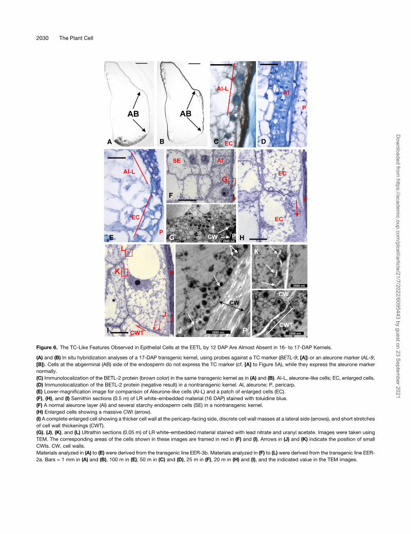
Figure 6. The TC-Like Features Observed in Epithelial Cells at the EETL by 12 DAP Are Almost Absent in 16- to 17-DAP Kernels.
(A) and (B) In situ hybridization analyses of a 17-DAP transgenic kernel, using probes against a TC marker (BETL-9; [A]) or an aleurone marker (AL-9;
[B]). Cells at the abgerminal (AB) side of the endosperm do not express the TC marker (cf. [A] to Figure 5A), while they express the aleurone marker
normally.
(C) Immunolocalization of the BETL-2 protein (brown color) in the same transgenic kernel as in (A) and (B). Al-L, aleurone-like cells; EC, enlarged cells.
(D) Immunolocalization of the BETL-2 protein (negative result) in a nontransgenic kernel. Al, aleurone; P, pericarp.
(E) Lower-magnification image for comparison of Aleurone-like cells (Al-L) and a patch of enlarged cells (EC).
(F), (H), and (I) Semithin sections (0.5 m) of LR white–embedded material (16 DAP) stained with toluidine blue.
(F) A normal aleurone layer (Al) and several starchy endosperm cells (SE) in a nontransgenic kernel.
(H) Enlarged cells showing a massive CWI (arrow).
(I) A complete enlarged cell showing a thicker cell wall at the pericarp-facing side, discrete cell wall masses at a lateral side (arrows), and short stretches
of cell wall thickenings (CWT).
(G), (J), (K), and (L) Ultrathin sections (0.05 m) of LR white–embedded material stained with lead nitrate and uranyl acetate. Images were taken using
TEM. The corresponding areas of the cells shown in these images are framed in red in (F) and (I). Arrows in (J) and (K) indicate the position of small
CWIs. CW, cell walls.
Materials analyzed in (A) to (E) were derived from the transgenic line EER-3b. Materials analyzed in (F) to (L) were derived from the transgenic line EER-
2a. Bars = 1 mm in (A) and (B), 100 m in (E), 50 m in (C) and (D), 25 m in (F), 20 m in (H) and (I), and the indicated value in the TEM images.
2030 The Plant Cell
Dow
nloaded from https://academ
ic.oup.com/plcell/article/21/7/2022/6095443 by guest on 23 Septem
ber 2021

on the differentiation of the abgerminal endosperm epidermal
cells were very clear. Aleurone cells expressingMRP-1 acquired
the features of endosperm TCs (Royo et al., 2007), forming what
has here been called an EETL (Figure 2). The development of an
extensive network of CWIs is among the best characterized of TC
features (for an ultrastructural characterization of maize TC
CWIs, see Davis et al., 1990). These ingrowths provide the
increase in plasma membrane surface area needed to enhance
the transport function of the TC (reviewed in Pate and Gunning,
1972; Offler et al., 2002) and are clearly observed in the EETL
(Figure 2; see diagram in Supplemental Figure 2 online). Their
reactivity to cell wall stains was indistinguishable from that
observed for the BETL CWI, although the extension of the
intracellular network of CWIs in the EETL never reached the
density observed in the BETL. TEM analyses (Figure 2) revealed
the presence in the EETL of the cellular elements previously
associated with the synthesis of CWIs (Offler et al., 2002) but also
indicated structural differences between the CWIs of the BETL
and EETL, which appear to develop in a less organized pattern in
the latter, where the CWIs acquired a floccular structure. This
suggests that additional factors, other than MRP-1, might be
required for the complete development of theCWI network found
in the BETL. These additional factors might implicate the ex-
pression of genes not regulated by MRP-1 and/or the require-
ment of an energy supply that cannot be obtained in cells
physically separated from the nutrient unloading zone at the
pedicel. Cell elongation is another remarkable feature of the
endosperm TCs compared with the round starchy endosperm
cells and the cubic aleurone cells (Becraft, 2001; Olsen, 2004).
Several layers of TCs elongate along an axis orthogonal to the
endosperm surface, the elongation rate being a function of TC
maturity (Royo et al., 2007). This gives a domed appearance to
the BETL, with the longest cells positioned at the center of the
layer and slightly closer to the germinal pole (as shown in Figures
3A, 5A, and 6A). The same cell elongation and general appear-
ance was observed for the EETL (Figures 3A, 5A, and 5B).
Interestingly, since the elongation axis of the EETL cells was
again orthogonal to the endosperm surface, it was also orthog-
onal to the elongation axis of the BETL. This, together with the
observed secretion of TC-specific peptides toward the sur-
rounding apoplast, provided a strongly polarized appearance to
both layers. While the achievement of cell polarity is a complex
process that probably involves the reorganization of the cyto-
skeleton and contacts with neighboring cells (as has been found
for animal epithelia [Gibson and Perrimon, 2003; Nelson, 2003]),
it is clear that the preexisting polar information in aleurone cells is
efficiently passed on via MRP-1 in the formation of highly
polarized TCs.
The availability of MRP-1–regulated TC-specific genes (i.e.,
BETL-1 and BETL-2 [Gomez et al., 2002], TCRR-1 [Muniz et al.,
2006], and BETL-9 and BETL-10 [this work]) allowed the tran-
scriptional regulatory activity of MRP-1 to be correlated with
morphological observations (Figure 3 for BETL-2; Figure 5 for
BETL-9). Furthermore, the induction of these genes by MRP-1
could be quantified (Figure 4). The real-time RT-PCR data
suggest that all TC markers respond in a quantitative mode to
the expression of MRP-1. The induction of TC markers in the
transgenic heterozygous kernel shown in Figure 4B resembles
that seen for a presumably heterozygous kernel of a different line
(Top and HET in Figure 4C), whereas a presumably homozygous
sample (Top and HOM in Figure 4C) expressed much higher
levels of both MRP-1 and the TC markers. Although sample-to-
sample variation might reflect the concurrent influence of other
factors regulating the expression of the transgene, for instance,
nutrient availability or small differences inmaturity, it is clear from
the data shown in Figure 4 that the expression levels of the TC
markers follow that of MRP-1. Interestingly, the analysis of the
expression of a TC-specific gene not previously related with the
MRP-1–regulated gene set, the TC-specific cell wall bound
invertase INCW-2 (for Cell Wall Invertase 2; Cheng et al., 1996),
produced results comparable to those found for the BETL genes.
The expression of INCW-2 at the EETL suggests that this gene is
part of the gene complement of TCs and is thus expressed as the
result of the partial TC differentiation process induced byMRP-1.
Alternatively, INCW-2might be another direct target of theMRP-1
transcriptional regulator. In this context, we have identified a se-
quence motif in the INCW-2 promoter (59-GAGATAGAGAAT-39)that strongly resembles the sequence identified as the target of
MRP-1 in the BETL promoters (59-GAGATAGAGATA-39; Barreroet al., 2006), although in the case of INCW-2, this motif would be
positioned 227 bp upstream of the position reported for the other
BETL promoters (as measured from the start codon of the
protein). Consistent with these results, we have found that the
expression level ofMRP-1 in theminiature-1mutant endosperms
(not expressing INCW-2; Cheng et al., 1996) is at least as strong
as in the wild-type endosperm (see Supplemental Figure 7
online), while the TCs develop their characteristic features
(Cheng et al., 1996), suggesting that INCW-2 acts downstream
of MRP-1.
The quantitative RT-PCR results indicate that the extension of
the EETL is limited to a discrete patch of cells right at the
abgerminal pole of the endosperm; hence, the almost normal
expression levels detected for AL-9 in the upper halves of the
kernels (Figures 4Band 4C)while the EETL cells showed reduced
AL-9 expression in in situ hybridization experiments (Figure 5D).
Further investigations are required to understand why cells at
the abgerminal pole of the endosperm can be induced to
differentiate into an EETL. This is very likely connected to the
fact (discussed above) that these cells are the only endosperm
epidermal cells that expressed detectable levels ofMRP-1 in the
transgenic material (Figure 1). Certainly, these cells are the last to
acquire aleurone morphological and molecular features. Studies
using transposon-induced gain- and loss-of-function sectors of
Deficient kernel1 (Dek-1; Becraft and Asuncion-Crabb, 2000), a
gene required for aleurone cell fate determination (Lid et al.,
2002), revealed that aleurone cell fate is not fixed until late in
development. Rather, aleurone and starchy endosperm fates are
interchangeable for a long period. Furthermore, positional cues
are required to specify and maintain the identity of an aleurone
cell; the differentiation gradient along the germinal-abgerminal
axis seems to dictate that the abgerminal cells are less surely
destined to acquire an aleurone cell identity. In addition, in situ
hybridization experiments using an aleurone marker in very
young material (AL-9 in Supplemental Figure 8 online) showed
that the abgerminal epithelial cells are the last to acquire aleurone
cell molecular features. We propose that the cells on the
MRP-1 Induces Transfer Cell Differentiation 2031
Dow
nloaded from https://academ
ic.oup.com/plcell/article/21/7/2022/6095443 by guest on 23 Septem
ber 2021
abgerminal side of the endosperm remain in an undifferentiated,
MRP-1–responsive stage because they are exposed to maternal
signals for a longer period than cells in other regions of the
aleurone (see Supplemental Figure 9 online for a picture of a
kernel section at 3 DAP, showing possible routes for the input of
maternal signals into the endosperm space). The strict depen-
dence on maternally derived signals for the expression of TC-
specific genes has been elegantly demonstrated by Gruis et al.
(2006); the expression of a fluorescent reporter under the control
of a BETL-specific promoter occurred normally in planta, but the
reporter switched off in in vitro–grown endosperms. As in in
planta endosperms, the in vitro–cultured endosperms expressed
aleurone- and starchy endosperm-specific reporter constructs,
indicating that these cell fates are correctly specified in the
absence of maternally derived signals. These maternal signals,
possibly derived from vascular tissues, are thus responsible for
the induction of the BETL at the base of the endosperm and
might participate in the formation of the EETL in a process
triggered by the expression ofMRP-1. In this context, analyses of
the MRP-1 promoter (Barrero et al., 2009) have shown that its
expression is upregulated by phloem components (mainly glu-
cose and sucrose). The role of sugars as signaling molecules in
plants is well established (reviewed in Smeekens, 2000; Rolland
et al., 2002). Changes in sugar concentration cause induction or
repression of gene transcription, providing a mechanism for
interaction between sink and source tissues for carbohydrate
metabolism (Koch, 1996). In addition, sugar sensing also affects
gene expression posttranscriptionally by changing mRNA sta-
bility, translation, or protein stability (Chan and Yu, 1998; Rook
et al., 1998; Cheng et al., 1999; Yanagisawa et al., 2003; Wiese
et al., 2004). Interestingly, the regulation of the maize cell wall–
bound invertase Incw-1 is included among these examples
(Cheng et al., 1999). Incw-1 is expressed in maize TCs, and
we reported the association between metabolizable sugars
and the increase in the steady state abundance of functional
Incw-1 RNA.
Regardless of the identity of the signals that maintainMRP-1
expression in the EETL area, it is clear that the EETL cells cease
to expressMRP-1 and TCmarkers later in development (from 17
DAP to maturity) and go on to acquire aleurone morphological
features, the accumulation of TC peptides at the apoplast being
the only remnant of the transient acquisition of TC identity (Figure
6C). We have shown that this process is gradual; very few cells
with modified cell walls remained at 16 to 17 DAP, and these cell
wall modifications were less evident than those observed at
earlier developmental stages (Figure 6), and no sign of modified
cells was found in an extensive analysis of the abgerminal
aleurones of mature plants. Within the model explained above,
this observation suggests that the exposure of the endosperm
abgerminal cells to the vascular tissue-derived substances
ceases in older kernels, which results in the cessation of MRP-1
expression. This also implies that continuous expression of
MRP-1 is required to maintain the TC differentiated stage, which
agrees with previous observations of MRP-1 expression in the
BETL as late as 29 DAP, when the TCs are completely differen-
tiated (Gomez et al., 2002). Furthermore, the results imply that
the TC phenotype is reversible, presenting a remarkable illustra-
tion of cell plasticity. In the absence of adequate levels ofMRP-1,
cells rapidly (in <5 d) change their shape and, more strikingly,
their cell wall structure. As discussed above, dynamic intercon-
versions between the aleurone and starchy endosperm cell fate
have also been demonstrated using unstable alleles of the
aleurone developmental regulator dek-1 (Becraft and Asuncion-
Crabb, 2000). EETL reversion notably implies the remobilization
of the cell wall modifications and ingrowths that characterize TCs
(Figure 3). Although the in vitro transformation of garden zinnia
(Zinnia elegans) mesophyll cells into highly differentiated vascu-
lar elements is a well-known process (Fukuda, 1997; McCann,
1997), no such remobilization of cell wall modifications has
previously been reported. The reversible modifications observed
here (cf. Figures 2 and 6; see Supplemental Figure 5 online) might
cast light on the biochemical components involved in cell wall
plasticity.
Many of the TC-specific genes identified so far encode small
hydrophilic proteins of unknown function, although there is
increasing evidence that these proteins might be involved in
the defense of the developing seed against mother plant-borne
pathogens (Serna et al., 2001). In addition, it has been shown that
MRP-1 can transactivate various TC-specific promoters, includ-
ing those of BETL-1 and BETL-2 (Gomez et al., 2002), Meg-1
(Gutierrez-Marcos et al., 2004), and the TC-specific response
regulator TCRR-1 (Muniz et al., 2006). This transactivation oc-
curs through the interaction of MRP-1 with a TC-box found in the
promoters of these genes (Barrero et al., 2006) and also in the
promoters of BETL-9 and BETL-10 used in this work.
The results presented here leave open the possibility that
these peptides have a role in cell-to-cell communication and
signaling in addition to or alternative to the role in defense
proposed above, as has been suggested for other TC-specific
peptides (Gutierrez-Marcos et al., 2004). Indeed, a role in cell-to-
cell communication and signaling has already been demon-
strated for the barley nucellar TC-specific gene Jekyll, which
plays a decisive role in the differentiation of nucellar projection
TCs (Radchuk et al., 2006).
METHODS
Plant Material
Transgenic maize (Zea mays) plants were produced from the variety
A188. All plants were grown at 248C under a 16-h-light/8-h-dark regimen.
Quantitative Real-Time RT-PCR
The Brilliant SYBR green quantitative RT-PCR kit (Stratagene) was used
for real time RT-PCR. Reactions contained 100 ng DNase-treated total
RNA from 12-DAP kernels and were performed in an ABI 7000 real-time
thermocycler. CT values were normalized against those obtained for a
ubiquitously expressed gene, FKBP-66 (Hueros et al., 1998), thus
obtaining the DCT value. Relative quantification was determined by
subtracting the DCT value for one of the lower kernel halves in the
experiment from all the normalized CT values. This provided the DDCT
value. Finally, since the efficiency of the PCR reactions was ;1 for all
genes, the expression 2e-DDCT was considered to be a function of the
starting transcript concentration. The mean (and SD) 2e-DDCT value of
three experiments (technical replicates) was recorded. The oligonucleo-
tides used are listed in Supplemental Table 1 online. In all cases, one of
2032 The Plant Cell
Dow
nloaded from https://academ
ic.oup.com/plcell/article/21/7/2022/6095443 by guest on 23 Septem
ber 2021

the primers used spanned an intron to avoid amplification from contam-
inating DNA.
Cell Wall Staining
The sections in Figure 2 were stained for cellulose using 0.1% calcofluor
Fluorescent Brightener 28 (Sigma-Aldrich). Under UV light, cellulose-
containing primary walls appear dark blue, whereas newly deposited wall
thickenings appear light blue.
In Situ Hybridization and Immunolocalization
Maize seeds at 10 to 12 or 16 to 17 DAP were fixed in 4% paraformal-
dehyde and 0.1% glutaraldehyde in 0.1 M sodium phosphate buffer, pH
7.2, for 12 to 24 h, depending on the tissue volume. Samples were
dehydrated, embedded in Fibrowax (Plano), and cut into 8-mm sections.
For in situ hybridization, slides were probed as previously described
(Hueros et al., 1995) with sense and antisense 35S labeled–riboprobes,
synthesized from cDNA clones in a pBluescript vector. Slides were
exposed to LM-1 silver grain emulsion for 2 to 4 d at 48C (Amersham).
After developing, sections were stained with calcofluor (0.01%).
In situ hybridization results were photographed under bright-field
(hybridization signals appear as black spots) or dark-field (hybridization
signals appear as white spots) illumination. In some cases, combined
dark-field and epifluorescence images were produced to reveal the
calcofluor white counterstaining.
For the immunolocalization experiments, endogenous peroxidase was
inhibited by incubating the sections in 0.3% v/v H2O2 in methanol for 10
min. The sections were then washed in PBS and blocked with 10%
normal donkey serum (Chemicon International) for 20 min before being
exposed to anti-BETL-1 or anti-BETL-2 rabbit sera (diluted 1:400) for 1 h
at room temperature. The immunoreaction was performed using a
biotinylated anti-rabbit antibody (Sigma-Aldrich) and a streptavidin-
peroxidase conjugate (Zymed). The peroxidase was detected using 39,
39-diaminobenzidine tetrahydrochloride with a metal enhancer (Sigma-
Aldrich). PBS was used as the washing buffer. Finally, the sections were
stained with Azure B, pH 4.0, for 3 min.
For confocal microscopy, the sections were rehydrated in an ethanol
series, exposed to Alexa enhancer (Invitrogen) for 30 min, and then
preincubated in 10% normal donkey serum diluted in 1% (w/v) BSA
(fraction V) in phosphate buffer (0.1 M, pH 7.5). After five washes in
phosphate buffer (0.1 M, pH 7.5), the sections were reacted with the
primary antibody (polyclonal rabbit anti Betl-2 1:400) for 2 h. The
antigen-antibody binding reaction was revealed by applying goat anti-
rabbit Alexa Fluor 488 antibodies (Invitrogen) diluted 1:1000 in 1% (w/v)
BSA (fraction V) in phosphate buffer (0.1 M, pH 7.5). After several
washes in phosphate buffer (0.1 M, pH 7.5), the sections were stained
with toluidine blue (0.01%; Sigma-Aldrich) to quench cell wall auto-
fluorescence. A Leica TCS-SL confocal microscope equipped with an
argon laser was used for the localization of Alexa 488 green fluores-
cence. Red cell wall autofluorescence was also recorded to visualize
tissues.
For LR-White embedding, fixed tissue was infiltrated with benzoyl
peroxide–activated LR white embedding media (Sigma-Aldrich). The
infiltration began with a graded infiltration series for 24 h at room
temperature: 1:3, 1:1, and 3:1 (LR white:100% ethanol), followed by
two changes of pure LR white at 48C for 1 week each. Samples were
positioned into aluminum weight boats, covered, and polymerized at
608C for at least 24 h. Samples were first sectioned at 1-mm thickness
using tungsten-carbide knives. These whole-kernel sections were
stained with toluidine blue and used to explore the aleurone, and blocks
were then carved and sectioned with glass knives. Semithin sections (0.5
mm) were stained with toluidine blue, and ultrathin (0.05 mm) sections in
nickel gridswere stainedwith Reynolds’ lead nitrate and uranyl acetate as
described by Hayat (2000). Ultrathin sections were examined using a
Zeiss EM-10 electron microscope.
Transgenes
To generate pAL-9-GUS, a 2.2-kbp AL-9 promoter fragment was ampli-
fied by PCR from a maize BAC clone hybridizing to AL-9, such that the
fragment contained the entire 59 untranslated region up to the ATG. The
PCR fragment was cloned in front of the GUS reporter gene and a
cauliflower mosaic virus 35S polyadenylation signal, in pSK, forming
plasmid AL-9GUS-sk(PmeI). Embryogenic type II maize calluses were
transformed with the AL-9GUS-sk(PmeI) construct by biolistic bombard-
ment and regenerated as described by Komari et al. (1996).
To generate pAL-9-MRP-1, a full-length MRP-1 cDNA was first
cloned into a GATEWAY entry vector (Invitrogen) and then transferred
via an LR clonase reaction into the plant binary destination vector
pBIOS873, forming a pAL-9-MRP-1 chimeric gene. The vector
pBIOS873 is a derivative of pSB12 (Komari et al., 1996) that contains
a selectable marker gene for maize transformation and the 2.2-kb AL-9
promoter in front of a GATEWAY cassette, which is followed by an
Arabidopsis thaliana polyadenylation sequence. The resulting plasmid,
pBIOS880, was transferred into Agrobacterium tumefaciens strain
LBA4404 (pSB1) as described by Komari et al. (1996). The maize
cultivar A188 was transformed with this clone essentially as described
by Ishida et al. (1996).
Transient Expression Assays
Plant material was surface sterilized, sectioned, and maintained on the
solid medium described below until bombardment with DNA-coated gold
particles. Coating and bombardment was performed according to
Knudsen and Muller (1991). Five micrograms of each plasmid to be
transformedwas used to prepare a gold particle batch that was then used
to bombard seven tissue samples. After bombardment, samples were
incubated at 258C for 24 h in the dark on solid (0.5% agarose) Murashige
and Skoog medium containing 100 mg/L myo-inositol, 2 g/L Asn, 2 g/L
Gln, 30 g/L sucrose, and Murashige and Skoog vitamins (Sigma-Aldrich).
Histochemical detection of GUS expression was performed by stain-
ing, according to Jefferson et al. (1987). Samples were stained for GUS
in a medium containing 0.5 mg/mL X-glucuronide (Clontech), 0.5 mM
K+-ferrocyanide, 0.5 mM K+-ferricyanide, 10 mM Na2EDTA, 50 mM
phosphate buffer, pH 7, 0.1% Triton X-100, and 20% (v/v) methanol.
Accession Numbers
Sequence data from this article can be found in the Arabidopsis Genome
Initiative or GenBank/EMBL databases under the following accession
numbers: BETL-10, FN400765; BETL-9, FN400766; AL-9, FN400767;
MRP-1, AJ318518; BETL-1, Z49203; BETL-2, NM_001112109; TCRR-1,
AM085299; INCW-2, AF050128; and ESR-6, AJ849917.
Supplemental Data
The following materials are available in the online version of this article.
Supplemental Figure 1. Expression Analyses of the AL-9 Gene.
Supplemental Figure 2. Interpretation of the Transformation of
Endosperm Epithelial Cells into Transfer Cells, Induced by the
Expression of MRP-1.
Supplemental Figure 3. The Ectopic Expression of MRP-1 Induces
the Expression of BETL-1.
Supplemental Figure 4. The Expression of MRP-1 in the Abgerminal
Aleurone Decreases as the Kernel Matures.
MRP-1 Induces Transfer Cell Differentiation 2033
Dow
nloaded from https://academ
ic.oup.com/plcell/article/21/7/2022/6095443 by guest on 23 Septem
ber 2021
Supplemental Figure 5. Morphological Analyses of the Mature
Endosperm Epidermis.
Supplemental Figure 6. The Presence of the MRP-1 Coding Se-
quence Decreases the Expression Efficiency of Reporter Constructs
in Transient Expression Experiments.
Supplemental Figure 7. Expression Analyses in miniature-1 and
Wild-Type Kernels.
Supplemental Figure 8. Expression Pattern of the Aleurone Marker
Gene AL-9 in Young Kernels.
Supplemental Figure 9. A Model Explaining How Maternal Signals
Might Be Perceived by the Abgerminal Side of the Endosperm at Its
Initial Developmental Stages.
Supplemental Table 1. Oligonucleotides Used for Quantitative
RT-PCR.
ACKNOWLEDGMENTS
We thank P. Rogowsky and R.D. Thompson for their critical review of
the original manuscript and their useful suggestions regarding its
content, Y. Sanz for excellent technical support, and the personnel at
Biogemma SAS who contributed towards the production of the maize
transformants. This work was supported by grants to G.H. from the
Spanish Ministerio de Educacion y Ciencia (BIO2003-03721 and
BIO2006-14190) and by internal funds from Biogemma SAS.
Received December 23, 2008; revised May 26, 2009; accepted June 18,
2009; published July 2, 2009.
REFERENCES
Allaway, W.G., Carpenter, J.L., and Ashford, A.E. (1985). Amplifica-
tion of intersymbiont surface by root epidermal transfer cells in the
Pisonia mycorrhiza. Protoplasma 128: 227–231.
Balandın, M., Royo, J., Gomez, E., Muniz, L.M., Molina, A., and
Hueros, G. (2005). A protective role for the embryo surrounding
region of the maize endosperm, as evidenced by the characterisation
of ZmESR-6, a defensin gene specifically expressed in this region.
Plant Mol. Biol. 58: 269–282.
Baranowskij, N., Frohberg, C., Prat, S., and Willmitzer, L. (1994). A
novel DNA binding protein with homology to MYB oncoproteins
containing only one repeat can function as a transcriptional activator.
EMBO J. 13: 5383–5392.
Barrero, C., Muniz, L.M., Gomez, E., Hueros, G., and Royo, J. (2006).
Molecular dissection of the interaction between the transcriptional
activator ZmMRP-1 and the promoter of BETL-1. Plant Mol. Biol. 62:
655–668.
Barrero, C., Royo, J., Grijota-Martinez, C., Faye, C., Paul, W., Sanz,
S., Steinbiss, H.-H., and Hueros, G. (2009). The promoter of
ZmMRP-1, a maize transfer cell-specifc transcriptional activator, is
induced at solute exchange surfaces and responds to transport
demands. Planta 229: 235–247.
Becraft, P.W. (2001). Cell fate specification in the cereal endosperm.
Semin. Cell Dev. Biol. 12: 387–394.
Becraft, P.W., and Asuncion-Crabb, Y. (2000). Positional cues specify
and maintain aleurone cell fate in maize endosperm development.
Development 127: 4039–4048.
Berger, F. (2003). Endosperm: The crossroad of seed development.
Curr. Opin. Plant Biol. 6: 42–50.
Chan, M.T., and Yu, S.M. (1998). The 39 untranslated region of a rice
alpha-amylase gene mediates sugar-dependent abundance of
mRNA. Plant J. 15: 685–695.
Cheng, W.H., Taliercio, E.W., and Chourey, P.S. (1996). The Miniature1
seed locus of maize encodes a cell wall invertase required for normal
development of endosperm and maternal cells in the pedicel. Plant Cell
8: 971–983.
Cheng, W.H., Taliercio, E.W., and Chourey, P.S. (1999). Sugars
modulate an unusual mode of control of the cell-wall invertase gene
(Incw1) through its 39 untranslated region in a cell suspension culture
of maize. Proc. Natl. Acad. Sci. USA 96: 10512–10517.
Cochrane, M.P., and Duffus, C.M. (1980). The nucellar projection and
modified aleurone in the crease region of developing caryopses of
barley (Hordeum vulgare L. var. Distichum). Protoplasma 103: 361–375.
Davis, R.W., Smith, J.D., and Cobb, B.G. (1990). A light and electron
microscopic investigation of the transfer cell region of maize caryop-
sis. Can. J. Bot. 68: 471–479.
Doan, D.N., Linnestad, C., and Olsen, O.A. (1996). Isolation of mo-
lecular markers from the barley endosperm coenocyte and the
surrounding nucellus cell layers. Plant Mol. Biol. 31: 877–886.
Farley, S.J., Patrick, J.W., and Offler, C.E. (2000). Functional transfer
cells differentiate in cultured cotyledons of Vicia faba L. seeds.
Protoplasma 214: 102–117.
Fukuda, H. (1997). Tracheary element differentiation. Plant Cell 9: 1147–
1156.
Gibson, M.C., and Perrimon, N. (2003). Apicobasal polarization: Epi-
thelial form and function. Curr. Opin. Cell Biol. 15: 747–752.
Gomez, E., Royo, J., Guo, Y., Thompson, R., and Hueros, G. (2002).
Establishment of cereal endosperm expression domains: Identifica-
tion and properties of a maize transfer cell–specific transcription
factor, ZmMRP-1. Plant Cell 14: 598–610.
Gruis, D., Guo, H., Selinger, D., Tian, Q., and Olsen, O.-A. (2006).
Surface position, not signaling from surrounding maternal tissues,
specifies aleurone epidermal cell fate in maize. Plant Physiol. 141:
898–909.
Gunning, B.E., Pate, J.S., Minchin, F.R., and Marks, I. (1974). Quan-
titative aspects of transfer cell structure in relation to vein loading in
leaves and solute transport in legume nodules. Symp. Soc. Exp. Biol.
28: 87–126.
Gutierrez-Marcos, J.F., Costa, L.M., Biderre-Petit, C., Khbaya, B.,
O’Sullivan, D.M., Wormald, M., Perez, P., and Dickinson, H.G.
(2004). Maternally expressed gene1 is a novel maize endosperm
transfer cell–specific gene with a maternal parent-of-origin pattern of
expression. Plant Cell 16: 1288–1301.
Hayat, M.A. (2000). Principles and Techniques of Electron Microscopy:
Biological Applications, 4th ed. (Cambridge, UK: Cambridge Univer-
sity Press).
Hueros, G., Rahfeld, J., Salamini, F., and Thompson, R. (1998). A
maize FK506-sensitive immunophilin, mzFKBP-66, is a peptidylproline
cis-trans-isomerase that interacts with calmodulin and a 36-kDa
cytoplasmic protein. Planta 205: 121–131.
Hueros, G., Royo, J., Maitz, M., Salamini, F., and Thompson, R.D.
(1999). Evidence for factors regulating transfer cell-specific expres-
sion in maize endosperm. Plant Mol. Biol. 41: 403–414.
Hueros, G., Varotto, S., Salamini, F., and Thompson, R.D. (1995).
Molecular characterization of BET1, a gene expressed in the endo-
sperm transfer cells of maize. Plant Cell 7: 747–757.
Ishida, Y., Saito, H., Ohta, S., Hiei, Y., Komari, T., and Kumashiro, T.
(1996). High efficiency transformation of maize (Zea mays L.) mediated
by Agrobacterium tumefaciens. Nat. Biotechnol. 14: 745–750.
Jefferson, R.A., Kavanagh, T.A., and Bevan, M. (1987). Gus-fusions:
b-Glucuronidase as a sensitive and versatile gene fusion marker in
higher plants. EMBO J. 6: 3901–3907.
2034 The Plant Cell
Dow
nloaded from https://academ
ic.oup.com/plcell/article/21/7/2022/6095443 by guest on 23 Septem
ber 2021

Jones, M.G., and Northcote, D.H. (1972). Nematode induced synci-
tium: A multinucleate transfer cell. J. Cell Sci. 10: 789–809.
Kiesselbach, T.A. (1949). The Structure and Reproduction of Corn.
(Lincoln, NE: University of Nebraska).
Knudsen, S., and Muller, M. (1991). Transformation of the developing
barley endosperm by particle bombardment. Planta 185: 330–336.
Koch, K.E. (1996). Carbohydrate-modulated gene expression in plants.
Annu. Rev. Plant Physiol. Plant Mol. Biol. 47: 509–540.
Komari, T., Hiei, Y., Saito, Y., Murai, N., and Kumashiro, T. (1996).
Vectors carrying two separate T-DNAs for co-transformation of higher
plants mediated by Agrobacterium tumefaciens and segregation of
transformants free from selection markers. Plant J. 10: 165–174.
Lid, S.E., Gruis, D., Jung, R., Lorentzen, J.A., Ananiev, E., Chamberlin,
M., Niu, X., Meeley, R., Nichols, S., and Olsen, O.-A. (2002). The
defective kernel 1 (dek1) gene required for aleurone cell development
in the endosperm of maize grains encodes a membrane protein of
the calpain gene superfamily. Proc. Natl. Acad. Sci. USA 99: 5460–
5465.
Ligrone, R., and Gambardella, R. (1988). The sporophyte–gameto-
phyte junction in bryophytes. Adv. Bryol. 3: 225–274.
McCann, M.C. (1997). Tracheary element formation: Building up to a
dead end. Trends Plant Sci. 2: 333–338.
Muniz, L.M., Royo, J., Gomez, E., Barrero, C., Bergareche, D., and
Hueros, G. (2006). The maize transfer cell-specific type-A response
regulator ZmTCRR-1 appears to be involved in intercellular signalling.
Plant J. 48: 17–27.
Nelson, W.J. (2003). Adaptation of core mechanisms to generate cell
polarity. Nature 422: 766–774.
Offler, C.E., Liet, E., and Sutton, E.G. (1997). Transfer cell induction in
cotyledons of Vicia faba L. Protoplasma 200: 51–64.
Offler, C.E., McCurdy, D.W., Patrick, J.W., and Talbot, M.J. (2002).
Transfer cells: Cells specialized for a special purpose. Annu. Rev.
Plant Biol. 54: 431–454.
Offler, C.E., and Patrick, J.W. (1993). Pathway of photosynthate
transfer in the developing seed of Vicia faba L. A structural assess-
ment of the role of transfer cells in unloading from the seed coat.
J. Exp. Bot. 44: 711–724.
Olsen, O.-A. (2004). Nuclear endosperm development in cereals and
Arabidopsis thaliana. Plant Cell 16: S214–S227.
Opsahl-Ferstad, H.G., Le Deunff, E., Dumas, C., and Rogowsky,
P.M. (1997). ZmEsr, a novel endosperm-specific gene expressed in a
restricted region around the maize embryo. Plant J. 12: 235–246.
Pate, J.S., and Gunning, E.S. (1972). Transfer cells. Annu. Rev. Plant
Physiol. 23: 173–196.
Radchuk, V., Borisjuk, L., Radchuk, R., Steinbiss, H.-H., Rolletschek,
H., Broeders, S., and Wobus, U. (2006). Jekyll encodes a novel
protein involved in the sexual reproduction of barley. Plant Cell 18:
1652–1666.
Rolland, F., Moore, B., and Sheen, J. (2002). Sugar sensing and
signaling in plants. Plant Cell 14 (suppl.): S185–S205.
Rook, F., Gerrits, N., Kortstee, A., van Kampen, M., Borrias, M.,
Weisbeek, P., and Smeekens, S. (1998). Sucrose-specific signaling
represses translation of the Arabidopsis ATB2 bZIP transcription
factor gene. Plant J. 15: 253–263.
Rose, A., Meier, I., and Wienand, U. (1999). The tomato I-box binding
factor LeMYBI is a member of a novel class of Myb-like proteins. Plant
J. 20: 641–652.
Royo, J., Gomez, E., and Hueros, G. (2007). Transfer cells. In Plant Cell
Monographs, O.-A. Olsen, ed (Berlin-Heidelberg, Germany: Springer-
Verlag), pp. 73–89.
Schaffer, R., Ramsay, N., Samach, A., Corden, S., Putterill, J., Carre,
I.A., and Coupland, G. (1998). The late elongated hypocotyl mutation
of Arabidopsis disrupts circadian rhythms and the photoperiodic
control of flowering. Cell 93: 1219–1229.
Serna, A., Maitz, M., O’Connell, T., Santandrea, G., Thevissen, K.,
Tienens, K., Hueros, G., Faleri, C., Cai, G., Lottspeich, F., and
Thompson, R.D. (2001). Maize endosperm secretes a novel antifun-
gal protein into adjacent maternal tissue. Plant J. 25: 687–698.
Smeekens, S. (2000). Sugar-induced signal transduction in plants.
Annu. Rev. Plant Physiol. Plant Mol. Biol. 51: 49–81.
Talbot, M.J., Franceschi, M.R., McCurdy, D.W., and Offler, C.E.
(2001). Wall ingrowth architecture in epidermial transfer cells of Vicia
faba cotyledons. Protoplasma 215: 191–203.
Thompson, R.D., Hueros, G., Becker, H.-A., and Maitz, M. (2001).
Development and functions of seed transfer cells. Plant Sci. 160:
775–783.
Wang, Z.-Y., Kenigsbuch, D., Sun, L., Harel, E., Ong, M.S., and
Tobin, E.M. (1997). A MYB-related transcription factor is involved in
the phytochrome regulation of an Arabidopsis Lhcb gene. Plant Cell 9:
491–507.
Weber, H., Borisjuk, L., Heim, U., Sauer, N., and Wobus, U. (1997). A
role for sugar transporters during seed development: Molecular
characterization of a hexose and a sucrose carrier in fava bean
seeds. Plant Cell 9: 895–908.
Wiese, A., Elzinga, N., Wobbes, B., and Smeekens, S. (2004). A
conserved upstream open reading frame mediates sucrose-induced
repression of translation. Plant Cell 16: 1717–1729.
Yanagisawa, S., Yoo, S.D., and Sheen, J. (2003). Differential regulation
of EIN3 stability by glucose and ethylene signalling in plants. Nature
425: 521–525.
MRP-1 Induces Transfer Cell Differentiation 2035
Dow
nloaded from https://academ
ic.oup.com/plcell/article/21/7/2022/6095443 by guest on 23 Septem
ber 2021