the metastatic cascade pathologic features of metastasis bone
TRANSCRIPT

1
1. The metastatic cascade
3. Pathologic features of metastasis
4. Therapeutic ramifications
Sir James Paget (1814-1899)
British Surgeon/ Pathologist
Paget’s disease of bone
Paget’s disease of the nipple (intraduc- tal Ca breast spread- ing to skin of nipple)
Stephen Paget, MD (1855-1926) METASTASIS: “Seed and Soil” Paget S. The distribution of secondary growths in cancer of the breast. Lancet 1889; 1: 571-573. “the best work in pathology of cancer is done by those who…are studying the nature of the seed…”… “the observations of the properties of the soil may also be useful.”
Carcinoma of breast
liver
Which malignant cells will metastasize? 1. Subclone theory
-a specific clone has the prop’s for metastasis
2. Cancer stem cell theory
-only the stem cells have the prop’s for metastasis
3. Genetic signature theory 70-gene signature for mets in breast Ca
-many malignant cells at different stages have the signature genes allowing metastasis
B.M.: laminin + coll. IV
TUM lungs
liver
bone brain
ECM
blood vessels: cap./venule/lymphatic
epithelium
lymph nodes
distant sites
TUM

2
B.M.: laminin + coll. IV
TUM lungs
liver
bone brain
ECM
blood vessels: cap./venule/lymphatic
epithelium
lymph nodes
distant sites
TUM
1. Invasion of B.M. & ECM
2. Intravasation & vasc. dissemination
3. Extravasation, colonization & growth
B.M.: laminin + coll. IV
TUM lungs
liver
bone brain
ECM
blood vessels: cap./venule/lymphatic
epithelium
lymph nodes
distant sites
TUM
Stage 1: Invasion of B.M. and ECM E-cadherin
1. Loosening of tumor cells
E-cadherin • Mutation of E-cadherin • Inappropriate expression of SNAIL & TWIST (which regulate E-cadh. expres.)
(Secondary effect: E-cadherin normally sequesters ß-catenin—cell proliferator— so with less sequestered, more tumor cells proliferate)
B.M.: laminin + coll. IV
TUM
ECM
blood vessels: cap./venule/lymphatic
epithelium
TUM
2. Degradation of B.M. & ECM
• MMP’s (esp. MMP9: gelatinase action on b.m. type IV collagen) • Cathepsin D • Urokinase plasminogen activator • Type IV collagenase
integrin with affinity for laminin and other ECM proteins
3. Change in attachment of tumor cells to ECM proteins
4. Locomotion of tumor cells
ECM
EMT: epithelial- mesenchymal transition
ECM
SMA (smooth muscle actin) vimentin
B.M.: laminin + coll. IV
TUM
ECM
epithelium
TUM platelet-tumor emboli
Vessel size Shear stress
NK cell
mac
HOSTILE ENVIRONMENT: Most tumor cells do not survive
Up-regulate adh. molecules for endothel.
TNF-α IFN-γ
Stage 2: Intravasation & vasc. dissemination

3
lungs
liver
bone brain
blood vessels: cap./venule/lymphatic
lymph nodes
Stage 3: Extravasation, colonization & growth
• Angiogenesis • EMT • Chemokine express. • Adhesion molecules • Down-regulate “metastasis suppressor proteins”
Alveolar space
Example: METASTASIS TO LUNG
Alveolar space
Example: METASTASIS TO LUNG
Vascular Sprouting
hypoxia
HIF-1α (hypoxia inducible factor)
VEGF
• TGF-α, ß • PDGF • FGF • EGF • angiogenin
A-B-C-D-E
lymphangiogenesis
VEGFR
Angiopoietins 1 & 2 Ang-1
A,B,D,E: • angiogenesis • endothelial mitosis
TIE-2 receptor
pericyte
Anti-angiogenic factors • thrombospondin-1 • p53 expression VEGF expression • Proteases (from tumor cells or from stromal cells):
Plasminogenangiostatin Collagenendostain Transthyretinvasculostatin
proteolytic cleavage
TUM
TUM TUM
Carcinoma of breast
liver liver

4
CXCR7
CXCL21 Chemokine receptors on tumor Cells: …ligands in metastatic sites
N Engl J Med 2001; 345: 833-835
Breast Carcinoma (invasive) Breast Carcinoma (intraductal and invasive)
fibrosis (desmoplasia)
necrosis
intraductal carcinoma invasive Ca
lymphatic
Ductal carcinoma In situ (DCIS)
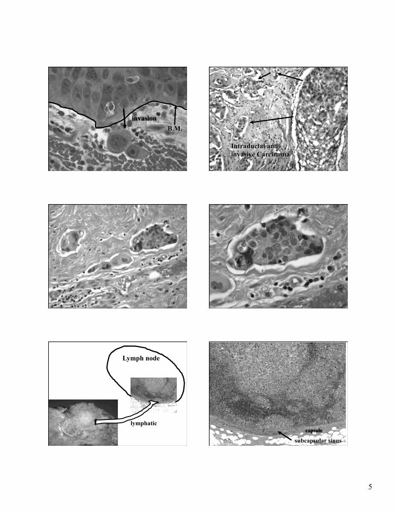
5
B.M. invasion
Intraductal and invasive Carcinoma
Lymph node
lymphatic capsule
subcapsular sinus

6
Blood vessels
Ingrowth of fibroblasts & stroma
inflammation
General Modes of Metastasis
lymphatics
regional lymph nodes carcinoma
sarcoma veins
bone lungs
Common Sites of Metastasis
Lung
• Colorectal • Breast • Renal cell Ca • Hepatocellular Ca
Liver
• Colorectal • Pancreas • Other GI: carcinoid, bile ducts • Lung • Breast
Common Sites of Metastasis
Bone
• Breast • Prostate • Renal cell Ca
Brain Neuroaxis
• Lung • Breast • Melanoma • Renal cell Ca
Vert. veins
Unique Sites of Metastasis
gastric Ca (signet ring Ca)
Virchow’s node (supraclavicular node)
Krukenberg tumor (bilateral ovarian mets)

7
Source: University of Alberta, Dep’t of Laboratory Medicine & Pathology
Lung Carcinoma metastasis to Adrenal
B.M.: laminin + coll. IV
TUM lungs
liver
bone brain
ECM
blood vessels: cap./venule/lymphatic
epithelium
lymph nodes
distant sites
TUM
Anti-angiogenic agents
Agent Mech. of action Neoplasm(s)
Bevacizumab against VEGF colorectal
Sunitinib Multi-kinase inhibitor of VEGFR, PDGFR renal cell; GIST
Sorafenib Multi-kinase inhibitor:
VEGFR, PDGFR, C-kit, Raf renal cell
Monoclonal Ab
Anti-chemokine receptors