the microscopist
TRANSCRIPT

7/29/2019 THE MICROSCOPIST
http://slidepdf.com/reader/full/the-microscopist 1/361
7/29/2019 THE MICROSCOPIST
http://slidepdf.com/reader/full/the-microscopist 2/361

7/29/2019 THE MICROSCOPIST
http://slidepdf.com/reader/full/the-microscopist 3/361

7/29/2019 THE MICROSCOPIST
http://slidepdf.com/reader/full/the-microscopist 4/361
2.

7/29/2019 THE MICROSCOPIST
http://slidepdf.com/reader/full/the-microscopist 5/361
Dr. Bennett F. Davenport,
751 TREMONT ST.
BOSTON, - - MASS.

7/29/2019 THE MICROSCOPIST
http://slidepdf.com/reader/full/the-microscopist 6/361
Digitized by the Internet Archive
in 2011 with funding from
Open Knowledge Commons and Harvard Medical School
http://www.archive.org/details/microscopistmanuOOwyth

7/29/2019 THE MICROSCOPIST
http://slidepdf.com/reader/full/the-microscopist 7/361
THE MIOROSCOPIST.

7/29/2019 THE MICROSCOPIST
http://slidepdf.com/reader/full/the-microscopist 8/361
ZENTMAYER'S LARGEST MICROSCOPE.
ONE-THIRD ACTUAL SIZE.

7/29/2019 THE MICROSCOPIST
http://slidepdf.com/reader/full/the-microscopist 9/361
THE
MICROSCOPISTi
MANUAL OF MICROSCOPY
COMPENDIUM OF THE MICROSCOPIC SCIENCES,
MICRO-MINERALOGY, MICRO-CHEMISTRY, BIOLOGY, HISTOLOGY,
AND PATHOLOGICAL HISTOLOGY.
THIRD EDITION.
KEWEITTEN AND GEEATLY ENLARGED.
WITH
TWO HUNDEED AND FIVE ILLU STE ATIO NS.
J. H. WYTHE, A.M., M.D.,
PROFESSOR OF MICROSCOPY AND BIOLOGY IN THE MEDICAL COLLEGE OF THE PACIFIC,
SAN FRANCISCO.
PHILADELPHIA:
LliSTDSAY & BLAKISTON.
187 7.

7/29/2019 THE MICROSCOPIST
http://slidepdf.com/reader/full/the-microscopist 10/361
Entered according to Act of Congress, in the year 1877,
By LINDSAY & BLAKISTON,
In the office of the Librarian of Congress at Washington, D. C.
PHILADELPHIA:
SHERMAN <t CO., PRINTER S.

7/29/2019 THE MICROSCOPIST
http://slidepdf.com/reader/full/the-microscopist 11/361
RESPECTFULLY DEDICATED
TO THE
SAN FRANCISCO MICROSCOPICAL SOCIETY,
AS A TESTIMONY
TO THE
ZEAL AND INDUSTRY OF ITS MEMBERS
IN THE PROSECUTION
OF
MICROSCOPIC SCIENCE.

7/29/2019 THE MICROSCOPIST
http://slidepdf.com/reader/full/the-microscopist 12/361

7/29/2019 THE MICROSCOPIST
http://slidepdf.com/reader/full/the-microscopist 13/361
PREFACE.
The progress of microscopic science may be well illus-
trated by a comparison between the present and former
editions of this book. The author's intention was to
place within the reach of the student of nature a com-
pendium of microscopy, free from unnecessary verbiage,
which should aid in every department of natural science.
It is no small compliment to such a work that for a
quarter of a century it should hold a place among works
of reference, although surrounded by larger and morepretentious volumes. In order to meet the request of
the publishers for another edition, it has been found
necessary to rewrite the entire book, and although the
original design has been kept in view, the numerous
additions to our science render considerable enlargement
needful, notwithstanding the effort made to concentrate
the material into the smallest compass consistent with
perspicuity.
The vision of microscopy sweeps over all the world,
and embraces all forms of organic and inorganic ex-
/

7/29/2019 THE MICROSCOPIST
http://slidepdf.com/reader/full/the-microscopist 14/361
Vm PREFACE.
isteuce. To give directions respecting most approved
methods, and to classify the most important facts, has
required labor, which it is hoped will result in rendering
the work a necessary companion to the student, and an
aid to the progress of real science.
Many of the figures illustrating the lower forms of life,
and normal and pathological histology, have been drawn
from the works of Carpenter, Beale, Frey, Strieker, Bill-
roth, and Rindfleisch, to which the more advanced stu-
dent is referred for further details.
January, 1877.

7/29/2019 THE MICROSCOPIST
http://slidepdf.com/reader/full/the-microscopist 15/361
CONTENTS.
CHAPTER I.
History anb Importance or Microscopy.
Application of the Microscope to Science and Art—Progress of Micros-
copy, 17-21
CHAPTER II.
The Microscope.
The Simple Microscope—Chromatic and Spherical Aberration—Com-
pound Microscope— Achromatic Object-glasses— Eye-pieces— Me-
chanical Arrangements—Binocular Microscope, . . . 21-32
CHAPTER III.
Microscopic Accessories.
Diaphragms—Condensers—Oblique Illuminators—Dark-ground ditto
Illumination of Opaque Objects—Measuring and Drawing Objects
Standards of Measurement—Moist Chamber—Gas Chamber—WarmStage—Polariscope—Microspectroscope—Nose-piece—Object-finders
—Micro-photography,. . . . . . . .
32-48
CHAPTER IV.
Use of the Microscope.
Care of the Instrument—Care of the Eyes—Table—Light—Adjustments
—Errors of Interpretation—Testing the Microscope, . . 48-58

7/29/2019 THE MICROSCOPIST
http://slidepdf.com/reader/full/the-microscopist 16/361
X CONTENTS.
CHAPTER V.
Modern Methods of Examination.
Preliminary Preparation of Objects—Minute Dissection—Preparation
of Loose Textures—Preparation by Teasing—Preparation by Section
—Staining Tissues—Injecting Tissues—Preparation in Viscid Media
—Fluid Media—Inditfercnt Fluids—Chemical Reagents—Staining
Fluids—Injecting Fluids—Preservative Fluids—Cements, . 58-76
CHAPTER VI.
Mounting and Preserving Microscopic Objects.
Opaque Objects— Cells—Dry Objects—Mounting in Balsam or Dammar
Mounting in Fluid—Cabinets—Collecting Objects—Aquaria, 76-83
CHAPTER VII.
The Microscope in Mineralogy and Geology.
Preparation of Specimens — Examination of Specimens— Crystalline
Forms—Crystals within Crystals—Cavities in Crystals—Use of Po-
larized Light—Origin of Rock Specimens—Materials of Organic
Origin—Microscopic Paleontology, ..... 84-98
CHAPTER VIII.
The Microscope in Chemistry.
Apparatus and Modes of Investigation—Preparation of Crj'stals for the
Polariscope—Use of the Microspectroscope—Inverted Microscope
General Micro-chemical Tests—Determination of Substances—Al-
kalies—Acids—Metallic Oxides—Alkaloids—Crystalline Forms of
Salts, . 98-115
CHAPTER IX.
The Microscope in Biology.
Theories of Life—Elementary Unit or Cell—Cell-structure and Formation
—Phenomena of Bioplasm—Movements of Cells—Microscopic Dem-
onstration of Bioplasm—Chemistry of Cells and their Products—Va-
rieties of Bioplasm—Cell-genesis—Reproduction in Higher Organ-
isms—Alternation of Generations—Parthenogenesis —Transforma-
tion and Metamorphosis—Discrimination of Living Forms, 116-127

7/29/2019 THE MICROSCOPIST
http://slidepdf.com/reader/full/the-microscopist 17/361
CONTENTS. XI
CHAPTEE X.
The Microscope in Vegetable Histology and Botany.
Molocular Coalescence—Cell-substance in Vegetables—Cell-wall or Mem-
brane—Ligneous Tissue—Spiral Vessels—Laticiferous Vessels—Si-
liceous Structures—Formed Material in Cells—Forms of Vegetable
Cells—Botanical Arrangement of Plants—Fungi—Protophytes
Desmids— Diatoms— Nostoc— Oscillatoria— Examination of the
Higher Cryptogamia—Examination of Higher Plants,.
128-157
CHAPTER XI.
The Microscope in Zoology.
3Ionera — Ehizopods— Infusoria—Kotatoria—Polyps—Hydroids—Aca-
lephs— Echinoderms— Bryozoa—Tunicata — Conchifera—Gastero-
poda— Cephalopoda— Entozoa— Annulata— Crustacea—Insects—
Arachnida—Classification of the Invertebrata,. . .
158-182
CHAPTER XII.
The Microscope in Animal Histology.
Histo-chemistry — Histological Structure— Simple Tissues — Blood—Lymph and Chyle—Mucus—Epithelium—Hair and jSTails—-Enamel—Connective Tissues—Compound Tissues—Muscle—Nerve—Glan-
dular and Vascular Tissue—Development of the Tissues—Digestive
and Circulatory Organs—Secretive Organs— Respiratory Organs
Generative Organs—Locomotive Organs—Sensory Organs—Organs
of Special Sense—Suggestions for Practice, . . . 182-226
CHAPTER XIII.
The Microscope in Practical Medicine and Pathology.
Microscopic Appearances after Death of the Tissues—Morbid Action in
Tissues—New Formations— Examinatit^n of Urinary Deposits
Human Parasites—Examination of Sputa— Microscopic Hints in
Materia Medica and Pharmacy, 226-245

7/29/2019 THE MICROSCOPIST
http://slidepdf.com/reader/full/the-microscopist 18/361

7/29/2019 THE MICROSCOPIST
http://slidepdf.com/reader/full/the-microscopist 19/361
THE MICROSCOPIST.
CHAPTER L
HISTORY AND IMPORTANCE OF MICROSCOPY.
The term microscopy, meaning t?ie use of the micro-
scope, is also applied to the knowledge obtained by this
instrument, and in this sense is commensurate with aknowledge of the minute structure of the universe, so far
as it may come under human observation. Physics and
astronomy treat of the general arrangement and motions
of masses of matter, chemistry investigates their constitu-
tion, and microscopy determines their minute structure.
The science of histology, so important to anatomy and
physiology, is wholly the product of microscopy, while
this latter subject lends its aid to almost every other
branch of natural science.
To the student of phj'sical phenomena this subject un-
folds an amazing variety developed from most simple
beginnings, while to the Christian philosopher it gives the
clearest evidence of that Creative Power and Wisdom
before whom great and small are terms without meaning.
In the arts, as well as in scientific investigations, the
microscope is used for the examination and preparation of
delicate work. The jeweller, the engraver, and the miner
find a simple microscope almost essential to their employ-
ments. This application of the magnifying power of lenses
was known to the ancients, as is shown by the glass lens
2

7/29/2019 THE MICROSCOPIST
http://slidepdf.com/reader/full/the-microscopist 20/361
18 THE MICROSCOPIST.
found at Nineveh, and by the numerous gems and tablets
so finely engraved as to need a magnifying glass to detect
their details.
In commerce, the microscope has been used to detect
adulterations in articles of food, drugs, and manufactures.
In a single year $60,000 worth of adulterated drugs was
condemned by the j!Sew York inspector, and, so long as
selfishness is an attribute of degraded humanity, so long
will the microscope be needed in this department.In agriculture and horticulture microscopy afibrds valu-
able assistance. It has shown us that mildew and rust in
wheat and other food-grains, . the " potato disease," and
the "vine disease," are dependent on the growth of minute
parasitic fungi. It has also revealed many of the minute
insects which prey upon our grain-bearing plants and fruit
trees. The damage wrought by these insects in the UnitedStates alone has been estimated by competent observers
as not less than three hundred millions of dollars in each
year. The muscardine, which destroys such large num-
bers of silk-worms in France and other places, is caused
by a microscopic fungus, the Botrytis hassiana.
The mineralogist determines the character of minute
specimens or of thin sections of rock, and the geologistfinds the nature of many fossil remains by their magnified
image in the microscope.
The chemist recognizes with this instrument excessively
minute quantities and reactions which would otherwise
escape observation. Dr. Wormley shows that micro-
chemical analysis detects the reaction of the 10,000th to
the 100,000th part of a grain of hydrocyanic acid, mer-cury, or arsenic, and very minute quantities of the vege-
table alkaloids may be known by a magnified view of their
sublimates. The micro-spectroscope promises still more
wonderful powers of analysis by the investigation of the
absorption bands in the spectra of difl:erent substances.
In biology the wonderful powers of the microscope find
7/29/2019 THE MICROSCOPIST
http://slidepdf.com/reader/full/the-microscopist 21/361
HISTORY AND IMPORTANCE OF MICROSCOPY. 19
their widest range. If we see not life itself, we see its
first beginnings, and the process of its development or
manifestation. If we see not I^atnre in her undress, we
trace the elementary warp and woof of her mystic drapery.
In vegetable and animal physiology we see, by its
means, not only the elementary unit—the foundation-stone
of the building—but also chambers and laboratories in
the animated temple, w^liich we should never have sus-
pected—tissues and structures not otherwise discoverable—not to speak of species innumerable which are invisible
to the naked eye.
In medical science and jurisprudence the contributions
of microscopy have been so numerous that constant study
in this department is needed by the physician who would
excel or even keep pace with the progress of his profes-
sion. Microscopy may be truly called the guiding geniusof medical science.
Even theology has its contribution from microscopy.
The teleological view of nature, which traces design, re-
ceives from it a multitude of illustrations. In this de-
partment the war between skeptical philosophy and theol-
ogy has waged most fiercely; and if the difterence between
living and non-living mattermay
be demonstratedby
the
microscope, as argued by Dr. Beale and others, theology
sends forth a peean of victory from the battlements of this
science.
The attempts made by early microscopists to determine
ultimate structure were of but little value from the im-
perfections of the instruments employed, the natural mis-
takes made in judging the novel appearances presented,
and the treatment to which preparations were subjected.
In late years the optical and mechanical improvements in
microscopes have removed one source of error, but other
sources still remain, rendering careful attention to details
and accuratejudgment of phenomena quite essential. Care-
ful manipulation and minute dissection require a knowledge

7/29/2019 THE MICROSCOPIST
http://slidepdf.com/reader/full/the-microscopist 22/361
20 THE MICROSCOPIST.
of the effects of various physical and chemical agencies, a
steady hand, and a quick-discerning eye. Above all,
microscop}' requires a cultured mind, capable of readily
detecting sources of fallacy, and such a love of truth as
enables a man to free himself from all preconceived no-
tions of structure and from all bias in favor of particular
theories and analogies. What result is it possible to draw
from the observations of those who boil, roast, macerate,
putrefy, triturate, and otherwise injure delicate tissues,
except for the purpose of isolating special structures or
learning the effects of such agencies ? Yet many of the
phenomena resulting from such measures have been de-
scribed as primary, and theories of development have been
proposed on the basis of such imperfect knowledge.
Borelli (1608-1656) is considered to be the first who
applied the microscope to the examination of animal
structure. Malpighi (1661) first witnessed the actual cir-
culation of tlie blood, which demonstrated the truth of
Harvey's reasoning. He also made many accurate obser-
vations in minute anatomy. Lewenhoeck, Swammerdam,
Lyonet, Lieberkuhn, Hewson, and others, labored also in
this department. AVhen we remember that these early
laborers u.-ed only simple microscopes, generally of their
own construction, we must admire their patient industry,
skilful manipulation, and accurate judgment. In these
respects they are models to all microscopists.
Within the last quarter of a century microscopic ob-
servers may be numbered by thousands, and some have
attained an eminent reputation. At the present day, in
German}', England, France, and the United States, the
most careful and elaborate investigations are being made,
older observations are repeated and corrected, new discov-
eries are rapidly announced, and the most hidden recesses
of nature are being explored.
It is proposed in this treatise to give such a resume of
microscopy as shall enable the student in any department

7/29/2019 THE MICROSCOPIST
http://slidepdf.com/reader/full/the-microscopist 23/361
THE MICROSCOPE. 21
to pursue original investigations witli a general knowl-
edge of what has heen accomplished bj others. To this
end a comprehensive view of the necessary instruments
and details of the art, or what the Germans call technol-
ogy, is first given, and then a brief account of the appli-
cation of the microscope to various branches of science,
especially considering the needs of physicians amd stu-
dents of medicine.
CHAPTER II.
THE MICROSCOPE.
The /Simple 3Hcroscope.—The magnifying power of aglass lens (from lens^ a lentil ; because made in the shape
of its seeds) was doubtless known to the ancients, but only
in modern times has it been applied in scientific research.
The forms of lenses generally used are the double convex^
with two convex faces; piano convex^ w'xth one face flat
and the other convex ; double concave^ with two concave
faces; plano-concave^ with one flat and one concave face ;
and the meniscus^ with a concave and a convex face.
In the early part of the seventeenth century very mi-
nute lenses were used, and even small spherules of glass.
Many of the great discoveries of that period were made
b}' these means. A narrow strip of glass was softened in
the flame of a spirit-lamp and drawn to a thread, on the
end of which a globule was melted and placed in a thinfolded plate of brass, perforated so as to admit the light.
Some of these globules were so small as to magnify sev-
eral hundred diameters. Of course, they were inconve-
nient to use, and larger lenses, ground on a proper tool,
were more common.
The magnifying power of lenses depends on a few simple

7/29/2019 THE MICROSCOPIST
http://slidepdf.com/reader/full/the-microscopist 24/361
22 THE MICROSCOPIST.
optical laws, concerning refraction of ligbt, allowing the
e3^e to see an object under a larger visual angle ; so tliat
the power of a simple microscope is in proportion to the
shortness of its focal leno-th, or the distance from the lens
to the point where a distinct image of the object is seen.
This distance niay be measured by directly magnifying
an object with the lens, if it be a small one, or by casting
an image of a distant window, candle, etc., upon a paper
or wall. The focus of the lens is the point where theimage is most distinct. Different persons see objects
naturally at different distances, but ten inches is consid-
ered the average distance for the minimum of distinct
vision. A lens, therefore, of two inches focal length,
magnifies five diameters ; of one inch focus, ten diameters;
of one-half inch, tw^enty diameters ; of one-eighth inch,
eighty diameters; etc.
Simple microscopes are now seldom used, except as
hand magnifiers, or for the minute dissection and prepa-
ration of objects. They are used for the latter purpose,
when suitably mounted with a convenient arm, mirror,
etc., because of the inconvenience of larger and otherwise
more perfect instruments.
Single lenses, of large size, are also used for concentra-ting the light of a lamp on an object during dissection, or
on an opaque object on the stage of a compound micro-
scope.
There are imperfections of vision attending the use of
all common lenses, arising from the spherical shape of the
surface of the lens, or from the separation of the colored
rays of light when passing through such a medium.These imperfections are called respectively spherical and
chromatic aberration. To lessen or destroy these aberra-
tions, various plans have been proposed by opticians. For
reducing spherical aberration, Sir John Herschel pro-
posed a doublet of two plano-convex lenses, whose focal
lengths are as 2.3 to 1, with their convex sides together;

7/29/2019 THE MICROSCOPIST
http://slidepdf.com/reader/full/the-microscopist 25/361
THE MICROSCOPE. 23
and i\Ir. Goddington invented a lens in the form of a
sphere, cnt away round the centre so as to assume the
shape of an hour-gLass. This latter, in a convenient set-
ting, is one of the best pocket microscopes. Dr. WoUas-
ton's doublet consists of two plano-convex lenses, whose
focal lengths are as 1 to 3, with the plane sides of each
and the smallest lens next the object. They should be
Fig. 1.
Hollaud's Triplet.
about the difference of their focal lengths apart, and a
diaphragm or stop—an opaque screen with a hole in it
placed just behind the anterior lens. This performs ad-
mirably, yet has been further improved by Mr. Holland
by making a triplet of plano-convex lenses (Fig. 1), with
the stop between the upper lenses.
The Compound Microscope consists essentially of two
convex lenses, placed some distance apart, so that the
image made by one may be magnified by the other.
These are called the object-glass and the eye-glass. In
Fig. 2, A is the object-glass, which forms a magnified
image at c, which is further enlarged by the eye-glass b.
An additional lens, d, is usually added, to enlarge the
field of view. This is called the field-glass. Its office, as
in the figure, is to collect more of the rays from the
object-glass and form an image at f, which is viewed by
the eye-glass.
Owing to chromatic aberration, an instrument ot this
kind is still imperfect, presenting rings of color round the
edge of the field of view as well as at the edge of the
magnified image of an object, together with dimness and

7/29/2019 THE MICROSCOPIST
http://slidepdf.com/reader/full/the-microscopist 26/361
24 THE MICROSCOPIST.
confusion of vision. This may be partly remedied by a
small bole or stop behind the object-glass, which reduces
the aperture to the central rays alone, yet it is still un-
FlG. 2.
Compound Microscope.
satisfactory. Some considerable improvement may result
from using Wollaston's doublet as an object-glass, but the

7/29/2019 THE MICROSCOPIST
http://slidepdf.com/reader/full/the-microscopist 27/361
THE MICROSCOPE. 25
achromatic object-glasses now supplied by good opticians
leave nothing to bedesired.
Objed-glasses.—A general view of an achromatic object-
glass is given in Fig. 3. It is a system of three pairs of
lenses, 1, 2, 3, each composed of a double convex of crown
glass and a plano-concave of flint. «, b^ c, represents the
angle of aperture, or the cone of rays admitted. It is
unnecessary to consider the optical principles which un-
derlie this construction. Difterent opticians have different
formulae and propose various arrangements of lenses, and
there is room for choice among the multitude of micro-
scopes presented for sale. For high powers, the German
Fig. 3.
Achromatic Object-glass. Hiiygenian Eye-piece.
and French opticians have lately proposed a principle of
construction which is known as the immersion system.
It consists in the interposition of a drop of water between
the front lens of the objective and the covering glass over
the object. This form of object-glass is coming into gen-
eral use. For the more perfect performance of an objec-
tive, it is necessary that it should be arranged for correct-
ing the effect of different thicknesses of covering glass.
This is accomplished by a flne screw movement, which
brings the front pair of lenses (1, Fig. 3) nearer or further
from the object. In this way the most distinct and accu-
rate view of an object may be obtained.
a4
7/29/2019 THE MICROSCOPIST
http://slidepdf.com/reader/full/the-microscopist 28/361
26 THE MICROSCOPIST.
Eye-])U'ces.—The eye-piece usually employed is the Hny-
genian, or negative eye-piece (Fig. 4). This is composedof two plano-convex lenses, with their plane sides next
the eye. Their focal lengths are as 1 to 3, and their
distance apart half the sum of their focal distances.
Several of these, having different magnifying powers, are
supplied wdth good microscopes. It is best to use a weak
eye-piece, increasing the power of the instrument by
stronger objectives when necessary. Ivellner's eye-piecehas the lens next the eye made achromatic. The peri-
scopic ej^e-piece of some of the German opticians has both
lenses double convex. This gives a larger field of view
with some loss of accurate definition. For high powers,
I have used a strong meniscus in place of the lower lens
in the Huygenian eye-piece. Dr. Royston Pigott has
suggested improvements in eye-pieces by using an inter-
mediate Huygenian combination, reversed, between the
objective and ordinary eye-piece. This gains power, but
somewhat sacrifices definition. 8till better, he has pro-
posed an aplanatic combination, consisting of a pair of
slightly overcorrected achromatic lenses, mounted mid-
way between a low eye-piece and the objective. This
hasa separating adjustment so as to traverse two or
three inches. The focal length of the combination varies
from one and a half to three-fourths of an inch. The
future improvement of the microscope must be looked for
in this direction, since opticians seem to have approached
the limit of perfection in high power objectives, some of
which have been made equivalent to g'othi or y^o^^ of an
inch focal length.
Asan amplifier, I have used a double
concave lens of an inch in diameter and a virtual focus of
one and a half inches between the object-glass and the
eye-piece. If the object-glass be a good one, this will
permit the use of a very strong eye-piece with little loss
of defining power, and greatly increase the apparent size
of the object.

7/29/2019 THE MICROSCOPIST
http://slidepdf.com/reader/full/the-microscopist 29/361
THE MICROSCOPE. 27
Mechmiical Anyangements.— The German and French
opticians devote their attention chiefly to the excellence
of their glasses, while the mechanical part of their instru-
ments is quite simple, not to sa}^ clumsy. They seem to
proceed on the principle that as little as possible should be
done by mechanism, which may be performed by the hand.
It is different with English and American makers, some
of whose instruments are the very perfection of mechan-
ical skill. The disparity in cost, however, for instrumentsof equal optical power is quite considerable.
Certain mechanical contrivances are essential to every
good instrument. The German and French stands are
usually vertical, but it is an advantage to have one which
can be inclined in any position from vertical to horizontal.
There should be steady and accurate, coarse and fine ad-
justments for focussing;
a large and firm stage with ledge,
etc., and with traversing motions, so as to follow an object
quickly, or readily bring it into the field of view ; also a
concave and plane mirror with universal joints, capable
of beino* brouo;ht nearer or farther from the stas-e, or of
being turned aside for oblique illumination. Steadiness,
or freedom from vibration, is of the utmost importance in
the construction, since every unequal vibration will bemagnified by the optical power of the instrument.
Among so many excellent opticians it would be impos-
sible to give a complete list of names whose workmanship
is wholl}^ reliable, yet among the foremost may be men-
tioned Tolles, of Boston ; Wales, of Fort Lee, I^. J. ; Gru-
now, of Xew York ; and Zentmayer, of Philadelphia;
Powell&Leland, Ross
and Smith, Beck &Beck, of
London;
Hartnack and Xachet, of Paris ; Merz, of Munich ; and
Gundiach, of Berlin. The optical performance of lenses
from these establishments is first class, and the mechanical
work of their various models good. The finest instru-
ments from these makers, with complete appliances, are
quite costly, except the Germans and French, whose ar-

7/29/2019 THE MICROSCOPIST
http://slidepdf.com/reader/full/the-microscopist 30/361
2S THE MICROSCOPIST.
rangements, as we have said, are more simple. Cheaper
instruments, however, are made by English and American
opticians, some of which are very fine.
Opticians divide microscopes into various classes, ac-
cording to the perfection of their workmanship or the
accessories supplied. The best first-class instruments have
Wenham's Prism for the Binocular Microscope.
a great varietj^ of objectives and eye-glasses, mechanical
stage with rack-work ; a sub-stage with rack for carrying
various illuminators : a stand of most solid construction;
and every variety of apparatus to suit the want or wish
of the observer. Tliey are great luxuries, although not
essential to perfect microscopic work. The second class,
or students' microscopes, have less expensive stands, but
equal optical powers, with first-class instruments. The

7/29/2019 THE MICROSCOPIST
http://slidepdf.com/reader/full/the-microscopist 31/361
Fig. 6.
Collins's Harley Binocular Microscope.

7/29/2019 THE MICROSCOPIST
http://slidepdf.com/reader/full/the-microscopist 32/361
30 THE MICROSCOPIST.
third or fourth classes of instruments are intended for
popular and educational use, and are fitted not only withstands of more simple workmanship, but with cheaper
lenses, although often very good. Some French achro-
matic objectives, adapted to this class, are suitable for all
but the very finest work.
Binocular Microsco-pes.—The principle of the stereoscope
has been applied to the microscope, so as to permit the
use of both eyes. The use of suchan
instrument with
low or medium powers is very satisfactory, but is less
available with objectives stronger than one-half inch focus.
There are two ways of accomplishing a stereoscopic effect
in the microscope. The first and most common is by
means of Wenham's prism (Fig. 5), placed above the ob-
jective, and made to slide so as to transform the binocular
into a monocular microscope.
The second mode is to place an arrangement of prisms
in the eye-piece, so as to refract one-half the image to the
right and the other half to the left, which are viewed by
the corresponding eyes. In either construction there is a
provision made for the variable distance between the eyes
of different observers. In the frontispiece is a representa-
tion of Zentmayer's grand American microscope, which
will afibrd a good idea of the external appearance of a
first-class binocular microscope. Students' and third-class
microscopes, as before said, are less complicated and of
more moderate cost. The mechanical and optical per-
formance of Zentmayer's large instrument leaves scarcely
anything to be desired. Instead of the more expensive
rack-work stage, a simple form, originally invented by Dr.
Keen, of Philadelphia, and copied by Nachet and others,
is often employed. It consists of a rotating glass disk, to
which is attached a spring, or a V-shaped pair of springs,
armed with ivory knobs, which press upon a glass plate
in the object-carrier. The motion is exceedingly smooth
and efiective.

7/29/2019 THE MICROSCOPIST
http://slidepdf.com/reader/full/the-microscopist 33/361
Fio. 7.
Fig. 8.
Beck's Large Compound Microscope.
Fig. 9.
Hartnack's Small Model Microscope. Kachet's Inverted Microscope.

7/29/2019 THE MICROSCOPIST
http://slidepdf.com/reader/full/the-microscopist 34/361
32 THE MICROSCOPIST.
Fig. 6 shows Collins's Harley binocular microscope, a
good second class instrument.
Fig. 7 represents Beck's large compound miscroscope
(monocular) ; and Fig. 8, Hartnack's small model micro-
scope, with the body made to incline.
Fig. 9, N^achet's inverted microscope, invented by Dr.
Lawrence Smith for chemical investigations.
CHAPTER III.
MICROSCOPIC ACCESSORIES.
In addition to the object-glasses, eye-glasses, mirror,
and mechanical arrangement of the microscope, to which
reference w^as made in the last chapter, several accessory
instruments will be useful and even necessary for certain
investigations.
The Dia'phragm^ for cutting off extraneous light when
vievving transparent objects, is generally needed. In some
German instruments it consists of a cylinder or tube, whoseupper end is fitted with a series of disks having central
openings of different sizes. The disk can be adjusted to
variable distances from the object on the stage so as to
vary its effects. English and American opticians prefer
the rotary diaphragm, which is of circular form, perforated
with holes of different sizes, and made to revolve under
the stage. The gradual reduction of light can be accom-plished by the cylinder diaphragm, since when it is pushed
up so as to be near the stage it cuts off' only a small part
of the cone of rays sent upwards by the concave mirror,
but, when drawn downwards, it cuts off" more.
Collins's Graduating Diajjhragm^ which is made with
four shutters, moving simultaneously by acting on a lever

7/29/2019 THE MICROSCOPIST
http://slidepdf.com/reader/full/the-microscopist 35/361
MICROSCOPIC ACCESSORIES. 33
handle, so as to narrow the aperture, accomplishes the
end most perfectl3\ (Fig 10.)
Fig. 10.
Culliiis's Nl-w (ji- (luating Diaphragm.
Beck's Ids Dia'phragm is a further improvement of this
sort.
Coyidensers.—The loss of light resulting from the em-
plojaiient of high powers has led to several plans for con-
densing light upon the object. Sometimes a plano-convex
lens, or combination of lenses, is made to slide up and
down under the stage. A Kellner's eye piece^ or some
Smith and Beck's Acliruuiatic Condfiiser.
similar arrangement, especially if fitted with a special
diaphragm, containing slits and holes, some of the latter
having central stops, is of very great use. First-class in-
struments are fitted up with achromatic condensers (Fig.
llj, carrj'ing revolving diaphragms, some of whose aper-
3

7/29/2019 THE MICROSCOPIST
http://slidepdf.com/reader/full/the-microscopist 36/361
34 THE MICROSCOPIST.
tures are more or less occupied by stops, or solid disks, so
as to leave but a ring of space for light to pass through.
The effect of these annular diaphragms is similar to an
apparatus for oblique illumination.
The Webster condenser is similar in its optical parts to
the Kellner eye-piece, and is provided with a diaphragm
plate, with stops for oblique illumination, as well as a
Fm.12.
Webster's Condenser, with Graduating Diaphragms.
graduating diaphragm for the regulation of the central
aperture. This is a most useful accessory. (Fig. 12)
Oblique Illuminators —Certain fine markings on trans-
parent objects can scarcely be made out by central illumi-
nation, but require the raj^s to come from one side, so as
to throw a shadow. Sometimes this is well accomplished
by turning the mirror aside from the axis of the micro-
scope, and sometimes by the use of one of the condensers
referred to above. Amici's prism, which has both plane
and lenticular surfaces, is sometimes used on one side and
under the stage, in lieu of the mirror. For obtaining
very oblique pencils of light the double hemispherical con-
denser of Mr. Reade has been invented. It is a henu-
spherieal lens of about one and a half inch diameter, with
its flat side next the object, surmounted by a smaller lens
of the same form, the flat side of which is covered with a
thin diaphragm, having an aperture or apertures close to

7/29/2019 THE MICROSCOPIST
http://slidepdf.com/reader/full/the-microscopist 37/361
MICROSCOPIC ACCESSORIES. 35
its margin. These apertures niaj be Y-shaped, extending
to about a quarter of an inch from the centre.
If the microscope has a mechanical stage, with rack-
work, or is otherwise too thick to permit the mirror to
be turned aside for very oblique illumination, Nachefs
prism will prove of service. I have also contrived a useful
oblique illuminator for this purpose, by cementing with
Dammar varnish a plano-convex lens on one face of a to-
tally-reflecting prism, and near the upper edge of the
other side (at 90°) an achromatic lens from a Frencli trip-
let. The prism is made to turn on a hinge, so that an
accurate pencil of light may fall on the object at any
angle desired.
Dark-ground Illuminators.—Some beautiful effects are
produced, and the demonstration of some structures aided,
by preventing the light condensed upon the object fromentering the object-glass. In this way the object appears
Fig. 13. Fig. U.
Nobert's Illuminator. Parabolic Illuminator.
self-luminous on a black ground. For low powers this
can be easily done by turning aside the concave mirror as
in oblique illumination, or by employing Nohert's illum.i-
nafor, which is a thick plano-convex lens, in the convex

7/29/2019 THE MICROSCOPIST
http://slidepdf.com/reader/full/the-microscopist 38/361
36 THE MICROSCOPIST.
surface of which a deeJD concavity is made. The plane
side is next the object. This throws an oblique light all
round the object. A substitute for this, called a spot lens,
is often used, and differs only from Robert's in having a
central black stop on the plane side instead of a concavity
(Fig. 13). A still greater degree of obliquity suitable for
high powers must be sought by the use of the parabolic
illuminator (Fig. 14). This is usually a paraboloid of glass,
which reflects to a focus the rays which fall upon its inter-
nal surface, while the central rays are stopped.
Illuminators for Opaque Objects.—Ordinary daylight is
hardly sufdcient for the illumination of opaque objects,
Fig. 15.
Bull's-eye Condenser.
so that microscopists resort to concentrated lamplight, etc.
Gas, paraffine, and camphene lamps, have been variously
modified for this purpose, but few are better than the Ger-

7/29/2019 THE MICROSCOPIST
http://slidepdf.com/reader/full/the-microscopist 39/361
MICKOSCOPIC ACCESSORIES. 37
man student's Argand lamp for petroleum or kerosene
oil, as it is called. To concentrate the light from such a
source a condensing lens is used, either attached to the
microscope or mounted on a separate stand. Sometimes
a hulVs-eye condenser is used for more etiective illumination
(Fig. 15). This is a large plano-convex lens of short focus,
mounted on a stand. For such a lens the position of least
spherical aberration is when its convex side is towards
parallel rays ; hence, in daylight, the plane side should be
next the object. But, if it is desired to render the diverg-
FiG. 16.
Parabolic Sppcuhim.
ing rays of a lamp parallel, the plane side should be next
the lamp, and rather close to it. The use of this con-
denser will also commend itself, when used as last referred
to, in microscopic dissection. It will throw a bright light
from the lamp directly on the trough, watch-glass, etc., in
which the specimen is being prepared. The Lieberkalm,
or a concave speculum attached to the object-glass, and
reflecting the light from the mirror directly upon the
object, is one of the oldest contrivances for the illumina-
tion of opaque objects; but the most convenient instru-
ment is the jmraholic specidum (Fig. 16), a side mirror with

7/29/2019 THE MICROSCOPIST
http://slidepdf.com/reader/full/the-microscopist 40/361
38 THE MICROSCOPIST.
a parabolic surface attached to the objective. For high
powers, a lateral aperture above the objective has been
made to throw the light down through the object-glass
itself by means of a small reflector, as devised by Prof.
Smith, or a disk of thin glass, as in Beck's vertical illumi-
nator. This latter is attached to an adapter interposed
between the objective and the body of the microscope.
Instruments far Measuring and Draimng Objects.—Screw
micrometers are sometimes used with the microscope, as
with the telescope, for the measurement of objects ; but
the less expensive and simpler glass micrometers have
generally superseded them. The latter are of two sorts,
the stage and the ocular micrometer. The stage micrometer
is simply a glass slide, containing fine subdivisions of the
inch, line, etc., engraved by means of a diamond point.
In case the rulings are yJo^hs and ,-j^'j^^ths of an inch, it
is evident that an object may be measured by comparison
Avith the divisions;yet, in practice, it is found incon-
venient to use an object with the stage micrometer in this
wa}^ and it will be found better to combine its use with
that of the drawing apparatus, as hereafter described.
The ocular, or eye-yiece micrometer, is a ruled slip of glass
in the e3'e-piece. Its value is a relative one, depending on
the power of the objective and the length of the micro-
scope tube. By comparing the divisions with those of the
stage micrometer their value can be readily ascertained.
Thus, if five spaces of the eye-piece micrometer cover one
space of the stage micrometer, measuringjQ'jjgth of an
inch, their value will be ooo^h of an inch each.
Different standards of measurement are used in different
countries. English and American microscopists use the
inch. In France, and generally in Germany, the Paris
line or the millimetre is used. The millimetre is 0.4433 of
a Paris line and 0.4724 of an English line(/oth of an
inch).
In the French system the fundamental unit is the metre,

7/29/2019 THE MICROSCOPIST
http://slidepdf.com/reader/full/the-microscopist 41/361
MICROSCOPIC ACCESSORIES. 39
which is the ten-millionth part of the quadrant of the
meridian of Paris. The multiples are made by prefixingGreek names of numbers, and the subdivisions by prefix-
ing Latin names. Thus, for decimal multiples, we have
deco^ hecto, kilo, and myrio ; and, for decimal subdivisions,
deci, centi, and milli. The following may serve for con-
verting subdivisions of the metre into English equiva-
lents :
A millimetre equals 0.03937 English inches.
A centimetre " 0.89371 "
A decimetre " 3.98708 "
One inch =2.539954 centimetres, or 25.89954 millimetres.
For drawing microscopic objects the camera lucida will
be found useful. This is a small glass prism attached to
the eye-piece. The microscope is inclined horizontally,
Oberhauser's Drawing Apparatus.
and the observer, looking into the prism, sees the object
directly under his eye, so that its outlines may be drawn
on a piece of paper placed on the table. Some practice,
however, is needed for satisfactory results. For the up-
right stands of German and French microscopes, the camera
lucida of Chevalier & Oberhauser is available. This is a
prism in a rectangular tube, in front of which is the eye-
piece, carrying a small glass prism (c, Fig. 17), surrounded

7/29/2019 THE MICROSCOPIST
http://slidepdf.com/reader/full/the-microscopist 42/361
40 THE MICROSCOPIST.
by a black metal ring A paper placed beneath is visible
tlirough the opening in the ring, and the image reflected
by the prism upon it can be traced by a pencil. It is neces-
sary to regulate the light so that the point of the pencil
may be seen.
Dr. Beale has recommended, in lieu of the camera lucida,
a piece of slightly tinted plate glass (Fig. 18), placed in a
short tube over the eye-piece at an angle of 45°. This is
a cheap and effective plan. A similar purpose is served
Beale's Tint-glass Camera. Soemraerinf>;'s Steel Disk.
by a little steel disk, smaller than the pupil of the eye,
placed at the same angle (Fig. 19).
The most simple method of measuring objects is to
employ one of the above drawing instruments, placing
first on the microsco[)e stage an ordinary micrometer, and
tracing its lines on the paper. Then the outline of the
object can be traced and compared with the lines. The
magnifying power of an object-glass can also be readily
found by throwing the image of the lines in a stage
micrometer upon a rule held ten inches below the eye-
piece, looking at the magnified image with one eye and
at the rule with the other. Dr. Beale strongly urges
observers to delineate their own work on wood or stone,
since they can do it more exactly and truthfully than the

7/29/2019 THE MICROSCOPIST
http://slidepdf.com/reader/full/the-microscopist 43/361
MICROSCOPIC ACCESSORIES. 41
most skilled artists who are unfamiliar with microscopic
manipulation.
Other accessory apparatus, such as a frog-plate, for uiore
readily observing the circulation in a frog's foot ; an
animalcule cage, or live box ; a eompressoriam, for apply-
ing pressure to an object ; fishing tubes ; watch-glasses;
growing-slides, etc., will commend themselves on personal
inspection.
For preventing the evaporation of fluids during obser-
vation, Recklinghausen invented the moist chamljer (Fig.
20), consisting of a glass ring on a slide, to which is fas-
tened a tube of thin rubber, the upper end of which is
fastened round the microscope tube with a rubber band.
Keckiiughiuiseii's JSlmst ( liainb^'i-.
A simpler form of moist chamber may be made by a
glass ring cemented on a slide. A few drops of water
cautiously put on the inner edge of the ring with a brush,
or a little moist blotting-paper may be placed inside. The
object (as a drop of frog's blood, etc.) may then be put on
a circular thin cover, which is placed inverted on the nng.
A small drop of oil round the edge of the cover keeps it
air and water-tight.
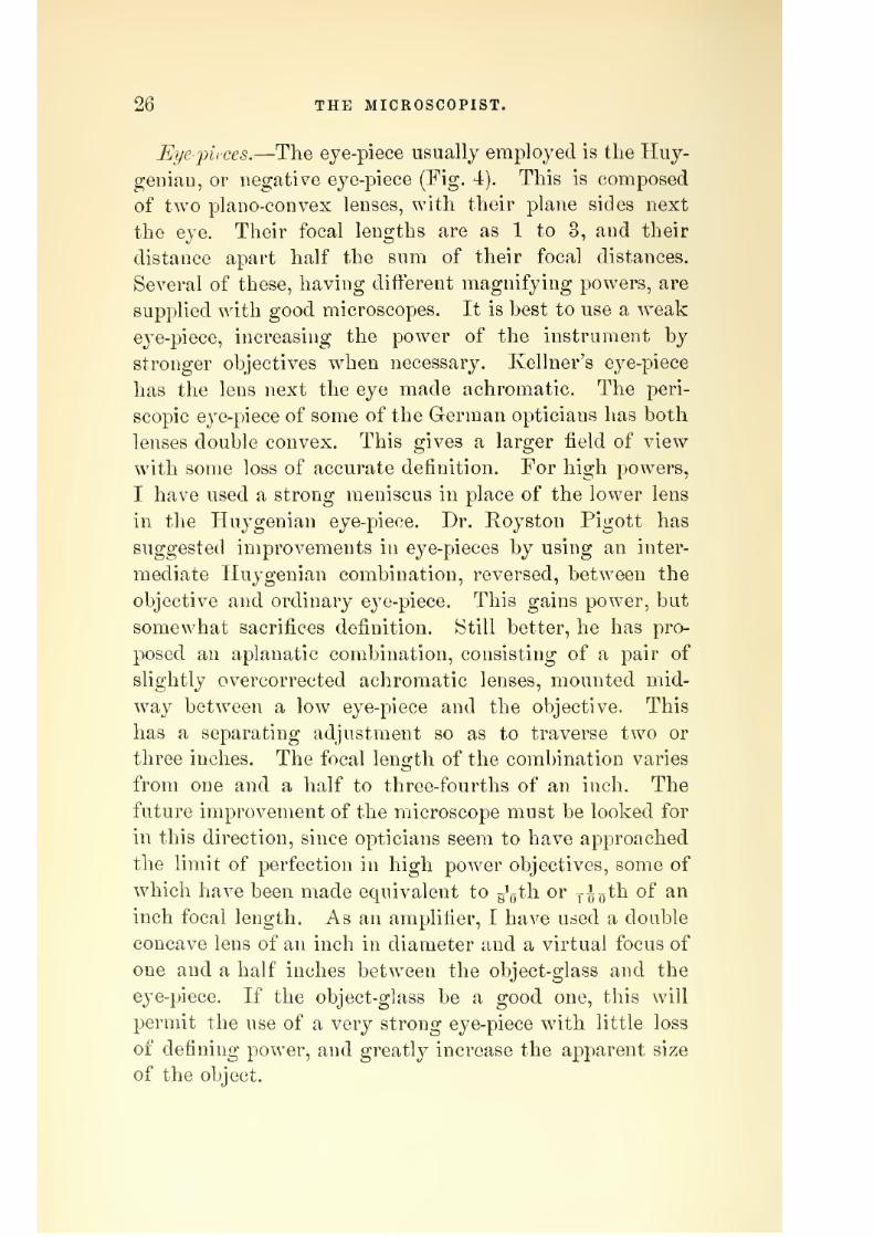
Somewhat similar to the above is Strieker's _^as c^/?a?n6er
(Fig. 21). On the object-slide is a ring of glass, or putty,
with its thin cover. Through this ring two glass tubes
are cemented, one of which is connected with a rubber
7/29/2019 THE MICROSCOPIST
http://slidepdf.com/reader/full/the-microscopist 44/361
42 THE MICROSCOPIST.
tube for the entrance of gas, while the other serves for
its exit.
For the studj^ of phenomena in the fluids, etc., of warm-
blooded animals, we need, in addition to the moist cham-
ber, some way of keeping the object warm. This may be
roughly done by a perforated tin or brass plate on the
stage, one end of which is warmed by a spirit-lamp. Apiece of cocoa butter or wax will show by its melting
when the heat is sufiicient. Schultze's warm stage is a
more satisfactory and scientific instrument. It is a brass
plate to fit on the stage, perforated for illumination, and
connected with a spirit-lamp and thermometer, so that
F(Ct. 21.
Strieker's Gas Chamber.
the amount of heat may be exactl}^ regulated. Other
arrangements have been proposed to admit a current of
warm water, or for the passage of electricity through an
object while under observation, which are scarcely neces-
sary to describe.
Tlie Polariscope.—The nature and properties of polarized
light belong rather to a treatise on optics or natural phi-
losophy than to a work like the present, yet a very brief
account may not be out of place. AYe premise, then, that
every ray or beam of common light is supposed to have
at least two sets of vibrations, vertical and horizontal.
As these vibrations have difterent properties, the ray when

7/29/2019 THE MICROSCOPIST
http://slidepdf.com/reader/full/the-microscopist 45/361
MICROSCOPIC ACCESSORIES. 43
divided is said to be i^olarized ^irom. a fancied resemblance
to tbe poles of a magnet. The division of the vibrations
may be effected [i. t'., the light may be polarized) in vari-
ous ways. For the microscope the jjolarizer is a Nichol's
prism, composed of a crystal of Iceland spar, which has
been divided and again cemented with Canada balsam, so
as to throw one of the doubly refracted rays aside from
the field of view (Fig. 22). Such a prism is mounted in
a short tube and attached to the under side of the stage.
In order to distinguish the effects of polarized light, an
analyze}^ is also needed. This usually consists of another
Fig. 22. Fig. 23.
Kichol's Pi ism. Polarizer and Analyzer.
similar l^ichol's prism, attached either to the eye-piece or
just above the objective. The latter position gives a
larger field, but the former better definition. Fig. 23
shows the polarizer and the analyzer. The polarizer is
improved by the addition of a convex lens next the object.
Hartnack has also improved theeye-]3iece analyzer
byadding a graduated disk and vernier.
When the polarizer and analyzer have been put in place,
they should be rotated until their polarizing planes are
parallel, and the mirror adjusted so as to give the most
intense light. If now the polarizing planes are placed at
right angles, by turning one of them 90°, the field is ren-

7/29/2019 THE MICROSCOPIST
http://slidepdf.com/reader/full/the-microscopist 46/361
44 THE MICROSCOPIST.
dered dark, and doubly refracting bodies on the stage of
the microscope appear either illuminated or in colors. If
a polarized ray passes through a doubly refracting film,
as of selenite, it forms two distinct rays, the ordinary
and the extraordinar}^ ray. Each of these will be of dif-
ferent colors, according to the thickness of the film. If
one be red, the other will be green, these colors being
complementary. By using the analyzer one of these rays
is alternately suppressed, so that on revolving the appa-ratus the green and red rays appear to alternate at each
quarter of a circle. Films of selenite are often mounted
so as to revolve between the polarizer and the stage.
Barker's selenite stage is sometimes used for this purpose
(Fig. 24). With such a stage a set of selenites is usually
Diuker's S^'lenite Stusre.
supplied, giving the blue, purple, and red, with their com-
plementary colors, orange, yellow, and green. By this
combination all the colors of the spectrum may be ob-
tained. The selenite disks generally have engraved on
them the amount of retardation of the undulations of
white light, thus: J, f, and \. If these are place<] so
that their positive axes (marked PA) coincide, they give
the sum of their combined retardations.
The Mic?'ospectrosco2)e. ^—Ordimiry spectrum analysis, by
determining the number and position of certain narrow
lines in the spectra of luminous bodies, called Fraunhofer's

7/29/2019 THE MICROSCOPIST
http://slidepdf.com/reader/full/the-microscopist 47/361
MICROSCOPIC ACCESSORIES. 45
lines, enables the chemist to identify different substances.
The object of the niicrospectroscope is different. It en-
ables us to distinguish substances by the absence of cer-
tain rajs in the spectrum, or, in other words, to judge of
substances by a scientific examination of their color. The
color of a body seen with the naked e^^e is the general
iivipression made by the transmitted light, and this may
be the same although the compound rays may differ
Fi,;. -ir.
\sr---^
The Sorby-Browning Microspectroscope.
greath', so that colors which seem absolutely alike may
be distinguished by their spectra.
Manysolutions are
seen to absorb different colors in very definite parts of the
spectrum, forming absorption bands or lines, varying in
width and intensity according to the strength of the so-
lution. The instrument usually employed consists of a
direct-vision spectrum apparatus attached to the eye piece
of the microscope, which shows the principal Fraunhofer

7/29/2019 THE MICROSCOPIST
http://slidepdf.com/reader/full/the-microscopist 48/361
46 THE MICROSCOPIST.
lines by da3'liglit, or a spectrum of the light transmitted
by any object in the field of view. A reflecting prism is
placed under one-half of the slit of the apparatus so as to
transmit from a side aperture a standard spectrum for
comparison. In Fig. 25, a is a brass tube carrying the
compound direct-vision system of five prisms and an
achromatic lens. This tube is moved by the milled head
Spectroscope with Micrometer.
B, SO as to bring to a focus the different parts of the
spectrum. This is important when the bands or lines to
be examined are delicate, d is the stage on which objects
for comparison are placed. The light passing through
them from the mirror i, goes through a side opening to a
reflecting prism which covers a part of a slit in the bot-
tom of the tube a. This slit is opened and shut by means
of the screws c and h Fig. 26 shows the internal ar-

7/29/2019 THE MICROSCOPIST
http://slidepdf.com/reader/full/the-microscopist 49/361
MICROSCOPIC ACCESSORIES. 47
rangement of tlie prisms and lens, together with a microm-
eter for measuring the position of lines or absorptionbands. To use the microspectroscope, remove the tube
A, with the prisms, and insert the tube g in the place of
the eye-piece of the microscope. With the lowest power
object-glass which is suitable, and the slit opened wide by
the screw h, the object on the stage of the microscope,
illuminated by the mirror or condenser, is brought to a
focus, the tube a replaced and adjusted for focus by thescrew B, while the slit is regulated by c and h until a well-
defined sjDectrum is seen. To determine the position of
the absorption lines, remove the upper cover of the tube
a and replace it with that carrying the micrometer repre-
sented in Fig. 26. The mirror illuminates a transparent
line or cross, whose image is refracted by a lens c, mov-
ableby
a screw b,
andreflected
at an angle of45°
fromthe upper surface of the prisms, so as to be seen upon the
spectrum. By means of the micrometer screw m, this is
made to move across the spectrum, so that the distance
between the lines may be determined. In order to com-
pare the results given by different instruments, the
observer should measure the position of the principal
Fraunliofer lines in bright daylight, and mark them on
a cardboard scale, which may be preserved for reference.
By comparing the micrometric measurement of lines in
the spectrum of any substance observed by artiflcial light
with such a scale, their position may readily be seen.
In using the microspectroscope some objects require a
diaphragm of small size, and others, especially with the
1| or 2-inch objective, a cap with a hole ,\;th of an inch
in diameter over the end of the microscope, to prevent
extraneous light from passing through the tube.
Nose-inece.—For the purpose of facilitating observations
with objectives of different powers a revolving nose-piece
has been contrived, carrying two, three, or four objectives,

7/29/2019 THE MICROSCOPIST
http://slidepdf.com/reader/full/the-microscopist 50/361
48 THE MICROSCOPIST
which may be brought quickly into the axis of the instru-
ment.
Object-finders.—It is sometimes tedious to find a small
object on a slide, particularly with high powers, and a
number of contrivances, as Maltwood's finder, have been
proposed for this end. A very simple method, however,
may serve. Mark on the stage two crosses, one like the
sign of addition +, and the other like the sign of multi-
plication X , and, when the object is found, mark the slide
to correspond with the marks below. If the stage be a
mechanical one it will be necessary to arrange it in the
previous position.
Microscopic Fliotography.—Many European experimen-
ters have succeeded in taking microscopic photographs,
but a great advance in this direction has been made under
the direction of the medical department of the UnitedStates army at Washington. Lieutenant-Colonel Wood-
ward has succeeded in furnishing permanent records of
many details of structure, which exhibit the very perfec-
tion of art. In a work like the present a full account of
the apparatus and methods employed would be out of place.
Dr. Beale's How to Work ivit/i the Uicroscope, and the re-
ports issued from the Surgeon-General's office at .Wash-ington, will give the details.
CHAPTER lY.
USE OF THE MICROSCOPE.
Care of the Instrument.—But little satisfaction will be
secured in microscopic work for any length of time with-
out scrupulous care of the lenses, etc., belonging to the
instrument, and habits of this kind should be early ac-
quired. When in frequent use the microscope should be

7/29/2019 THE MICROSCOPIST
http://slidepdf.com/reader/full/the-microscopist 51/361
USE OF THE MICROSCOPE. 49
seldom packed away in its case, as a certain necessary
stiffness of motion in its various parts might thereby be
lessened. Yet it should be kept free from dust and damp.
A bell-glass cover, or glass case, or a cabinet which will
admit the reception of the instrument in a form ready for
immediate use, is desirable. Before using, the condition
of objective and eye-piece should be examined as well as
of the mirror, and dust or dampness removed. Another
examination should be made before the microscope is put
away.
Stains on the brass-work may be removed by a linen
rag, and dust on the mirror and lenses by a fine camel's-
hair brush, or very soft and clean chamois skin. Frequent
wiping will injure the polish of the lenses.
The npper surfaces of the lenses in the eye-pieces and
the mirror will need the most frequent attention Theobjectives, if carefully handled and kept in their boxes
when not in use, will seldom require cleaning. If the front
of the objective becomes accidentally wet with fluid it
should be at once removed, and, when reagents are used,
great care should be taken to prevent contact with the
front of the lens.
Care of the Eyes.— Continuous observation, especially bylamplight, and with high powers, has doubtless a ten-
dency to injure the sight. To cease work as soon as
fatigue begins is, however, a simple but certain rule for
protection. This time will vary greatly, according to the
general tone and vigor of the observer. It is also impor-
tant to use the eyes alternately if a monocular instrument
is emploj'ed, as otherwise great difference both in thefocus and in the sensitiveness of the eyes will result. The
habit of keeping the unemployed eye open is a good one,
and, though troublesome at first, is not diificult to ac-
quire. It is well to protect the eye from all extraneous
light, and to exclude every part of the object except that
which is under immediate observation. The diaphragm
4

7/29/2019 THE MICROSCOPIST
http://slidepdf.com/reader/full/the-microscopist 52/361
60 THE MICROSCOPIST.
will serve this end as well as modify the quality of the
light. For very delicate observations a dark shade over
the stage, which may be fastened by an elastic ring to
the microscope-tube, so as to shut off extraneous light,
will be useful.
Tahle^ etc.—The microscopist's work-table should be
large and massive, so as to be convenient and free from
vibration. Drawers for accessories and materials used in
preparing and mounting objects are also desirable, as well
as a few bell-glasses for secluding objects from dust. Re-
agents should always be removed from the table after use
and kept in another place.
Light.—Dr. Carpenter has well said, "Good daylight is
to be preferred to any other kind of light, but good lamp-
light is preferable to bad daylight." A clear blue sky
gives light enough for low powers, but a dull white
cloudiness is better. The direct rays of the sun are too
strong, and should be modified by a white curtain, reflec-
tion from a surface of plaster of Paris, or, still better, by
passing through a glass cell containing a solution of am-
monio-sulphate of copper.
Various kinds of lamps have been contrived for micro-
scopic use ; among the best are the German and French" student's reading lamps," which burn coal oil or petro-
leum. It is often useful to moderate such a light by the
use of a chimney of blue glass, or by a screen of blue glass
between the flame and the object. Dr. Curtis contrived
a useful apparatus, consisting of a short petroleum lamp
placed in an upright, oblong box. On one side of the box
is an opening occupied with blue glass ; on another side
the opening has ground-glass, as well as a piece colored
blue, and a plano-convex lens so placed as to condense the
light thus softened to a suitable place on the table.
As a general rule the light should come from the left
side, and that position assumed or inclination given to the
instrument which is most comfortable to the observer.
APR 281917 _ I

7/29/2019 THE MICROSCOPIST
http://slidepdf.com/reader/full/the-microscopist 53/361
USE OF THE MICROSCOPE. 51
Englisli aud American microscopists prefer an inclined
microscope, while the German and French instruments
heing usually vertical do not permit this arrangement.
Adjustment.—The details of microscopic adjustments
are only to be learned by practice, yet a few directions
may be instructive. The selection of the objectives and
eye-pieces depends on the character of the object. As a
general rule, the lowest powers which will exhibit an
object are the best. It is best to use weak eye-pieces with
the stronger objectives, yet much depends on the perfec-
tion of the glasses employed.
The focal adjustment can be made with the coarse ad-
justment or quick motion when low powers are employed
but for higher powers the line adjustment screw is essen-
tial. Care must be taken not to bring the objective into
close or sudden contact with the thin glass cover over the
object, and, in changing object-glasses, the microscope
body should be raised from the stage by the coarse adjust-
ment.
The actual distance between the object and object-glass
is much less than the nominal focal length, so that the
1 inch objective has a working distance of about | an
inch, the |th of about 4'oth of an inch, while shorter ob-
jectives require the object to be covered with the thinnest
glass.
Sometimes, in high powers, and especially with immer-
sion-lenses, an adjustment of the object-glass is necessary
in order to suit the thickness of the glass cover. With
thick covers the individual lenses must be brought nearer
to each other, and, with very thin covers, moved farther
apart.
If immersion-objectives be employed a drop of water is
placed on the glass cover with a glass rod or camel's-hair
pencil, and a second drop on the lens. The lens and object
are then approximated till the drops flow together and the
focus is adjusted. By turning the fcrew of the objective

7/29/2019 THE MICROSCOPIST
http://slidepdf.com/reader/full/the-microscopist 54/361
OZ THE MICROSCOPIST.
and using the fine adjustment the best position will he
shown by the sharper and more delicate image of the
object.
For other details respecting adjustment the reader is
referred to the chapter on Microscopic Accessories.
J^rro?-s of Ititerpretation.—True science is hindered most
of all by speculation and false philosophy, which often
assume its garb and name, but it is also retarded by im-
perfect or false observation. It is much less easy to see
than beginners imagine, and still less easy to know what
we see. The latter sometimes requires an intellect of sur-
passing endowments. The sources of error are numerous,
but some require special caution, and to these we now
refer.
The nature of microscopic images causes error from
imperfect focal adjustment. We see distinctly only that
stratum of an object which lies directly in focus, and it is
seldom that all parts of an object can be in focus together.
Hence we only recognize at once the outline of an object,
but not its thickness, and, as the parts which are out of
focus are indistinct, we may readily fall into error. Glasses
vary much in this respect. Sonie have considerable pene-
trating and defining power even with moderate angular
aperture, and are better for general work than those more
perfect instruments which give paler images and only re-
veal their excellencies to the practiced microscopist.
Sometimes the focal adjustment leads to error on ac-
count of the reversal of the lights and shadows at difter-
ent distances. Thus the centres of the biconcave blood-
disks appear dark when in focus, and bright when a little
within the focus; while the hexagonal elevations of a dia-
tom, as the Pleurosigma aiigulatiim,a,Ye light when in focus,
with dark partitions, and dark when just beyond the
focus. From this we gather a means of discrimination,
since a convex body appears lighter by raising the micro-
scope, and a concave by lowering it.

7/29/2019 THE MICROSCOPIST
http://slidepdf.com/reader/full/the-microscopist 55/361
USE OF THE MICROSCOPE. 53
The refractive power of the object, or of the medium in
which it lies, is sometimes a source of error. Thus a
human hair was long thought to be tubular, because of
the convergence of the rays of light on its cylindrical con-
vexity. A glass cylinder in balsam appears like a flat
band, because of the nearly equal refractive powers of
object and medium. Tlie lacunae and canaliculae of bone
were long considered solid, because of the dark appear-
ance presented on account of the divergence of the rayspassing through them. Their penetration with Canada
balsam, however, proves them to be cavities. Air-bubbles,
from refraction, pre-ent dark rings, and, if present in a
specimen, seldom fail to attract the first attention of an
inexperienced observer. The diiference between oil-globules
in water and water in oil, or air-bubbles, should be early
learned, as in some organized structures oil-particles andvacuoles (or void spaces) are often interspersed. A globule
of oil in water becomes darker as the object-glass is de-
pressed, and lighter when raised ; while the reverse is the
case with water in oil, since the difference of refraction
causes the oil particles to act as convex lenses, and those
of water like concave lenses.
Other errors arise from the phenomena of motion visibleunder the microscope. A dry filament of cotton, or other
fabric absorbing moisture, will often oscillate and twist
in a curious way.
If alcohol and water are mixed, the particles suspended
acquire a rapid motion from the currents set up, which
continues till the fluids are thoroughly blended. ISTearly
all substances in a state of minute division exhibit, when
suspended in fluid, a movement called the " Brownonian
motion," from Dr. Robert Brown, who first investigated
it. It is a peculiar, uninterrupted, dancing movement, the
cause of which is still unexplained. These movements,
as all others, appear more energetic when greatly magni-
fied by strong objectives. It requires care to discriminate

7/29/2019 THE MICROSCOPIST
http://slidepdf.com/reader/full/the-microscopist 56/361
54 THE MICROSCOPIST.
between such motions and the vital or voluntary motions
of organized bodies.The inflection or diffraction of light is another source
of error, since the sharpness of outline in an object is thus
impaired. The shadow of an opaque object in a divergent
pencil of light presents, not sharp, well-defined edges, but
a gradual shading off, from which it is inferred that the
rays do not pass from the edge of the object in the same
line asthey come
to it. This is inconsequence of
the
undulator}^ nature of light. When any system of waves
meets with an obstacle, subsidiary systems of waves will
be formed round the edge of the obstacle and be propagated
simultaneously with the original undulations. For a cer-
tain space around the lines in which the rays, grazing the
edge of the opaque body, would have proceeded, the two
systems of undulation will intersect and produce the phe-
nomena of interference. If the opaque body be very small,
and the distance from the luminous point proportionally
large, the two pencils formed by inflection will intersect,
and all the phenomena of interference will become evident.
Thus, if the light be homogeneous, a bright line of light
will be formed under the centre of the opaque object, out-
side of which will be dark lines, and then bright and dark
lines alternately. If the light be compound solar light, a
series of colored fringes will be formed. In addition to
the results of inflection, oblique illumination at certain
angles produces a double image, or a kind of overlying
shadow, sometimes called the "diftraction spectrum,"
although due to a diflerent cause. !No rules can be o-iven
for avoiding errors from these optical appearances, but
practice will enable one to overcome them, as it were,
instinctively.
Testing the 3Iicroscope.—The defining power of an in-
strument depends on the correction of its spherical and
chromatic aberrations, and excellence may often be ob-
tained with objectives having but a moderate angle of

7/29/2019 THE MICROSCOPIST
http://slidepdf.com/reader/full/the-microscopist 57/361
USE OF THE MICROSCOPE. 55
aperture. It may be known by the sharp outline given
to the image of an object, which is not much impaired bythe use of stronger eye pieces. _
Eesolving power is tlie capability an instrument has of
bringing out the fine details of a structure, and depends
mainly on the angle of aperture of the objective, or the
angle formed hy the focus and the extremities of the
diameter of the lens. On this account the increase of the
angleof aperture has been
achief
aim with practicalopticians.
Penetrating power is the degree of distinctness with
which the parts of an object lying a little out of focus
may be seen. Objectives which have a large angle of
aperture, and in consequence great resolving power, are
often defective in penetration, their very perfection only
permitting accurate vision of what is actually in focus.
Hence for general purposes a moderate degree of angular
aperture is desirable.
Flatness of field of view is also a necessity for accurate
observation. Many inferior microscopes hide their im-
j)erfection in this respect by a contracted aperture in the . /v
eye-pieee,^ by which, of course, only a part of the rays
transmitted by the objective are available.
Object-glasses whose focal length is greater than half
an inch are called low powers. Medium powers range
from one-half to one-fifth of an inch focal length, and all
objectives less than one-fifth are considered high powers.
For definition with low power objectives, the pollen
grains of hollyhock, or the tongue of a fly, or a specimen
of injected animal tissue, will be a sufficient test. The
aperture should be enough to give a bright image, and
the definition sufficient for a clear image. A section of
wood, or of an echinus spine, will test the flatness of the
field.
Medium powers are seldom used with ojDaque objects
unless they are very small, but are most useful with
"r'h^^'

7/29/2019 THE MICROSCOPIST
http://slidepdf.com/reader/full/the-microscopist 58/361
56 THE MICROSCOPIST.
properly prepared transparent objects. A good half-inch
objective should show the transverse markings betweenthe longitudinal ribs on the scales of the Hi'p'parchia
janira, butterfly (Plate I, Fig. 27), and the one-fourth or
•one-fifth should exhibit markings like exclamation points
on the smaller scales of Podura ylumbea (Plate I, Pig. 28)
or Lepidocyrtis.
High power objectives are chiefly used for the most
delicate and refined investigations of structure, and are
not so suitable for general work. It is with these glasses
that angular aperture is so necessary to bring out striae,
and dots, and other delicate structures, under oblique
illumination. For these glasses, the best tests are the
siliceous envelopes of diatoms, as the Pleurosigma angu-
latum, Surirella gemma, Grammataphora subtilissima ; or
the wonderful plates of glass artificially^ ruled by M. ]^o-
bert, and known as Nobert's test.
The latter test is a series of lines in bands, the distance
between the lines decreasing in each band, until their
existence becomes a matter of faith rather than of sight,
since no glass has ever revealed the most difiicult of them.
The test plate has nineteen bands, and their lines are
ruled at the following distances: Band 1, y,/„^,th of aParis line (to an English inch as .088 to 1.000, or as 11 to
125). Band 2, fg'goth. Band 3, ^p'^^^th. Band 5, g^^^oth.
Band 9, g^'goth. Band 13, ^g'^pth. Band 17, -go'^Qth.
Band 19, ^^l^j^th.
It is said that Ilartnack's immersion system ]^o. 10
and oblique light has resolved the lines in the 15th band,
in which the distance of lines is about ^ y Joo^^ ^^ ^^^ inch.
The surface markings of minute diatoms are also ex-
cessively fine. Those of Pleurosigma formosam. being from
20 to 32 iny^j'^j^th of an inch ; of P. hippocampus and P.
attenuatum, about 40 ; P. angulatum 46 to 52 ; Navicula
rhomboides 60 to 111 ; and Atnphipleura pellucida 120 to
130. This latter has been variously estimated at 100,000

7/29/2019 THE MICROSCOPIST
http://slidepdf.com/reader/full/the-microscopist 59/361
PLATE I,
Fig. 28.
l^"'''
Scale of Hippnrc.hUi Janira.
Fig. 29.
Scales of Poibira plumben —a, large
strongly marked scale; u, small scalemore faintly marked; c, jiortion of aninjured scale, showing the nature of the
markings.
Plfuroxigmn angiilatnw :—A, entire frustiile, as seen
under a power of .500 diam.: b, hexagonal aerolation,
as seen under a power of 1.300 diam.; c, the same,
as seen under a power of 1.5,000 diam.

7/29/2019 THE MICROSCOPIST
http://slidepdf.com/reader/full/the-microscopist 60/361

7/29/2019 THE MICROSCOPIST
http://slidepdf.com/reader/full/the-microscopist 61/361
USE OF THE MICROSCOPE. 57
to 180,000 in an inch. It has been resolved by Dr. Wood-
ward with the y'gth immersion of Powell and Lealand,using oblique sunlight through a solution of ammonio-
sulphate of copper.
The longitudinal lines (between the transverse) of the
Fig. 30.
Valve of Surirella Gemma.
a. Transverse ridges. 6. Longitudinal lines, c. The same, resolved into areolations.
Surirella gemma are estimated at 30 to 32 in yoo^^^ ^^ ^
millimetre, and the markings on Grammata-phora suhtilis-
sima at 32 to 34 in the same distance.
Fig. 31.
Grammataphora Subtilissima.
a. Valve, b. Transverse lines.
J. D. MoUer has produced a very excellent test-plate,
containing twenty diatoms, with descriptions, according
to their value as tests.

7/29/2019 THE MICROSCOPIST
http://slidepdf.com/reader/full/the-microscopist 62/361
58 THE MICROSCOPIST.
The Pleurosigma angulatum (Plate T, Fig. 29), with suit-
able power and illumination, should show distinct hexag-
onal areolations. The Surirella gemma (Fig. 30) show^s a
series of fine transverse lines across the ridges which run
from the edge to the central line. The finest of these
ridges are not always readily seen, and the transverse
ones are only to be mastered by toil and patience.
The Grammataj)hora subtilissima (Fig. 31) shows trans-
verse lines (or rows of dots) along the edge, and sometimesa double series of oblique lines.
CHAPTER y.
MODERN METHODS OF EXAMINATION.
Microscopy does not limit its researches to optical
enlargement, but seeks to comprehend elementary struc-
ture, and its methods vary according to the object imme-diately in view. It may seek merely to discern the form
or morphology of the elementary parts or their peculiar
functions. It may be concerned with inorganic forms,
normal or pathological anatomy, or with physiology.
Each department of pursuit will suggest some variation,
yet a general plan of operation is possible.
Coarse, and moderately large objects, as a small insect,a piece of vegetable tissue, etc., may be observed by plac-
ing it in the forceps, or on the stage of the instrument,
under an objective of low power, but ordinarily a consid-
erable degree of preparation is needed in order to acquire
a true idea of structure.
Most of the tissues to be examined are in a moist con-

7/29/2019 THE MICROSCOPIST
http://slidepdf.com/reader/full/the-microscopist 63/361
MODERN METHODS OF EXAMINATION. 59
dition, and many require to be dissected or preserved in
fluid. This has miicli to do with the appearance of theobject in the microscope. If fibres or cells are imbedded
in connective tissue or in fluids, of which the refractive
power is the same as their own, they cannot be perceived
even with the best glasses, and artificial means must be
resorted to that they may become visible. The refractive
power of ditt'erent media causes different appearances.
Thus a glass rod lying in Avateris
easily seen, but inCanada balsam, whose refractive power is nearly the same
as glass, it is barely seen as a flat band, and in the more
highly refractive anise oil it presents the appearance of a
cavity in the oil.
During life the cavities and fissures in animal tissues,
in consequence of the different refractive power of their
contents and the change which takes place soon after
death, exhibit a sharpness and softness of outline which
is seldom seen in preparations.
There are two methods of microscopic investigation or
of preparation preliminary to direct observation: 1. Me-
chanical, for the separation and isolation of the elemen-
tary parts. 2. Chemical, which dissolve the connecting
material, or act on it differently than on other elements.
For minute dissection a great variety of instruments
have been proposed, but by practiced hands more can be
accomplished in shorter time by simple means than with
complicated ones. Two or three scalpels, or small ana-
tomical knives, a pair of small scissors, such as is used in
operations on the eye, and fine-pointed forceps, will be
found useful. But the most serviceable instruments are
dissecting-needles, such as the raicroscopist may make for
himself. A common sewing-needle, with the eye end
thrust into a cedar stick about three inches long and one-
fourth of an inch diameter, will answer the purpose. The
point should not project so far as to spring, and if desired,
a cutting edge can be given to it by a hone.

7/29/2019 THE MICROSCOPIST
http://slidepdf.com/reader/full/the-microscopist 64/361
60 THE MICROSCOPIST.
The light should be concentrated on the work by means
of a bulFs-eye condenser, and as far as possible, the dis-
section should be carried on with the unassisted eye.
Very often the work is so fine that a magnifying glass,
or simple microscope, fixed to a suitable arm, will be
needed. A large Coddington lens, an inch and a half in
diameter, such as is used frequently by miners, will be
useful. Sometimes it is necessary to resort to the dissect-
ing microscope, which is a simple lens, of greater or less
power, arranged with rack and pinion, mirror, etc.
The specimen may be dissected under water, in a glass
or porcelain dish, or a trough made of gutta-percha, etc.
Dr. Lawson's binocular dissecting microscope (Fig. 32) is
Fig. 32.
Lawson's Binocular Dissecting Microscope.
a most useful form, as both eyes may be used. Loaded
corks, with sheet lead fastened to their under surface,
may be used to pin the subject on for greater facility in
dissection. Rests, or inclined planes of wood, one on each
side of the trough, will give steadiness to the hands.
Camels'-hair pencils for the removal of dust and extrane-
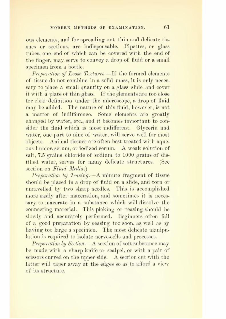
7/29/2019 THE MICROSCOPIST
http://slidepdf.com/reader/full/the-microscopist 65/361
MODERN METHODS OF EXAMINATION. 61
ous elements, and for spreading out thin and delicate tis-
sues or sections, are indispensable. Pipettes, or glass
tubes, one end of which can be covered with the end of
the finger, may serve to convey a drop of fluid or a small
specimen from a bottle.
Preparation of Loose Textures.—If the formed elements
of tissue do not combine in a solid mass, it is only neces-
sarj^ to place a small quantity on a glass slide and cover
it with a plate of thin glass. If the elements are too close
for clear definition under the microscope, a drop of fluid
may be added. The nature of this fluid, however, is not
a matter of indiflerence. Some elements are greatly
changed by water, etc., and it becomes important to con-
sider the fluid which is most indifferent. Glycerin and
water, one part to nine of water, will serve well for most
objects. Animal tissues are often best treated with aque-ous humor, serum, or iodized serum. A weak solution of
salt, 7.5 grains chloride of sodium to 1000 grains of dis-
tilled water, serves for many delicate structures. (See
section on Fluid Media.)
Pre-paraiion by Teasing.—A minute fragment of tissue
should be placed in a drop of fluid on a slide, and torn or
unravelled by two sharpneedles. This is accomplished
more easily after maceration, and sometimes it is neces-
sary to macerate in a substance which will dissolve the
connecting material. This picking or teasing should be
slowly and accurately performed. Beginners often fail
of a good preparation by ceasing too soon, as well as by
having too large a specimen. The most delicate manipu-
lation is required to isolate nerve-cells and processes.
Preparation by Section.—A section of soft substance may
be made with a sharp knife or scalpel, or with a pair of
scissors curved on the upper side. A section cut with the
latter will taper away at the edges so as to aftbrd a view
of its structure.

7/29/2019 THE MICROSCOPIST
http://slidepdf.com/reader/full/the-microscopist 66/361
62 THE MICROSCOPIST.
Valentin's double knife (Fig. 33) is used for soft tissues
where only a moderate degree of thinness is needed. The
blades should be wet, or the section made under water.
Soft substances often require hardening before sections
can be made. The most simple and best method is that
of freezing, by surrounding the specimen with a freezing-
mixture, when it may be cut with a cold knife. Small
pieces of tissue may be hardened in absolute alcohol, fre-
quentij' renewed. Cli^romic acid, in solution of one-fourth
to two per cent., is often used for animal tissues, or bichro-
mate of potash of the same strength. A solution of one-
fifth to one-tenth per cent, of perosmic acid or of chloride
of palladium is also recommended.
Soft tissues often require imbedding in a concentrated
solution of gum or of wax, spermaceti, or paraffin tem-
pered with oil. In this case sections may be made readily
by means of a section-cutter. For imbedding in wax, etc.,
Fig. 3-3.
Valentin's Knife.
the specimen must be hardened in alcohol, then treated
with oil of cloves or turpentine, and the section should be
mounted in Canada balsam or Dammar varnish.
Sections of hard substances, or of those imbedded, are
often made by machines invented for the purpose. One
of the simplest is (Fig. 34) an upright hollow cylinder,
with a kind of piston, pushed upwards by a fine screw.
The upper end of the cylinder carrying the specimen ter-
minates in a flat table, along which a sharp knife or flat
razor is made to slide. At one side of the tube is a
binding-screw for holding the specimen steady. A sec-

7/29/2019 THE MICROSCOPIST
http://slidepdf.com/reader/full/the-microscopist 67/361
MODERN METHODS OF EXAMINATION. 63
tion may be cut by snch an instrument after inserting
the structure desired in a piece of carrot, etc., which maybe phiced in the tube ; or the tube may be filled with wax,
etc., and the specimen imbedded. Bones, teeth, shells,
corals, minerals, etc., require to be cut with fine saws, or
a disk of thin iron on a lapidary's wheel, and filed or
ground down to the requisite thinness, then polished with
emery, rouge, etc. The green oxide of chromium has
been suggested to me as a useful polishing powder for
hard substances. For calcareous substances, files and
hones will sufiice to reduce the thickness, and putty
Fig. 34.
Section-Cutter.
powder or jewellers' rouge for polishing. They should
be mounted in Canada balsam.
Staining Tissues.—Certain elements, not previously visi-
ble, can often be made evident by certain coloring matters,
by w^hich some constituents become more quickly or more
thoroughly stained than others. The " germinal matter,"
or "bioplasm" of Dr. Beale, identical with the "proto-
plasm " or " sarcode " of other observers, may thus be dis-
tinguished from the " formed materials " or " tissue ele-

7/29/2019 THE MICROSCOPIST
http://slidepdf.com/reader/full/the-microscopist 68/361
64 THE MICROSCOPIST.
ment," which are the products of its activity. Carmine,
anilin, h?ematoxylin, and picric acid, are used for staining
by imparting their own color to tissues; while nitrate of
silver, chloride of gold, chloride of palladium, and peros-
mic acid stain, by their chemical action, often under the
reducing influence of light. (See Fluid Media.)
Injecting Tissues.—Injections of the vessels in animal
tissues are resorted to either to exhibit their course or the
structure of the vascular walls. For the latter purpose a
solution of nitrate of silver is commonly employed, for the
former either opaque or transparent coloring matter. (See
Fluid Media.)
The injecting syringe (Fig. 35) is made of brass or Ger-
Injecting Syringe.
man silver. One of the pipes should be inserted into the
principal vessel, as the aorta of a small animal, the um-
bilical vein of a foetus, or the arterj^, etc ,of an organ,
and should be securely fastened by a thread. All other
open vessels should be tied. The solution of gelatin, or
other matter used, should be strained, so as to be free
from foreign particles, and should be forced into the ves-
sels with a gentle, steady pressure on the syringe.
Injections should be made soon after the death of the
animal, or else after the rigor mortis has subsided.

7/29/2019 THE MICROSCOPIST
http://slidepdf.com/reader/full/the-microscopist 69/361
MODERN METHODS OF EXAMINATION. 65
Sometimes the syringe is substituted by a self-acting
apparatus, consisting of a Wolfe's bottle, containing the
fluid, which is pressed upon by a column of air from
another source, and driven through a flexible tube to the
pipe in the bloodvessel.
The older anatomists used colored plaster or v^ax to
demonstrate the arteries and veins, but modern histology
requires finer materials. Isinglass or gelatin, colored, and
injected warm, or a solution of colored glycerin, are nowresorted to. The former serves for opaque, and the latter
for fine, transparent injections.
The art of injecting can only be learned by practice,
yet perseverance, in despite of many failures, will insure
success.
The liver, kidney, etc., may be injected separately, and
it- is often desirable to use various colors for the diflerentsets of vessels. After injection thin slices may be cut off
and mounted in fluid or balsam.
Preparation in Viscid Media.—Dr. Beale has proposed a
method of preparing animal and vegetable tissues for ex-
amination with the very highest powers, which has led to
valuable results. It consists in using pure glycerin or
strong syrup, instead of watery solutions. In this wayan amount of pressure may be applied to sections, in order
to render them thin enough for examination, which would
be destructive to specimens in water, while the preserva-
tive action of the media prevents change in the structure.
It is necessary to soak the specimen some time, and the
strength of the fluid should be gradually increased until
,the tissue is permeated by the strongest that can be ob-
tained. Dr. Beale has found that minute dissection is
much more readily performed in such fluids, and that
even very hard textures, as bone and teeth, may be softened
by them, especially if acetic acid is added, so as to permit
thin sections to be made with a knife. He recommends
5

7/29/2019 THE MICROSCOPIST
http://slidepdf.com/reader/full/the-microscopist 70/361
66 THE MICROSCOPIST.
vessels to be first injected, as with fine, transparent blue,
and the germinal matter to be stained with carmine. Afew drops of a solution of chromic acid, or bichromate of
potash, so as to impart to the glycerin a pale straw color,
serves to harden even the finest nerve-structures. Acetic
acid, and other reagents also, are much more satisfactorily
used with glycerin than with water. If syrup is used,
camphor, carbolic acid, etc , must be employed to prevent
the growth of fungi, but pure glycerin is free from this
inconvenience.
A great advantage of this mode of investigation con-
sists in the fact that a specimen thus prepared is already
mounted, and needs but a proper cement to the glass cover
and a finish to the slide, when it is ready for the cabinet.
fluid media.
1. Indifferent Fluids.
The vitreous humor, amniotic liquor, serum, etc., which
form the usual fluids termed indifterent, always contain
what Prof. Graham designated colloid and crystalloid
substances. In 1 000 parts there are about 4 parts of col-
loid (albumen) and 7.5 of crystalloid substance (chloride
of sodium).
The iodine serum of Schultze consists of the amniotic
fluid of the embryo of a ruminant, to which about 6 drops
of tincture of iodine to the ounce is added A small piece
of camphor will preserve this from decomposition a long
time. A substitute for this is composed of 1 ounce of
white of egg, 9 ounces of water, 2 scruples chloride of
sodium, with the corresponding quantity of tincture of
iodine.

7/29/2019 THE MICROSCOPIST
http://slidepdf.com/reader/full/the-microscopist 71/361
MODERN METHODS OF EXAMINATION. 67
2. Chemical Reagents.
The greatest care should be used with these, that the
instrument and glasses maybe preserved. A small drop,
applied by a glass rod drawn out to a point to the edge of
the glass cover, will suffice in most cases.
Sulphuric Acid.—Concentrated is used to isolate the
cells of horny structures, as hair, nails, etc. Dilute (1 partto 2-3 of water) gives to cellulose, previously dyed with
iodine, a blue or purple color, and, when mixed with
sugar, a rose-red to nitrogenous substances and bile. 0.1
to 1000 of water, at a temperature of 35-40° C, resolves
connective tissue into gelatin and dissolves it, so as to be
useful in isolating muscular fibres.
Nitric Acid.—Diluted with 4 or 5 parts water, separatesthe elementary' parts of many vegetable and animal tis-
sues when they are boiled or macerated in it. With chlo-
rate of potash it is still more energetic, but caution is
needed in its use.
Bluriatic Acid, Strong.—Used for dissolving intercellular
substance, as in the tubes of the kidney, etc. Dilute for
dissolvingcalcareous matter.
Chromic acid, J to 2 per cent, solution for hardening
nerves, brain, etc.
Oxcdic acid, to 15 parts water, causes connective tissue
to swell and become transparent, while albuminoid ele-
ments are hardened. Preserves well delicate substances,
as rods of retina, etc.
Acetic acid makes nuclei visible and connective tissue
transparent, so as to exhibit muscles, nerves, etc., other-
wise invisible.
Iodine (1 grain of iodine, 3 grains iodide of potassium,
1 ounce of water) turns starch blue and cellulose brown.
Caustic potash or soda renders many structures trans-
parent.

7/29/2019 THE MICROSCOPIST
http://slidepdf.com/reader/full/the-microscopist 72/361
68 THE MICROSCOPIST.
Lime-water or baryta-water is used for investigating con-
nective structures, especially tendon, as maceration en-
ables the needle to divide its fibrilla.
Chloride ofSodium.—Solutions of this salt for indifferent
media should always have some colloid, as albumen or
gum-arabic added (7.5 grains in 1000 grains of water for
delicate structures).
Bichromate of ])otash is used in stronger solution for the
same purposes as chromic acid.
MUllei'^s eye-fluid for hardening the retina, and preserv-
ing delicate embryos, etc., consists of bichromate of potass,,
2 grammes ; sulphate of soda, 1 gramme ; distilled water,
100 grammes.
Alcohol dissolves resins and many vegetable coloring
matters ; renders most vegetable preparations more trans-
parent, and albuminous animal tissues more opaque.
Acetic acid and, cdcohol, 1 part of each to 2 of water,
renders connective tissue transparent, and albuminoid tis-
sue prominent. The proportions can be varied.
Alcohol and soda (8-10 drops of strong solution of caustic
soda to each ounce) renders many tissues very hard and
transparent. Beale recommends it for embryonic struc-
tures.
Ether dissolves resins, oils, and fat.
Turpentine renders dried animal sections transparent.
Oil of cloves acts as turpentine.
Solution of chloride of zinc, iodine, and iodide ofpotassium,
is recommended by Schacht as a substitute for iodine and
sulphuric acid to color vegetable cells, etc. Zinc is dis-
solved in hydrochloric acid, and the solution is evaporated
to syrupy consistence in contact with metallic zinc. This
is saturated with iodide of potassium, iodine added, and
the solution diluted with water. Wood cells, after boiling
in caustic potash, are stained blue by it.
JBoracic acid, used by Prof, Brucke to separate the ele-
ments of red blood-corpuscles.
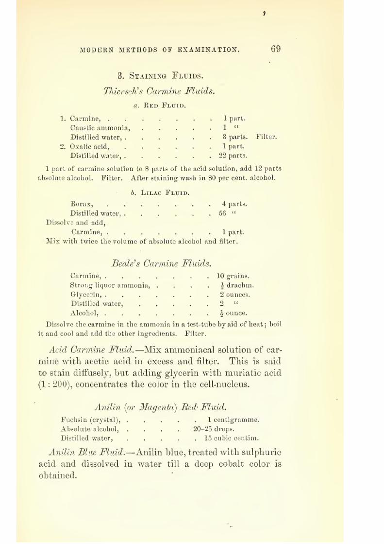
7/29/2019 THE MICROSCOPIST
http://slidepdf.com/reader/full/the-microscopist 73/361
MODERN METHODS OF EXAMINATION. 69
3. Staining Fluids.
Thiersch's Carmine Fluids.
a. Red Fluid.
1. Carmine, 1 part.
Caustic ammonia, . . . . . 1 "
Distilled water, 3 parts. Filter.
2. Oxalic acid, 1 part.
Distilled water, 22 parts.
1 part of carmine solution to 8 parts of the acid solution, add 12 parts
absolute alcohol. Filter. After staining wash in 80 per cent, alcohol.
b. Lilac Fluid.
Borax, 4 parts.
Distilled water, 56 "
Dissolve and add,
Carmine, ....... 1 part.
Mixwith
twice the volume of absolute alcohol andfilter.
Beetle's Carmine Fluids.
Carmine, .
Strong liquor ammonia,
Glycerin, .
Distilled water.
Alcohol, .
Dissolve the carmine in the ammoniait and cool and add the other ingredien
10 grains.
J drachm.
2 ounces.
^ ounce.
n a test-tube by aid of heat; boil
ts. Filter.
Acid Carmine Fluid.—Mix ammoniacal solution of car-
mine with acetic acid in excess and filter. This is said
to stain difi'usely, but adding glycerin with muriatic acid
(1 : 200), concentrates the color in the cell-nucleus.
Anilin {or Magenta) Red- Fluid.
Fuchsin (crystal), ..... 1 centigramme.
Absolute alcohol, .... 20-25 drops.
Distilled water, 15 cubic centim.
Anilin Blue Fluid.—Anilin blue, treated with sulphuric
acid and dissolved in water till a deep cobalt color is
obtained.

7/29/2019 THE MICROSCOPIST
http://slidepdf.com/reader/full/the-microscopist 74/361
70 THE MICROSCOPIST.
Blue Fluidfrom Indigo Carmine.
Oxalic acid, 1 part.
Distilled water, 20-30 parts.
Indigo carmine to saturation.
Logwood Violet Fluid.
1. "Haematoxylin, ...... 20 grains.
Absolute alcohol, ..... ^ ounce.
2. Solution of 2 grains of alum to 1 ounce of water.
A few drops of the first solution to a little of the second in a watch-
ss, etc.
Picro-Carmine Fluid.— Filter a saturated solution of
picric acid, and add, drop by drop, strong ammoniacal
solution of carmine till neutralized.
Nitrate of Silver Fluid.— Fresh membranous tissues,
exposed to 0.5 to 0.2 per cent, solution of nitrate of silver,
washed and exposed to light, often show a mosaic of epi-
thelium, etc.
Osniic Acid.— j'yth to 1 per cent, solution stains the
medulla of nerves, etc., black.
Chloride of Gold.—The solution should be similar to that
of nitrate of silver. Exposure to light stains the nerves,
etc., a violet or red color.
Prussian Blue.—After immersing a tissue in 0.5 to 1
per cent, solution of a protosalt of iron, dip it in a 1 per
cent, solution of ferrocjanide of i^otassiura.
Other Staining Fluids.—Marked effects are often pro-
duced by the use of the violet, blue, and other inks in the
market. Thus I succeeded in some demonstrations of
nerve plexuses in muscle better than in any other way.
I suspect the particular ink employed contained a large
per cent, of soluble Prussian blue.
4. Injecting Fluids.
For opaq}ie injection several plans have been devised.
Resinous and gelatinous substances, variously colored, are

7/29/2019 THE MICROSCOPIST
http://slidepdf.com/reader/full/the-microscopist 75/361
MODERN METHODS OF EXAMINATION. 71
most usual. Lieberkulm used tallow, varnish, and tur-
pentine, colored with cinnabar;
and Hyrtl, whose prepa-rations have been much admired, follows a similar plan.
He evaporates pure copal or mastic varnish to the consis-
tence of sjrup, and grinds one-eighth as much cinnabar
and a little wax with it on a slab. For fine injections this
is diluted with ether.
For a bright red, the cinnabar may be mixed with a
little carmineFor a yellow color, the chromate of lead, prepared by
mixing solutions of acetate of lead (3U parts to 2 ounces
of water), and red chromate of potash (15 parts).
White may be made with zinc-white or carbonate of
lead—ij ounces of acetate of lead in 16 ounces of water,
mixed with 3i ounces carbonate of soda in 16 ounces.
Forgelatinous injections
thecoloring
matteris
com-bined with jelly, prepared by soaking fine gelatin in cold
water for several hours, then dissolving in a w^ater-bath
and filtering through flannel.
B}" injecting gelatinous fluid solutions of various salts,
the coloring matter may be left in the vessels by double
decomposition.
Ared precipitate, with iodide of potassium and bichlo-
ride of mercury.
A blue, by ferrocyanide of potassium and peroxide of
iron, etc.
Dr. Goadby's formula for a yellow color is
Saturated solution of bichromate of potassium, . 8 ounces.
Water, * 8 "
Gelatin, . . 2 "
Saturated solution of acetate of lead, . . .8 ounces.
Water, 8 "
Gelatin, 2 "
For gelatinous injections, both the fluid and the subject
should be as warm as may consist with convenience.
Camphor also should be added to prevent mould.
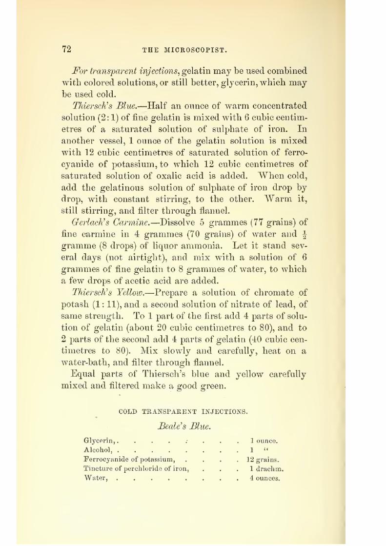
7/29/2019 THE MICROSCOPIST
http://slidepdf.com/reader/full/the-microscopist 76/361
72 THE MICROSCOPIST.
For transjparent injections^ gelatin may be used combined
with colored solutions, or still better, glycerin, whicb maybe used cold.
Thiersch's Blue.—Half an ounce of warm concentrated
solution (2:1) of fine gelatin is mixed with 6 cubic centim-
etres of a saturated solution of sulphate of iron. In
another vessel, 1 ounce of the gelatin solution is mixed
with 12 cubic centimetres of saturated solution of ferro-
cyanide of potassium, to which 12 cubic centimetres of
saturated solution of oxalic acid is added. When cold,
add the gelatinous solution of sulphate of iron drop by
drop, with constant stirring, to the other. Warm it,
still stirring, and filter through fiannel.
GerlacKs Carmine.—Dissolve 5 grammes (77 grains) of
fine carmine in 4 grammes (70 grauis) of water and \
gramme (8 drops) of liquor ammonia. Let it stand sev-
eral days (not airtight), and mix with a solution of 6
grammes of fine gelatin to 8 grammes of water, to which
a few drops of acetic acid are added.
Thiersch's Yellow.—Prepare a solution of chromate of
potash (1 : 11), and a second solution of nitrate of lead, of
same strength. To 1 part of the first add 4 parts of solu-
tion of gelatin (about 20 cubic centimetres to 80), and to
2 parts of the second add 4 parts of gelatin (40 cubic cen-
timetres to 80). Mix slowly and carefully, heat on a
water-bath, and filter through flannel.
Equal parts of Thiersch's blue and yellow carefully
mixed and filtered make a good o-reen.
COLD TRANSPARENT INJECTIONS.
BeaWs Blue.
Glycerin,........ 1 ounce.
Alcohol, 1 "
Ferrocyanide of potassium, .... 12 grains.
Tincture of perchloride of iron, . . . 1 drachm.
Water, . 4 ounces.

7/29/2019 THE MICROSCOPIST
http://slidepdf.com/reader/full/the-microscopist 77/361
MODERN METHODS OF EXAMINATION. 73
Dissolve the ferrocyaiiide in 1 ounce of water and glyc-
erin, and the muriated tincture of iron in another ounce.
Add the latter very gradually to the other, shaking often
then gradually add the alcohol and water.
BcaWs Finest Blue
Price's glj'cerin, . .
Tincture of perchloride of iron,
Ferrocyanide of potassium,
Strong hydrochloric acid,
.
Water,
2 ounces.
10 drops.
3 grains.
3 drops.
1 ounce.
Mix the tincture of iron with 1 ounce glycerin and the
ferrocyanide, after dissolving in a little water, with the
other ounce. Add the iron to the other solution gradu-
ally, shaking well. Lastly, add the water and hydro-
chloric acid. Sometimes about 2 drachms of alcohol are
added.
3Iiiller's Blue.—This is made by precipitation of soluble
Prussian blue from a concentrated solution by means of
90 per cent, alcohol.
Beale's Carmine.—]\Iix 5 grains of carmine with a few
drops of water, and when well incorporated, add 5 or 6
drops of liquor ammonia. To this addJounce of glyc-
erin, and shake well. Another J ounce of glycerin con-
taining 8 or 10 drops of acetic or hydrochloric acid is
gradually added. It is then diluted with | ounce of glyc-
erin, 2 drachms of alcohol, and 6 drachms of water.
Nitrate of Silver Injection.—For demonstrating the struc-
ture of the bloodvessels, the animal is bled, and a solution
of 0.25 to 1 per cent, of nitrate of silver, or a mixture of
gelatin with such a solution, is used.
5. Preservative Fluids.
Canada Balsam.—This is perhaps the most common
medium used. "When an object is not very transparent,
and drying will not injure it, balsam will do very well,

7/29/2019 THE MICROSCOPIST
http://slidepdf.com/reader/full/the-microscopist 78/361
74 THE MICROSCOPIST.
but it is not adapted to moist preparations. Colonel
Woodward, of Washington, uses a solution of dried or
evaporated Canada balsam in chloroform or benzole.
Dammar Varnish.—Dr. Klein and other German his-
tologists prefers this to Canada balsam. Dissolve | to 1
ounce of gum Dammar in 1 ounce of turpentine ; also J
to 1 ounce of mastic in 2 ounces of chloroform. Mix and
filter.
Glycerin.—This fluid is universally useful to the micros-
copist. (See Preparation in Viscid Media, page 65.) Vege-
table and animal substances may be preserved in glycerin,
but if it is diluted, camphor or creasote must be added
to prevent confervoid growths. It is said, however, to
dissolve carbonate of lime.
Gelatin and Glycerin.—Soak gelatin in cold ^vater till
soft, then melt in warm water, and add an equal quantity
of glj^cerin.
Gum and Glycerin.—Dissolve 1| grains of arsenious
acid in 1 ounce of water, then 1 ounce of pure gum arable
(without heat), and add 1 ounce of glycerin.
Deane's Compouyid.—Soak 1 ounce of gelatin in 5 ounces
of water till soft ; add 5 ounces of honey at a boiling heat.
Boil the mixture, and when cool, add tj drops of creasote
in \ ounce of alcohol; filter through flannel. To be used
warm.
Carbolic Acid.—1 : 100 of water is a good preservative.
Thvxdte's Fluid.—To 16 parts of distilled water, add 1
part of rectified spirit and a few drops of creasote ; stir in
a little prepared chalk, and filter. Mix an equal measure
of camphor-water, and strain before using. For preserva-
tion of algae.
Solution of Naphtha and Creasote.—Mix 3 drachms of
creasote with 6 ounces of wood naphtha ; make a thick,
smooth paste with prepared chalk, and add graduall}^,
rubbing in a mortar, 64 ounces of water. Add a few
lumps of camphor, and let it stand several weeks before

7/29/2019 THE MICROSCOPIST
http://slidepdf.com/reader/full/the-microscopist 79/361
MODERN METHODS OP EXAMINATION. 75
pouring off or filtering the clear fluid. Dr. Beale i^ecom-
mends this highly for the preservation of dissections of
nerves and morbid specimens.
Half's Fluid.—As a substitute for Thwaite's fluid in
the preservation of alg?e. 1 grain of alum and 1 of bay
salt to 1 ounce of distilled water.
Goadhy's Solution.—Bay salt, 4 ounces; alum, 2 ounces;
corrosive sublimate, 4 grains; boiling water, 4 pints.
This is the strength most generally useful, although it
maj" be made stronger or more dilute. It is a useful
fluid. If the specimen contain carbonate of lime, the
alum must be left out, and the quantity of salt may be
quadrupled.
Dr. Beale discards all solutions containing salts for
microscopic purposes, as they render the textures opaque
and granular.
Soluble Glass, or a solution of silicate of soda or potash,
or of both, has been proposed, but it is apt to render
specimens opaque.
Chloride of Calcimn in saturated aqueous solution has
been much recommended, especially by botanists.
Acetate of Potash, a nearly saturated solution, is useful
for vegetable preparations and for specimens of animaltissue which have been stained with osmic acid. The
latter do not bear glycerin.
Facinian Fluid.—This is variously modified, but may
consist of corrosive sublimate,! part; chloride of sodium,
2 parts; glycerin, 13 parts; distilled water, 113 parts.
Sometimes acetic acid is substituted for chloride of so-
dium.6. Cements.
Gold Size is recommended by Dr. Carpenter as most
generally useful for thin covers. It is made by boiling
25 parts of linseed oil for three hours with 1 part of red
lead and J of as much umber. The fluid part is then
mixed with yellow ochre and white lead in equal parts,

7/29/2019 THE MICROSCOPIST
http://slidepdf.com/reader/full/the-microscopist 80/361
76 THE MICROSCOPIST.
SO as to thicken it, the whole boiled again, and the fluid
poured off for use.
BelVs Cement is said to be best for glj-cerin specimens.
It appears to be shellac dissolved in strong alcohol.
Brunswick Black is asphaltum dissolved in turpentine.
A little india-rubber dissolved in mineral naphtha is some-
times added.
Canada Balsam in chloroform or Dammar varnish (page
74) is often used as a cement.
Ma?i7ie Glue.—This is most useful in building glass
cells, etc. It consists of equal parts of shellac and india-
rubber dissolved in mineral naphtha by means of heat.
Electrical Cement is made by melting together 5 parts
of rosin, 1 of beeswax, and 1 of red ochre. 2 parts of
Canada balsam added make it more adhesive to glass.
White^ hard Varnish., or gum sandarac, dissolved in
alcohol and mixed with turpentine varnish, is sometimes
colored by lampblack, sealing-wax, etc.
White Zinc Cement.—Oxide of zinc rubbed up with
equal parts of oil of turpentine and 8 parts of solution of
gum Dammar in turpentine of a syrupy consistence, or
Canada balsam, chloroform, and oxide of zinc.
CHAPTER YL
MOUNTING AND PRESERVING OBJECTS FOR THE MICROSCOPE.
For the permanent preservation of specimens, various
means are employed, according to the nature of the object
and the particular line of investigation desired. Few, if
any, objects show all their peculiarities of structure or
adaptation to function, and for scientific work it is often
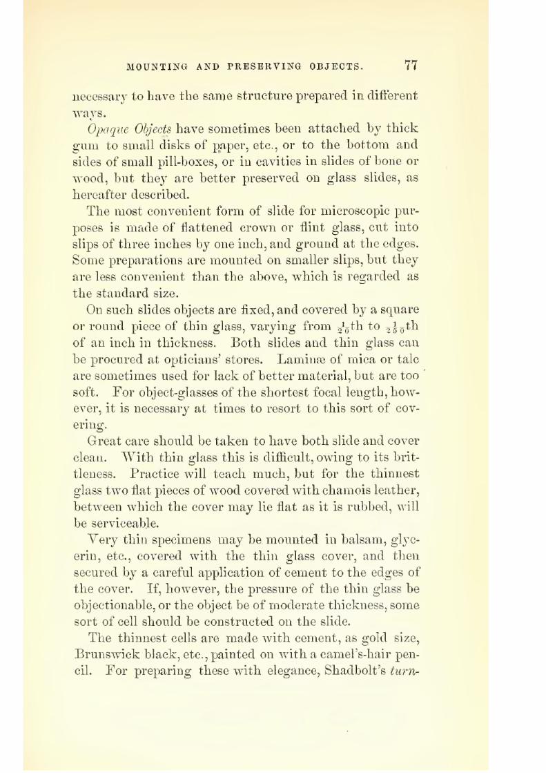
7/29/2019 THE MICROSCOPIST
http://slidepdf.com/reader/full/the-microscopist 81/361
MOUNTING AND PRESERVING OBJECTS. 77
necessary to have the same structure prepared in difterent
Opaque Objects have sometimes been attached by thick
gum to small disks of paper, etc., or to the bottom and
sides of small pill-boxes, or in cavities in slides of bone or
wood, but they are better preserved on glass slides, as
hereafter described.
The most convenient form of slide for microscopic pur-
poses is made of flattened crown or flint glass, cut into
slips of three inches by one inch, and ground at the edges.
Some preparations are mounted on smaller slips, but they
are less convenient than the above, which is regarded as
the standard size.
On such slides objects are fixed, and covered by a square
or round piece of thin glass, varying from ggth to 05 o^^
of an inch in thickness. Both slides and thin glass canbe procured at opticians' stores. Laminae of mica or talc
are sometimes used for lack of better material, but are too
soft. For object-glasses of the shortest focal length, how-
ever, it is necessary at times to resort to this sort of cov-
ering.
Great care should be taken to have both slide and cover
clean. "With thin glass this is difiicult, owing to its brit-
tleness. Practice will teach much, but for the thinnest
glass two flat pieces of wood covered with chamois leather,
between which the cover may lie flat as it is rubbed, will
be serviceable.
Very thin specimens may be mounted in balsam, glyc-
erin, etc., covered with the thin glass cover, and then
secured by a careful application of cement to the edges of
the cover. If, however, the pressure of the thin glass be
objectionable, or the object be of moderate thickness, some
sort of cell should be constructed on the slide.
The thinnest cells are made with cement, as gold size,
Brunswick black, etc., painted on with a camel's-hair pen-
cil. For preparing these with elegance, Shadbolt's turn-

7/29/2019 THE MICROSCOPIST
http://slidepdf.com/reader/full/the-microscopist 82/361
78 THE MICROSCOPIST.
table has been contrived (Fig. 36). The slide is placed
between the springs, and while rotated, a ring of varnish
of suitable breadth is made on the glass.
A piece of thin glass (or even of thick glass) may be
perforated and cemented to the slide with marine glue by
Fig. 36.
Sbadbolt's Turntable for making Geiuent-Cells.
the aid of heat; or vulcanite, lead, tin, gutta percha, etc.,
may be made into a cell in a similar way as seen in Fig.
37.
The perforation of thin glass may be easily performed
by cementing it over a hole in a brass plate, etc., with
marine glue, and punching it through with the end of a
Fig. 37.
Cell of Glass, Vulcanite, etc.
file. The edges may then be filed to the size of the hole,
and the glass removed by heating the brass. Thicker
glass may be bored with a file by moistening it with
turpentine.
Dry objects^ especially if they are transparent, as dia-
toms, thin sections of bone, crystals, etc., may be attached
to the slide with Canada balsam, etc., covered with thin

7/29/2019 THE MICROSCOPIST
http://slidepdf.com/reader/full/the-microscopist 83/361
MOUNTING AND PRESERVING OBJECTS. 79
glass, whicli should be cemented at ttie edges, and gummed
over all a strip of colored or lithographed paper, in whichan aperture has been made with a punch.
Mounting in Balsam, or Dammar Varnish is suitable for
a very large proportion of objects. It increases the trans-
parency of many structures, filling up interstices and cavi-
ties, and giving them a smooth, beautiful appearance.
Yer}^ delicate tissues, as fine nerves, etc., are rendered in-
visibleby
it,
and require otherfluids,
as gl3"cerin.
Before mounting in balsam, the object should be thor-
oughlj^ dry, otherwise a milky appearance will result. It
should then be placed in oil of cloves or of turpentine to
remove greasiness and increase the transparency. A clean
slide, warmed over a spirit-lamp or on a hot plate, should
then have a little balsam placed on its centre, and the
object carefully lifted from the turpentine is put into the
balsam and covered with another drop. The slide should
then be gently warmed, and any air-bubbles pricked with
a needle-point or drawn aside. The thin glass cover should
be warmed and put on gently, in such a way as to lean
first on one edge and fall gradually to a horizontal posi-
tion. The slide may be w- armed again, and the superflu-
ous balsam pressed from under the cover by the pressure
of a clean point upon it.
If the object is full of large air-spaces and is not likely
to be injured by heat, the air may be expelled by gently
boiling it in the balsam on the slide. If the object be one
which will curl up, or is otherwise injured by heat, the
air-pump must be resorted to. A cheap substitute for the
air-pump may be made by fitting a piston into a tolerably
wide glass tube closed at one end. The piston should
have a valve opening outwards. The preparation in bal-
sam may be placed at the bottom of the tube, and a few
strokes of the piston w^ill exhaust the air.
To fill a deep cell wdth Canada balsam, it may be well
to fill it first wdth turpentine and place the specimen in

7/29/2019 THE MICROSCOPIST
http://slidepdf.com/reader/full/the-microscopist 84/361
80 THE MICROSCOPIST.
it. Then pour in the balsam at one end, the slide being
inclined so that the turpentine may run out at the other.
Lay the cover on one edge of the cell and gradually lower
it till it lies flat. In this way air may be excluded.
The solution of balsam in chloroform needs no heat,
and has little liability of air-bubbles.
The excess of balsam round the edge of the glass cover
may be removed with a knife and cleaned with turpentine
or benzine, etc.
For animal tissues, the oil of cloves is sometimes used
instead of turpentine to increase the transparency, and a
wet preparation, as a stained or injected specimen, may
be mounted in balsam or Dammar by flrst placing it in
absolute alcohol to extract the water, then transferring
to oil of cloves or turpentine, and lastly, to the balsam.
In a reverse order, a specimen from balsam may be cleaned
and mounted in fluid.
Mounting in Fluid is necessary for the preservation of
the most delicate tissues and such as may be injured by
Fig. 38.
Spring Clip.
drying. Glycerin is perhaps the most generally useful
fluid. (See Preservative Fluids^ page 73.)
For mounting in fluid, it is safer to have a thin cell of
varnish prepared first than to risk the running in of the
cement under the cover, as will be likely to occur other-
wise.
The air-pump is sometimes needed in mounting in fluid
to get rid of air-bubbles. A spring clip (Fig. 38) is also

7/29/2019 THE MICROSCOPIST
http://slidepdf.com/reader/full/the-microscopist 85/361
MOUNTING AND PRESERVING OBJECTS. 81
a useful instrument for making moderate pressure on the
glass cover until the cement on its edge is dry. A drop-
ping-tube with a bulbous funnel, covered with thin india-
rubber, for taking up and dropping small quantities of
fluid, will also be of service.
Superfluous fluid may be removed from the edge of the
cover b}^ a piece of blotting-paper, care being used not to
draw away the fluid beneath the cover.
As soon as objects are mounted, the slides should be
labelled before cementing is finished, otherwise time will
be lost in searching for a particular object among others,
or the name may be forgotten.
Boxes of wood or of pasteboard, with grooved racks at
the sides, are occasionally used for preserving a collection
of specimens. It is better, however, to have a cabinet
with drawers or trays so that the specimens may lie flat,
with their ends towards the front of the drawer. A piece
of porcelain on the end of the drawer is convenient for
the name of the class of objects contained, to be written
on with lead-pencil.
Collecting Oljjects.—The methods pursued by naturalists
generally will sufiice for a large proportion of the objects
which are matters of microscopic inquiry, but there areothers which, from their minuteness, require special search.
Many fresh-water species of microscopic organisms inhabit
pools, ditches, and streams. Some attach themselves to
the stems and leaves of aquatic plants, or to floating and
decajung sticks, etc. Others live in the muddj^ sediment
at the bottom of the water. A 'pond stick has been con-
trived for the collection of such organisms, consisting oftwo lengths, sliding one within the other, so that it may
be used as a walking-cane. In a screw socket at one end
may be placed a curved knife for cutting portions of plants
which contain microscopic parasites ; or a screw collar for
carrying a screw-topped bottle, which serves to bring up
a sample of liquid ; or it may have a ring for a muslin net.
6

7/29/2019 THE MICROSCOPIST
http://slidepdf.com/reader/full/the-microscopist 86/361
82 THE MICROSCOPIST.
The net should be confined by an india-rubber band in a
groove, so as to be slipped off readily and emptied into abottle. The collector should have enough bottles to keep
organisms from each locality separate, and when animal-
cules are secured enough, air should be left to insure their
safety.
Marine organisms may be obtained in a similar way if
they inhabit the neighborhood of the shore, but others
can only be secured by means of the dredge or tow-net.
The latter may be of fine muslin sewn to a wire ring of
twelve inches diameter. It may be fastened with strings
to the stern of a boat, or held by a stick so as to project
from the side. For the more delicate organisms, the boat
should be rowed slowly, so that the net may move gently
through the water. Firmer structures may be obtained
by attaching a wide-mouthed bottle to the end of a netmade conical, and double, so that the inner cone may act
as a valve. The bottle may be kept from sinking by a
piece of cork. Such a net may be fixed to the stern of a
vessel, and drawn up from time to time for examination.
Minute organisms may be examined on the spot by
fishing them out of the bottle with a pipette, or small
glass tube, and placing them on a slide. A Coddingtonor other pocket lens will suflice to show which are desir-
able for preservation.
Many of the lower animals and plants may be kept
alive in glass jars for some time. Frogs, etc., may be
kept under wire covers with a large piece of moist sponge.
Aquaria of various sorts may be procured and stocked
at small expense, and will afi:brd a constant source of in-
struction. For fresh-water aquaria the bottom of the jar,
etc., should be covered with rich black earth, made into
a paste, and this should be surmounted with a layer of
fine washed sand. Roots of Valisneria, Anacharis^ or
Ghara may then be planted in the earth and the vessel
filled with water. As soon as the water is clear, put a

7/29/2019 THE MICROSCOPIST
http://slidepdf.com/reader/full/the-microscopist 87/361
MOUNTING AND PRESERVING OBJECTS. 83
few fresh-water molluscs in to keep down the growth of
coiifervne, especially such as feed on decayed vegetable
matter, as Flanorbis carinaius, Paludina vivipara^ or A'm-
phibia glutinosa. When bubbles of oxj-gen gas appear,
fish, water insects, etc., may be introduced.
Marine aquaria require more skill than those for fresh
water, but for temporary purposes, the plan described by
Mr. Highley, in Dr. Beale's How to Work ivith the Micro-
scope^ is excellent. He fills a number of German beakerglasses with fresh sea-water, and places them in a sunny
window. He then drops in each one or two limpet shells
from which the animals have been removed, and upon
which small plants of Enteromorpha and Ulva are growing.
In a short time the sides of the jars next the light become
coated with spores. He keeps the other sides clean with
a piece of wood or sponge, so as to observe the smallmarine animals which may now be placed in the beakers.
In this way a collection will keep healthy for months.
After the sides are covered with spores, the sea-weeds
may be removed, and the jars placed on a table at such a
distance from the window that the light impinges only
on the coated half, taking care that there is sufficient light
to stimulate the spores to throw off l)ubbles of oxygendaily.
Prawns, fish, actinise, etc., may be fed on shreds of beef
which has been pounded and dried, and then macerated
in sea-water for a few minutes. All dead animals, slime,
or effete matter should be removed by wooden forceps,
etc , as soon as noticed.

7/29/2019 THE MICROSCOPIST
http://slidepdf.com/reader/full/the-microscopist 88/361
84 THE MICROSCOPIST.
CHAPTER YIL
THE MICEOSCOPE IN MINERALOGY AND GEOLOGY.
Microscopic examination of minute fossil organisms, as
Diatoms, Foraminifera, spicules of sponge, etc., has long
been a subject of interest. Latterly, however, the micro-
scope has been found to be essential to the study of phys-
ical geology and petrology. How many crude and verbose
theories respecting cosmogony will disappear by this means
of investigation time must reveal, but the animal nature
of the Eozoon Canadense found in the Serpentine Lime-
stone of the Lauren tian formation of Canada, parallel with
the Fundamental Gneiss of Europe, and the discovery by
Mr. Sorby'"' of minute cavities jfiUed with fluid in quartz
and volcanic rocks, are indications that speculations based
upon a merely external or even chemical examination of
rock structures are immature and inadequate.
The systematic study of raicroscopic mineralogy and
geology will require a large outlay of time and patience,
and the field is one which is scarcely trodden. The plan
of this work will only permit a brief outline, sufiicient to
aid a beginner, and indicating the value and the methods
of minute investigation.
Preparation of S-pecimens.—Examination of the outer
surface of a mineral specimen, viewed as an opaque body
with a low power and by condensed light, is sometimes
useful. The metals and their alloys, with most of their
combinations with sulphur, etc , admit of no other method.
Occasionally, as in iron and steel, the microscopic structure
is best seen by polishing the surface, and then allowing
the action of very dilute nitric acid. Mr. Forbesf states
* See Beale's How to Work with the Microscope.
f The Microscope in Geology, Popular Science Keview, No. 25.

7/29/2019 THE MICROSCOPIST
http://slidepdf.com/reader/full/the-microscopist 89/361
THE iMICROSCOPE IN MINERALOGY AND GEOLOGY. 85
that many vitreous specimens (quite transparent) show no
trace of structure until the surface has been carefully acted
on by hydrofluoric acid.
It is generally necessary to have the specimens flat and
smooth, and thin enongh to transmit light. Sometimes
fragments may be thin enough to show structure when
mounted in balsam, as in tbe case of quartz, obsidian, pitch-
stone, etc., but usually thin sections must be ground and
polished.
Chip off a fragment of the rock as flat and thin as pos-
sible, or cut with a lapidary's wheel, or a toothless saw of
sheet-iron with emery. Grind down the specimen on an
iron or pewter plate in a lathe until perfectly flat. Then
grind with finer emerj^ on a slab of fine-grained marble or
slate, and finish with water on a fine hone, avoiding all
polishing powders or oil. When perfectly smooth, cementthe specimen on a square of glass with Canada balsam,
and grind the other side until as thin as necessary, finish
as before, remove it from the glass, and mount on a glass
slide in balsam.
In this way, most silicates, chlorides, fluorides, carbo-
nates, sulphates, borates, many oxides, sulphides, etc., may
be prepared for examination by transmitted light. Verysoft rocks may be soaked in turpentine, then in soft balsam,
and afterwards heated until quite hard. The deep scratches
on hard minerals, like quartz, left by the use of coarse
emery, may be removed by using fine emery paper held
flat on a piece of plate glass, and Anally polished with
rouge on parchment. Perhaps oxide of chromium from
its hardness will be found the best polishing material.Crystals of soluble salts may be ground on emery paper
and polished with rouge. Sometimes much may be learned
by acting on one side only of a specimen with dilute acid.
Examination of Specimens.—The object of microscopic
examination of minerals is to determine not only the nature
of the material of which they are composed, but also, and

7/29/2019 THE MICROSCOPIST
http://slidepdf.com/reader/full/the-microscopist 90/361
86 THE MICROSCOPIST.
chiefly, their structure, whether homogeneous, derived
from the debris of previous rocks, or from the agency of the
organic world. Ordinary mineralogical characteristics, as
to hardness, specific gravity, color, lustre, form, cleavage,
and fusibility, and above all, chemical composition, may
suffice to show the material, but the microscope will give
valuable assistance to this end, and is essential to a knowl-
edge of structure.
CryslalUne Forms.—The laws of crystallography teach
that each chemical combination corresponds to a distinct
relation of all the angles which can possibly arise from
the primary form, so that the angular inclination of the
facets of a crystal is a question of importance. This can
be ascertained by a microscope having a revolving stage,
properly graduated, or by the use of a goniometer^ which
is a thread stretched across the focus of the eye-lens, and
attached to a movable graduated circle and vernier. The
eye-piece attached to the polariscope of Hartnack is thus
arranged, so as to act also as a goniometer.
Crystals are assumed to possess certain axes, and the
form is determined by the relation of the plane surface to
these axes. Although the forms of crystals are almost
infinitely varied, they may be classified into seven crystal-
lographic systems,
1. The Regular Cubic or 31onoinetric System (Fig. 39).
These crystals are symmetrical, about three rectangular
axes. The simplest forms are the cube and octahedron.
Examples, diamond, most metals, chloride of sodium, fluor
spar, alum.
2. The Quadratic or Dimetric System (Fig. 40).—Crystals
symmetrical, about three rectangular axes, but only two
axes of equal length. Examples, sulphate of nickel, tung-
state of lead, and double chloride of potassium and cop-
per.
3. Hexagonal or Rhomhohedral Systeyn (Fig. 41).—Crys-
tals with four axes ; three equal in length, in one plane,
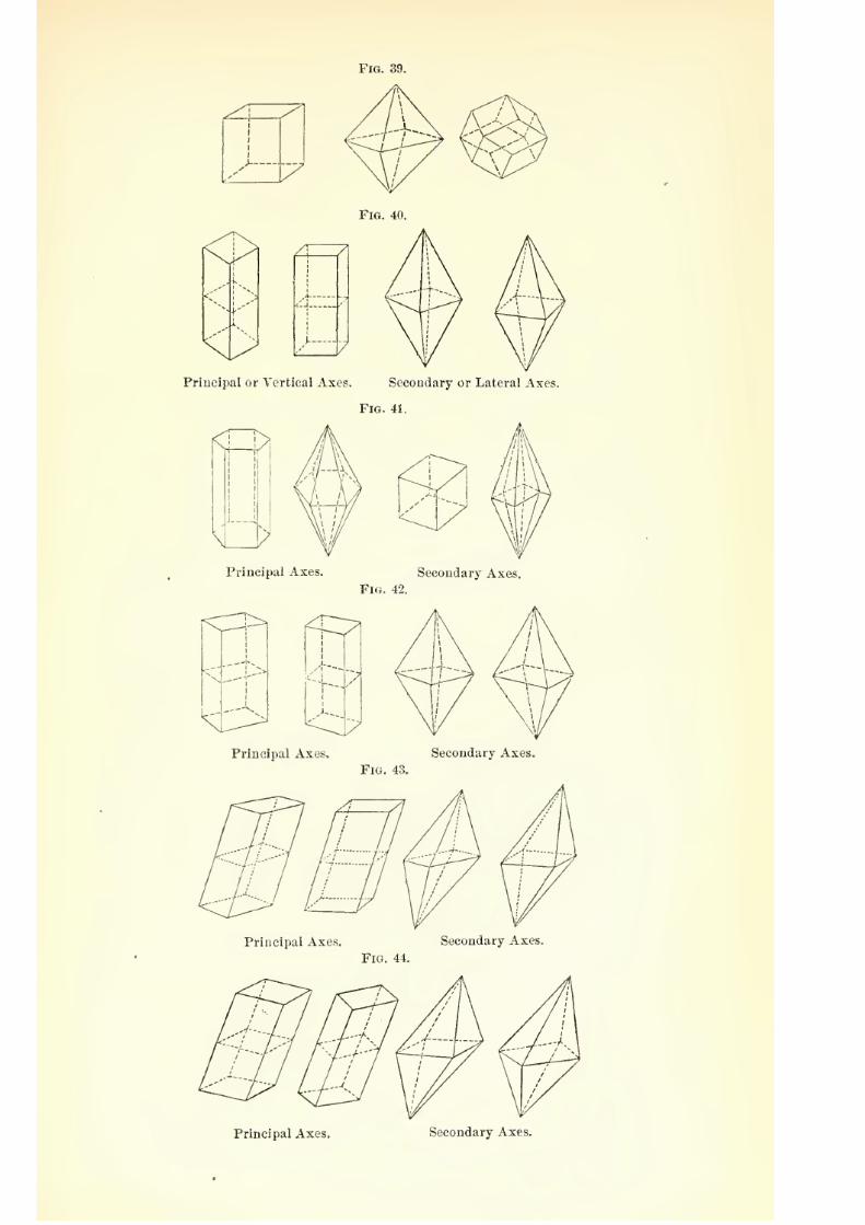
7/29/2019 THE MICROSCOPIST
http://slidepdf.com/reader/full/the-microscopist 91/361
Principal or Vertical Axes. Secondary or Lateral Axes.
Fig. 41.
Principal Axes. Secondary Axes.
Fig. 42.
^C^
Principal Axes, Secondary Axes.
Fig. 43.
Principal Axes. Secondary Axes.
Fig. 44.
Principal Axes. Secondary Axes.
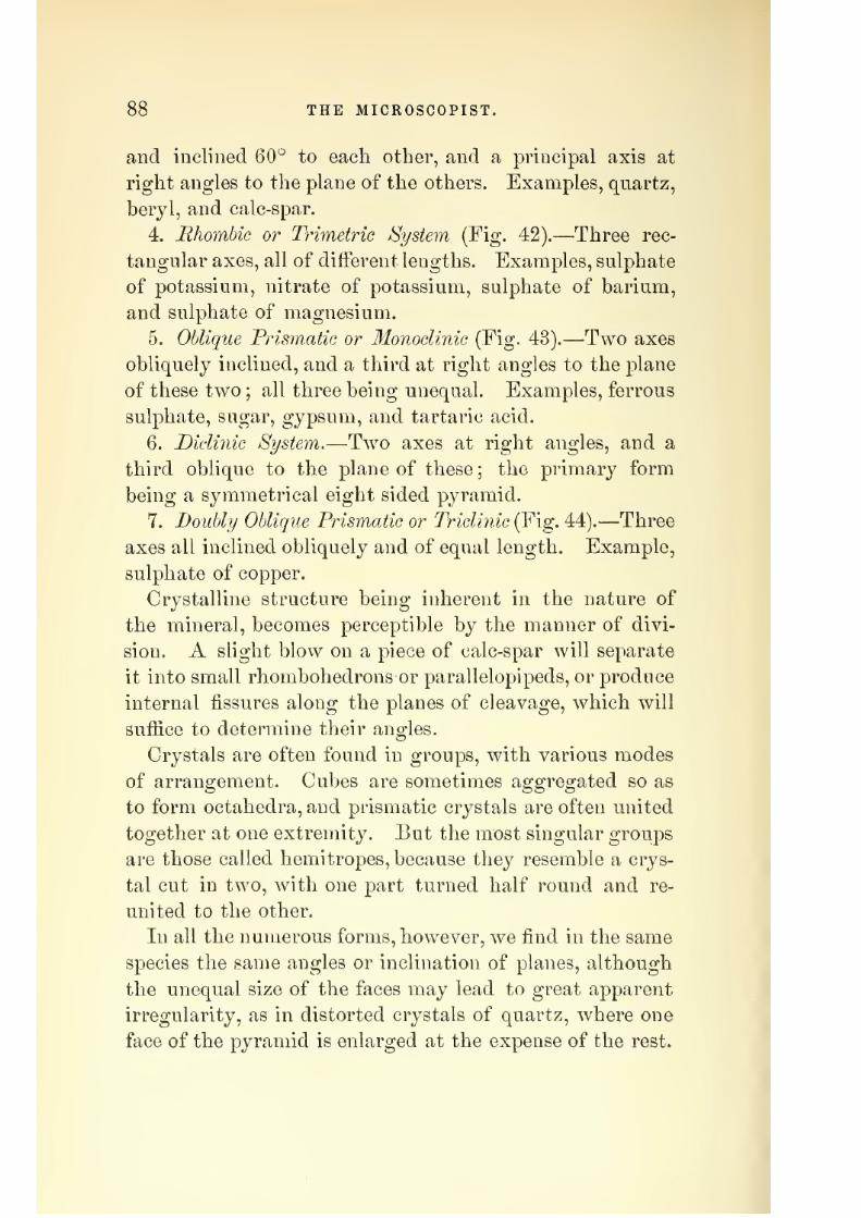
7/29/2019 THE MICROSCOPIST
http://slidepdf.com/reader/full/the-microscopist 92/361
»5 THE MICROSCOPIST.
and inclined 60*^ to each other, and a principal axis at
right angles to the plane of the others. Examples, quartz,
beryl, and calc-spar.
4. Rhombic or Trimetric /System (Fig. 42).—Three rec-
tangular axes, all of different lengths. Examples, sulphate
of potassium, nitrate of potassium, sulphate of barium,
and sulphate of magnesium.
5. Oblique Prismatic or Monoclinic (Fig. 43). Two axes
obliquely inclined, and a third at right angles to the plane
of these two ; all three being unequal. Examples, ferrous
sulphate, sugar, gypsum, and tartaric acid.
6. Diclinic System.—Two axes at right angles, and a
third oblique to the plane of these ; the primary form
being a symmetrical eight sided pyramid.
7. Doubly Oblique Prismatic or Triclinic (Fig. 44).—Three
axes all inclined obliquely and of equal length. Example,sulphate of copper.
Crystalline structure being inherent in the nature of
the mineral, becomes perceptible by the manner of divi-
sion. A slight blow on a piece of calc-spar will separate
it into small rhombohedrons or parallelopipeds, or produce
internal fissures along the planes of cleavage, which will
suffice to determine their angles.
Crystals are often found in groups, with various modes
of arrangement. Cubes are sometimes aggregated so as
to form octahedra, and prismatic crystals are often united
together at one extremity. But the most singular groups
are those called hemitropes, because they resemble a crys-
tal cut in two, with one part turned half round and re-
united to the other.
In all the numerous forms, however, we find in the same
species the same angles or inclination of planes, although
the unequal size of the faces may lead to great apparent
irregularity, as in distorted crystals of quartz, where one
face of the pyramid is enlarged at the expense of the rest.

7/29/2019 THE MICROSCOPIST
http://slidepdf.com/reader/full/the-microscopist 93/361
THE MICROSCOPE IN" MINER ALOQY AND GEOLOGY. o9
An apparent distortion may also be produced by an oblique
section.
The following examples may be of service, as showing
the value of angular measurement in minerals:
Quartz. Ehombohedral system. Inclination of two
adjoining faces 9-1:° 15'.
Felspar. Monoclinic. Cleavage planes at right angles.
Albite or soda felspar. Triclinic. Angle 93° 36'.
Mica. Oblique prisms.
Magnesian mica. Right, rhombic, or hexagonal prisms.
Garnet. Dodecahedrons or trapezohedrons.
Idocrase. Square prisms.
Epidote. Oblique prisms.
Scapolite. Square and octagonal prisms.
Andalusite. Prisms of 90° 44'.
Staurotide. Ehombic prisms of 129° 20'.
Tourmaline. Three, six, nine, or twelve-sided prisms.
Topaz. Ehombic prisms of 124'^ 19'.
Beryl. Six-sided prisms.
Hornblende. Monoclinic. 124° 30'.
Augite or pyroxene. Monoclinic. 87° 5'.
Calcite or carbonate of lime. Forms various, but 105°
5' between the cleavage faces.
Magnesite. Angle 107° 29'.
Dolomite. 106" 15'.
Gypsum. Monoclinic.
Crystals within Crystals.—Many specimens which appear
perfectly homogeneous to the naked eye are shown by the
microscope to be very complex. The minerals of erupted
lavas are often full of minute crystals, leading to veryanomalous results of chemical analysis. Some care is
needed at times to distinguish such included minerals
from cavities filled with fluid. The use of polarized light
will sometimes determine this point.
Cavities in Crystals.—Mr. Sorby has shown that the
various cavities in minerals containing air, water, glass,

7/29/2019 THE MICROSCOPIST
http://slidepdf.com/reader/full/the-microscopist 94/361
90 THE MICROSCOPIST.
or stone will often indicate under what conditions the
rock was formed. Thus crj^stals with water cavities were
formed from solution ; those with stone or glass cavities
from igneous fusion ; those with both kinds hj the com-
bined influence of highly heated water and melted rock
under great pressure; while those that contain no cavi-
ties were formed very slowly, or from the fusion of homo-
geneous substance.
Use of Polarized Light.—Mr. Sorby states that the
action of crystals on polarized light is due to their double
refraction, which depolarizes the polarized beam, and
gives rise to colors by interference if the crystal be not
too thick in proportion to the intensity of its power of
double refraction. This varies much, according to the
position in which the crj^stal is cut, yet we may form a
general opinion, since it is the intensity and not the char-
acter of the depolarized light which varies according to
the position of the crystal. There are two axes at right
angles to each other, and when either of them is parallel
to the plane of polarization, the crystal has no depolariz-
ing action, and if the polarizing and analyzing prisqis are
crossed, it looks black. On rotating the crystal or the
plane of polarization, the intensity of depolarizing action
increases until the axes are at 45°, and then diminishes
till the other axis is in the plane. If, therefore, this takes
place uniformly over a specimen, we know that it has one
simple crystalline structure, but if it breaks up into de-
tached parts, we know it is made up of a number of sepa-
rate crystalline portions.
The definite order that may occur in the arrangement
of a number of crystals may indicate important differences.
Some round bodies, for example, like oolitic grains, have
been formed by crystals radiating from a common nu-
cleus; whilst others, as in meteorites, have the structure
of round bodies which crystallized afterwards.
Sir D. Brewster discovered that many crystals have

7/29/2019 THE MICROSCOPIST
http://slidepdf.com/reader/full/the-microscopist 95/361
THE MICROSCOPE IN MINERALOGY AND GEOLOGY. 91
two axes of double refraction, or rather axes around which
double refraction occurs. Thus nitre crystallizes in six-
sided prisms, with angles of about 120°. It has two axes
of double refraction inclined about 2|° to the axes of the
prism, and o'^ to each other, so that a piece cut from such
a crystal perpendicular to the axes, shows a double system
of rings when a ray of polarized light is transmitted.
"When the line connecting the axes is inclined 45° to the
plane of polarization, a cross is seen, which gradually
assumes the form of two hyperbolic curves on rotating
the specimen If the analyzer be revolved, the black cross
will be reijlaced by white, the red rings by green, the yel-
low by indigo, etc. These rings have the same colors as
thin plates, or a sj'stem of rings round one axis. Mica
has two sets of rings, with the angle between the axes of
60° to 75°. Magnesian mica gives an angle of 5° to 20°.
jDetennination of the Origin of Rock Specimens.—Mr.
Forbes has shown that the primary or eruptive rocks,
consisting chiefly of crystallized silicates, with small
quantities of other minerals, are developed as more or
less perfect crystals at all angles to one another, indicat-
ing the fluid state of the mass at some previous time.
The secondary or sedimentary rocks consist of rocksformed by the immediate products of the breaking up of
eruptive rocks, or are built of the debris of previous erup-
tive or sedimentary rocks, or composed of extracts from
aqueous solution by crystallization, precipitation, or the
action of organic life. The accompanying figures, selected
from Mr. Forbes's article in the Popular Science Heview,
well illustrate this method of investigation. Plate II,
Fig. 45, is a section of lava from Vesuvius, magnified
twelve diameters, showing crystals of augite in a hard
gray rock. Plate II, Fig. 46, is a volcanic rock from
Tahiti, consisting of felspar, with olivine and magnetic
oxide of iron, and numerous crystals of a pj^roxenic min-
eral. Plate II, Fig. 47, is pitchstone from a dyke in new
7/29/2019 THE MICROSCOPIST
http://slidepdf.com/reader/full/the-microscopist 96/361
92 THE MICKOSCOPIST.
red sandstone, magnified seventy-five diameters. Exter-
nally it resembles dirty green bottle-glass, but shows in
the microscope an arborescent crystallization of a green
pyroxenic mineral in a colorless felspar base. Plate II,
Fig. 48, shows auriferous diorite from Chili, consisting of
felspar, with hornblende and crystals of iron pyrites, mag-
nified thirty diameters. Plate II, Fig. 49, is a section of
granite from Cornwall, with crystals of orthoclase, hexag-
onal crystals of brown mica, and colorless quartz, which
a higher power shows to contain fluid cavities, magnified
twenty-five diameters. Plate II, Fig. 50, a volcanic rock
from Peru, composed of felspar, dark crystals of augite,
hexagonal crystals of dark mica, and a little magnetic
oxide of iron, magnified six diameters. Plate II, Fig.
51, lower silurian roofing-slate, cut at right angles to the
cleavage, showing that the latter is not due to crystalline
but to mechanical arrangement, magnified two hundred
diameters. Plate II, Fig. 52, is an oolitic specimen from
Peru, regarded as an eruptive rock by D'Orbigny, but
shown in the microscope to be a mere aggregation of
sand, etc., without the crystalline character of eruptive
rocks.
Materials of Organic Origin.—Rocks and strata derived
from plants or animals may be arranged in four groups
1. The calcareous, or those of which limestones have been
formed, as corals, corallines, shells, crinoids, etc. 2. The
siliceous, which have contributed to the silica, and may
have originated flints, as the microscopic shields of dia-
toms and siliceous spicule of sponges. 3. The phosphatic,
as bones, excrement, etc. Fossil excrements are called
coprolites, and those of birds in large accumulations,
guano. 4. The carbonaceous, or those which have afi'orded
coal and resin, as plants.
To examine the structure of coal, it is necessary to have
very thin sections. From its friability, this is a process
of great difliculty. The 31icrograijhic Dictionary recom-

7/29/2019 THE MICROSCOPIST
http://slidepdf.com/reader/full/the-microscopist 97/361
PJj^'E TT.
He. i5 12
^^H |^^^^^ 'y yf ^^H
^V y^iuj*^
1
^^,r^'/
#1
t%^
/ ^J^^^^ n .1 flHI
1^ >% ]\*^ H
Tig. 46 > 12
Fig- ^r'
Tie- 51x200
^^^^^^.50 ^ 6
1^^^^s^3^%^^l^^j^
ig. 52 X 30

7/29/2019 THE MICROSCOPIST
http://slidepdf.com/reader/full/the-microscopist 98/361

7/29/2019 THE MICROSCOPIST
http://slidepdf.com/reader/full/the-microscopist 99/361
THE MICROSCOPE IN MINERALOGY AND GEOLOGY. 93
mends the maceration of the coal for about a week in a
solution of carbonate of potassium, when thin slices maybe cut with a razor. These should be gently heated in
nitric acid, and when they turn j^ellow, washed in cold
water and mounted in glycerin, as spirit and balsam ren-
der them opaque. Sometimes, as in anthracite, casts of
vegetable fibres may be obtained in the ash after burning
and mounted in balsam.
The lignites of the tertiary period show a vegetal^le
structure similar to the woods of the present period, but
the older coal of the palaeozoic series is a mass of decom-
posed vegetable matter chiefly derived from the decay of
coniferous wood, analogous to the araucarise, as is seen
from the peculiar arrangement of the glandular dots on
the woody fibres. Traces of ferns, sigillaripe, calamites,
etc., such as are preserved in the shales and sandstones ofthe coal period, are also met with, but their structure has
not been preserved.
Professor Heer, of Zurich, has described and classified
several hundred species of fossil plants from the miocene
beds of Switzerland by the outlines, nervation, and micro-
scopic structure of the leaves and character of sections of
the wood. Several hundred kinds of insects also havebeen found in the same strata. It is remarkable that a
great part of this fossil flora is such as is now common
to America, as evergreen oaks, maples, poplars, ternate-
leaved pines, and the representatives of the gigantic
sequoias of California.
The researches of palaeontologists have brought to light
nearlytwo
thousand species of fossil plants, of which
about one-half belong to the carboniferous and one-fourth
to the tertiary formations.
The rapid multiplication of the minute microscopic
organisms called diatoms^ is such that Professor Ehren-
berg afiirms it to have an important influence in blocking
up harbors and diminishing the depth of channels. These

7/29/2019 THE MICROSCOPIST
http://slidepdf.com/reader/full/the-microscopist 100/361
94 THE MICROSCOPIST.
organisms, now generally regarded as plants, are exceed-
ingly small, and are usually covered by loricEe or shields
of pure silica, beautifully marked, as if engraved. These
loric?e or shells having accumulated in great quantities,
have given rise to very extensive siliceous strata. Thus
the "infusorial earth" of Virginia, on which Richmond
and Petersburg are built, is such a deposit eigliteen feet
in thickness. The polishing material called Tripoli, and
Fig. 53.
Fossil Diatomacese, etc., from Mourne Mountain. Ireland: a, a, a, Gaillondla (Melo-
seira) procera, and G. granulata; rf, d, d, G. biseriata (side view); 6, 6, Surirella plicata
c, S, crat.icula; k, S, caledonica; e, Goniplionema gracile; /, Cocoonenia fusidium; g,
Tabellaria vulgaris; h, I'inuularia dactylus; i, P. nobilis; /, Synedra ulna. (From
Carpenter.)
the deposit called in Sweden and Xorway herg-mehl or
mountain flour, because used in times of scarcity to mix
with ilour for bread, are similarly composed, strata of
white rock in the anthracite region of Pennsjdvania, and
from the sides of the Sierra iS'evada and Cascade rang-es
in California and Oregon, have also been found to consist
of such remains (Fig. 53).

7/29/2019 THE MICROSCOPIST
http://slidepdf.com/reader/full/the-microscopist 101/361
THE MICROSCOPE IN MINERALOGY AND GEOLOGY. 95
The lowest type of animal life, consisting of minute
portions of sarcode or animal jelly, having the power of
putting forth prolongations of the body at will, contain
some forms which cover themselves with shells, usually
many-chambered, of carbonate of lime. From the pores
in these shells, through which tlie root-like processes of
sarcode are protruded, they are called Foraminifera.
Fig. 54.
Fossil Polycystina, etc., from Barbadoes: o, Podocyrtis mitra; 6, Rliabdolithus scep-
trum;
c, Lychnocanium falciferum; d, Encyrtidium tubulus; e, Flustrella concentrica;
/, Lycbnocanium lucerna; g, Encyrtidium elegan's ; h, Dictyospyris clathrus; ?, Encyr-
tidium mongolfieri; A;, Steplianolithis spinescens; /,S, nodosa; «i, Lithocyclia ocellus;
71, Cephalolithis sylvina; o, Podocyrtis coihurnata; p, Rhabdolithes pipa. (From Car-
penter.)
Another class, the Polycystina^ secrete a siliceous shell,
usually of one chamber. The accumulations of the Fo-
raminifera have formed our chalk beds, while the Polycys-
tina have contributed to siliceous strata, like the Diato-
macece (Fig. 54).
The origin of white chalk strata has been illustrated

7/29/2019 THE MICROSCOPIST
http://slidepdf.com/reader/full/the-microscopist 102/361
96 THE MICROSCOPIST.
by the deep-sea soundings made preparatory to laying
the telegraph cable across the Atlantic Ocean. Professor
Huxley found the mud composing the floor of the ocean
to consist of minute Rhizopods or Foraminifera, of the
genus Globigeri?ia, together with Polycystina and Dia-
toms, and a few siliceous spiculpe of sponges. These were
connected by a mass of living gelatinous matter, to which
he has given the name of Bathyhius^ and which contains
minute bodies termed Coccoliths and Coccospheres, which
have also been detected in fossil chalk. It is said that
95 per cent, of the mud of the ISTorth Atlantic consists of
Globigerina shells.
To examine Foraminifera in chalk, rub a quantity to
powder in water with a soft brush, and let it settle for a
variable time. The first deposits will contain the larger
specimens, with fragments of shell, etc. ; the smaller fall
next, while the amorphous particles suspended in the
water may be cast aside. After drying such specimens
as may be selected by the use of a dissecting microscope
or Coddington lens, etc., they may be mounted in balsam.
The flint found in chalk often contains Xanthidia,
which are the sporangia of Desmidiacese, as well as speci-
mens of sponge, Foraminiferal shells, etc. They must be
cut as other hard minerals.
There are other deposits besides chalk which are seen
by the microscope to consist of minute shells, corals,
etc. A section of oolitic stone will often show that each
rounded concretion is composed of a series of concentric
spheres inclosing a central nucleus which may be a forami-
niferal shell. The green sand formation is composed of
the casts of the interior of minute shells which have them-
selves entirely disappeared. The material of these casts,
chiefly silex colored with iron, has not only filled the cham-
bers of the shells, but has penetrated the canals of the
intermediate skeleton.
The more recent discovery by Drs. Dawson and Carpen-

7/29/2019 THE MICROSCOPIST
http://slidepdf.com/reader/full/the-microscopist 103/361
THE MICKOSCOPE IN MINERALOGY AND GEOLOGY. 9T
ter of the organic nature of those serpentine limestones
in the Laurentian formations of Canada and elsewhere,,
which are products of the growth of the gigantic forami-
nifei'al Eozoon Canadense^ over immense areas of the
ancient sea-bottom, is one of still greater interest both to
the student of Geolog}^ and of Biology.
This immense rhizopod appears to have grown one layer-
over another, and to have formed reefs of limestone as do
the living coral-polj^ps. Parts of the original skeleton,,
consisting of carbonate of lime, are still preserved, while
certain interspaces have been filled up with serpentine and
white augite.
Microscopic PalcEontology.—As a general rule it is only
the hard parts of animal bodies tliat have been preserved
in a fossil state.
It wnll olten occur that the inspection of a microscopic
fragment of such a fossil will reveal with certainty the
entire nature of the organism to which it belong-ed. Thus
minute fossil corals, the spines of Echinodermata, the
eyes of Trilobites, etc., will determine the position to
which we should ascribe the specimen, or a section of tooth
or bone will enable the microscopist to assign the fossil to
its proper class, order, or family. Thus Professor Owenidentified by its fossil tooth, the Lahyrinthodon of AVar-
wicksbire, England, with the remains in the Wittemberg
sandstones, and declared it to be a gigantic frog with some
resemblances both to a lish, and a crocodile. This predic-
tion the subsequent discovery of the skeleton confirmed.
The minute structure of teeth differs greatly in ditter-
ent animals. In the shark tribe of fishes the dentine is
very similar to bone, excepting that the lacunae of bone
are absent. In man and in the Carnivora the enamel is a
superticial layer of generally uniform thickness, while in
many of the IJerbivora the enamel forms with the cemen-
tura a series of vertical plates which dip into the substance
of the dentine. Enamel is wanting in serpents, Edentata,

7/29/2019 THE MICROSCOPIST
http://slidepdf.com/reader/full/the-microscopist 104/361
98 THE MICKOSCOPIST.
and Cetaeea. Such differences make it quite possible to
distinguish the affinities of a fossil specimen from a small
fragment of tooth.
In a similar way the microscopic characters of bone vary.
The bones of reptiles and fishes have the cancellated struc-
ture throughout the shaft, while the lacunae present very
great varieties, so that an animal tribe may be determined
by their measurement. In this way many contributions
have alreadj^ been made to palaeontology.
CHAPTER VIII.
THE MICROSCOPE IN CHEMISTRY.
The value of microchemical analysis, and the simplicity
of its processes, commend this department of microscopy
to general favor.
A large proportion of the actions and changes produced
by reagents may be observed as satisfactorily in drops as
in larger quantities. The decompositions effected by a
galvanic battery far smaller than that contained in a lady's
silver thimble, which deflected the mirror at the other end
of the Atlantic Telegraph Cable, may be readily observed
with a microscope.
Apparatus and Modes of Investigation.—A few flat and
hollow glass slides, thin glass covers, test-tubes, small
watch-glasses, a spirit-lamp or Bunsen's burner, constitute
nearly all the furniture which is essential.
Dr. Wormley* directs that a drop of the solution to be
examined should be placed in a watch-glass, and a small
portion of reagent added with a pipette. The mixture
* The Microchemistry of Puiscms, by Dr. Wormley.

7/29/2019 THE MICROSCOPIST
http://slidepdf.com/reader/full/the-microscopist 105/361
THE MICROSCOPE IN CHEMISTRY. 99
may then be examined with, the microscope. If there is
no precipitate, let it stand several hours and examine again.
Dr. Beale prefers a flat or concave slide, and suggests that
if a glass rod be used for carrying the reagent, it must be
washed each time, or a portion may be transferred from
the slide to the bottle. He also advises the use of small
bottles with capillary orifices for reagents. Dr. Lawrence
Smith uses small pipettes with the open end covered by
india-rubber.
If heat be required, the drop may be boiled on the slide
over a spirit-lamp, or a strip of platinum-foil or mica may
be held with forceps so as to get a red or white heat from
the lamp -or a Bunsen burner. This is especially needed
to o-et rid of oro;anic matters.
For the examination of earthy materials, as carbonate
or phosphate of lime, phosphate of ammonia and magne-
sia, sulphates or chlorides, a small fragment may be placed
on a slide and covered with thin glass. A drop of nitric
acid is then put near the edge of the cover. If bubbles
escape a carbonate is indicated. iSTeutralize the acid with
ammonia ; let the flocculent precipitate stand awhile
cover and examine with the microscope. After a time,
amorphous granules and prisms will show phosphates of
ammonia, magnesia, and lime. Sulphates are shown by
adding to the nitric acid solution nitrate of barytes, and
chlorides by nitrate of silver.
Dr. Beale recommends adding glycerin to the test solu-
tions. The reactions are slower but more perfect, and the
crystalline forms resulting are more complete.
If a sublimate be desired, a watch-glass can be inverted
over another, and the lower one containing the material,
as biniodide of mercury, etc., heated over a spirit-lamp,
or the sublimation may be made in a reduction-tube.
Preparation of Crystalsfor the Polariscope.—Many speci-
mens may be prepared by concentrating the solution w^ith
heat and allowing it to cool. It should not be evaporated

7/29/2019 THE MICROSCOPIST
http://slidepdf.com/reader/full/the-microscopist 106/361
i
100 THE MICROSCOPIST.
to cliyness. Many salts may be preserved in balsam, but
some are injured by it, and need glycerin or castor oil as
a preserving fluid.
The method of crystallization may be modified in vari-
ous ways so as to obtain special results. Thus if a solu-
tion of sulphate of iron is sufi'ered to dry on a slide, the
crystals will be arborescent and fern-like, but if the liquid
is stirred with a glass rod or needle while evaporating,
separate rhombic prisms will form, which give beautiful
colors in the polariscope. Pyrogallic acid also crystallizes
in long needles, but a little dust, etc., as a nucleus, brings
about a change of arrangement resembling the "eye" of
the peacock's tail.
A saturated solution dropped into alcohol, if the salt is
insoluble in alcohol, will produce instantaneous crystals.
To obtain the best results, some crystals, as salicin,
should be fused on a slide over the lamp, and the matter
spread evenly over the surface. This may be done with
a hot needle. The temperature greatly aiiects the char-
acter of the crystallization. If very hot, the crystals run
in lines from a common centre. A medium temperature
produces concentric waves.
Many new forms result from uniting different salts in
different proportions. The knowledge of these different
effects can only be attained by experience.
Sections of crystals, as nitrate of potash, etc., to show
the rings and cross in the polariscope, are difficult to
make. After cutting a plate with a knife to about one-
fourth of an inch thick, it may be filed with a wet file to
one-sixth of an inch, smoothed on wet glass with fine
emery, and polished on silk strained over a' piece of glass,
and rubbed with a mixture of rouge and tallow. The
nitre must be rubbed till quite dry, and the vapor of the
fingers prevented by the use of gloves.
For a general account of the use of polarized light, see
Chapter VI.

7/29/2019 THE MICROSCOPIST
http://slidepdf.com/reader/full/the-microscopist 107/361
THE MICROSCOPE IN CHEMISTRY. 101
The Use of the 3Iicrospectroscope.—We Jiave already de-
scribed ihiii accessory in Chapter Ill.-'^-If. promises im-
portant results in chemical analysis, but requires delicate
observation and exact measurements, together with a
careful and systematic study of a large number of colored
substances.
In usiug the microspectroscope, much depends on the
regulation of the slit. It should be just wide enough to
give a clear spectrum without irregular shading. As a
general rule, it should be just wide enough to show Frau-
enhofer's lines indistinctly in daylight. The slit in the
side stage should be such that the two spectra are of
equal brilliancy. ISTo light should pass up the microscope
but such as has passed through the object under exam-
ination. This sometimes recjuires a cap over the object-
glass, perforated with an opening of about one-sixteenth
of an inch for a one and a half inch objective.
The number, position, width, and intensity of the ab-
sorption-bands are the data on which to form an opinion
as to the nature of the object observed, and Mr. Sorby
has invented a set of symbols for recording such observa-
tions. (See Dr. Beale's Hoio to Work with the Microscope.)
These bands, however, do not relate so much to the ele-
mentary constitution as to the physical condition of the
substance, and vary according to the nature of the solvent,
etc., yet many structures give such positive eifects as to
enable us to decide with confidence what they are.
Colored beads obtained by ordinary blowpipe testing,
sections of crystals, etc., cut wedge-shaped so as to vary
their thickness, often give satisfactory results. Butminute quantities of animal and vegetable substances, as
blood-stains, etc., dissolved and placed in short tubes
fastened endwise on glass slides, or in some other conve-
nient apparatus, offer the most valuable objects of re-
search.
To measure the exact position of the absorption-bands,

7/29/2019 THE MICROSCOPIST
http://slidepdf.com/reader/full/the-microscopist 108/361
102 THE MICROSCOPIST.
the micrometer already described may be used, or Mr.
Sorby's apparatus, giving an interference spectrum with
twelve divisions, made by two Mcol's prisms, with an
intervening plate of quartz of the required thickness.
The value of this mode of investigation in medical
chemistry, and for purposes of diagnosis or jurisprudence,
may be seen by the following illustrations:*
Pettenkofer''s Test for Bile (Fig. 55).—To a few drops of
bile in a porcelain dish, add a drop of solution of cane-
A a B a D K h
FtG. 55.
F G IT W\
i
I
Pettenkofer's Bile-Test.
sugar, and then concentrated sulphuric acid drop by drop,
with agitation. The mixture becomes a purple-red color,
and shows a spectrum as in the figure. The color will
be destroyed by water and alcohol.
Tests for Blood.—H?eraatocrystalline, or cruorin, com-
posed of an albuminoid substance and hasmatin, generally
crystallizes in tetrahedra or octahedra. In blood from
Blood.
the horse and from man only an amorphous deposit is
found. The watery solution of this substance properly
diluted, shows two remarkable bands of absorption, and
obscuration of the blue and violet end of the spectrum
(Fig. 56). As the blood of all vertebrates shows the same
* See Thudiclium's Manual of Chemical Physiology. New York, 1872.
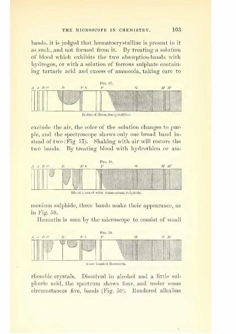
7/29/2019 THE MICROSCOPIST
http://slidepdf.com/reader/full/the-microscopist 109/361
THE MICROSCOPE IN CHEMISTRY. 103
bands, it is judged that hfematocrystalline is present in it
as such, and not formed from it. Bj^ treating a solution
of blood which exhibits the two absorption-bands with
hydrogen, or with a solution of ferrous sulphate contain-
ing tartaric acid and excess of ammonia, taking care to
Fig. 57.
F
Reduced Haiuuitocrystalliue.
exclude the air, the color of the solution changes to pur-
ple, and the spectroscope shows only one broad band in-
stead of two (Fig 57). Sliaking with air will restore the
two bands. By treating blood with hydrothiou or am-
FlG. 58.
F
iiiiiiliiliil infix
Bloud Healed with Aniniumuin ^iiljiliide.
monium sulphide, three bands make their appearance, as
in Fig. 58.
Haematin is seen by the microscope to consist of small
Fig. 59.
F
11(5 f ilflfljl
iJ___
l^uur bauded lltematin.
rhombic crystals. Dissolved in alcohol and a little sul-
phuric acid, the spectrum shows four, and under some
circumstances five, bands (Fig. 59). Rendered alkaline
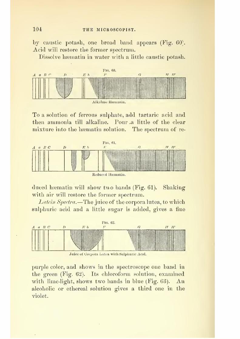
7/29/2019 THE MICROSCOPIST
http://slidepdf.com/reader/full/the-microscopist 110/361
104 THE MICROSCOPIST.
by caustic potash, one broad band appears (Fig. 60\
Acid will restore the former spectrum.
Dissolve hsematiri in water with a little caustic potash.
Alk.iluju ULuuiiaiu.
To a solution of ferrous sulphate, add tartaric acid and
then ammonia till alkaline. Pour ,a little of the clear
mixture into the hsematin solution. The spectrum of re-
Fi<;. ei.
A a B C T)
J
ReducKl Ha'iiiaLii
duced hsematin will show two bands (Fig. 61). Shaking
with air will restore the former spectrum.
hiitein Spectra.—The juice of the corpora lutea, to whichsulphuric acid and a little sugar is added, gives a fine
A a B c
liiiliP
Juice of Cuipura Lutea with Sulphuric Acid.
jDurple color, and shows in the spectroscope one band in
the green (Fig. 62). Its chloroform solution, examined
with lime-light, shows two bands in blue (Fig. 63). An
alcoholic or ethereal solution gives a third one in the
violet.

7/29/2019 THE MICROSCOPIST
http://slidepdf.com/reader/full/the-microscopist 111/361
THE MICROSCOPE IN CHEMISTRY. 105
Cj'Sto lutein, or the j'ellow fluid of an ovarian cyst,
shows with the linie-lio'ht three bands in blue, in the
A T R r n n h
Pig. 63.
F G H H'
MiC HiiF i
: ,
i
')
Chloroloim SuUitiuu ul Coipora Lutea.
same position as the chloroform solution of lutein (Fig.
64).
The serum of blood, etc., shows the bands of hiemato-
A a B r
Fl«. 61.
F
mm
Cystn-Lutein lium uii Uvaiiau Cyst.
crj^stalline and one or two doubtful bands, as in the figure
(Fig. 65).
Dr. Richardson, of Philadelphia, gives the following
directions for examining blood-stains:
Procure a glassslide with a circular excavation, and moisten the edges
of the cavity with a small drop of diluted glycerin. Lay
A n B C B b
Fra. 65.
F
^mi{J siiiip
1|' ''ill
Sero-Luteiii.
a clean glass cover, a little larger than the excavation, on
white paper, and put on it the smallest visible fragment
of blood-clot. With a needle, put on the centre of the
cover a speck of glycerin, not larger than a full stop (.),

7/29/2019 THE MICROSCOPIST
http://slidepdf.com/reader/full/the-microscopist 112/361
106 THE MICROSCOPIST.
and with a dry needle push the blood to the edge that it
may be just moistened with the glycerin. Place the slide
on the cover so that the glycerin edges of the cavity may
adhere, and turning it over, transfer it to the stage of the
microscope. Thus a minute quantity of a strong solution
of hsemoglobulin is obtained, the point of greatest density
of which may be found by a one-fourth objective, and
tested by the spectroscopic eye-piece and with high powers.
The tiny drop may be afterwards wiped oft' with moist
blotting-paper, and a little fresh tincture of guaiacum
added, showing the blue color of the guaiacum blood-test.
Inverted Microscope of Dr. Laiorence Smith.—In ordinary
chemical investigations there is some- risk of injuring the
polish of the lenses, as well as the brass work of the mi-
croscope, without very great care. This is particularly
the case in observing the eftects of heat or of strong acids.
To obviate this difticulty, Dr. Lawrence Smith contrived
a plan for an inverted microscope, which has been con-
structed by Xachet of Paris. The optical part of the in-
strument is below the stage, and is furnished with a pecu-
liar prism, by which the rays from the objective are bent
into a conveniently inclined body. The illuminating ap-
paratus is above the stage. This construction renders the
instrument well adapted to chemical investigations.
General Microchemical Tests.
Dr. Wormley has directed attention to some necessary
cautions. He shows that many substances which may
readily be detected in a pure state, even in very minutequantities by the microscope, are difficult to detect Vk^hen
mixed with complex organic materials. This is especially
applicable to the alkaloids, which should be separated
from such mixtures by the use of the dialyzer—a hoop
with a bottom of parchment-paper, etc.— or extracted
with ether or chloroform.

7/29/2019 THE MICROSCOPIST
http://slidepdf.com/reader/full/the-microscopist 113/361
THE MICROSCOPE IN CHEMISTRY. 107
The purity of all reagents should be carefully estab-
lisbed, aud they should be kept in hard German glass
bottles, and only distilled water used in all our researches.
The true nature of a reaction that is common to several
substances may often be determined with the microscope.
Thus a solution of nitrate of silver becomes covered with
a white film when exposed to several different vapors, but
hydrocyanic acid is the only one which is crystalline.
Tins will detect 100,000th of a grain of the acid. A slip
of clean copper boiled in a hydrochloric acid solution of
arsenic, mercury, antimony, etc., becomes coated with the
metal, but when heated in a reduction-tube, arsenic only
yields a sublimate of octahedral crystals, and mercury
only, will furnish metallic globules.
A solution of iodine produces distinct reaction with
100,000th of a grain of strychnine in solution in 1 grainof water, but as this is common to other alkaloids, other
tests are needed. Yet the absence of such a reaction
shows the absence of the alkaloid.
The degree of dilution is important. Thus bromine with
atropin yields a crystalline deposit from 1 grain of a
20,000th or stronger dilution, but not with diluter solu-
tions. A limited quantity of sulphuretted hydrogenthrows down from corrosive sublimate a white deposit,
while excess produces a black precipitate.
Blue and Reddened Litmus Paper are used as tests for
acids and alkalies. It is a bibulous paper dyed in infu-
sion of litmus. The red is made by adding a little acetic
acid to the infusion. Dry substances and vapors require
the paper to be moistened with distilled water. If theacid reaction depends on carbonic acid, warming the paper
on a slide over a lamp will restore the color. So if a
volatile alkali, ammonia or carbonate of ammonia, have
made the red paper blue, its color will be restored by a
gentle heat. Sometimes the infusion of litmus is more
convenient than the paper.

7/29/2019 THE MICROSCOPIST
http://slidepdf.com/reader/full/the-microscopist 114/361
108 THE MICROSCOPIST.
Alcohol coagulates albuminous matter.
Ether dissolves fat.
Acetic Acid will dissolve phosphate or carbonate of lime,
but not the oxalate.
Nitrate of Barytes in cold saturated solution is a test for
sulphuric and phosphoric acids. The precipitated sulphate
of baryta is insoluble in acids and alkalies. The phosphate
is soluble in acids and insoluble in ammonia.
Nitrate of Silver.—A solution of 60 grains to the ounceof water is a convenient test for chlorides and phosphates.
Chloride of silver is white, soluble in ammonia and insolu-
ble in nitric acid. The tribasic phosphate of silver is yel-
low, and soluble in excess of ammonia or of nitric acid.
Oxalate of Ammonia is a test for salts of lime. Dissolve
the material in nitric acid, and add excess of ammonia.
Dissolve the flocculeut precipitate in excess of acetic acid,
and add the oxalate of ammonia. Oxalate of lime is in-
soluble in alkalies and acetic acid, but soluble in strong
mineral acids.
Iodine is a test for starch, coloring it blue. Albuminous
tissues are colored yellow, and vegetable cellulose a brown-
ish-yellow. The addition of sulphuric acid turns cellulose
blue.
Determination of Substances.
alkalies.
Bichloride of platinum precipitates from salts of potash
or ammonia a yellow double chloride, which crystallizes
in beautiful octahedra. It has no precipitating efiect on
solutions of soda. Polarized light will distinguish the
800,000th of a grain of double chloride of sodium and
platinum by its beautiful colors from the chloride of
potassium and platinum, or of platinum alone. The
double chloride of platinum and potassium may be dis-
tinguished from that of ammonia by heating to redness,
treating with hot water, and acting on with nitrate of

7/29/2019 THE MICROSCOPIST
http://slidepdf.com/reader/full/the-microscopist 115/361
THE MICROSCOPE IN CHEMISTRY. 109
silver. The ammonium compound after ignition leaves
only the platinum, which gives no precipitate with nitrate
of silver, while the potassium chloride yields a white pre-
cipitate of chloride of silver.
Antimoniate of potash throws down from solutions of
soda and its neutral salts a white crystalline antimoniate
of soda, the forms of which vary according to the strength
of the solution;generally they are rectangular plates and
octahedra.
ACIDS.
Sulphuric.—In solutions acidulated with hydrochloric
or nitric acid, the chloride or nitrate of baryta produces
a white precipitate. Yeratrin added to a drop of concen-
trated sulphuric acid produces a crimson solution, or de-
posit if evaporated.
Nitric.—Heated with excess of hydrochloric acid elimi-
nates chlorine, M'hich will dissolve gold leaf. A blood-
red color is produced when nitric acid or a nitrate is mixed
with a sulphuric acid solution of brucin.
Hydrochloric.—Mtrate of silver precipitates amorphous
chloride of silver; soluble in ammonia, but insoluble in
nitric and sulphuric acid.
Oxalic.—jSTitrate of silver precipitates amorphous oxa-
late of silver ; soluble in nitric acid and also in solution
of ammonia.
Hydrocyanic.—Put a drop of acid solution in a watch-
glass, invert another over it containing a drop of solution
of nitrate of silver, and a crystalline lilm will form. A
solution of hydrocyanic acid treated with caustic potashor soda and then with persulphate of iron yields Prussian
blue.
Phosphoric.—A mixture of sulphate of magnesia, chlo-
ride of ammonium, and free ammonia produces in solu-
tions of free phosphoric acid and alkaline phosphates
white feathery or stellate crystalline precipitate of ammo-

7/29/2019 THE MICROSCOPIST
http://slidepdf.com/reader/full/the-microscopist 116/361
110 THE MICROSCOPIST.
nio-phosphate of magnesia. A slower crystallization gives
prisms.
METALLIC OXIDES.
These may usually be determined by treating a small
portion of solution, acidulated with hydrochloric acid, by
sulphuretted hj'drogen ; another, and neutral portion w^ith
sulphuret of ammonium ; and a third with carbonate of
soda.
Antimony.—Sulphuretted hydrogen throws down or-
ange-red precipitate from tartar-emetic solutions, etc.
Arsenic yields white octahedral crystals of arsenious
acid when sublimed. Arsenious acid may be reduced to
metallic arsenic by heating to redness in a tube with
charcoal and carbonate of soda. A solution of arsenious
acid yields octahedral crystals by evaporation, so as to
determine with the microscope 1000th to 10,000th of a
grain.
Ammonio-nitrate of silver throws down from an aque-
ous solution of arsenious acid a bright yellow precipitate,
ammonio-sulphate of copper a green precipitate, and sul-
phuretted hydrogen a bright yellow.
Mercury.—Bichloride of mercury, moistened with a drop
of solution of iodide of potassium, assumes the bright
scarlet color of biniodide of mercury. A strong solution
of caustic potash or soda turns bichloride of mercury yel-
low from the formation of protoxide ; but calomel or chlo-
ride of mercury is blackened from formation of suboxide.
Heated in a rednction-tube with dry carbonate of soda,
the sublimate shows under the microscope small, opaque,
spherical globules of mercury. Dr. Wormley states that
a globule of mercury or "artificial star" may be discrim-
inated by the one-eighth objective if it be but the
25,000th of an inch in diameter, weighing about tlie
9,000,000,000th of a grain; globules of so'ootti of an inch
diameter weigh about 70,000,000th of a grain.

7/29/2019 THE MICROSCOPIST
http://slidepdf.com/reader/full/the-microscopist 117/361
THE MICROSCOPE IN CHEMISTRY. Ill
Lead.—Sulphuretted hydrogen gives a black amor-
phous deposit. Sulphuric and hydrochloric acids yield a
white precipitate. Chloride of lead crystallizes in needles.
Iodide of potassium gives a bright yellow precipitate, sol-
uble in boiling water, and crystallizing in six-sided plates.
Bichromate of potassium yields a bright yellow amor-
phous deposit.
Copper.—Sulphuretted hydrogen gives a brown or black-
ish deposit ; ammonia a blue or greenish-blue amorphousprecipitate, or in dilute solutions a blue color to the liquid
caustic alkali, a similar precipitate, which on boiling in
excess of reagent becomes black, but if grape-sugar, or
some other organic agents, be present, a yellow or red
precipitate of suboxide of copper occurs. Arsenite of
potassium produces a bright green.
Zinc-—^Sulphuretted hydrogen gives a white amorphousdeposit—the only white sulphuret. Alkalies produce a
white hydrated oxide of zinc.
ALKALOIDS.
The editors of the Blicrogrcqihic Dictionary refer to a paper
of Dr. T. Anderson, in the Edinburgh Monthly Journal.^
where he shows that the microscope readily distinguishes
the more common alkaloids from each other by the form
of their crystals and of their sulphocyanides. The alka-
loids are first dissolved in dilute hydrochloric acid, then
precipitated on a glass plate with a solution of ammonia,
or if the sulphocyanide is required, with a strong solution
of sulphocyanide of potassium. It may then be placedunder the microscope. The solution should not be too con-
centrated. This branch of investigation has been greatly
promoted by the elegant work of Dr. Wormley, already
referred to, on the Microcliemistry of Poisons.
Atroirln.—Ammonia throws down an amorphous pre-
cipitate. One grain of a yoo^^ grain solution yields to

7/29/2019 THE MICROSCOPIST
http://slidepdf.com/reader/full/the-microscopist 118/361
112 THE MICROSCOPIST.
caustic potash or soda a precipitate which, when stirred
with a glass rod, becomes a mass of crystals, as in Plate
TIT, Fig. 60. The sulphocjanide of potassium gives no
precipitate,
Aconitin.—l^o characteristic test, except the physiologi-
cal one;^
Q'^^th of a grain produces on the end of the
tongue a peculiar tingling and numbness, lasting for an
hour; ygo^^ grain in alcohol, rubbed on the skin, pro-
duces temporary loss of feeling.
Brucin or Brucia.—Potash or ammonia produces stellar
crystals. Sulphocyanide of potassium, feathery, or sheaf-
like. (Plate III, Fig. 67.) Nitric acid produces a blood-
red color, changing to yellow by heat. On cooling the
latter and adding protochloride of tin, it becomes a beau-
tiful purple. Ferricyanide of potassium, with j- ^pth grain
of brucin yields the most brilliant polariscope crystals.
(Plate III, Fig. 68).
Cinchonine.—Ammonia produces granular radiating
crystals. (Plate III, Fig. 69.) Sulphocyanide of potas-
sium six-sided plates, some irregular. (Plate III, Fig. 70.)
Conine.—This alkaloid and nicotin are distinguished
from other alkaloids by being liquid at ordinary tempera-
tures, and by their peculiar odor. Conine may be knownfrom nicotin by its odor and sparing solubility in water,
by yielding crystalline needles to the vapor or solution of
hydrochloric acid, a white precipitate with corrosive sub-
limate, and a dark-brown precipitate with nitrate of silver.
Codein.—Ammonia or alkalies give a white amorphous
deposit. Sulphocyanide of potassium, crystalline needles.
A solution of iodine in iodide of potassium, a reddish-
brown precipitate, which becomes crystalline. This is
soluble in alcohol, from which it se})arates in plates (Plate
III, Fig. 71), which appear beautiful in the polariscope.
Daturin.-—-According to Dr. Wormley, this is identical
with atropin.
Narcotin.—In its pure state crystallizes in rhombic

7/29/2019 THE MICROSCOPIST
http://slidepdf.com/reader/full/the-microscopist 119/361
PLATE in.
Fig. G6.
^
Fig. 71.
©(>
Fig. 67.
t&w- ^^'^^-
-V, -i
Fig. 72.
<fe-
t^" '%;^
Fig. 73.
Fig.
/
Fig. 69.
Fig. 74.
H^ ^
jij,* as- "*
W Mf
Fig. 70.
\j
P'IG. 75.

7/29/2019 THE MICROSCOPIST
http://slidepdf.com/reader/full/the-microscopist 120/361
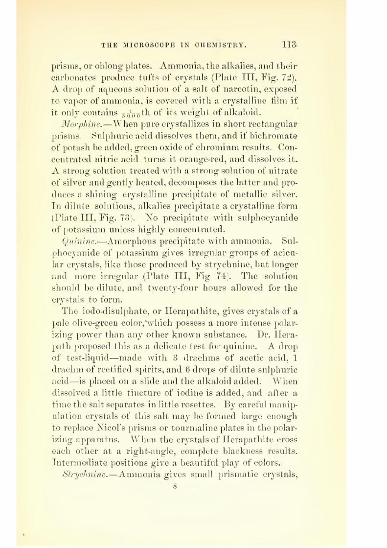
7/29/2019 THE MICROSCOPIST
http://slidepdf.com/reader/full/the-microscopist 121/361
THE MICROSCOPE IN CHEMISTRY. 113.
prisms, or oblong plates. Ammonia, the alkalies, and their
carbonates produce tufts of crystals (Plate III, Fig. 72).
A drop of aqueous solution of a salt of narcotin, exposed
to vapor of ammonia, is covered with a crystalline film if
it only contains gf^'j^^th of its weight of alkaloid.
3Iorphine.—When pure crystallizes in short rectangular
prisms. Sulphuric acid dissolves them, and if bichromate
of potash be added, green oxide of chromium results. Con-
centrated nitric acid turns it orange-red, and dissolves it..
A strong solution treated with a strong solution of nitrate
of silver and gently heated, decomposes the latter and pro-
duces a shining cr^'stalline precipitate of metallic silver.
In dilute solutions, alkalies precipitate a crystalline form
(Plate III, Fig. 73). ISTo precipitate with sulphocyanide
of potassium unless highly concentrated.
Quinine.
—Amorphous precipitate with ammonia. Sul-
phocyanide of potassium gives irregular groups of acicu-
lar crystals, like those produced by strychnine, but longer
and more irregular (Plate III, Fig 74;. The solution
should be dilute, and twenty-four hours allowed for the
crystals to form.
The iodo-clisulphate, or Ilerapathite, gives crystals of a
pale olive-green color,'which possess a more intense polar-
izing power than any other known substance. Dr. Ilera-
path proposed this as a delicate test for quinine. A drop
of test-liquid—made with 3 drachms of acetic acid, 1
drachm of rectified spirits, and 6 drops of dilute sulphuric
acid—is placed on a slide and the alkaloid added. Whendissolved a little tincture of iodine is added, and after a
time the salt separates in little rosettes. By careful manip-
ulation crystals of this salt may be formed large enough
to replace Xicol's prisms or tourmaline plates in the polar-
izing apparatus. When the crystals of Ilerapathite cross
each other at a right-angle, complete blackness results.
Intermediate positions give a beautiful play of colors.
Strychnine.—Ammonia gives small prismatic crystals,
8

7/29/2019 THE MICROSCOPIST
http://slidepdf.com/reader/full/the-microscopist 122/361
114 THE MICROSCOPIST.
some crossed at 60° (Plate III, Fig. 75). Sulphocjanide
of potassium produces flat needles, often in groups. Iodine
in iodide of potassium gives a reddish-brown amorphous
precipitate, crystalline in dilute solutions. When pure,
strychnine appears in colorless octahedra, lengthened
prisms or granules. To a solution of the alkaloid or its
salts in a drop of pure sulphuric acid, which produces no
color, add a small crystal of bichromate of potash, and
stir slowly with a pointed glass rod. A blue color will
appear, passing into purple, violet, and red. The bright
yellow crystals of chromate of strychnia, if dried and
touched with sulphuric acid, will also show the color test.
This is said to be delicate enough to show too'ooo^^^^' ^
grain of strychnine. The tetanic convulsions of frogs im-
mersed in a solution of strychnine, or after injections of
the solution in lungs or stomach, etc , is also a very deli-
cate test.
Veratrin and its salts treated in the dry state with con-
centrated sulphuric acid, slowly dissolve to a reddish-yel-
low, or pink solution, wdiich becomes crimson-red. The
process is accelerated by heat.
Naixein, touched with the cold acid, becomes brown,
brownish-yellow, and greenish-yellow, and if heated, adark purple-red.
. Solanin turns orange-brown, and later purplish-brown.
Piperin turns orange-red to brown.
Salicin gives to the acid a crimson pink, changing to
black.
Papaverin gives a fading purple.
CRYSTALLINE FORMS OF VARIOUS SALTS.
Our limits forbid extended description, yet a few forms
of frequent recurrence will be useful to the student. For
crystals in plants or from animal secretions reference ma}^
be made also to succeeding chapters.
jSalis of Lime.—The carbonate sometimes occurs in ani-

7/29/2019 THE MICROSCOPIST
http://slidepdf.com/reader/full/the-microscopist 123/361
PLATE IV.
Fig. 76.
IP
^s^^A
tS^
Fig. 80.
-V
Fig. 77.
^
Fig. 81.
Fig. 7S.
1
'%)
J
^~::i
Fl^. 82.
Fig. 79.
#^'^//0 #^^^r<^ Jl I
Fig. 83.
op
77o
Fig. 84.
i n

7/29/2019 THE MICROSCOPIST
http://slidepdf.com/reader/full/the-microscopist 124/361

7/29/2019 THE MICROSCOPIST
http://slidepdf.com/reader/full/the-microscopist 125/361
THE MICROSCOPE IN CHEMISTRY. 115
mal secretions in the form of little spheres or disks, con-
sisting of groups of radiating needles. In otoliths it is
often in minute hexagonal prisms with trilateral summits.
It is deposited from water in irregular forms, all of which
are grouped needles. Sometimes it assumes the rhombo-
hedral form, as in the oyster shell (Plate lY, Fig. 76).
In anj^ doubtful case, test as described at pages 99 and
108.
Lactate of Lime gives microscopic crystals, consisting of
delicate radiating needles (Plate lY, Fig. 77).
Oxalate of Lime occurs as square flattened octahedra, as
square prisms with quadrilateral pyramids, as fine needles,
and as ellipsoidal flattened forms, sometimes constricted
so as to resemble dumb-bells (Plate lY, Fig. 78).
Phosphate of Lime is usually in the form of thin rhombic
plates (Plate lY, Fig. 79).
Sulphate of Lime rapidly formed, as in chemical testing,
gives minute needles or prisms (Plate lY, Fig. 80). When
more slowly formed, these are larger and mixed with
rhombic plates.
Soda Salts.—Chloride of Sodium or common salt gener-
ally forms a cube, terminated by quadrangular pyramids
or depressions (Plate lY, Fig. 81). The crystals do notpolarize light.
Plate lY, Fig. 82, represents crystals of oxalate of soda,
and Plate lY, Fig. 83, those of nitrate.
Magnesia Salts.—Ajnmonio-phosjjhate, or triple phosphate,
is often found in animal secretions. The most common
form is prismatic, but sometimes it is feathery or stellate
(Plate lY, Fig. 84).
Sulphate of 31agnesia forms an interesting polarizing
object.
A most instructive series of salts may be made by
rapidly crystallizing some on glass slides, and allowing
others to deposit more slowly. In this way a set of speci-
mens may be prepared for comparison.

7/29/2019 THE MICROSCOPIST
http://slidepdf.com/reader/full/the-microscopist 126/361
116 THE MICKOSCOPIST.
CHAPTER IX.
THE MICROSCOPE IN BIOLOGY.
The science of biology (from /?jo?, life), which treats of
the forms and functions of living beings, would be crude
and imperfect without the aid of the microscope. What-
ever might be learned bj general observation, we should
miss the fundamental laws of structure and the unity
which we now know pervades distant and apparently
different organs, as well as distinct species, if we were
deprived of the education which microscopy gives the
eye and hand.
The evident differences between living and non-living
bodies led to ancient theories of life which are still influ-
ential in modern thought, but neither microscope nor
scalpel nor laboratory have revealed the mystery which
seems ever to beckon us onward to another and entirely
difi'erent sphere of existence. Hippocrates invented the
hypothesis of a principle {(putrcq, or nature) which influences
the organism and superintends it with a kind of intelli-
gence, and to which other principles ('Jova.ae:?, powers) are
subordinated for the maintenance of various functions.
This was also the theory of Aristotle, who gave the name
of soul {<l'ox-^) to the animating principle.
Paracelsus and the chemical philosophers, from the
fifteenth to the seventeenth century, maintained that all
the phenomena of vitality may be explained by chemical
laws. To these succeeded the mathematical school under
Bellini (A.D. 1645), who taught that all vital functions
may be explained by gravity and mechanical impulse.
These theories were supplanted by those of the physiolo-
gists. Van Helmont revived the Hippocratian idea of a
specific agent, which he called archeus. This was more
fully elaborated by Stahl, who taught that by the opera-

7/29/2019 THE MICROSCOPIST
http://slidepdf.com/reader/full/the-microscopist 127/361
THE MICROSCOPE IN BIOLOGY. 117
tion of an immaterial animating principle or soul (anima),
all vital functions are produced. The vis medicatrix na-
turce of Cullen was an attempt to compromise between
the rival theories of a superadded principle and a special
activity in organized matter itself.*
Harvey, Hunter, Miiller, and Prout proposed hypotheses
similar to those of Aristotle and Hippocrates, and many
modern scientific men accept similar views. The recent
doctrine of the correlation of physical forces has, however,
revived the mechanical and chemical theories, and the
industry with which these views have been propagated
has gained many adherents.
It is to be regretted that philosophy should assume the
name of science and dogmatize under that appellation.
The object of science is to state facts, and not to dream,
yet such is the nature of man's intellect that it will seek
to account for facts, and is thus drawn into metaphysical
speculation. If the age-long controversy between the
phj'sicists and the vitalists is ever to cease, it will prob-
abl}^ be through the microscopic demonstration of the
absolute diiierence between livino- and non-livino- matter.
lu the present chapter it is designed to set forth briefly
the principal facts of elementary biology as they have
been brought to light by microscopy. For further illus-
trations in vegetable and animal histology, reference nisiy
be made to following chapters.
1. All biologists agree that the elementary unit in living
bodies is the cell. This, according to the most recent in-
vestigations, is a soft, transparent, colorless, jelly-like par-
ticle of matter, which may be large enough to be just dis-
cernible to the naked eye, or so small as to be invisible
wdth our best instruments. The simplest or most elemen-
tary forms of vegetable or animal life consist of single
cells, while the more complex organisms are built up of
* Compare Bostock's History of Medicine.

7/29/2019 THE MICROSCOPIST
http://slidepdf.com/reader/full/the-microscopist 128/361
118 THE MICROSCOPIST.
great numbers of these cells with the materials which
they have produced and deposited.
Haller, who has been called the father of modern physi-
ology, seems first to have conceived, though vaguely
(A.D. 1766), the idea of the essential unity of vital struc-
ture.
In 1838, Schleiden and Schwann wrote on the elemen-
tary cell, the former treating of the vegetable, and the
latter of the animal cell. From this time may be dated
the origin of the cell doctrine. Much importance was
assigned to the distinction between cell-wall, cell-contents,
nuclei, and nucleoli.
In 1835, Dujardin discovered in the lower animals a
contractile substance capable of movement, to which he
gave the name of sarcode.
In 1861, Max Schultze showed that sarcode is analo-
gous to the body or contents of animal cells, and that on
this account the infusorial animalcules possessed of inde-
pendent life were simple or compound.
Examinations of this structure were made by numerous
observers, and the identity of many of its properties in
animals and vegetables established. To this structure
the name of iirotoplasm^ rather than sarcode, has been
assigned. As this term has been somewhat loosely used,
so as to refer to it either in the dead or living state, Dr.
Beale has proposed the term bioplasm for elementary struc-
ture while living, and has given a generalization from
observed facts which has attracted much attention. He
distinguishes in all organic forms three states of matter:
First. Germinal matter or bioplasm^ or matter which is
living. Second. Matter which was living, ovformed mate-
rial. Third. Matter about to become living, or pabulum.
Schleiden and Schwann considered the cell as a growth
from a nucleus, and to consist of a cell-wall and cavity.
In vegetable cells there seemed to be an external wall of
cellulose, within w^iich was another, the primordial utri-

7/29/2019 THE MICROSCOPIST
http://slidepdf.com/reader/full/the-microscopist 129/361
THE MICROSCOPE IN BIOLOGY. 119
cle. But it has since been shown that the appearance of
the primordial utricle is caused by the protoplasm or bio-
plasm Ij'ing in apposition Avith the inner surface of the
cell-wall. In the cryptogamia, cells are known to occur
in which no nucleus is visible. Max Schultze and Hackel
have also discovered non-nucleated forms of animal life.
The idea of nucleus and cell-wall as essential to a cell is
therefore abandoned. I^uclei are regarded as new centres
of living matter, or minute particles of such matter capa-
ble of independent existence. Some of these masses are
so small as to be barely visible witli the one-fiftieth objec-
tive under a magnifying power of five thousand diameters.
2. The structure and formation of a simple cell may be
illustrated by Plate V, Figs. 85 to 89, after Beale.* The
earliest condition of such a living particle is shown in
Plate V, Fig. 85. If the external membrane of a fully
developed spore or any of the growing branches (Plate V,
Figs. 86 to 89) be ruptured, such particles would be set
free in vast numbers.
The surface of such a particle becomes altered by con-
tact with external agencies. A thin layer of the external
surface is changed into a soft membrane or cell-wall,
through which pabulum passes and undergoes conversion
into living matter, which thus increases. The increase
of size is not owing to the addition of new matter upon
the external surface, but to the access of new matter in-
teriorly. The thickness of the formed material depends
on external circumstances, as temperature, moisture, etc.
If these be unfavorable to the access of pabulum, layer
after layer of living matter will die or be deposited, as in
Plate Y, Figs. 87 and 88. If such a cell be exposed to
circumstances favorable to growth, the accession of fresh
pabulum will cause portions of living matter to make
^ Physiological Anatomy and Physiology of Man, by Drs. Todd, Bow-
man, and Beaie. New edition.

7/29/2019 THE MICROSCOPIST
http://slidepdf.com/reader/full/the-microscopist 130/361
120 THE MICROSCOPIST.
their way through natural pores or chance fissures and
protrude, as in the figures.
3. The peculiar 'phenomena of living cells or bioplasms
may be classified as follows: Active or spontaneous move-
ment, nutrition and growth, and the power of reproduc-
tion. These vital actions, according to Dr. Beale, occur
in the bioplasm only, while the formed material, or non-
living matter, is the seat of physical and chemical changes
exclusively. Physical processes, as diffusion and osmose,
occur in bioplasmic particles, but the peculiar phenomena
referred to, and which are properly termed vital, do not
occur in non-living matter.
Movements of Cells.—Granules imbedded in the bioplasm,
either formed material or accidental products, enable our
microscopes to observe internal movement, while change
of form and of place exhibit the movement of the entire
cell.
The granular movement is either vibratory or continu-
ous. The vibrations of the granules appear similar to the
molecular movement described by Dr. Robert Brown in
1827, and which is common to all small masses of matter,
organic or inorganic. Minute cells may thus dance in
fluid as well as fine powders, etc. Such movements occur,
however, in the interior of living cells, and may possibly
be connected with vitality. In the salivary corpuscles,
the dancing motion ceases on the addition of a solution
of one-half to one per cent, of common salt, while such
addition has no influence of the kind on fresh pus or
lymph.
The continuous granular motion is either a relatively
slow progression, corresponding to the change of form in
the cell, or a swifter flowing movement. Max Schultze
thus describes this motion in the threads of sarcode pro-
jected from the apertures of a Foraminiferal shell: "As
the passengers in a broad street swarm together, so do
the granules in one of the broader threads make their

7/29/2019 THE MICROSCOPIST
http://slidepdf.com/reader/full/the-microscopist 131/361
PLATE V.
Fig. S5.
m..«.••,
Minute particles of Bioplasm. FromMildew. Jotli i"- Obj. Passage of Germinal-matter through
pores in the formed material. X 1800.
Fig. 89.
Production of forraed-material on
surface of Bioplasm. X ISOO.
Production and accumulation of
Formed-material on Bioplasm. Epi-
thelium of cuticle. X 700.
Fig. 87. Fig. 90.
Further production of formed-
material. At a is the budding
P.ioplasm, passing through pores
in the forraed-material. X 1800.
Amceboe from organic infusion.

7/29/2019 THE MICROSCOPIST
http://slidepdf.com/reader/full/the-microscopist 132/361

7/29/2019 THE MICROSCOPIST
http://slidepdf.com/reader/full/the-microscopist 133/361
THE MICROSCOPE IN BIOLOGY. 121
way b}' one another, oftentimes stopping and hesitating,
yet always pnrsning a determinate direction correspond-
ing to the long axis of the thread. They frequently be-
come stationary in the middle of their course, and then
turn round, but the greater number pass to the extreme
end of the thread, and then reverse the direction of their
movement." 'No physical or chemical action with which
we are acquainted will account for such motions, which
have no analogy in unorganized bodies.
Changes of form are most strongly marked in the lower
forms of animal life, although occurring also in the sim-
pler vegetables, as the volvox. The Ammba or Proteus is
typical of such changes, which have hence been termed
Amoeboid (Plate V, Fig. 90). AVhen an Amoeba meets
another animal which is too slow to escape, it sends out
projections which encircle its prey ; these coalesce, and
invest the whole mass with its bioplasm. It maintains
its grasp till it has abstracted all the portions which are
soluble, and then relaxes its hold.
Ama?boid cells in higher animals rarely move so rapidly
as the Amoeba itself. Their motions are limited to a
gradual change of form or to the protrusion of processes
in the form of threads, or tuberosities, or tufts, whicheither drag the rest of the body after them or are again
withdrawn.
Cells of bioplasm may not only change their form, but
may wander from place to place by protruding a portion
of their mass, which drags the rest after it. The discov-
ery of wandering cells in the higher organisms, as man,
lias opened quite a new and important field of physiologi-
cal and pathological research.
The movements of bioplasm may be changed, acceler-
ated, retarded, or stopped by a variety of stimuli, mechani-
cal, electrical, chemical, and nervous. Gentle warmth and
moisture are necessarj^ to their perfection.*
* See Strieker's Manual of Histology.

7/29/2019 THE MICROSCOPIST
http://slidepdf.com/reader/full/the-microscopist 134/361
122 THE MICROSCOPIST.
The nutrition and growth of the living cell has already
been described as the conversion of pabulum into bioplasm
or living matter. The subject of reproduction will be ex-
amined below under the head of cell-genesis.
4. The microscopic demonstration of bioplasm may be
effected by the use of an alkaline solution of coloring
matter, as carmine. (See Chapter V.) As bioplasm pos-
sesses an acid reaction, the alkali is neutralized and the
color retained. This process, however, is rather a dem-
onstration of the protoplasm which was recently alive.
For living cells or bioplasm, we must dejDcnd on supplying
them artificially w^th colored food. Thus indigo, carmine,
etc., in fine particles, added to the pabulum of cells or
liquid media in which they float, will be taken into the
interior of the bioplasm by the nutritive process. In this
w^ay Recklinghausen showed the migration of pus-corpus-
cles.-
Welcker and Osborne were the first to use a solution
of carmine in order to stain the nuclei of tissues. They
w^ere followed by Gerlach and Beale, the latter of whom
has greatly improved the process and shown its signifi-
cance.
5. The chemistry of cells and their products is an essential
part of biology, but would lead us too far from our subject
to discuss, yet a few points may not be irrelevant.
The chemical composition of bioplasm consists essen-
tially of oxygen, hydrogen, nitrogen, and carbon. Other
elements are often present and important, but not essen-
tial. Of the relation of these elements we know nothing,
save that they are in a state of constant vibration or
change. Dr. Beale considers it doubtful if ordinary chemi-
cal combination is possible while the matter lives. Analy-
sis in the laboratory is only possible with the compounds
resulting from the death of the cell.
When living or germinal matter is converted into formed
material, a combination of its elements takes place, often

7/29/2019 THE MICROSCOPIST
http://slidepdf.com/reader/full/the-microscopist 135/361
THE MICROSCOPE IN BIOLOGY. 123
with verj complex results, the nature of which has hitherto
baiRed the eiibrts of chemists to determine. When the
life of germinal matter, however, is suddenly destroyed,
or rather when the matter is first transformed, the com-
pounds resulting from various species have similar chemi-
cal composition and properties, and an acid reaction is
developed. Fibrin, albumen, water, and certain salts may
thus be obtained from every kind of germinal matter.
Fatty matters also result, which continue to increase in
quantity for some time after death. In slow molecular
death, a certain amount of oxygen is taken into combina-
tion, which o-ives rise to ditFerent results from those which
occur when life is suddenly destroyed. Still other com-
binations are due to vital actions which are not yet under-
stood. Thus some bioplasm produces muscle; other par-
ticles originate nerve structure, cartilage, bone, connective
tissue, etc. Many chemical changes occur also in formed
material after its production. It may become dry or fluid,
or split up into gaseous or soluble substances as soon as
produced. Imperfect oxidation may lead to the formation
of fatty matters, uric acid, oxalates, sugar, etc. At the
earliest period of development, the formed material con-
sists principally of albuminous and fatty matters, with
chlorides, alkaline and earthy 2:)hosphates. At a later
period gelatin, with amyloid or starchy matter, is pro-
duced.*
6. Varieties in the Form and Function of Bioplasm..—Mutability of shape is characteristic of amoeboid cells,
and no conclusions can be drawn from their appearance
after death. Where numbers of them are accumulated,
they are flattened by mutual pressure so as to appear
polyhedral, laminated, or prismatic. The upper layers of
laminated epithelium are usually flattened. Where cells
line the interior of cavities in a single layer, they form
* See Physiological Anatomy, by Todd, Bowman, and Beale.
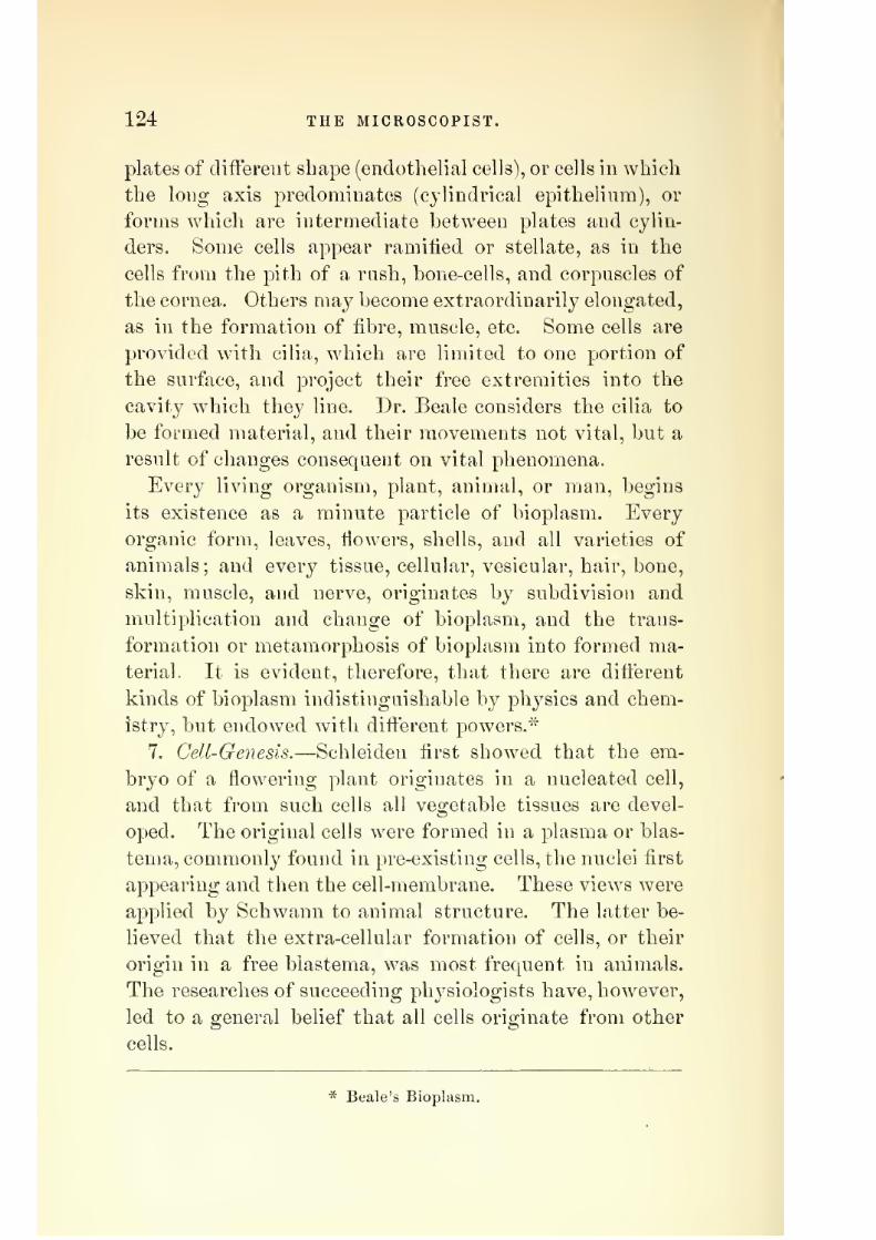
7/29/2019 THE MICROSCOPIST
http://slidepdf.com/reader/full/the-microscopist 136/361
124 THE MICROSCOPIST,
plates of different shape (endothelial cells'), or cells in which
the long axis 23redoniinates (cylindrical epithelium), or
forms which are intermediate between plates and cylin-
ders. Some cells appear ramified or stellate, as in the
cells from the pith of a rush, hone-cells, and corpuscles of
the cornea. Others may become extraordinarily elongated,
as in the formation of fibre, muscle, etc. Some cells are
provided with cilia, which are limited to one portion of
the surface, and project their free extremities into the
cavity which they line. Dr. Beale considers the cilia to
be formed material, and their movements not vital, but a
result of changes consequent on vital phenomena.
Every living organism, plant, animal, or man, begins
its existence as a minute particle of bioplasm. Every
organic form, leaves, flowers, shells, and all varieties of
animals; and every tissue, cellular, vesicular, hair, bone,
skin, muscle, and nerve, originates by subdivision and
multiplication and change of bioplasm, and the trans-
formation or metamorphosis of bioplasm into formed ma-
terial. It is evident, therefore, that there are different
kinds of bioplasm indistinguishable by physics and chem-
istry, but endowed with different powers.*
7. Cell-Genesis.—Schleiden first showed that the em-
bryo of a flowering plant originates in a nucleated cell,
and that from such cells all vegetable tissues are devel-
oped. The original cells were formed in a plasma or blas-
tema, commonly found in pre-existing cells, the nuclei first
appearing and then the cell-membrane. These views were
applied by Schwann to animal structure. The latter be-
lieved that the extra-cellular formation of cells, or their
origin in a free blastema, was most frequent in animals.
The researches of succeeding physiologists have, however,
led to a general belief that all cells originate from other
cells.
* Beale's Bioplasm.

7/29/2019 THE MICROSCOPIST
http://slidepdf.com/reader/full/the-microscopist 137/361
THE MICROSCOPE IN BIOLOGY. 125
The doctrine of spontaneous generation or ahiogenesis
has been the object of considerable research, but the bril-
liant experiments of Pasteur have shown that when all
access of living organisms into tluids is prevented, no de-
velopment of such organisms can be proved in any case
to occur. If the access of air, for instance, to a liquid
which has been boiled, is filtered through a plug of cotton-
wool, no living forms will appear in the liquid, but on
examination, such forms wnll be found in considerable
numbers in the cotton-wool, proving the presence of these
forms or their germs in the external air. Recent experi-
ments also render it probable that some cell-germs are
indestructible by a heat far exceeding that of boiling
water.
There are three forms of cell-multiplication, by fission,
by germination or budding, and by internal division. The
latter mode is termed endogenous. In it new cells are
produced within a parent-cell by the separation of the
bioplasm into a number of distinct masses, each of which
ma}^ become a new cell, as in the fecundated ovum.
Fission, or the division b}^ cleavage of a parent-cell into
two or four parts, may be regarded as a modification of
endogenous cell-multiplication. A good example of it
may be seen in cartilage.
Budding or germination consists in the projection of a
little process or bud from the mass of bioplasm, which is
separated by the constriction of its base, and becomes an
independent cell.
8. Hejjrod'uctmi in the higher organisms consists essen-
tially of the production of two distinct elements, a germ-
cell or ovum, and a sperm-cell or spermatozoid, by the
contact of which the ovum is enabled to develop a new
individual. Sometimes these elements are produced by
dififerent parts of the same organism, in which case the
sexes are said to be united, and the individual is called
hermaphrodite, androgynous, or monoecious. In other in-
7/29/2019 THE MICROSCOPIST
http://slidepdf.com/reader/full/the-microscopist 138/361
126 THE MICROSCOPIST.
stances the sexes are distinct, and the species are called
dioecious.
9. The alternation of generations is a term given in bi-
ology to express a form of multiplication which occurs in
some of the more simple forms of life. It consists really
of the alternation of a true sexual generation with the
phenomenon of budding. Thus a fern spore gives rise, by
budding and cell-division, to a prothallium ; this produces
archegonia and antheridia, as the sexual elements are
called, and the embryo which results from sexual union
produces not a prothallium but a fern. This phenomenon
is better seen in the Hydrozoa. In these the Qgg produces
a minute, ciliated, free-swimming body, which attaches
itself, becomes tapering, develops a mouth and tentacles,
and is known as the Hydra tuba. This multiplies itself,
and produces extensive colonies by germination, but under
certain circumstances divides by fission and produces
Medusce, which develop ova.
10. Parthenogenesis designates the production of new
individuals by virgin females without the intervention of
a male. It has also been applied to germination and
fission in sexless beings. In the Aphides, ova are hatched
in spring, but ten or more generations are produced vi-
viparously and without sexual union throughout the sum-
mer. In autumn, however, the final brood are winged
males and wingless females, from whose union ova are
produced in the ordinary manner.
11. Transformation and metamorphosis relate to certain
changes or variations of development in the structure and
life history of an individual. Thus an insect is an egg or
ovum, a caterpillar or larva, a pupa or chrysalis, and an
imago or perfect insect, and these changes of condition
and structure constitute its development. Much difificulty
is caused by the phenomena of metamorphosis in assign-
ing the place of difi:erent species, transformations being
often mistaken for specific differences. It was formerly
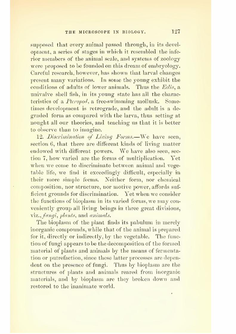
7/29/2019 THE MICROSCOPIST
http://slidepdf.com/reader/full/the-microscopist 139/361
THE MICROSCOPE IN BIOLO&Y. 127
supposed that every animal passed througli, in its devel-
opment, a series of stages in which it resembled the infe-
rior members of the animal scale, and systems of zoology
were proposed to be founded on this dream of embryology.
Careful research, however, has shown that larval changes
present many variations. In some the young exhibit the
conditions of adults of lower animals. Thus the Eolis, a
univalve shell fish, in its young state has all the charac-
teristics of a Pterojiod, a free-swimming mollusk. Some-
times development is retrograde, and the adult is a de-
graded form as compared with the larva, thus setting at
nought all our theories, and teaching us that it is better
to observe than to imagine.
12. Discrimination of Living Forms.—We have seen,
section 6, that there are different kinds of living matter
endowed with different powers. We have also seen, sec-
tion 7, how varied are the forms of multiplication. Yet
when we come to discriminate between animal and vege-
table life, we find it exceedingly difficult, especially in
their more simple forms. ISTeither form, nor chemical
composition, nor structure, nor motive power, affords suf-
ficient grounds for discrimination. Yet when we consider
the functions of bioplasm in its varied forms, we may con-
veniently group all living beings in three great divisions,
Yiz., fungi^plants, and animals.
The bioplasm of the plant finds its pabulum in merely
inorganic compounds, while that of the animal is prepared
for it, directly or indirectly, by the vegetable. The func-
tion of fungi appears to be the decomposition of the formed
material of plants and animals by the means of fermenta-
tion or putrefaction, since these latter processes are depen-
dent on the presence of fungi. Thus by bioplasm are the
structures of plants and animals reared from inorganic
materials, and by bioplasm are they broken down and
restored to the inanimate world.

7/29/2019 THE MICROSCOPIST
http://slidepdf.com/reader/full/the-microscopist 140/361
128 THE MICROSCOPIST.
CHAPTER X.
THE MICROSCOPE IN VEGETABLE HISTOLOGY AND BOTANY.
Histology (from ((rroq^ a tissue) treats of formed mate-
rial, or the microscopic structure resulting from the trans-
formation of germinal or living matter. The nature of
this transformation is partly physical and partly vital,
and, as alread}^ stated, is often so complex as to baffle all
chemical analysis. Some light, however, has been thrown
on this subject by the modification of ordinary crystalline
forms when inorganic particles aggregate in the presence
of certain kinds of organic matter. To this mode of form-
ation the name of molecular coalescence has been given.
Mr. Rainey and Professor Harting contemporaneously
experimented with solutions of organic colloids, and found
that the crystallization of certain lime salts, as the car-
bonate, was so modified by such solutions as to resemble
many of the calcareous deposits found in nature. These
researches leave little doubt but that a majority of calca-
reous and silicious organic forms may be thus accounted
for. Such changes are rather physical than vital.
Cell-substance in Vegetables.—The protoplasm or bio-
plasm in vegetable-cells cannot be distinguished from ani-
mal "sarcode" or protoplasm except by the nature of the
pabulum or aliment necessary to its nutrition. The vege-
table, under the stimulus of light, decomposes carbonic
acid, and acquires a red or green color from the compounds
which it forms, while the animal requires nutriment from
pre-existing organisms. Yet this definition fails to apply
to fungi, which resemble primitive animals even in this
respect. So difficult is it to discriminate that the simpler
forms of vegetables have often been classed by naturalists
among animals, and vice versa. Amoeboid movements
have been observed in the bioplasm of vegetable-cells,

7/29/2019 THE MICROSCOPIST
http://slidepdf.com/reader/full/the-microscopist 141/361
THE MICROSCOPE IN HISTOLOGY AND BOTANY. 129
especially in the Volvo.i\ and some have considered it
probable that an organism may live a truly vegetable life
at one period and a truly animal life at another.
Analogous to amoeboid movements, is the motion of
bioplasmic fluid in the interior of undoubtedly vegetable
cells. This movement is called cydosis^ and may be de-
tected under the microscope by the granules or particles
which the current carries with it in the transparent cells
of CA(2rfl,T^a^/is7?ma, etc., and in the epidermic hairs of
many plants, as Tradescantia, Plantago, etc. (Plate VI^
Fig. 91).
The bioplasm of plants may be stained with carmine-
solution without affecting the cell-wall or other formed
material.
Cell-wall or 3Iemhrane.—Plants, whether simple or
complicated in structure, are but cells or aggregations of
cells. In the simplest vegetables or Protophytes^ each cell
lives, as it were an independent life, performing every
function; while in the higher plants, as the palm or oak^
the cells undergo special modifications, and serve various-
functions subsidiary to the life of the plant as a whole.
Cell-membrane, or the envelope of formed material, wa&
formerly thought to be composed of two^ layers, to the
inner one of which the name of primordial utrirle was
given, but this is now considered to be but the external
surface of the bioplasm or germinal matter.
The chemical nature of cell-membrane is nearly identi-
cal with starch, being composed of cellulose. The presence
of cellulose may be shown by the blue color which is
produced by applying iodine and sulphuric acid, or the
iodized solution of chloride of zinc.
Endosmose will take place in cell-membrane, allowing
solutions to pass through, as pabulum, and the manner of
this passage may in some instances determine the subse-
quent deposit of formed material. Sometimes actual pores
are left in the membrane, as in Sphagnum (Plate VI, Fig.
•J

7/29/2019 THE MICROSCOPIST
http://slidepdf.com/reader/full/the-microscopist 142/361
130 THE MICROSCOPIST.
92). The walls of vegetable-cells are often thickenedby
deposit. If this is in isolated patches, the cells are called
dotted (Plate YI, Fig. 93), and it is sometimes difficult to
distinguish them from porous cells. Many cells have a
spiral fibre (Plate VI, Fig. 94), which appears to have-
been detached from the outer membrane. In the seeds of
Collomia, etc., the cell-wall is less consolidated than the
deposit, so that on softening the cells by water, the spiral
fibres suddenly spring out, making a beautiful object for
a half-inch object-glass (Plate VI, Fig. 95).
The tendency of formed material to arrange itself in a
spiral is seen in the endochrome of many of the simpler
plants, as Zygnema, and the cell-wall sometimes tears most
readily in a spiral direction.
If the. spiral deposit is broken and coalesces at some of
its turns, it forms an annulus or ring. Some cells show
both rings and spirals.
For the production of a spiral movement or growth,
another force is needed in addition to the centripetal and
centrifugal forces which are necessary for curvilinear mo-
tion. The centripetal point must be carried forward in
space by a progressive force. When we consider that a
spiral form is so frequently seen in morphology, that the
secondary planets move in spirals round their primaries,
and that even in distant nebulae the same law prevails,
we are struck with the unity of plan which is exhibited
throughout the universe, and can scarcely fail to observe
that even a microscopic cell shows the tracings of the same
divine handiwork which swings the stars in their courses.
Sderogen— Ligneous Tissue.— Sometimes the deposit
within the cell-wall is of considerable thickness, and often
in concentric rings, through which a series of passages is
left so that the outer membrane is the only obstacle to
the access of pabulum, as in the stones of fruit, gritty tis-
sue of the pear, etc. (Plate VII, Fig. 96). The nature of
this deposit is similar to cellulose, although often contain-

7/29/2019 THE MICROSCOPIST
http://slidepdf.com/reader/full/the-microscopist 143/361
PLATE VI.
Fig. ||!, Fig. 93.
Dotted cells— pith of Klder.
Fig. 94.
Circulation of fluid in hairs of Tradescanda
Vlrginica.
Spiral cells : a, Balsam ; b, c,
Pleurothallis.
Fig. 92. Fig. 95.
P
I
Portion of the leaf oi Sphagnum. Spiral fibres of seed-coat of Colhmin.

7/29/2019 THE MICROSCOPIST
http://slidepdf.com/reader/full/the-microscopist 144/361

7/29/2019 THE MICROSCOPIST
http://slidepdf.com/reader/full/the-microscopist 145/361
THE MICROSCOPE IN HISTOLOGY AND BOTANY. 131
ing resinous and other matters. Woody fibre or ligneous
tissue is quite similar, save that the cells have become
elongated or fusiform, and when completely filled up with
internal deposit, fulfil no other purpose than that of me-
chanical support (Plate VII, Fig. 97). The woody fibres
of the ConifercB exhibit peculiar markings, which have
been called glandular (Plate VII, Fig. 98). In these the
inner circle represents a deficiency of deposit as in other
porous cells, wdiile the outer circle is the boundary of a
lenticular cavity between the adjacent cells. This ar-
rangement is so characteristic as to enable us to determine
the tribe to which a minute fragment, even of fossil wood,
belonged.
S-piral Vessels.—If spiral cells are elongated, or coalesce
at their ends, they become vessels, some of which convey
air and some fluid (Plate VII, Fig. 99). As in cells, the
want of continuity in the spiral fibre sometimes produces
rings, when the duct is called annular. In other instances
the spires are still more broken up by the process of growth,
so as to form an irregular network in the duct, which is
then said to be reticulated. A still greater variation in
the deposit produces dotted ducts. ISTot infrequently we
find all forms in the same bundle of vessels.
Laticiferous Vessels (Plate VII, Fig. 100).—These con-
vey the milky juice or latex of such plants as possess it,
as the Euphorbiacese, india-rubber plant, etc., and clifter
from the ducts above described by their branching, so as
to form a network, while ducts are straight and parallel
with each other.
The laticiferous vessels resemble the capillary vessels of
animals, while the spiral ducts remind us of the trachea
of insects.
Siliceous Structures.—The structures of many plants,
especially the epidermis, often become so permeated with
a deposit of silica, that a complete skeleton is left after
the soft vegetable matter is destroyed. The frustules of

7/29/2019 THE MICROSCOPIST
http://slidepdf.com/reader/full/the-microscopist 146/361
132 THE MICROSCOPIST.
Diatoms have in this way heen preserved in vast numbersin the rocky strata of the earth. The markings on these
siliceous shells are so delicate as to be employed as a test
of microscopic power and definition. In a species of
Equisetum or Dutch rush, silica exists in such abundance
that the stems are sometimes employed by arti'sans as a
substitute for sand-paper. If such a stem is boiled and
macerated in nitric acid until all the softer parts are de-stroyed, a cast of pure silica will exhibit not only the
forms of the epidermic cells, but details of the stomata or
pores. The same also is true of the husk of a grain of
wheat, etc., in which even the fibres of the spiral vessels
are silicified. The stellate hairs of the siliceous cuticle
from the leaf of DeAitzia scahra fornis a beautiful polari-
seope object.
Formed Material within Vegetable Cells.
1. Haphides.—These are crystalline mineral substances,
principally oxalate, citrate, and phosphate of lime. They
occur in all parts of the plant, sometimes in the form of
bundles of delicate needles, sometimes in larger crystals,
and sometimes in stellate or conglomerate form. Mr. E.
Quekett produced such forms artificially by filling the
cells of rice-paper with lime-water under an air-pump, and
then placing the paper in weak solutions of phosphoric or
oxalic acid,
2. Starch.—This performs in plants a similar function
to that of fat in animals, and is a most important ingre-
dient in human food, since two-thirds of mankind subsist
almost exclusively upon it. It is found in the cells of
plants in the form of granules or secondary cells. Each
granule under the microscope shows at one extremity a
circular spot or hilum, around which are a number of
curved lines, supposed to be wrinkles in the cell-membrane.
When starch is boiled in water, this membrane bursts and

7/29/2019 THE MICROSCOPIST
http://slidepdf.com/reader/full/the-microscopist 147/361
PLATE VI r.
Flo. tm.
Gritty tissue—Pear.
Spiral vessels: a, reticulated; B, old
vessel, with perforations; c, d, spiral
vessels, Ijecuming annular.
Fig. 97.Fiii. 100.
Wood-fibre—flax.Lactiferous vessels
Fig. 101.
.Section of Coniferous Wood in the
direction of the fibres.
Cubical parenchj'nia, with stellate cells
from petiole of Ntiphar luUa.

7/29/2019 THE MICROSCOPIST
http://slidepdf.com/reader/full/the-microscopist 148/361

7/29/2019 THE MICROSCOPIST
http://slidepdf.com/reader/full/the-microscopist 149/361
THE MICROSCOPE IN HISTOLOGY AND BOTANY. 133
the amylaceous matter is dissolved. Iodine stains starch
blue. Starch shows in the polariscope a black cross in
each grain, changing to white as the prism is revolved.
3. Chlorophyll is the green coloring matter of plants.
It is usuall_y seen in the form of granules of bioplasm in
the interior of cells. These green granules yield their
chlorophyll to alcohol and ether. It seems to be neces-
sary to nutrition, since green plants under the stimulus
of light break up carbonic acid into oxygen and carbon,
the latter of which is absorbed.
The red and yellow color of autumn leaves is owing to
the chemical metamorphosis of chlorophyll, as also is the
red color of many of the lower Algte, etc. In the latter
it seems to be in some w^ay connected with the vital pro-
cesses.
4. The coloring matter offlowers is various, and ordinarily
depends on the colored fluid contained in cells subjacent
to the epidermis, although sometimes it is in the form of
solid corpuscles. White patches on leaves, etc., arise from
absence of chlorophyll.
5. Milkyjuices are true secretions contained in the lati-
ciferous ducts. The juice of the dandelion, caoutchouc
or india-rubber, which is the concrete juice of the Ficus
elastica^ and gutta-percha, from Isonandra gutta, are exam-
ples.
6. Fixed oils are found in the cells of active tissues, and
notably in seeds, w^here they serve to nourish the embryo.
Cocoanut, palm, castor, poppy, and linseed oils are exam-
ples.
7. Volatile oil, sometimes called essential oil, is chiefly
found in glandular cells and hairs of the epidermis.
Many of them yield a resinous substance by evaporation.
8. Camphor is analogous to volatile oil, although solid
at ordinary temperatures. It abounds in the Lauracese.
9. Resin, wax, and tallow are also found in plants. The
bloom of the plum and grape is due to wax.

7/29/2019 THE MICROSCOPIST
http://slidepdf.com/reader/full/the-microscopist 150/361
134 THE MICROSCOPIST.
10.
Gumis a viscid secretion. What is called gum
tragacanth, is said to be partially decomposed cell-mem-
brane, and is allied to amyloid matter.
Forms of Vegetable Cells.—From the account given in
the chapter on biology, page 128, it is evident that the
form of cells is quite varied, and often depends on the
amount of pressure from aggregation, yet function also
has much to do in the determination of shape. Thus
while most elongated cells are lengthened in the direction
of plant-growth, hi which is least resistance, the medul-
lary rays of Exogenous stems are elongated in a horizon-
tal direction. Some cells are cubical, as in the leaves of
the yellow water-lily, Nuphar luiea (Plate VII, Fig. 101).
Others are stellate, as in the rush (Plate VIII, Fig. 102).
In many tissues are large cavities or air-chambers alto-
gether void of cells, and in leaves such cavities communi-
cate with the external air by means of siomata or pores
(Plate VIII, Fig. 103), which are usually provided with
peculiar cells for contracting or widening the orifice.
The Botanical Arrangement of Plants.—Considered with
reference to their general structure, plants are divided by
botanists into cellular and vascular. The first of these
classes is of greatest interest to the microscopist, as em-
bracing the minuter forms of vegetable life.
The classification and natural grouping of plants is yet
far from being perfect, although microscopic examinations
have largely contributed to an orderly arrangement of the
multitudinous varieties in this field of research. In the
present work we propose only a brief outline of typical
subjects of interest, with the methods of microscopic ex-
amination.
Fungi.—At page 127 it was stated that all living beings
may be grouped in three divisions, fungi, plants, and
animals. Botanists generally class fungi among cellular
flowerless plants. They cannot assimilate inorganic food
as other plants, but live upon the substance of animal or

7/29/2019 THE MICROSCOPIST
http://slidepdf.com/reader/full/the-microscopist 151/361
PLATE VIII.
Section of cellular parenchyma of Rush. Portion of the cuticle of the leaf of the Iris
Gennanica, torn from its surface.
Fig. 104.
Cells from the petal of the Geranium{Pelargoniuvij. Cuticle of leaf of Indian Corn (Zeamais).

7/29/2019 THE MICROSCOPIST
http://slidepdf.com/reader/full/the-microscopist 152/361

7/29/2019 THE MICROSCOPIST
http://slidepdf.com/reader/full/the-microscopist 153/361
THE MICROSCOPE IN HISTOLOGY AND BOTANY. 135
vegetable tissue. They also cliifer from ordinary vegeta-
bles by the total absence of chlorophyll or its red modifi-
cation. A laro-e number of this strange class are micro-
scopic, and require high powers for their observation.
Eecent investigations show that individual fungi are de-
veloped in very dissimilar modes, and are subject to a
great variety of form, rendering it probable that those
which seem most simple are but imperfectly developed
forms. Amceboid motions also in the cell-substance of
certain kinds of fungi, and the projection of threads of
bioplasm, show a great resemblance to some of the lower
forms of animal life, as the Rhizojpods.
All fungi exhibit two well-defined structures, a myce-
lium or vegetative structure, which is a mass of delicate
filaments or elongated cells ; and a /rH«7 or reproductive
structure, which varies in different tribes. In Tonda^ one
or more globular cells are produced at the ends of fila-
ments composed of elongated cells ; these globules drop
off" and become new mycelia. The " yeast plant," or Torula
cerevisia (Plate IX, Fig. 107), receives its name from its
habitat. Fermentation depends upon its presence, as pu-
trefaction does upon the minute analogous bodies called
Bacteria and Vibriones. Bacteria are minute, moving,
rod-like bodies, sometimes jointed ; and vibriones are
moniliform filaments, havino- a vibratile or wrio-o-lino; mo-
tion across the field of view in the microscope. The re-
searches of Madame Luders render it probable that the
germs of fungi develop themselves into these bodies when
sown in water containing animal matter, and into yeast
in a saccharine solution. The universal diff'usion of spor-
ules of fungi in the atmosphere readilj^ accounts for their
appearance in such fluids, and Pasteur's exjDeriments are
quite conclusive.
The minute molecules called microzymes, present in va-
rious products of disease, as the vaccine vesicle, fluid of
glanders, etc. ; the minute corpuscles which cause the dis-

7/29/2019 THE MICROSCOPIST
http://slidepdf.com/reader/full/the-microscopist 154/361
136 THE MICROSCOPIST.
ease among silkworms called "pebrine;" etc.; have a
strong analogy in their rapid multiplication to the yeast-
eells.
The spornles of any of the ordinary moulds, as Penidl-
lium, Mueor, or Aspergillus, will develop into yeast-cells
ill a moderately warm solution of cane-sugar, showing
how diflterently the same type of bioplasm may develop
under different conditions. The term j^olymorphism has
been given to this phenomenon. Yery many species, and
even genera, so called, may after all be only varieties of
the same kind of organism.
In many morbid conditions of the skin and mucous
membranes, there is not only an alteration or morbid
growth of the part, but a vegetation of fungi. Thrown-
off scales of epithelium from theniouth and fauces exhibit
fibres of lejjtothrix, and the false membrane of diphtheria,
as well as the white patches of aphtha or thrush, show
the mj^celia and spores of fungi. The disease in silkworms
called muscardiue is due to a fungus, the Botrytis bassiana
(Plate VIII, Fig. 104), whose spores enter and develop in
the air-tubes. The filamentous tufts seen about dead flies
on window-panes, etc., arise from a similar growth of
Achyla. In certain Chinese or Australian caterpillars,
this sort of growth becomes so dense as to give them the
appearance of dried twigs. Even shells and other hard
tissues may become penetrated by fungi. The dry rot in
timber is a form of fungus.
The mildew which attacks the straw of wheat, etc.,
arises from the Pucciiiia graminis, whose spores find their
way through the stomata or breathing pores of the epi-
dermis. Rust, and smut, and bunt, originate in varieties
of TJredo. The "vine disease" and the "]30tato disease,"
as they are called, have similar origin.
Various methods have been proposed to destroy fungi
in growing plants, but it must be remembered that the
function of these organisms is chiefly to remove formed

7/29/2019 THE MICROSCOPIST
http://slidepdf.com/reader/full/the-microscopist 155/361
PLATE IX.
Fig. 107.
O/^
.^f
Torida Cereviske, or Yeast-Plaiit.
Fig. 109.
ri^.^^
Genu aud Sperm-cells in Achyla.
Fig. 108.
Development of fungi: a, mycelium;b, liyplia; c, conidiophores; d, a mngnified branch.
m '^3 MA fimil >=< /;^^;Sl^ [f.
Various pliases of development of Paimoglcea macrococca.

7/29/2019 THE MICROSCOPIST
http://slidepdf.com/reader/full/the-microscopist 156/361

7/29/2019 THE MICROSCOPIST
http://slidepdf.com/reader/full/the-microscopist 157/361
THE MICROSCOPE IN HISTOLOGY AND BOTANY. 137
material in a state of decay, which is more or less com-
plete. The prevalence of atmospheric changes, variations
in light, heat, moisture, and electricity, etc., have much
to do in predisposing vegetable as well as animal tissues to
disease and producing epidemics. The agriculturist, there-
fore, as well as the physician, must discriminate between
those diseased conditions which provide a habitat for
fungi, and the effects produced by the fungi themselves.
Impregnating wood with corrosive sublimate or chlo-
ride of zinc has been used to prevent dry rot in wood, and
soaking seeds, in alkaline solutions or sulphate of copper
is said to remove smut and similar fungus spores.
The development of fungi is from spores or coniJia.
Plate IX, Fig. 107, represents the Torula vegetating by
the budding of its spores. These buds rapidly fall ofi" and
become independent cells. In other varieties self-division
gives rise to the mycelium., a mass of fibres often inter-
laced so as to form a sort of felt. Some branches of this
mycelium (Jiyphce) hang down, while others rise above the
surface (conidiophores) and bear conidia, which fall off and
develop into new hyphfe (Plate IX, Fig. 108). In the
"blight" of the potato' the mycelium is loose, and the
hyph?e ramify in the intercellular spaces and give off pro-
jections into the cells of the plant. The conidia germinate
by bursting the sac which contains them, putting forth
cilia, moving awhile, then resting and enveloping them-
selves with membrane and growing into hyphfe. In the
autumn, parts of the hyphaj assume special functions.
One part develops a. spherical mass called oogonium, while
another becomes a smaller mass or antheruliwn. Whenthe first is ripe, it is penetrated by the latter, and the
bioplasms of each are fused together. The antheridium
then decays, while the oogonium grows and becomes au
oospore, in which the bioplasm divides and subdivides.
!Next season each segment escapes ciliated, and moves
about till it finds a place to germinate. In Achyla two

7/29/2019 THE MICROSCOPIST
http://slidepdf.com/reader/full/the-microscopist 158/361
138 THE MICROSCOPIST.
sacs are formed, one of which contains "germ-cells," and
the other (2n^Aero2:ozc?s or "sperm-cells." When both are
ripe the sac opens, and the ciliated antherozoids pass into
the neighboring sac and fertilize its contents (Plate IX,
Fig. 109).
In other fungi the reproductive cells are undistinguish-
able from the rest, and the coalescence takes place in a
new cell formed by the union of the other two.
Mr. Berkeley divides fungi into six orders, as follows
1. Hymenomycetes or Agaricoidece {Mushrooms, etc.).
Mycelium flocuose, inconspicuous, bearing fleshy fruits
which expand so as to expose the hymenium or sporifer-
ous membrane to the air. Spores generally in fours on
short pedicles.
2. Gasteromycetes or Lycoperdoidece {Puff balls, etc.).
Fruit globular or oval, with convolutions covered by the
hymenium, which bears the spores in fours on distinct
pedicles. The convolutions break up into a pulverulent
or gelatinous mass.
3. Coyiiom.ycetes or Uredoidece {Smuts, etc.).—Mycelium
filamentous, parasitic. Microscopic fructification of ses-
sile or stalked spores in groups, sometimes septate.
4. Hyphomycetes or Botrytoidece {Mildews, etc.).—Micro-
scopic. Mycelium filamentous, epiphytic, with erect fila-
ments bearing terminal, free, single, simple, or septate
spores.
5. Ascomycetes or Helvelloidece [Truffles, etc.).—Myce-
lium inconspicuous. Fruit fleshy, leathery, horny, or ge-
latinous, lobed, or warty, with groups of elongated sacs
{asci or thecce) in which the spores (generally eight) are
developed.
6. Pliysomycetes or Mucoroidece {Moulds).— Mycelium
(microscopic) filamentous, bearing stalked sacs containing
numerous minute sporules.
Protophytes, or primitive plants, afibrd many forms and
groups of great interest to the microscopist as well as to

7/29/2019 THE MICROSCOPIST
http://slidepdf.com/reader/full/the-microscopist 159/361
THE MICROSCOPE IN HISTOLOGY AND BOTANY. 139
the biologist. The plan of the present work permits us
only to indicate a few particulars, the details of which
would form a volume of considerable size.
The Algfe are divided into three orders: I. Rhodos'per-
mece or Florida'. {JRed-spored Alga). Marine plants, with
a leaf-like or filamentous rose-red or purple thallus. II.
Melanosporea or Facoidece [Daidc-siJored Algce). Marine.
Thallus leaf-like, shrubby, cord-like, or filamentous, of
olive-green or brown color. III. Chlorosporece or Confer-
voidece(Gtren-spored Algce). Plants marine or fresh water,
or growing on damp surfaces. Thallus filamentous, rarely
leaf-like, pulverulent, or gelatinous. These have been
subdivided into families, viz.
I. Rhodospermece.—1. Rhodomelacese. 2, Laurenciace?e.
3. Corallinaceae. 4. Delesseriacese. 5. Rhodymeniace?e.
6. Cryptonemiace?e. 7. Ceramiacese. 8. Forphyraceae.
II. 3Ielanospore(E.—1. Fucacese. 2. Dictyotaceae. 3.
Cutleriacese. 4. Laminariacese. 5. Dictyosiphonacese. 6.
Functariacese. 7. Sporochnacese. 8. Chordariaceee. 9.
Mja'ionemaccEe. 10. Ectocarpace?e.
III. Chlorosporece.—1. Lemaneese. 2. Batrachospermese.
3. Choetophoracepe. 4. ConfervaceaB. 5. Zj^gnemacefe.
6. OEdogoniace^e. 7. Siphonaceje. 8. Oscillatoriaceee. 9.
JN'ostochacese. 10. Ulvace?e. 11. Falmellacea3. 12. Des-
midiace^B. 13. Diatomaeeae. 14. Volvocine^s.
For fuller information, we refer to the MicrograpJnc
Dictionary by Griffith and Henfrey.
In the family of Pcdinellacece we find the simplest forms
of vegetation in the form of a powdery layer of cells, or a
slimy film, or a membranous frond. The green mould on
damp walls and the red snow of alpine regions are exam-
ples.
In the green slime on damp stones, etc., is found the
Pcdmoglcea macrococca. The microscope shows it to con-
sist of cells containing chlorophyll, surrounded by a ge-
latinous envelope. These cells multiply by self-division.

7/29/2019 THE MICROSCOPIST
http://slidepdf.com/reader/full/the-microscopist 160/361
140 THE MIOROSCOPIST.
Sometimes a conjugation or fusion of cells occurs,and
the
product is a spore or primordial cell of a new generation
(Plate IX, Fig. 110). During conjugation oil is produced
in tlie cells, and the chlorophyll disappears or becomes
brown, and when the spore vegetates, the oil disappears
and green granular matter takes its place. This is analo-
gous to the transformation of starch into oil in the seeds
of the higher plants.
Most of the lower forms of vegetable life pass through
what is called the motile condition, which depends on the
extension of the bioplasm into thread-like filaments, whose
contractions serve to move the cell throuo-h the water.
Many of these forms were formerly mistaken for animal-
cules, and the transformation of a portion of green chlo-
rophyll into the red form was represented as an ej-e. The
multiplication of the "still" cells is by self-division, as in
PalmogUea, but after this has been repeated about four
times, the new cells become furnished with cilia and pass
into the "motile" condition, and their multiplication goes
on in diflerent ways, as by binary or quaternary segmen-
tation, or the formation of a compound, mulberry-like
mass, the ciliated individual cells of which, becoming free,
rank as zoosjjores (Plate X, Fig. 111).
The Volvox is a beautiful example of the composite
motile form of elementary vegetation. It is found in
fresh water, and consists of a hollow pellucid sphere,
studded with green spots, connected together often by
green threads. Each of these spots has two cilia, whose
motions produce a rolling movement of the entire mass.
Within the sphere there are usually from two to twenty
smaller globes, which are set free by the bursting of the
original envelope. Sometimes one of the masses of endo-
chrome enlaro-es, but instead of undero-oino; subdivision
becomes a moving mass of bioplasm, which cannot be dis-
tinguished from a true AiiKEha or primitive animal cell.
The Desmidiacece are a family of minute g;.'een plants

7/29/2019 THE MICROSCOPIST
http://slidepdf.com/reader/full/the-microscopist 161/361
PLATE X.
Fig. 111.
Various phases of development of Protococcus pluvialis.
//-(
Fig. 112.
c
n^\o^
V
i. &\3 m
Formation of Zoospores in P/ii/coseri.s giganlea{Vlvn latissima).

7/29/2019 THE MICROSCOPIST
http://slidepdf.com/reader/full/the-microscopist 162/361

7/29/2019 THE MICROSCOPIST
http://slidepdf.com/reader/full/the-microscopist 163/361
THE MICROSCOPE IN HISTOLOGY AND BOTANY. 141
of great interest. Generally the cells are independent,but a filament is sometimes formed hy binary subdivision.
Their symmetrical shape, and frequently spinous projec-
tions and peculiar movements, render them beautiful ob-
jects. By conjugation a spore-cell or sporangium, is pro-
duced, T^diich in some species is spinous, and reseitibles
certain fossil remains in flint, which have been described
as animalcules under the name of Xanthidia.The family of Diatomacece aftbrds more occupation to
microscopists than other protophytes. Like the Desmids,
they are simple cells with a firm external coating, but in
Diatoms this coating is so penetrated with silex, that a
cast of the frustule is left after the removal of the organic
matter. Eeference has already been made to the number
of these organisms in a fossil state, as well as to their
utility as tests of the defining power of microscopic ol)ject-
glasses.
Some species inhabit the sea, and others fresh water.
Thej^ are so numerous that scarcely a ditch or cistern is
free from specimens, and they multiply so rapidly as to
actually diminish the depth of channels and block up
harbors. They may be sought for in the slimy masses
attached to rocks and plants in water, in the scum of the
surface, in mud or sand, in guano, in the stomachs of
molluscs, etc., and on sea-weeds.
To separate the shields or siliceous frustules from foreign
matter, either fresh or fossil, they should be washed sev-
eral times in water, and the sediment allowed to subside.
The deposit should then be treated in a test-tube with
hydrochloric acid, sometimes aided by heat. This should
be repeated as often as any effect is produced, and then
the sediment should be boiled in strong nitric acid, and
washed several times in water. They may be mounted
dry or in balsam.
The classification of Diatoms is not yet perfected, but
Muller's type slides, containing from one hundred to five

7/29/2019 THE MICROSCOPIST
http://slidepdf.com/reader/full/the-microscopist 164/361
142 THE MICROSCOPIST.
hundred characteristic forms, is a valuable assistance.
The following table, from the Micrograiihic Dictionary^
gives an analysis of tribes and genera : Fr. denotes the
frustules in front view; v. the valves; granular strise
means strise resolvable into dots ; and continuous strise
signify cost^e or canaliculse.
A. Fnishdes not contained in a Gelatinous Mass or Tube.
Tribe I. Striata.—Frustules usually transversely stri-
ate, but neither vittate nor areolate.
f Valves without a Median Nodule.
Cohort 1. Eunotie^.—Fr. arcuate, single, or united
into a straight filament.
1. JEpithemia.—Fr. single or binate, wnth transverse or
slightly radiant strife, some continuous ; no terminal nod-
ules; aquatic and marine.
2. Eunotia.—Fr. single or binate; v. with slightly ra-
diant granular strife and terminal nodules; aquatic.
3. Himanticlium..—Fr. as in Eujiotia, but united into a
filament; strise parallel, transverse; aquatic.
Cohort 2. Meeide^.—Fr. cuneate, single, or united
into a curved or spihal band ; v. with continuous or gran-
ular stripe.
4. Meridion.—Fr. cuneate, united into a spiral band;
striae continuous ; aquatic.
5. JEucampia.—Fr. united into an arched band ; v. punc-
tate ; marine.
6. Oncosphenia.—Fr. single, cuneate, uncinate at the
narrow end; strife granular; aquatic.
Cohort 3. FRAOiLLARiEiE.—Fr. quadrilateral, single, or
united into a filament or chain ; v. wdth continuous or
granular strife.
7. Diatoma.—Fr. linear or rectangular, united by the

7/29/2019 THE MICROSCOPIST
http://slidepdf.com/reader/full/the-microscopist 165/361
THE MICROSCOPE IN HISTOLOGY AND BOTANY. 143
angles so as to form a zigzag chain ; striae continuous
aquatic and marine.
8. Asierionella.—Fr. adherent by adjacent angles into
a star-like filament ; v. inflated at one or both ends
aquatic.
9. Fragillaria.—Fr. linear, united into a straight, close
filament ; stride granular, faint ; aquatic and marine.
10. Denticula.—Fr. linear, simple, or binate, rarely more
united ; strise continuous ; aquatic.
11. Odontidium.—As Denticula, but fr. forming a close
filament; aquatic and marine.
Cohort 4. MELOSiREiE.—Fr. cylindrical, disk-shaped or
globose; v. punctate, or often with radiate continuous
or granular striae.
12. Cyclotella.—Fr. disk-shaped, mostly solitary; v.
M'ith radiate marginal striae ; aquatic.
13. Melosira.—Fr. cylindrical or spherical, united into
a filament; v. punctate, or with marginal radiate granu-
lar striEe; aquatic and marine.
14. Podosira.—Fr. united in small numbers, cylindrical
or spherical, fixed by a terminal stalk; v. hemispherical,
punctate; marine.
15. JIastogonia.—Fr. single; v. unequal, angular, mam-miform, circular at base, without umbilical processes
angles radiating; fossil.
16. Pododiscus.—Fr. single or united, with a marginal
stalk ; V. circular, convex.
17. Pyxidicula.—Fr. single or binate, free or sessile;
V. convex ; aquatic and marine.
18. Stephanodiscus.—Fr. single, disk-shaped; v. circu-
lar, equal, punctate, or striate, with a fringe of minute
marginal teeth ; aquatic.
19. Steiihanogonia.—Fr. as in Mastogonia, but ends of
valves truncate, angular, and spinous; fossil.
20. Hercotheca.—Fr. single, turgid laterally ; v. with
marginal free setae.

7/29/2019 THE MICROSCOPIST
http://slidepdf.com/reader/full/the-microscopist 166/361
144 THE MICROSCOPIST.
21. Goniothecium.
—Fr. single, constricted in the middle,
suddenly attenuate and truncate at the ends (hence appear-
ing angular).
Cohort 5. SuRiRELLEiE.—Fr. single or binate, quadri-
lateral, oval, or saddle-shaped, sometimes constricted in
the middle; v. with transverse or radiating continuous
or granular stride, interrupted in the middle, or with one
or more longitudinal rows of puncta; often keeled.
22. Bacillaria.—Fr. prismatic, straight, at first forming
a filament; v. with a median longitudinal row of puncta;
marine.
23. Campylodiscus.—Fr. single, free, disk-shaped; v.
curved or twisted (saddle-shaped); aquatic and marine.
24. Doryphora.—Fr. single, stalked ; v. lanceolate or
elliptical, with transverse granular strise.
25. PoJocysiis.—Fr. attached, sessile; v. with a median
line, transverse continuous, and intermediate granular
strise.
2(3. Nitzschia.—Fr. free, single, compressed, usually elon-
gate, straight, curved, or sigmoid, with a not-median
keel, and one or more longitudinal rows of puncta
aquatic and marine.
27. Spli indocystls(Cymatopleura).—Fr. free, single, linear,
with undulate margins; v. ohlong or elliptical, sometimes
constricted in the middle; aquatic.
28. Sarirella.—Fr. free, single, ovate, elliptical, oblong,
cuneate, or broadly linear ; v. with a longitudinal median
line or clear space, margins winged, and with transverse
or slightly radiating continuous striae ; aquatic and marine.
29. Synedra.—Fr. prismatic, rectangular, or curved ; at
first attached to a gelatinous-lobed cushion, often becom-
ing free; v. linear or lanceolate, usuallj^ with a median
pseudo-nodule and longitudinal line; aquatic and marine.
30. Tryhlionella.—Fr. free, linear, or elliptical; v. plane,
with a median line, transverse striae, and submarginal or
obsolete aloe; aquatic and marine.

7/29/2019 THE MICROSCOPIST
http://slidepdf.com/reader/full/the-microscopist 167/361
THE MICROSCOPE IN HISTOLOGY AND BOTANY. 145
81. JRaphoneis.—Dorj/pJiora without a stalk.
Cohort 6. AmphipleurExE.—Fr. free, single, straiglit,
or slightly signioid ; v. lanceolate, or linear-lanceolate,
with a median longitudinal line.
32. Amphipleura.—Characters as above.
ft Valves with a Median Nodule.
Cohort 7. Cocconeid.e.—Fr. straight or bent, attached
by the end or side; v. elliptical, equilateral.
33. Cocconeis.—Fr. single,. compressed, adnate; v. ellip-
tical, one of them with a median line.
Cohort 8. Achxanthe^e.—Fr. compressed, single, or
rarely united into a straight filament, curved, attached
by a stalk at one angle; uppermost v. with a longitudinal
median line, lower v. the same, and a stauros or transverse
line; marine.
35. Achnanthidium.—Fr. those of Achnanthes, bnt free
aquatic.
36. Cymbosira.—Fr. as Achnanthes^ solitary or binate,
stipitate, and attached end to end ; marine.
Cohort 9. CYMBELLEiE.—Fr. straight or curved, free or
stalked at the end ; v. inequilateral, not sigmoid.
37. Cymhella.—Fr. free, solitary; v. navicular, with a
subcentral and two terminal nodules, and a submedian
longitudinal line; aquatic.
38. Cocconema.—Fr. as Cymhella., but stalked; aquatic.
Cohort 10. Gomphoneme^.—Fr.wedge-shajDed, straight,
free, or stalked; v equilateral.
39. Gomphonema.—Fr. single or binate, wedge-shaped,
attached by their ends to a stalk; v. wdth a median line,
and a median and terminal nodules; aquatic.
40. Sphenella.—Fr. free, solitar3^ wedge-shaped, invo-
lute; aquatic.
30'

7/29/2019 THE MICROSCOPIST
http://slidepdf.com/reader/full/the-microscopist 168/361
146 THE MICROSCOPIST.
41. Sphenosira.—Fr. united into a straight filament; v.
wedge-shaped, at one end rounded, suddenly contracted
and produced ; aquatic.
Cohort 11. NAVicuLEiE.—-Fr. free, straight; v. equilat-
eral, or sometimes sigmoid.
42. Navicula.—Fr. single, free, straight; v. oblong, lan-
ceolate, or elliptical, with a median line, a central and two
terminal nodules, and transversely or slightly radiant lines
resolvable into dots ; aquatic, marine, and fossil.
43. Gyroslgma {Pleurosigm.a).—Fr. as Navicula^ but v.
sigmoid ; aquatic and marine.
44. Pinnularia.—Fr. as Navicula^ but transverse lines
continuous ; aquatic and marine.
45. Stauroneis.—Fr. as Navicula^ but the median line
replaced l)y a stauros; aquatic and marine.
46. Diadesmis.—Fr. as Navicula^ united into a straight
filament; aquatic.
47. Amyhiprora.—Fr. free, solitary, or in pairs, con-
stricted in the middle ; v. with a median keel, and a
median and terminal nodules, often twisted ; marine.
48. Aniphora.—Fr. plano-convex, elliptical, oval or ob-
long, solitary, free or adnate, with a marginal line, and a
nodule or stauros on the fiat side ; aquatic and marine.
Tribe II. YiTTATiB.—Fr. with vittse.
f Valves without a Median Nodule.
Cohort 12. Licmophore^.—Fr. cuneate; vittfe arched.
49. Licmo2)hora.—Fr. cuneate, rounded at the broad
end, radiating from a branched stalk; vittse curved (by
inflection of upper margins of valves) ; marine.
50. Podosphenia.—Fr. as Licino-phora, but single or in
pairs, sessile on a thick but little branched pedicle; ma-
rine.

7/29/2019 THE MICROSCOPIST
http://slidepdf.com/reader/full/the-microscopist 169/361
THE MICROSCOPE IN HISTOLOGY AND BOTANY. 147
51. Ehiindophora.—Fr. as Licmophora, single or in pairs,
on a branched stipes ; marine.
52. Climacosphenia.—Fr. cuneate, ronnded at broad end,
divided into loculi by transverse septas or vitt?e ; marine.
Cohort 13. STRiATELLEiE.—Fr. tabular or filamentous;
vittse straight (not arched).
53. Striatella.—Fr. compound, stalked at one angle;
vittse longitudinal and continuous ; v. elliptic-lanceolate,
not striated ; marine.
54. JRhabdonema.—Fr. as Striatella., but vittee inter-
rupted ; v. with transverse granular strise ; marine.
55. Tetracyclus.—Fr. compound, filamentous ; vittfe al-
ternate, interrupted ; v. inflated at the middle ; strife trans-
verse, continuous;aquatic.
56. Tabellaria.—Fr. united into a filament, subsequently
breaking up into a zigzag chain ; vittfe interrupted, alter-
nate ; V. inflated at middle and ends ; aquatic.
57. Pleurodesmiuin.—Fr. tabular, united into a filament,
and with a transverse median hyaline band; marine.
58. Hyalosira.—Fr. tabular, fixed by a stalk at one
angle ; vittfe alternate, interrupted, bifurcate at the end;
marine.
59. Anaulus.—Fr. rectangular, single, compressed, with
lateral inflections, giving the valves a ladder-like appear-
ance ; marine.
60. Bihlarium.—Fr. as Tetracyclus., but single ; fossil.
61. Teriosince.—Fr. tabular, obsoletely stalked, subse-
quently connected by isthmi ; vittfe transverse, short, in-
terrupted, and capitate ; aquatic and marine.
62. Stylobihlium.—Fr, compound; v. circular, sculptured
with continuous strife ; fossil.
tt With a 3Iedian crpimrent [yseudo) Nodule.
63. Gramraatophora.—Fr. at first adnate, afterwards

7/29/2019 THE MICROSCOPIST
http://slidepdf.com/reader/full/the-microscopist 170/361
l-iS THE MICROSCOPIST.
forming a zigzag chain ; vittse two, longitudinal, inter-
rupted, and more or less figured ;marine.
Tribe III. AREOLATiE.—Valves circular, witli cell-like
(areolar) markings, visible hy ordinary illumination.
Sub-tribe 1. Disciformes.—Valves alike, without ap-
pendages or processes.
Cohort 14. Coscinodisce^..—Valves circular.
64. Actinocydus.—Fr. solitary; v. circular, undulate,
the raised portions like rays or bands radiating from the
centre, which is free from markings ; marine and fossil.
65. Actinoptychus.—Fr. as Actinocydus, but radiating
internal septpe, as well as rays.
66. Coscinodiscus.—Fr. single ; v. circular, areolar all
over ; marine and fossil.
67. Arachnoidiscus.—Fr. single ; v. circular, not undu-
late, with concentric and radiating lines, and intermediate
areola absent from the centre (pseudo-nodule) ; marine
and fossil.
68. Asterolamjyra.—Fr. single; v. circular, finely areolar,
except in the centre and at equidistant clear marginal rays
radiating from the centre, which is traversed by radiating
dark lines (septa), alternating with the marginal rays;
fossil.
69. Asterom-phalos.—-As Asterolampra, but two of the
central dark lines parallel, and the corresponding mar-
ginal ray obliterated ; fossil.
70. JJalionyx.—Fr. single ; v. circular, without septa,
with rays not reaching the centre, and with intermediate
shorter rays ; between the rays transverse areolar lines
fossil.
71. Odontodiscus.—Fr. single, lenticular ; v. covered
with puncta (areolae), arranged in radiating rows on ex-
centrically curved lines, and with erect marginal teeth;
fossil.
72. Omrphalojjelta.—As Actinoptychus, but upper part of
margin of valves with a few erect spines ; fossil.
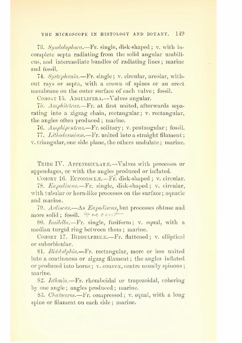
7/29/2019 THE MICROSCOPIST
http://slidepdf.com/reader/full/the-microscopist 171/361
THE MICROSCOPE IN HISTOLOGY AND BOTANY. 149
73. Si/nibolophora.—Fr. single, disk-shaped ; v. witli in-
complete septa radiating from the solid angular umbili-
cus, and intermediate bundles of radiating lines ; marine
and fossil.
74. Systephania.—Fr. single ; v. circular, areolar, with-
out rays or septa, with a crown of spines or an erect
membrane on the outer surface of each valve ; fossil.
Cohort 15. Angulifera.—Valves angular.
75. Amphitetras.—Fr. at first united, afterwards sepa-
rating into a zigzag chain, rectangular; v. rectangular,
the angles often produced ; marine.
76. Amphipentras.—Fr. solitary; v. pentangular ; fossil.
77. Lithodesmiinn.—Fr. united into a straight filament
V. triangular, one side plane, the others undulate ; marine.
Tribe IV. AppENDicuLATyE.—Valves with processes or
appendages, or with the angles produced or inflated.
Cohort 16. Eupodisce^.—Fr. disk-shaped ; v. circular.
78. Eupodiscus.—Fr. single, disk-shaped ; v. circular,
with tubular or horn-like processes on the surface ; aquatic
and marine.
79. Aidiaciis.—As Eupodiscus^hwt processes obtuse and
more solid ; fossil. "^ •*-€ o ^-^tf—
80. Insilella.—Fr. single, fusiform ; v. equal, with a
median turgid ring between them ; marine.
Cohort 17. Biddulphie^.—Fr. flattened ; v. elliptical
or suborbicular.
81. Biddidphia.—Fr. rectangular, more or less united
into a continuous or zisTzag; filament ; the angles inflated
or produced into horns ; v. convex, centre usually spinous;
marine.
82. Isthmia.—Fr. rhomboiclal or trapezoidal, cohering
by one angle ; angles produced ; marine.
83. Chceioceros.—Fr. compressed ; v. equal, with a long
spine or filament on each side ; marine.

7/29/2019 THE MICROSCOPIST
http://slidepdf.com/reader/full/the-microscopist 172/361
150 THE MICROSCOPIST.
84. lihizoselenia.—Fr. elongate, subcylindrical, markedwith transverse or spiral lines, ends oblique or conical,
and with one or more terminal bristles ; marine.
85. Hemiaidus.—Fr. single, compressed, rectangular
angles produced into tubular direct processes, those on
one valve longer than on the other ; fossil.
86. Syringidium.—Fr. single, terete, acuminate at one
end, two-horned at the other;
marine.87. Periptera.—Fr. single, compressed ; v. unequal, one
simply turgid, the other with marginal wings or spines;
fossil.
88. Didadia.—Fr. single ; v. unequal, one turgid and
simple, the other two-horned ; fossil.
Cohort 18. AjsrauLATiE.—Valves ano;ular.
89.Triceraii'um.
—Fr. free;
v. triangular, each anglewith a minute tooth or horn ; marine.
90. Syndendrium.—Fr. single, subquadrangalar ; v. un-
equal, slightly turgid, one smooth, the other with numer-
ous median spines, or little horns branched at the ends.
B. Frustides enveloijed in a massof
Gelatin, or contained in
Gelatinous Tubes, forming a Frond.
91. Mastogloia.—Frond mamrailate ; fr. like Navicula,
but hoops with loculi ; aquatic and marine.
92. Dickieia.—Frond leaf-like ; fr. like Navicida or
Stamoneis ; marine.
93. Berkeleya.—Frond rounded at base, filamentous at
circumference ; fr. navicular ; marine.
94. Homoeocladia.—Frond sparingly divided, filiform;
fr. like Nitzschia ; marine.
95. Colletonenia. — Frond filamentous, filaments not
branched ; fr. like Navicula or Gyrosigma ; aquatic.
96. Schizonema.—Frond filamentous, branched ; fr. like
Navicula ; marine.

7/29/2019 THE MICROSCOPIST
http://slidepdf.com/reader/full/the-microscopist 173/361
THE MICROSCOPE IN HISTOLOGY AND BOTANY. 151
97. Encyonema.—Frond filamentous,but little branched;
fr. like Gymhella ; aquatic.
98. Syncyclia.—Fr. those of Cymhella^ united in circular
bands, immersed in an amorphous gelatinous frond ; ma-
rine.
99. Frustulia.—Fr. as Navicula^ irregularly scattered
through an amorphous gelatinous mass ; aquatic.
100. Micromega.—Fr. as Navicula^ arranged in rows in
gelatinous tubes, or surrounded by fibres, these being in-
closed in a filiform branched frond ; marine.
The family of Nostochince, is allied to the Palmellaeece.
It consists of beaded filaments suspended in a gelatinous
frond. The gelatinous masses of Nostoc often appear quite
suddenly in damp places, and have been called "fallen
stars." Thej^ attracted the notice of the alchemists, and
enter into manj^ of their recipes for the transmutation of
metals. What have been termed showers of flesh or of
blood, originated in all probability in the rapid develop-
ment of similar masses. Many botanists regard them as
the "gonidia" of Collema and other lichens.
The Osfdllatoria, so called from the singular oscillatory
motion of their filaments, consist also of cells which mul-
tiply in a longitudinal direction by self-division. The
Ulvacece, to which the grass-green sea-weeds belong, in-
crease in breadth as well as length by the subdivision of
cells, so as to produce a leaf-like expansion (Plate X, Fig.
112). An illustration of the simpler forms of reproduction
in Protophytes is seen in Zygnema, so called from the sin-
gular manner in which the filaments are yoked together
in pairs. In an early stage of growth, while multiplica-
tion of cells proceeds by subdivision, the endochrome is
generally difi"used, but about the time of conjugation it
arranges itself usually into a spiral. Adjacent cells put
forth protuberances, which unite and form a free passage
between them, and the endochrome of one cell passes over
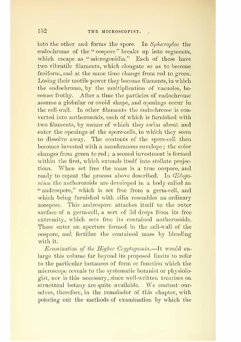
7/29/2019 THE MICROSCOPIST
http://slidepdf.com/reader/full/the-microscopist 174/361
152 THE MICROSCOPIST. /
into the otber and forms the spore. In Sphoeroi^lea the
endochrome of the " oospore " breaks up into segments,
which escape as " microgonidia." Each of these have
two vibratile filaments, which elongate so as to become
fusiform, and at the same time chano;e from red to screen.
Losing their motile powder they become filaments, in which
the endochrome, hj the multiplication of vacuoles, be-
comes frothy. After a time the particles of endochrome
assume a globular or ovoid shape, and openings occur in
the cell- wall. In other filaments the endochrome is con-
verted into antherozoids, each of which is furnished with
two filaments, by means of which they swim about and
enter the openings of the spore-cells, in which they seem
to dissolve away. The contents of the spore-cell then
becomes invested with a membranous envelope ; the color
changes from green to red ; a second investment is formed
within the first, which extends itself into stellate projec-
tions. When set free the mass is a true oospore, and
ready to repeat the process above described. In (Edogo-
nium the antherozoids are developed in a body called an
" androspore," which is set free from a germ-cell, and
which being furnished with cilia resembles an ordinary
zoospore. Tliis androspore attaches itself to the outer
surface of a germ-cell, a sort of lid drops from its free
extremity, which sets free its contained antherozoids.-
These enter an aperture formed in the cell-wall of the
oospore, and fertilize the contained mass by blending
with it.
Examination of the Higher Gryptogamia.—It would en-
large this volume far beyond its proposed limits to refer
to the particular instances of form or function which the
microscope reveals to the systematic botanist or physiolo-
gist, nor is this necessary, since well-written treatises on
structural botany are quite available. We content our-
selves, therefore, in the remainder of this chapter, with
pointing out the methods of examination by which the

7/29/2019 THE MICROSCOPIST
http://slidepdf.com/reader/full/the-microscopist 175/361
THE MICROSCOPE IN HISTOLOGY AND BOTANY. 153
views of other observers may be verified, or additions
made to our knowledge of vegetable life.
The lower forms of algfB and fnngi, to wdiich we have
already referred, need scarcely any preparation, save the
disentanglement of twisted threads under the simple mi-
croscope, or a gentle teasing witli needles, or rinsing with
water. The solution of iodine, and of iodine and sulphuric
acid, will suffice to exhibit the nature of the cell-wall and
cell-contents. In more highly developed plants it will be
necessary to take thin sections from different parts, and
indifferent but definite directions. These sections may
be made by hand, or between pieces of pith or cork by
means of a section-cutter. Iii some instances some of the
methods of staining will also be useful. Dr. Hunt, of
Philadelphia, has proposed a plan of staining which is
well adapted to all vegetable tissues. lie first soaks the
part or section in strong alcohol to dissolve the chloro-
phyll, then bleaches it in a solution of chlorinated soda.
It is then placed in a solution of alum, and afterwards in
one of extract of logwood. By transferring it to w^eak
alcohol and afterwards to stronger, it is deprived of its
water, and after being made transparent with oil of cloves,
it is ready for mounting in balsam or dammar varnish.
Care must be taken to wash it well after each of the
preliminary steps before staining.
In the higher algpe, the layers of cells assume various
sizes and shapes, and the nature of their fructification is
of great interest. Sections may be made of the " recep-
tacles " at the extremities of the fronds, which contain
filaments, whose contents become antherozoids. The pear-
shaped sporangia in the receptacles subdivide into clusters
of eight cells, called octospores, which are liberated from
their envelopes before fertilization.
The red sea-weeds, or Bhodospennece, afford many beau-
tiful forms for the microscope. The " tetraspores " are
imbedded in the fronds.

7/29/2019 THE MICROSCOPIST
http://slidepdf.com/reader/full/the-microscopist 176/361
154 THE MICROSCOPIST.
Ill lichens^ the cqjothecia form projections from the thal-
lus, or general expansion produced by cell-division. Avertical section shows them to contain asci or spore-cases
amid straight filaments, or elongated cells called 'para-
physes.
The fronds of He-paticm or liverworts bear stalks with
shield-like disks, which carry antheridia, and others with
radiating bodies bearing archegonia, which afterwards
give place to the sporangia or spore-cases. The spores
are associated with elaters^ or elastic spiral fibres, which
suddenly extend themselves and disperse the spores.
The Cliaracem are often incrusted with carbonate of
lime, wdiich may be removed with dilute sulphuric acid.
The motion of the bioplasm in the cells of the stem is
often well seen. The cells in which the spiral filaments
or antheridia are developed, are strung together like a
row of pearls. The position and construction of the spores
also should be examined, as well as the mode of growth
in the plant by division of the terminal cell (Plate XI,
Fig. 113).
Stems of mosses and liverworts should be examined by
means of transverse and longitudinal sections. Similar
sections through the half-ripe fruit of a moss will show
the construction of the fruit, the peristome, the calyptra,
etc. The ripe spores may be variously examined dry, in
water, in oil of lemons, and in strong sulphuric acid. The
capsules or urns of mosses are not now regarded as their
fructification, but its product.
The true antheridia and 'pistillidia are found among the
bases of the leaves, close to the axis. The fertilized " em-
bryo-cell " becomes gradually developed by cell-division
into a conical body or spore-capsule, elevated on a stalk.
The jw/ristome^ or toothed fringe, seen around the mouth
of the urn when the calyptra or hood, and operculum or
lid, are removed, furnishes a beautiful object for the bi-
nocular microscope.

7/29/2019 THE MICROSCOPIST
http://slidepdf.com/reader/full/the-microscopist 177/361
PLATE Xr.
Fig. 113.
Antheridia of Chnra fragilU —.\, antln ridium or "globule" developed at tlie base of pistillidium or
"nucule;" b, nucule enhirsed, globule laid open by the separation of its valves; C, one of the valves,
with its group of antheridial filaments, each composed of a linear series of cells, ^vithin every one of
which an antherozoid is formed ; in d, k, and F, the successive stages of this formation are seen ; and atG is shown the escape of the mature antherozoids, n. (From Carpenter.)
Development of Prothalium of Pleris serrulata:—A, spore .set free from the thcca; u, spore beginning
to germinate, putting forth the tubular prolongation n, from the principal cell b ; c, first formed linear
series of cells; D, prbthallium taking the form of a leaf-like expansion; a first and b second radical
fibre ; c, (/, the two lobes, and e the indentation between them; /. f, tir.st-formed part of the prothaliium
g, external coat of the original spore ; A, A, antheridia. (From Carpenter.)

7/29/2019 THE MICROSCOPIST
http://slidepdf.com/reader/full/the-microscopist 178/361

7/29/2019 THE MICROSCOPIST
http://slidepdf.com/reader/full/the-microscopist 179/361
THE MICROSCOPE IN HISTOLOGY AND BOTANY. 155
The SjjJmgnum, or bog-moss, has large and elongated
leaf-cells, with loosely-coiled spiral fibres, and their mem-
branous walls have large apertures. Their spores are of
two kinds, and when germinating in water, produce a long
filament with root-fibres at the lower end and a nodule
at the upper, from which the young plant is formed. If
grown on wet peat, instead of a filament there is evolved
a lobed foliaceous prothalliiim^ resembling the frond of
liverworts.
In ferns the structure approximates to true flowering
plants, while the reproductive organs are those of crj'pto-
gamia. Thin sections of the stem, cut obliquely, show
the scalariform or ladder-like vessels. The fructification
is usually found on the under side of the frond in isolated
spots called sori. Each of these contains a number of cap-
sules or thecce, and each capsule is surrounded by an an-
nulus or ring, whose elasticity opens the capsule when
ripe and permits the spores to escape. The spores are
somewhat angular, and when vegetating give rise to a
leaf-like expansion called a prothallium. In this the an-
theridia and archegonia^ which represent the true flower
of higher plants, are developed. The ciliated anthero-
zoids from the antheridia penetrate the cavity of the
archegonium and fertilize the " germ-cell," which subdi-
vides and becomes a young fern, while the prothallium,
having discharged the functions of a nurse, withers away
(Plate XI, Fig. 114). The group of Equisetacem or horse-
tails is interesting from the siliceous skeletons of the epi-
dermis, already referred to, page 131, as well as for the
elastic filaments attached to their spores.
Examination of Higher Plants.
The elementary tissues described in the beginning of
this chapter are chiefly characteristic of phanerogamic
plants, yet some additional particulars remain to be no-

7/29/2019 THE MICROSCOPIST
http://slidepdf.com/reader/full/the-microscopist 180/361
156 THE MICROSCOPIST.
ticed in connection witli the axis or stem, the leaves,
flowers, and fruit.
1. The Stem.—The arrangement of fibro-vascular bun-
dles, i. e.. woody fibres and ducts, differs widely in the
two botanical divisions of Monocotyledons and Dicotyle-
dons. In the first the growth is eiulogenoiis.,2a\di a section
exhibits the bundles of fibres and ducts disposed without
regularity in the mass of cellular tissue which forms the
basis of the fabric. In the second, or exogenous stems,
the fibro-vascular bundles are wedge-shaped, and inter-
posed between the bark and the pith, being kept apart by
plates of cellular tissue, called medullary rays, proceeding
from the pith.
The course of the vascular bundles in monocotyledons
should be carefully followed, either by maceration or
minute dissection. In the dicotyledonous stem, sections
must be made in three directions, transversely, longitu-
dinally across the diameter, and at a, tangent from the
bundles of fibres. The section-cutter, described page 63,
will be serviceable, although a sharp razor or scalpel may
serve. The size, form, and contents of the pith-cells
should be noticed, and their transition to wood-cells.
The arrangement of the medullary rays, of the wood-cells,
and of the ducts must also be observed, and in the Coni-
ferse the position of the pits. The cambium layer, between
the bark and wood, may have its cells rendered more
transparent by weak alkalies, and their contents tested
with iodine solution. The course and construction of
laticiferous vessels in the bark, when present, and of the
cork-cells of the tuberous layer, may be noted.
Fossil woods may be cut with a watch-spring saw, and
ground on a hone like bone or teeth. Sometimes it is
best to break ofi" small lamella by careful strokes with a
steel hammer. It is sometimes useful to digest fossil
wood in a solution of carbonate of soda for several days
before cuttino-.

7/29/2019 THE MICROSCOPIST
http://slidepdf.com/reader/full/the-microscopist 181/361
THE MICROSCOPE IN HISTOLOGY AND BOTANY. 157
2. Leaves.
—These should be examined by thin longitu-
dinal and transverse sections. The epidermis of both sides
should be detached, and the position and arrangement of
the stomata observed (Plate VII, Fig. 100). The hairs
of the epidermis, the arrangement of the parenchyma, and
the distribution of the vascular bundles in the form of
nerves, are also of importance.
3. FloiDers.—For ascertaining the number and position
of the parts of the flower, transverse sections at different
heights through an unopened bud may be taken, together
with a longitudinal section exactly through the middle.
The general structure of sepals and petals corresponds with
that of leaves, but there are some peculiarities. Thus the
cells of the petal of the geranium exhibit wdien deprived
of epidermis, dried and mounted in balsam, a peculiar
mammillated appearance with radiating hairs (Plate VIII,
Fig. 102). Anthers and pollen grains are also interesting
microscopic objects. The protrusion of the inner mem-
brane through the exterior pores in pollen may be stimu-
lated by moistening with water, dilute acid, etc. The
penetration of the pollen tubes through the tissue of the
style may be traced by sections or careful dissection. The
heartsease, viola tricolor., and the black and red currant,
vibes nigrura and rubrum, have been recommended for this
purpose.
4. Seeds.—The reticulations or markings on various
kinds of seeds render them frequent objects for observa-
tion with the binocular microscope. Adulterations may
also be detected in this way, as well as imperfect seeds in
any sample, a subject of much importance to the practical
farmer.

7/29/2019 THE MICROSCOPIST
http://slidepdf.com/reader/full/the-microscopist 182/361
158 THE MICROSCOPIST.
CHAPTER XL
THE MICROSCOPE IN ZOOLOGY.
We have already seen that both animal and vegetable
structures originate in a jelly-like mass or cell, and that
in the simple forms it is difficult, if not impossible, to
determine whether the object is an animal or a vegetable.
The mode of alimentation, and not structure, is our only
guide in the discrimination of the Protozoa or elementary
animal forms from Protopliytes or simple vegetables.
It has been proposed by Professor Hseckel to revive the
idea of a kingdom of nature intermediate between plants
and animals, but it does not appear that any gain to sci-
ence would result from such an arrangement.
I. MoNERA.—The simplest types of Protozoa are mere
particles of living jelly (Plate XII, Fig. 115), yet they
possess the power of contraction and extension, and of
absorbing alimentary material into their own substance
for its nutrition. The Bathybiiis^ from the "globigerina
mud," referred to on page 96, seems to have been an in-
definite expansion of such protoplasm or bioplasm.
II. Rhizopods.—This term (meaning root-footed) is ap-
plied to such masses of sarcode or bioplasm as extend long
processes, called j^seudopodia, as prehensile or locomotive
organs (Plate XII, Fig. 116). The Rhizopods are either
indefinitely organized jelly, like Mo7iera, or attain a cov-
ering or envelope of membrane called ectosarc^ while the
thin contents are termed endosarc. The first order of
Rhizopods, Peticidaria, consist of indefinite extensions of
freely branching and mutually coalescing bioplasm. The
second order, Padiolaria^ have rod-like radiating exten-
sions of the ectosarc, which do not coalesce. The order
Lobosa are lobose extensions of the bodj^ itself, as in the

7/29/2019 THE MICROSCOPIST
http://slidepdf.com/reader/full/the-microscopist 183/361
THE MICROSCOPE IN ZOOLOGY. 159
Ammba princeps already described. Some of tliis latter
order, as Arcella and Difflugia^nvQ testaceous. In Arcella
the test is a horny membrane, analogous to the ehitine
which hardens the integuments of insects. In Difflugia
the test is made up of minute particles of gravel, shell,
etc., cemented together. From the opening the amoeboid
body puts forth its pseudopodia (Plate XII, Fig. 117).
Connected with Ehizopods are three remarkable series of
forms, generally marine, and distinguished by skeletons
of greater or less density, which ailbrd many objects of
interest to the microscopist. These are the Foraminifera^
the Polycystina^ and the Sponges or Porifera. The shells
of the Foraminifera are calcareous, and those of Polycys-
tina siliceous ; both are perforated with numerous aper-
tures, which in Polycystina are often largo. We have
previousl}^ referred to these forms as occurring in a fossil
state.
Some Foraminifera have porcellanous, and others vitre-
ous or hyaline shells, usually many-chambered, and of every
shape between rectilinear and spinal. Most of them are
microscopic, but some are of considerable size, as the Or-
bitolites, which are found in tertiary limestones in Malabar.
The Xummulitic limestone, which extends over large areas
of both hemispheres, and of which the pyramids of Egypt
are built, is composed of the remains of the genus Num-
mulina; and the Eozoon Canadeyise has been shown by
Drs. Dawson and Carpenter to belong to the Foramini-
feral type.
In some Foraminifera the true shell is replaced by a
sandy envelope, whose particles are often cemented by
phosphate of iron. Dr. Carpenter, whose researches have
largely extended our knowledge of this group, pertinently
remarks that "there is nothing more wonderful in nature
than the building up of these elaborate and symmetrical
structures by mere jelly specks, presenting no trace what-
ever of that definite 'organization' which we are accus-

7/29/2019 THE MICROSCOPIST
http://slidepdf.com/reader/full/the-microscopist 184/361
160 THE MICROSCOPIST.
tomed to reo;ard as necessary to the manifestations of
conscious life."*
The Polycystina^ like the Foraminifera, are beautiful
objects for the binocular microscope, with the black-
ground illumination by the Webster condenser, the spot-
lens, or the paraboloid.
The Porifera or sponges begin life as solitary Amoeba,
and amid aggregations formed by their multiplication,
the characteristic spicules of sponge-structure make their
appearance. In one group, the skeleton is a siliceous
framework of great beauty. In Hyalonenia, the silica is
in bundles of long threads like spun glass. Sometimes
sponge spicules are needle-like, straight or curved, pointed
at one or both ends; sometimes with a head like a pin,
furnished with hooks, or variously stellate. Dr. Carpen-
ter thinks it probable that each spicule was originally a
segment of sarcode, which has undergone either calcifica-
tion or silicification (Plate XII, Fig. 118).
III. Infusorial Animalcules.—From the earliest his-
tory of the microscope, the minute animals found in vari-
ous infusions or in stagnant pools, etc., have attracted
attention. We owe to Professor Ehrenberg the jfirst sci-
entific arrangement of this class, and althou2:h more ex-
tended observations have changed his classification, yet
many of his views are still accepted by the most recent
investigators. Ehrenberg divided this class into two
groups, which represent very different grades of organi-
zation. The first he called Polygasirica (many-stomached)
from a view of their structure, which subsequent examin-
ations have not confirmed. The other group is that of
Rotifera or Poiatoria, a form of animal life which is most
appropriately classed among worms. The term Infusoria
is now applied to those forms which Professor Ehrenberg
* The Microscope and its Revelations, by W. B. Carpenter, M.D.,
LL.D., etc.

7/29/2019 THE MICROSCOPIST
http://slidepdf.com/reader/full/the-microscopist 185/361
PLATE XII.
Fig. 115.
Monera (Amceba),
A, Difflugia proteijoimis ; s,Diffluglaoblonga; c, ArceUa
acuminata ; D, ArceUa dentata.
Gromia oviformis,w[th its pseudopodia extended.
Structure of Granrtf; compressa : B, small portion highly luaguified.

7/29/2019 THE MICROSCOPIST
http://slidepdf.com/reader/full/the-microscopist 186/361

7/29/2019 THE MICROSCOPIST
http://slidepdf.com/reader/full/the-microscopist 187/361
THE MICROSCOPE IN ZOOLOGY. 161
called polvgastric animalcules. Yet a large section de-scribed bj him in this connection, including the Desmidi-
acece, Diatomacecc, Volvocmeiv, and other protophytes, have
been transferred b}^ naturalists to the vegetable kingdom.
The bodies of the Infusoria consist of sarcode or bio-
plasm, having an outer layer of firmer consistence. Some-
times the integument is hardened on one side so as to form
ashield,
andin other cases it is so prolonged and doubled
upon itself as to form a sheath or cell, within which the
animalcule lies. The form of the body is more definite
than that of Amceba, so as to be characteristic of species.
It may be oblong, oval, or round ; and some kinds, as
VorticeUa^ are attached to a footstalk, which has the power
of contracting in a spiral coil. No distinct muscular
structure can be detected in the Infusoria, yet the general
substance of the body is contractile. In most species
short hair-like filaments or cilia project from the surface,
sometimes arranged in one or more rows round the mouth,
and moving to all appearance under the influence of voli-
tion. In others there are one or two flagelliform filaments,
or long anterior cilia with vibratile ends. Others, again,
have setfe or bristles, which assist in locomotion. The
motions of some are slow, and of others quite rapid.
The interior of the sarcode body exhibit certain round-
ish spots, sometimes containing Diatoms or other foreign
substances. They have been called gastric vesicles, cells,
spaces, or sacculi. They are only visible from their con-
tents, and seem to be mere spaces without a living mem-
brane. If a little indigo or carmine is diffused in the
water which contains the Infusoria, the cavities will soon
be filled and become distinct. If watched carefully they
will appear to move round the body of the animal, and as
the pigment escapes at some part of the surface, the spots
will disappear. Ehrenberg regarded these spots as so
many stomachs arranged about a common duct, but the
common opinion at present regards them as temporary
11

7/29/2019 THE MICROSCOPIST
http://slidepdf.com/reader/full/the-microscopist 188/361
162 THE MICROSCOPIST.
digestive sacs made by the inclosure of food by the soft
bioplasm.
In addition to the "vacuoles" described, contractile
vesicles are seen which contract and dilate rh^^thmically,
and do not change their position. They have been con-
sidered to serve for respiration.
Most of the Infusoria multiply by self-division (Plate
XIII, Fig. 119), and at certain times undergo an encyst-
ing process, much resembling the "still" condition of Pro-
tophytes, and like that serving for preservation under
circumstances which are unfavorable to ordinary vital
activity. The gemmules or progeny which result from
the bursting of the cyst do not always resemble the parent
in form. The recent researches of Drs. Dallinger and
Drysdale have shown considerable variety in the life his-
tory of the Infusoria. In some instances the product of
the encysting process was not a mass of granules, but an
aggregation of minute germinal particles not more than
Tjoo'ooyth of an inch in diameter, and capable of resisting
heat, either by boiling or by dry heating up to 300° F.
The observations of M. Balbiani show that in many of
the Infusoria, male and female organs are combined in
the same individual, but that a congress of two is neces-
sary for the impregnation of the ova, those of each being
fertilized by the spermatozoa of the other.
There is also a curious tribe of suctorial animalcules
called Acinetce, which put forth tubular prolongations
which penetrate the bodies of other species and grow in
their interior as parasites.
The systematic arrangement of the Infusoria is yet
unsettled. Ehrenberg's families, excluding those now
placed among Algre or Rhizopods, are as follows

7/29/2019 THE MICROSCOPIST
http://slidepdf.com/reader/full/the-microscopist 189/361
THE MICROSCOPE IN ZOOLOGY. 163
A. Intestinal tube absent.
Body variable, without cilia. _Carapace absent, . . . . " . Astasi.^a.
Carapace present, ..... Dinobryina.
Cilia or setre present.
Carapace absent, Cyclidina.
Carapace present, ..... Peridin/EA.
B. Intestinal tube present.
Orifice single.
Carapace absent,
.....Voeticellina.
Carapace present, ..... Ophkydina.
Two opposite orifices.
Carapace absent, ..... Enchelia.
Carapace present, ..... Colepina.
Orifices differently placed.
Carapace none.
No tail, but a proboscis, . . Trachelina.
Tail present, mouth anterior, . Ophryocercina.
Carapace present, Aspidiscina.Orifices ventral.
Carapace absent.
Motion by cilia, .... Colpodea.
Motion by organs, .... Oxytrichina.
Carapace present, ..... Euplota.
IV. Rotatoria or Wheel Animalcules.—These are
microscopic, aquatic, transparent animals, of a higher
organization than the Infusoria, and belonging in all
probability to the class Vermes. Their chief interest to
the microscopist is derived from the possession of a more
or less lobed, retractile disk, covered vs'ith cilia, which,
when in motion, resemble revolving wheels. They have
also a complicated dental apparatus, and generally a dis-
tinct alimentary canal, and are reproduced by ova. Someare more or less covered by a carapace, and in most there
is a retractile tail-like foot, sometimes terminated by a
suctorial disk or a pair of claw-like processes. The ner-
vous and vascular systems are not well known, although
traces of them are seen. The young of some possess an
eye which often disappears in the adult. They are re-

7/29/2019 THE MICROSCOPIST
http://slidepdf.com/reader/full/the-microscopist 190/361
164 THE MICROSCOPIST.
markably tenacious of life, having in some instances re-
vived after having been kept dry for several years.
M. Dujardin divides the Rotifera into four groups or
natural families
1. Those attached by the foot, vehich is prolonged into
a pedicle. It includes two families, the Floscularians and
the Melieertians, in the first of which the sheath or cara-
pace is transparent, and in the other composed of little
rounded pellets (Plate XIII, Fig. 120).
2. The common Rotifer and its allies, wdiich swim freely
or attach themselves by the foot at will (Plate XIII, Fig.
121).
3. Those which are seldom or never attached, the Bra-
chionians and the Furcularians. The former are short,
broad, and flat, and inclosed in a sort of cuirass ; the latter
are named from a bifurcated, forcep-like foot (Plate XIV,
Fig. 122).
4. The Tardigrada or water bears. These have no
ciliated lobes, but are in other respects like their allies,
and seem to be a connecting link between the Rotifers
and worms. The segments of the body, except the head,
bear two fleshy protuberances furnished with four curved
hooks.''^
Y. Polyps.—The animals of this class were formerly
called Zoophytes^ or animal flowers. They are the most'
important of coral-making animals, although the Hydroids
and Bryozoa, together with some Alga3, as the Xullipores,
share with them the formation of coral, which is a secre-
tion of calcareous matter. Dana's work on corals gives a
classification, of which we present a summary.
A good idea of a polyp may be had from comparison
with the garden aster, the most common form of a polyp
flower being a disk fringed with petal-like organs called
tentacles.
The internal structure, like the external, is radiate, and
*Carpenter on the Microscope.

7/29/2019 THE MICROSCOPIST
http://slidepdf.com/reader/full/the-microscopist 191/361
PLATE XIII.
Fig. 119.
ill I
Fig. 121.
Fissiparous multiplication of Clulodon cuciiUulus.
Stephanoceros Eichorn ii.
Rodifer vulgaris, as seen at a, 'witli
the wheels drawn in, and at b with
the wheels expanded; a, mouth; 6,
eye-spots ; c, wheels ; d, calcar (an-
tenna?); e, jaws and teeth; /, ali-
mentary canal ; g, glandular (?) massenclosing it; /;, longitudinal mus-
cles; i, i, tubes of water vascular
system; /i, young animal; Z, cloaca.

7/29/2019 THE MICROSCOPIST
http://slidepdf.com/reader/full/the-microscopist 192/361

7/29/2019 THE MICROSCOPIST
http://slidepdf.com/reader/full/the-microscopist 193/361
THE MICROSCOPE IN ZOOLOGY. 165
the cavity of the body is divided by septa into narrow
compartments. The walls contain circular and longitu-
dinal muscles, which serve for contraction of the body,
which is afterwards expanded by an injection or absorp-
tion of water b}^ the mouth.
The most interesting part of the structure of these
animals, to the microscopist, is the multitude of lasso-cells,
called also nettling-cells^ thread capsules^ and cnuhe, which
stud the tentacles and other parts of the body, and by
means of which the prey of the polyp is at once pierced
and poisoned. A small piece of the tentacle of a sea
anemone placed in a compressorium under the microscope,
and subjected to gentle pressure, will show the protrusion
of many little dart-like processes attached to thread-like
filaments. Many observations indicate the injection of a
poison through these darts, which is instantly fatal to
small animals (Plate XIV, Fig. 123).
The polyp has no circulating fluid but the results of
digestion mixed with salt water, no bloodvessels but the
vacuities among the tissues, and no passage for excrements
except the mouth and the pores of the body. Eeproduc-
tion is both by ova and by buds.
I. Actinoid jyolyps are related to the Actinea or sea
anemone. The number of tentacles and interior septa
is a multiple of six.
II. Cyaihophylloid polyps have the number of tentacles
and septa a multiple of four.
III. Alcyonoid polyps have eight fringed tentacles. The
Alcyonium tribe are among the most beautiful of coral
shrubs. The Gorgonia tribe has reticulated species like
the sea fan, and bears minute calcareous spicules, often
brilliantly colored. The Pennatula tribe is unattached,
and often rod-like, with the polyps variously arranged.
YI. Hydroids.—The type of this class is the common
Hydra, which is often found attached to leaves or stems
of aquatic plants, etc. It is seldom over half an inch long.

7/29/2019 THE MICROSCOPIST
http://slidepdf.com/reader/full/the-microscopist 194/361
166 THE MICROSCOPIST,
It has the form of a polyp, with long slender tentacles.
Besides these tentacles with their lasso-cells, it has no
special organs except a mouth and tubular stomach. Like
the fabled Hydra, if its head be cut off another will grow
out, and each fragment will in a short time become a per-
fect animal, supplying whatever is wanting, hence its name
(Plate XIV, Fig. 124). The Hydra has the power of lo-
comotion, bending over and attaching its head until the
tail is brought forward, somewhat after the manner of a
leech.
Compound Ilydroids may be likened to a Hydra whose
buds remain attached and develop other buds until an
arborescent structure, called a ]}olypary^ is produced. The
stem and branches consist of fleshy tubes with two layers,
the inner one having nutritive functions, and the outer
secreting a hard, calcareous, or horny layer. The indi-
viduals of the colony are of two kinds, the 'polypite or
nutritive zooid, resembling the Hydra, and the gonozooid,
or sexual zooid, developed at certain seasons in buds of
particular shape.
To mount compound Hydrozoa, or similar structures,
place the specimen alive in a cell, and add alcohol drop
by drop to the sea-water; this will cause the animals to
protrude and render their tentacles rigid. Then replace
the alcohol with Groadby's solution, dilute glycerin, or
other preserving fluid.
VII. AcALEPiis, or sea-nettles, are of all sizes, from an
almost invisible speck to a yard in diameter. They swarm
in almost every sea, and are frequently cast upon the
beach by the waves. They are transparent, floating free,
discoid or spheroid, often shaped like a mushroom or um-
brella, and their organs are arranged radiately round an
axis occupied by the pedicle or stalk. They are furnished
with muscular, digestive, vascular, and nervous systems.
They were formerly divided into
1. Pulmonigrada, from their movements being effected

7/29/2019 THE MICROSCOPIST
http://slidepdf.com/reader/full/the-microscopist 195/361
PLATE XIV.
Fig. 122.
Fig. 123.
Noteus quadricornis :—A,doTsa.] view; b, side view.
Filiferous capsules of Helianthoid Polypes ;—a,b,Corynactis Allmanni; c, E, f, Caryopliyllia Smilhii]T),G, Actinia crassicornis; n, Actinia Candida.
Fig. 124.
\ ^11
<?] .—
Hydra fusca in gemination.

7/29/2019 THE MICROSCOPIST
http://slidepdf.com/reader/full/the-microscopist 196/361

7/29/2019 THE MICROSCOPIST
http://slidepdf.com/reader/full/the-microscopist 197/361
THE MICROSCOPE IN ZOOLOGY. 167
hy a rhythmical contraction and dilation, as in RJdzistoma^
etc. 2. Cilograda^ moving by narrow bands of vibratile
cilia varionsly disposed over the body. In Beroe the cilia
are transformed into flat fln-like shntters, arranged in
eight longitndinal bands. InYenns's givdilQ, Cestum Ve-
neris^ the margins of a gelatinous ribbon are fringed with
cilia. 3. Physograda^ which move by means of an expan-
sile bladder, as the Phi/salia, or Portuguese man of war.
4. Cirrigrada, possessing a sort of cartilaginous skeleton,
and furnished with appendages called cirri, serving as oars
and for prehension, as Porpita and Velella. In the latter
there is also a subcartilaginous plate rising at right angles
from the surface supporting a delicate membrane, which
acts as a sail.
This classification has been laid aside since the micro-
scopic discover}'- of the close relationship between the
Hj'drozoa and the Medusoid Acalephs, and the latter are
now subdivided into the " naked-eyed " and the " covered-
eyed " Acalephs. The alternation of generations, page
126, is fully illustrated in this class. The embryo emerges
as a ciliated gemmule, resembling one of the Infusoria.
One end contracts and attaches itself so as to form a foot,
while the other enlarges and becomes a mouth, from which
four tubercles sprout and become tentacles. Thus a Hy-
dra-like polyp is formed, which acquires additional tenta-
cles. From such a polyp many colonies may rise by gem-
mation or budding, but after a time the polyp becomes
elongated, and constricted below the mouth. The con-
stricted part gives origin to other tentacles, while similar
constrictions are repeated round the lower parts of the
body, so as to divide it into a series of saucer-like disks,
which are successively detached and become Medusae
(Plate XY, Figs. 125, 126).
YIII. EcHixoDERMS.—This class includes the star-fishes,
the sea-urchins or sea-eggs, the sea-slugs, and the crinoids
or stone lilies of former ages. If we imagine a polyp with

7/29/2019 THE MICROSCOPIST
http://slidepdf.com/reader/full/the-microscopist 198/361
168 THE MICROSCOPIST.
a long stem to secrete calcareous matter, not merely exter-
nally, but in the substance of its body and tentacles, such
polyp when dried would present some such appearance as
the fossil Encrinoid Echinoderras of past times. The im-
agination of such a polyp without a stem, and having
sucker-like disks on its arms, will give us the picture of
a star-fish {Asterias). Imagine the rays diminished and
the central part extended, either flat or globular, and we
have the form of Echini with the spines removed. The
Holoihurice have elongated membranous bodies, with im-
bedded spicule
.
The structure of Echinoderms is quite complex, and
belongs to comparative anatomy rather than microscopy,
yet some directions for the study of these forms is essen-
tial to our plan.
Thin sections of the shells, spines, etc., may be made by
first cutting with a fine saw, and rubbing down with a
flat file. They should be smoothed by rubbing on a hone
with water, cemented to a glass slip with balsam, and
carefully ground down to the required thickness. They
may be mounted in fluid balsam.
Many Echinoderms have a sort of internal skeleton
formed of detached plates or spiculse. The membranous
integument of the Ilolothurife have imbedded calcareous
plates with a reticulated structure, and they are often
furnished with appendages, as prickles, spines, hooks, etc.,
which form beautiful microscopic objects.
The larva of an Echinoderm is a peculiar zooid, which
develops by a sort of internal gemmation. One of the
most remarkable of these larvse has been called Bvpin-
naria.
IX. Bryozoa or Polyzoa.—Microscopic research has
removed this class from the polyps, which they resemble,
to the molluscan sub-kingdom. They have a group of
ciliated tentacles round the mouth, but have a digestive
system far more complex than polyps. They form delicate

7/29/2019 THE MICROSCOPIST
http://slidepdf.com/reader/full/the-microscopist 199/361
PLATE XV.
Fig. 125. Fig. 127.
Development of Jledusa buds in Syn-
choryna Sarsii.
A, Portion of Cellularia ciliata, enlarged ; b,
one of the "bird's-head" of 5?/(7w/ffl avimlaria,
more highly magnified, and seen in the act of
grasping another.
Fig 126. Fig. 128.
/^^l
Successive stages of development of Medusa
buds from StrobUa larva.
Seitularia cupresHaa:—.\, natural size;
B, portion magnified.

7/29/2019 THE MICROSCOPIST
http://slidepdf.com/reader/full/the-microscopist 200/361

7/29/2019 THE MICROSCOPIST
http://slidepdf.com/reader/full/the-microscopist 201/361
THE MICROSCOPE IN ZOOLOGY. 169
corals, either membranous or calcareous, made up of minute
cabin-like cells, which are either thin crusts on sea-weeds,
rocks, etc., or slender moss-like tufts, or groups of thin
curving plates, or net-like fronds, and sometimes thread-
like lines or open reticulations. The cells of a group have
no connection with a common tube, as the Hjdroids, but
the alimentary system of each little Bryozoon is indepen-
dent.
Many of the Polyzoa have curious appendages to their
cells, of two kinds ; the first are called birds'-head pro-
cesses or avicidaria. They consist of a body, a hinge or
lower jaw-like process, and a stalk. The lower portion is
moved by an elevator and depressor muscle, and during
life the motion is constant. The second kind, or vibracula^
is a hollow process from which vibratile filaments project
(PlateXY, Figs. 127, 128).
X. TuNiCATA.—These molluscs are so named from the
leathery or cartilaginous tunic which envelops them, and
which often contains calcareous spicula. Like the Bryo-
zoa they tend to produce composite structures by gemma-
tion, but they have no ciliated tentacles. They are of
most interest to the microscopist from the peculiar actions
of their respiratory and circulatory organs, which may be
seen through the transparent walls of small specimens.
The branchial or respiratory sac has a beautiful network
of bloodvessels, and is studded with vibratile cilia for dif-
fusing water over the membrane. The circulation is re-
markable from the alternation of its direction.
The smaller Tunicata are usually found aggregate, in-
vesting rocks, stones and shells, or sea-weeds ; a few are
free.
Synopsis of the Families.
A. Attached ; mantle and test united only at the ori-
fices.
1. Botryllidce.—Bodies united into systems.

7/29/2019 THE MICROSCOPIST
http://slidepdf.com/reader/full/the-microscopist 202/361
170 THE MICROSCOPIST.
2. Clavelinidm.—Bodies distinct, but connected by a
common root thread.3. AscidiadcE.—Bodies unconnected.
B. Free ; mantle and test united throughout.
4. PeloncEadcE.—Orifices near together.
5. SalpadcB.—Orifices at opposite ends.
XI. CoNCHiFERA.—This class consists of bivalve mol-
luscs, and is chiefly interesting to the microscopist from
the ciliary motion on their gills and the structure of theshell.
The ciliary motion may be observed in the oyster or
mussel, by detaching a small piece of one of the bands
which run parallel with the edge of the open shell, placing
it on a glass slide in a drop of the liquid from the shell,
separating the bars with needles, and covering it with
thin glass; or the fragment may be placed in the live boxand submitted to pressure. The peculiar movement of
each cilium requires a high magnifying power. It appears
to serve the double purpose of aeration of the blood and
the production of a current for the supply of aliment.
Dr. Carpenter has shown that the shells of molluscs
possess definite structure. In the Margaritacece the exter-
nal layer is prismatic,and the internal nacreous. The
nacreous or iridescent lustre depends on a series of grooved
lines produced by laminae more or less oblique to the plane
of the surface. The shells of Terehratulce are marked by
perforations, which pass from one surface to another.
The rudimentary shell of the cuttle-fish (of the class
Cephalopoda), or " cuttle-fish bone," is a beautiful object
either opaque or in the polariscope. Sections
maybe
made in various directions with a sharp knife, and
mounted as opaque objects or in balsam.
XII. Gasteropoda.—These molluscs are either naked,
as the slug, or have univalve shells, as the snail, the lim-
pet, or the whelk. As in the other classes referred to,
the details of anatomical structure are full of interest

7/29/2019 THE MICROSCOPIST
http://slidepdf.com/reader/full/the-microscopist 203/361
PLATE XVI.
Fig. 130.
y/ y
Palate of J>oris hibereulata.
A, female of Cyclops quadricornis —a, body ; 6, fail
;
c, antenna; d, antennule; e, feet; /, pluraose setae of
tail;—B, tail, with external egg-sacs; c, d, e, f, g,
successive stages of development of young.
Fig. 131.
Metamorphosis of Carcinvs mcenas:—a, first stage; b, second stage ; c, third stage,
in which it begins to assume the adult form ; d, perfect form.
7/29/2019 THE MICROSCOPIST
http://slidepdf.com/reader/full/the-microscopist 204/361

7/29/2019 THE MICROSCOPIST
http://slidepdf.com/reader/full/the-microscopist 205/361
THE MICROSCOPE IN ZOOLOGY. 171
but to the microscopist the palate, or tongue as it is called
—a tube which passes beneath the mouth, opening ob-
liquely in front, and which is covered with transverse
rows of minute teeth set upon plates—presents characters
of great value in classification. These palates require
careful dissection, and when mounted in balsam become
beautiful polariscope objects (Plate XVI, Fig. 129).
XIII. Cephalopoda.—The crystalline lens in the eye of
the cuttle-fish is said to be of the same form as the well-known " Coddington lens." The skin of this class con-
tains a curious provision for changing its hue, consisting
of large pigment-cells containing coloring matter of vari-
ous tints.
The suckers, or prehensile disks, on the arms of cephal-
opods often make interesting opaque objects when dried.
XIV. Entozoa.
—These are parasitic animals belonging
to the class of worms. They are characterized by the
absence or low development of the nutritive system, and
the extraordinary development of their reproductive or-
gans. Thus the Tmiia or tapeworm has neither mouth
nor stomach, the so-called " head" being merely an organ
for attachment, while each segment of the " body " con-
tains repetitions of a complex generative apparatus.
Among the Xematoid or roundworms, the Anguillulce^
or little eel-like worms, found in sour paste, vinegar, etc.,
as well as the Trichina spiralis, inhabiting the voluntary
muscles, are generally classified.
Order I. Sterelmintha.—Alimentary canal absent or
indistinct.
Family 1. Cestoidea.—Tapeworms; body strap-shaped,
divided into transverse joints ; alimentary canal indistinct.
The cystic Entozoa [Echinococcus, etc.) are nurse or larval
forms of Cestoidea.
Family 2. Trernatoda.—Body mostly flattened; alimen-
tary canal distinct ; branched.
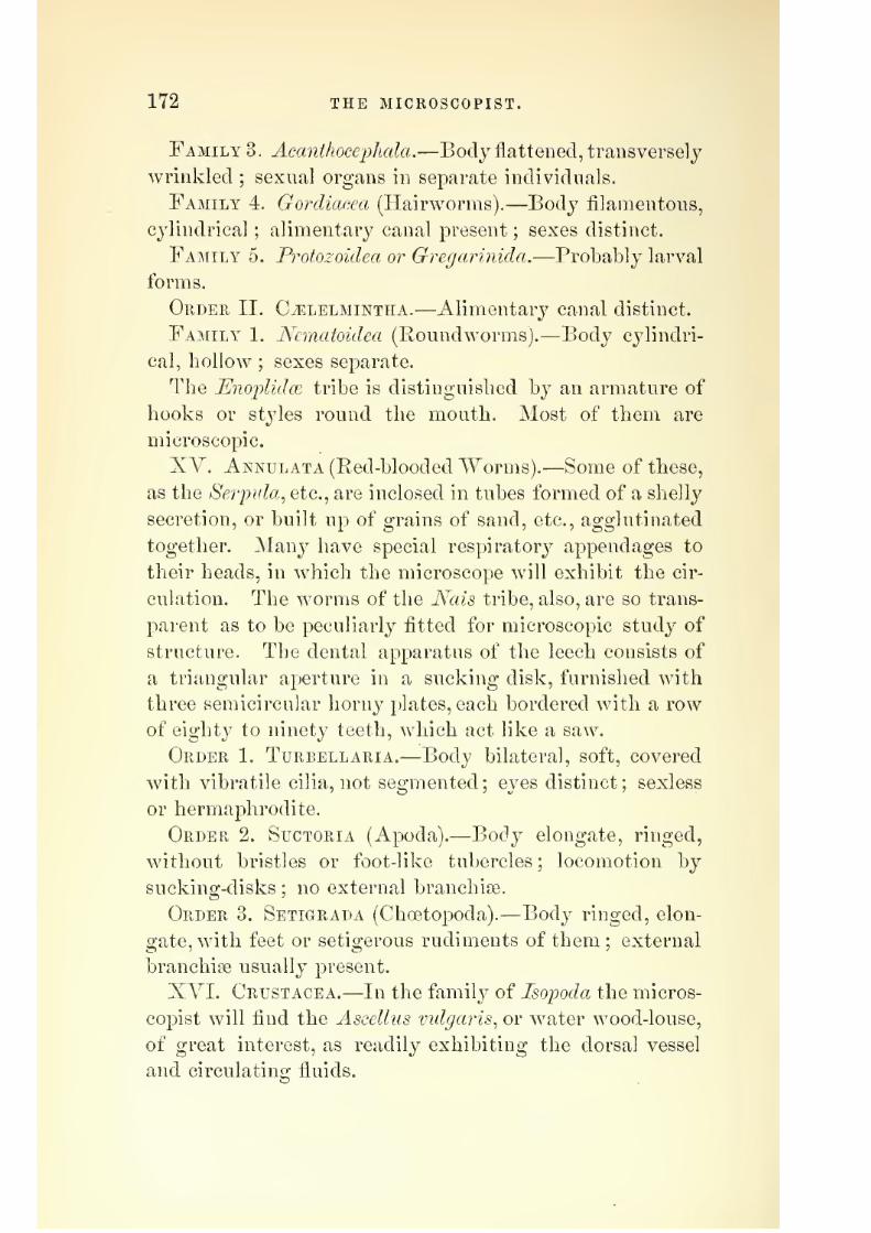
7/29/2019 THE MICROSCOPIST
http://slidepdf.com/reader/full/the-microscopist 206/361
172 THE MICROSCOPIST.
Family 3. Acanthocejjhala.—Body flattened, transversely
wrinkled; sexual organs in separate individuals.
Family 4. Gordiacea (Hairworms).—Body filamentous,
cylindrical ; alimentary canal present ; sexes distinct.
Family 5. Protozoidea or Gregarinida.—Probably larval
forms.
Order II. C^lelmintiia.—Alimentary canal distinct.
Family 1. Nematoidea (Roundworms).—Body cylindri-
cal, hollow;
sexes separate.The EnojplidcB tribe is distinguished by an armature of
hooks or styles round the mouth. Most of them are
microscopic.
XY. Annulata (Red-blooded Worms).—Some of these,
as the Serinda^ etc., are inclosed in tubes formed of a shelly
secretion, or built up of grains of sand, etc., agglutinated
together. Many have special respiratory appendages totheir heads, in which the microscope will exhibit the cir-
culation. The worms of the Nais tribe, also, are so trans-
parent as to be peculiarly fitted for microscopic study of
structure. The dental apparatus of the leech consists of
a triangular aperture in a sucking disk, furnished with
three semicircular horny plates, each bordered with a row
of eighty to ninety teeth,which
act likeasaw.
Order 1. Turbellaria.—Body bilateral, soft, covered
with vibratile cilia, not segmented ; eyes distinct ; sexless
or hermaphrodite.
Order 2. Suctoria (Apoda).—Body elongate, ringed,
without bristles or foot-like tubercles ; locomotion by
sucking-disks ; no external branchioB.
Order 3. Setigrada (Choetopoda).
—Body ringed, elon-
gate, with feet or setigerous rudiments of them ; external
branchiffi usually present.
XYI. Crustacea.—In the family of Isopoda the micros-
copist will find the AsceUiis vidga?is^ or water wood-louse,
of great interest, as readily exhibiting the dorsal vessel
and circulating fluids.
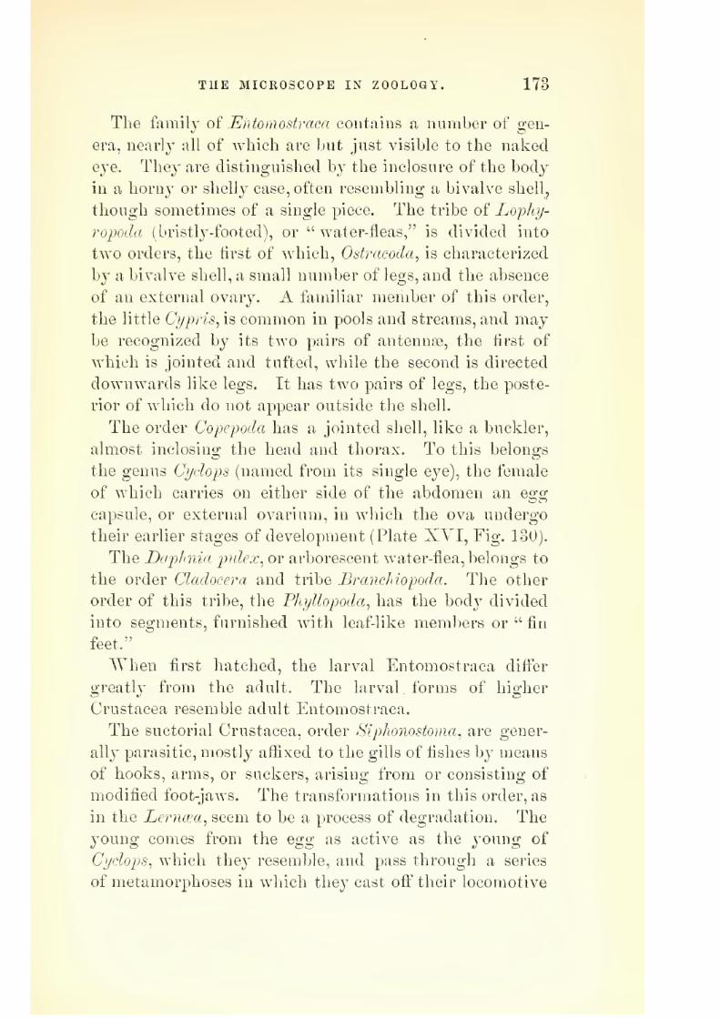
7/29/2019 THE MICROSCOPIST
http://slidepdf.com/reader/full/the-microscopist 207/361
THE MICROSCOPE IN ZOOLOGY. 173
The family oi Entomostraca contains a number of gen-
era, near]}' all of which are but just visible to the nakedQye. They are distinguished by the inclosure of the body
in a horny or shelly case, often resembling a bivalve shellj,
though sometimes of a single piece. The tribe of Lophj-
ropoda (bristlj'-footed), or " water-fleas," is divided into
two orders, the first of which, Ostracoda, is characterized
by a bivalve shell, a small number of legs, and the absence
of au external ovary. A familiar member of this order,the little Ci/pris, is common in pools and streams, and may
be recognized by its two pairs of antennae, the first of
which is jointed and tufted, while the second is directed
downwards like legs. It has two pairs of legs, the poste-
rior of which do not appear outside the shell.
The order Copepoda has a jointed shell, like a buckler,
almost inclosing the headand
thorax.To
this belongs
the genus Cydops (named from its single eye), the female
of which carries on either side of the abdomen an egg
capsule, or external ovarium, in which the ova undergo
their earlier stages of development (Plate XYI, Fig, 130).
The Daphnia pidex^ or arborescent water-flea, belongs to
the order Cladocera and tribe Branckiopoda. The other
order of this tribe, the Phyllopoda^ has the body divided
into segments, furnished with leaf-like members or " fin
feet."
When first hatched, the larval Entomostraca differ
greatly from the adult. The larval forms of higher
Crustacea resemble adult Entomostraca.
The suctorial Crustacea, order Siphonostoma, are gener-
ally parasitic, mostly aflixed to the gills of fishes by means
of hooks, arms, or suckers, arising from or consisting of
modified foot-jaws. The transformations in this order, as
in the Lerncea, seem to be a process of degradation. The
young comes from the egg as active as the young of
Cydops, which they resemble, and pass through a series
of metamorphoses in which they cast off their locomotive

7/29/2019 THE MICROSCOPIST
http://slidepdf.com/reader/full/the-microscopist 208/361
174 THE MICROSCOPIST. '
members and their e^^es. The males and females do not
resemble each other.The order CuThipeda consists of the harnades and their
allies. In their early state they resemble the Entomos-
traca, are unattached, and have eyes. After a series of
metamorphoses they become covered with a bivalve shell,
which is thrown off; the animal then attaches itself by
its head, which in the barnacle becomes an elongated
pedicle,
andin
Balanus expandsinto a disk.
Thefirst
thoracic segment produces the "multivalve" shell, while
the other segments evolve the six pairs of cirrhi, which
are slender, tendril-like appendages, fringed with ciliated
filaments.
In the order Amjyhipoda, the Gammarus pidex, or fresh-
water shrimp, and the Talitrus saltaior, or sandhopper,
may be interesting to the microscopist.
The order Decapoda^ to which belong the crab, lobster,
shrimp, etc., is of interest, from the structure of the shell
and the phenomena of metamorphosis. The shell usually
consists of a horny structureless layer exteriorly, an areo-
lated stratum, and a laminated tubular substance. The
difference between the adult and larval forms in this order
is so great that the young crab was formerly considered
a distinct genus, Zoea (Plate XVI, Fig. 131).
For the preservation of specimens of Crustacea, Dr.
Carpenter recommends glycerin jelly as the best medium.
XVII. Insects..—Many insects may be mounted dry,
as opaque objects. They may be arranged in position by
the use of hot water or steam. Those which are trans-
parent enough may be mounted in balsam, and very deli-
cate ones in fluid. To display the external chitinous cov-
ering of an entire insect, it may be soaked in strong liquor
potass?e, and the internal parts squeezed out in a saucer
of water by gently rolling over it a camel's-hair brush.
It may be put on a slide, and the cover fastened by tying
with a thread. It should then be soaked in turpentine

7/29/2019 THE MICROSCOPIST
http://slidepdf.com/reader/full/the-microscopist 209/361
THE MICROSCOPE IN ZOOLOGY. 175
until quite transparent, when it may be removed, the tur-
pentine partiallj^drained off,
anda solution of balsam in
chloroform allowed to insinuate itself by capillary attrac-
tion. Gentle heat from a spirit-lamp will be useful at this
stage of the mounting.
Small insects hardly need soaking in caustic potash, as
turpentine or oil of cloves will render them after awhile
quite transparent, and their internal organs are beautifully
seen in the binocular microscope. Thin sections of insects
are instructive, and may be made with a section-cutter by
jfirst saturating the body with thick gum mucilage, and
then incasing in melted paraffin.
Man}' insects and insect preparations are well preserved
in glycerin.
The eggs of insects are often interesting objects, and
should be mounted in fluid.
Wing cases of beetles are often very brilliant opaque
objects. Some are covered with iridescent scales, and
others have branching hairs. Many are improved by
balsam, and this may be determined by touching with
turpentine before mounting.
Scales of Lepidopfera, etc., may be exhibited in their
natural arrangement by mounting a small piece of wing
dry. If desired as test objects, a slide or thin cover, after
having been breathed on, may be slightly pressed on the
wing or body of the insect. The scales are really flattened
cells, analogous to the epidermic cells of higher animals.
Some have their walls strengthened by longitudinal ribs,
while others, as the Podurce, show a beaded appearance
under high powers from corrugation. Dr. Carpenter be-
lieves the exclamation marks in the scales of the latter to
be the most valuable test of the excellence of an objective.
Hairs of insects are often branched or tufted. The hair
of the bee shows prismatic colors if the chromatic aberra-
tion of the object-glass is not exactly neutralized.
Antennce vary greatly in form, and are often useful in

7/29/2019 THE MICROSCOPIST
http://slidepdf.com/reader/full/the-microscopist 210/361
176 THE MICROSCOPIST.
classification (Plate XYII, Fig. 132). Thus in the Cole-
optet^a we have the Serricomes, or serrated antennse; the
Clavicornes, or clubbed ; the Palpieorms^ with antennae no
larger than palpi ; the Lamellicornes, with leaf-like appen-
dages to the antennse, ; and the Longicornes, with antennae
as long or longer than the body. ITerve-fibres, ending in
minute cavities in the antennee, have been traced, which
are supposed to be organs of hearing. The antennae should
be bleached to exhibit them. The bleaching process is
also useful for other parts of insects. The bleaching fluid
consists of a drachm of chlorate of potass in about two
drachms of water, to which is added about a drachm of
hydrochloric acid.
Compound eyes of insects are always interesting. They
are quite conspicuous, and often contain thousands of
facets, or minute eyes, called ocelli (Plate XVII, a b, Fig.
133). Besides these, insects possess rudimentary single
eyes, like those of the Arachnidce. These are at the top
of the head, and are termed stemmata (Plate XYII, «,
Fig. 133). To display the " corneules," or exterior layer
of the compound eye, the pigment must be carefully
brushed away after maceration. A number of notches
may then be made around the edge, the membrane flat-
tened on a slide, and mounted in balsam. A^ertical sec-
tions may be made while fresh, so as to trace the relations
of the optic nerve, etc. The dissecting microscope and
needles will be found useful (Plate XVII, Fig. 132).
Mouths of insects present great varieties. In the beetles
the mouth consists of a pair of mandibles, opening later-
ally ; a second pair, called maxillfe ; a labrum or upper
lip ; an under lip or labium ; one or two pairs of jointed
appendages to the maxillse, termed maxillary palpi ; and
a pair of labial palpi. The labium is often composed of
distinct parts, the first of which is called the mentum or
chin, and the anterior part the ligula or tongue. This
latter part is greatly developed in the fly, and presents
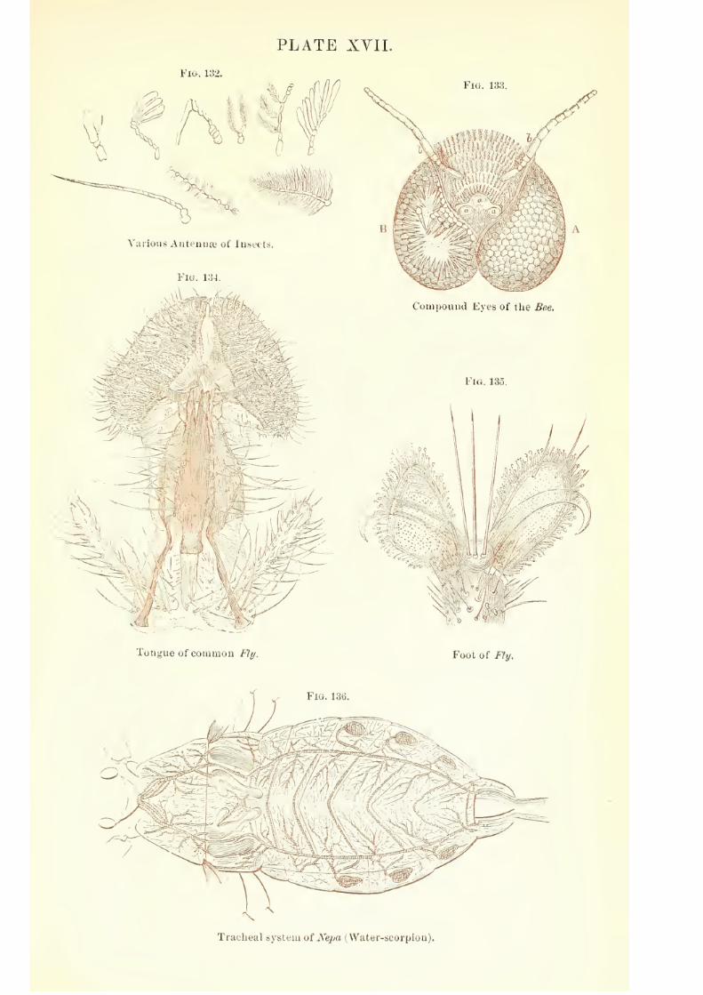
7/29/2019 THE MICROSCOPIST
http://slidepdf.com/reader/full/the-microscopist 211/361
PLATE XVII.
Fig. 133.
Compound Eyes of the Bee.
\ >:^
/e-
,i'#_
f V
Tongue of coiuinou Fly.
Fig. 135.
\^W .
'eb^'
I i /
^^4if
Foot of /'/y.
Tracheal system ot Nepa (Water-scorpion).

7/29/2019 THE MICROSCOPIST
http://slidepdf.com/reader/full/the-microscopist 212/361

7/29/2019 THE MICROSCOPIST
http://slidepdf.com/reader/full/the-microscopist 213/361
THE MICROSCOPE IN ZOOLOGY. 177
a curious modilication of traclical structure, which is
thought to serve the functiou of suction (Plate XYII,
Fig. 134). The tongue of the bee is also an interesting
object. In the Diptera the labrum, maxillfe, mandibles,
etc., are converted into delicate lancets, termed setpe, and
are used to puncture the epidermis of animals or plants,
from which the juices may be drawn by the proboscis.
In the Lepidoptera the labrum and mandibles are reduced
to minute plates, while the maxillfe, are greatly elongated,
and are united to form the haustellum, or true proboscis,
which contains a tube for suction.
Feet.—These organs vary with the habits of life in dif-
ferent species. The limb consists of five divisions : the
coxa or hip, the trochanter, the femur or thigh, the tibia
or shank, and the tarsus or foot. This last has usually
five joints, but sometimes less. The ColeojAei-a are subdi-
vided into groups, according as the tarsus consists of five,
four, or three segments. The last joint is furnished with
hooks or claws, and in the fiy, etc., the foot is also
furnished with membranous expansions, called pul villi.
These latter have numerous hairs, each of which has a
minute disk at its extremity. By these, probably by the
secretion of a viscid material, the insect is enabled to
walk on glass, etc., in opposition to gravity (Plate XVII,
Fig. 135). In the Dutiscns^ the inner side of the leg is
furnished with disks or suckers of considerable size.
They may be mounted as opaque objects. Stings and
Ovipositors also present a great variety of structure, and
may be best mounted in balsam.
The alimentary canal in insects presents many diversi-
ties. As in higher animals, it is shorter in flesh-eaters
than in feeders on vegetables. It consists of: 1. The
oesophagus, which is sometimes dilated to form a crop.
2. The muscular stomach, or gizzard, whose lining mem-
brane is covered with plates, or teeth, for trituration.
3. A cylindrical true stomach, in which digestion takes
12

7/29/2019 THE MICROSCOPIST
http://slidepdf.com/reader/full/the-microscopist 214/361
178 THE MICROSCOPIST.
place. 4. The small intestine, terminating in 5, the large
intestine or colon. The colon of most insects in the
imago or perfect state^ never in larvse or pupse, contains
from four to six organs of doubtful nature arranged in
pairs. They are transparent, round, or oval tubercles
projecting inside the colon, traversed by tufts of trache[e,
and sometimes with a horny ring at the base.
The salivciiy glands are sacs or tubes of variable form
and length, terminating near the mouth. A distinct liver
is absent, its function being performed by glandular cells
in the walls of the stomach. Many insects, however,
have csecal appendages to the stomach which secrete bile.
Some have tubular cseca appended to the small intestine,
probably representing a ixnicreas. In the interspaces of
the various abdominal organs, is found a curious organ
called the fatty body, which attains its development at
the close of the larval period, and appears to form a res-
ervoir of nourishment for the pupa. It consists of fat-
cells imbedded in a reticular tissue, and is traversed by
slender tracheae.
The Malyighian vessels are slender, mostly tubular
glands, caecal or uniting with each other, which open into
the pyloric end of the stomach, and as uric acid has been
found in them, are thought to serve the functions of a
kidney. Some consider the renal organ to be represented
by certain Jong vessels convoluted on the colon, and open-
ing near the anus.
Other glandular organs occur in insects, as cysts in the
integument, called glandulse odoriferse;poison glands,
attached to the sting in many females ; and silk-secreting
glands, coiled in the sides of the body and opening out-
side the mouth.
The heai't is a long contractile vessel situated in the
back. It is constricted at intervals. The posterior part
acts as a heart, and the anterior represents an aorta, and
conveys blood to the body. From the anterior end the

7/29/2019 THE MICROSCOPIST
http://slidepdf.com/reader/full/the-microscopist 215/361
THE MICROSCOPE IN ZOOLOGY. 179
dIoocI passes in currents in all directions, witliout vascular
walls, running into the antennee, wings, extremities, etc.,
and returning as a venous current, forming two lateral
currents towards the end of the abdomei], it is brought
by the diastole of the heart through lateral lissures ex-
isting in it.
The respiration is effected by means of trachecE^ two
or more large vessels running longitudinally, giving off
branches in all directions, and opening to the air by short
tubes, connected at the sides of the body with orifices
called sjrirades. Aquatic larvae often have branchiae in
the form of plates, leaves, or hairs, through which the
tracheae ramify (Plate XVII, Fig. 136).
The nervous system consists of a series of ganglia ar-
ranged in pairs, one for each segment of the body. They
are situated between the alimentary canal and the under
surface of the body, and are usually connected by longi-
tudinal nervous cords. From the ganglia nerves are dis-
tributed to all parts.
The muscular sysiern of insects is quite extensive. Ly-
onet dissected and described more than four thousand in
the caterpillar of the goat-moth [Cossus ligniperda).
XYIII. Arachnida.—This class of animals includes
mites, ticks, spiders, and scorpions. They are destitute
of antennae ; the head and thorax are united ; they have
simple eyes (ocelli), and eight jointed legs.
The cheese-mite, the " ticks," the itch-insect [Sarcoptes
scabies), and the Dertioclex foUiculonim, which is parasitic
in the sebaceous follicles of the skin of the face, are com-
mon examples of Acari. They are best mounted in fluid.
The respiratory apparatus in spiders differs from that
of insects, the spiracles opening into respiratory sacs, which
contain leaf-like folds for aeration of blood. The sj^inning
apparatus is also interesting.
The minute anatomy of vertebrated animals affords the

7/29/2019 THE MICROSCOPIST
http://slidepdf.com/reader/full/the-microscopist 216/361
180 THE MICROSCOPIST.
microscopist numerous specimens, but the details will be
best understood from the following chapter.
As the classification of the Invertebrata is subject to
great variation, the following table, after Nicholson, is
added for the sake of comparison :
INYEKTEBEATE ANIMALS.
SUB-KINGDOM I.
—PROTOZOA.
Class L Gregarinid^.—Parasitic Protozoa, destitute
of a mouth, and destitute of pseudopodia. Ex., Gregarina.
Class II. Rhizopoda.—Simple or compound ; destitute
of a mouth ; capable of putting forth pseudopodia.
Class III. Infusoria.—Generally with a mouth ; no
pseudopodia ; with vibratile cilia or contractile filaments.
SUB-KINGDOM II.—C(ELENTERATA.
Class I. Hydrozoa.—Walls of the digestive sac not
separated from those of the body cavity ; reproductive
organs external.
Sub-class 1. Hydroida.—Ex., Hydra. Tubularia (pipe-
coralline). Sertularia (sea-fir).
Sub-class 2. Svphonoj^hora.—Ex., Diphyes. Physalia
(Portuguese man-of-war).
Sub-class 3. Discopliora.—Ex., IsTaked-eyed Medusae, or
Jelly-fish.
Sub-class 4. Lucernarida.—Ex., Sea-nettles, or " Hidden-
eyed " Medusae.
Class II. Agtinozoa.—Digestive sac distinct from the
general cavity, but opening into it ; reproductive organs
internal.
Order 1. Zoantharia.—Ex., Sea-Anemones (Actinia).
Reef-building corals.
Order 2. Alcyonaria.—Ex., Sea-pen. Red coral.
Order 3. Ctenophora.—Ex., Cestum (Venus's girdle).

7/29/2019 THE MICROSCOPIST
http://slidepdf.com/reader/full/the-microscopist 217/361
THE MICROSCOPE IN ZOOLOGY. 181
SUB-KINGDOM III.—ANNULOIDA.
Class I. Echinodermata.^Integument calcareous or
leathery ; adult radiate.
Order 1. Grinoidea.—Ex., Comatula.
Order 2. Blastoidea.—(Extinct.)
Order 3. Gystoidea.—(Extinct.)
Order 4. Ophiuroidea.—Ex., Brittle-star.
Order 5. Asteroidea.—Ex., Star-fish.
Order 6. Echinoidea.—Ex., Sea-urchins.
Order 7. Ilolothuroidea.—Ex., Sea-cucumbers.
Class II. Scolecida.—Soft-bodied, cylindrical, or flat
nervous system not radiate ; of one or two ganglia.
Order 1. Tceniada.—Ex., Tapeworms,
Order 2. Trematoda.—Ex., Flukes.
Order 3. Tarhellaria
—Ex., Planarians.
Order 4. Acanthoceiohala.—Ex., Echinorynchus.
Order 5. Gordiacea.—Ex., Hairworms.
Order 6, Nematoda.—Ex., Roundworms.
Order 7. JRotifera.—Ex., Wheel animalcules.
sub-kingdom IV. annulosa.
Division A. Anarthropoda.—Locomotive appendagesnot distinctly jointed or articulated to the body.
Class I. Gephyrba.—Ex., Spoon-worms.
Class II. Annelida. — Ex., Leeches (Hirundinidse).
Earth-worms (Oligocheeta). Tube-worms (Tubicola).
Sand-worms and Sea-worms (Errantia).
Class III. Ch^tognatha.—Ex., Sagitta.
Division B.Arthropoda- — Locomotive appendages
jointed to the body.
Class I. Crustacea.—Ex., Decapoda. Isopoda. Xi-
phosura. Cirripedia.
Class II. Arachnida.—Ex., Podosomata (sea-spiders).
Acarina (mites). Pedipalpi (scorpions). Araneida (spi-
ders).

7/29/2019 THE MICROSCOPIST
http://slidepdf.com/reader/full/the-microscopist 218/361
182 THE MICROSCOPIST.
Class III. Myriapoda.—Ex., Centipedes.
Class IY, Insecta.— Ex., Anoplura (lice). Mallophaga
(bird lice). Thj^sanura (spring-tails). Hemiptera. Or-
thoptera. J^europtera. Diptera. Lepidoptera. Ilynie-
noptera. Coleoptera.
SUB-KINGDOM V.—MOLLUSCA.
Division A. Molluscoida.—A single ganglion, or pair
of ganglia ; heart imperfect, or none.
Class I. Polyzoa.—Ex., Sea-mats (Flustra).
Class II. Tunicata.—Ex., Ascidia (Sea-squirts).
Class III. Brachiopoda.—Ex., Terebratula.
Division B. Mollusca Proper.—Three pairs of gan-
glia ; heart of at least two chambers.
Class I. Lamellibranchiata.—Ex., Oyster. Mussel.
Class II. Gasteropoda. — Ex., Buccinium. Helix.
Doris.
Class III. Pteropoda.—Ex., Cleodora.
Class IV. Cephalopoda.
Order 1. Dihranchiata.—Ex., Poulp. Paper ISTautilus.
Order 2, Tetrabrancliiata.—Ex., Pearly ISTautilus.
CHAPTER XII.
THE microscope IN ANIMAL HISTOLOGY.
In Chapter IX we described the elementary living
substance, or bioplasm, from which all organized struc-
tures proceed, with an outline of its morphology, chemis-
try, and physiology. In Chapter X we treated of Vege-
table Histology, or the elementary tissues and organs
which pertain to vegetable life. We now consider the

7/29/2019 THE MICROSCOPIST
http://slidepdf.com/reader/full/the-microscopist 219/361
THE MICROSCOPE IN ANIMAL HISTOLOGY. 183
structure of formed material in animals, with special
reference to the minute anatomy of the human body.Following the generalization of Dr. Beale, page 118, we
may classify histological structures as follows :
A. Inorganic and Organic Elements or Pabulum.
Eesultiug in
B. Bioplasm; or, 0. II. C. and 1^., with other chemical
elements, ]ilus^ The cause of life.
From this results
C. Formed Material, consisting of,
I. Chemical Products ; Organic Compounds, etc.
II. Morpijological Products. 1. Granules; 2. Globules;
3. Fibres ; -l. Membrane.
Forming Tissues. 1. Simple ; 2. Compound.
Arranged in Organs. 1. Vegetative ; 2. Animal.
I. The Chemical Products of Bioplasm are very nu-
merous, and belong to the science of Histo-Chemistry.
Our plan allows us to do little more than to enumerate
the principal groups. It has already been stated that the
true chemical structure of bioplasm, or living sarcode,
(protoplasm in a living state) is unknown, since it is only
possible to analyze the dead cell substance. Of the rela-
tion of the oxygen, hydrogen, carbon, and nitrogen, etc.,
which constitute its "physical basis," we can only specu-
late, or imagine. See Chemistry of Cells and their Products^
page 122.
The chemical transformations of cell-substance into
" formed material " consist chiefly, with water and min-
eral matter, of certain groups of organic principles, some,
times called albuminous or "protein" substances, and
their nearer derivatives, as glutin-yielding and elastic
matter, with fat and pigments. These materials are sub-
ject to constant secondary changes or transformations.

7/29/2019 THE MICROSCOPIST
http://slidepdf.com/reader/full/the-microscopist 220/361
184 THE MICROSCOPIST.
since they are not laid down in the living body once for
all.
Theyare also subject
toconstant decay,
or ultimatedecomposition. Histo-Chemistrymnst^ therefore, be always
a difficult study, since we can rarely isolate the tissues for
examination, nor always tell when a substance is super-
fluous aliment, formative or retrogressive material. From
a limited number of formative or histogenic materials, we
have a host of changed or decomposition products.
Frey's Histology and Histo-Chemistry^ Strieker's Hand-
Book of Histology^ and Beale's ^zop/asm, are among the
most useful books to the student in this department.
Frey subdivides the groups of organic principles as fol-
lows :
I. Albuminous or Protein Compounds.—Albumen. Fi-
brin. Myosin. Casein. Globulin. Peptones. Ferments ?
II. .Hmmoglohulin.
III. Formative [Histogenic) Derivatives frorin Albuminous
Substances.—Keratin. Mucin. Colloid. Glutin-yielding
substances. Collagin and Glutin. Chondrigen and Chon-
drin. Elastin.
lY. Fatty Acids and Fats.—Glycerin. Formic acid.
Acetic acid. Butyric acid. Capronic acid. Palmitic acid.
Stearic acid. Oleic acid. Cerebrin. Cholesterin.
V. Carbohydrates.—-The Grape-sugar group, Cane-sugar
group, and Cellulose group; or Glycogen. Dextrin.
Grape-sugar. Muscle-sugar. Sugar of milk.
VI. Non-Nitrogenous Acids.—Lactic. Oxalic. Succinic.
Carbolic. Taurylic.
VII. Nitrogenous Acids.—Inosinic. Uric. Hippurie.
Glycocholic. Taurocholic.
VIII. Amides., Araido Acids., and Organic Bases.—Urea.
Guanin. Xanthin. Allantoin. Ivreatin. Leucin. Ty-
rosin. Glycin. Cholin (ISTeurin). Taurin. Cystin.
IX. Animal Coloring Matters.—Hsematin. H?emin.
H^Tematoidin. Urohsematin. Melalin. Biliary pigments.
X. Cyanogen Compounds.—Sulpho-cyanogen.
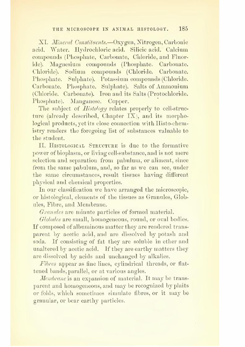
7/29/2019 THE MICROSCOPIST
http://slidepdf.com/reader/full/the-microscopist 221/361
THE MICROSCOPE IN ANIMAL HISTOLOGY. 185
XI. 31iner(d Constituents.—Oxygen, Mtrogen, Carbonic
acid. AVater. H^'drocliloric acid. Silicic acid. Calciumcompounds (Phosphate, Carbonate, Chloride, and Fluor-
ide). jSIagnesium compounds (Phosphate. Carbonate.
Chloride). Sodium compounds (Chloride. Carbonate.
Phosphate. Sulphate). Potassium compounds (Chloride.
Carbonate. Phosphate. Sulphate), Salts of Ammonium
(Chloride. Carbonate). Iron and its Salts (Protochloride.
Phosphate). Manganese. Copper.The subject of Histology relates properly to cell-struc-
ture (already described, Chapter IX), and its morpho-
logical products, yet its close connection with liisto-chem-
istry renders the foregoing list of substances valuable to
the student.
II. Histological Structure is due to the formative
power of bioplasm, or living cell-substance, and is not mere
selection and separation from pabulum, or aliment, since
from the same pabulum, and, so far as we can see, under
the same circumstances, result tissues having difierent
physical and chemical properties.
In our classification we have arranged the microscopic,
or histological, elements of the tissues as Granules, Glob-
ules, Fibre, and Membrane.
Granules are minute particles of formed material.
Globules are small, homogeneous, round, or oval bodies.
If composed of albuminous matter they are rendered trans-
parent b}^ acetic acid, and are dissolved by potash and
soda. If consisting of fat they are soluble in ether and
unaltered by acetic acid. If they are earthy matters they
are dissolved by acids and unchanged by alkalies.
Fibres appear as fine lines, cylindrical threads, or flat-
tened bands, parallel, or at various angles.
Membrane is an expansion of material. It may be trans-
parent and homogeneous, and may be recognized by plaits
or folds, which sometimes simulate fibres, or it may be
granular, or bear earthy particles.

7/29/2019 THE MICROSCOPIST
http://slidepdf.com/reader/full/the-microscopist 222/361
186 THE MICROSCOPIST.
From these elements result the simple and compound
tissues.
The Simple Tissues may be divided into
1. Cells with intermediate fluid, as Blood, Lymph,
Chyle, Mucus, and Pus.
2. Epithelium and its appendages.
3. Connective Substances.—Cartilage. Fat. Connec-
tive tissue. Bone. Dentine.
The Compound Tissues are Muscle, ISTerve, Gland, and
Vascular tissues.
These are formed into Organs.
1. Vegetative.—The Circulatory, Respiratory, Diges-
tive, Urinarj^, and Generative organs.
2. Animal.—The Bony, Muscular, ITervous, and Sensory
apparatus.
AVe shall attempt a brief description of these tissues
and organs, as illustrated by the microscope and modern
methods of research.
I. SIMPLE TISSUES.
1. Cells with Intermediate Fluid.
I. The Blood.
The microscope shows blood to consist, especially in
man and the higher animals, of red corpuscles, colorless
corpuscles, and the fluid in which they are suspended.
1. Blood Plasma., or Liquor Sanguinis.—This is a color-
less and apparently structureless fluid, but when removed
from the body, fibrin separates from it in solid form. In
small quantities of blood this is seen in delicate fibres
crossing each other at various angles.
2. Red-blood Corpuscles.—These were first discovered by
Swammerdam, in 1658, in frog's blood, and in that of man
by Lewenhoek, in 1673. Malpighi is said to have first
seen the actual circulation of blood in the web of a frog:'s
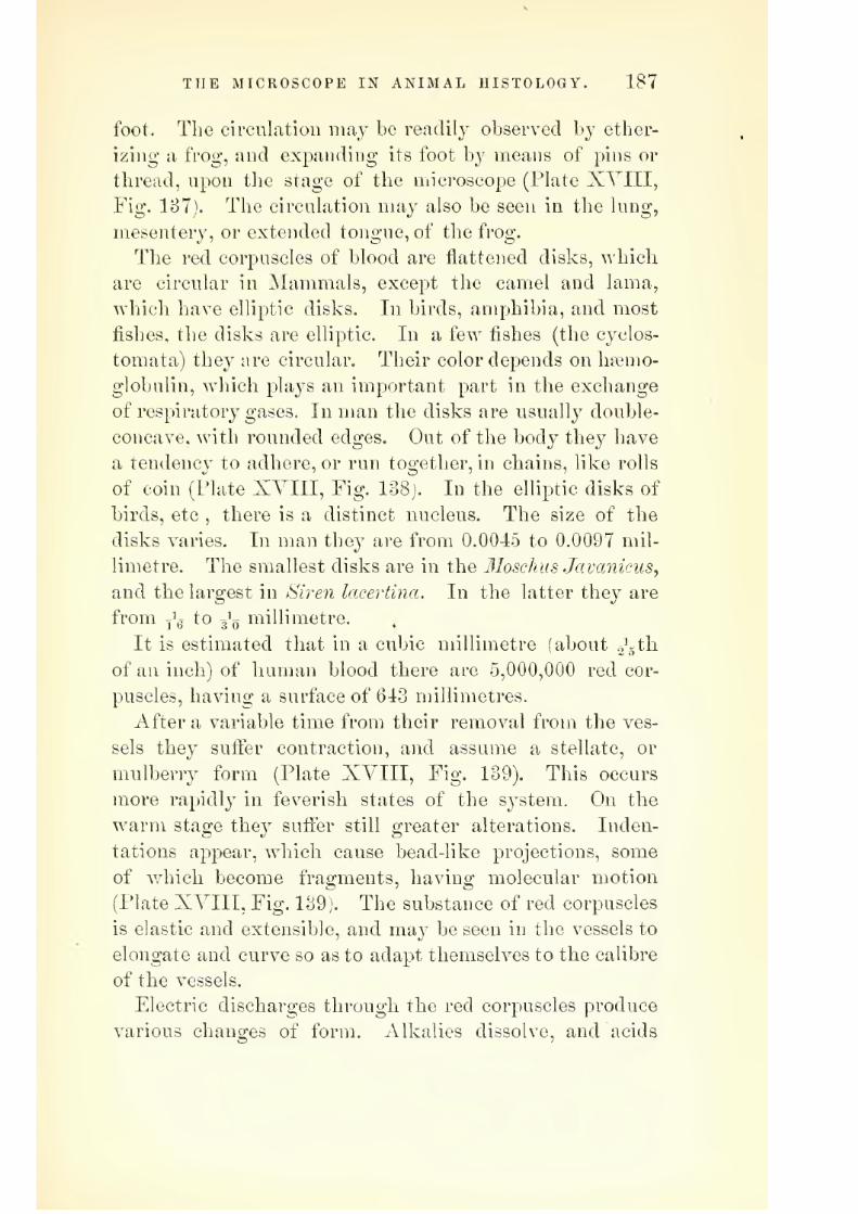
7/29/2019 THE MICROSCOPIST
http://slidepdf.com/reader/full/the-microscopist 223/361
THE MICROSCOPE IN ANIMAL HISTOLOGY. 187
foot. The circulation may be readily observed by ether-
izing a frog, and expanding its foot by means of pins or
thread, upon the stage of the microscope (Plate XVIII,Fig. 187). The circulation may also be seen in the lung,
mesenterj^ or extended tongue, of the frog.
The red corpuscles of blood are flattened disks, which
are circular in Mammals, except the camel and lama,
which have elliptic disks. In birds, amphibia, and most
fishes, the disks are elliptic. In a few fishes (the cyclos-
tomata) they are circular. Their color depends on ha?mo-globulin, which plays an important part in the exchange
of respiratory gases. In man the disks are usuallj- double-
concave, with rounded edges. Out of the body they have
a tendency to adhere, or run together, in chains, like rolls
of coin (Plate XVIII, Fig. 138). In the elliptic disks of
birds, etc , there is a distinct nucleus. The size of the
disks varies. In man they are from 0.0045 to 0.0097 mil-
limetre. The smallest disks are in the lloschusJavanicus,
and the largest in Siren lacertina. In the latter they are
from y'g to 3^0 millimetre.
It is estimated that in a cubic millimetre (about ..'gth
of an inch) of human blood there are 5,000,000 red cor-
puscles, having a surface of 643 millimetres.
After a variable time from their removal from the ves-
sels they suffer contraction, and assume a stellate, or
mulberry form (Plate XVIII, Fig. 139). This occurs
more rapidly in feverish states of the system. On the
warm stage they sufier still greater alterations. Inden-
tations appear, which cause bead-like projections, some
of which become fragments, having molecular motion
(Plate XVIII, Fig. 139). The substance of red corpusclesis elastic and extensible, and may be seen in the vessels to
elongate and curve so as to adapt themselves to the calibre
of the vessels.
Electric discharges through the red corpuscles produce
various changes of form. Alkalies dissolve, and acids

7/29/2019 THE MICROSCOPIST
http://slidepdf.com/reader/full/the-microscopist 224/361
188 THE MICROSCOPIST.
cause a precipitate in them. Thej are tinged by neutral
solutions of carminate of ammonia. One-half to 1 per
cent, of salt added to the staining fluid causes the nuclei
only of Amphibian corpuscles to be stained. Chloroform,
tannin, and other reagents, produce various changes,
which suo;o;est a wide field of research connected with
Therapeutics.
The old opinion of the structure of red corpuscles rep-
resented them as vesicles consisting of a membrane and its
contents, but Max Schultze, in 1861, showed that a mem-
brane was not constant. This may be verified by break-
ing them underpressure.
Briicke's experiment on the astringent action of boracic
acid on the blood of 7r?7o?i, repeated by Strieker and Lan-
kester, shows the red corpuscles to possess a double struc-
ture. There is a body, called (Ecoid ; a porous, non-con-
tractile, soft, transparent mass ; and a retractile substance,
or Zooid, containing the hsemoglobulin, which fills the in-
terspaces of the Q3coid. The Zooid seems identical with
simple cell-substance, or bioplasm.
3. Colorless, or White Corpuscles.—These appear to be
simply masses of bioplasm of various sizes. Some are
quite small, and many are larger than the red corpuscles.
Their number is much smaller than the red disks, being
about 1 to 350, or even less. In leucaemia and other dis-
eases their relative number is much greater. In the blood
of cold-blooded animals, and in that of vertebrata, if the
normal temperature is continued by means of a warm
stage, the amoeboid motions are quite perceptible with a
high magnifying power (Plate XYIII, Fig. 139). They
may also be seen to take up small particles of matter into
their interior, such as cinnabar, carmine, milk-globules,
and even portions of the red globules.
Both red and white cells are forced through the unin-
jured walls of small vessels by impeded circulation, but
the white cells thus migrate, by virtue of their vital con-

7/29/2019 THE MICROSCOPIST
http://slidepdf.com/reader/full/the-microscopist 225/361
PLATE XVIII.
Fig. 13
Cupillary circulatiou iu a portion of the web of a Frog's foot.
Fig. 138. Fig. 139
M #<&
Alterations in form in blood-discs;—1, Stellate or mul-berry form; 2, On warm stage; 3, Amceboid white-cell
forms.
Fig. 140.
S)
Blood-discs ;—1, Eliptic Discs of Amphibia;
2, Human red-corpuscles ; 3,White or lymph-
corpuscle ; 4, Rouleaux of red-discs.© 1
Pus-corpuscles —a with acetic acid
Fig. 142.
Mucous corpuscles and epithelium.
Varieties of Epithelium ; a, Tessalated ; 6, Squamous
;
c, Glandular; d, Columnar; e. Ciliated.

7/29/2019 THE MICROSCOPIST
http://slidepdf.com/reader/full/the-microscopist 226/361

7/29/2019 THE MICROSCOPIST
http://slidepdf.com/reader/full/the-microscopist 227/361

7/29/2019 THE MICROSCOPIST
http://slidepdf.com/reader/full/the-microscopist 228/361
190 THE MICROSCOPIST.
digestion a mixture of albuminous and fatty matters,
which is known as chyle^ and these vessels have obtained
the name of ladeals. The cells in this fluid are leucocytes,
identical with white cells in blood. They originate in the
lymphatic glands and "Peyer's patches" of the intestine,
and are the corpuscles of these organs which have been
carried off by the fluid stream.
III. Mucus.
Is a tenacious semifluid substance which covers the
surface of mucous membranes. It contains cast-off epi-
thelial and gland-cells, and the mucus corpuscle^ which, as
we have before said, is identical with other leucocytes.
Synovial fluid is of similar nature. It is now regarded as
a transformation product of the epithelial cells, and not
to originate as a secretion from special glands (Plate
XYIII, Fig. 141).
2. Epithelium and its Appendages.
Epitlielium (from e-j, upon, and Oa/dw^ to sprout) is so
called since it was formerly supposed to sprout from mem-
brane. It is a tissue formed of cells more or less closely
associated, which is found in layers upon external and
internal surfaces. The cells are generally transparent,
with vesicular, homogeneous, or granular nuclei, the lat-
ter being the remains of the original leucocyte or bio-
plast. In the older cells the nucleus is absent, the entire
mass having been transformed.
The forms of epithelial cells vary according to situation
or function. The original form is spheroidal, but changes
by compression, etc.
1. Tessellated or pavement epithelium (Plate XVIII,
a, Fig. 142). These are cells whose formed material is
flattened, and which are united at their edges. They are
sometimes hexagonal, and often polyhedral, in form.
Examples : Serous and synovial membranes ; the pos-

7/29/2019 THE MICROSCOPIST
http://slidepdf.com/reader/full/the-microscopist 229/361
THE MICROSCOPE IN ANIMAL HISTOLOGY. 191
terior layer of the cornea ; the peritoneal surface ; the
interior 'of bloodvessels, and shut sacs generall}-.
2. Squamous or scaly epithelium. The cells are flat,
and overlap each other at the edges (Plate XVIII, 6, Fig.
142).
Examples : Epidermis ; many parts of mucous mem-
branes, as the mouth, fundus of bladder, vagina, etc,
3. Glandular epithelium (Plate XVIII, c, Fi|. 142).
The cells are round or oval bioplasts, often polyhedral
from pressure, and the formed material is often soft.
Examples : Liver cells, convoluted tubes of kichiey, and
interior of glands generally.
4. Columnar epithelium (Plate XVIII, d, Fig. 142}.
Cells cylindrical or oblong, arranged side by side. Abird's-eye view shows them similar to the tessellated form,
hence they should be seen from the side.
Examples : Villi and follicles of intestine, ducts of
glands, urethra, etc.
Some of the columnar or cylinder-cells have a thickened
border or lid perforated with minute pores (Plate XVIII,
/, Fig. 142). They are found in the small intestine, gall-
bladder, and biliary ducts.
5. Ciliated epithelium (Plate XVIII, e, Fig. 142).
These are cylindrical cells having vibratile cilia, whose
motions produce a current in the surrounding fluid.
Examples : The upper and back nasal passages, the
pharynx, bronchi. Fallopian tubes, etc.
The Hair.—Hairs are flliform appenchiges, composed of
a modiflecl epithelial tissue of rather complex structure.
They originate in a follicle, which is a folding in of the
skin. The shaft of the hair is the portion projecting
above the skin, and the root is concealed in the hair-fol-
licle. The bulb of the root is the rounded terminal part,
which is hollow below, and rests on a papilla which rises
from the floor of the follicle (Plate XIX, Fig. 143). Be-
tween the follicle and hair is a sheath, which is divided
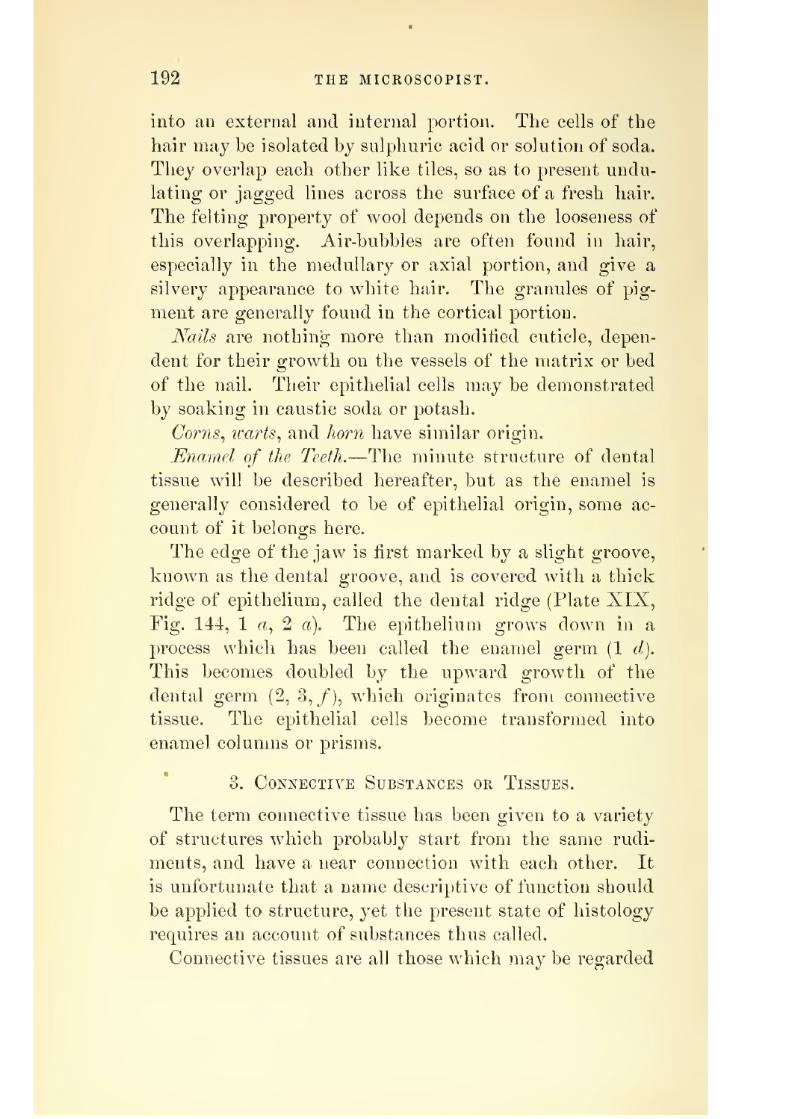
7/29/2019 THE MICROSCOPIST
http://slidepdf.com/reader/full/the-microscopist 230/361
192 THE MICROSCOPIST.
into an external and internal portion. The cells of the
hair may be isolated by sulphuric acid or solution of soda.
They overlap each other like tiles, so as to present undu-
lating or jagged lines across the surface of a fresh hair.
The felting property of wool depends on the looseness of
this overlapping. Air-bubbles are often found in hair,
especially in the medullary or axial portion, and give a
silvery appearance to white hair. The granules of pig-
ment are generally found in the cortical portioi].
Nails are nothing more than modified cuticle, depen-
dent for their growth on the vessels of the matrix or bed
of the nail. Their epithelial cells may be demonstrated
by soaking in caustic soda or potash.
Corns^ icarts^ and horn have similar origin.
Enamel of the Teeth.—The minute structure of dental
tissue will be described hereafter, but as the enamel is
generally considered to be of epithelial origin, some ac-
count of it belongs here.
The edge of the jaw is first marked by a slight groove,
known as the dental groove, and is covered with a thick
ridge of epithelium, called the dental ridge (Plate XIX,
Fig. 144, 1 rt, 2 a). The epithelium grows down in a
process which has been called the enamel germ (1 d).
This becomes doubled by the upward growth of the
dental germ (2, 3,/), which originates from connective
tissue. The epithelial cells become transformed into
enamel columns or prisms.
3. Connective Substances or Tissues.
The term connective tissue has been given to a variety
of structures which probably start from the same rudi-
ments, and have a near connection with each other. It
is unfortunate that a name descriptive of function should
be applied to structure, yet the present state of histology
requires an account of substances thus called.
Connective tissues are all those which may be regarded

7/29/2019 THE MICROSCOPIST
http://slidepdf.com/reader/full/the-microscopist 231/361
Fig. 143,
PLATE XIX.
Struct lire of Human Hah:
Fig. 145.
Conneftice-tisniip elements. From the Frog's Thigh :
a, coutractfd cell; 6, ramified; c, rf, motionless gran-
ular cells: /, tibrillce; g, connective-tissue bundle;
h, elastic fibre net-work.
1 r^-^
Fig. 144. a.
Development of the enamel: a, dental ridge;
6, young layer of epithelium; c, deep layer; d,
enamel germ; e, enamel organ
; /, dental germ.
Fig. 146.
// 7^-f
%kmim
IV /
w
W/iite Fibrous Tissue, from Ligament.
Fig. 148.
Yellow Fibrous Tissue, from Ligameutum Niichje
of Calf.
Fully Tissue.

7/29/2019 THE MICROSCOPIST
http://slidepdf.com/reader/full/the-microscopist 232/361

7/29/2019 THE MICROSCOPIST
http://slidepdf.com/reader/full/the-microscopist 233/361
THE MICROSCOPE IN ANIMAL HISTOLOGY. 193
as basement-membranes, supporting layers or investments
for epithelial structures, blood, lymph, muscle, and nerves.
It includes ordinary connective tissue (white and yellow
fibrous tissues), cartilage, bone, corneal tissue, dentine^
and fatty tissue.
Most of the difficulty found in the consideration of
these tissues arises from discussions relative to the inter-
cellular substance. Max Schultze and Beale agree in re-
garding it to originate from the protoplasm or bioplasm
of cells.
The cells are, according to Frey, originally spheroidal,
with vesicular nuclei, and between them is an albuminous
intercellular substance—a product of the cells, or trans-
formed cells—which usually undergoes fibrillation, while
the cells become stunted, or develop into spindle-shaped
or stellate elements. Calcification of the intercellular sub-
stance occurs in some of these tissues, as bone and dentine..
The cells of connective tissue present many varieties.
Eecklinghausen first observed migrating lymphoid cells
or bioplasts in the cornea of the eye, the tail of the tad-
pole, the peritoneum, and in various other places. The
exit of white corpuscles from the vascular walls renders
it probable that these amoeboid cells originate in the blood.
Granular cells, of various forms—rounded, fusiform, and
stellate—are also observed. Some of the stellate cells
give oif anastomosing branches. Pigment cells, filled with
granular pigment, are also met with (Plate XIX, Fig.
145).
In its earliest stages, connective tissue consists of closely-
compressed cells, but in the adult two principal forms
have been distinguished ; first, those networks and trabec-
ul?e, developed from cells, which do not yield gelatin on
boiling, and, secondly, fibrillar connective tissue composed
of a gelatin-yielding substance. Of the first kind we notice
the following varieties
1. Independent masses of gelatinous or mucous tissue,
13

7/29/2019 THE MICROSCOPIST
http://slidepdf.com/reader/full/the-microscopist 234/361
194 THE MICROSCOPIST.
'Consisting of nucleated cells, giving off smooth anasto-
mosing trabecnlpe, as in the early stage of the yitreous
liumor of the eye and of the gelatinous tissue of the um-
bilical cord, etc.
2. Very delicate reticular tissue found in the eye and in
the interior of nerve-centres.
3. A network filled with lymphoid cells (adenoid or
cytogenous tissue) in the glands of the lymphatic system,
and around the fasciculi of fibrillar coimective tissue.
4. A coarser network in the ligamentum pectinatum
of the human eye.
5. A tissue formed of fusiform and stellate cells, as in
the interior of the kidneys
The second form referred to, or the fibrillar connective
•'tissue, was the only form to which the term connective
tissue was formerly applied. It is composed of gelatin-
yielding fibrillse, which may be split into skein-like por-
tions of various breadth. (Plate XIX, Fig. 146.) Per-
imanganate of potash stains it brown. Acetic and dilute
mineral acids cause the tissue to swell so that the appear-
ance of fibrillation is lost through compression, and the
cells, or nuclei, are made manifest. Chloride of gold
staining exhibits both fibrillfe and cells.
Masticfibres (yellow elastic) (Plate XIX, Fig. 147) are
apparent in all forms of connective tissue which have been
made transparent by boiling, or acetic acid. They are
non-gelatinizing, cylindric, slightly branched, or forming-
plexuses. In some fasciculi of fibrillar connective tissue, as
seen after the action of acetic acid, elastic fibres appear in
hoops, or spirals, around them. In the ligamentum nu-
clei of the giraffe the elastic fibres are marked by trans-
verse striae, or cracks. Elastic fibres often form flattened
trabeculse, or are fused into elastic plates, or membranes,
with foramina, as in arterial tunics.
The ligaments of the skeleton, the periosteum, peri-
chondrium, aponeuroses, fasciae, tendons, and generally all

7/29/2019 THE MICROSCOPIST
http://slidepdf.com/reader/full/the-microscopist 235/361
THE MICROSCOPE IN ANIMAL HISTOLOGY. 195
the tunics of the body, afford examples of the tlbrillar
connective tissue.
Fatty Tissue.—The loose connective tissue contains in
various parts great numbers of cells filled with fat. Their
form is round, or oval, and are often divided into groups,
or lobules, by trabeculfe. (Plate XIX, Fig. 148.) Each
lobule has its own system of bloodvessels, which divide
into such numerous capillaries that the smaller groups,
and even individual fat-cells, are surrounded by vascular
loops. Sometimes the contents of the cells appear in
needle-shaped crystals, often collected in a brush-like form.
Fat-cells seem to be chiefly receptacles for the deposit of
superabundant oleaginous nutriment, and are analogous
to the starch-cells in vegetables.
Cartilage.—This is formed of cells in an originallj" homo-
geneous intercellular substance. The only difference be-
tween what was formerly distinguished as cartilage and
fibro-cartilage is that the matrix or intercellular substance
of the latter is fibrous.
The cells, or cartilage-corpuscles, are nucleated, and lie
in cavities of various sizes and form in the matrix (Plate
XX, Fig. 149). Two nuclei often appear in one cell. It
is yet a question whether the capsule and matrix are the
secretion of the cells which has become solid, or a part of
the body of the cell which has undergone metamorphosis.
The multiplication of cartilage-cells is endogenous. By
segmentation, two, four, or a whole generation of daughter-
cells, so called, may lie in the interior of a capsule. In this
way growing cartilage may acquire a great number of
elements.
In the ear of the mouse, etc, we observe a form of car-
tilage which is wholly cellular, and possesses no matrix
(Plate XX, Fig. 150).
Bone., or osseous tissue, is formed secondarily from meta-
morphosed descendants of cartilage or connective-tissue
cells, and is the most complex structure of this group. It
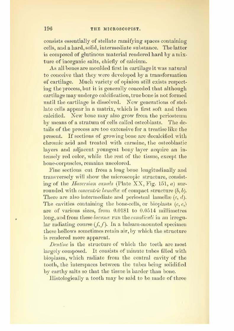
7/29/2019 THE MICROSCOPIST
http://slidepdf.com/reader/full/the-microscopist 236/361
196 THE MICROSCOPIST.
consists essentially of stellate ramifj-ing spaces containing
cells, and a hard, solid, intermediate substance. The latter
is composed of glutinous material rendered hard by a mix-
ture of inorganic salts, chiefly of calcium.
As all bones are moulded first in cartilage it was natural
to conceive that they were developed by a transformation
of cartilage. Much variety of opinion still exists respect-
ing the process, but it is generally conceded that although
cartilage raa^^ undergo calcification, true bone is not formed
until the cartilage is dissolved. IS'ew generations of stel-
late cells appear in a matrix, which is first soft and then
calcified. New bone may also grow from the periosteum
by means of a stratum of cells called osteoblasts. The de-
tails of the process are too extensive for a treatise like the
present. If sections of growing bone are decalcified with
chromic acid and treated with carmine, the osteoblastic
layers and adjacent youngest bony layer acquire an in-
tensely red color, while the rest of the tissue, except the
bone-corpuscles, remains uncolored.
Fine sections cut from a long bone longitudinally and
transversely will show the microscopic structure, consist-
ing of the Haversian canals (Plate XX, Fig. 151, a) sur-
rounded with concentric lamellw of compact structure {b, b).
There are also intermediate and periosteal lamellae (c, d).
The cavities containing the bone-cells, or bioplasts (e, e,)
are of various sizes, from 0.0181 to 0.0514 millimetres
Ions:, and from these lacunce run the canaliculi in an irreg-u-
lar radiating course (/,/). In a balsam-mounted specimen
these hollows sometimes retain air, by which the structure
is rendered more apparent.
Dentine is the structure of which the teeth are most
largely composed. It consists of minute tubes filled with
bioplasm, which radiate from the central cavity of the
tooth, the interspaces between the tubes being solidified
by earthy salts so that the tissue is harder than bone.
Histologically a tooth may be said to be made of three

7/29/2019 THE MICROSCOPIST
http://slidepdf.com/reader/full/the-microscopist 237/361
PLATE XX.
Fig. 149.
^ 'cK' Fig. 150.
W( /v
O
a.^ll
Section of the Brauchial Cartilage of Tadpole.
Cellular Cartilage of JMouse's Ear.
Vertical section q^HumanMolar Tooth:—1, enamel ; 2,
cementumorcrustapetrosa;
3, dentine, or ivory; 4, osse-
ous excrescence, arising fromliypertropliy of cementum :
5, pulp-cavity; 6, osseous
lacunse at outer part of den-tine. Involuntary Muscular-fibre.
Sarcolemma.

7/29/2019 THE MICROSCOPIST
http://slidepdf.com/reader/full/the-microscopist 238/361
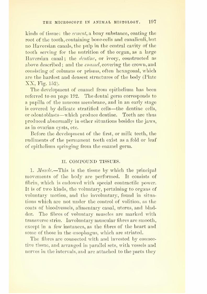
7/29/2019 THE MICROSCOPIST
http://slidepdf.com/reader/full/the-microscopist 239/361
THE MICROSCOPE IN ANIMAL HISTOLOGY. 197
kinds of tissue: the cement., a bonj substance, coating the
root of the tooth, containing bone-cells and canaliculi,but
no Haversian canals, the pulp in the central cavity of the
tooth serving for the nutrition of the organ, as a large
Haversian canal ; the elenti7ie, or ivory, constructed as
above described; and the enamel, coYermg the crown, and
consisting of columns or prisms, often hexagonal, which
are the hardest and densest structures of the body (Plate
XX, Fig. 152).
The development of enamel from epithelium has been
referred to on page 192. The dental germ corresponds to
a papilla of the mucous membrane, and in an early stage
is covered by delicate stratified cells—the dentine cells,
or odontoblasts—which produce dentine. Teeth are thus
produced abnormally in other situations besides the jaws,
as in ovarian cysts, etc.
Before the development of the first, or milk teeth, the
rudiments of the permanent teeth exist as a fold or leaf
of epithelium springing from the enamel germ.
II. C0MP0UI!^D TISSUES,
1. llusde.—This is the tissue by which the principal
movements of the body are performed. It consists of
fibrin, which is endowed with special contractile power.
It is of two kinds, the voluntarj^, pertaining to organs of
voluntary motion, and the involuntary, found in situa-
tions which are not nnder the control of volition, as the
coats of bloodvessels, alimentary canal, uterus, and blad-
der. The fibres of voluntary muscles are marked with
transverse strife. Involuntary muscular fibres are smooth,
except in a few instances, as the fibres of the heart and
some of those in the oesophagus, which are striated.
The fibres are connected w^ith and invested by connec-
tive tissue, and arranged in parallel sets, with vessels and
nerves in the intervals, and are attached to the parts they

7/29/2019 THE MICROSCOPIST
http://slidepdf.com/reader/full/the-microscopist 240/361
198 THE MICROSCOPIST.
are designed to move by tendon, aponeuroses, or some form
of fibrous tissue. The organs or muscles thus formed are
generally solid and elongated, but sometimes expanded.
Involuntary or unsfriped muscular fibres are flat bands
or spindle-shaped fibres with nuclei, which may be re-
garded as the remains of the formative bioplasm (Plate
XX, Fig. 153). They are usually transverse, or interlace
with each other on the walls of cavities and vessels. In
the heart the fibres, though involuntary, are striped and
branching. Striyed fibre varies from g'^th to ys'oTjth inch
in diameter. It is largest in insects, in which individual
fibrils may be readily obtained, especially from the thoracic
muscles. They are generally found in bundles of fibrils,
splitting longitudinally or in disks, and each bundle is
inclosed in a sheath or sarcolemma (Plate XX, Fig. 154).
The transverse striation of muscle is subject to much
variation, and the precise nature of the sarcous elements
which produce the appearance is yet a matter of dispute,
but in all probability the ultimate elements are sarcous
prisms or particles imbedded in a homogeneous mass, and
by their mutual attraction, excited by various stimuli,
the contraction of the fibre takes place.
For the purpose of observation, the connective tissue
may be removed from muscular fibre by gelatinizing it
with dilute sulphuric acid, and dissolving it at a temper-
ature of 104° F. The nuclei of muscular fibre are seen
after treating with acetic acid, and may be stained with
carmine fluid, etc.
2. Nerve-tissue.—The term nerve was applied by the
ancients to tense cords, as bow-strings, musical strings,
etc., and was appropriated to the fibres now called nerves,
because they deemed them to operate by tremors, vibra-
tions, or oscillations, another instance of wrong naming
of structure from an opinion respecting function. Plip-
pocrates, Galen, and others, however, thought nerves were
7/29/2019 THE MICROSCOPIST
http://slidepdf.com/reader/full/the-microscopist 241/361
THE MICROSCOPE IN ANIMAL HISTOLOGY. 199
hollow tubes, conveying fine ethereal fluids, termed ani-
mal spirits. • ' '
- T^"--
JS'ervous matter is soft, unctuous, and easil}^ disturbed,
hence it is necessary to examine it while fresh. Histo-
logically it is divided into fibres and cells, imbedded in
connective tissue.
Nerve-fibres are of two kinds, the medullated, or dark-
bordered threads, and the pale, or non-medullated. Med-
ullated fibres consist of a delicate envelope of connective
tissue, called the neurilemma or primitive sheath, an axis-
cjdinder or albuminous portion, extending down the cen-
tre, and a portion composed of a mixture of albumen,
cerebral matter, and fat, surrounding the axis-cylinder
(Plate XXI, Fig. 155, a, b, g). This latter is the medul-
lary sheath, or white substance of Schwann. It changes
rapidly, so as to coagulate and become granular. Alka-
lies render it fluid, so as to exude in fat-like drops. Ab-
solute alcohol, chromate of potass and collodion, contract
the sheath, so as to permit the axis-cylinder, which is the
essential part of the nerve, to protrude (Plate XXI, e,
Fig. 155). Anilin, carmine, nitrate of silver, and chloride
of gold stain the axis, while osmic acid blackens only the
medullary sheath.
Xon-medullary or pale nerve-fibres are regarded as Qva-
bryonic or developmental forms (Plate XXI, d, Fig. 155).
The ganglionic fibres of the sympathetic (Remak's fibres)
are flat, homogeneous bands, with round or oval nuclei.
Some have considered them as formed of connective tis-
sue, but their nervous character is generally conceded.
Schultze and others regard the axis-cylinder as made up
of extremely delicate fibrillte.
Nerve-cells^ or ganglion corpuscles, are of two kinds,
those without and those with processes. The first are
called apolar, and the latter unipolar, bipolar, or multi-
polar, according to the number of ramifications. The
cells are nucleated, and inside the nucleus is usually

7/29/2019 THE MICROSCOPIST
http://slidepdf.com/reader/full/the-microscopist 242/361
200 THE MICROSCOPIST.
another, the nucleolus. Dr. Beale discovered certain gan-
glion-cells in the sympathetic of the tree-frog (in the au-
ricular septum of the heart), one of whose poles is encir-
cled spirally by the others (Plate XXI, Fig. 156).
The ultimate structure of ganglia or nervous knots,
and the relation of the fibres to the cells, opens a wide
field of research. In the muscle of the heart, etc., many
of these ganglia seem to form special nervous systems.
Dr. Beale has described the nerves ramifying on the capil-
laries and involuntary muscular fibrils of the terminal ar-
teries as a self-regulating mechanism for the distribution
of blood (Plate XXI, Fig. 157). Thus, if a tissue receives
excess of pabulum, the capillary nerve-fibre is disturbed
and transmits a change to the ganglion, and thence
throusrh the efterent nerve to the muscular fibres of the
:artery, and vice versa.
Meissner has shown many ganglionic plexuses in the
submucous coat of the alimentary canal. Another system
of the same kind, called the ijlexus myentericus, was dis-
<;overed by Auerbach between the muscular layers of the
intestinal tube. Similar plexuses exist in other organs.
As to the peripheral termination of nerve-fibres, there
as still considerable discussion.
Mostof the
Germanhis-
ftologists consider the nerves of voluntary muscles to ter-
.minate in end plates, in which the neurilemma becomes
•continuous with the sarcolemma of the muscular fibre.
Dr. Beale maintains that there is a plexus of minute nerves
'=0ver the fibrils. In some of my own preparations, espe-
cially some stained with soluble Prussian blue, a disk
formed of a plexus of excessively minute nerve-fibres is
observed, from which tortuous branches go to other mus-
ele-fibres.
In the cornea, Cohnheim and Klein have traced fine
nerve-fibres to the epithelial cells of the conjunctiva, by
means of chloride of gold staining.
-3. 'Qlandular tissue consists of a fine transparent mem-

7/29/2019 THE MICROSCOPIST
http://slidepdf.com/reader/full/the-microscopist 243/361
PLATE XXI.
Fig. 155. Fig. 15G.
(-' v)
amm
Nerve-fibres.
Fig. 159.
Vascular Tissue.
Various Ganglionic Nerve-cells.
Fig. 157.
Self-regulating System of Ganglia—nerves,arteries, and capillaries.
Layers of Blastoderm.

7/29/2019 THE MICROSCOPIST
http://slidepdf.com/reader/full/the-microscopist 244/361

7/29/2019 THE MICROSCOPIST
http://slidepdf.com/reader/full/the-microscopist 245/361
THE MICROSCOPE IN ANIMAL HISTOLOGY. 201
brane, tbroiigli which the plasma transudes, and cells of
glandular epithelium. A vascular network exists on the
surface of the membrane, from which the material of the
secretion is obtained. This membrane may be a simple
follicle, or tube, as in the mucous membrane, or system of
tubes, as in the kidneys, a convoluted tube, a simple open
vesicle, a racemose aggregation of vesicles, or a close cap-
sule which discharges itself by bursting. (Plate XXI,
Fig. 158).
4. Vascular Tissue.-—The smallest bloodvessels and lym-
phatics, called capillaries, are minute tubes, consisting of a
series of flattened epithelial cells, and containing stomata,
or openings through which white or red blood-corpuscles
may occasionally pass (Plate XX, Fig. 159, a, b). The
larger trunks have, in addition to the cellular layer, one
of longitudinally striated connective tissue, a middle coat
containing transverse muscular fibres, and an external coat
of connective tissue (Plate XXI, Fig. 159). The distribu-
tion of the capillary bloodvessels is various, according to
the nature or function of the organ or tissue in which they
are found.
Development of the Tissues.
It has been stated, page 125, that reproduction in the
higher animals consists of an ovum fecundated by contact
with a sperm-cell, or spermatozoid.' The ovum consists
of a germinal vesicle, containing one or xwoyq germinal spots,
and included within a vitellus (a yelk) which is surrounded
by SI vitelline memhran e, which may have additional invest-
ments in the form of layers of albumen and of an outer
coriaceous or calcified shell.
The first step in the development of the embryo is the
division of the vitelline substance into cleavage-masses, at
first two, then four, then eight, etc. This process of yelk-
division may aftect the whole yelk or a part of it, and re-
sults in the formation of a blastoderm, or embryogenic

7/29/2019 THE MICROSCOPIST
http://slidepdf.com/reader/full/the-microscopist 246/361
202 THE MICROSCOPIST.
tissue. This riiclinientary embryonic tissue consists of
three laj'ers of cells, or germinal plates. The upper is the
corneous layer, or epiblast, the middle one the intermediate
plate, or mesoblast, and the lower the intestinal glandular
layer, or hypoblast (Plate XXI, Fig. 160). From these
the various tissues and organs are developed.
The outer plate produces the epithelium of the skin and
its appendages, with the cellular elements of the glands of
the skin, mammse, and lachrymal organs. B}^ a peculiar
folding over the axis this plate also produces the elements
of the brain and spinal cord, and the internal parts of the
organs of special sense. The physiological significance of
this laj^er is, therefore, very great.
Tlie low^er stratum of the blastoderm supplies the epi-
thelium of the digestive tract, and the cellular constituents
of its various glands, together with the liver, lungs, and
pancreas.
The middle layer supplies the material for many struc-
tiires. The whole group of connective substances, or
tissues of support; muscular tissue; blood and lymph,
with their containing vessels ; lymph-glands, including
the spleen, etc., all arise from this. The epithelial cells
of such tubes and cavities as originate in this layer are
regarded as diti^erent from those of true glands, and are
more permeable to fluids. They have been termed false
epithelium, or endothelium.
The following description, by Professor Huxley, will
enable the student to form an idea of the general process
of development. A linear depression, the primitive groove.,
makes its appearance on the surface of the blastoderm,
and the substance of the mesoblast along each side of this
groove grows up, carrjnng with it the superjacent epiblast.
Thus are produced the two dorsal lamince^ the free edges
of which arch over toward one another, and eventually
unite, so as to convert the primitive groove into the cere-
bro-spinal canal. The portion of the epiblast which lines

7/29/2019 THE MICROSCOPIST
http://slidepdf.com/reader/full/the-microscopist 247/361
THE MICROSCOPE IN ANIMAL HISTOLOGY. 203
this, cut otF from the rest, becomes thickened, and takes
on the structure of the brain, or encephalon^ in the region
of the head ; and of the spinal cord, or myelon^ in the
region of the spine. The rest of the epiblast is converted
into the epidermis.
The part of the blastoderm which lies external to the
dorsal laminae forms the ventral lamince; and these bend
downward and inward, at a short distance on either side
of the dorsal tulje, to become the walls of a ventral or
visceral tube. The ventral laminjB carry the epiblast on
their outer surfaces, and the hypoblast on their inner sur-
faces, and thus, in most cases, tend to constrict off" the
central from the peripheral portions of the blastoderm.
The latter, extending over the yelk, incloses it in a kind
of bag. This bag is the first formed and the most con-
stant of the temporary, or fa3tal appendages of the young
vertebrate, the umbilical vesicle.
AVhile these changes are occurring, the mesoblast splits,
throughout the regions of the thorax and abdomen, from
its ventral margin, nearly up to the notochord (which has
been developed, in the meanwhile, by histological differen-
tiation of the axial indifterent tissue, immediately under
the floor of the primitive groove) into t^vo lamellce. One
of these, the visceral lamella^ remains closely adherent to
the hypoblast, forming with it the splancknopleure, and
eventually becomes the proper wall of the enteric canal;
while the other, ih.Q parietal lamella, follows the epiblast,
forming with it the sornatopleure, which is converted into
the parietes of the thorax and abdomen. The point of
the middle line of the abdomen at which the somato-
pleures eventually unite, is the umbilicus.
The walls of the cavity formed by the splitting of the
ventral laminae acquire an epithelial lining, and become
the great jpleurope.ritoneal serous membranes (Huxley's
Anatomy of Vertebraied Animals).
In addition to the umbilical vesicle, above described as

7/29/2019 THE MICROSCOPIST
http://slidepdf.com/reader/full/the-microscopist 248/361
204 THE MICROSCOPIST.
a temporary appendage, the foetus lias other special struc-
tures, derived from the blastoderm. Thus the somato-
pleure grows up over the embryo and forms a sac filled
with clear fluid, the amnion. The outer layer of the sac
coalesces with the vitelline membrane to form the chorion.
The allantois begins as an outgrowth from the mesoblast.
It becomes a vesicle, and receives the ducts of the primor-
dial kidneys or Wolffian bodies, and is supplied with blood
from the two hypogastric arteries which spring from the
aorta. Tbe allantois is afterwards cast oW by the contrac-
tion of its pedicle, but a part of its root is usually re-
tained, and becomes the permanent urinary bladder. In
the Mammalia the allantois conveys the embryonic ves-
sels to the internal surface of the chorion, whence they
draw supplies from the vascular lining of the uterus.
Foster and Balfour recommend that the study of em-
bryonic development should commence with the egg of a
fowl taken at difterent times from a broodins: hen, or an
artificial incubator. The egg should be placed on a hol-
low mould of lead in a basin, and covered with a warm
solution of salt (7.5 per cent.). It should be opened with
a blow, or by filing the shell. With the naked eye or
simple lens, lying across the long axis of the egg., may be
seen the pellucid area, in which the embryo appears as a
white streak. The mottled vascular area, with the blood-
vessels, and the opaque area spreading over the yelk, may
be observed. The blastoderm may be cut out with a sharp
pair of fine scissors, floated into a watch-glass, freed from
vitelline membrane and yelk, and removed (under the salt
solution) to a glass slide, A thin ring of putty may then
be placed round the blastoderm, which is covered with
salt solution, and the thin glass cover put on. With a
low-power objective many of the details of structure may
be seen in an embryo of thirty-six to forty-eight hours
incubation, as the heart, the neural tube, the first cere-

7/29/2019 THE MICROSCOPIST
http://slidepdf.com/reader/full/the-microscopist 249/361
THE MICROSCOPE IN ANIMAL HISTOLOGY. 205
bral vesicles, the folds of the soraatopleure and splanch-
nopleure, the provertebr?e, etc.
To prepare sections of the embryo, it must be first hard-
ened by placing the slide containing it in a sokition of 1
per cent, chromic acid for twenty-four hours. From this
it should be removed to one of 3 per cent, for twenty-four
hours more ; then for a similar time in alcohol of 70 per
cent., then in alcohol of 90 per cent., and lastly in abso-
lute alcohol, where it may remain till reciuired for section.
Sometimes picric or osmic acid is used for hardening.
The embryo may be stained by placing it in Beale's car-
mine fluid for twenty-four hours, and then replacing it in
absolute alcohol for a day before it is cut. It maj also
be stained with hsematoxylin if preferred. The specimen
may be imbedded in paratiin, wax, and oil, or a mixture
of four parts of spermaceti to one part of cocoa butter or
castor oil. If there are cavities in the object, it is best
to saturate it first with oil of bergamot. A little melted
spermaceti mixture is poured on the bottom of a small
paper box, and when solid the embryo is placed flat on
itj the superfluous oil removed as far as possible, and the
warm mixture poured on. Bubbles can be removed with
a hot needle. A mark should be made of the exact posi-
tion of the embryo. Sections may be cut with the sec-
tion-cutter or a sharp razor, and if the spermaceti mix-
ture is used, the razor should be moistened with olive oiL
The sections should be floated from the razor to the slide,
and treated with a mixture of four parts turpentine and
one of creasote. They may then be mounted in balsam
or dammar varnish.
The most instructive transverse sections of an early
embryo will be through the optic vesicles, the hind brain,
the middle of the heart, the point of divergence of the
splanchnopleure folds, the dorsal region, and a point where
the medullary canal is still open. For the unincubated
blastoderm only one section, through the centre, is re-

7/29/2019 THE MICROSCOPIST
http://slidepdf.com/reader/full/the-microscopist 250/361
206 THE MICROSCOPIST.
quired to show the formative layers. In the later stages
dissection is required, and is best performed with embryo
preserved in spirit. If living embryos are placed in spirit,
a natural injection of the vessels may be obtained.
III. OEGAI^S OF THE BODY.
Anatomists usuall}^ group the organs into systems, as
the osseous, muscular, nervous, vascular systems, etc., but
for histological study a classification based on physiologi-
cal considerations may be more convenient for the student.
I. Vegetative Organs.
1. Natritive^OY organs pertaining to the absorption and
distribution of pabulum, including the digestive and cir-
culatory organs.
The mucous memhrane of the intestiyial canal contains
many follicles and glands, whose secretions serve impor-
tant offices in the preparation of the food. These will be
referred to in the next section. The epithelium of the
intestinal canal is columnar, except in the oesophagus,
where it is laminated. Beneath the glandular la^'er of
the stomach is a stratum of fibrous connective tissue and
muscle fibres in two layers, an interntil with transverse,
and an external with longitudinal fibres. The tissue of
the small intestine beneath the epithelium is reticular
connective, entangling Ijmiphoid cells. The structure of
the large intestine is similar to that of the stomach. The
villi of the small intestine begins at the pylorus, flat and
low at first, but becoming conical, and finally finger-like
in shape. The epithelium of the villi are columnar, with
a thickened and perforated edge (Plate XXII, Fig. 161).
Between the epithelial cells of the villi, peculiar "goblet-
cells" are often found, which Frey supposes to be decay-
ing cells. The reticular connective tissue of each villus
is traversed by a vascular network, a lymphatic canal or
lacteal, and delicate longitudinal muscular fibres. If the

7/29/2019 THE MICROSCOPIST
http://slidepdf.com/reader/full/the-microscopist 251/361
THE MICROSCOPE IN ANIMAL HISTOLOGY. 207
villus is unusually broad, there may be more than one
lacteal. The lacteals absorb the fluid known as chyle.
The}' are blind ducts, and nitrate of silver injections show
them to have the same structure as other lymphatics.
The lymphatic radicles are widely disseminated through
all the tissues and organs of the body. They take up nu-
tritive fluids, either from the alimentary canal, or such as
have transuded from the capillaries into the interstices of
the body, mingled wdth the products of decomposition,
and convey them into the general circulation. Hyrtl's
method of demonstrating these radicles is hy passing a
fi.ne canula into the tissue containing lymphatics and forc-
ing the injection by gentle pressure. They are either net-
works, analogous to cai^illaries, or blind passages which
nnite in reticulations. The structure of the vessels has
already been described, page 201. Ljmiphatics and capil-
laries do not communicate directly. A lymph-canal maybe surrounded by capillaries, or run alongside of a capil-
lary, or a lymphatic sheath may envelop a bloodvessel.
This latter plan is seen in the nervous centres, and has
been called by His the perivascular canal system.
The larger lymphatic trunks are interrupted by nodular
and very vascular organs, the lyin.phatic glands. These
consist of the reticular connective tissue already described,
surrounded by an envelope of ordinary fibrous tissue. One
or more afl^'erent lymphatic vessels penetrate the capsule,
or envelope, and similar efferent vessels make their exit
from the other side. Frey describes these glands as con-
sisting of a cortical portion, follicles, and a medullary
portion composed of the tubes and reticular prolongations
of the follicles (Plate XXII, Fig. 1(J2). There is a com-plicate system of communication between the follicles.
The afferent vessel opens into the investing spaces of the
follicle. These lead into the lymph-passages of the med-
ullary portion, from the confluence of which the radicles
of the efferent vessels are formed. The linorual follicular

7/29/2019 THE MICROSCOPIST
http://slidepdf.com/reader/full/the-microscopist 252/361
208 THE MICROSCOPIST.
glands and tonsils, tlie solitary and agminated glands of
the intestine (Peyer's patches), the thymus, and the spleen
have a similar structure, and are called lymphoid organs.
In the thoracic duct the epithelium is inclosed in several
layers of fibrous ixiembrane. The latter contains trans-
verse muscular fibres. The hearty although an involuntary
muscular organ, has striated muscular fibres. These fibres
are not, like other striped muscles, collected into bundles,
but are reticular. The heart, like other organs, is supplied
with lymphatics and bloodvessels. The cardiac plexas of
nerves consists of branches from the vagus and sympa-
thetic. JSTumerous microscopic nervous ganglia also occur,
especially near the transverse groove and septum of the
ventricles. It is thought that these are the chief centres
of energy, so that the heart pulsates after its removal from
the body. It has also been shown recently that the sym
pathetic and vagus filaments are in antagonism, so that
stimulation of the vagus interrupts the motor influence of
the sympathetic, and may bring the heart to a standstill
in a condition of diastole.
The structure of bloodvessels has been described under
the head of vascular tissue. ISTo special boundary exists
between capillaries and the arteries and veins. The ar-
rangement of the capillaries, however, is various, and
often so characteristic that a practiced eye can generally
recognize an organ or tissue from its injected capillaries.
(Plate XXII, Figs. 163 to 1G8.) For methods of inject-
ing, see page 61. Capillaries form either longitudinal or
rounded meshes. The muscular network, etc., is extended,
while fat-cells, the alveoli of the lungs, lobules of liver,
capillary loops of papillae in skin and mucous membranes,
outlets of follicles, etc., present a more or less circular in-
terlacement. The capillary tube lies external to the ele-
mentary structure, and never penetrates its interior.
2. Secretive Organs.—True secretions serve important
oflices in the organism : as the materials of reproduction;
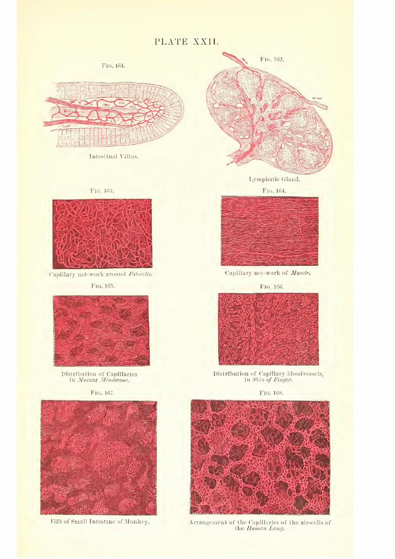
7/29/2019 THE MICROSCOPIST
http://slidepdf.com/reader/full/the-microscopist 253/361
PLATE XXII.
Fig. 1G2.
Fig. IGi;
Intestinal Villns.
Capillary net-worlv aruiaid Fat-cells.
Fig. 1155.
I'istriljution ot Capillanes
in Mucous Membrane.
V\i.. ir,7.
Lymphatic Gland.
Fi<;. 1C,4.
Capillary nel-\vorl< of Muscle.
Via ir.i;.
I)istribLiti<in ot t;a])illaiy bloodvf.ssels,
in Skin (/ Finger.
Villi of Small Intestine of Monkey. Arrangement of the Capillmif < oli hr aii-r(ll>> of
the Human Latuj.
7/29/2019 THE MICROSCOPIST
http://slidepdf.com/reader/full/the-microscopist 254/361

7/29/2019 THE MICROSCOPIST
http://slidepdf.com/reader/full/the-microscopist 255/361
THE MICROSCOPE IN ANIMAL HISTOLOGY. 209
milk from the mammary glaud; saliva, gastric juice and
pancreatic ilnid for digestion ;mucus, sebaceous matter,
tears, etc. Excretions result from waste or decomposi-
tion, and are incapable of further use; as carbonic acid,
separated by the lungs ; urea, uric acid, etc., by the kid-
nej^s ; saline matters, from kidneys and skin ; lactic acid,
portions of bile, and some of the components of fseces.
The sweat glands in the skin are simply convoluted tubes
lined with glandular epithelium and surrounded by a
basket-like plexus of capillaries. The sebaceous glands are
racemose, and often open into the hair-follicles.
The salivary glands are complex mucous glands, and
the saliva secreted by them is a complex mixture. The
terminal nerves of the submaxillary gland have been traced
to the nuclei of the gland-cells.
The lingual glands, and parotid, partake of the nature
of lymphoid organs. The glands of the oesophagus are
racemose. In the stomach there are two kinds, the peptic,
and gastric mucous glands. The i3eptic glands are blind
tubes closely crowded together over the mucous mem-
brane, lined with columnar epithelium near their open-
ings, and gland-cells below. The mucous glands are nu-
merous near the pylorus, and are usually branching tubes.
The capillaries are arranged in long meshes about the
peptic glands, and form a delicate network in the submu-
cous tissue. J^umerous lymphatic radicles communicate
with lymph-vessels below the peptic glands.
The small intestine contains the racemose glands of
Brunner and the tubular follicles of Lieberkuhn, together
with the lymphoid follicles known as the solitary and
agminated glands of Feyer. The glands of Brunner are
confined to the duodenum, and their excretory duct and
gland vesicle are lined by columnar epithelium. Lieber-
kuhn's follicles are found in great numbers all over the
small intestine. Peyer's patches are most numerous in
the ileum. They are accumulations of solitary glands,
14

7/29/2019 THE MICROSCOPIST
http://slidepdf.com/reader/full/the-microscopist 256/361
210 THE MICROSCOPIST.
aud their structure is similar to tlie follicles of a lymphatic
glancL The gland vesicles of the pancreas are roundish,and like other salivary glands it is invested with a vascu-
lar network with rounded meshes.
The liver \% the largest gland connected with nutrition.
Few animals are without a liver or its structural equiva-
lent. In polyps the liver is represented by colored cells
in the walls of the stomach cavity. In annelids the biliary
eells cluster round csecal prolongations of the digestive
cavity. In Crustacea the liver consists of follicles, and in
insects of tubes, opening into the intestine. In all cases
the essential elements are glandular cells containing col-
oring matter, oil, etc. In vertebrates some parts of the
structure have not been decided upon without controversy.
In man the liver is a large, solid, reddish-brown gland,
about twelve inches across, and six or seven inches from
anterior to posterior edge, and weighing three or four
pounds, situated in the right hypochondrium, and reach-
ins; over to the left. It is divisible into rio-ht and left
lobes by the broad peritoneal ligament above, and the
longitudinal fissure beneath. From the latter a groove
passes transversely on the right side, lodging the biliary
ducts, sinus of the portal vein, hepatic artery, lymphatics,
and nerves, which are enveloped in areolar tissue, called
the capsule of Glisson. From this groove ramifications
of the portal canal extend through the liver, so numerous
that no part of the hej)atic substance is further than one-
thirtieth of an inch from them. These ramifications
carry the branches of the jjortal vein from which the
capillary plexus surrounding the lobules begin, together
with the bile-ducts, hepatic artery, etc.
The hepatic lobules are readily distinguished by the
naked eye in many mammals, as the hog, but less easily
in human liver. They consist essentially of innumerable
gland-cells, and a complex network of vessels which tend
towards the centre of the lobule, where their confluence

7/29/2019 THE MICROSCOPIST
http://slidepdf.com/reader/full/the-microscopist 257/361
PLATE XXIII.
Fig. 169. Fig. 170.
Lobule of Liver.
Fig. 171.
Urinlferous Tubes of Kidney.
Tactile Papillse.
Blood-vessels of Kidney. Fig. 174.
Fig. 172
Alveoli of Lung. Taste-buds.

7/29/2019 THE MICROSCOPIST
http://slidepdf.com/reader/full/the-microscopist 258/361
7/29/2019 THE MICROSCOPIST
http://slidepdf.com/reader/full/the-microscopist 259/361
THE MICROSCOPE IN ANIMAL HISTOLOGY. 211
forms the radicle of the hepatic vein ; while externally
the lol)ules are bounded by branches of the portal vein
and biliary canals (Plate XXIII, Fig. 169). The hepatic
artery nourishes the proper connective tissue of the organ,
and its venous radicles return the blood to the portal
vein. The liver or bile-cells lie between the meshes of
the capillaries, and are irregularly polyhedral from j)res-
sure, soft, granular, and nucleated. Brown pigment-gran-
ules and fatty globules are also found in the cells, and indisease in increased quantity. These bile-cells are inclosed
in a delicate reticulated membrane, and Ilering considers
them to have a plexus of fine bile-ducts around them.
The kidneys are two large bean-shaped organs, each
covered with a thin but strong fibrous envelope or tunic,
which is continuous round the organ to the hilus, where
the ureter leaves the gland and the bloodvessels enter.Even with the naked eye we may distinguish in a section
of kidney the external granular cortex and the fibrous or
striped medullary portion. The lines of the latter con-
verge towards the hilus, and generally in a single conoid
mass; but in man and some other animals this is divided
into sections, called the pyramids, and between them the
corticalsubstance
is prolonged inthe form of
septse,while
both portions contain interstitial connective tissue. Both
the cortical and medullary portions contain long branch-
ino' crlandular tubes, called the uriniferous tubes. In the
medullary part these tubes are straight and divide at
acute angles, while in the cortex they are greatly convo-
luted and terminate in blind dilatations, the capsules of
Bowman. Staining with nitrate of silver shows the cap-
sules to be lined with delicate pavement-epithelium. The
convoluted tubes proceeding from the capsules, containing
thick granular gland-cells, after numerous windings in the
cortex, arrive at the medullary portion, where each pur-
sues a straight course, and is lined wnth flat pavement-
epithelium similar to the endothelium of vascular tissue.

7/29/2019 THE MICROSCOPIST
http://slidepdf.com/reader/full/the-microscopist 260/361
212 THE MICROSCOPIST.
IsTear the base of the pyramids these tubes curve upwards,
forming the looped tubes of Henle. The recurrent tubesenlarge, and exhibit the ordinary cubical gland-cell.
These tubes also become more tortuous, and empty into
others of larger calibre, called collecting tubes. These
are lined with low columnar epithelium, and uniting with
similar tubes at acute angles, give exit to the urine at the
apex of the papillae in the pyramids (Plate XXIII, Fig.
170).
The bloodvessels of the kidney are as complex as the
glandular tissue. Both vein and artery enter at the hilus
of the kidney, and after giving twigs to the external
tunic, proceed between the pyramids as far as their bases.
Here they give off curving branches, forming imperfect
arches among the arteries, and complete anastomosing
rings on the veins. From the arterial arches spring thebranches which bear the glomeruli of the cortical sub-
stance or Malpighian tufts (Plate XXIII, a, Fig. 171).
The afferent vessel of the glomerulus subdivides, and after
coiling and twisting within the capsule of Bowman, gives
origin to the efferent vessel, by the union of the small
branches thus formed. This efferent vessel breaks up into
a network of fine capillaries, with elongated meshes sur-rounding the straight uriniferous canals. From the periph-
ery of this network somewhat wider tubes are given off,
which surround with rounded meshes the convoluted tubes
of the cortex.
The long bundles of vessels between the uriniferous tubes
of the medulla, communicating in loops or forming a deli-
cate networkround the mouths of the canals at the
apexof the papillfe are called the vasa recta.
The ureters, like the pelvis of the kidney, consist of an
external fibrous tunic, a middle layer of smooth muscular
fibres, and an internal mucous membrant; with a layer of
epithelium. The bladder is covered externally with a
serous membrane, the peritoneum. The female urethra is

7/29/2019 THE MICROSCOPIST
http://slidepdf.com/reader/full/the-microscopist 261/361
THE MICROSCOPE IN ANIMAL HISTOLOGY. 213
lined by mucons membrane, with vascular walls full of
folds, and containing, near the bladder, a number of mu-cous glands.
3. Bespiralory Organs.—The lungs receive air by the
trachea and venous blood from the right side of the heart
to transmit to the left side. They may be compared, as
to form and development, to racemose glands. The ex-
cretory ducts are represented by the bronchial ramifica-
tions, and the acini
bythe air-vesicles.
The ciliated mucous membrane of the bronchial twigs
gradually loses its laminated structure until only a single
layer remains. Their muscular layer also ceases before
arriving at the air-cells. At the end of the last bronchial
tubules we find thin-walled canals called alveolar j^assages.
These are again subdivided and end in peculiar dilatations
called primary pulmonary lobules, or iiifandibula (Plate
XXIII, Fig. 172). The air-cells^ vesicles^ or alveoli^ are
saccular dilatations in the walls of the primary lobules,
opening directly into a common cavity. Their walls con-
sist of delicate membrane of connective tissue, often con-
taining black pigment, probably from inhalation of car-
bonaceous matter, or a deposit of melanin.
The pulmonary artery sulxlivides, and follows the rami-
fications of the bronchi to the pulmonary vesicles. Here
a multitude of capillary tubes form a network over the
alveoli, only separated from the air by the most delicate
membrane (Plate XXII, Fig. 168). In the frog we find
the whole respiratory portion lined with a continuous
layer of flattened epithelia. A similar lining is found in
the mammalian foetus, but in the adult the number and
character of the epithelial scales is greatly changed. Large
non-nucleated plates are seen with occasional traces of the
original bioplasm. In inflammatory alFections, however,
these may multiply, giving rise to catarrhal desquamation.
4. Generative Organs.—The histology of the organs of
reproduction is quite elaborate, and the plan of this work

7/29/2019 THE MICROSCOPIST
http://slidepdf.com/reader/full/the-microscopist 262/361
214 THE MICROSCOPIST.
only permits us to glance at the essential structures,
wMcli are the seminiferous tubules for the secretion of
spermatozoa, in the male, and the ovary for the production
of the germ, or ovum, in the female.
The tiibuli seminifei-i are a multitude of fine and tortuous
tubules contained in the testis, with its accessory epididy-
mis. They lie in the interstices of sustentacular connec-
tive tissue, and consist of membranous tubes filled with
cells, which are said to possess amoeboid motion. During
the virile period these glandular tubes generate the sper-
matozoa, or microscopic seminal filaments. The shape of
these spermatozoa is filiform in all animals, but vary in
different species. In man they consist of an anterior oval
portion, or head, and a posterior flexible filament, or tail.
Different observers have taken different views as to the
origin of these structures. Some suppose them the product
of special cells, others trace them to the nuclei of the
glandular epithelium, while others regard them as ciliated
elements formed by the metamorphosis of entire cells.
Their motions baffle all attempts at explanation, although
quite similar to those of ciliated epithelium. The sperma-
tozoa penetrate by their movements into the interior of
the ovum, in order to impregnate it, and in the mammalia
iu considerable numbers.
The ovaiy may be divided into two portions: a medul-
lary substance, which is a non-glandular and very vascular
connective tissue, and a glandular parenchyma enveloping
the latter. The surface of the ovar}'- uncovered by peri-
toneum is coated with a layer of low columnar cells, called
the germinal epithelium. Immediately under this is a
stratum called the zone of the primordial follicles, or cor-
tical zone. Here the young ova lie crowded in layers.
They consist of granular bioplasm, containing fatty mol-
ecules and a spherical nucleus. They are probably de-
veloped by a folding in of the germinal epithelium. To-
ward the internal portion of the ovary the follicles become

7/29/2019 THE MICROSCOPIST
http://slidepdf.com/reader/full/the-microscopist 263/361
THE MICROSCOPE IN ANIMAL HISTOLOGY. 215
more highly developed, and the ovani contained in them is
also increased in size and enveloped in a distinct mem-
brane. There are from twelve to twenty mature follicles
in the ovarium, named, from their discoverer, Graafian
follicles. Each has an epithelial lining, in which the ovum
is imbedded. The capsule of the ovum is known as the
zona, pellucida, or cJwrion^ and the albuminous cell-body is
the vitellus. The nucleus is situated excentrieally, and is
called the vesicida germinativa., or germinal vesicle of Pur-
kinje. Within it is a round and highly refractive nucle-
olus, the macula germinativa, or germinal spot of Wagner.
A Graafian vesicle bursts and an ovum is liberated at
every menstrual period. During the progress of the latter
down the Fallopian tube to the uterus, impregnation may
take place by the penetration of spermatozoa into its yelk.
Then the inherent vital energies of the cell are aroused,
and the process of segmentation begins. Unimpregnatedova are destroyed by solution. The ruptured and emptied
Graafian vesicle becomes filled up wnth cicatricial connec-
tive tissue, Avhich constitutes wdiat is called the corpus
luleum, after which it gradually disappears.
II. Organs of Animal Life.
1. Locomotive.—The microscopic structure of bone and
muscle has been described in connection with elementary
tissues. Tendons and fascias belong to the connective
tissues.
2. Sensory.—The nervous apparatus of the body, whose
histological elements were treated of on a previous page,
has been classified physiologically into:1. The sympathetic system, consisting of a chain of gan-
glia on each side of the vertebral column, wnth commu-
nicating cords or extensions of ganglia, visceral nerves,
arterial nerves, and nerves of communication with the
cerebral and spinal nerves. The chief structural difter-

7/29/2019 THE MICROSCOPIST
http://slidepdf.com/reader/full/the-microscopist 264/361
216 THE MICROSCOPIST.
ence between tliis and the cerebro-spinal system is that in
the latter the nerve-cells form large masses, and the union
of its parts is effected by means of central fibres, while in
the sympathetic the cells are more widely separated, and
nnion between them and with the cerebro-spinal axis is
by means of peripheral fibres. The sympathetic is con-
sidered a motor and sensitive nerve to internal viscera,
and to govern the actions of bloodvessels and glands.
2. The cerehrO'Sjnnal system, divided into
(1.) A sj^stem of ganglia subservient to reflex actions,
the most important of which is the spinal cord, where the
gray or vesicular nervous matter forms a continuous tract
internally.
(2.) A ganglionic centre for respiration, mastication,
deglutition, etc., with a series of ganglia in connection
with the organs of special sense : the medulla oblongata,
with its neighboring structures; the mesocephalon, cor-
pora striata, and optic thalami.
(3.) The cerebellum, a sort of offshoot from the upper
extremity of the medulla, for adjusting and con:ibining
voluntary motions.
(4.) The cerebrum, cerebral hemispheres, or ganglia,
which are regarded as the principal organs of voluntary
movements. In the lower vertebrates the hemispheres
are comparatively small, so as not to overlap the other
divisions of the brain; but in the higher Mammalia they
extend over the olfactory lobes and backward over the
optic lobes and cerebellum, so as to cover these parts,
while they also extend downward toward the base of the
brain. In the lower vertebrates, also, the surface of the
hemispheres is smooth, while in the higher it is compli-
cated by ridges and furrows.
(5.) The cerebral and spinal nerves. The spinal nerves
arise in pairs, generally corresponding with the vertebrae.
Each has two roots, one from the dorsal, and one from
the ventral reo'ion of its half of the cord. The former

7/29/2019 THE MICROSCOPIST
http://slidepdf.com/reader/full/the-microscopist 265/361
THE MICROSCOPE IN ANIMAL HISTOLOGY. 217
root has a ganglionic enlargement, and contains only sen-
sory fibres ; the latter has no ganglion, and contains only
motor fibres.
The cerebral nerves are those given off from the base
of the brain. Some of these minister to special sensation,
as the olfactory, optic, auditory, part of the glosso-pha-
rj'ngeal, and the lingual branch of the trifacial nerves.
Some are nerves of motion, as the motor oculi, patheti-
cus, part of the third branch of the fifth pair, the abdu-
cens, the facial and the hypoglossal nerves. Others are
nerves of common sensation, as the fifth, and part of the
glosso-pharyngeal nerves. Others, again, are mixed, as
the pneumogastric and spinal accessory nerves.
The minute structure of the central organs of the ner-
vous system is excessively complicate and full of details.
Hardening with chromic acid and bichromate of potash
is generally advisable before examination. This should
be done with small pieces in a large quantity of the fluid.
One-eighth to one-half grain of bichromate, or 0.033 to
0.1 grain of chromic acid, to the ounce of water should
be used, the strength gradually increased from day to
day. After such maceration for several days, a drop of a
28 per cent, solution of caustic potash may be added to
one ounce of water, and the specimen soaked in it for anhour, to macerate the connective tissue. After again soak-
ing in graduated solutions of the bichromate, up to two
grains to the ounce, the tissue may be carefully picked
apart under the dissecting microscope. In such manner
Deiters discovered the two kinds of processes in the multi-
polar ganglion-cells. Gerlach placed thin sections for two
or three days in 0.01 to 0.02 per cent, solutions of bichro-
mate of ammonia, and picked them apart after staining
with carmine.
Lockhart Clarke placed parts of the spinal cord in equal
parts of alcohol and water for a day, then for several days
in pure alcohol, till thin sections could be made. These

7/29/2019 THE MICROSCOPIST
http://slidepdf.com/reader/full/the-microscopist 266/361
218 THE MICROSCOPIST.
were immersed for an hour or two in a mixture of one part
acetic acid and three j^arts alcohol, to render the gray
matter transparent and the fibrous elements prominent.
Sections may be stained with carmine and mounted in
glycerin or balsam (see Chapter Y).
(6.) Organs of special sense
a. Organs of Touch.—The tactile papillae of the skin
and Pacinian corpuscles may be studied in thin sections
of fresh or dried skin. Treatment with dilute acetic
acid, or acetic acid and alcohol, and staining with car-
mine, or chloride of gold, is recommended. The papillae
are made up of connective tissue, into which nervous fila-
ments enter, and end in peculiar tactile corpuscles (Plate
XXIII, Fig. 173). The structure of the skin itself, with
its various layers and sudoriparous glands, may be seen
in such sections.
h. Organs of Taste.—The terminations of the gustatory
nerves of the tongue are yet imperfectly known. In the
circumvallate papillae, on the side walls, certain structures
are found, called gustatory buds or taste-cups (Plate XXIII,
Fig. 174). They consist of flattened lanceolate-cells, ar-
ranged like the leaves of a flower-bud, and containing
within them fusiform gustatory cells, which end in rods,
and filaments projecting from the rods above the buds
are seen in some animals. Underneath is a plexus of pale
and medullated nerve-fibres. The mode of nervous ter-
mination in the fungiform papillae is not known. For pri-
mary examination, sections of the dried tongue may be
softened in dilute acetic acid and glycerin, or hardened in
osmic acid. For the finer structure, maceration in iodine
serum, and immersion in one-half per cent, chromic acid,
with an equal quantity of glycerin, is recommended.
Careful picking under the simple microscope is necessary.
Sections may also be stained with chloride of gold.
c. Organs of Smell.—In the olfactory regions, which are
patches of yellowish or brownish color on the upper and

7/29/2019 THE MICROSCOPIST
http://slidepdf.com/reader/full/the-microscopist 267/361
THE MICROSCOPE IN ANIMAL HISTOLOGY. 219
deeper part of the nasal cavity, we find nucleated cylin-
drical cells taking the place of ordinary ciliated epithe-
lium, and sending processes downward, which communi-
cate with each other, forming a delicate network (Plate
XXIY, Fig. 175). Between these cells we find the olfac-
tory cells, spindle-shaped nucleated bodies, extending up-
ward into a fine rod and downward into a varicose fila-
ment. In birds and amphibia these rods are terminated
by delicate hairs, some of which have ciliary motion.
Beneath these structures are peculiar glands, consisting
of pigmented gland-cells. They are called Bowman's
glands. The branches of the olfactory nerve proceed be-
tween these glands and branch out into fine varicose fila-
ments, which are supposed to communicate with the
olfactory cells. Hardening in chromic acid, or Muller's
fluid, or a concentrated solution of oxalic acid, or one-
half to one per cent, solution of sulphuric acid, is neces-
sary for the preservation of these delicate structures.
d. Organs of Sight.—As in the sense of touch certain
tactile papillee detect deviations from the general surface;
and in that of taste special rod-like end organs and their
covering bulbs distinguish the solutions of different sapid
substances ; and as in smelling, not the whole organ but
olfactory regions, with peculiar cells and nervous rods,
discriminate mechanical or chemical odors, so in vision a
special apparatus is ];^^ovided to perceive the wonderful
variety of colors and forms. The minute structure of
organs becomes more complex in proportion as they serve
the higher functions of mind.
The various tunics and accessory structures of the eye
are described in most text-books ; we here limit ourselves
to a brief reference to those refracting and receptive struc-
tures whose ofiiee it is to translate the phenomena of light
into those of nervous conduction.
Externally, we have in front of the eye the transparent
cornea. This is made of connective tissue with cells, bun-

7/29/2019 THE MICROSCOPIST
http://slidepdf.com/reader/full/the-microscopist 268/361
220 THE MICROSCOPIST.
dies of fibres, and cavities containing cells. Its tissues are
in layers, as follows : 1. External epithelium, flat and
laminated. 2. Anterior basement-membrane or lamina.
3. True corneal tissue. 4. Membrane of Descemet or
Demours. 5. Endothelium with flat cells (Plate XXIY,
Fig. 176). The cells of corneal tissue are of two forms.
The first are wandering or amoeboid cells, and may be
seen in a freshly extirpated frog's cornea placed underside
up, with aqueous humor in a moist chamber, on the stage
of the microscope. If a small incision be made at the
margin of the cornea of a living frog a few hours before
its extraction, and vermilion, carmine, or aniliu blue is
rubbed in, the cells which have absorbed the coloring
matter will be found at some distance afterwards, having
wandered like leucocytes or pus-cells elsewhere. Their
origin may be from blood or true corneal corpuscles, or
both. The second form, or corneal corpuscles, are im-
movable, flat, with branching or stellate processes. They
may be demonstrated by staining with chloride of gold
or nitrate of silver. The bundles of fibrillar substance in
the cornea pass in various directions, and the natural
cavities in it contain the corneal cells. As stated, the
nerves of the cornea have been traced to the external
epithelium, which sometimes contains serrated (rifi:' or
stachell) cells.
The aqueous humor is structureless, but the vitreous
humor is supposed to have delicate membranous septa.
The crystalline lens consists of a capsule inclosing a tissue
of fine transparent fibres or tubules, which are of epithe-
lial origin. These fibres are flat, and often have serrated
borders, especially in fishes.
The retina^ or nervous portion of the eye, is the most
important, as its delicacy and liability to decomposition
render it the most difiicult object of microscopic exami-
nation.
We must dismiss the popular notion of minute images

7/29/2019 THE MICROSCOPIST
http://slidepdf.com/reader/full/the-microscopist 269/361
THE MICROSCOPE IN ANIMAL HISTOLOGY. 221
produced on the retina by the lens to be viewed by the
mind. The lens does, indeed, form an image on the mem-
brane, SO it would on glass or paper, but the real action
of the vibrations of light upon the nervous conductors is
not thus to be explained.
The complex structure of the retina is only recently
known, and it may be that many laws of light yet un-
known are to be exhibited by its means, as well as much
that relates to the connection of the perceiving thinking
mind and the external world.
MuUer's fluid, concentrated solution of oxalic acid, 0.6
per cent, solution of sulphuric acid, and 0.1 to 2 per cent,
solutions of osmic acid, may be used for hardening, but
very delicate dissection is required for demonstration.
Rutherford recommends chromic acid and spirit solution,
1 gramme of chromic acid in 20 c.c. of water, and 180 c.c.
of methylated spirit added slowly.
The retina consists of the following layers : 1. The
columnar layer, or layer of rods and cones. 2. Membrana
limitans externa. 3. External gi-anular layer. 4. Inter-
granular layer. 5. Internal granular layer. 6. Molecular
layer. 7. Ganglionic cell layer. 8. Expansion of optic
nerve. 9. ^lembrana limitans interna. To these may be
added : 10. The pigment layer, often described as the
pigmented epithelium of the choroid, into which the rods
and cones project. These layers are composed of two
different elements, mutually blended, a connective-tissue
framework of varying structure in the different layers,
and a complex nervous tissue of fibres, ganglia, rods, and
cones. Plate XXIV, Fig. 177, is a diagram of these
separate structures, after M. Shultze, in Strieker's 31an-
ual of Histology.
The structure of the rods and cones is complex, and
varies in different animals. The rods readily decompose,
becoming bent and separated into disks, but examination
of well-preserved specimens shows them to have a fibril-

7/29/2019 THE MICROSCOPIST
http://slidepdf.com/reader/full/the-microscopist 270/361
222 THE MICROSCOPIST.
lated outer covering. In addition, certain globular or
lenticular refractive bodies, of different shape and color
in different animals, are found in these structures (Plate
XXIV, Fig. 178), which doubtless are designed to give
the rays of light such a direction for final elaboration in
the outer segment as they could not receive from the
coarser refractive apparatus in the front of the eye.
e. Organs of Searing.—These are most intimately con-
nected with mental functions, because of language, which
is the highest sensual expression of mind. Hence the
structure of these organs is most delicate and complex.
The labyrinth is the essential part of the organ, con-
sisting in man of the vestibule, the semicircular canals,
and the cochlea. Sonorous undulations are propagated
to the fluid in the labyrinth through the tympanum and
chain of otic bones.
The auditory nerves are distributed to the ampullte and
sacculi of the vestibule, and to the spiral plate of the
cochlea. At the terminal filaments in the sac of the
vestibule, crystals, called otoliths., of shapes differing in
various animals, are inclosed in membrane. Hasse con-
siders them to be vibrating organs, but Waldeyer regards
their function to be that of dampening sound.
As we distinguish in sounds the various qualities of
pitch, intensity, quality, and direction, it is probable that
there is a special apparatus for each, but histology has
not 3^et established this fully. Kolliker thinks the gan-
glionic termination of the cochlear nerve renders it proba-
ble that it only receives sonorous undulations. The ex-
periments of Flourens seem to show that the semicircular
canals influence the impression of direction of sound.
In the sacs of the vestibule and ampullae, the nerve-
fibres are confined to a projection of the walls called the
septum nerveum. Here are found cylinder- and fibre-cells,
Avith rods, basal-cells, and nerves. But it is in the lamina
spiralis of the cochlea that the most elaborate organ.

7/29/2019 THE MICROSCOPIST
http://slidepdf.com/reader/full/the-microscopist 271/361
PLATE XXIV.
Fig. 175. Fig. 176.
Olfactory cells. Section of Cornea.
Fig. 177. Fig. 179.
Connective-tissue and nerve-elements of Eetina.
Showing rods and cones.
Section of Cochlea:—v, scala vestibuli; t, scala
tympani ; c, canal of Cochlea; e, Reissner's mem-
brane, attached at a to the habenula sulcata; b,
connective-tissue layer; c, organ of Corti.
Fig. 178. Fig. 180.
Refractive bodies in the rods and cones. Corti's organ, from above.
Fig. 181.
Section of Corti's organ.

7/29/2019 THE MICROSCOPIST
http://slidepdf.com/reader/full/the-microscopist 272/361

7/29/2019 THE MICROSCOPIST
http://slidepdf.com/reader/full/the-microscopist 273/361
THE MICROSCOPE IN ANIMAL HISTOLOGY. 223
called from its discoverer the organ of Corti^ is found.
IvoUiker considers the free position of the expanded por-tion of tlie nerve, and the extent of surface over which
its terminal fibres are spread, to constitute it an organ of
great delicacy, enabling us to distinguish several sounds
at once and to determine their pitch. There is a striking
analogy between the visual and auditorj^ apparatus in the
ganglionic structure of the nerve-structure. Plate XXIV,
Fig. 179, represents a vertical section through the tubeof the cochlea ; and Plate XXIV, Figs. 180 and 181, the
vestibular aspect and a vertical section of Corti's organ.
Waldeyer recommends examination of the cochlea in a
fresh state and in aqueous humor. Preparations in osmic
acid and chloride of gold are also useful. For sections he
removes much of the bony substance of large coehlese with
cutting pliers, opens themembrane
in several places,
andplaces the specimen in 0.001 per cent, of chloride of palla-
dium, or 0.2 to 1 per cent, osmic acid solution for twenty-
four hours, then for the same time in absolute alcohol.
It is then treated with a fluid composed of 0.001 per cent,
chloride of palladium with one-tenth part of J to 1 per
cent, muriatic or chromic acid, to deprive it of earthy
salts. It is then w^ashed in absolute alcohol, and inclosed
in a piece of marrow or liver, and placed to harden in
alcohol again. The hollows of the cochlea may be filled
with equal parts of gelatin and glycerin before they are
inclosed. Sections must be cut with a sharp knife.
Rutherford advises the softening of the bone and hard-
ening of other tissues by maceration in chromic acid and
spirit (1 gramme of chromic acid in 20 c.c. of water, and
180 c.c. of methylated spirit slowly added). For sections
he commends Strieker's mode of imbedding in gum. Place
the cochlea in a small cone of bibulous paper, containing
a strong solution of gum arable, for four or five hours
then immerse the cone in methylated spirit for forty-eight

7/29/2019 THE MICROSCOPIST
http://slidepdf.com/reader/full/the-microscopist 274/361
224 THE MICROSCOPIST.
hours, or until the gum is hard enough. The sections
may bestained with carmine, logwood, silver, or gold.
The following suggestions from Rutherford's Outlines
of Practical Histology^ will be of service to the student in
this department
Most of the tissues required may be obtained from the
cat or guinea-pig. Feed the cat, and an hour or so after
place it in a bag ; drop chloroform over its nose until it is
insensible. Open the chest by a linear incision through
the sternum, and allow the animal to bleed to death from
a cut in the right ventricle.
Divide the trachea below the cricoid cartilage and in-
ject it with \ per cent, chromic acid fluid ; tie it to prevent
the escape of fluid, and place the distended lungs in the
same fluid, and cover them with cotton-wool. Change
the fluid at the end of eighteen hours. Allow them to
remain in this fluid for a month, then transfer to methy-
lated spirit till needed for mounting.
Open by a linear incision the oesophagus, stomach, large
and small intestines, and wash them with salt solution (|
per cent.). Place a portion of small intestine in chromic
and bichromate fluid (1 gramme chromic acid and 2
grammes potassium bichromate in 1200 c.c. water) for two
weeks (change the fluid at the end of eighteen hours), and
then in methylated spirit till required. Act similarly
with parts of oesophagus, stomach and large intestine, in
\ per cent, chromic acid for three or four weeks. A por-
tion of stomach may be placed in Muller's fluid till re-
quired for preparation of non-striped muscle, and of the
gastric follicles.
The bladder may be treated as the small intestine.
Divide one kidney longitudinally, and the other trans-
versely, and place in Muller's fluid. Change the fluid in
eighteen hours, and after four weeks transfer to methy-
lated spirits. They will be ready for use in two weeks
after.

7/29/2019 THE MICROSCOPIST
http://slidepdf.com/reader/full/the-microscopist 275/361
THE MICROSCOPE IN ANIMAL HISTOLOGY. 225
Cut one-half of the liver into small pieces and prepare
as the kidneys. The tongue, divided transversely into five
or six pieces, the spleen, uterus, and thin muscles from
limbs or abdomen, in ^ per cent chromic acid. Change as
before, and in a month to methylated spirit.
Testis of dog, freely incised, and ovaries of cat or dog, in
MuUer's fluid, and after three weeks to methylated spirits.
Divide the eyes transversely behind the lens. Remove
the vitreous. Place posterior halves in chromic and spirit
solution. Change in eighteen hours. Transfer to methy-
lated spirit in ten days. Place the lens in MuUer's fluid
for five weeks, and then in methylated spirits. The cor-
nea may remain in ^ per cent, chromic acid for a month,
and then in meth3^1ated spirit.
Cautiously open the cranial and spinal cavities. Re-
move brain and cord, and strip oiF arachnoid. Partially
divide tlra cord into pieces a half inch long. Partially
divide the brain transversely into a number of pieces.
Place in a cool place in methylated spirits for eighteen
hours. Transfer cord fo ^ per cent, chromic acid for six
or seven weeks. Change in eighteen hours. Prepare the
sciatic nerve in the same manner. Place the brain in
chromic and bichromate fluid. Chano;e in eio;hteen hours,
and then once a week, until the brain is hard. If not
leathery in six weeks place in ^ per cent, chromic acid for
two weeks, and then in methylated spirits. Support the
brain and cord on cotton-wool in the hardening fluid.
Remove muscles, but not periosteum from bones of
limbs, and both from the lower jaw. Divide the bones
transversely in tw^o or three places, and put them in chro-
mic and nitric fluid (chromic acid, 1 gramme ; water, 200
c.c. ; then add 2 c.c. nitric acid). Change the fluid often
until the bone is soft enough, and transfer to methylated
spirits. If not complete in a month, double the quantity
of nitric acid in the fluid.
15

7/29/2019 THE MICROSCOPIST
http://slidepdf.com/reader/full/the-microscopist 276/361
226 THE MICROSCOPIST.
Place a piece of human scalp, skin from palmar surface
of finger, and skin of dog (for muscles of hair-follicles) in
chromic and spirit fluid. In a month transfer to methy-
lated spirit.
Remove the petrous portion of temporal bone, open the
tympanum, pull the stapes from the oval fenestra, and place
the cochlea in chromic and spirit fluid. Change in eighteen
hours, and at the end of seven days, if a brown precipi-
tate falls, change fluid every third day. On the tenth or
twelfth day transfer to chromic and nitric fluid. Change
frequently till the bone is soft. Then place it in methy-
lated spirit. The cochlea of the guinea pig projects into
the tympanum, and is, therefore, convenient for enabling
the student to see how the cone is to be sliced when sec-
tions are made.
Too long exposure to chromic acid renders tissues friable,
and prevents staining with carmine.
Methylated spirit is ordinary alcohol containing 10 per
cent, of wood-naphtha, and is used in England as a substi-
tute for alcohol, since it is free of duty for manufacturing
purposes.
CHAPTER XIII.
THE MICROSCOPE IN PATHOLOGY AND PRACTICAL MEDICINE,
Pathological Histology, though yet imperfect, has at-
tained an extended literature. Paget, Jones, Sieveking,
Rokitansky, Virchow, Rindfleisch, and Billroth are names
of investigators in this department, well known to medi-
cal students. As in our former chapters, we aim only to
present the briefest and most elementarj^ outline of the
subject as introductory to more extended research.

7/29/2019 THE MICROSCOPIST
http://slidepdf.com/reader/full/the-microscopist 277/361
THE MICROSCOPE IN PATHOLOGY AND MEDICINE. 227
I. Microscopic Appearances after Death of the
Tissues, or ISTecrosis.
1. Blood.—This undergoes decomposition more rapidly
than other tissues. The colorless corpuscle or bioplast,
after slightly swelling, dissolves, and entirely disappears.
The coloring matter leaves the red corpuscles shortly after
death, and is diffused through the tissues, then the cor-
puscledisintegrates,
andbreaks
upinto granules.
2. Nucleated Cells.—In these the protoplasm coagulates,
forming a solid albuminate, which becomes cloudy, and
breaks up into granules.
3. Cell-membrane resists decomposition in proportion as
it has become horny. Hence the outer layers of epithe-
lium last longer than the inner ones.
4. Smooth musclefibres
are first filled with dusty parti-
cles, which unite into elongated masses, then they assume
a striated appearance, and finally soften into a slimy mat-
ter.
5. Striated Muscular Fibre.—The muscle-juice coagu-
lates to a solid albuminate, giving rise to rigor mortis in
twelve or fourteen hours after death, except in death from
charcoal or sulphuretted hydrogen vapor, lightning, or
from putrid fevers or long debility. This stifthess of the
muscle lasts about twenty-four hours. Under the micro-
scope the transverse strise and nuclei first disappear, then
fat- and pigment-granules show themselves, the fibres melt
away from the edges and became gelatinous. If gelatin-
ous softening is marked, the fibres may disintegrate into
Bowman's disks.
6. Nerve-tissue.—Little is known of its necrosis, beyond
the fact that the white substance of Schwann first coagu-
lates, then there is a collection of drops of myelin within
the neurilemma, producing varicosity before complete
dissolution.

7/29/2019 THE MICROSCOPIST
http://slidepdf.com/reader/full/the-microscopist 278/361
228 THE MICROSCOPIST.
7. Adipose Tissue.—The fluid fat leaves the cells and
gives an appearance of emulsion to the mass.
8. Fibres of loose connective tissue swell, become stained
with the coloring matter of blood, granulate, and liquefy,
or they may desiccate by evaporation.
9. Elastic fibres and networks resist longer than the last.
Hence elastic fibres may be found in expectorated matter
from gangrene of the lungs, etc. Later, they break into
granular strise, then into molecules, and vanish.
10. Cartilage resists long, but melts away at the edge,
first becoming transparent and reddish. The cells fill
with fat-globules from fatty degeneration of bioplasm.
11. Bone retains its structure in necrosis, and hence is
recognized by the surgeon in sequestr?e, yet it decays in
patches. The bioplasm becomes flat in the cells, acid fluids
dissolve the lime salts, and the remaining structure disin-
tegrates like cartilage.
The ichorous fluid into which all tissues are resolved,
finally ends in carbonic acid, ammonia, and water, but
the metamorphic substances resulting from the various
preceding changes are not yet well known to histo-chem-
istry. Some of theni are volatile, sometimes giving- rise
to emphysematous or crepitant gangrene, and are of bad
odor ; others are more solid, and produce interesting mi-
croscopic objects, as leucin, tyrosin, margarin, ammonio-
magnesian phosphate, and pigment-granules. At page
135 we have referred to the presence of bacteria and vib-
riones, and their origin from fungi, in decaying animal
matters.
II. Morbid Action in Tissues.
1. Infiltration.—This consists in the deposition or filtra-
tion of material from the blood, and is caused by adultera-
tion of the blood or certain peculiarities of tissues. Thus
for infiltration of fat, the liver and areolar connective
tissue is most fitted ; for superfluous salts of lime, the

7/29/2019 THE MICROSCOPIST
http://slidepdf.com/reader/full/the-microscopist 279/361
THE MICROSCOPE IN PATHOLOGY AND MEDICINE. 229
lungs ; amj^oid matter seeks first the kldnej's, next tlie
spleen, liver, etc. In addition, there maybe local
causes,as pigment deposits from hypemeraia, haemorrhage, etc.
(1.) Amyloid infiltration of tissues. This is a waxy,
lardaceous, or vitreous albuminate, but may be distin-
guished from fibrin, albumen, etc., by becoming blue,
violet, or red, with iodine. Sometimes it has concentric
layers. Its likeness to starch led Virchow to call it
amyloid.
Deposits of fibrin in blood extravasations in the lungs
show a change into am^^loicl, round, small heaps of blood-
globules, fragments of tissue, particles of charcoal, etc.
An amyloid infiltrated cell is larger than natural, and
deformed, often coalescing with others.
The small arteries and capillaries, as the Malpighian
tufts of the kidney, etc., are generally the first to suffer
infilti^ation, which extends to the outer coat and surround-
ing tissue of the artery, even obliterating it. The degen-
eration of vessels leads to anremia, as in lardaceous liver.
(2.) Calcification is the infiltration of tissue with solid
phosphate and carbonate of lime. Free carbonic acid is
the solvent of these salts, and by its capacity for diffusion
it escapes, leaving the insoluble salts in the stagnating
nutritive fluid. Thus cartilage becomes bone, and under
peculiar circumstances other tissues calcify, as the pleu-
ritic false membrane, etc.
(8.) Pigmentation. Under necrosis we referred to col-
oring matter of the blood impregnating tissues in soluble
form ; under this head we refer to it in solid form, as
granules or particles, often without much depreciation of
the functions of a part. The color of bile is derived from
blood, and jaundice is an infiltration of fiuid pigment from
absorption of bile color. The black pigment of the lungs
is from charcoal or inhaled carbon. Amoeboid cells or
leucocytes may imbibe solid particles and carry them in
their wanderings.
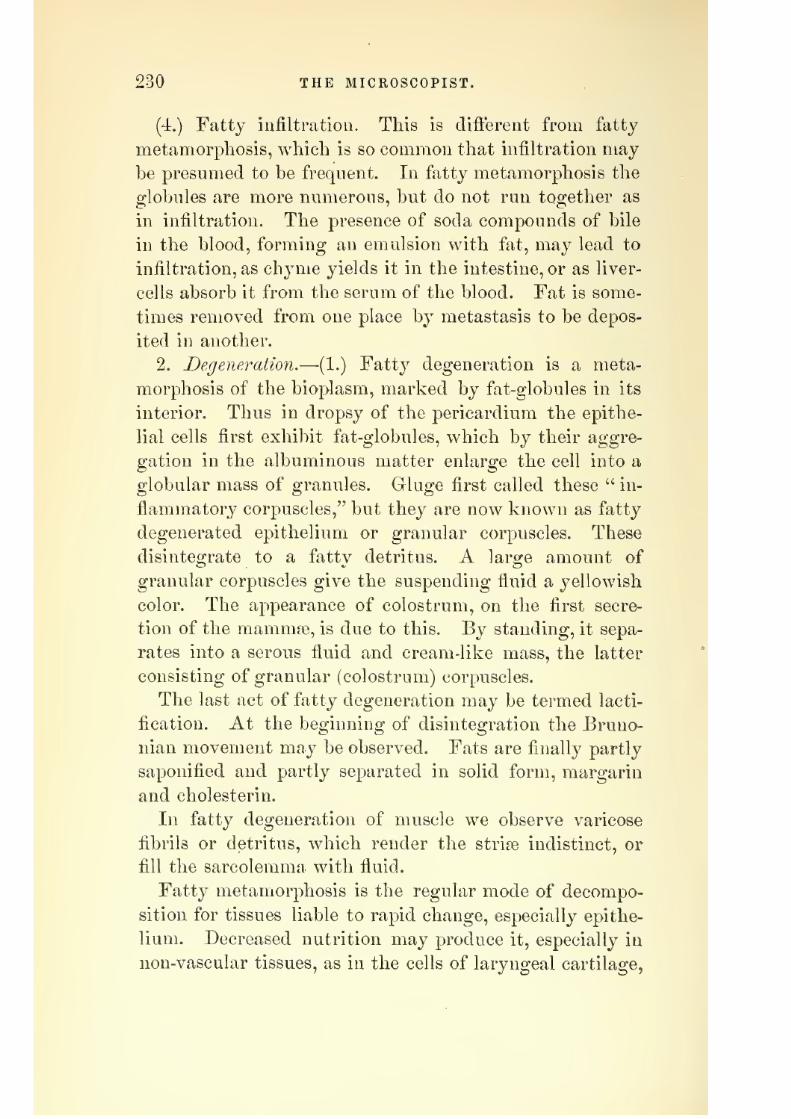
7/29/2019 THE MICROSCOPIST
http://slidepdf.com/reader/full/the-microscopist 280/361
230 THE MICROSCOPIST.
(4.) Fatty infiltration. This is different from fatty
metamorphosis, which is so common that infiltration may
be presumed to be frequent. In fatty metamorphosis the
globules are more numerous, but do not run together as
in infiltration. The presence of soda compounds of bile
in the blood, forming an emulsion with fat, may lead to
infiltration, as chyme yields it in the intestine, or as liver-
cells absorb it from the serum of the blood. Fat is some-
times removed from one place by metastasis to be depos-
ited in another.
2. Degeneration.—(1.) Fatty degeneration is a meta-
morphosis of the bioplasm, marked by fat-globules in its
interior. Thus in dropsy of the pericardium the epithe-
lial cells first exhibit fat-globules, which by their aggre-
gation in the albuminous matter enlarge the cell into a
globular mass of granules. Gluge first called these " in-
flammatorj^ corpuscles," but they are now known as fatty
degenerated epithelium or granular corjiuscles. These
disintegrate to a fatty detritus. A large amount of
granular corpuscles give the suspending fluid a yellowish
color. The appearance of colostrum, on the first secre-
tion of the marnniEe, is due to this. By standing, it sepa-
rates into a serous fluid and cream-like mass, the latter
consisting of granular (colostrum) corpuscles.
The last act of fatty degeneration may be termed lacti-
fication. At the beginning of disintegration the Bruno-
nian movement may be observed. Fats are finally partly
saponified and partly separated in solid form, margarin
and cholesterin.
In fatty degeneration of muscle we observe varicose
fibrils or detritus, which render the strise indistinct, or
fill the sarcolemma with fluid.
Fatty metamorphosis is the regular mode of decompo-
sition for tissues liable to rapid change, especially epithe-
lium. Decreased nutrition may produce it, especially in
non-vascular tissues, as in the cells of laryngeal cartilage,

7/29/2019 THE MICROSCOPIST
http://slidepdf.com/reader/full/the-microscopist 281/361
THE MICROSCOPE IN PATHOLOGY AND MEDICINE. 231
and in arcus senilis. Of vascular organs, tlie muscular
tissue of the heart is most liable to it.
The cheesy degeneration of Yirchow is a variety of
fatty degeneration. It is a yellowish, compact, friable,
or smeary mass, like cheese. It was formerly believed to
be the product of tuberculosis, and regarded as the sepa-
ration of morbid matter (crude tubercle) from diseased
blood. It is now regarded as a fatty degeneration prod-
uct, with less water present than usual. Sometimes salts
of lime are infiltrated in such masses. Real tubercle is a
gray, translucent, compact nodule, about the size of a
millet-seed (miliary), found in great numbers together.
Cheesy inflammation and miliary tuberculosis are often
found side by side, and Cohnheim, etc., have shown that
inoculation with clieesy detritus will produce tubercle.
Tubercle is found in many organs, especially the lungs.
Its structure consists of: 1. Large rounded cells of finely
granular substance, and small strongly-shining nucleus
or nuclei. 2. Small cells, with shining, darkly-contoured
nuclei. 3. Mother-cells, with clear areas round the small
ones. 4. A fine fibrous network (Plate XXV, Fig. 182).
(2.) Mucoid softening. Mucus is a colloid substance,
capable of swelling by imbibition, but of little capacity
for diffusion. It is a local production from epithelia of
mucous membrane, yet is a structural element in many
tumors. In the fcetus, the entire subcutaneous cellular
tissue is mucous tissue. The fibrinous pseudo-membranes
of the respiratory organs soften by mucoid metamorpho-
sis, and in cartilage the intercellular substance dissolves
in the same way, producing fibres near the surface.
(3.) Colloid degeneration. This is similar to the last,
but clifiers in having peculiar cells, colloid globules, begin-
ning in a normal cell by a change in its bioplasm, while
mucoid softening occurs between the fibres of connective
tissue. Like mucus, colloid enlarges by imbibition, and
ends in soda albuminate, more soluble than common albu-
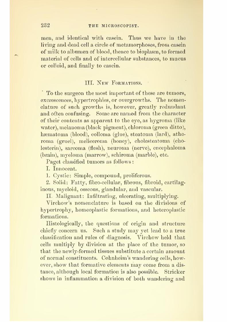
7/29/2019 THE MICROSCOPIST
http://slidepdf.com/reader/full/the-microscopist 282/361
232 THE MICROSCOPIST.
men, and identical with casein. Thus we have in the
living and dead cell a circle of metamorphoses, from casein
of milk to albumen of blood, thence to bioplasm, to formed
material of cells and of intercellular substances, to mucus
or colloid, and finally to casein.
III. IS'ew Formations.
To the surgeon the most important of these are tumors,
excrescences, hypertrophies, or overgrowths. The nomen-
clature of such growths is, however, greatly redundant
and often confusing. Some are named from the character
of their contents as apparent to the eye, as hygroma (like
water), melanoma (black pigment), chloroma (green ditto),
hEematoma (blood), coUoma (glue), steatoma (lard), athe-
roma (gruel), meliceroma (honey), cholesteatoma (cho-
lesterin), sarcoma (flesh), neuroma (nerve), encephaloma
(brain), myeloma (marrow), schiroma (marble), etc.
Paget classified tumors as follows :
I. Innocent.
1. Cystic: Simple, compound, proliferous.
2. Solid: Fatty, fibro-cellular, fibrous, fibroid, cartilag-
inous, myeloid, osseous, glandular, and vascular.
II. Malignant: Infiltrating, ulcerating, multiplying.
Yirchow's nomenclature is based on the divisions of
hypertrophy, homeoplastic formations, and heteroplastic
formations.
Histologically, the questions of origin and structure
ctiiefl}^ concern us. Such a study may yet lead to a true
classification and rules of diagnosis. Yirchow held that
cells multiply by division at the place of the tumor, so
that the newly-formed tissues substitute a certain amount
of normal constituents. Cohnheim's wandering cells, how-
ever, show that formative elements may come from a dis-
tance, although local formation is also possible. Strieker
shows in inflammation a division of both wanderino; and

7/29/2019 THE MICROSCOPIST
http://slidepdf.com/reader/full/the-microscopist 283/361
THE MICKOSCOPE IN PATHOLOGY AND MEDICINE. 233
local cells. This favors Beale's view that nuilti[>l3'ing
bioplasm are the true disease germs.The general plan on which new formations occur has
been shown by Rindfleisch as : 1. The uniform enlargement
of an organ by increase of structural elements (hypertro-
phy). 2. A node, or roundish tumefaction, by interstitial
deposit, stretching the parenchyma. 3. Infiltration, which
condenses tissue in small depots. 4. Desquamation, as in
epithelial catarrh. 5. Flat tumefaction. 6. Tuberosity,which, when narrow or finger-like, is a papillae or wart.
7. Fungus, spongy or ulcerated. 8. Polypus, a papilla
with a narrow base. 9. Dendritic vegetation, or new
papillae from the sides of others. 10. Cysts of retention,
from occlusion of ducts. 11. Exudation cysts, in closed
cavities. 12. Etravasation cysts. 13. Softening cysts.
Histologically,
we maj^divide
tumors intoI. Histoid, whose elements correspond with normal
tissues.
II. Carcinomatous, dependent on abnormal growth of
epithelial elements. These are generally in the skin, the
mucous membranes, or glands.
The following list, after Billroth, may serve for brief
reference
1. Fil)roma, composed of developed connective tissue.
(1.) Soft fibrous tumors, almost exclusively in the skin.
(2.) Firm fibroma. Most often in the uterus, where they
may calcify or form tibrous polypi ; sometimes on the
periosteum and on nerves.
2. Lipoma, or fatty tumor.
3. Chondroma (cartilaginous). These occur on bones,
are vascular, and often ossify. The softened and cystic
forms have been called colloid tumors, gelatinous cancer,
etc.
4. Osteoma (exostosis). These may be spongy or com-
pact.
5. Myoma. Billroth doubts if true muscular tumors

7/29/2019 THE MICROSCOPIST
http://slidepdf.com/reader/full/the-microscopist 284/361
234 THE MICROSCOPIST.
exist. He objects to calling the spindle-celled fibroma of
the uterus myoma.
6. ISTeuroma (nerve tumor). This also is misapplied to
all tumors or nerves, many of which are fibroma.
7. Angioma (vascular tumor), as n?evi and erectile tu-
mors. They may be plexiform or cavernous.
8. Sarcoma, a large and uncertain group, containing (1)
granulation sarcoma, or round-celled sarcoma of Yirchow
(Plate XXV, Fig. 183). (2.) Spindle-celled sarcoma (Plate
XXY, Fig. 184). (3.) Giant-celled sarcoma (Plate XXY,Fig. 185). (4.) Net-celled or mucous sarcoma (Plate XXY,Fig. 186). (5.) Alveolar sarcoma, often resembling carci-
noma (Plate XXY, Fig. 187). (6.) Pigmentary sarcoma,
or melanoma (Plate XXY, Fig. 188).
9. Lymphoma, or enlarged lymph-glands.
10. Papilloma, or hypertrophied papillse.
11. Adenoma, or glandular hypertrophy, as in goitre
sometimes forming mucous polypi.
12. Cystic tumors. If in connection with other tumors
they are named accordingly, as cysto-fibroma, etc. They
may be simple, compound, or proliferous.
13. Carcinoma, or cancers, so called from the distended
veins sometimes appearing as crabs' feet.
The old division of cancers was into scirrhus or hardcancer, the outline well defined, the aspect of cut surface
glistening, and yielding no jnice; colloid, or soft cancer;
encephaloid, or brain-like, yielding a milky juice full
of cells and nuclei, generally soft, aflecting neighboring
glands, and associated with cancerous cachexia; fungus
hsematodes, when protruding and bleeding. Billroth
treats of cancers (1) of skin, a glandular ingrowth of the
rete Malpighi, and showing globular cells. (2.) Mam-
mary cancers. (3.) Of mucous membranes with cylindric
epithelium. (4.) Of lachrymal, salivary, and parotid
glands. (5.) Of the thyroid gland and ovary.
It was thought by the early microscopists that a pecu-

7/29/2019 THE MICROSCOPIST
http://slidepdf.com/reader/full/the-microscopist 285/361
PLATE XXV.
Granulation Sarcoma.
Fig. 184.
Spindle-celled Sarcoma.
Fig. 185.
Giant celled Sarcoma \Mth ossifying foci
Fig. 186.
^//^
S^uV
Fig. 187.
Alveolar Sarcoma.
Fig. 188.
Pigmentary Sarcoma.
Fig. 189.
Epithelial Cancer of the Cheeks.
Ingrowth of the rete Malpighi.
Fig. 190.
-f.-::
il
MmEpithelial Cancer of the Stomach.
Fig. 191.
Net-celkd Sarcoma., Soft Glandular Cancer.

7/29/2019 THE MICROSCOPIST
http://slidepdf.com/reader/full/the-microscopist 286/361

7/29/2019 THE MICROSCOPIST
http://slidepdf.com/reader/full/the-microscopist 287/361
THE MICROSCOPE IN PATHOLOGY AND MEDICINE. 235
liar caudate cell was characteristic of cancer, but constant
research has shown that there is no special form of cell
in this disease. The greater number of cancers may be
characterized as a proliferation and ingrowing of epithe-
lial elements, either of the surface or of the glands. Figs.
189 to 191, Plate XXY, illustrate several varieties of
carcinoma.
The anomalies of the various tissues and organs are
well delineated byRindfleisch in his
PathologicalHistology.
TV. Microscopic Examination of Urinary Deposits,
Healthy urine holds in solution a variety of organic and
inorganic substances, as urea, uric acid, alkaline and earthy
salts, animal extractive, vesical mucus, and epithelial de-
bris. A few drops allowed to evaporate on a glass slide
will exhibit the crystalline matters, consisting of urea,
•urate of soda, chloride of sodium, phosphates and sul-
phates.
The amount passed each twenty-four hours varies from
20 to 50 ounces, holding in solution from 600 to 700 grains
of solid matter. Of this, about three-fourths consists of
organic, and one-fourth of saline substances, the largest
amount being urea, comprising nearly two-thirds of the
whole. The amount, both of solids and fluids, is subject
to great variation, according to the amount of fluids im-
bibed, the action of the skin, etc.
The average specific gravity of healthy urine is 1.020.
It may be measured with the urinometer, a loaded glass
bulb, with a graduated stem. According to a table cal-
culated by Dr. G. Bird, after Dr. Christison's formula,
each degree of the urinometer represents 2.33 grains of
solids in 1000. Thus the specific gravity 1.020 represents
46.60 grains of solid matter in 1000 of urine. By weigh-
ing all the urine passed in twenty-four hours, it is easy,
therefore, to calculate the amount of solids secreted.

7/29/2019 THE MICROSCOPIST
http://slidepdf.com/reader/full/the-microscopist 288/361
236 THE MICROSCOPIST.
Dr. Bird has also given another table, from which it
appears that the figures ofspecific
gravitywill
indicatenearly the amount of solids in each fluid ounce. Thus,
specific gravity 1010 shows a little more than 10 grains
of solids to the ounce; 1020 equals a little over 20 grains
above 1030 a grain or two more must be added, as 1030
equals 3l| grains ; 1035 gives about 37 grains.
The proportion of urinary excretion to the weight of
the bodyis often an important consideration. It may be
stated, as an average, to consist of about 149 grains of
water and 6| grains of solids to each pound weight in
twenty-four hours.
The tables given by different observers vary, but the
above may serve as an average approximation.
Urea is the vehicle by which nearl}^ all the nitrogen of
the exhausted tissues is removed from the system, and its
retention is often attended with fatal ur?emic poisoning of
the blood. In health, 400 to 500 grains are excreted iji
twenty-four hours, but in some cases of kidney diseases
not more than 100 grains are eliminated, while in some
fevers over 1000 grains are removed in the same period.
If urea be suspected in excess, a drop of urine added to a
drop of nitric acid may be placed under the microscope,
when the characteristic crystals of nitrate of urea will
appear (Plate XXYI, Fig. 192).
Volumetric analysis is the best means of ascertaining
the quantity of urea, as of other chemical ingredients, but
the practitioner may approximately estimate hj weighing
the crystals of nitrate of urea formed by adding nitric
acid to double the quantity of urine, wdiich has been con-
centrated to half its bulk by boiling.
The proportion of uric add varies from 0.3 to 1 part in
1000 of healthy urine. It may be obtained by adding a
few drops of hydrochloric acid to urine concentrated to
half its bulk, and allowing it to stand in a cool place.
The estimation of the chlorides in urine is sometimes

7/29/2019 THE MICROSCOPIST
http://slidepdf.com/reader/full/the-microscopist 289/361
THE MICROSCOPE IN PATHOLOGY AND MEDICINE. 237
Deeessarv in disease. It may be done by acidnlating the
urine with a few drops of nitric acid, and then addingnitrate of silver. The precipitate should then be dried
and fused in a porcelain capsule before weighing.
Alhumen in suspected urine may be tested by boiling in
a test-tube, when it will be coagulated. As a white pre-
cipitate sometimes occurs from an excess of earthy phos-
phates, a few drops of nitric acid should be added, which
dissolves phosphates, but coagulates albumen.Diabetic sugar is recognized by several tests. Moore's
test is made by mixing the urine with half its bulk of
liquor potassie, and boiling gently for five minutes. Sugar
gives the liquid a brown or bistre tint. Trommer's test
consists in boiling the urine with a mixture of caustic
potash and sulphate of copper, when if sugar be present
the suboxide of copper w^ill be reduced to a reddish-brownor ochre-colored powder. Fehling's test solution is a
modification of the last, and is made by dissolving 69
grains of sulphate of copper in 345 grains of distilled
water ; to this is added a concentrated solution of 268
grains of tartrate of potash, and then a solution of 80
grains of carbonate of soda to 1 ounce of water, and the
whole diluted to 1000 grains. The fermentation test
consists in filling a test-tube with urine, to which a little
3'east is added. The tube is then inverted over a saucer
containing a little urine, and placed in a warm place for
twenty-four hours. If sugar is present, it undergoes
vinous fermentation, yielding alcohol and carbonic acid.
The latter rises in the tube and displaces the liquid.
Thecoloring
matter ofbile in
urine may be detected bythe nitric-acid test. A few drops of biliary urine are
poured on a white plate, and a drop of nitric acid allowed
to fall upon it. As the acid mixes with the fluid, a play
of colors, commencing in green, passing through various
shades, and terminating in red, will be observed.
"We necessarily omit many chemical details respecting

7/29/2019 THE MICROSCOPIST
http://slidepdf.com/reader/full/the-microscopist 290/361
238 THE MICROSCOPIST.
urinary examinations, but the foregoing will be found of
practical use to the student.
Mici^oscopic dejjosits in urine are either organic mixtures
or precipitates from solution. They should be collected
by standing several hours in a conical vessel.
1. Organic Mixtures.—(1.) Epithelium. The character
of the scales will serve often to show the locality of dis-
ease in the urinary organs. The appearance of cells from
various parts is shown in Fig. 142.
(2.) Mucus and pus-cells. Mucus is deposited as a floc-
culent cloud, entangling a few round or oval delicately
granular cells, a little larger than a red blood-globule.
In disease this increases and contains numerous ill-defined
cells. A very thick glairy deposit in disease of the blad-
der may be mistaken for mucus, although it is pus altered
by the action of carbonate of ammonia.
As pus is often formed from the germinal matter of
epithelial cells, a small quantity in the urine is not neces-
sarily a sign of serious disease. In large quantities, pus
forms an opaque cream-colored deposit, which becomes
glairy and tenacious on the addition of liquor potassse.
Pus-globules under the microscope, if long removed from
the body, are granular, and show from one to four nuclei
when treated with acetic acid. In fresh pus-corpuscles,
especially in warm weather, amoeboid motion is often seen.
In a late period of catarrh of the bladder, but little epi-
thelium will accompany the discharge, but crystals of
triple phosphates generally occur in pus derived from the
bladder.
(3.) Blood-disks usually form a reddish-brown deposit.
A smoky appearance of the urine is produced in blood
derived from the kidney. A brown deposit resembling
blood, but showing granules and no disks, is supposed to
be derived from blood. The epithelium associated with
blood-disks will often point out the source of haemorrhage.
(4.) Spermatozoa in urine are not uncommon in perfect

7/29/2019 THE MICROSCOPIST
http://slidepdf.com/reader/full/the-microscopist 291/361
PLATE XXVI.
Fig. 192. -, , Fig. 193.
m j'fj m _...
Tube-casts.
Nitrate of Urea.
Fig. 194.
Fig. 195.
^V-;
Urate of Ammonia.
.. II' /~ ^^ ^
Uric Acid. Uric Acid.
Fig. 197.
^ o
/vUric Acid—re-precipitated.

7/29/2019 THE MICROSCOPIST
http://slidepdf.com/reader/full/the-microscopist 292/361

7/29/2019 THE MICROSCOPIST
http://slidepdf.com/reader/full/the-microscopist 293/361
THE MICROSCOPE IN PATHOLOGY AND MEDICINE. 239
health, but nervous patients are often dekiclecl by quacks
on account of them.
(5.) Accidental products. A great variety of things
may accidentally get into urine, and the observer must
guard against them by studying the appearance of various
objects in the microscope. Pieces of feathers, fibres of
wood, vegetable-cells, wool, cotton, silk, dust, etc., will
almost always attract the attention first of one unused to
them.
(6.) Sarcinse are minute vegetable organisms, in the
form of cubes, often subdividing into groups of four or
their multiples. They were detected by Goodsir in the
stomach in a case of obstinate vomiting, and have been
occasionally found in urine. They seem associated with
some dyspeptic cases.
(7.) Torahc. This fungus is developed in urine whichcontains even minute traces of sugar. It is identical with
the yeast plant (see pages 135, 137).
(8.) The ijenicilliam. glaucum, a fungus allied to the last,
commonly makes its appearance in acid urine when ex-
posed to the air.
(9.) Yibriones. What was said at page 185 will readily
account for the presence of vibriones in decaying urine.
In perfectly fresh urine it may be regarded as a sign of
debility.
(10.) Tube-casts. In many cases of congestion and in-
flammation a coagulable material is eifused into the tubes
of the kidney, forming a cast or mould of the tube. This
may be ejected, bringing with it pus, blood, epithelium,
or other material with which it is associated. In Bright'sdisease these casts, in addition to albuminous urine, as-
sume considerable clinical importance. In the acute form
of the disease the cylinders or casts are fibrinous, with
blood, mucus or pus-cells, and epithelium. Towards the
close the casts become homogeneous or hyaline. In chronic
desquamative nephritis the cylinders are without blood,

7/29/2019 THE MICROSCOPIST
http://slidepdf.com/reader/full/the-microscopist 294/361
240 THE MICROSCOPIST.
and towards the close waxy or fatty, often containing
manyoil-globu]es'(Plate
XXVI,Fig. 193).
2. Precipitates from Solution.—(1.) Urate of ammonia.
This is generally an amorphous deposit, in irregular groups
of molecules, but with an alkaline fermentation sometimes
crystallizes (Plate XXVI, Fig. 194). Some regard this
as urate of soda, or a mixture of urates of potash, soda,
and ammonia.
Its color varies from light pink to brickdust color. It
is deposited in all concentrated urine, and is often a
" critical discharge " in fevers, etc. It is found in gouty
concretions, and dissolves with heat and acids.
['2.) Uric acid may occur from an acid fermentation
dissolving urates of soda or ammonia. It is a yellow,
reddish, or brown sediment of crystals, wdiich assume
different forms, as rhomboid tablets with obtuse angles,
or of the shape of a whetstone (Plate XXVI, Fig. 195).
When slowly precipitated, it may form druses of four-
sided prisms (Plate XXVI, Fig. 19ij). When precipitated
from fresh urine by the addition of muriatic acid, the
crystals are large, and often of varying shapes.
They may be tested by dissolving in potassa, and re-
precipitating by muriatic acid, wdien they assume the
shape of Fig. 197, Plate XXVI.
They originate from waste, excess of nitrogenous food,
defective assimilation, congestions of the kidnej^, or chronic
disease of the respiratory organs.
(3.) Ammonio-phosphate of magnesia, triple phosphate,
may be precipitated from fresh urine in stellate crystals
(Plate XXVII, Fig. 198) by adding ammonia. When
more slowly deposited from alkaline urine, or in diseased
states of the system, the crystals are prismatic, generally
triangular, with obliquely truncated ends. Sometimes the
terminal edges are bevelled, and the varying lengths of
the prisms give rise to a variety of forms (Plate XXVII,
Fig. 199j. They are generally thought to proceed from

7/29/2019 THE MICROSCOPIST
http://slidepdf.com/reader/full/the-microscopist 295/361
Fig. 19S.
h
i!:\
Av^j
PLATE XXVIT.
v.. v^i
..:m^
^.«^'
.- CHi«-
Amiuonio-phosphate of Magnesia.
Fig 199
Amiuonio-phosphate of Magnesia.
Fig. 200.
\j w,
Phosphate of Lime.
Fig. 202.
^^:*i
<;^\j»^
Cliloride of Sodium.
Fig. 201.
?g9 ^.
E^ Ag
^i^ K'
Oxalate of Lime.
Fig. 203.
o% oO<i)qO
Cystine.
Fig. 204.
.Salivary corpuscles, epithelial scales and granules.

7/29/2019 THE MICROSCOPIST
http://slidepdf.com/reader/full/the-microscopist 296/361

7/29/2019 THE MICROSCOPIST
http://slidepdf.com/reader/full/the-microscopist 297/361
THE MICROSCOPE IN PATHOLOGY AND MEDICINE. 241
disintegrated albuminous, and chiefly nervous, matter, but
their clinical importance is not fully settled. They are
found in cases of nervous depression, various forms of
dyspepsia, shock of the spinal cord, irritation of the blad-
der, etc.
In highly alkaline urine, the triple phosphates are
usually accompanied with pus and phosphate of lime.
The latter occurs as minute granules or dumb-bells, or in
groups of crystals (Plate XxVlI, Fig. 200).
(4:,) Oxalate of lime is de[)Osited in small octahedra,
generally appearing under the microscope as minute
squares, with crossed lines proceeding from the angles,
the upper angle being next the eye (Plate XXYII, Fig.
201). Dumb-bell forms, and circular or oval crystalline
masses, are often seen.
Oxalate of lime is found as a urinary deposit in various
conditions, as pulmonary and dyspeptic affections. It is
usually associated with hypochondriasis, and in cases of
overfatigue, particularly from mental work, it is very
common. Its association with calculous affections ren-
ders it interesting to the suro-eon.
(5.) Chloride of sodium never crystallizes from fluid
urine. On evaporation it crystallizes in stellar form or
in cubes (Plate XXVII, Fig. 202). The presence of urea
sometimes disposes it to assume the form of a regular
octahedron.
It is useful to investigate this excretion in typhoid
fevers and inflammations of the respiratory organs, etc.
In commencing hepatization of the lung it is absent, but
returns on resolution of the inflammation. The methodof testing has been given before.
(6.) Cystin (Plate XXVII, Fig. 203) crystallizes in
characteristic six-sided plates. It contains a large pro-
portion of sulphur, 26 per cent., and is considered a
product of decomposition. It is often associated with
16

7/29/2019 THE MICROSCOPIST
http://slidepdf.com/reader/full/the-microscopist 298/361
242 THE MICROSCOPIST.
calculus. Some regard it as indicating a strumous and
ill-nourished system.
Y. List of Parasites Infecting the Human Body.
I. Epiphytes^ or vegetable parasites. Parasitic lesions
of the surface are denoted by the infiltration or destruc-
tion of hairs and epithelial textures by the sporules of a
fungus, which by unionor
growthform elongated branches
or mycelia. Reference has been made, page 136, to poly-
morphism, or the varieties of form produced by the same
fungus germ, so that the names ascribed to these parasites
must be regarded as only provisional. The diagnosis of
fungi on the skin, hair, or epithelium requires care and
skill in microscopic manipulation, and the use of liquor
potassge long enough to render the specimen transparent,
1. The Trichophyton tonsurans^ present in ring-worm of
the body, scalp, or beard. Its anatomical seat is the
interior of the roots of the hairs, but it also covers the
epidermis between the hairs, and invests them in a white
sheath, producing infiammation of the follicles and sur-
rounding tissues, and subsequent baldness.
2. The Trichophyton sporidoides, present in the disease
called plica polonica.
3. Achorion Schonlelnii and the Puccinia favi, 'present in
the honeycomb ring-worm,
4. Microsporon mentagraphyta., present in the Mentagra.
5. Microsporon furfur^ the cause of liver-colored spots,
or Pityriasis versicolor.
6. Microsporon Audoitini, occurring in Porrigo decal-
vans, or bald patches.
7. Mycetoma Garteri, the cause of the " fungous foot of
India."
8. Oidium cdbicans, in diphtheria and aphtha.
9. Cryptococcus (or Torula) cerevisice^ j-east plant in blad-
der or stomach.

7/29/2019 THE MICROSCOPIST
http://slidepdf.com/reader/full/the-microscopist 299/361
THE MICROSCOPE IN PATHOLOGY AND MEDICINE. 243
10. Sarcina veniricuH, in the stomacli.
11. Epizoa^ or animals living upon the skin and hair.
1. Pedicidus, or louse, three forms: P. coryoris^ P. capi-
tis^ and P. pubis.
2. Acarus scabiei, or itch insect.
3. DemodexfoUicidonim, mhiihiting sebaceous and hair-
follicles.
III. Entozoa, or internal parasites. On page 171 we
have given a general account of the Entozoa. At least
thirty different forms have been described as infesting
the human body ; eight species of Tcenia and two of
Bothriocephalic genera of the family Cestoidea or tape-
worms, of which the Cysticerci and Echinococci (vesicu-
lar cysts containing an embryo head provided with a
circle of booklets, and giving rise to the appearance of
measly flesh in animals) are larval forms; nine species of
Trematoda, or fluke-like parasites, existing in an encysted
and a free state; and eleven species of Xematoid, or round-
worms, including the common round-worms or Asearis,
the Trichina spbrdis, and the FUaria ocidi, etc.
Students who have not access to Dr. Cobbold's great
work on parasites, may find an excellent resume in Dr.
Aitken's /Science and Practice of Medicine.
\1. Examination of Sputa.
The microscopic examination of sputa is important in
practical medicine, but requires familiarity with the ap-
pearance of different structures under different magnify-
ing powers, as fragments of food, muscular fibre, starch,
etc.
We may usually expect to find mucus, entangling air-
bubbles, and pavement-epithelium from the mouth (Plate
XXVII, Fig. 204). In catarrhal aft'ections, ciliated epi-
thelium from the nasal or respiratory passages may also
be seen, and perhaps molecules of fat, pus-globules, blood.

7/29/2019 THE MICROSCOPIST
http://slidepdf.com/reader/full/the-microscopist 300/361
244 THE MICROSCOPIST.
or transformed epithelial cells (formerly called granule-
cells, or inflammatory corpuscles). In phthisis, the soft-
ened tubercle or gangrene may be early detected by the
fibres of elastic tissue from the walls of the pulmonary
vesicles. The sputa should be first liquefied by boiling
with an equal bulk of caustic soda, and then allowed to
settle in a conical glass, when a small quantity may be
removed by a pipette to a glass slide, covered by thin
glass, and placed under the microscope.The occurrence of fungi in sputa is to be expected
whenever there is decay. The Leptothrix buccalis, one
form of Pencillium, is common on old epithelial scales of
the mouth, and in the later stages of phthisis the sputa
will often show fungi in difterent stages of development.
Bacteria and vibriones are also frequent in pus.
In catarrhal pneumonia we may find fibrinous casts ofthe alveoli of the lungs and epithelial elements.
VII. Hints on the Application of the Microscope to
Materia Medica and Pharmacy.
The observations of Dr. Hassall on the detection of
adulterations in food," have prompted similar investiga-
tions respecting the purity of medicinal substances. Suchexaminations cover a wide field of research, chiefly related
to micro-chemistry and botany.
The student in this department will do well to provide
himself with undoubted specimens of various articles for
comparison, although much may be learned from a gen-
eral examination of any particular drug, etc.
In addition to the recognition of genuine forms of leaves,
seeds, roots, etc., and their adulterations, the microscope
will often be serviceable in exhibiting the deteriorations
to which such articles are subject if kept too long.
Dr. Hale, in the American Journal of Microscoj:)!/, shows
* Food and its Adulterations. By A. H. Hassall, M.D.

7/29/2019 THE MICROSCOPIST
http://slidepdf.com/reader/full/the-microscopist 301/361
THE MICROSCOPE IN PATHOLOGY AND MEDICINE. 24:5
that various causes combine to effect the deterioration
of drugs. Tliey may become infested with animalculje.
Leaves and roots may be eaten by insects until all vestige
of medicinal power is destroyed. Fungi of various kinds
may destro}" the tissue. The uncertain action of some
pharmaceutical preparations may be thus accounted for.
For the method of examination in this department, as
well as in medical jurisprudence, we refer to the foregoing
chapters, contenting ourselves here with the remark, that
investigations involving life or reputation should never be
undertaken without a thorough practical acquaintance
with microscopic manipulation and the microscopic sci-
ences. He to whom such work is intrusted, should be
able to exhibit and to explain to an intelligent jury what
he has seen and what he ought to see.

7/29/2019 THE MICROSCOPIST
http://slidepdf.com/reader/full/the-microscopist 302/361

7/29/2019 THE MICROSCOPIST
http://slidepdf.com/reader/full/the-microscopist 303/361

7/29/2019 THE MICROSCOPIST
http://slidepdf.com/reader/full/the-microscopist 304/361
248 GLOSSARY.
from its focal point. A very wide angle of aperture only
allows distinct vision of what is exactly in focus, so thatwdien penetration is needed most, as in physiological work,
a smaller angle is better than one used for resolution of
diatoms and other minutife.
Aplanatic.—Destitute of spherical aberration.
Archeus.—The term applied by Yan Helmont to the
specific agent which, according to his theory, presided
over vital functions.Bacteria.—Minute, transparent, rod-like bodies, some-
times jointed, and often exhibiting a vacillating motion.
It is probable that they are produced by the germs of
fungi in a solution of animal matter.
Bathyhius.—A term given by Professor Huxley to the
slimy matter from the ocean bottom. Doubts have been
expressedas to its animal nature.
Binocular {binus, two, and oculiis, an eye).—An ar-
rangement of a prism (Wenham's) and eye-pieces for
two eyes, so as to produce a stereoscopic effect with the
microscope.
Biology (Gr. Bios, life, and logos, a discourse).— The
study of living beings, including zoology and botany.
BiopUism.
—Aterm proposed for the elementary sub-
stance of organic bodies when actually alive, or living
protoplasm.
Blastema.—The fluid matter, or plasma, in which, ac-
cording to the theory of Schleiden and Schwann, nuclei
first make their appearance, and then organic cells.
Brunonian Motion.—The molecular movement of fine
particles suspended in fluid, first observed by Dr. R.
Brown in 1827.
Calcification.—The infiltration of animal tissues with
salts of lime.
Cell.—The elementary unit of organic structure. From
the time of Schleiden and Schwann (1838) the researches
of biologists have been greatly aided by the demoustra-
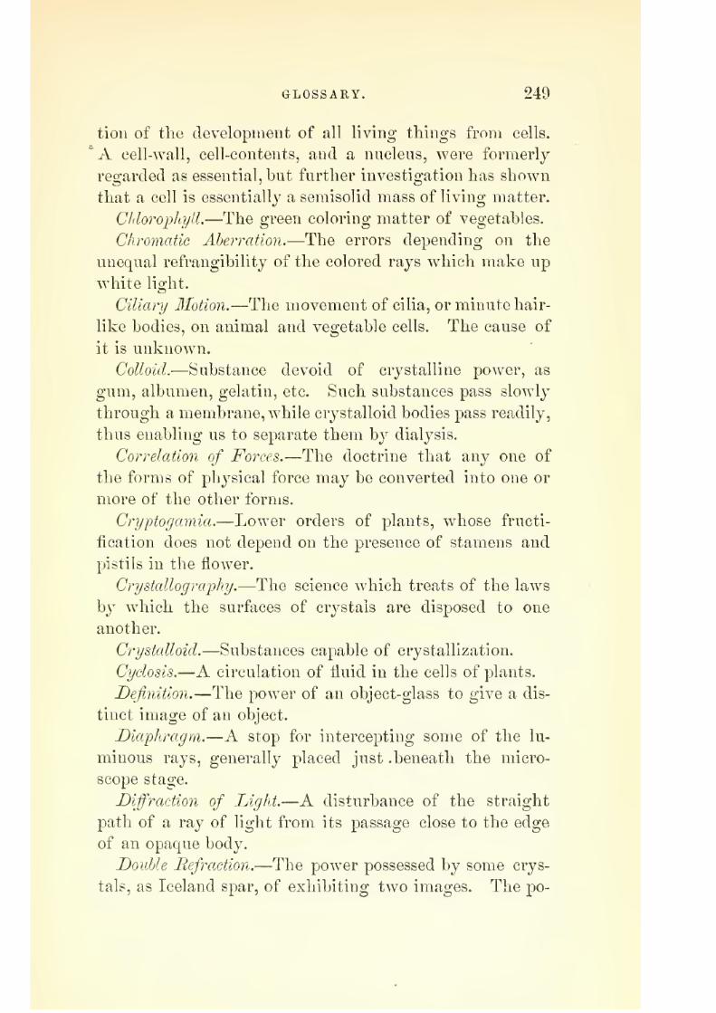
7/29/2019 THE MICROSCOPIST
http://slidepdf.com/reader/full/the-microscopist 305/361
GLOSSARY. 249
tion of tlie development of all living things from cells.
'A cell-wall, cell-contents, and a nucleus, were formerly
regarded as essential, but further investigation has shown
that a cell is essentially a semisolid mass of living matter.
Chloropliyll.—The green coloring matter of vegetables.
Chromatic Aberration.—The errors depending on the
unequal refrangibility of the colored rays which make up
white light.
Ciliary Motion.—The movement of cilia, or minute hair-
like bodies, on animal and vegetable cells. The cause of
it is unknown.
Colloid.—Substance devoid of crystalline power, as
gum, albumen, gelatin, etc. Such substances pass slowly
through a membrane, while crj^stalloid bodies pass readily,
thus enabling us to separate them by dialysis.
Correlation of Forces.—The doctrine that any one of
the forms of physical force may be converted into one or
more of the other forms.
Cryptogamia.—Lower orders of plants, whose fructi-
fication does not depend on the presence of stamens and
pistils in the flower.
Crystallography.—The science which treats of the laws
by which the surfaces of crystals are disposed to one
another.
Crystalloid.—Substances capable of crystallization.
Cyclosis.—A circulation of fluid in the cells of plants.
Definition.—The power of an object-glass to give a dis-
tinct image of an object.
Diaphragm.—A stop for intercepting some of the lu-
minous rays, generally placed just .beneath the micro-
scope stage.
Diffraction of Lighi.—A disturbance of the straight
path of a ray of light from its passage close to the edge
of an opaque body.
Double Refraction.—The power possessed by some crys-
tals, as Iceland spar, of exhibitiug two images. The po-

7/29/2019 THE MICROSCOPIST
http://slidepdf.com/reader/full/the-microscopist 306/361
250 GLOSSARY.
lariscope tests this property and exhibits it in many sub-
stances.
Athin film of selenite intensifies it if it is not*
strong enough to show color otherwise.
Epiphytes.—Parasitic plants.
Epithelium.—The layer or layers of cells covering ex-
ternal or internal surfaces of animal bodies.
Epizoa.—Parasitic animals.
Focal Distance.—The distance from the centre of a lens
to the focus, or point of distinct vision.
Foraminifera.—Shells of minute animals, chiefly calca-
reous, which are perforated with minute pores for the pro-
trusion of threads of sarcode or bioplasm.
Formed Matter.—The structure produced by the action
of bioplasm, or by the influence of external agents upon it.
Fravnhqfer^s Lines.—The dark lines which cross the
solar spectrum, corresponding to the chemical nature of
the burning substance. The blackness of the lines de-
pends on the incandescent vapor of the substance.
Gemmation., or Budding.—A term given to the repro-
duction of cells by the protrusion of a part of their sub-
stance, which becoming constricted, falls oft' and lives an
independent life.
Germinal Matter.—Another name for bioplasm, or " cell-
stuft'."
Herapathite.—The iodo-disulphate of quinia.
Histo-Chemistry.—The science which investigates the
chemistry of the tissues.
Histology.—The science of tissues.
Immersion Lens.—An objective arranged so as to re-
quire a drop of fluid interposed between its front lens
and the covering glass of the object.
Indifferent Fluids.—Fluids which produce little or no
change in animal or other tissues.
Infiltration.—The deposition of material from the blood
into various tissues.
Lens.—A piece of glass ground and polished so as to

7/29/2019 THE MICROSCOPIST
http://slidepdf.com/reader/full/the-microscopist 307/361
GLOSSARY. 251
refract the light to a focus, or causing the rays to di-
verge.
Leucocytes.—White cells, whether in blood or elsewhere.
They are simply masses of bioplasm.
Lideiiie Spectra.—The spectra produced by light passing
through juice from the corpora lutea in the ovary.
Metamorphosis.—Change of form, as from the caterpil-
lar to the butterfly.
JlJetric Measure.—The system first adopted in France,
based on the metre, which is the ten millionth of the
quadrant of the meridian of Paris. The unit of surface,
is the arc of one hundred square metres. The unit of
weight is the gramme, weighing yQ^^gth of a cubic
metre of wa<^er. The multiples are indicated by Greek
prefixes, deca (10), ImAo (100), kilo (1000), myrio (10,000).
The subdivisions are named by Latin prefixes, deci, ce7iti,
and milli.
Micro-gonidia.—Bodies resulting from the segmentation
of motile cells in the lower order of vegetables. When
possessing active movement they rank as zoospores.
Micrometer. — An instrument for measuring minute
spaces.
Microscopy.—The use of the microscope, and the knowl-edge attained by it.
Microzymes.—Minute molecules found in the vaccine
vesicles, glanders, and other disease products.
Molecular Coalescence.—A name given to the action of
various chemical substances, in a nascent state, upon an
organic colloid.
Monera.
—The namegiven by Professor
Hackelto
thesimplest forms of animal life.
3Iorphology.— The science of form. Applied to the
structures of organized beings.
Motile Cells.—Minute vegetable cells, moving by means
of vibratile cilia. After a time they lose their cilia and
become still cells, which multiply by self-division.

7/29/2019 THE MICROSCOPIST
http://slidepdf.com/reader/full/the-microscopist 308/361
252 GLOSSARY.
Necrosis.—The deatli of tissue.
Nucleus.—A concentration of vital power in a mass of
bioplasm.
Objective.—The object-glass of a microscope.
Oblique Illumination.—The illumination of microscopic
objects by light thrown from the side, either by the mirror
or some special contrivance.
Oolites.—Certain rocks which present a granular struc-
ture resembling the roe of a fish.
Orbitolites.—Foraminiferous shells of considerable size
occurring in tertiary limestones.
Otoliths.—Small crystalline bodies from the inner ear.
Pabulum.—The nutritive material supplied to animal
cells.
Parthenogenesis.—Reproduction without sexual union.
Pathology.—The science of diseased structure and func-tion.
Penetration.—The property of an objective which ex-
hibits layers of structure below the focus. It seems to
depend on moderate angular aperture.
Physical Movements.—A peculiar vibratile motion in mi-
nute particles suspended in fluid. See Brunonian Motion.
Polymorphism.—The development of similar germs intodifferent forms by various agencies.
Protophytes.—Plants of simplest forms.
Protoplasm.—The elementary cell-material, or "physi-
cal basis of life."
Protozoa.—Simplest forms of animals.
Raphides.—Crystals occurring in vegetable tissues.
Pesolution.
—The propertyin
amicroscope of exhibiting
minute details, as lines, etc.
Sarcode.—A synonym of protoplasm, or cell-material.
Sclerogen.—Woody tissue.
Selenite.—Crystallized sulphate of lime.
Spectrum. Analysis.—The analysis of incandescent sub-
stances by means of the spectroscope.

7/29/2019 THE MICROSCOPIST
http://slidepdf.com/reader/full/the-microscopist 309/361
GLOSSARY. 253
Spherical Aberration.—The errors of lenses arising from
the spherical surface, so that the raj-s from centre and
edge do not accurately combine in focus.
Spontaneous Generation.—The theory of the spontaneous
or independent origin of minute organisms. Bastian's
views in favor of this theorj^ seem to have been over-
thrown b}^ the experiments of Paste|i^and Tyndall, and ^^-*-
biologists now generally agree to the doctrine that all
living bodies are derived from pre-existent life.
Torula.—The "yeast-plant" fungus.
Vernier.—A short graduated scale, made to slide along
a large scale so as to read to fractions of divisions.
Vibriones.—Vibi-atile filaments, or bacteria, sometimes
moniliform, or bead-like, frequently found in decaying
animal infusions.
Wandering Ce^^s.—Leucocytes, or white blood-cells,
which pass through the coats of vessels and thence into
neighboring tissues.

7/29/2019 THE MICROSCOPIST
http://slidepdf.com/reader/full/the-microscopist 310/361

7/29/2019 THE MICROSCOPIST
http://slidepdf.com/reader/full/the-microscopist 311/361
INDEX.
Aberration of lenses, 22
Abiogenesis, 125
Absorption bands, 18, 101
Acalephs, 166
Acari, 177
Achromatic condenser, 33
Acinetse, 162
Accessories, microsopic, 32
Adjustment, 51
Agriculture, microscope in, 18
Air-pump, 79Alternation of generations, 126
AlgtB, 139, 153
Albumen in urine, 237
Aliraentar}' canal in insects, 177
Amici"s prism, 34
Ama'ba, 121
Amoeboid motion, 121
Amplifier, 26
Amyloid infiltration, 229
Amphlipleura pellucida, 56
Analysis of urine, 235
of earths, etc., 99. 108Anatomy of insects, 177
Animal histology, 182
Animalcule cage, 41
Angulnr aperture, 25, 55
measurement of crystals, 89
Annular vessels, 131
Annulata, 172
Antennae of insects, 175
Antiquity of microscope, 17
Aquaria, 82
Arachnida, 179
Aristotle on life, 116
Archeus, 116
Bacteria, 135
Bathybius, 96, 158
Beale's generalization, 118
tint-glass camera, 40
Beck's microscope, 32
iris diaphragm, 33
illuminator, 38
Biology, microscope in, 116
Binocular microscope, 30
Bioplasm, 118
varieties of, 123
Blood, 186
Blood-tests, 102
Blastema. 124
Bone, 195
Brunonian movement, 53, 120
Bryozoa, 168
Bull's-eye condenser, 36
Cabinet, 81
Carmine staining, 69
Care of microscope, 48
of the eyes, 49
Calcification, 229
Camera lucida, 39
Camphor, 133
Cancer, 234
Capillary structure, 201
Cavities in crystals, 89
Cell, 117formation of, 119
phenomena of, 120
movements of, 120
chemistry of, 122
forms of, 123
genesis, 124
multiplication, 125
wall, vegetable, 129
Cellulose, 129
Cements, 75
Cephalopoda, 171
Cerebro-spinal nerves, 216
Characeae, 154
Chalk strata, 95
Chemical isolation, 59
reagents, 67
products, 183
Chlorophyll, 133
Chromatic aberration, 22
Chyle, 190
Cilia, 124
Cirrhipeds, 174
Classes of microscopes, 28
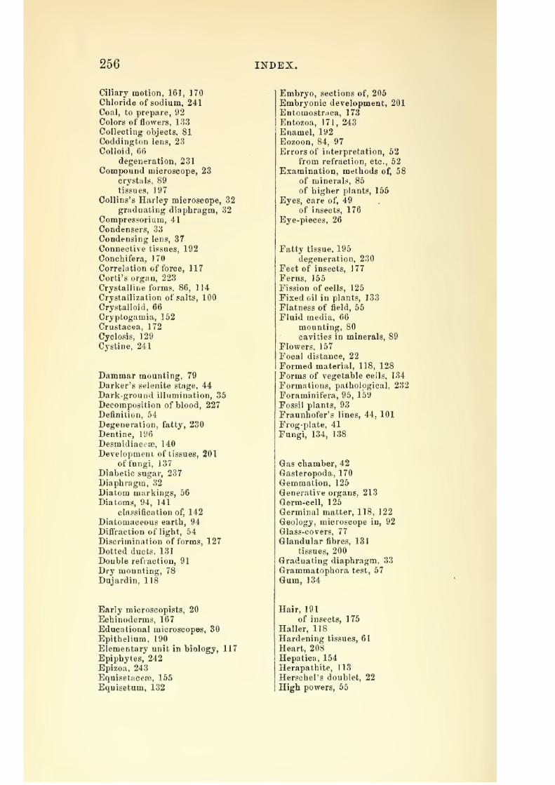
7/29/2019 THE MICROSCOPIST
http://slidepdf.com/reader/full/the-microscopist 312/361
256 INDEX.
Ciliary motion, 161, 170
Chloride of sodium, 241
Coal, to prepare,92
Colors of flowers, 133
Collecting objects, 81
Coddington lens, 23
Colloid, 66
degeneration, 231
Compound microscope, 23
crystals, 89
tissues, 197
Collins's Harley microscope, 32
graduating diaphragm, 32
Compressorium, 41
Condensers, 33
Condensing lens, 37
Connective tissues, 192
Conchifera, 170
Correlation of force, 117
Corti's organ, 223
Crystalline forms, 86, 114
Crystallization of salts, 100
Crystalloid, 66
Cryptogamia, 152
Crustacea, 172
Cyclosis, 129
Cystine, 241
Dammar mounting, 79
Darker's selenite stage, 44
Dark-ground illumination, 35
Decomposition of blood, 227
Definition, 64
Degeneration, fatty, 230
Dentine, 196
Desmidiacese, 140
Development of tissues, 201
of fungi, 137
Diabetic sugar, 237
Diaphragm, 32
Diatom markings, 56
Diatoms, 94, 141
classification of, 142
Diatomaceous earth, 94
Diffraction of light, 54
Discrimination of forms, 127
Dotted ducts, 131
Double refraction, 91
Dry mounting, 78
Dujardin, 118
Early microscopists, 20
Echinoderms, 167
Educational microscopes, 30
Epithelium, 190
Elementary unit in biology, 117
Epiphytes, 242
Epizoa, 243
Equisetacese, 155
Equisetum, 132
Embryo, sections of, 205
Embryonic development, 201
Entomostr.ica, 173Entozoa, 171, 243
Enamel, 192
Eozoon, 84, 97
Errors of interpretation, 52
from refraction, etc., 52
Examination, methods of, 58
of minerals, 85
of higher plants, 155
Eyes, care of, 49
of insects, 176
Eye-pieces, 26
Fatty tissue. 195
degeneration, 230
Feet of insects, 177
Ferns, 155
Fission of cells, 125
Fixed oil in plants, 133
Flatness of field, 65
Fluid media, 66
mounting, 80
cavities in minerals, 89
Flowers, 157
Focal distance, 22
Formed material, 118, 128
Forms of vegetable cells, 134
Formations, pathological, 232
Foraminifera, 95, 159
Fossil plants, 93
Fraunhofer's lines, 44. 101
Frog-plate, 41
Fungi, 134, 138
Gas chamber, 42
Gasteropoda, 170
Gemmation, 125
Generative organs, 213
Germ-cell, 125
Germinal matter, 118, 122
Geology, microscope in, 92
Glass-covers, 77
Glandular fibres, 131
tissues, 200
Graduating diaphragm, 33
Grammatophora test, 57
Gum, 134
Hair, 191
of insects, 175
Haller, 118
Hardening tissues, 61
Heart, 208
Hepatica, 154
Herapathite, 113
Herschel's doublet, 22
High powers, 55
7/29/2019 THE MICROSCOPIST
http://slidepdf.com/reader/full/the-microscopist 313/361
INDEX. 257
Hipparcbia Janira, 56
Histo-chemistry, 185Histology, 182
Histologists, early, 20
Histological structures, 185
Holothurite, 168
Hunt, Dr., on staining, 153
Hydrozoa, 165
Imbedding tissues, 62
Immersion lenses, 25, 51
Indifferent fluids, 66
Infiltnition, 228Infusoria, 160
families of, 163
Injecting, 64
syringe, 64
material, 65, 70
Insects, 174
scales of, 1 75
hairs of, 175
eyes of, 1 76
mouths of. 176
feet of, 177
anatomy of, 177changes of, 126
Intestinal canal, 206
Interpretation, errors of, 52
Inverted microscope, 106
Invertebrata, classes of, 180
lod-serum, 66
Iris diaphragm, 33
Kellner's eye-piece, 26
Kidney, 211
Labyrinthodon, 97
Laticiferous vessels, 131
Lamps, 50
Leaves of plants, 157
Lasso-cells, 166
Lenses, 21
Lernoea, 173
Leptothrix, 136
Leucocytes, 189
Lieberkuhn, 37
Light for microscopes, 50
Life, theories of, 116
Litmus-paper, 107
Living bodies, element of, 117
Ligneous tissue, 130
Lichens, 154
Liver, 210
Locomotive organs, 215
Low powers, 55
Lymph, 189
Lymphatics, 207
Luteine spectra, 105
Magnifying power, 22
Max Schultze, 118Measuring objects, 38
Medium powers, 55
Metric system, 39
Medicine, microscope in, 226,
Metamorphosis, 126
Microscope, compound, 23
mechanism of, 27
in the arts, 17
in commerce, 18 '
in agriculture, 18
Micro-spectroscope, 44
Micro-mineralogy, 84Micro -chemistry, 98
Micro-chemiciil analysis, 106;
Microscopic slides, 77
Microzymes, 135
Milky juice in plants, 131
Mildew, 136
Minute lenses, 21
dissection, 59
Micro-gonidia, 152
Micrometer, 38
Moist-chamber, 41
Moller's test-plate, 57
Mounting objects, 76
Molecular movements, 120
coalescence, 138
Monera, 158
Mosses, 154
Morbid actions, 227
Morphological products, 183
Motile cells, 120
Mounting in balsam, 79
Mucus, 190
Mucoid softening, 231
Muller's eye-fluid, 68
Muscardine, 136
Muscle, 197
Mycelia of fungi, 137
Naehet's inverted microscope, 32
prism, 35
Navicula rhomboides, 56
Nerve tissue, 198
cells, 199
preparations, 217
Nichol's prism, 43
Nobert's illuminator, 35
test, 56
Nose-piece, 47
Nostochinse, 151
Nucleus of cells, 119
Nutritive organs, 206
Oberhauser's drawing apparatus, 39
Objects of microscopy, 58
Object glasses, 25
finders, 48
17

7/29/2019 THE MICROSCOPIST
http://slidepdf.com/reader/full/the-microscopist 314/361
258 INDEX.
Oblique illumination, 34
Ocular micrometer, 38
Opaque injections, 65
objects, 77
Opticians, list of, 27
Orbitolites, 159
Organs of touch, 218
of taste, 2 IS
of smell, 218
of sight, 219
of hearing, 222
Origin of rocks, 92
Organic principles, 184
Oolites, 90
Otoliths, 222Oscillatorice, 151
Oxalate of lime, 241
Pabulum, 118, 183
Paleontology, 97
Palmellaceaa, 139
Parasites, human, 242
Parthenogenesis, 126
Pathology, microscope in, 226
Pathological formations, 232
Parabolic illuminator, 36speculum, 37
Pettenkofer's test, 102
Penetration, 55
Periscopic eye-piece, 26
Preserving objects, 76
Pleurosigma test, 58
Pigott's, Dr. R. . eye-piece, 26
Pigmentation, 229
Phosphates, 240
Physical movements, 53
Photography, microscopic, 48
Phenomena of cells, 120Polariscope, 42, 99
in mineralogy, 90
Polycystina, 95, 160
Polymorphism, 136
Polyps, 164
Polyzoa, 168
Popular microscopes, 30
Porifera, 160
Preparation, 58
by teasing, 61
by section, 6 I
of minerals, 84of crystals, 85, 99
in viscid media, 65
of vegetables, 156
embryonic, 204
Preservative fluids, 73
Preserving objects, 76
Primordial utricle, 129
Protoplasm, 1 18
Progressive force, 130
Prothallium of ferns, 126
Prism for oblique light, 35
Protopbytes, 129
Protozoa, 158
Pus, 189
Raphides, 132
Reade's condenser, 34
Recklinghausen's moist-chamber, 41
Red blood-corpuscles, 186
Reproduction, 125
Resolution, 65
Resin, 133
Reticulated vessels, 131
Respiratory organs, 213
Retina, 221
RhodospermeEe, 153
Rhizopods, 168
Rotatoria, 163
Sarcode, 118
Salivary glands, 209
corpuscles, 189
Scbleiden and Schwann, IIS
Sclerogen, 130
Schultze's warm stage, 42
Scales of Lepidoptera, 175Section-cutter, 63
Secretory organs, 208
Sections of hard tissues, 62
Sensory organs, 218
Selenite stage, 44
Seeds, 167
Shadbolt's turntable, 78
Shell structure, 170
Silica in plants, 131
Simple tissues, 186
Soemmering's steel disk, 40
Spectra of substances, 102Sperm-cell, 125
Spectrum analysis, 101
Spiral movements, 130
vessels, 131
Spot lens, 36
Sphagnum. 155
Spherical aberration, 22
Sphajroplea, 152
Sponges, 160
Spontaneous generation, 125
Stage micrometer, 38
Staining cells, 122tissues, 63
fluids, 69
Starch, 132
Stems of plants, 156
Still cells, 140
Students' microscopes, 28
Strieker's gas chamber, 41
Sublimation, 99
Surirella gemma, 56
Stomata, 157
Sputa, 243

7/29/2019 THE MICROSCOPIST
http://slidepdf.com/reader/full/the-microscopist 315/361
INDEX. 259
Sweat glands, 209
Sympathetic nerve, 215
Tiible. 50
T.ipeworm, 171
Tactile papilh*?, 218
Tests, microscopic, 54
micro-chemic;il. 106
for alkalies, 108
for acids, 109
for oxides, 110
for blood, 102
Theology and the microscope, 19Thin cells, 77
Tissue elements, 185
Torula, 135
Tow net, 82
Transformations, 126
Transparent injections, 72
Triple phosphates, 240
Tumors, 232
Turntable, 78
Tunicata, 169
Uric acid, 240
Urinary deposits, 238
Ulvacea?, 151
Varieties of bioplasm, 123
Vascular tissue, 201Valentine's knife, 62
Vernier, 43
Vegetable cells, 128
Vibriones, 135
Vinegar eels, 171
Vorticella, 161
Volatile oil, 133
Volvox, 140
Warm stage, 42
Wandering cells, 121Water bears, 164
fleas, 173
Wenham's prism, 30
Webster condenser, 34
White blood-cells, 188
Wollaston's doublet, 23
Xanthidia, 96
Zentmayer's microscope, 30Zoology, microscope in, 158
Zoophytes, 164
Zygnema, 151
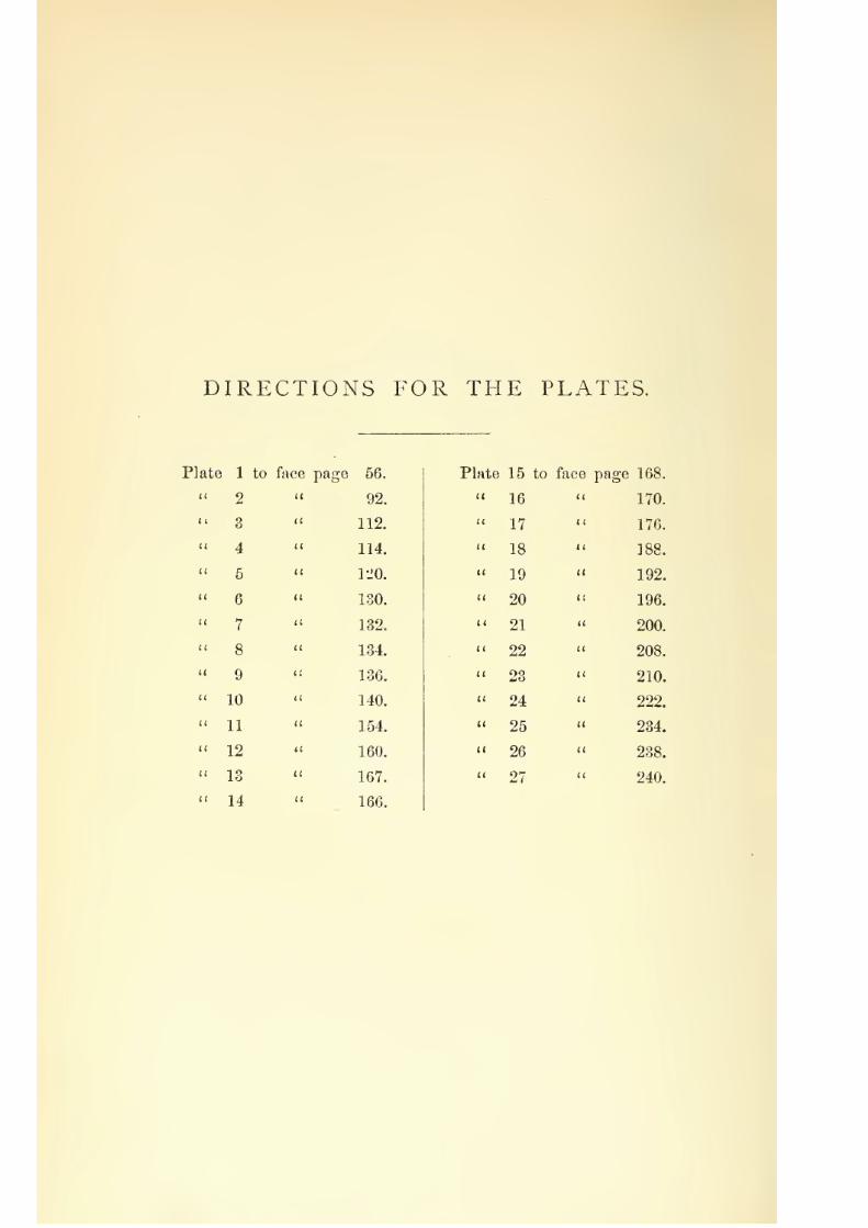
7/29/2019 THE MICROSCOPIST
http://slidepdf.com/reader/full/the-microscopist 316/361
DIRECTIONS FOR THE PLATES.
Plate 1
2
3
4
5
6
7
8
9
10
11
12
13
14
to face page 56.
92.
112.
114.
rjo.
130.
132.
134.
136.
140.
154.
160.
167.
166.
Plate 15 to face page 168
16 170
17 176
18 188
19 192
20 196
21 200
22 208
23 210
24 222
25 234
26 238
27 240

7/29/2019 THE MICROSCOPIST
http://slidepdf.com/reader/full/the-microscopist 317/361
THE PHYSICIAN'S VISITING LIST.
PUBI^ISIIED ANNUALLY.^OJ* Various Styles and Prices, see page 35 of this Catalogue.
No. 25 South Sixth Street,
PHILADELPHIA,January, 1S77,
OF
DENTAL AND PHARMACEUTICAL
PUBLICATIONS.
MESSRS. LINDSA Y &- BLAKISTON ask the attention of the Medical Profes-
sion to the extensive list and varied character of theirpublications and to the
NEWBOOKS now added.
ALL THEIR PUBLICATIONS can be hadfrom or through BOOKSELLERSin any of the large cities of the United States or Canada. When, for a7iy reason,
it is inconve}iie7it thus to procure theni, they will befurnished dij'cct by mail or
. express ztpon receipt of a Post-office order, draft or checkfor the amount ordered.
IN ADDITION to their own publicatio?ts, they keep on hand a full and complete
asso7-tment of all Medical Books published in the United States; and, by special
a,rra7igeme7it with Messrs. J.&^ A. CHURCHILL a7id other Medical Pub-
lishej's of Lo7ido7i, they ca7i supply ma7iy i77iporta7it English Medical Works at
g7'eatly reducedprices ; such as are 7tot 07i hand they ca7i iiiport p7'077iptly to
order.
AS SPECIAL AGENTS OF THE SYDENHAM SOCIETY in the Uiitcd
States, they a7-e prepared to receive Subscribe7'S at Ten Dollars per Aj.7inu77i,
ajid supply a7iy of the back years. Complete lists of works published will be
fur7iished upon applicaJioii.
DEALERS IN MEDICAL BOOKS will be supplied on the most favorable
terms, a7id will be furnished with copies of this Catalogue, without charge,for
distribtitio7i among their ctistotners if desired.

7/29/2019 THE MICROSCOPIST
http://slidepdf.com/reader/full/the-microscopist 318/361
STUDENT'S SERIES OF MANUALS.
This Series of Works has been prepared by Authors of estab-
Hshed reputation in the specialties upon which they have written.
They are compact in size; brief
andexplicit in their contents, but
containing all that is essential under ordinary circumstances, and are
admirably adapted to the wants of the Busy Practitioner and Student
who'have neither time nor leisure to consult or read the more elab-
orate and expensive treatises.
WaggstaiFe's gtMclc-nt's Ciiiidet® MiiTmaiB Osteology. 23 Litho-
graphic Plates and 66 other Illustrations, Price, $3.50.
Clay's CoMipleie IIaHcl-Il®®li ®f ©testetric Surgery. With numer-
ous Illustrations. Price, $2.2o.
SwaiM's ManMal ©f Surgical Emergencies. With numerous Illus-
trations. Price, '?2.00.
Alliill°s dinical jLectiares ©m ©iseases Peculiar to Women.With Illustrations. Price, $2.25.
BillsafeeFger's Maii!<Jly-B«>®& ®f W^mesi's aiad Claildren's I>is«eases. Price, $1.75.
Iiaivs®ia's diseases aaicl IiajMrDes ®f tlae Eyes their Medical andSurgical Treatment. V/ith Illustrations. Price, $2.50.
Coliesa ©la Iiilaalat.i®ii : its Therapeutics and Practice. With Illustrations.Price, $2,75.
Ilarley ©is tlae Urls^e aaad ite ©efi'aEBgemeists. With Engravings.New Enlare-ed Edition. Price,
s Practical ^iside t© tlse Esaiiiiiiati®!! ®f UrSiie. Illus-
trated. Price, $1.50.
Haiid"ll®®k ©f Medical Electricity. With 65 Illustra-
tions. Price, $2 00.
•m tlae diseases and lejiiries of tlie Ear. With Illustra-
tions. Price, $1.50.
Meafla's MaisMal ®f Miis®r Swrgery and Baiislagiisg. With nu-merous Illustrations. Price, $2.00.
Wilsffis2's MaBBd-lld®fe ®f HygieBie aaad SaBaitary Science. WithEngravings. Third Revised Edition.
Alllngliam ®3J Fisiiila, Il£esfii®rrli®icls, aiad ©ilaer Uiseascs ®filie KectiiaMc Price, $2.00.
liewin ©11 tlae Trealisaeiat ®f Sypliilis by Subcutaneous Sublimate
Injections. With Plates. Price, $2.2:5. ^
Brad lily's Maaisiial ®f C®MiFfWith Illustrations. Price, $2.60.
KeyiiwM'S Cllsiical Uses ®f Electricity. Price, $1.25.
Blac&'s Eiiiicti©iraal Hi^eases ®f tlae Keiial, Urinary, asad He-productive ©FgaHS. Price, $2.50.
C®Ieg' Mamiial ®f ©eaatal Meeliasiics. 140 Illustrations. Price, $2.50,
ElMs's Maiiiial ®f tlie Biseases ®f Cliildreia. With Formulary.Price, $2.y5.
Kotoerts's Stedeiit's Guide t© tlse I^ractice ®f Midwfiferyo With
numerous Illustrations. Price, $2.25.
Sewell's StigdeaHt^s CSmide t® I>eiital AHat®Miy asid Surgery.With 77 Illustrations. Price, $1.75.

7/29/2019 THE MICROSCOPIST
http://slidepdf.com/reader/full/the-microscopist 319/361
Medical Text-Books and Works of Reference,
PUBLISHED BY
LINDSAY & BLAKISTON, PHILADELPHIA.
Roberts's Hand-Book of the_ Practice of Medicine. Uniformly commended by the Pro-
fession and the Press. Octavo. Price, bound in cloth, fc.oo ; leather, $6.00.
TroLisseau's Clinical Medicine. Complete in two volumes, octavo. Price, in cloth, $10.00;
leather, f12.00.
Aitken's Science and Practice of Medicine. Third American, from the Sixth LondonEdition. Two volumes, royal octavo. Price, in cloth, ^fia.00 ; leather, ^14.00.
Sanderson's Hand-Book for the Physiological Laboratory. E.iierci.=;es for Students
in Physiolog^r and Histology. 353 Illustrations. Price, in one volume, cloth, $5.oo ; leather, S7.00.
Cazeaux's Text-Book of Obstetrics. From the Seventh French Edition, Revised and greatly
Enlarged. With Illustrations. Cloth, $6.50; leather, $7.50.
VVaring's Practical Therapeutics, From the Third London Edition. Cloth, $5.00 ; leather,
§6.00.
Rindfleiseh's Pathological Histology. Containing 208 elaborately executed Microscopical
Illustrations. Cloth, S6.00 ; leather, $7.00.
Meigs and Pepper's Practical Treatise on the Diseases of Children. Sixth Edition.
Cloth, $6.00 ; leather, ?7.oo.
Tanner's Practice of Medicine. The Sixth American Edition, Revised and Enlarged. Cloth,
§6.00 ; leather, $7.00.
Tanner and Meado\Ar's Diseases' of Infancy and Childhood. Third Edition.
Cloth, $3.50.
Biddle's Materia Mediea for Students. The Seventh Revised and Enlarged Edition. WithIllustrations. Price, §4.00.
Harris's Principles and Practice of Dentistry. The Tenth Revised and Enlarged Edition.
Cloth, $6.50; leather, $7.50.
Soelberg Wells on Diseases of the Eye. The Third London Edition. Illustrated byOphthalmoscopic Plates and other Engravings. Cloth, $5.00 ; leather, $6.00.
Byford's Practice of -Medicine and Surgery. Applied to the Diseases of Women. SecondEdition. Cloth, .§5. 00; leather, :$6.00.
Byford on the Uter'US. A New, Enlarged, and thoroughly Revised Edition. Numerous Illustra-
tions. Price, S3.00.
He'witt's Diagnosis and Treatment of the Diseases of Women. Third Edition.
Cloth, f5.00 ; leather, f6.oo.
Headland on the Action of Medicines. Sixth American Edition. Price, S3. 00.
Harley on Urine and its Derangements. With Illustrations. Price,
Meadov^''.s Manual of Midwifery. Illustrated. Third Enlarged Edition, including the Signs
and Symptoms of 1 regnancy, etc. Price, 5^3.25.
Bloxom's Chemistry. Inorganic and Organic. Second Edition. 276 Illustrations. Cloth, ^4.00 ;
leather, $5.00.
Walton's Practical Treatise on Diseases of the Eye. The Third Revised and EnlargedEdition.
Numer us Illustrations, Test Types, etc. Price, $9.00.
Jones and Sieveking's Pathological Anatomy. A New Enlarged Edition, edited bjfJ.
F. Payne, JaI. D. With Illustrations. Price, $6.00.
Wilks and Moxon's Pathological Anatomy. Second Edition, Enlarged and Revised.
Price, S5 50.
Carpenter's Microscope and its Revelations. The Fifth Edition, very much Enlarged.
With 500 Illustrations. Price, S5.50,
Wilson's Anatomist's Vade Meeum. The Ninth Enlarged London Edition. Price, Sts-so.
Parke's Manual of Practical Hygiene. The Fourth Enlarged Edition. Price, f5.oo.
Richardson's Mechanical Dentistry. Second Edition, much Enlarged. With over 150
Illustrations. Price, in cloth, ?4.oo ; leather, S4. 50.
Beale's Use of the Microscope in Practical Medicine. Fourth Edition. 500 Illustra-
tions. Preparing.
Sweringen's Dictionary of Pharmaceutical Science. Octavo. Price, $5.00.
Mackenzie's Growth in the Larynx. With Numerous Colored Illustrations. Price, $3.00.
COMPLETE DESCRIPTIVE CATALOGUES FURNISHED FREE UPONAPPLICATION.

7/29/2019 THE MICROSCOPIST
http://slidepdf.com/reader/full/the-microscopist 320/361
IMPORTANT NEW ILLUSTRATED WORKS.
FOX'S ATLAS OF SKIN DISEASES.
To be completed in Eighteen Monthly Parts, each containiiig 4 Chromo-Lithographic
Plates, with Descriptive Text and Notes upon Treatment. By Tilbury Fox, M. D.,
F. R. C. P., Physician to the Department for Skin Diseases in University College
Hospital. Royal 410. Price, $2.00 each. Thirteen Parts now Ready.
No Atlas of Skin Diseases has been issued in this country for many years, and no com-
plete work of the kind is now. procurable by the Profession. This one, brought out under
the editorial supervision and care of Dr. Tilbury Fox (the most distinguished, writer on
Cutaneous Medicine now in the English language), is partly based upon the classical
work of WiHan and Bateraan (now entirely out of print), but completely remodelled, so
as to represent fully the Dermatology of the .present day.
" Preference will be given to this work over Hebra ; not simply, however, because it is a home pro-
duction, but by reason of the manner of its e.xecution, the excellent delineation of disea.se, and the naturaf.
coloring of the plates. . . . The letter-press is entirely new. In the as'curacy of the latter the subscribermay have the fullest confidence, since it is from the pen of Dr. Tilbury Fox." British and Fo7-eigii Medico-
Chiriirgical Review.
HUTCHINSON'S ILLUSTRATIONS OF CLINICAL SURGERY.
Consisting of Plktes, Photographs, Woodcuts, Diagrams, etc., illustrating Surgical Diseases,
Symptoms, and Accidents ; also. Operations and other Methods of Treatment. With
Descriptive Letter-press. By Jonathan Hutchinson, F. R. C. S., Senior Surgeon to
the London Hospital, Surgeon to the Moorfields Ophthalmic Hospital, and to the
Hospital for Diseases of the Skin, Blackfriars. In Quarterly Fasciculi. Imperial
4to. $2.50 each. Four Fasciculi Now Ready.
BENTLEY AND TRIMEN'S MEDICINAL PLANTS.
Being Descriptions, with Original Figures, of the Principal Plants employed in Medicine,
and an Account of their Properties and Uses. By R. Bentley, F. L. S., Professor of
Botany in Kirfg's College, London; Professor of Botany and Materia Medica to the
Pharmaceutical Society; and H. Trimen, M. B., F. L. S., Department of Botany^
British Museum; Lecturer on Botany in St. Mary's Hospital Medical School. To
be completed m about Thirty-six Monthly Parts, With Colored Illustrations (natural
size). Large 8vo.' Price, $2.00 each. Thirteen Parts now Ready.
*^* This work will serve especially as an illustrated Botanical Guide to the British, United States-, andIndian Pharmacopoeias ;
it will also include other species employed, or in common use though not ofAcial.
Some others which afford food substances, of value chiefly to invalids, have also been added.Each Plate will be accompanied by letter-press comprehending a full description of the plant in plain
scientific language, its nomenclature, geographical distribution, etc., as well as an account of its properties
and uses, with full references to pi-evious descriptions and figures and to more specis.! treatises.
HEATH'S COURSE OF OPERATIVE- SURGERY.With Plates drawn from Nature by M. Leveill^ and Colored by hand under his direc-
tion. By Christopher Heath, F. R. C. S., Surgeon to University College Hospital,
and Holme Professor of Clinical Surgery in University College. To he completed in
Five Bi-Monthly Parts, at $2.50 each, each Part containing 4 Plates. Imperial Quarto,
Three Parts are now ready, and the whole work will be completed within the year.
SUBSCRIPTIONS will be received for these works by the PUBLISHERS, or any
of their AGENTS, and remittances can be made for five or more Parts, to be delivered or
sent free by mail as published. Full Prospectuses also furnished upon application.
GOOD CANVASSING AGENTS are wanted for these and other important
Medical Worlds now in course of publication.

7/29/2019 THE MICROSCOPIST
http://slidepdf.com/reader/full/the-microscopist 321/361
MEDICAL, DENTAL,PHARMACEUTICAL AND SCIENTIFIC BOOKSPUBLISHED BY
LINDSAY & BLAKISTON, Philadelphia.
AITKEN (WILLIAM), M. D.,
Professor of Pathology in the Army Medical School,'&c,
THE SCIENCE AND PRACTICE OF MEDICINE. THIRDAmerican, from the Sixth London Edition. Thoroughly Revised,
Remodelled, many portions Rewritten, with Additions almost equal to
a Third Volume, and numerous additional Illustrations, without any
increase in bulk or price. Containing a Colored Map showing the
Geographical Distribution of Disease over the Globe, a Lithographic
Plate, and nearly 200 Illustrations on Wood.
Two volumes, royal octavo, bound in cloth, price, . . ^12.00
." " " " leather, . . 14.00
For eighteen months Dr. Aitken has been engaged in again carefully revising this G^^eat
Work, and adding to it many valuable additions and improvements, amounting in the ag-
gregate almost to a volume of new matter, included in which will be found the adoption and
incorporation in the text of the "New Nomenclature of the Royal College of Physicians of
London;" to which are added the Definitions and the Foreign Equivalents for their English
names ; the ±^ew Classification of Disease as adojDted by the Royal College of Physicians, &c.
The American editor, Meredith Clymer, M. D., has also added to it manyvaluable articles, with special reference to the' wants of the American" Prag-"
TITIO^'ER.
The work is now, by almost universal consent, both in England and the United States,
acknowledged to be in advance of all other works on The Science and Practice of 3Iedicine.
It is a most thorough and complete Text-book: for students of medicine, following such a
systematic arrangement as. will give them a consistent view of the main facts, doctrines, and
practice of medicine, in accordance with accurate physiological and pathological principles
and the present state of science. For the i^ractitioner it will be found equally acceptable as
a vfork of reference.
ALLINGHAM (william), F. R. C. S.,
'Surgeon to St. Mark's Hospital for Fistula, &,c,
FISTULA, HEMORRHOIDS, PAINFUL ULCER, STRICT-LRE, PROLAPSUS, and other Diseases of the Rectum, their Diagnosis
and Treatment. Second Edition, Revised and Enlarged by the
Author. Price . . . . . . . . . ;^2.oo
This book has been well received by the Profession ; the first edition sold rap >
idly ; the present one has been revised by the author, and some additions made,
chiefly as to the mode of treatment.
The Medical Press and Circular, siDeaking of it, says: "No book on this special subject
can at all approach ilr. Allingham's in jDrecision, clearness, and practical good sense."
The London Lancet : " As a practical guide to the treatment of afi'ections of the lower
bowel, this book is worthy of all commendation."
T/'e Edinburgh Monthly :" We cordially recommend it as well deserving the careful study
of Physicians and Surgeons."

7/29/2019 THE MICROSCOPIST
http://slidepdf.com/reader/full/the-microscopist 322/361
ATTHILL (lombe), M. D.,
Fellow and Examiner in Midwifery, King and Queen's College of Physicians, Dublin.
CLINICAL LECTURES ON DISEASES PECULIAR TO WO-MEN. Second Edition, Revised and Enlarged, with Six Lithographic
Plates and other Illustrations onWood.
Price . . ,
^2.2:^
The value and popularity of this book is proved by the rapid sale of the first edition,
which was exhausted in less than a year from the time of its publication. It appears to
possess three great merits : First, It treats of the diseases very common to females. Second,
It treats of them in a thoroughly clinical and practical manner. Third, It is concise, orig-
inal, and illustrated by numerous cases frm the author's own experience. His style is clear
aud the volume is the result of tiie author's large and accurate clinical observation recorded
in a remarkable, perspicuous, and terse manner, and is conspicuous for the best qualities of
a practical guide to the student and practitioner. •— British Medical Jout'nal.
ADAMS (WILLIAM), F. Pv. C.S.,Surgeon to the Royal Orthopedic and Great Northern Hospitals.
CLUB-FOOT: ITS CAUSES, PATHOLOGY, AND TREAT-MENT. Being the Jacksonian Prize Essay of the Royal College of
Surgeons. A New Revised and Enlarged Edition, with 106 Illustrations
engraved on Wood, and Six Lithographic Plates. A large Octavo
Volume. Price ......... ^6.00
ADAMS (ROBERT), M. D.,
Regius Professor of Surgery in the University of Dublin, &.C., &c.
RHEUMATIC GOUT, or CHRONIC RHEUMATIC ARTHRI-TIS OF ALL THE JOINTS. The Second Edition. Illustrated by
numerous Woodcuts, and a quarto Atlas of Plates. 2 Volumes.
Price .'. ^8.50
ALTHAUS (JULIUS), M.D.,Physician to the Infirmary of Epilepsy and Paralysis.
A TREATISE ON MEDICAL ELECTRICITY, Theoretical and
Practical, and its Use in the Treatment of Paralysis, Neuralgia, and other
Diseases. Third Edition, Enlarged and Revised, with One Hundred
and Forty-six Illustrations. In one volume octavo. Price . ^6.00
In this work both the scientitic and practical aspects of the subject are ably, concisely, and
thoroughly treated. It is much the best work treating of the remedial effects of electricity
in the English language. JVew York Medical Record.
ARNOTT (henry), F.R.C.S.
CANCER : its Varieties, their Histology and Diagnosis. With Five
Lithographic Plates and Twenty-two Wood Engravings. Price ^2.25
AGNEW (d. HAYES), M.D.,
Professor of Surgery in the University of Pennsylvania.
THE LACERATIONS OF THE FEMALE PERINEUM, AND
VESICO-VAGINAL FISTULA, their History and Treatment, withnumerous Illustrations. Octavo. Price . . . . $2.00
Prof. Agnew has been a most indefatigable laborer in this department, and his work stands
deservedly high in tlie estimation of the ])rofession. It is well illustrated, and full descrip-
tions of the operations aud instruments employed are given.— Canada Lancet.

7/29/2019 THE MICROSCOPIST
http://slidepdf.com/reader/full/the-microscopist 323/361
ACTON (WILLIAM), M.R.C.S., etc.
THE FUNCTIONS AND DISORDERS OF THE REPRODUC-TIVE ORGANS. In Childhood, Youth, Adult Age, and Advanced
Life, considered in their Ph^^siological, Social, and Moral Relations.
Fourth American from the Fifth London Edition, Carefully revised bythe Author, with additions. . . . , > . . . ^3.00
Mr. Acton has done good service to society by grappling manfully with sexual vice, and
we trust that others, whose position as men of science and teachers enable them to sjieak
with authority, will assist in combathig and arresting the evils which it entails. Tlie spirit
which pervades his book is one which does credit equally to the head and to the heart of the
author.— British and Foreign Medico-Chiruryical Review.
SAME AUTHOR.PROSTITUTION: Considered in its Moral, Social, and Sanitary As-
pects. Second Edition, Enlarged. Price , ,, ,
^5.0.0
ANSTIE (FRANCIS E.), M.D.,Lecturer on Materia Medica and Therapeutics, etc.
STIMULANTS AND NARCOTICS. Their Mutual Relations, with
Special Researches on the Action of Alcohol, Ether, and Chloroform
on the Vital Organism. Octavo. . , , . > $3'Od
ANDERSON (m'call), M.D.,Professor of Clinical Medicine in the University of Glasgow, &Ci
ECZEMA. The Pathology and Treatment of the various Eczema-tous Affections or Eruptions of the Skin. The Third Revised and En-
larged Edition. Octavo. Price . . . , . - ^2.75
BUZZARD (THOMAS), M. D., '
Physician to the National Hospital for Paralysis and Epilepsy.
CLINICAL ASPECTS OF SYPHILITIC NERVOUS AFFEC-TIONS. i2mo. Cloth. Price $1.75
BASHAM (w. R.), M.D., F.R.C.R,Senior Physician to the Westminster Hospital, &c,
AIDS TO THE DIAGNOSIS OF DISEASES OF THE KID-NEYS. With Ten large Plates. Sixty Illustrations. Price . ^2.00
SAME AUTHOR.
ON DROPSY, AND ITS CONNECTION WITH DISEASES OFTHE KIDNEYS, HEART, LUNGS AND LIVER. With Sixteen
Plates. Third Edition. Octavo. . . . . . I5.00
M. BARTH AND M. HENRI ROGER.A MANUAL OF AUSCULTATION AND PERCUSSION. A
new Translation, from the Sixth French Edition. . . . ^1.25
S. M. BRADLEY, F. R. C. S.
Senior Assistant Surgeon Manchester Royal Infirmary,
A MANUAL OF COMPARATIVE ANATOMY AND PHYSI-OLOGY. With 60 Illustrations. Third Edition. Price . $2.50

7/29/2019 THE MICROSCOPIST
http://slidepdf.com/reader/full/the-microscopist 324/361

7/29/2019 THE MICROSCOPIST
http://slidepdf.com/reader/full/the-microscopist 325/361
BENNETT (j. henry), M. D.
NUTRITION IN HEALTH AND DISEASE. A Contribution to
Hygiene and to Clinical Medicine. Second Edition, Revised and En-
larged. Octavo. Cloth. Price...... $2.50.
BIRCH (s. B.), M.D.,Member cv xhe Royal College of Physicians, &c.
CONSTIPATED BOWELS ; the Various Causes and the Different
Means of Cure. Third Edition. Price . . . . ^i.oo
BUCKNILL(joHN CHARLES), M.D., & TUKE (daniel h.),M.D.
A MANUAL OF PSYCHOLOGICAL MEDICINE: containing the
Lunacy LaAvs, the Nosology, GEtiology, Statistics, Description, Diagno-
sis, Pathology (including Morbid Histology), and Treatment of Insanity.Third Edition, much enlarged, with Ten Lithographic Plates, and nu-
merous other Illustrations. Octavo. Price .... ^8.00
This edition contains upwards of 200 pages of additional matter, and, in consequence of
recent advances in Psychological Medicine, several chapters have been rewritten, bi-inging
the Classification, Pathology, and Treatment of Insanity up to the present time.
BROWNE (j. H. BALFOUR), Esq.
MEDICAL JURISPRUDENCE OF INSANITY. Second Edition,
very much Enlarged. With References to the Scotch and AmericanDecisions, etc., etc. Octavo. Price . . . . . ^5.00
BIDDLE "(jomTB.), M. D.,
Professor of Materia Medicaand Tlierapeutics in the Jefferson IViedical College, Philadelphiai &lc.
MATERIA MEDICA, FOR THE USE OF STUDENTS. WithIllustrations. Seventh Edition, Revised and Enlarged. Price ^4.00
This new and thoroughly revised edition of Professor Biddle's work has incorporated in
it ail the improvements as adopted by the New United States Pharmacopceia just issued. It
is designed to present the leading facts and principles usually comprised under this head asset forth by the standard authorities, and to fill a vacuum which seems to exist in the wantof an elementary work on the subject. The larger works usually recommended as text-books
in our Medical schools are too voluminous for convenient use. This will be found to contain,
in a condensed form, all that is most valuable, and will supply students with a reliable guide
to the course of lectures on Materia Medica as delivered at the various Medical schools in
the United States,
BALFOUrTgTw.), m. d.,
Physician to the Royal Infirmary, Edinburgh |Lecturer on Clinical Medicine, &c.
'
- CLINICAL LECTURES ON DISEASES OF THE HEART ANDAORTA. With Illustrations. Octavo. Price . . . ^5.00
BYFORD (wTT), A.m., M.D.,Professor of Obstetrics and Diseases of Women and Children in the Chicago Medical College, &c.
PRACTICE OF MEDICINE AND SURGERY. Applied to the
Diseases and Accidents incident to Women. Second Edition, Revised
and Enlarged. Octavo. Price, cloth, ^5.00; sheep . . ,0.oo
SAME AUTHOR.
ON THE CHRONIC INFLAMMATION AND DISPLACEMENTOF THE UNIMPREGNATED UTERUS. A New, • Enlarged, and
Thoroughly Revised Edition, with Numerous Illustrations. Octavo. $3
Dr. Byford writes the exact present state of medical knowledge on the subjects presented;
and does this so clearly, so concisely, so truthfully, and so completely, that his book on the
uterus will always meet the approval of the profession, and be everywhere regarded as a
popular standard work. — Buffalo lledical and Surgical Journal.

7/29/2019 THE MICROSCOPIST
http://slidepdf.com/reader/full/the-microscopist 326/361
10
BLACK (d. CAMPBELL), M. D.,
L. R. C. S. Edinburgh, Member of the General Council of tlie University of Glasgow, &c., Sic,
THE FUNCTIONAL DISEASES OF THE RENAL, URINARY/and Reproductive Organs, with a General View of Urinary Pathology.
Price ........... ^2.50
The style of the author is clear, easy, and agreeable, . . . his work is a valuable contri-
bution to medical science, and being penned in that disposition of unprejudiced philosophical
inquiry which should always guide a true physician, admirably embodies the spuit of its
opening quotation from Professor Huxley.— Fkilada. Med. Times.
BY SAME AUTHOR.LECTURES ON BRIGHT'S DISEASE OF THE KIDNEYS.
Delivered at the Royal Inirrmary of Glasgow, With 20 Illustrations,
engraved on Wood. One volume, octavo, in Cloth. Price . ^2.00
BENTLEY and TRIMEN'SMEDICINAL PLANTS. A New Illustrated Work, now Publish-
ing in Monthly Parts. Thirteen Parts nov/ ready. Eight Colored,
Plates in each Part. Price, each, . . . . . . $2.00
This work includes full botanical descriptions, and an account of the properties and uses
of tlie principal plants employed in medicine, especial attention being paid to those which
are ofHcinal in the British and United States Pharraacopceias. The plants which su})j)]y
food and substances required by the sick and convalescent will be also included. Each spe-cies will be illustrated by a colored plate drawn from nature.
BEASLEY (henry).
THE BOOK OF PRESCRIPTIONS. Containing over 3000
Prescriptions, collected from the Practice of the mos't Eminent Physi-
cians and Surgeons— English, French, and American; comprising also
a Compendious History of the Materia Medica, Lists of the Doses of all
Officinal and Established Preparations, and an Index of Diseases and
their Remedies. Fifth Edition, Revised and Enlarged. Price ^2.50
BY SAME AUTHOR.
THE POCKET FORMULARY: A Synopsis of the British and
Foreign Pharmacopoeias. Ninth Revised Edition. Price . ^2.50
THE DRUGGIST'S GENERAL RECEIPT BOOK and VETERI-NARY FORMULARY. Seventh Edition. Price. ^2.50
BRANSTON(thomas f.).
HAND-BOOK OF PRACTICAL RECEIPTS. For the Chemist,
Druggist, &c. ; with a Glossary of Medical and Chemical Terms. $1.50
BRAUNE—BELLAMY.AN ATLAS OF TOPOGRAPHICAL ANATOMY. After Plane
Sections of Frozen Bodies, containing Thirty-four Full-page Photo-
graphic Plates and numerous other Illustrations on Wood. By AVilhelm
Braune, Professor of Anatomy in the University of Leipzig. Trans-lated and Edited by Edward Bellamy, F. R. C. S. ,
Senior Assistant Sur-
geon to, and Lecturer on Anatomy and Teacher of Operative Surgery
at, the Charing Cross Hospital, London. A large quarto volume.
Price in cloth, gi2. 00 ; half morocco, ..... I14.00

7/29/2019 THE MICROSCOPIST
http://slidepdf.com/reader/full/the-microscopist 327/361
11
COHEN (i. soLis), M.D.Lecturer on Laryngoscopy and Diseases of the Throat and Chest in Jefferson Medical College,
ON INHALATION. ITS THERAPEUTICS AND PRACTICE.Including a Description, of the Apparatus employed, &c. With Cases
and Illustrations. A New Enlarged Edition. Price . . ^2.75
SAME AUTHOR.CROUP. In its Relations to Tracheotomy. Price . . ^i.oo
CARSON (JOSEPH), M.D.,
Professor of Materia Medica and Pharmacy in the Univprslty.
A HISTORY OF THE MEDICAL DEPARTMENT OF THEUNIVERSITY OF PENNSYLVANIA, from its Foundation in 1765:
with Sketches of Deceased Professors, &c. . . . . $2.00
Tiie history of the University of Pennsylvania has a national as well as a local interest,
from the early date of its origination, and the connection with it of men of illustrious public
reputation, such as Drs. Franklin, Rush, Physick, Gibson, Uewees, Chapman, Wood, &c,, &c.
For the labor and love which he has spent in preparing this most interesting and valuable
work, Prof. Carson has earned the gratitude of the alumni of the University, and of all otners
interested in medical education in this country. — American Jour^ial of Medical Science.
CARPENTER (w. b.), M.D., F.R.S.
THE MICROSCOPE AND ITS REVELATIONS. The Fifth
London Edition, Revised and Enlarged, with more than 500 Illustra-tions ^5.50
SAME AUTHOR.PRINCIPLES OF HUMAN PHYSIOLOGY. The Eighth Revised
and Enlarged Edition. With nearly 400 Illustrations on Steel and
Wood. Edited by Mr. Henry Power. 1200 pages. Octavo. $5.50
CHAVASSE (p. henry), F.R.C.S.,
Author of Advice to a Wife, Advice to a Mother, &c,
APHORISMS ON THE MENTAL CULTURE AND TRAIN-ING OF A CHILD, and on various other subjects relating to Health
and Happiness. Addressed to Parents. Price . . . ,$1.50
Dr. Chavasse's works have been very favorably received and had a large circulation, the
value of his advice to WIVES and MOTHEPtS having thus been very generally recognized.
This book is a sequel or companion to them, and it will be found both valuable and important
to all who have the care of families, and who want to bring up their children to become useful
men and women. It is full of fresh thoughts and graceful illustratioas.
CLARKE (w. fairlie),
M. D.,"^
Assistant Surgeon to Charing Cross Hospital.
CLARKE'S TREATISE ON DISEASES OF THE TONGUE.With Lithographic and Wood-cut Illustrations. Octavo. Price $5.00
It contains The Anatomy and Physiology of the Tongue, Importance of its Minute Exam-ination, Its Congenital Defects, Atrophy, Hypertrophy, Parasitic Diseases, Inflammation,
Syphilis and its effects, Various Tumors to which it is subject, Accidents, Injuries, &c., &c.
COOPER (s.).
A DICTIONARY OF PRACTICAL SURGERY AND ENCY-CLOPEDIA OF SURGICAL SCIENCE. New Edition, brought
down to the present time. By Samuel A. Lane, F.R.C.S., assisted by
other eminent Surgeons. In two vols., of over 1000 pages each. ^15.00

7/29/2019 THE MICROSCOPIST
http://slidepdf.com/reader/full/the-microscopist 328/361
12
CLAY (CHARLES), M. D.
Fellow of the London Obstetrical Society, &c.
THE COMPLETE HAND-BOOK OF OBSTETRIC SURGERY,or, Short Rules of Practice in Every Emergency, from the- Simplest to
the most Formidable Operations in the Practice of Surgery. First
American from the Third London Edition. With numerous Illustra-
tions. In one volume. $2.25
CHAMBERS (thomas k.), M. D.,
LECTURES, CHIEFLY CLINICAL. Illustrative of a Restorative
System of Medicine.
CORMACK (sir john i^os^K. B., F. R. S. E., M. D.
Edinburgh and Paris, Fellow Royal College of Physicians, Physician to the Hertford British Hospital, Paris, &c,
CLINICAL STUDIES, Illustrated by Cases observed in Hospital and
Private Practice. With Illustrative Plates. 2 Volumes. Octavo. $6.50
COBBOLD (t. spencer), M.D., F.R.S.
WORMS: a Series of Lectures delivered at the Middlesex Flospital
on Practical Helminthology. Post Octavo. . . . . ^2.00
^
CLEAVELAND (c. H.), M.D.,Member of the American Medical Association, &.c.
A PRONOUNCING MEDICAL LEXICON. Containing the Cor-
rect Pronunciation and Definition of Terms used in Medicine and the
Collateral Sciences. Improved Edition, Cloth, ^1.25 ; Tucks, ^1-50
This work is not only a Lexicon of all the words in common use in Medicine, but it is
also a Pronouncing Dictionary, a feature of great value to Medical Students. To the Dis-
penser it will prove an excellent aid, and also to the Pharmaceutical Student. It has received
strong commendation both from the Medical Press and from the profession.
COLES (oakley), D.D.S.Dental Surgeon to the Hospital for Diseases of the Throat, &c,
A MANUAL OF DENTAL MECHANICS. Containing muchinformation of a Practical Nature for Practitioners and Students.
INCLUDINGThe Preparation of the Mouth for Artificial Teeth, on Taking Impressions, Various
Modes of Applying Heat in the Laboratory, Casting in Plaster of Paris and Metal,
Precious Metals used in Dentistry, Making Gold Plates, Various Forms of Porcelain
used in Mechanical Dentistry, Pivot Teeth, Choosing and Adjusting Mineral Teeth, the
Vulcanite Base, tlie Celluloid Base, Treatment of Deformities of the Mouth, Receipts
for Making Gold Plate and Solder, etc., etc.
With 140 Illustrations. Price . . . . . . ^2.50
SAME AUTHOR.ON DEFORMITIES OF THE MOUTH, CONGENITAL AND
ACQUIRED, with their Mechanical Treatment. Second Edition, Re-
vised and Enlarged. With Illustrations. Price, . . . ^2.50
CURLING (T.B.), F.R.S.
Consulting Surgeon to London Hospital, Slc,
OBSERVATIONS ON DISEASES OF THE RECTUM. WithIllustrations. Fourth Edition, Revised and Enlarged. Octavo. Cloth.
Price ........... ^2.75

7/29/2019 THE MICROSCOPIST
http://slidepdf.com/reader/full/the-microscopist 329/361
CLARK (f. le gros), F. R. S.,
Senior Surgeon to St. Thomas's Hospital.
OUTLINES OF SURGERY AND SURGICAL PATHOLOGY,including the Diagnosis and Treatment of Obscure and Urgent Cases,
and the Surgical Anatomy of some Important Structures and Regions.
Assisted by W. W. Wagstaffe, F. R. C. S., Resident Assistant-Surgeon
of, and Joint Lecturer on Anatomy at, St. Thomas's Hospital. Second
Edition, Revised and Enlarged. Price . . . . ^3.00
This edition brings the work up to the highest level of our present knowledge, incorporat-
ing all that is sound and recent in Physiology so far as it relates to subjects requiring its
aid. It is not alone an admirable exposition of the principles of Surgery, but a trusty guide
to the emergencies of Practice. We cannot too highly estimate the ability to condense andthe results of a ripened experience furnished to us here in a readable and practical form. —Med. Times and Oazette.
COOLEY (a. j.).
CYCLOP/EDIA OF PRACTICAL RECEIPTS. Containing Pro-
cesses and Collateral Information in the Arts, Manufactures, Profes-
sions, and Trades, including Medicine, Pharmacy, and Domestic
Economy ; designed as a General Book of Reference for the Manufac-
turer, Tradesman, Amateur, and Pleads of Families. The Fifth Edi-
tion, Revised and partly Rewritten by Richard V. Tuson, F. C.S. , &c.
Over 1000 royal-octavo pages, double columns. With Illustrations.
Price
...........|io.oo
Every part of this edition has been subjected to a thorough and comiolete revision by the
editor, assisted by other scientific gentlemen. In the cliemical portion of the book, every
subject of practical importance has been retained, corrected, and added to ; to the name of
every substance of established composition a formula has been attached ; while to the Phar-
maceutist its value has been greatly increased by the additions which have been made from
the British, Indian, and United States Pharmacopoeias.
CAZEAUX (p.), M. D.,
Adjunct Professor of the Faculty of Medicine, Paris, etc.
A THEORETICAL AND PRACTICAL TREATISE ON MIDWIFERY,including the Diseases of Pregnancy and Parturition. Translated from
the Seventh French Edition, Revised, Greatly Enlarged, and Improved,
. by S. Tarnier, Clinical Chief of the Lying-In Hospital, Paris, etc.,
with numerous Lithographic and other Illustrations. Price, in Cloth,
16.50; in Leather, '^7.50.
M. Cazeaux's Great Work on Obstetrics has become classical in its character, and almost
an Encyclopsedia in its fulness. Written expressly for the use of students of medicine, its.
teachings are plain and explicit, presenting a condensed summary of the leading j)rinciplei'
established by the masters of the obstetric art, and such clear, practical directions for the
management of the pregnant, parturient, and puerperal states, as have been sanctioned by
the most authoritative practitioners, and confirmed by the author's own experience.
DOBELL (HORACE), M. D.,
Senior Pliysician to the Hospital,
WINTER COUGH (CATARRH, BRONCHITIS, EMPHYSEMA,ASTHMA). Lectures Delivered at the Royal Hospital for Diseases of the
Chest, The Third Enlarged Edition, with Colored Plates. Octavo.
Price '$3-5°
This work has been thoroughly revised. Two new Lectures have been added— viz.,
Lecture IV., " On the Xatural Course of Neglected Winter Cough, and on the Interdepen-
dence of Winter Cough with other Diseases ;" Lecture IX., " On Change of Climate in ¥/inter
Cough." Also additional matter on Post-nasal Catarrh, Ear-Cough, Artificial Eespiration as
a means of Treatment, Laryngoscopy, New Methods and Instruments in Treating of Emphy=sema, a good Index, and Colored Plates, with appended Diagnostic Physical signs.

7/29/2019 THE MICROSCOPIST
http://slidepdf.com/reader/full/the-microscopist 330/361
14
DIXON (jAMEs), F.R.C.S.Surgeon to the Royal London Ophtha'mic Hospital, &.c,
A GUIDE TO THE PRACTICAL STUDY OF DISEASES OFTHE EYE, with an Outline of their Medical and Operative Treatment,
with Test Types and Illustrations. Third Edition, thoroughly Revised,and a great portion Rewritten, Price ..... $2.50
Mr. Dixon's book is essentially a practical one, written by an observant author, who brinj^s
to his special subject asouud knowledge of general Medicine and Surgery. DvubLin Quavterly.
DILLNBERGER (dr. emil).
A HANDY-BOOK OF THE TREATMENT OF WOMEN ANDCHILDREN'S DISEASES, according to the Vienna Medical School
Part I.
TheDiseases of
Women.Part 11.
The Diseases of Children.Translated from th6 Second German Edition, by P. Nicol, M. D.
Price .........'.. ^1.75
Many practitionei-s will be glad to possess this little manual, which gives a large massof practical hints on the treatment of diseases which probably make up the larger half of
every-day practice. The translation is well made, and explanations of reference to Germais
medicinal preparations are given with proper fulness. — The Fractitioner.
DARLINGTON (william), M.D.
FLORA CESTRICA; OR, HERBORIZING COMPANION. Con-taining all the Plants of the Middle States, their Linnsean Arrangement,
a Glossary of Botanical Terms, a complete Index, &c. Third Edition.
i2mo. . . . . , . . . . . . ;^2.25
DUCHENNE (dr. g. b,).
LOCALIZED ELECTRIZATION AND ITS APPLICATIONTO PATHOLOGY AND TPIERAPEUTICS. Ti-anslated by Her-
bert Tibbits, M.D. With Ninety-two Illustrations. Price . ^3.00Duehenne's great work is not only a well-nigh exhaustive treatise on the medical uses of
Electi'icity, but it is also an elaborate expnsition of the different diseases in which Electric-
ity has proved to be of value as a therapeutic and diagnostic agent.
Part II., illustrated by chromo-lithographs and numerous wood-cuts, is preparing.
DURKEE (SILAS), M.D.,Fellow of the Massachusetts Medical Society, &c.
GONORRHCEA AND SYPHILIS. The Fifth Edition, Revised
and Enlarged, with Portraits and Eight Colored Illustrations. Octavo.
Price ........... I5.00
Dr. Durkee's work impresses the reader in favor of the author by its general tone, the
thorougli honesty everywhere evinced, the skill with which the book-is arranged, the man-ner in which the facts are cited, the clever wav in which the author's experience is brought
in, the lucidity of the reasoning, and the care with which the therapeutics of venereal com-
plaints are treated.— Lancet.
DRUITT (ROBERT), F.R.C.S.
THE SURGEON'S VADE-MECUM. A Manual of Modern Sur-gery. The Tenth Revised and Enlarged Edition, with 350 Illustra-
tions. ........... ^5-°<^

7/29/2019 THE MICROSCOPIST
http://slidepdf.com/reader/full/the-microscopist 331/361
15
DALBY (w. B.), F.R. C.S.,
Aural Surgeon to St. George's Hospital.
LECTURES ON THE DISEASES AND INJURIES OF THEEAR. Delivered at St. George's Hospital. With Illustrations.
Price
...........$1.50
This admirable little volume by llr. Dalby, the accomplished aural surgeon to St. George's
Hospital, consists of eleven lectures delivered by him at that institution. With a modest
aim, this work, the latest issued by tlie English i)ress on Aural Surgery, is happy in concej)-
tion and pleasantly written ; further, it shows that its author is thoroughly «« /ai^ in his
speeialt_v. The subject of which tlie volume treats is handled in a terse style, and this, if
we mistake not, will make it acceptable to the student and practitioner who have a just
horror of tinneeessary details. In conclusion, we hope that we have succeeded in interesting
our readers in the volume. We cordially reconnneiid it as a trustworthy guide in the treat-
ment of the atfections of tite ear. The book is moderate in j^rice, beautifully illustrated by
wood-cuts, and got up in the best style.— Glasgow Medical Journal.
DUNGLISON (robley), M.D.,Late Professor of Institutes of Medicine, dtc, in the Jefferson Medical College,
A HISTORY OF MEDICINE, from the Earliest Ages to the Com-mencement of the Nineteenth Century. Edited by his son, Richard
J. DuNGLisoN, M.D. . . . . . , . . ^2.50
ELLIS (EDWARD), M.D.
Physician to the Victoria Hospital for Sick Children, &c,
A PRACTICAL MANUAL OF THE DISEASES OF CHIL-DREN, with a Formulary. .Second Edition, Revised and Improved.
One vohmie. ......... $2.75
The AUTHOR, in issuing this new edition of his book, says: "I have very carefully revised
each chapter, adding several new sections, and making considerable additions wliere the
subjects seemed to reqtiire fuller treatment, without, however, sacrificing conciseness or
anduly increasing the bulk of the volume."
ELAM (CHARLES), M.D., F.R.C.P.
ON CEREBRIA AND OTHER DISEASES OF THE BRAIN.Octavo. .......... ^2.50
FOTHERGILL (j. milner), M. D.
THE HEART AND ITS DISEASES, AND THEIR TREAT-MENT. With Illustrations. Octavo. Price . . . ^5.00
DIGITALIS. Its Mode of Action and its Use, illustrating theEffect of Remedial Agents over Diseased Conditions of the Heart.
Pi^ce ........... ^1.25
FOX (tilbury), M. D., F. R. C. P.
Physician to the Department for Skin Diseases in University College Hospital,
ATLAS OF SKIN DISEASES. Consisting of a Series of Colored
Illustrations, in Monthly Parts, together with Descriptive Text andNotes upon Treatment ; each Part containing Four Plates, reproduced byChromo-Lithography from the work of Willan & Bateman, or taken from
Original Sources. Thirteen Parts now ready. Price, per Part, ^2.00Full Pj'osptciusesfuj'nished upon appli.ation.

7/29/2019 THE MICROSCOPIST
http://slidepdf.com/reader/full/the-microscopist 332/361
16
FENNER (a s.), M.D., &aVISION: ITS OPTICAL DEFECTS, and the Adaptation of Spec-
tacles ; embracing Physical Optics, Physiological Optics, Errors of Re-
fraction and Defects of Accommodation, or Optical Defects of the Eye.
With 74 Illustrations. Selections from the Test Types of Jaeger andSnellen, etc. Octavo. Price . . . . . . ^3.50
FOSTER (BALTHAZAR), M. D.,
Professor of Medicine in Queen's College.
LECTURES AND ESSAYS ON CLINICAL MEDICINE. Re-
vised and Enlarged by the Author. With Engravings. Octavo.
Price . . . . « . . ' .' • $d>-S°.
FRANKLAND (e.), M. D., F. R. S., &aHOW TO TEACH CHEMISTRY, being the substance of Six
Lectures to Science Teachers. Reported, with the Author's sanction,
by G. George Chaloner, F. C. S., &c. With Illustrations . ^1.25
FENWICK (SAMUEL), M.D., F R.C.P.
THE MORBID STATES OF THE STOMACH AND DUO-
DENUM, AND THEIR RELATIONS TO THE DISEASES OFOTHER ORGANS. With Ten Plates. .... ^5.00
FLINT (AUSTIN), M.D.,Professor of the Principles and Practice of Medicine, &c., Bellevue Hospital College, New York.
CLINICAL REPORTS ON CONTINUED FEVER. Based on
an Analysis of One Hundred and Sixty-four Cases, with Remarks on
the Management of Continued Fever; the Identity of Typhus and
Typhoid Fever; Diagnosis, &c., &c. Octavo. Price . . ^2.00
GANT (FREDERICK J.), F. R. C. S.,
Surgeon to the Royal Free Hospital, &,c.
DISEASES OF THE BLADDER, PROSTATE GLAND, ANDURETHRA, including a Practical View of Urinary Diseases, Deposits,
and Calculi. Fourth Edition, Revised and Enlarged. With New Il-
lustrations. Now Ready. Price ...... ^4.00
Thefact
that a fourthedition
ofthis book has been required seems to be sufficient proof
of its vahie. The author has carefully revised and added such additional matter as to makeit more complete and practically useful.
GODFREY (benjamin), M.D., F.R.A.S.-
THE DISEASES OF HAIR: a Popular Treatise upon the Affec-
tions of the Hair System. . . . . . . .^1.50
GROSS (SAMUEL D.), M.D.,
Professor of Surgery in tiie Jefferson Medical College, Philadelphia, &.c,
AMERICAN MEDICAL BIOGRAPHY OF THE NINETEENTHCENTURY. With a Portrait of Benjamin Rush, M.D. Octavo. I3.50

7/29/2019 THE MICROSCOPIST
http://slidepdf.com/reader/full/the-microscopist 333/361
17
GREENHOW (e. headlam), M.D,Fellow of the Royal College of Physicians, &c.
ON CHRONIC BRONCHITIS, Especially as Connected with Gout.
Emphysema, and Diseases of the Heart. Price . . . ^2.00
Of all works yet written on Chronic Bronchitis, this is undoubtedly the best. The style
is clear and to the point, and the principles of pathology and treatment eminently correct
and practical. It is a positive addition to our medical literature.— Journal Psychological
Medicine.
BY SAME AUTHOR.ADDISON'S DISEASE. Being the Cronian Lectures for 1875.
Dehvered before the Royal College of Physicians. Revised, and Illus-
trated by numerous Cases and 5 full-page Colored Engravings. Onevolume, octavo. Price......,, ^3.50
HARLEY (GEORGE), M. D., F. R. C. R,
Physician to University College Hospital.
THE URINE AND ITS DERANGEMENTS: With the Applica-
tion of Physiological Chemistry to the Diagnosis and Treatment of
Constitutional as well as Local Diseases. New Revised and Enlarged
Edition preparing. With Engravings.
Wehave here a valuable addition to the library of the practising physician
not only for the information which it contains, but also for the suggestive way in whichmany of the subjects are treated, as well as for the fact that it contains the ideas of one whothoroughly believes in the future capabilities of Therapeutics bas.ed on Physiological facts,
and in the important service to be rendered by Chemistry to Physiological investigation.
Aiuerican Journal of the Medical /Science.
HEATH (CHRISTOPHER), F. R. C. S.,
Surgeon to University College Hospital and Holme Professor of Clinical Surgery in University Cafclege.
OPERATIVE SURGERY. Elegantly Illustrated by 20 Large Col-
ored Plates, Imperial Quarto Size, each Plate containing several Fig-
ures, drawn from Nature by M. Leveille, of Paris, and Colored by handunder his direction. To be issued in Five Quarterly Parts. Three Parts
now ready. Price, per Part, . ...... $2.50
1^^ Full Prospectuses furnished upon application.
HEWITT (graily), M. D.,
Physician to the British Lying-in Hospital, and Lecturer on Diseases of Women and Children, &c.
THE DIAGNOSIS, PATHOLOGY, AND TREATMENT OFDISEASES OF WOMEN, .including the Diagnosis of Pregnancy.Founded on a Course of Lectures delivered at St. Mary's Hospital
Medical School. The Third Edition, Revised and Enlarged, v/ith
new Illustradons. Octavo. Price in Cloth . . . I5.00" Leather . . . 6.00
This ney edition of Dr. Hewitt's book has been so much modified, that it may be consideredsubstantially a new book
; very much of the matter has been entirely revfritten, and the Avholevrork has been rearranged in such a manner as to present a most decided improvement overpre\^ous editions. Dr. Hewitt is the leading clinical teacher on Diseases ofWomen in London,
and the characteristic attention paid to Diagnosis by him has given his work great popularitythere. It may unquestionably be considered the most valuable guide to correct Diai;nosis to
be found in the Enghsh language. „
7/29/2019 THE MICROSCOPIST
http://slidepdf.com/reader/full/the-microscopist 334/361
18
HILLIER (THOMAS), M.D.,Physician to tile Hospital for Sick Children, &c,
A CLINICAL T5 EATISE ON THE DISEASES OF CHILDREN.Octavo. Price ......... ^3.00
We have said enough to indicate and illustrate the excellence of Dr. Hillier's volume. Itis eminently the kind of book needed by all medical men who wish to cultivate clinical
accuracy and sound practice. — London Lancet.
HOLDEN (LUTHER), F.R.C.S.
HUMAN OSTEOLOGY, comprising a Description of the Bones
with Dehneations of the Attachments of the Muscles, &c. With
numerous IHustrations. Fifth Edition, carefully Revised. Price, $6.00
HOLDEN'S MANUAL OF DISSECTIONS. Price . ^5.00
HARRIS (cHAPiN A.), M. D., D. D. S.
Late President of and Professor of the Principles and Practice of Dental Surgery in the Baltimore Coileg-e, Slc.
THE PRINCIPLES AND PRACTICE OF DENTISTRY. Tenth
Revised Edition. In great part rewritten, rearranged, and with many' new and important Illustrations. Including— i . Dental Anatomy and
Physiology. 2. Dental Pathology and Therapeutics, 3. Dental Sur-
gery. 4. Dental Mechanics. Edited by P. H. Austen, M.D., Pro-
fessor of Dental Science and Mechanism in the Baltimore College ofDental Surgery. With nearly 400 Illustrations, iiicluding many new
ones made especially for this edition. Royal octavo. Price, in cloth,
^6.50; in leather ........ $7.50
This new edition of Dr. Harris's work has been thoroughly revised in all its parts— more
so than any previous edition. So great have been the advances in many branches of dentistry,
that it was found necessary to rewrite the articles or subjects, and this has been done in the
most efficient manner by Professor Austen, for many years a,n associate and friend of Dr.
Harris, assisted by Professor Gorgas and Thomas S. Latimer^ M. D. The publishers feel
assured that it will now be found the most complete text-book for the student and guide lor
the practitioner in the English language.
SAME AUTHOR.A DICTIONARY OF MEDICAL TERMINOLOGY, DENTAL
SURGERY, AND THE COLLATERAL SCIENCES. Third Edition,
Carefully Revised and Enlarged, by FerdinandJ. S. Gorgas, M. D.y
D.D.S.) Professor of Dental Surgery in the Baltimore College, &:c., &c.
Royal octavo. Price, in cloth, |6. 50; in leather . . IV-So
The many advances in Dental Science rendered it necessary that this edition should be
thoroughly revised, which has been done in the most satisfactory manner by Professor Gorgas^
Dr. Harris's successor in the Baltimore Dental College, he having added nearly three thou-
sand new words, besides making many additions and corrections. The doses of the moreprominent medicinal agents have also been added, and in every way the book has been greatly
improved, and its value enhanced as a work of reference.
• HANDY (WASHINGTON R.), M.D.Late Professor of Anatomy, &c., in the Baltimore College.
A TEXT-BOOK OF ANATOMY, AND GUIDE TO DISSEC-TIONS. For the Use of Students of Medicine and Dental Surgery.
With 312 Illustrations. Octavo. Price . . ... $4.00
Dr. Handy's work was prepared with special reference to the wants of the Student andPractitioner of Dental Surgery. Directing particular attention to the Mouth, it shoAvs step
by step the important Anatomical and Physiological relations which it lias with each andall the organs and functions of the general system.

7/29/2019 THE MICROSCOPIST
http://slidepdf.com/reader/full/the-microscopist 335/361
19
HARDWICH AND DAWSON.HARDWICH'S MANUAL OF PHOTOGRAPHIC CHEMISTRY.
With Engravings. Eighth Edition. Edited and Rearranged by G.
Dawson, Lecturer on Photography, &c., &c. i2mo. . . ^2.00
The object of the Editor has been to give practical instruction in this fascinating art, and
to lead the novice from first principles to the higher branches, impressing him with the valueof care and exactness in every operation.
HEADLAND (f. w.), M. D.,
Fellow of the Royal College of Physicians, &c., &c.
ON THE ACTION OF MEDICINES IN THE SYSTEM. Sixth
American irom the Fourth London Edition. Revised and Enlarged.
Octavo. Price ......... $3.00
Dr. Headland's work gives the only scientific and satisfactory view of the action of medi-
cine; and this not in the way of idle s2)ecu]ation, but by demonstration and exj^eriments,
and inferences almost as indisputable as demonstrations. It is truly a great scientific workin a small compass, and deserves to be the hand-book of every lover of the Profession. It
has received the approbation of the Medical Press, both in this country and in Eurojae, and
is pronounced by them to be the most orixjinal and practically useful work that has been
issued for many 3rears.
HILLES (m. w.),
Formerly Lecturer on Anatomy, &c,, at Westminster Hospital,
THE POCKET ANATOMIST. Being a Complete Description ofthe Anatomy of the Human Body; for the Use of Students. Price, in
cloth, ^i.oo; in Pocket-book form . . . . . ^1.25
HEATH (Christopher), F.R. C.S.,
Surgeon to University College Hospital, &.c.
INJURIES AND DISEASES OF THE JAWS. The Jacksonian
Prize Essay of the Royal College of Surgeons of England, 1867. Sec-
ond Edition, Revised, with over 150 Illustrations. Octavo. Price,
^5.00
SAME AUTHOR.A MANUAL OF MINOR SURGERY AND BANDAGING, for
the Use of House Surgeons, Dressers, and Junior Practitioners. Vv^ith
a Formulae and Numerous Illustrations. i6mo. Price . ^2.00
HAYDEN (THOMAS), M. D.,
Fellow of the King and Queen's College of Physicians, &.c., dtc.
THE DISEASES OF THE HEART AND AORTA. With 81Illustrations. In two volumes, Octavo, of over 1 200 pages. Price, ^8. 00
HUFELAND (c. w.), M.D.THE ART OF PROLONGING LIFE. Edited by Erasmus Wil-
son, M. D., F. R.S., &c. i2mo. Cloth $1.25
The highly practical character of Dr. Hufeland's book, the sound advice which it eon-
tains, and its elevated moral tone, recommend it for extensive circulation both amongprofessional and non-professional readers.

7/29/2019 THE MICROSCOPIST
http://slidepdf.com/reader/full/the-microscopist 336/361
20
HEWSON (addinell,) M. D.
Attending Surgeon Pennsylvania Hospital, &.c.
EARTH AS A TOPICAL APPLICATION IN SURGERY.Being a full Exposition of its use in all the Cases requiring Topical
Applications admitted in the Surgical Wards of the Pennsylvania Hospi-
tal during a period of Six Months. With Illustrations. Price ^2.50
HUTCHINSON (Jonathan), R R. C. S.
Senior Surgeon to the London Hospital.
ILLUSTRATIONS OF CLINICAL SURGERY. Consisting of
Plates, Photographs, Wood-cuts, Diagrams, etc., Illustrating Surgical
Diseases, Symptoms and Accidents, also Operations and other Methods
of Treatment. With Descriptive Letter-press. 4 Part's now ready.
Price. . . . . . . . . . .
^2.50'^Prospectuses furnished upon application.
^
HODGE (HUGH L.), M.D.Emeritus Professor in the University of Pennsylvania.
HODGE ON FCETICIDE, OR CRIMINAL ABORTION.Fourth Edition. Price, in paper, 30 cents; in cloth, . . .50
HODGE'S (h. lenox) NOTE-BOOK FOR CASES OF OVARIAN
TUMORS. With Diagrams, etc. Price, 50
HOLDEN (edgar), A. M., M. D.,
Of Newark, New Jersey.
CONTAINING THEEE HUNDRED ILLUSTRATIONS.
THE SPHYGMOGRAPH. Its Physiological and Pathological In-
dications. The Essay to which was awarded the Stevens Triennial
Prize in the College of Physicians and Surgeons in New York, April,
1873. Illustrated by Three Hundred Engravings on Wood. One vol-ume octavo. Price. . . . . . . . . ;^2.oo
HOOD (p.), M.D.
A TREATISE ON GOUT, RHEUMATISM, AND THE ALLIEDAFFECTIONS. Crown octavo. ^4.25
HANCOCK (henry), F.R.CS.
ON THE OPERATIVE SURGERY OF THE FOOT ANDANKLE. Numerous Illustrations. Octavo. . . . ^6.00
JONES (t. WHARTON), F.R.S.
DEFECTS OF SIGHT AND HEARING. Their Nature, Causes,
Prevention, &c. Second Edition. Price ... - ^1-25
JONES, SIEVEKING, and PAYNE. -
A MANUAL OF PATHOLOGICAL ANATOMY. By C. Hand-field Jones, M. D., F. R. S., Physician to St. Mary's Hospital; and
Edward PI. Sieveking, M.D. , F.R.C.P., Physician to St. Mary's hWpital. A New and Enlarged Edition. Edited by J.
F. Payne, M.B.,
F.R.C.P., Assistant Physician and Lecturer on Morbid Anatomy at St.

7/29/2019 THE MICROSCOPIST
http://slidepdf.com/reader/full/the-microscopist 337/361
21
KIRBY (e. a.), M. D., M. R. C. S. Eng.,
Late Physician to the City Dispensary.
ON THE ADMINISTRATION AND VALUE OF PHOSPHO-RUS, as a Remed}^ for Loss of Nerve Power, Neuralgia, Hysteria, etc.
With Formulje for Combinations with Iron, etc. Second Edition. .50
LAWSON (GEORGE), F.R.C.S.,
Surgeon to the Royal London Ophthalmic Hospital.
DISEASES AND INJURIES OF THE EYE, THEIR MEDICALAND SURGICAL TREATMENT. Containing a Formulary, Test
Types, and Numerous Illustrations. Price .... ^2.50
This Manual is admirably clear and eminently practical. The reader feels that he is in
the hands of a teacher who has a right to speak with authority, and who, if he may be said
to be positive, is so from the fulness of knowledge and experience, and who, while" well ac-
quainted with the writings and labors of other authoritie's on the matters he treats of, has
himself practically worked out what he teaches. — London Medical Twies and Gazette.
LEBER & ROTTENSTEIN (drs.).
DENTAL CARIES AND ITS CAUSES. An Investigation into
the Influence of Fungi in the destruction of the Teeth, translated by
Thomas H. Chandler, D.M.D., Professor of Mechanical Dentistry in
the Dental School of Harvard University.' "With Illustrations. Octavo.
Price . . . . . . . . . . . ^1.50
This work is now considered the best and most elaborate work on Dental Caries. It is
everywhere quoted and relied upon as authority by the profession, who have seen it in. the
original, and by authors writing on the subject.
LEGG (j, wickham), M. D.
Member of the Royal College of Physicians, &c.
A GUIDE TO THE EXAMINATION OF THE URINE. Forthe Practitioner and Student. Fourth Edition. i6mo. Cloth. Price, |o. 75
Dr. Legg's little manual has met with remarkable success; the speedy exhaustion of two
editions has enabled the author to make certain emendations which add greatly to its value,
rt can confidently be commended to the student as a safe and reliable guide.
LEARED (ARTHUR), M.D., RR.C.RIMPERFECT DIGESTION: ITS CAUSES AND TREATMENT.
The Sixth Edition, Revised and Enlarged $i-75
LESCHER (f. harwood),
THE ELEMENTS OF PHARMACY. For Students. The Fourth
Edition, Revised and Enlarged. Octavo ^3-oo
KOLLMEYER~(r^.), A. M., M. D.
Professor of Materia Medica and Therapeutics, Montreal CoHege.
CHEMIA COARTATA ; or, The Key to Modern Chemistry. WithNumerous Tables, Tests, &c., &c. Price, . . . . ^2.25
LIVEING (EDWARD), M. D.
ON MEGRIM, SICK-HEADACHE, AND SOME ALLIEDDISORDERS. With Colored Plate. Octavo . . . ^6.00

7/29/2019 THE MICROSCOPIST
http://slidepdf.com/reader/full/the-microscopist 338/361
22
LEWIN (dr. GEORGE).
Professor at the Fr,-Wil!i. University, and Surgeon-in-Chief of tlie Syphilitic Wards and Skin Diseases of
the Charity Hospital, Berlin.
THE TREATMENT OF SYPHILIS by Subcutaneous Sublimate
Injections. With a Lithographic Plate illustrating the Mode and Proper
Place of administering the Injections, and of the Syringe used for thepurpose. Translated by Carl Prcegler, M.D., late Surgeon in the
Prussian Service, and E. H. Gale, M.D., late Surgeon in the United
States Army. Price ........ I2.25The great number of cases treated, some fourteen hundred, within a period of four years,
in tlie wards of the Charity Hospital, Berlin, only twenty of whicli were returned onaccount of Syphilitic relapses, certainly entitles the method of treatment advocated by this
distinguished syjjhilograi^her to the attention of all physicians under whose notice syphilitic
cases come.
LIZ.ARS (JOHN), M. D.Late Professor of Surgery in the Royal College of Surgeons, Edinburgh.
THE USE AND ABUSE OF TOBACCO. From the Eighth
Edinburgh Edition. i2mo. Price, in flexible cloth, . ^0.60
This little work contains a History of the introduction of Tobacco, its general characteris-
tics;practical observations upon its effects on the system ; the opinion of celebrated j^rofes-
sional men in regard to it, together with cases illustrating its deleterious influence, &c., &c.
MACNAMARA (c).
Surgeon to the Ophthalmic Hospital, and Professor of Ophthalmic Medicine in the Medical College, Calcutta.
MANUAL OF THE DISEASES OF THE EYE. The Third
Edition, carefully Revised j with Additions, and numerous Colored
Plates, Diagrams of the Eye, many Illustrations on Wood, Snellen's
Test Types, &c., &c. Price ...... ^4.50" This work wiien first published took its place in medical literature as the most complete,
condensed, and well-arranged maniial on oi:)hthalmic surgery in the English language.
Arranged especially for medical students, it became, however, the work of reference for the
busy practitioner, who could obtain nearly all that was best worth knowing on this subject,
tersely stated, and easily found by the aid of tlie escellent marginal notes on the contents
of the paragraphs."— Philadelphia Medical Times.
MACKENZIE (morell), M. D.
Physician to the Hospital for Diseases of the Throat, London, &c,
GROWTHS IN THE LARYNX. Their History, Causes, Symp-toms, Diagnosis, Pathology, Prognosis, and Treatment. With Reports
and Analysis of One Hundred Consecutive Cases treated by the Author
and a Tabular Statement of every published case treated since the in-
vention of the Laryngoscope. With numerous Colored and other
Illustrations. Octavo. Price . . . . . . ^3.00
Dr. Mackenzie's position has given him great advantages and a large experience in the
treatment of Diseases of the Throat, and for many years lie has been regarded as a leading
authority in this de23artment of Surgery. The Illustrations have been j^repared witll great
care and expense.
OTHER AVORKS BY SAME AUTHOR.THE LARYNGOSCOPE IN DISEASES OF THE THROAT.
With an Appendix on Rhinoscopy, and an Essay on Hoarseness and
Loss of Voice. With Additions by J.Solis Cohen, and Numerous
Illustrations on Wood and Stone. Price
....PHARMACOPCEIA OF THE HOSPITAL for Diseases of the
Throat; with One Hundred and Fifty Formulse for Gargles, &c., &c.
Price $1.25
9

7/29/2019 THE MICROSCOPIST
http://slidepdf.com/reader/full/the-microscopist 339/361
23
MEIGS AND PEPPER.
A PRACTICAL TREATISE ON THE DISEASES OF CHIL-DREN. By J.
Forsyth Meigs, M.D., Fellow of the College of Physi-
cians of Philadelphia, &c., &c., and William Pepper, M.D., Physician
to the Philadelphia Hospital, &c. Sixth Edition, thoroughly Revised
and greatly Enlarged, forming a Royal Octavo Volume of over looopages. Price, bound in cloth, ^6.00; leather . . . ^7.00
It is t'ue most complete work on the subject in our language. It contains at once the re-
siilts of personal, and the exi^erience of others. Its quotations from the most recent author-
ities, at liome and abroad, are amjile, and we think the authors deserve congratulations for
having produced a book unequalled for the use of the student and indispensable as a workof reference for the practitioner.— American Medical Journal.
MURPHY (JOHN G.), M.D.
A REVIEW OF CHEMISTRY FOR STUDENTS. Adapted to
the Courses as Taught in the Principal Medical Schools in the United
States. . . . . |i-25
MENDENHALL (george), M.D.,Professor of Obstetrics in tlie IViedical College of Oliio, &.c.
MEDICAL STUDENT'S VADE MECUM. A Compendium of
Anatomy, Physiology, Chemistry, the Practice of Medicine, Surgery,
Obstetrics, Diseases of the Skin, Materia Medica, Pharmacy, Poisons,
&c., &c. Eleventh Edition, E.evised and Enlarged, with 224 Illustra-
tions. ........... ^2.50
MAXSON (EDWIN R.), M.D.,Formerly Lecturer on the Practice of f^Hedicine in the Geneva Medical College, &c.
THE PRACTICE OF MEDICINE ^4.00
MARSHALL (john), F.R.S..
Professor of Surgery, University College, London.
PHYSIOLOGICAL DIAGRAMS. Life-size, and Beautifully Col-
ored. An Entirely New Edition, Revised and Improved, illustrating
the whole Human Body, each Map printed on a single sheet of paper,
seven feet long and three feet nine inches broad.
No. 1. The Skeleton and Ligaments. No. 7. The Brain and Nerves,
No. 2. The Muscles, Joints, and Auimal Me- No. 8. The Organs of the Senses and Organs
chanics. of the Voice. Plate 1.
No. 3. The Viscera in Position. — The Struc- No. 9. The Organs of the Senses. Plate '2.
ture of the Lungs. No. 10. The Microscopic Structure of the
No. 4. The Organs of Circulation. Textures. Plate 1.
No. 5. The Lymphatics or Absorbents. No. 11. The Microscopic Structure of the
No. 6. The Digestive Organs. Textures. Plate 2.
Price of the Set, Eleven Maps, in Sheets, ..... ^50.00" " " " handsom.ely Mounted on
Canvas, with Rollers, and varnished, ..... |8o.co
An Explanatory Key to the Diagram. Price . . . . 50
MADDEN (t. m.), M. D.
Author of " Climatology and the Use of Mineral Waters,"
THE HEALTH RESORTS OF EUROPE AND AFRICA for the
Treatment of Chronic Diseases. A Hand -Book the result of the
Author's own Observations during several years of Health-Travel in
many Lands, containing, also, the substance of the Author's former
Work on Climatology and the Use of Mineral Waters. Octavo.
Price

7/29/2019 THE MICROSCOPIST
http://slidepdf.com/reader/full/the-microscopist 340/361
24
MAUNDER (c. f.), R R. C. S.
Surgeon to the London Hospital; formerly Demonstrator of Anatomy at Guy's Hospital.
OPERATIVE SURGERY. Second Edition, with One Hundredand Sixty-four Engravings on Wood. Price . . . ^2.50
BY SAME AUTHOR.SURGERY OF THE ARTERIES, including Aneurisms, Wounds,
Hemorrhages, Twenty-seven Cases of Ligatures, Antiseptic, etc. Witli
18 Illustrations. Price . . . . . . . . ^2.00
MAYNE (r. g.), M. D., and MAYNE (j.), M. D.
MEDICAL VOCABULARY: An Explanation of all Names,
Synonyms, Terms, and Phrases used in Medicine and the Relative
Branches of Medical Science, giving their correct Derivation, Meaning,
Application, and Pronunciation. Intended especially as a book of
reference for Physicians and Students. Fourth Edition, Revised and
Enlarged. Post 8vo. 450 pages. Price .... $3-oo
MARTIN (JOHN H.).
Author of Microscopic Objects, &c,
A MANUAL OF MICROSCOPIC MOUNTING. With Notes on
the Collection and Examination of Objects, and upwards of One Hun-
dred Illustrations on Stone and Wood, drawn by the Author.
Price ........... ^3.00
MEADOWS (ALFRED), M.D. '
Pliysician to the Hospital for Women, and to the General Lying-in HospitsI, &,c,
MANUAL OF MIDWIFERY. A New Text-Book. Including the
Signs and Symptoms of Pregnancy, Obstetric Operations, Diseases of
the Puerperal State, &c., &c. Second American from the Third Lon-
don Edition. Revised and Enlarged. With 145 Illustrations. $3.25
This book is especially valuable to the Student as containing in a condensed form a large
amount of valuable information on the subject which it treats. It is also clear and methodi-
cal in its arrangement, and therefore useful as a work of reference for the practitioner. Tlie
Illustrations are numerous and well executed.
MILLER (jAMEs),F.R.C.S.
Professor of Surgery University of Edinburgh.
ALCOHOL, ITS PLACE AND POWER. From the Nineteenth
Glasgow Edition. i2mo. Cloth flexible. Price . . . ^0.75
This work was prepared by Professor Miller at the special request of the" Scottish Temper-ance League, who were anxious to have a work of high authority, presenting the medical
view of the subject that could be freely disseminated among all classes.
MILLER AND LIZARS.ALCOHOL: Its Place and Power. By James Miller, F.R:S.E., late
Professor of Surgery in the University of Edinburgh, &c.—THE USEAND ABUSE OF TOBACCO. By John Lizars, late Professor to the
Royal College of Surgeons, &c. The Two Essays in One Volume.
i2mo. «i.oo

7/29/2019 THE MICROSCOPIST
http://slidepdf.com/reader/full/the-microscopist 341/361
25
MARSDEN (ALEXANDER), M.D.
A NEW AND SUCCESSFUL MODE OF TREATING CERTAINFORMS OF CANCER. Second Edition, Colored Plates. . ^3.50
MACDONALD (j. d.), M. D.,
Deputy Inspector-General of Hospitals, Assistant Professor of Hygiene, Army Medical School, &.c,
A GUIDE TO THE MICROSCOPICAL EXAMINATION OFDRINKING WATER. With Twenty Full-page Lithographic Plates,
References, Tables, etc., etc. Octavo. Price . . . ^3.00
NORRIS (GEORGE w.), M. D.,
Late Surgeon to the Pennsylvania Hospital, &c,
CONTRIBUTIONS TO PRACTICAL SURGERY, incl-uding
numerous Clinical Histories, Dravi^n from a Hospital Service of Thirty-
Years. In one Volume, Octavo. Price .... ^4.00
OVERMAN (FREDERICK),
Mining Engineer, &c,
PRACTICAL MINERALOGY, ASSAYING AND MINING.
With a Description of the Useful Minerals, and Instructions for Assay-ing, according to the Simplest Methods. . . . . ^1.25
PHYSICIAN'S VISITING LIST, PUBLISHED ANNUALLY.SIZES AND PRICES.
Tucks, pockets, and pencil, . . . ^i.oo
" " "... 1.25
" " "... 1.50
. . . 2.00
For 2=; Patients weekly.
SOa a
75
[OO
1 { Ian. to Line }^°^^-
I July to Dec. j
2.50
ICO,
f Jan. to June ) ^^
^°^^-
I July to Dec.I
" • ••
3-oo
INTERLEAVED EDITION.
For 25 Patients weekly, interleaved, tucks, pockets, &c., . . 1.50
5' 1-75
cc Lc if I^i'i- to June) cc a ^ „„CO " " 2 vols, i i 1 ^ V-, F
•• 3-°°
^( July to Dec.
j
^
This Visiting List has now been publislied Twenty-Jour Yeari, and has met with such uni-
f- ivni and hearty approval from the Profession, that the demand for it has steadily increased
from year to year.
POWER, HOLMES, ANSTIE, and BARNES.REPORTS ON THE PROGRESS OF MEDICINE AND SUR-
GERY, PHYSIOLOGY, OPHTHALMIC MEDICINE, MID-WIFERY, DISEASES OF WOMEN AND CHILDREN, MATERIAMEDICA, &c. Edited for the Sydenham Society of London. Octavo.
Price
...........^2.00

7/29/2019 THE MICROSCOPIST
http://slidepdf.com/reader/full/the-microscopist 342/361
26
PARKES (EDWARD A.), M. D.,
Professor of Military Hygiene in tiie Army Medical School, &.c.
A MANUAL OF PRACTICAL HYGIENE. The Fourth Revised
and Enlarged Edition, for Medical Officers of the Army, Civil Medical
Officers, Boards of Health, &c., &c. With many Illustrations. One
Volume Octavo. Price . . . . . . . |6.oo
This work, previously unrivalled as a text-book for medical officers of the army, is nowequally unrivalled as a text-book for civil medical officers. The first book treats in succes-
sive chapters of water, air, ventilation, examination of air, food, quality, choice, and cooking
ofibod, beverages, and condiments; soil, habitations, removal of excreta, warming of houses,
exercise, clothing, climate, meteorology, individual hygienic management, disposal of the
dead, the prevention of some common diseases, disinfection, and statistics. The second
book is devoted to the service of the soldier, but is hardly less instructive to the civil officer
of health. It is, in short, a comprehensive and trustworthy text-book of hygiene for the
scientific or general reader.— London Lancet,
POWER (henry), M. B., F. R.C.S.,
Senior Ophthalmic Surgeon to St. Bartholomew's Hospital.
THE STUDENT'S GUIDE TO THE DISEASES OF THE EYE.With Engravings. Preparing.
PENNSYLVANIA HOSPITAL REPORTS.EDITED BY A COMMITTEE OF THE HOSPITAL STAFF.
J. M. Da Costa, M. D., and William Hunt, M. D. Vols, i and 2 ; eachvolume containing upwards of Twenty Original Articles, by former
and present Members of the Staff, now eminent in the Profession, with
Lithographic and other Illustrations. Price per volume . ^4.00
The first Reports were so favorably received, on both sides of the Atlantic, that it is hardly
necessary to speak for them the universal welcome of which they are deserving. The papers
are all valuable contributions to the literature of medicine, reflecting great credit upon their
authors. The work is one of which the Pennsylvania Hospital may well be proud. It will
do much towards elevating the profession of this country.— American Journal of Obntetrics.
PAGET (jAMEs), F.R.S.,
Surgeon to St. Bartholomew's Hospital, &c.
SURGICAL PATHOLOGY. Lectures delivered at the Royal Col-
lege of Surgeons of England. Third London Edition, Edited and
Revised by William Turner, M. D. With Numerous Illustrations.
Price, in cloth, $7.50; in leather ...... ^S.50
A new and revised edition of Mr. Paget's Classical Lectures needs no introduction to our
readers. Commendation would be as superfluous as criticism out of place. Every page bears
evidence that this edition has been " carefully revised." American Medicai Journal.
PEREIRA (JONATHAN), M. D., F. R. S., &c.
PHYSICIAN'S PRESCRIPTION BOOK. Containing Lists of
Terms, Phrases, Contractions, and Abbreviations used in Prescriptions,
vi^ith Explanatory Notes, the Grammatical Constructions of Prescrip-
tions, Rules for the Pronunciation- of Pharmaceutical Terms, a Proso-
diacal Vocabulary of the Names of Drugs, &c., and a Series of Abbre-
viated Prescriptions illustrating the use of the preceding terms, &c. ; to
which is added a Key, containing the Prescriptions in an unabbreviated
Form, with a Literal Translation, intended for the use of Medical and
Pharmaceutical Students. From the Fifteenth London Edition. Price,
in cloth, ^1.25 ; in leather, with Tucks and Pocket, . . ^1.50

7/29/2019 THE MICROSCOPIST
http://slidepdf.com/reader/full/the-microscopist 343/361
27
PROCTOR (BARNARD S.).
PRACTICAL PHARMACY. A Course of Lectures comprisingDescriptions of General Processes, Lessons in Dispensing, Pharmaco-
pceial Testing, Qualitative and Quantitative, &c. With Illustrations.
Octavo. Price . . , . . . . . .
^5.00
PARKER (langston), F. R. C. S. L.
THE MODERN TREATMENT OF SYPHILITIC DISEASES.Containing the Treatment of Constitutional and Confirmed Syphilis,
with numerous Cases, Formulae, &c.,&c. Fifth Edition, Enlarged. ^4.25
PRINCE (david), M.D.
PLASTIC AND ORTHOPEDIC SURGERY. Containing i. AReport on the Condition of, and Advancesmade in. Plastic and Ortho-
pedic Surgery up to the Year 1871. 2. A New Classification and Brief
, Exposition of Plastic Surgery. With numerous Illustrations. 3. Ortho-• pedics: A Systematic Work upon the Prevention and Cureof Delormities.
With numerous Illustrations. Octavo. Price . . . ^4.50
This is a good book upon an important practical subject; carefully written and abun-
dantly illustrated. It goes over the whole ground of deformities — from cleft-palate andclub-foot to spinal curvatui'es and ununited fractures. Itaj^pears, moreover, to be an original
book. — Jledical and Surgical Reporter.
SAME AUTHOR.GALVANO-THERAPEUTICS. A Revised reprint of A Report
made to the Illinois State Medical Society. With Illustrations. Price,
$1-25
PIESSE (g. w. Septimus),
Analytical Chemist,
WHOLE ART OF PERFUMERY. And the Methods of Obtainingthe Odors of Plants ; the Manufacture of Perfumes for the Handkerchief,
Scented Powders, Odorous Vinegars, Dentifrices, Pomatums, Cosmet-
ics, Perfumed Soaps, &c. ; the Preparation of Artificial Fruit Essences,
&c. Second American from the Third London Edition. With Illus-
trations. ..........PIGGOTT (a. snowden), M. D.,
Practical Chemist,
COPPER MINING AND COPPER ORE. Containing a full Descrip-
tion of some of the Principal Copper Mines of the United States, the Art
of Mining, the Mode of Preparing the Ore for Market, &c., &c. $1.50
PAVY (f.w.),M. D., F. R.S,
DIABETES. Researches on its Nature and Treatment. Third Re-vised Edition. Octavo .......
PHYSICIAN'S PRESCRIPTION BLANKS, with a Margin for
Duplicates, Notes, Cases, &c., &c. Price, per package,
Price, per dozen .

7/29/2019 THE MICROSCOPIST
http://slidepdf.com/reader/full/the-microscopist 344/361
RINDFLEISCH (dr. edward).
Professor of Pathological Anatomy, University of Bonn.
TEXT-BOOK OF PATHOLOGICAL HISTOLOGY. An Intro,
duction to the Study of Pathological Anatomy. Translated from the
German, by Wm. C. Kloman, M.D., assisted by F. T. Miles, M.D.,Professor of Anatomy, University of Maryland, &c., &c. Containing
Two Hundred and Eight elaborately executed Microscopical Illustra-
tions. Octavo. Price, bound in Cloth, .... ^6.00" '^ Leather, .... 7.00
This is now confessedly the leading book, and the only complete one on the subject in
the English language. The London Lancet says of it :" Rindfleisch's work forms a mine
which no pathological writer or student can atlbrd to neglect, who desires to interpret aright
pathological structural changes, and his book is consequently well known to readers of Ger-
man medical literature. What makes it especially valuable is the fact that it was originated,
as its autlior himself tells us, more at the microscope than at the writing-table. Altogetherthe book is tlie result of honest hard labor. It is admirably as well as profusely illustrated,
furnished with a capital Index, and got up in a way that is worthy of what must continue
to be the standard book of the kind."
ROBERTS (FREDERICK T.)., M. D., B. Sc.
Assistant Physician and Teaciierof Clinical Medicine in the University College Hospitalj Assistant Physician
Brompton Consumption Hospital, &c.
A HAND-BOOK OF THE THEORY AND PRACTICE OFMEDICINE. Second Edition, Revised and Enlarged. Cloth, ^5.00
Leather, 6.00
This work has been prepared mainly for the use of Students, and its object is to
present in as condensed a form as the present extent of Medical Literature will permit,
and in one volume, such information with regard to the Principles and Practice of
Medicine, as shall be sufficient not only to enable them to prepare for the various
examinations v^hich they may have to undergo, but also to guide them in acquiring
that Clinical Knowledge which can alone properly fit them for assuming the active
duties of their profession. The work is also adapted to the wants of very manymembers of the profession
whoare
already busilyengaged in general Practice,
andconsequently have but little leisure and few opportunities for the perusal of the larger
works on Practice or of the various special monographs.
REYNOLDS (j. russell), M. D., F. R. S.,
Lecturer on the Principles and Practice of Medicine, University College, London.
LECTURES ON THE CLINICAL USES OP^ ELECTRICITY.
Delivered at University College Hospital. Second Edition, Revisedand Enlarged. Price . . . . . . . . ;^i.25
RYAN (MICHAEL), M. D.
Member of the Royal College of Physicians,
PHILOSOPHY OF MARRIAGE, in its Social, Moral, and Physi^
cal Relations ; with an Account of the Diseases of the Genito-Urinary
Organs, &c. Price . . . . . . . . $1.00
This is a philosophical discussion of the whole subject of Marriage, its influences andresults in all their varied asj^eets, together with a medical history of tiie reproductive func-
tions of the vegetable and animal kingdoms, and of the abuses and disorders resulting from
it in the latter. It is intended both for the professional and general reader.
11

7/29/2019 THE MICROSCOPIST
http://slidepdf.com/reader/full/the-microscopist 345/361
29
RADCLIFFE (charles bland), M.D.,Fellow of the Royal College of Physicians of London, &c,
LECTURES ON EPILEPSY, PAIN, PARALYSIS, and otherDisorders of the Nervous System. With Illustrations. . . ^2.00
The reputation which Dr. Eadclififp j^ossesses as a very able authority on nervousatFections^yi]l comiueud liis work to every medical practitioner. We recommend it as a Avork that will
throw much light upon the Physiology and Pathology of the Nervous System,— CanadaMedical Journal.
ROBERTSON (a.), M,D, D.D.S.
A MANUAL ON EXTRACTING TEETH. Founded on the
Anatomy of the Parts involved in the Operation, the kinds and proper
construction of the instruments to be used, the accidents likely to occur
from the operation, and the proper remedies to retrieve such accidents.
A New Revised Edition.
.......The author is well known as a contributor to the literature of the profession, and as a
clear, terse, and practical writer. The subject is one to which he has devoted considerable
attention, and is treated with his usual care and ability. The work is valuable not only
to the dental student and practitioner, but also to the medical student and surgeon.— Denial
Cosmos.
REESE (JOHN J.), M.D.,Professor of Medical Jurisprudence and Toxicolosy in the University of Pennsylvania.
AN ANALYSIS OF PHYSIOLOGY. Being a Condensed Viewof the most important Facts and Doctrines, designed especially for the
Use of Students. Second Edition, Enlarged. . . . I1.50
SAME AUTHOR.
THE AMERICAN MEDICAL FORMULARY. Price . I1.50
A SYLLABUS OF MEDICAL CHEMISTRY. Price . ^i.oo
RICHARDSON (joseph), D.D S.
Late Professor of Mechanical Dentistry, &,c., &c,
A PRACTICAL TREATISE ON MECHANICAL DENTISTRY.Second Edition, much Enlarged. With over 150 beautifully executed
Illustrations. Octavo. Price, in cloth, ^4.00 j in leather, . I4.50
This work does infinite credit to its author. Its comprehensive style has in no way in-
terfered with most elaborate details Avhere this is necessary ; and the numerous and beautifully
executed wood-cuts witli which it is illustrated make the volume as attractive as its instruc-
tions are easily understood.—Edhibm^gh 3Ied. Journal.
ROBERTS (LLOYD D.), M.D.,
Vice-President of the Obstetrical Society of London, Physician to St, Mary's Hospital, Manchester.
THE STUDENT'S GUIDE TO THE PRACTICE OF MID-WIFERY. With 95 illustrations. Price . . . . ^2.25
RUTHERFORD (william), M. D., F. R. S. E.
Professor of the Institutes of Medicine in the University of Edinburgh.
OUTLINES OF PRACTICAL HISTOLOGY FOR STUDENTSAND OTHERS. Second Edition, Revised and Enlarged. With Illus-
trations, &c. Price . . . . . . . . ^2.00

7/29/2019 THE MICROSCOPIST
http://slidepdf.com/reader/full/the-microscopist 346/361
30
RIGBY AND MEADOWS.DR. RIGBY'S OBSTETRIC MEMORANDA. Fourth Edition,
Revised and Enlarged, by Alfred Meadows, M. D., Author of ''A
Manual of Midwifery," &c. Price . . . . . . .50
ROYLE'S MANUAL OF MATERIA MEDICA AND THERA-PEUTICS. The Sixth Revised and Enlarged Edition. Containing
all the New Preparations according to the New British, American,
French, and German Pharmacopeias, the New Chemical Nomencla-
ture, etc., etc. Edited by John Harley, M. D., F. R. C. P., Assistant
Physician and Lecturer on Physiology at St. Thomas's Hospital. With
139 Illustrations, many of them new. One vol., Demy Octavo. ^6.00
RUFPANER (antoine). M. D.
THE PRINCIPLES AND PRACTICE OF LARYNGOSCOPYAND RHINOSCOPY IN DISEASES OF THE THROAT, &c.
Fifty-nine Illustrations, Price . . . . . . ^1.50
SANDERSON, KLEIN, FOSTER, and BRUNTON.A HAND-BOOK FOR THE PHYSIOLOGICAL LABORATORY.
Being Practical Exercises for Students in Physiology and Histology, byE. Klein, M. D.
, Assistant Professor in the Pathological Laboratory
ot the Brown Institution, London;J. Burdon-Sanderson, M. D.,
F. R. S., Professor of Practical Theology in University College, Lon-don; Michael Foster, M.D., F.R.S., Fellow of and Praelector of Phys-
iology in Trinity College, Cambridge; and T. Lauder Brunton, M.D.,D.Sc, Lecturer on Materia Medica in the Medical College of St. Bar-
tholomew's Hospital. Edited byJ. Burdon-Sanderson. The Illus-
trations consist of One Hundred and Twenty-three octavo pages,
including over Three Hundred and Fifty Figures,with appropriate
letter-press explanations attached and references to the text. Price, in
one volume. Cloth, ^6.00; in Leather, I7.00; or in two volumes.
Cloth, ^7.00.
We feel that we cannot recommend this work too highly. To those engaged in physiologi-
cal work as stuilents or teachers, it is almost indispensable ; and to those who are not, a
perusal of it will by no means be nnproiitable. Tiie execution of tiie plates leaves nothing
to be desired. They are mostly original, and their arrangement in a separate volume has
great and obvious advantages. — Dublin Journal of Medical Sciences.
SIEVEKING (e. h.), M.D., F.R.C.S.THE MEDICAL ADVISER IN LIFE ASSURANCE. Price ^2.25
This book sujjplies, in a concise and available form, sucli facts and figures as are required
by the Pliysician or Examiner to assist liim in arriving at a correct estimate of tlie manycontingencies upon which life insurance rests.
SWAIN (WILLIAM PAUL), F.R.C.S.,
Surgeon to the Royal Albert Hospital, Devonport,
SURGICAL EMERGENCIES: A MANUAL CONTAININGCONCISE DESCRIPTIONS OF VARIOUS ACCIDENTS ANDEMERGENCIES, WITH DIRECTIONS FOR THEIR IMME-DIATE TREATMENT. With numerous Wood Engravings, In one
volume, 1 2 mo. Cloih. Price . . . . . . $2.00

7/29/2019 THE MICROSCOPIST
http://slidepdf.com/reader/full/the-microscopist 347/361
31 ^
STILLE (ALFRED), M. D.Professor of the Theory and Practice of Medicine in the University of Pennsylvania, &c.
EPIDEMIC MENINGITIS; or, Cerebro-Spinal Meningitis. In onevolume, Octavo. . . . . . _ . . . . ^2.00
This monograph is a timely publication, compreliensive in its sco23e, and jiresenting withina small compass a fair digest of our existing liaowledge of the disease, particularly accept-
able at the present time, It is just such a one as is needed, and may be talien as a modelfor similar works.— American Journal lledical Sciences.
SMITH (wiLLiAM Robert),
Resident Surgeon, Hants County Hospital.
LECTURES ON THE EFFICIENT TRAINING OF NURSESFOR HOSPITAL AND PRIVATE WORK. With Illustrations.
i2mo. Cloth. Price
.,....,.^2.25
SCHULTZE (dr. b. s.),
Professor of Midwifery at the University of Jena,
LECTURE DIAGRAMS FOR INSTRUCTION IN PREG-NANCY AND MIDWIFERY. Twenty Plates of the largest Imperial
size, printed in colors. Drav^m and Edited with Explanatory Notes,
and a 4to volume of letter-press. Prices, in sheets, ^15.00. Hand-
somely mounted on rollers for hanging up. . . . $30.00
SANSOM (ARTHUR ERNEST), M.B.,
Physician to King's College Hospital, &.c.
CHLOROFORM. Its Action and Administration. Price ^2.00
BY SAME AUTHOR.LECTURES ON THE PHYSICAL DIAGNOSIS OF DISEASES
OF THE HEART, intended for Students and Practitioners, $1-50
SCANZONI (f. w. von),
Professor in the University of Wurzbur^,
A PRACTICAL TREATISE ON THE DISEASES OF THESEXUAL ORGANS OF WOMEN. Translated from the French.
By A. K. Gardner, M.D. With Illustrations. Octavo. . ^5.00
STOKES (william),
Regius Professcr of Physic in the University of Dublioi
THE DISEASES OF THE HEART AND THE AORTA.Octavo. . . . . . . . . . . I3-00
SYDENHAM SOCIETY'S PUBLICATIONS. New Series, 1859
to 1875 inclusive, 17 years, 67 vols. Subscriptions received, and back
years furnished at $10.00 per year. Full prospectus, with the Reports
of the Society and a list of the Books published, furnished free upon
application.
SANKEY (w. H. o.), M.D., F.R.C.R '
LECTURES ON MENTAL DISEASES. Octavo. . . ^3-25

7/29/2019 THE MICROSCOPIST
http://slidepdf.com/reader/full/the-microscopist 348/361
SWERINGEN (hiram v.).
Member American Pharmaceutical Association, &,c.
PHARMACEUTICAL LEXICON. A Dictionary of Pharmaceu-
tical Science. Containing a concise explanation of the various subjects
and terms of Pharmacy, with appropriate selections from the collateral
sciences. P'ormulas for officinal, empirical, and dietetic preparations;
selections from the prescriptions of the most eminent physicians of
Europe and America; an alphabetical list of diseases and their defini-
tions; an account of the various modes in use for the preservation of
dead bodies for interment or dissection; tables of signs and abbrevia-
tions, weights and measures, doses, antidotes to poisons, &c., &c.
Designed as a guide for the Pharmaceutist, Druggist, Physician, &c.
Royal Octavo. Price in cloth . . . . . . ^5.00"
leather. . . . .
.6.00
SEWILL (h. e.), M. R. C. S., Eng., L. D. S.,
Dental Surgeon to the West London Hospital.
THE STUDENT'S GUIDE TO DENTAL ANATOMY ANDSURGERY. With 77 Illustrations. Price . . . . ^1.75
SHEPPARD (EDGAR), M. D.
Professor of Psycliological iiledicine in King's College, London.
MADNESS, IN ITS MEDICAL, SOCIAL, AND LEGAL AS-PECTS. A series of Lectures delivered at King's College, London.
Octavo. Price ......... $2.50
SAVAGE (henry), M. D.,_ F. R. C. S.
Consulting Physician to the Samaritan Free Hospital, London.
THE SURGERY, SURGICAL PATHOLOGY, and Surgical Anat-omy of the Female Pelvic Organs, in a Series of Colored Plates
taken from Nature : with Commentaries, Notes, and Cases. Third
Edition, greatly enlarged. A quarto volume. Price . ;gi4.oo
SAME AUTHOR.AN EXPOSITION OF THE NATURE OF THE SURGICAL
DISEASES OF THE FEMALE PELVIC ORGANS. With a View
to their Rational Treatment. Preparing.
SUTTON (FRANCIS), F. C. S.
A SYSTEMATIC HAND-BOOK OF VOLUMETRIC ANALYSIS,or the Quantitative Estimation of Chemical Substances by Measure,
Applied to Liquids, Solids, and Gases. Third Edition, enlarged.
With numerous Illustrations. Now Ready.,
Price . . $5-5°
SMITH (EUSTACE), M.D.Physician to the East London Hospital for Diseases of Children, &c.
CLINICAL STUDIES OF DISEASES OF THE LUNGS IN
CHILDREN. Price ^2.50

7/29/2019 THE MICROSCOPIST
http://slidepdf.com/reader/full/the-microscopist 349/361
33
TANNER (THOMAS hawkes), M.D., F.R.C.P., &p.
THE PRACTICE OF MEDICINE.' Sixth American from the last
London Edition. Revised, much Enlarged, and thoroughly brought up
to the present time. With a complete Section on ihe Diseases Peculiar
to Women, an extensive Appendix of Formulae for Medicines, Baths,
&c., &c. Royal Octavo, over iioo pages. Price^ ip cloth, ^6.00;leather . . . . . . . . . . ^7.00
There is a common character about the writings of Dt. Tanner— a characteristic which
constitutes one of their chief values : they are all essentially and thoroughly practical. Dr,
Tanner never, for one moment, allows this utilitarian end to escape his mental view. Heaims at teaching how to recognize and how to cure disease, and in this he is thoroughly suc-
cessful. ... It is, indeed, a wonderful mine of knowledge. Medical Times.
SAME AUTHOR.
A PRACTICAL TREATISE ON THE DISEASES OF IN-
FANCY AND CHILDHOOD.Third
Americanfrom the last
Lon-don Edition, Revised and Enlarged. By Alfred Meadows, M.D.j
London, M.R.C.P., Physician to the Hospital for Women and to the
General Lying-in Hospital, &c., &c. Price .... I3.50
This book of Dr. Tanner's has been much enlarged and the plan altered by Dr. Meadows.
As it now stands, it is probably one of the most complete in our language. It no longer
deals with children's diseases only, but includes the peculiar conditions of childhood, both
normal and abnormal, as well as the therapeutics sisecially api3licable to that class of patients.
The articles on Skin Diseases have been revised by Dr. Tilbury Fox, and those on Diseases
of the Eye by Dr. Brudenell Carter, both gentlemen distinguished in these specialties.—Medical Times and Gazette.
A MEMORANDA OF POISONS. A New and much Enlarged
Edition. Price . . . . . . . . . .75
TYSON (jAMEs), M.D.,
Lecturer on Microscopy in tiie University of Pennsylvania, &c.
THE CELL DOCTRINE. Its History and Present State, with a
Copious Bibliography of the Subject, for the use of Students of Medi-
cine and .Dentistry. With Colored Plate, and numerous Illustrations
on Wood. Price . . . . . . . . . |2.oo
BY SAME AUTHOR.
A PRACTICAL GUIDE TO THE EXAMINATION OF URINE.For the use of Physicians and Students. With a Colored Plate and
numerous Illustrations Engraved on Wood. A i2mo Volume. Price, ^'1.50
TAFT (JONATHAN), D.D. S.,
Professor of Operative Dentistry in the Oliio College, &c.
A PRACTICAL TREATISE ON OPERATIVE DENTISTRY.Third Edition, thoroughly Revised, with Additions, and fully brought
up to the Present State of the Science. Containing over 100 ilhistra-
tions. Octavo. Price, in cloth, $4. 25. In leather, , . $4-75
Professor Taft has done good service in thus embodying, in a separate volume, a compre-
hensive view of operative dentistry. This gentleman's position as a teacher must have ren-
dered him familiar with the most recent views which are entertained in Amei'ica 011 this
matter, while his extensive experience and well-earned reputation in practice must have
rendei'cd him a competent judge of their merits. We willingly commend Professor Taft's
able and useful work to the profession.— London Dental Revieiv.
3

7/29/2019 THE MICROSCOPIST
http://slidepdf.com/reader/full/the-microscopist 350/361
34
TROUSSEAU (a.),
Professor of Clinical i\fl.edicine to tlie Faculty of Medicine, Paris, &c,
LECTURES ON CLINICAL MEDICINE. Delivered at the H6tel
Dieu, Paris. Translated from the Third Revised and Enlarged Edition
by P. Victor Bazire, M.D., London and Paris 3 and John Rose Cor-
MACK, M.D., Edinburgh, F.R.S., &c. With a full Index, Table of Con-
tents, &c. Complete in Two volumes, royal octavo, bound in cloth.
Price $10.00 j in leather ....... $12.00
Trousseau's Lectures have attained a reputation both in England and this country far
greater than any Avork of a similar character heretofore written ; and, notwithstanding but few
medical men could afford to purchase the expensive edition issued by the Sydenham Society,
it has had an extensive sale. In order, however, to bring the work within the reach of ail
the profession, the publishers now issue this edition, containing all the lectures as contained
in the flve-voluaie. edition, at one-half the price. The London Lancet, in speaking of the
work, says: ' It treats of diseases of daily occurrence and of the most vital interest to the
practitioner. And we should think any medical library absurdly incomplete now which did
not have alongside of Watson, Graves, and Tanner, the 'Clinical Medicine' of Ti'ousseau."
The Sydenham Society's Edition of Trousseau can also be furnished in sets, or in separatevolumes, as follows : Volumes I., II., and III., i-b.W each. Volumes IV. and V., $4.00 each.
TILT (EDWARD John), M.D.
THE CHANGE OF LIFE IN HEALTH AND DISEASE. APractical Treatise on the Nervous and other Affections incidental to
Women at the Decline of Life. From the Third London Edition.
Price ........... $3-oo
The work is rich in personal exi^erience and observation, as well as in ready and sensible
reflection on the experience and observation of others.
Thebook is one
that nopractitioner
should be without, as the best we have on a class of diseases that makes a constant demandupon our care, and requires very judicious management on the part of the practitioner.
London Lancet.
TOYNBEE (j.), F.R.S.
ON DISEASES OF THE EAR. Their Nature, Diagnosis, andTreatment. A new London Edition, with a Supplement. By James
HiNTON, Aural Surgeon to Guy's Hospital, &c. And numerous Illus-
trations. Octavo. . , . . , . , . ^5.00
THOMPSON (sir henry), F.RX.S., &c.
ON THE PPvEVENTIVE TREATMENT OF CALCULOUSDISEASE, and the Use of Solvent Remedies. Second Edition, ^i.oo
PRACTICAL LITHOTOMY AND LITITOTRITY. Second Edi-
tion, with Illustrations. ........ ^4.00
THORNTON (w. pugin), M. D.
Surgeon to Hospital for Diseases of tlie Throat, &c.
ON TRACHEOTOMY, Especially in Relation to Diseases of the
Larynx and Trachea. With Photographic and other Illustrations.
Price •
I1.75
THOROWGOOD (john c), M.D.,Lecturer on IViateria IVIedicaat the IVliddlesex Hospital.
THE STUDENT'S GUIDE TO MATERIA MEDICA. With
Engravings on Wood, . I2.50
TYLER SMITH (w.), M.D., •
Physician, Accoucheur, and Lecturer on Midwifery, &c.
ON OBSTETRICS. A Course of Lectures. Edited by A. K.Gardner, M.D. With Illustrations. Octavo. . . , $>k.oq

7/29/2019 THE MICROSCOPIST
http://slidepdf.com/reader/full/the-microscopist 351/361
35
THOROWGOOD (j. c), M. D.
Physician to the City of London Hospital for Diseases of the Chest, and to the West London Hospital, &c,
NOTES ON ASTHMA. Its various Forms, their Nature and
Treatment, including Hay Asthma, with an Appendix of Formulae, &c.
Second Edition. Price . . . , , . • ^i-75
TOMES (JOHN), F. R.S.
Late Dental Surgeon to the Uliddlesex and Dental Hospitals, &c.
A SYSTEM OF DENTAL SURGERY. The Second Revised and
Enlarged Edition, by Charles S. Tomes, M.A., Lecturer on Dental
Anatomy and Physiology, and Assistant Dental Surgeon to the Dental
Hospital of London. With 263 Illustrations. Price . , ^5- 00
This book has been for some time out of print in this country. The material progress madein the science of Dental Surgery since its first publication has rendered large additions andmany revisions necessary to the New Edition : in order to biing it fully up to the time; this
has been done without increasing the size of the book more than possible. Many imjn-ove-
ments, however, will be found added to the Text, and some Sixty new illustrations are in-
corporated in the volume.
TOMES (c.s.),M.A.
Lecturer on Anatomy and Physiology, and Assistant Surgeon to the Dental Hospital of London,
A MANUAL OF DP:NTAL ANATOMY, HUMAN AND COM-
PARATIVE. With 179 Illustrations. Now Ready. Price.
$3.50
TRANSACTIONS OF THE COLLEGE OF PHYSICIANSOF PHILADELPHIA. New Series.
VOLUME I. contains A Report of an Autopsy on the Bodies of the
Siamese Twins, Chang and Eng, Surgical and other Papers, by leading
Members of the College, and numerous Illustrations.
VOLUME II. contains Memoirs of Dr. George W. Norris and Dr.
John S. Parry, with Surgical and other Papers, by Members of the
College. Illustrated by Chromo-Lithographs and Wood-cuts.
Price, per volume, ........ ^2.50
TUKE (DANIEL H.), M. D.
Associate Author of "A I'^anual of Psychological P/ledicine," &.c.
ILLUSTRATIONS OF THE INFLUENCE OF THE MINDUPON THE BODY. Octavo. Price . , . -$^-00
The author shows very clearly in this book the curative influence of the mind, as well asits efi'ect in causing disease, and the use of the imagination and emotions as tiierapeutie
agents. His object is also to turn to the use of legitimate medicine the means so iretpieutly
employed successfully iu many systems of cjuackery.
TIBBITS (HERBERT), M. D.
Medical Superintendent of the National Hospital for the Paralyzed and EpKeptic, &c.
A HANDBOOK OF MEDICAL ELECTRICITY. With Sixty-
four large Illustrations. Small octavo. Price . . . ^2.00
The author of this volume is the translator of Duchenne's great work on " Locali; eo Elec-
trization." Avoiding contested points in electro-f)hysiology and therapeutics, he l:as pre-
pared this handbook as containing all that is essential for the busy practidciner to know, not
only when, but in explicit a>-d full detail, how to use Electricity in the treatment of
disease," and to make tlie practitioner as much at home in the use of his eJectncal as hw
other medical instruments.
7/29/2019 THE MICROSCOPIST
http://slidepdf.com/reader/full/the-microscopist 352/361
36
VIRCHOW (rudolphe),
Professor, University of Berlin.
CELLULAR PATHOLOGY. Translated from the Second Edition,
with Notes and Emendations, by Frank Chance, B.A., M.A., 144.
Illustrations. . . . . . . . . . ^q.oo
VAN DER KOLK (j. l. c. schroeder),
THE PATHOLOGY AND THERAPEUTICS OF MENTALDISEASES. Translated by Mr. Rudall, F.R.C.S. Octavo. $3.00
WARING (EDWARD JOHN), F.R.C.S., F.L.S., &c., &€.
PRACTICAL THERAPEUTICS. Considered chiefly with refer-
ence to Articles of the Materia Medica. Third American from the last
London Edition. Price, in cloth, I5.003 leather . . ^6.00
There are many features in Dr. Waring's Therapeutics which render it especially valuable
to the Practitioner and Student of Medicine, much important and reliable information being
found in it not contained in similar works; also in its completeness, the convenience of its ar-
rangement, and the greater prominence given to the medicinal application of the various
articles of the Materia Medica in the .treatment of morbid conditions of the Human Body, &c.
It is divided into two parts, the alphabetical arrangement being adojjted throughout. It
contains also an excellent Index of Diseases, with a list of the medicines applicable as
remedies, and a full Index of the medicines and preparations noticed in the work.
WYTHE (JOSEPH pi), A.m., M.D., &c.
THE PHYSICIAN'S POCKET, DOSE, AND SYMPTOM BOOK.Containing the Doses and Uses of all the PrincipalArticles of the Materia
Medica, and Original Preparations; A Table of Weights and Measures,
Rules to Proportion the Doses of Medicines, Common Abbreviations
used in Writing Prescriptions, Table of Poisons and Antidotes, Classifi-
cation of the Materia Medica, Dietetic Preparations, Table of Symptom-
atology, Outlines of General Pathology and Therapeutics, &c. The
Eleventh Revised Edition. Price, in cloth, ^1.25; in leather, tucks,
with pockets, . . . . . . . . . ^1.50
BY SAME AUTHOR.THE MICROSCOPIST, a Compendium of Microscopic Science,
Micro-Mineralogy, Micro-Chemistry, Biology, Histology, and Patho-
logical Plistology. Elegantly Illustrated. Price . . . ,^4- 50
:.KS AND MOXON.LECTURES ON PATHOLOGICAL ANATOMY. By Samuel
WiLKS, M.D., F.R.S., Physician to, and Lecturer on Medicine at, Guy's
Hospital. Second Edition, Enlarged and Revised. By Walter Moxon,M.D., F.R.S., Physician to, and late Lecturer on Pathology at, Guy's
Hospital. .......... ^6.50
WILSON (ERASMUS), F.R.S.
HEALTHY SKIN. A Popular Treatise on the Skin and Hair, their
Preservation and Management. Eighth Edition. Cloth. . ^1.25

7/29/2019 THE MICROSCOPIST
http://slidepdf.com/reader/full/the-microscopist 353/361
37
WILSON (GEORGE), M. A., M. D.
Medical Officer to the Convict Prison at Portsmouth,
A HANDBOOK OF HYGIENE AND SANITARY SCIENCE.With Engravings. Second Edition, carefully Revised. Containing
Chapters on Public Health, Food, Air, Ventilation and Warming,
Water, Water Analysis, Dwellings, Hospitals, Removal, Purification,
Utilization of Sewage and Effects on Public Health, Drainage, Epi-
demics, Duties of Medical Officers of Health, &c., &c. Price ^2.50
WAGST/\FFE (william Warwick), F. R. C S.
Assistant-Surgeon ^nd Lecturer on Anatomy at St. Thomas's Hospital,
THE STUDENT'S GUIDE TO HUMAN OSTEOLOGY. With
Twenty-three Lithographic Plates and Sixty Wood Engravings. i2mo.Cloth. Price . . . ^3-5°
WARD (STEPHEN H.), M.D., F. R. C RPhysician to the Seaman's Hospital, &c., &c,
ON SOME AFFECTIONS OF THE LIVER and Intestinal Canal;
with Rem_arks on Ague and its Sequelse, Scurvy, Purpura, Src.
Price . ^3.00
" Dr. Ward's book is of a purely practical character, embodying the author's experience,
from his long connection as physician to the Seaman's Hospital. His accurate description
of the diseases treated will amply repay the reader."— Dublin Medical Journal.
WILSON (ERASMUS), F. R. C. S., &c.
CONTAINING THREE HUNDRED AND SEVENTY-ONE ILLUSTRATIONS.
THE ANATOMIST'S VADE MECUM. A Complete System of
Human Anatomy. The Ninth Revised and Enlarged London Edition.Edited and fully brought to the Science of the day by Prof George
Buchanan, Lecturer on Anatomy in Anderson's University, Glasgow,
with many New Illustrations, prepared expressly for this Edition.
Price . . . . . . . . . . $S-Z°
WEDL (carl), M. D.
Professor of Histology, &,c,, in the University of Vienna.
DENTAL PATHOLOGY. The Pathology of the Teeth. WithSpecial Reference to their Anatomy and Physiology. First American
Edition, translated by W. E. Boardman, M.D., with Notes by Thos,
B. Hitchcock, M.D., Professor of Dental Pathology and Therapeutics
in the Dental School of Harvard University, Cambridge. With 105
Illustrations. , . . Price, in Cloth, ^^4. 50 ; Leather, I5.50
This work exhibits laborious research and medical culture of no ordinary character. It
covers fte entire field of Anatomy, Physiology, and Pathology of the Teeth. The author,
Pi-of. V/edl, has thoroughly mastered the subiect, using with great benefit to the book the
very valuable material left "by the late Dr. Heider, Professor of Dental Pathology in the Uni-
versity of Vienna, the resultof the life-long work of this eminent man.

7/29/2019 THE MICROSCOPIST
http://slidepdf.com/reader/full/the-microscopist 354/361
38
WOODMAN AND TIDY.
A HANDY-BOOK OF FORENSIC MEDICINE AND TOXI-COLOGY. By W. Bathurst Woodman, M. D. St. And., Assistant
Physician and Lecturer on Physiology at the London Hospital ; and C,
Meymott Tidy, M. A., M. B., Lecturer on Chemistry, and Professor of
Medical Jurisprudence and Public Health, at the London HospitaLWith Numerous Illustrations. Preparing.
WELLS (j. sgelberg),
Ophthalmic Surgeon to King's College Hospital, &c,
TREATISE ON THE DISEASES OF THE EYE. Illustrated byOphthalmoscopic Plates and numerous Engravings on Wood. The
Third London Edition. Cloth, ^5.00; leather . . . ^6'. 00
This is the author's own edition, printed in London undei* his supervision, and issued in
this country by special arrangement with him.
SAME AUTHOR.
ON LONG, SHORT, AND WEAK SIGHT, and their Treatment
by the Scientific Use of Spectacles. Third Edition Revised, with Ad-
ditions and numerous Illustrations. Price . ; . . ^3.00
WRIGHT(henry G.), M.D.,
Member of the Royal College of Physicians, &C,
ON HEADACHES. Their Causes and their Cure. Frotn the FounhLondon Edition. i2mo. Cloth. , . . . . ^1.25
The autlior's plan is simple and i3ractical. He treats of headaches in childhood and youth,
in adult life and old age, giving in each their varieties and symptoms, and their causes and
treatment. It is a most satisfactory monograph, as the mere fact that this is a reprint of the
fourth edition testihes.
WALTON(haynes),
Surgeon in Charge of the Ophthalmic Department of, and Lecturer on Ophthalmic Medicine and Surgery
in, St, l^lary'sHcspitil.
A PRACTICAL TREATISE ON DISEASES OF THE EYE,Third Edition. Pvewritten and enlarged. With five plain, and three
colored full-page plates, numerous Illustrations on Wood, Test Types,
&c., &c. Octavo volume of nearly 1200 pages. Price , ^9-0o
WATERS (a. t. h.), M.D., F.R.C.P., &c.
DISEASES OF THE CHEST. Contributions to their Clinical His-
tory, Pathology, and Treatment. Second Edition, Revised and Enlarged.
With numerous Illustrative Cases and Chapters on Hsemoptysis, Hay
Fever, Thoracic Aneurism, and the Use of Chloral in certain Diseases
of the Chest, and Plates. Octavo. Price . . . . ^5.00
WALKER (ALEXANDER),
Author of "Woman," ".Beauty," &c,
INTERMARRIAGE; or, the Mode in which, and the Causes why,
Beauty, Health, Intellect result from certain Unions, and Deformity,
Disease, and Insanity from others. With Illustrations. i2mo. ^i-S©

7/29/2019 THE MICROSCOPIST
http://slidepdf.com/reader/full/the-microscopist 355/361
It is eminently a book which will teach the Student. — Practitioner.
It forms one of the most convenient, practical, and concise books yet
published on the subject.— London Lancet.
MEADOWS' MANUAL OF OBSTETRICS.
THE THIRD EEVISED AND ENLARGED EDITION, NOW READY.
WITH ONE HUNDRED AND FORTY-FIVE ILLUSTRATIONS.
INCLUDING THE SIGNS AND SYMPTOMS OF PREGNANCY,
Obstetric Operations, Diseases of the Puerperal State, &c., &c. By
Alfred Meadows, M. D., Physician to the Hospital for Women, to
the General Lying-in Hospital, &c., &c. Second American from the
Third London edition. With numerous Illustrations. Price . $T).2^
In this ne^Y edition, .. .not merely is the practical treatment of Labor, and also of the Dis-
eases and Accidents of Pregnancy, well and clearly taught, but the anatomical machinery
of parturition is more eiFectively exjilained than in any other treatise that we remember
besides this, the book is honorably distinguished among manuals of Midwifery by the ful-
ness with which it goes into the subject of the structure and development of the ovum. On
all questions of treatment, whether by medicines, by hj'gienic regimen, or by meclianical or
operative ap!)liances, this treatise is as satisfactory as a work of manual size could be ; students
and practitioners can hardly do better than adopt it as their vade-mecum. The Practitioner.
Upwards of ninety new engravings ha.ve been inserted in this edition, and, with a view to
facilitate reference, the author has furnished it with a very full and complete table of contents
and index. We can cordially recommend this manual as accurate and practical, and as con-
taining in a small compass a large amount of the kind of information suitable alike to the
student and practitioner. London Lancet.
It is concise, well arranged, and remarkably complete, as a guide to the student during his
lecture term ; and as a ready reference to the Physician, no work of similar character equals
it in value. Buffalo Ifedical Journal.
The systematic arrangement of subjects, and the concise, practical style in which it is
writte::, make the work especially valuable as a student's manual, while a very full table
of contents and index renders it easily accessible as a work of reference.— Chicago Medical
Examiner.
There can be no doubt that this manual will be generally accepted as a brief, convenient,
and compendious guide to the study and practice of the Obstetric Art. Richmond and
Louisville 3Iedical Journal.
We cannot but feel that every teacher of obstetrics has good cause to congratulate liiraself
on being able to put in the hands of the student a book which contains so much valuable
and reliable information in so condensed a form. Philadelphia Medical Times.
It is concisely and clearly written, and the information is on the whole on a level with tlie'
most recent knowledge of the d^y.—British and Foreign Medical Review.
A work which embodies a larger amount of practical information than any other book on
the subject. Pacific Ifedical and Surgical Journal.
It is with great gratification that we are enabled to class Dr. Meadows' j\Ianual as a rare
exception, and to pronounce it an accurate, practical, and creditable work, and to unhesi-
tatingly recommend it to both student and practitioner.—.4mprt«'m Journal of Ojsletrics.
It is a book of decided merit : every page teems with sound, practical common sense, advice
and suggestions. Kansas City Medical Journal, *

7/29/2019 THE MICROSCOPIST
http://slidepdf.com/reader/full/the-microscopist 356/361
I i «
A New Enlarged Edition,
JUST KEAOY,Uniformly oommended by the Profession and tlie Press.
A HAND-BOOK OF THE THEORY AND PRACTICE OF MEDI-CINE. By Frederick T. Roberts, M.D., M.R.C.P., Assistant Pro-
fessor and Teacher of Clinical Medicine in University College Hospital,
Assistant Physician in Brompton Consumptive Hospital, &c., &c.
Second Edition. Octavo. Price, in cloth .... $5.00
leather .... 6.00
Tlie Publishers are in receipt of numerous letters from Professors in the various Med-
ical Schools, uniformly commending this book ; whilst the following extracts from the
Medical Press, both English and American, fully attest its superiority and great value
not only to the student, but also to the busy practitioner.
This is a good book, yea, a very good book. It is not so full in its Pathology as " Aitken,"
so charming in its composition as " Watson," nor so decisive in its treatment as " Tanner;
but it is more comiiendious than any of them, and therefore more useful. VVe know of no
other work in the Englisli language, or in any other, for that matter, which competes with
this one. —Edinburgh Medical Journal.
We have much pleasure in expressing our sense of the author's conscientious anxiety to
make his work a faithful repyresentation of modern medical beliefs and practice. In this he
has succeeded in a degree that will earn the gratitude of very many students and practition-
ers : it is a remarkable evidence of industry, exj^erience, and research. Practitioner.
That Dr. Roberts's book is admirably fitted to supply the want of a good hand-book of
medicine, so much felt by every medical student, does not admit of a question.— Students'Journal and Hospital Gazette.
Dr. Eoberts has accomplished his task in a satisfactory manner, and has produced a workmainly intended for students that will be cordially welcomed by them
; most of the observa-
tions on treatment are carefully written and worthy of attentive study; the arrangement is
good, and the style clear and simple. — London Lancet.
It contains a vast deal of ca2>ital instruction for the student, much valuable matter in it to
commend, and merit enough to insure for it a rapid sale. London Medical Times and Gazette.
There are great excellencies in this book, which will make it a ftivorite both with, the
accurate student and busy practitioner. The author has had ample exjierienee. Richmondand, Louisville Journal.
We confess ourselves most favorably imytressed with this work. The author has performedhis task most creditably, and we cordially recommend the book to our i-eaders.— CanadaMedical and Surgical Journal.
A careful reading of the book has led us to believe that the author has written a workmore nearly up to the times than any that we have seen ; to the student, it will be a gift of
priceless value. — Detroit Review of 3Iedicine.
Our opinion of it is one of almost unqualified praise. The style is clear, and the amount of
useful and, indeed, indispensable information which it contains is marvellous. We heartily
recommend it to students, teachers, and practitioners.— Boston Med. and Surgical Journal.
It is of a much higher order than the usual compilations and abstracts placed in the hands
of students. It embraces many suggestions and hints from a carefully compiled hospital
experience ; the style is clear and concise, and the plan of the work very judicious. Medicaland Surgical Reporter.
It is unsurpassed by any work that has fallen into our hands as a compendium for students
preparing for examination. It is thoroughly practical and fully up to the times. The Clinic.
We find it an admirable book. Indeed, we know of no hand-book on the subject just nowto be preferred to it. We particularly commend it to students about to enter upon the
practice of their profession. — St. Louis Medical and Surgical Journal.
If there is a book in the whole of medical literature in which so much is said in so
few Avords, it has never come within our reach. So clear, terse, and pointed is the style;
so accurate the diction, and so varied the matter of this book, that it is almost a dictionary
of practical medicine.— Chicago Medical Journal.
The £H.ithor's style is clear, concise, and methodical. Chicago Medical Examiner.
Dr. Roberts has given us a work of real value, and especially for the use of students is
the book a good one. — Lancet and Observer.

7/29/2019 THE MICROSCOPIST
http://slidepdf.com/reader/full/the-microscopist 357/361

7/29/2019 THE MICROSCOPIST
http://slidepdf.com/reader/full/the-microscopist 358/361
7/29/2019 THE MICROSCOPIST
http://slidepdf.com/reader/full/the-microscopist 359/361

7/29/2019 THE MICROSCOPIST
http://slidepdf.com/reader/full/the-microscopist 360/361

7/29/2019 THE MICROSCOPIST
http://slidepdf.com/reader/full/the-microscopist 361/361