the molecular mechanism of nitrogen-containing ... molecular mechanism of nitrogen-containing...
TRANSCRIPT
The molecular mechanism of nitrogen-containingbisphosphonates as antiosteoporosis drugsKathryn L. Kavanagh†‡, Kunde Guo†, James E. Dunford†§¶, Xiaoqiu Wu†, Stefan Knapp†, Frank H. Ebetino�,Michael J. Rogers§, R. Graham G. Russell¶, and Udo Oppermann†,††
†Structural Genomics Consortium, University of Oxford, Oxford OX3 7LD, United Kingdom; �Procter & Gamble, Mason, OH 45040; §Bone Research Group,Institute of Medical Sciences, University of Aberdeen, Aberdeen AB25 2ZD, United Kingdom; and ¶Nuffield Department of Orthopaedic Surgery,Oxford University, Institute of Musculoskeletal Sciences, Nuffield Orthopaedic Centre, Headington, Oxford OX3 7LD, United Kingdom
Communicated by Anthony J. Pawson, University of Toronto, Toronto, ON, Canada, March 1, 2006 (received for review December 15, 2005)
Osteoporosis and low bone mass are currently estimated to be amajor public health risk affecting >50% of the female populationover the age of 50. Because of their bone-selective pharmacoki-netics, nitrogen-containing bisphosphonates (N-BPs), currentlyused as clinical inhibitors of bone-resorption diseases, target os-teoclast farnesyl pyrophosphate synthase (FPPS) and inhibit pro-tein prenylation. FPPS, a key branchpoint of the mevalonatepathway, catalyzes the successive condensation of isopentenylpyrophosphate with dimethylallyl pyrophosphate and geranylpyrophosphate. To understand the molecular events involved ininhibition of FPPS by N-BPs, we used protein crystallography,enzyme kinetics, and isothermal titration calorimetry. We reporthere high-resolution x-ray structures of the human enzyme incomplexes with risedronate and zoledronate, two of the leadingN-BPs in clinical use. These agents bind to the dimethylallyl�geranyl pyrophosphate ligand pocket and induce a conformationalchange. The interactions of the N-BP cyclic nitrogen with Thr-201and Lys-200 suggest that these inhibitors achieve potency bypositioning their nitrogen in the proposed carbocation-bindingsite. Kinetic analyses reveal that inhibition is competitive withgeranyl pyrophosphate and is of a slow, tight binding character,indicating that isomerization of an initial enzyme–inhibitor com-plex occurs with inhibitor binding. Isothermal titration calorimetryindicates that binding of N-BPs to the apoenzyme is entropy-driven, presumably through desolvation entropy effects. Theseexperiments reveal the molecular binding characteristics of animportant pharmacological target and provide a route for furtheroptimization of these important drugs.
farnesyl pyrophosphate synthase � osteoclast � slow, tight inhibition �farnesyl diphosphate synthase � trans-prenyltransferase
Farnesyl pyrophosphate synthase (FPPS) is a key regulatoryenzyme in the mevalonate pathway. This pathway, ubiquitous in
mammalian cells, provides essential lipid molecules, such as cho-lesterol and isoprenoids, with the latter necessary for posttransla-tional prenylation of small GTPases (1). The blockade of thispathway is a concept that has found widespread clinical use, withstatins as drugs that inhibit hydroxymethylglutaryl-CoA reductaseand reduce cholesterol biosynthesis, and nitrogen-containingbisphosphonates (N-BPs) as drugs for osteoporosis therapy thattarget FPPS and inhibit protein prenylation. In the case of N-BPs,the unique bone-targeting pharmacokinetic properties of thesecompounds cause selective inhibition of FPPS and loss of preny-lated proteins in osteoclasts, thereby inhibiting the bone-destroyingfunction of these cells (2).
FPPS catalyzes the sequential condensation of isopentenyl py-rophosphate (IPP), first with dimethylallyl pyrophosphate(DMAPP) and then with the resultant geranyl pyrophosphate(GPP) to produce the C15 farnesyl pyrophosphate (FPP). FPP is asubstrate for geranylgeranyl pyrophosphate synthase, which pro-duces the C20 isoprenoid geranylgeranyl pyrophosphate (GGPP).Posttranslational prenylation of small GTPases with FPP or GGPPis crucial for their correct subcellular localization and function (2).
It is now clear that FPPS is the major enzyme target of N-BPs, suchas risedronate (RIS) and zoledronate (ZOL) (3). These agents arecurrently used to treat postmenopausal and steroid-induced osteo-porosis, Paget’s disease, hypercalcemia, and osteolysis associatedwith multiple myeloma and metastatic cancers (4, 5). Because oftheir ability to bind calcium ions in a multidentate manner, bisphos-phonates (BPs) accumulate rapidly in bone tissue, where theyinhibit the activity of bone-resorbing osteoclasts. The antiresorptiveactivity of BPs was described more than 30 years ago, but theirmolecular mode of action has only become apparent recently.Whereas some of the earlier BPs such as etidronate and clodronateappear to act by reversing pyrophosphorylytic reactions catalyzedby aminoacyl-tRNA synthetases, thereby producing the corre-sponding bisphosphonate analogs of ATP (6), the action of themore potent N-BPs involves a different mechanism (7–9). Theirhigher potency results from two key properties: their affinity forbone mineral and their ability to inhibit osteoclast function (10).Several years ago we and others demonstrated that N-BPs inhibitbone resorption by inhibiting FPPS, thereby preventing the preny-lation of small GTPases, such as Rho, Rac, and Rabs, which areessential for osteoclast function (3, 11, 12).
There is a highly significant correlation between the order ofpotency of N-BPs for inhibiting human FPPS in vitro and theirantiresorptive potency in vivo, with ZOL being an extremely potentinhibitor of FPPS. Importantly, minor modifications to the N-BPside chain that were known to affect antiresorptive potency havealso now been shown to affect the ability to inhibit FPPS (3).Furthermore, the high degree of evolutionary conservation ofFPPS explains why N-BPs also inhibit the growth of Dictyosteliumand other eukaryotic microorganisms such as Leishmania andTrypanosoma parasites (13–15).
The exact mechanism by which N-BPs inhibit FPPS remainsunclear, however. Recent structural studies of prokaryotic FPPSdemonstrated that N-BPs can bind to the GPP�DMAPP sub-strate pocket (16). Studies of RIS in complex with Escherichiacoli FPPS revealed that the side chain is positioned in thehydrophobic cleft that normally accommodates an isoprenoidlipid, and the phosphonate groups are bound to a cluster of threeMg2� ions, chelated by two aspartate-rich motifs that areconserved in FPPS sequences (17).
Previous analyses based on molecular modeling and bindingstudies examined the potential mode of inhibition of vertebrate
Conflict of interest statement: No conflicts declared.
Abbreviations: BP, bisphosphonate; FPPS, farnesyl pyrophosphate synthase; N-BPs, nitro-gen-containing bisphosphonates; IPP, isopentenyl pyrophosphate; DMAPP, dimethylallylpyrophosphate; FPP, farnesyl pyrophosphate; GPP, geranyl pyrophosphate; GGPP, gera-nylgeranyl pyrophosphate; ITC, isothermal titration calorimetry; RIS, risedronate; ZOL,zoledronate.
Data deposition: The atomic coordinates and structure factors have been deposited in theProtein Data Bank, www.pdb.org [PDB ID codes 1YV5 (FPPS�RIS) and 1ZW5 (FPPS�ZOL�IPP)].
‡To whom correspondence may be addressed. E-mail: [email protected].
††To whom correspondence may be addressed. E-mail: [email protected].
© 2006 by The National Academy of Sciences of the USA
www.pnas.org�cgi�doi�10.1073�pnas.0601643103 PNAS � May 16, 2006 � vol. 103 � no. 20 � 7829–7834
MED
ICA
LSC
IEN
CES
FPPS (13, 18). Several studies suggested that N-BPs bind to theGPP substrate-binding site because N-BPs might mimic the struc-ture of the enzyme’s natural substrates GPP�DMAPP and act ascarbocation transition state analogs (18). However, kinetic studieswith recombinant human FPPS indicated that both the GPP andIPP substrate-binding sites might be occupied by N-BPs (19). Atwo-site binding model was further considered in in silico studiesbecause docking analysis of N-BPs into the GPP pocket of ahomology model of human FPPS based on the avian structure didnot offer a full qualitative explanation for the binding differencesof compounds with dramatic differences in potency (19, 20).
To clarify the mode of N-BP drug binding to its human target, wedetermined high-resolution structures of human FPPS in com-plexes with the clinically used N-BPs ZOL and RIS and its substrateIPP, and we studied in detail the mode of inhibition and binding byusing isothermal titration calorimetry (ITC) and kinetic analysis.
ResultsStructure Determination of Human FPPS. Human FPPS was crystal-lized, and its structure was determined in complex with Mg2� andRIS and in complex with Mg2�, ZOL, and IPP at resolutions of 2.0and 2.3 Å, respectively (Table 3, which is published as supportinginformation on the PNAS web site).
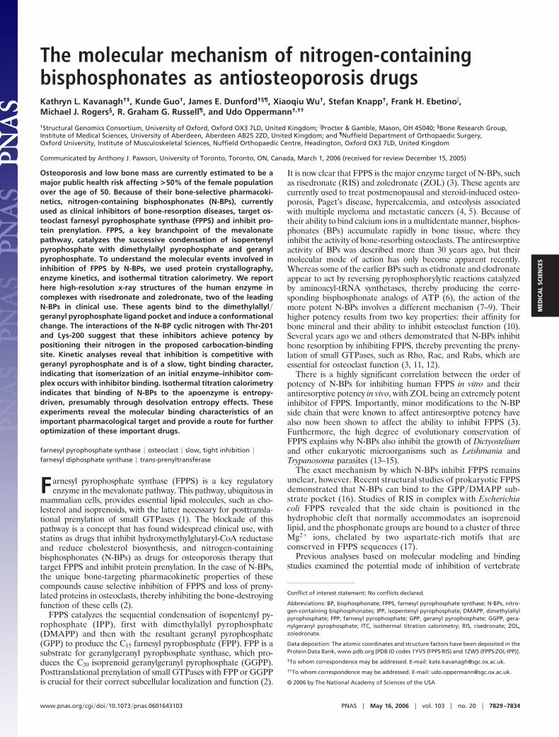
Human FPPS exhibits the all �-helical prenyltransferase folddescribed earlier for the avian form of FPPS (17). FPPS is a dimerwith 13 �-helices and connecting loops per subunit (Fig. 1A).Extended loop regions on the top of the protein include thoseconnecting �4–�5 (residues 107–127) and �8–�9 (residues 249–268). A large central cavity located within the helical bundle formsan elongated partly hydrophobic ligand-binding site. The bottom of
this cavity is delimited by the side chain of Phe-113. Conservedaspartate-rich motifs found in helices �4 (103DDIMD107) and �8(243DDYLD247) face the central cavity, and a pronounced kink inhelix �7 directs the carbonyl of Lys-200 toward the cavity as well.
The BP moieties of RIS and ZOL are ligated through a clusterof three Mg2� ions with phosphonate oxygens, aspartate residues,and water molecules contributing to the octahedral Mg2� coordi-nation (Fig. 1B). Compared with the avian apo structure (ProteinData Bank ID code 1FPS) and the Staphylococcus aureus structure(Protein Data Bank ID code 1RTR), the ligand-bound humanstructures have a more closed conformation (16, 17). This obser-vation suggests that N-BP binding causes a structural rearrange-ment accompanied by a decrease in the size of the internal cavity(Fig. 1C). This movement is likely mediated by the Asp-rich motifson �4 and �8 being drawn together to participate in ligand binding.The region connecting �8–�9 (residues 249–268) creates a lid overthe N-BP and is held in place by several polar interactions: loopresidue Lys-257 contacts Asp-243 and a phosphonate oxygen;Asp-247 at the end of �8 forms bidentate hydrogen bonds withmain-chain amides of Thr-260 and Asp-261; and the main-chainamide of Ile-258 is within hydrogen-bonding distance of Thr-111O�. The heterocyclic ring structures of RIS and ZOL are sur-rounded mainly by hydrophobic side chains of residues Phe-99,Leu-100, Thr-167, Lys-200, and Tyr-204, and the nitrogen atom ofthe ring system is found within hydrogen-bonding distance of O� ofThr-201 (Fig. 2A: RIS, 3.0 Å; ZOL, 2.8 Å) and the carbonyl oxygenof Lys-200 (�3.0 Å).
The production of GPP�FPP is proposed to proceed by athree-step ionization–condensation–elimination reaction in whichthe double bond of IPP attacks the C1 atom of a dimethylallyl�
Fig. 1. Structure of human FPPS. (A) Stereoview of the superimposition of FPPS in complex with RIS and Mg2� (blue) with the avian apo structure 1FPS (gray),made by superimposing the �-carbons in helices �1–�7. (B) Specific polar interactions of RIS and Mg2�. Amino acid numbering in this paper is offset by �14residues compared with the Protein Data Bank depositions. (C) Stereoview detail of RIS binding to FPPS (blue). Three Mg2� ions mediate the interaction betweenphosphates and conserved aspartate residues. A semitransparent avian apo structure is superimposed in gray to highlight the structural differences. (D) Chemicalstructures of RIS and ZOL.
7830 � www.pnas.org�cgi�doi�10.1073�pnas.0601643103 Kavanagh et al.
geranyl carbocation. The carbocation intermediate is predicted tobe stabilized through electrostatic interactions with a magnesium-bound pyrophosphate moiety and through interactions with thecarbonyl of Lys-200 and the side-chain oxygen of Thr-201. TheLys-200 and Thr-201 interactions are analogous to the N-BPnitrogen binding described above. The catalytic base necessary toabstract the IPP 2R proton is likely to be a pyrophosphate oxygenbound through interactions with conserved Arg-112 and Lys-257side chains, located on the flexible loops shielding the active sitefrom bulk solvent during catalysis (16).
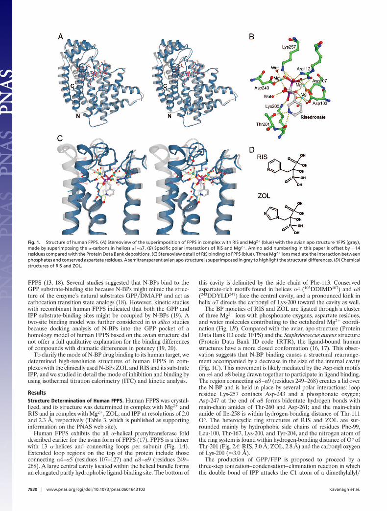
The N-BPs bind in a nearly identical manner, and root-mean-square deviations for �-carbon positions between the RIS and ZOLstructures are 0.44 Å. The largest conformational change involvesthe basic residues at the C terminus, 350KRRK353, which becomeordered at IPP binding. Although they were predicted to bind theIPP phosphates, a direct interaction between these residues and theIPP is not observed. Alternatively, the IPP phosphates are coordi-nated by six water molecules, Gln-96, Arg-113, Arg-60, and Lys-57.Additionally, there is a network of salt links involving Lys-57, theterminal carboxylate of Lys-353, and Arg-351. This conformation ofthe C terminus sequesters the active site from bulk solvent, locks theLys-57 side chain into position to make two polar interactions withthe IPP pyrophosphate moiety, and directs the hydrophobic tail ofIPP toward ZOL (Fig. 2B).
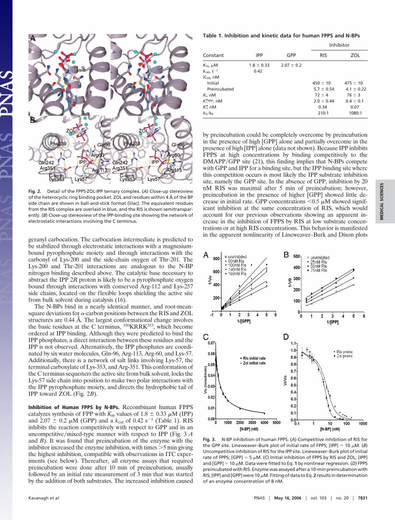
Inhibition of Human FPPS by N-BPs. Recombinant human FPPScatalyzes synthesis of FPP with Km values of 1.8 � 0.33 �M (IPP)and 2.07 � 0.2 �M (GPP) and a kcat of 0.42 s�1 (Table 1). RISinhibits the reaction competitively with respect to GPP and in anuncompetitive�mixed-type manner with respect to IPP (Fig. 3 Aand B). It was found that preincubation of the enzyme with theinhibitor increased the enzyme inhibition, with times �5 min givingthe highest inhibition, compatible with observations in ITC exper-iments (see below). Thereafter, all enzyme assays that requiredpreincubation were done after 10 min of preincubation, usuallyfollowed by an initial rate measurement of 3 min that was startedby the addition of both substrates. The increased inhibition caused
by preincubation could be completely overcome by preincubationin the presence of high [GPP] alone and partially overcome in thepresence of high [IPP] alone (data not shown). Because IPP inhibitsFPPS at high concentrations by binding competitively to theDMAPP�GPP site (21), this finding implies that N-BPs competewith GPP and IPP for a binding site, but the IPP binding site wherethis competition occurs is most likely the IPP substrate inhibitionsite, namely the GPP site. In the absence of GPP, inhibition by 20nM RIS was maximal after 5 min of preincubation; however,preincubation in the presence of higher [GPP] showed little de-crease in initial rate. GPP concentrations �0.5 �M showed signif-icant inhibition at the same concentration of RIS, which wouldaccount for our previous observations showing an apparent in-crease in the inhibition of FPPS by RIS at low substrate concen-trations or at high RIS concentrations. This behavior is manifestedin the apparent nonlinearity of Lineweaver–Burk and Dixon plots
Fig. 2. Detail of the FPPS�ZOL�IPP ternary complex. (A) Close-up stereoviewof the heterocyclic ring binding pocket. ZOL and residues within 4 Å of the BPside chain are shown in ball-and-stick format (lilac). The equivalent residuesfrom the RIS complex are overlaid in blue, and the RIS is shown semitranspar-ently. (B) Close-up stereoview of the IPP-binding site showing the network ofelectrostatic interactions involving the C terminus.
Table 1. Inhibition and kinetic data for human FPPS and N-BPs
Constant IPP GPP
Inhibitor
RIS ZOL
Km, �M 1.8 � 0.33 2.07 � 0.2kcat, s�1 0.42IC50, nM
Initial 450 � 10 475 � 10Preincubated 5.7 � 0.54 4.1 � 0.22
Ki, nM 72 � 4 76 � 3K*i app, nM 2.0 � 0.44 0.4 � 0.1K*i, nM 0.34 0.07k5�k6 210:1 1080:1
Fig. 3. N-BP inhibition of human FPPS. (A) Competitive inhibition of RIS forthe GPP site. Lineweaver–Burk plot of initial rate of FPPS; [IPP] � 10 �M. (B)Uncompetitive inhibition of RIS for the IPP site. Lineweaver–Burk plot of initialrate of FPPS; [GPP] � 5 �M. (C) Initial inhibition of FPPS by RIS and ZOL; [IPP]and [GPP] � 10 �M. Data were fitted to Eq. 1 by nonlinear regression. (D) FPPSpreincubated with RIS. Enzyme was assayed after a 10-min preincubation withRIS; [IPP] and [GPP] were 10 �M. Fitting of data to Eq. 2 results in determinationof an enzyme concentration of 8 nM.
Kavanagh et al. PNAS � May 16, 2006 � vol. 103 � no. 20 � 7831
MED
ICA
LSC
IEN
CES
and, when interpreted as classical linear inhibition, suggested thattwo molecules of inhibitor bind cooperatively. For the abovereasons, it is now apparent that our previous interpretation of thekinetic data (19) was incorrect. Given that the equilibrium amongenzyme, inhibitor, and substrate takes many minutes to be achievedand that the inhibition of FPPS by RIS occurs at such a low inhibitorconcentration (�50% inhibition of 10 nM FPPS by 10 nM RIS), theinhibition mechanism can be considered to be ‘‘slow, tight binding’’rather than classical competitive. Because the N-BP can act as aclassic competitive inhibitor in the early stages of the enzymereaction and increases in potency after preincubation, a two-stepreaction scheme involving a rate-limiting isomerization step fits theobserved enzyme behavior best. The vast majority of knownexamples of slow, tight binding adhere to this mechanism (22).
Based on our observations of the kinetics of inhibition of FPPSwe considered the following scheme. First, the inhibitor appears tocompete directly with GPP, to form an enzyme–inhibitor complex.This binding step occurs rapidly and causes inhibition. A secondstep involving the enzyme–inhibitor complex undergoing somechange, possibly an isomerization, occurs slowly and increases theinhibition of the enzyme over time (22), which gives rise to thefollowing mechanism:
E � I L|;k3
k4
EIL|;k5
k6
EI*.
The Ki for the first step is the dissociation constant for E�I. Theoverall dissociation constant is K*i, and k6 is the rate constant forconversion of FPPS�RIS* back to FPPS�RIS.
The calculated values of Ki suggest that both RIS and ZOLinitially bind the enzyme with a similar affinity (Table 1). Values forK*i indicate that given enough time to allow formation of the E�I*complex, inhibition is increased significantly, with ZOL beingsomewhat more potent than RIS. The isomerization constant k5�k6gives an indication of how likely the FPPS�RIS* complex is to returnto the FPPS�RIS state. Using the values of Ki and K*i calculatedabove, the value of k5�k6 was found to be 210:1 for RIS and 1080:1for ZOL, which suggests that the equilibrium lies on the side of theFPPS�N-BP* complex, even more so with ZOL than with RIS(accounting for the greater overall potency of ZOL).
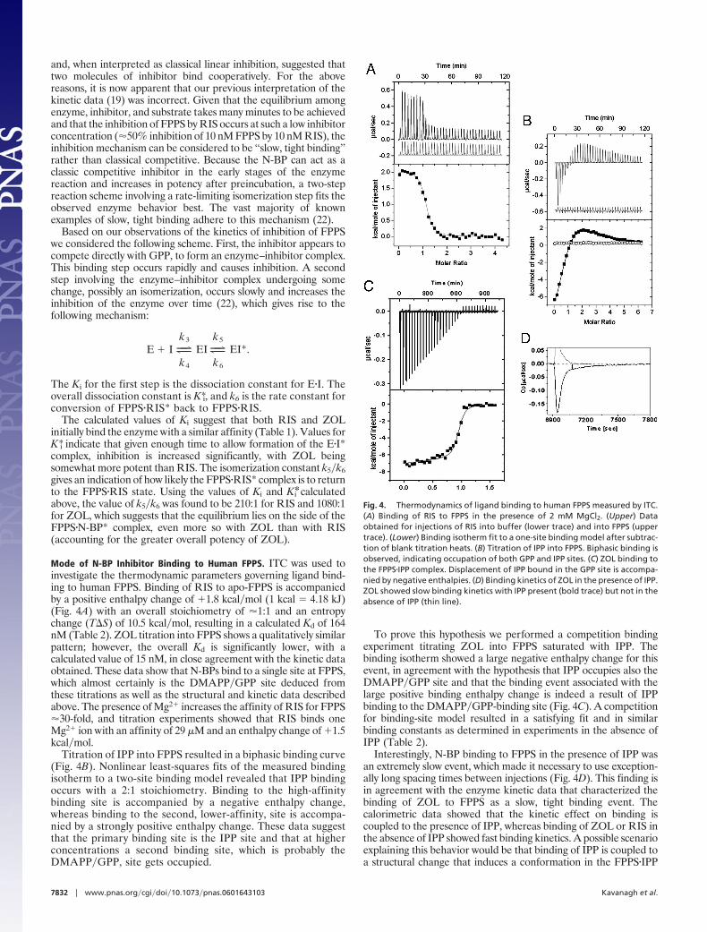
Mode of N-BP Inhibitor Binding to Human FPPS. ITC was used toinvestigate the thermodynamic parameters governing ligand bind-ing to human FPPS. Binding of RIS to apo-FPPS is accompaniedby a positive enthalpy change of �1.8 kcal�mol (1 kcal � 4.18 kJ)(Fig. 4A) with an overall stoichiometry of �1:1 and an entropychange (T�S) of 10.5 kcal�mol, resulting in a calculated Kd of 164nM (Table 2). ZOL titration into FPPS shows a qualitatively similarpattern; however, the overall Kd is significantly lower, with acalculated value of 15 nM, in close agreement with the kinetic dataobtained. These data show that N-BPs bind to a single site at FPPS,which almost certainly is the DMAPP�GPP site deduced fromthese titrations as well as the structural and kinetic data describedabove. The presence of Mg2� increases the affinity of RIS for FPPS�30-fold, and titration experiments showed that RIS binds oneMg2� ion with an affinity of 29 �M and an enthalpy change of �1.5kcal�mol.
Titration of IPP into FPPS resulted in a biphasic binding curve(Fig. 4B). Nonlinear least-squares fits of the measured bindingisotherm to a two-site binding model revealed that IPP bindingoccurs with a 2:1 stoichiometry. Binding to the high-affinitybinding site is accompanied by a negative enthalpy change,whereas binding to the second, lower-affinity, site is accompa-nied by a strongly positive enthalpy change. These data suggestthat the primary binding site is the IPP site and that at higherconcentrations a second binding site, which is probably theDMAPP�GPP, site gets occupied.
To prove this hypothesis we performed a competition bindingexperiment titrating ZOL into FPPS saturated with IPP. Thebinding isotherm showed a large negative enthalpy change for thisevent, in agreement with the hypothesis that IPP occupies also theDMAPP�GPP site and that the binding event associated with thelarge positive binding enthalpy change is indeed a result of IPPbinding to the DMAPP�GPP-binding site (Fig. 4C). A competitionfor binding-site model resulted in a satisfying fit and in similarbinding constants as determined in experiments in the absence ofIPP (Table 2).
Interestingly, N-BP binding to FPPS in the presence of IPP wasan extremely slow event, which made it necessary to use exception-ally long spacing times between injections (Fig. 4D). This finding isin agreement with the enzyme kinetic data that characterized thebinding of ZOL to FPPS as a slow, tight binding event. Thecalorimetric data showed that the kinetic effect on binding iscoupled to the presence of IPP, whereas binding of ZOL or RIS inthe absence of IPP showed fast binding kinetics. A possible scenarioexplaining this behavior would be that binding of IPP is coupled toa structural change that induces a conformation in the FPPS�IPP
Fig. 4. Thermodynamics of ligand binding to human FPPS measured by ITC.(A) Binding of RIS to FPPS in the presence of 2 mM MgCl2. (Upper) Dataobtained for injections of RIS into buffer (lower trace) and into FPPS (uppertrace). (Lower) Binding isotherm fit to a one-site binding model after subtrac-tion of blank titration heats. (B) Titration of IPP into FPPS. Biphasic binding isobserved, indicating occupation of both GPP and IPP sites. (C) ZOL binding tothe FPPS�IPP complex. Displacement of IPP bound in the GPP site is accompa-nied by negative enthalpies. (D) Binding kinetics of ZOL in the presence of IPP.ZOL showed slow binding kinetics with IPP present (bold trace) but not in theabsence of IPP (thin line).
7832 � www.pnas.org�cgi�doi�10.1073�pnas.0601643103 Kavanagh et al.

complex that is not adequate for ZOL or RIS binding. Theextremely large positive enthalpy change associated with the occu-pation of DMAPP�GPP site by IPP supports such a structuralrearrangement on IPP binding.
DiscussionThe structural changes occurring in FPPS with N-BP binding canbe seen by three methods: (i) preincubation of FPPS with N-BPsincreases the degree of inhibition; (ii) long equilibration times arerequired between ITC injections; and (iii) a more closed confor-mation is observed in the N-BP-bound crystal structures comparedwith apo-FPPS structures. Binding of N-BPs is magnesium-dependent and occurs in the GPP�DMAPP site. The cyclic nitro-gen interactions linking ZOL and RIS with Thr-201 and Lys-200support the hypothesis that N-BPs gain potency by positioning theirnitrogen in the proposed carbocation-binding site.
A second conformational change is revealed by comparing theN-BP-bound crystal structures. N-BP binding induces a closedconformation, creates a more ordered IPP-binding site, and pro-vides a surface for IPP to pack against. The second isomerizationoccurs once IPP is bound and entails the C terminus curling backtoward the pyrophosphate moiety, securing the ligands into positionand sequestering the site from bulk solvent.
Structure determination, detailed kinetic analysis of N-BP inhi-bition, and the thermodynamic characterization of ligand binding tohuman FPPS provide a unique framework for understanding themode of action of this widely used, important class of antiosteo-porosis drugs. From both kinetic and thermodynamic experimentsit is evident that RIS and ZOL binding to FPPS is a slow, tightbinding process, accompanied by large positive enthalpies whenbinding to apo-FPPS. These enthalpic components describe largelythe ligand–enzyme interactions, and it is likely that these positiveenthalpic changes are derived from breaking polar interactions withwater molecules both within the enzyme active site and around theligand. This ‘‘counterproductive’’ event is more than compensatedby the gain in entropy. Entropy changes reflect two distinct con-tributions: changes in solvation entropy and changes in conforma-tional entropy (23, 24). The structural changes occurring withoccupation of the DMAPP�GPP site, i.e., closing of the active-sitecavity, reduce the large solvation entropy effect occurring at theligand site, but the overall change is still large enough to drivehigh-affinity binding, likely by the primarily hydrophobic ringsystem of the N-BPs. Another example of this unusual drug-bindingbehavior is given by the first generation of HIV-1 protease inhib-itors, displaying enthalpically unfavorable characteristics, with bind-ing driven by large, positive entropy changes (25).
In titration experiments of N-BPs to apo-FPPS and FPPS�IPPcomplexes, the slow component in binding to FPPS is observed forN-BPs but not for IPP, indicating that specific rearrangementsoccur around the N-BP site. The exact nature of the slow compo-nent remains obscure; however, it is possible that any of theligand-induced changes (i.e., desolvation effects and structuralchanges within FPPS) forms the basis for the slow binding kinetics.
On the basis of the data described, it appears feasible to increasebinding affinity by designing novel N-BPs targeted to human FPPS.These N-BPs could be conformationally restrained, partly hydro-phobic, with functional groups branching further into the DMAPP�GPP site. This site has additional polar residues (Tyr-204 andThr-167) that could serve as interacting residues for a pharma-cophore model. These features would possibly enhance both theentropy and enthalpy effects to drive N-BP ligand binding.
During the course of the evaluation of this paper, an independentreport on structure determination of human FPPS was released(26), confirming our conclusions on the structural basis of N-BPbinding.
MethodsExpression and Purification of Recombinant Human FPPS. A cloneencoding human FPPS residues 1–353 (P14324) as an N-terminallyHis6-tagged fusion protein with a tobacco etch virus (TEV) pro-tease cleavage site was expressed in E. coli BL21(DE3). Cells werelysed by using a high-pressure cell disruptor, and the protein waspurified to near homogeneity by using nickel nitrilotriacetate resin(Qiagen). Initial crystallization experiments were performed di-rectly with this protein. For further experiments, the histidine tagwas removed by incubation with TEV protease, and gel filtrationchromatography was performed with a Superdex 200 column(GE�Amersham Pharmacia).
Crystallization and Data Collection of Human FPPS�RIS. FPPS includ-ing the N-terminal tag was concentrated to 13 mg�ml in 10 mMHepes (pH 7.5)�0.5 M NaCl�5% (vol�vol) glycerol�2 mM RIS�2mM MgCl2. Crystals were grown at 20°C in sitting drops by mixing100 nl of protein solution and 50 nl of precipitant consisting of 40%(vol�vol) polyethylene glycol 300 and 0.1 M phosphate�citratebuffer, pH 4.2. Diffraction data were collected from a single crystalat 100 K at the Advanced Light Source Beamline 8.3.1 (LawrenceBerkeley Laboratory, Berkeley, CA).
Crystallization and Data Collection of Human FPPS�ZOL�IPP. Tobaccoetch virus protease-cleaved FPPS was concentrated to 16 mg�ml,and ZOL, IPP, and MgCl2 were added to final concentrations of 2
Table 2. Thermodynamic data determined by isothermal titration calorimetry
Sample Kb, 106 M�1 Kd, �M�H,
kcal�molT�S,
kcal�mol�G,
kcal�mol n
RIS�Mg* 6.1 � 2 0.16 �1.8 � 0.4 10.5 �8.8 1.14RIS† 0.2 � 0.5 5 �3.9 � 0.2 10.9 �7 1.2Mg RIS‡ 0.035 � 0.004 290 �1.5 � 0.04 6.2 �4.7 0.99ZOL�Mg§ 64 � 2.0 0.015 �2.1 � 0.03 12.1 �10 0.93IPP 1¶ 0.33 � 0.06 3 �13.7 � 1.3 �6.6 �7.1 0.85IPP 2� 0.068 � 0.008 15 �13.4 � 1.3 19.7 �6.3 1.2ZOL�IPP** 55 � 1.2 0.018 �2.4 � 2 ND ND 1RIS�IPP†† 30 � 10 0.03 �4.4 � 2 ND ND 1.1
ND, not determined.*RIS titrated into FPPS with Mg2� present in both samples.†RIS titrated into FPPS in the absence of Mg2�.‡Mg2� titrated into RIS (no FPPS present).§ZOL titrated into FPPS with Mg2� present in both samples.¶IPP titrated into FPPS with Mg2� present in both samples, higher-affinity site.�IPP titrated into FPPS with Mg2� present in both samples, lower-affinity site.**ZOL titrated into an IPP-saturated FPPS, Mg2� present in both samples.††RIS titrated into an IPP-saturated FPPS, Mg2� present in both samples.
Kavanagh et al. PNAS � May 16, 2006 � vol. 103 � no. 20 � 7833
MED
ICA
LSC
IEN
CES
mM each. Crystals were grown at 20°C in sitting drops by mixing150 nl of protein solution and 150 nl of precipitant consisting of 14%(vol�vol) polyethylene glycol 6000, 0.7 M LiCl, and 70 mM citrate,(pH 4.0). A single crystal was transferred to a solution composedof 20% (vol�vol) polyethylene glycol 300 and 80% well solution andflash-cooled in liquid nitrogen. Diffraction data were collected atthe Swiss Light Source Beamline X10SA (Paul Scherrer Institute,Villigen, Switzerland).
Data Processing and Refinement. The data sets were processed withMOSFLM and SCALA (27). Initial phases for the FPPS�RIS complexwere calculated by molecular replacement implemented in PHASER(28) using the avian FPPS structure 1UBV as a search model (20).The FPPS�RIS structure minus ligands and solvent molecules wasused as a starting model for the FPPS�ZOL�IPP structure. Iterativerounds of model building in COOT (29) and refinement usingREFMAC5 (30) resulted in the final models for which statisticsappear in Table 3.
Kinetics and Inhibition of Human FPPS by N-BPs. FPPS was assayed bythe method of Reed and Rilling with modifications (31). For kineticanalysis, 40 ng of pure FPPS was assayed in a final volume of 100�l buffer containing 50 mM Tris (pH 7.7), 2 mM MgCl2, 1 mMtris(2-carboxyethyl)phosphine, and 5 �g�ml BSA. The concentra-tions of GPP and IPP ([14C]IPP, 400 kBq��mol) were as describedin the text but were typically 0.2–20 �M. Reactions were startedwith the addition of enzyme and allowed to proceed for anappropriate period at 37°C. When a preincubation of the enzymewith the inhibitor was required, the enzyme was incubated withinhibitor in a 60-�l volume. After 10 min, 40 �l of substrate in waterwas added to start the reaction. Assays were terminated by theaddition of 0.2 ml of concentrated HCl�methanol (1:4) and incu-bated for 10 min at 37°C. The reaction mixtures were then extractedwith 0.4 ml of ligroin, and the amount of radioactivity in the upperphase was determined by mixing 0.2 ml of the ligroin with 4 ml ofgeneral-purpose scintillant. The radioactivity was measured byusing a Packard Tri-Carb 1900CA scintillation counter. At theconcentration of FPPS used, half the amount of enzyme gives halfthe activity. Data were fitted to the following kinetic models bynonlinear regression using PRISM (GraphPad, San Diego).
To calculate the Ki for the formation of the E�I complex, theenzyme inhibition data from initial rate experiments were fitted tothe equation for classical linear competitive inhibition (22):
Vo �V[GPP]
Km,GPP� 1 �[I]K i� � [GPP]
. [1]
Data from experiments that included preincubation of enzymeand inhibitor were fitted to the following equation (32):
Vi
Vo�
1 �[E] � [I] � K*i app� � �[E] � [I] � K*i app�2 � 4[E][I]
2[E].
[2]
The overall dissociation constant, K*i, was calculated from K*i app
by using the model for competitive inhibition (32):
K*i �K*i app
1 �[GPP]Km,GPP
. [3]
The isomerization constant, k5�k6, was calculated by using thefollowing equation (32):
k5
k6�
Ki � K*i�K*i
. [4]
ITC. Calorimetric measurements were carried out by using a VP-ITCtitration calorimeter from Microcal (Amherst, MA). Samples weredialyzed extensively against 10 mM Hepes (pH 7.5)�100 mM NaCl.Each titration experiment consisted of a first (5-�l) injection of theligand followed by 8-�l injections. Heats of dilution were measuredin blank titrations by injecting the ligand into buffer, and thedilution heats were subtracted from the binding heats. Data wereanalyzed by using single and multiple binding site models imple-mented in the ORIGIN software package (OriginLab, Northampton,MA) provided with the instrument. Competition experiments wereanalyzed by using a competition for binding site model (33)implemented in the software package.
We thank Frank von Delft at the Swiss Light Source for data collection.The DNA was amplified from I.M.A.G.E. Consortium Clone ID4132071. The Structural Genomics Consortium is a registered charity(Number 1097737) funded by the Wellcome Trust, GlaxoSmithKline,Genome Canada, the Canadian Institutes of Health Research, theOntario Innovation Trust, the Ontario Research and DevelopmentChallenge Fund, the Canadian Foundation for Innovation, Vinnova, theSwedish Strategic Research Foundation, the Knut and Alice WallenbergFoundation, and the Karolinska Institutet.
1. Goldstein, J. L. & Brown, M. S. (1990) Nature 343, 425–430.2. Rogers, M. J. (2003) Curr. Pharm. Des. 9, 2643–2658.3. Dunford, J. E., Thompson, K., Coxon, F. P., Luckman, S. P., Hahn, F. M., Poulter, C. D.,
Ebetino, F. H. & Rogers, M. J. (2001) J. Pharmacol. Exp. Ther. 296, 235–242.4. Rodan, G. A. & Reszka, A. A. (2002) Curr. Mol. Med. 2, 571–577.5. Russell, R. G. & Rogers, M. J. (1999) Bone 25, 97–106.6. Frith, J. C., Monkkonen, J., Auriola, S., Monkkonen, H. & Rogers, M. J. (2001) Arthritis
Rheum. 44, 2201–2210.7. Fisher, J. E., Rogers, M. J., Halasy, J. M., Luckman, S. P., Hughes, D. E., Masarachia, P. J.,
Wesolowski, G., Russell, R. G., Rodan, G. A. & Reszka, A. A. (1999) Proc. Natl. Acad. Sci.USA 96, 133–138.
8. Luckman, S. P., Hughes, D. E., Coxon, F. P., Graham, R., Russell, G. & Rogers, M. J. (1998)J. Bone Miner. Res. 13, 581–589.
9. van Beek, E., Lowik, C., van der Pluijm, G. & Papapoulos, S. (1999) J. Bone Miner. Res. 14,722–729.
10. van Beek, E. R., Lowik, C. W., Ebetino, F. H. & Papapoulos, S. E. (1998) Bone 23, 437–442.11. Bergstrom, J. D., Bostedor, R. G., Masarachia, P. J., Reszka, A. A. & Rodan, G. (2000) Arch.
Biochem. Biophys. 373, 231–241.12. van Beek, E., Pieterman, E., Cohen, L., Lowik, C. & Papapoulos, S. (1999) Biochem. Biophys.
Res. Commun. 255, 491–494.13. Martin, M. B., Grimley, J. S., Lewis, J. C., Heath, H. T., III, Bailey, B. N., Kendrick, H.,
Yardley, V., Caldera, A., Lira, R., Urbina, J. A., et al. (2001) J. Med. Chem. 44, 909–916.14. Sanders, J. M., Gomez, A. O., Mao, J., Meints, G. A., Van Brussel, E. M., Burzynska, A.,
Kafarski, P., Gonzalez-Pacanowska, D. & Oldfield, E. (2003) J. Med. Chem. 46, 5171–5183.15. Szabo, C. M., Martin, M. B. & Oldfield, E. (2002) J. Med. Chem. 45, 2894–2903.16. Hosfield, D. J., Zhang, Y., Dougan, D. R., Broun, A., Tari, L. W., Swanson, R. V. & Finn,
J. (2004) J. Biol. Chem. 279, 8526–8529.
17. Tarshis, L. C., Yan, M., Poulter, C. D. & Sacchettini, J. C. (1994) Biochemistry 33,10871–10877.
18. Martin, M. B., Arnold, W., Heath, H. T., III, Urbina, J. A. & Oldfield, E. (1999) Biochem.Biophys. Res. Commun. 263, 754–758.
19. Ebetino, F. H., Roze, C. N., McKenna, C. E., Barnett, B. L., Dunford, J. E., Russell, R. G. G.,Mieling, G. E. & Rogers, M. J. (2005) J. Organomet. Chem. 690, 2679–2687.
20. Tarshis, L. C., Proteau, P. J., Kellogg, B. A., Sacchettini, J. C. & Poulter, C. D. (1996) Proc.Natl. Acad. Sci. USA 93, 15018–15023.
21. Barnard, G. F. & Popjak, G. (1981) Biochim. Biophys. Acta 661, 87–99.22. Morrison, J. F. & Walsh, C. T. (1988) Adv. Enzymol. Relat. Areas Mol. Biol. 61,
201–301.23. Velazquez-Campoy, A., Kiso, Y. & Freire, E. (2001) Arch Biochem. Biophys. 390,
169–175.24. Velazquez-Campoy, A. & Freire, E. (2001) J. Cell Biochem. Suppl. 37, 82–88.25. Velazquez-Campoy, A., Luque, I., Todd, M. J., Milutinovich, M., Kiso, Y. & Freire, E.
(2000) Protein Sci. 9, 1801–1809.26. Rondeau, J. M., Bitsch, F. M., Boirgier, E., Geiser, M., Hemmig, R., Kroemer, M., Lehmann,
S., Ramage, P., Rieffel, S., Strauss, A., et al. (2006) Chem. Med. Chem. 1, 267–273.27. Collaborative Computational Project 4 (1994) Acta Crystallogr. D 50, 760–763.28. Storoni, L. C., McCoy, A. J. & Read, R. J. (2004) Acta Crystallogr. D 60, 432–438.29. Emsley, P. & Cowtan, K. (2004) Acta Crystallogr. D 60, 2126–2132.30. Murshudov, G. N. (1997) Acta Crystallogr. D 53, 240–255.31. Reed, B. C. & Rilling, H. C. (1976) Biochemistry 15, 3739–3745.32. Copeland, R. A. (2000) Enzymes: A Practical Guide to Structure, Mechanism, and Data
Analysis (Wiley, New York), pp. 305–349.33. Sigurskjold, B. W. (2000) Anal. Biochem. 277, 260–266.
7834 � www.pnas.org�cgi�doi�10.1073�pnas.0601643103 Kavanagh et al.