the mri study of normal brain development mri study of normal brain development ... developed as...
TRANSCRIPT
The MRI Study of normal Brain Development Page 1 of 27 Protocol Release Document Version: November 2006
The MRI Study of Normal Brain Development
November 2006
To Interested Researchers,
Thank you for your interest in the protocol for the multi-site study: MRI Study of Normal Brain Development. The NIH and the investigators of the Steering Committee welcome the opportunity to share the protocol with other investigators doing related work.
The attached protocol has been developed as a collaborative effort of the seven involved pediatric study centers and the two coordinating centers
Please complete the attached registration form so we can be aware of who is interested in or implementing portions of this protocol. This registration will be used to send updated information in the future. Thank-you again for your interest! Sincerely, Kelly Botteron M.D. Principal Investigator Clinical Coordinating Center Alan Evans, Ph.D. Principal Investigator Data Coordinating Center
National Institute of Neurological Disorders
and Stroke National Institute on Drug Abuse
National Institute of Mental Health National Institute of
Child Health and Human Development
National Institutes of Health
United States Department of Health and Human Services
The MRI Study of normal Brain Development Page 2 of 27 Protocol Release Document Version: November 2006
Lisa Freund, Ph.D. Project Officer National Institute of Child Health and Human Development Katrina Gwinn-Hardy, MD Project Officer National Institute of Neurological Disorders and Stroke Judith Rumsey, Ph. D. Project Officer National Institute of Mental Health Laurence Stanford, Ph. D. Project Officer National Institute on Drug Abuse Karen Sirocco Ph. D. Project Officer National Institute on Drug Abuse
The MRI Study of normal Brain Development Page 3 of 27 Protocol Release Document Version: November 2006
The MRI Study of Normal Brain Development
Sponsored By
The National Institute of Child Health and Human Development The National Institute on Drug Abuse
The National Institute of Mental Health The National Institute of Neurological Disorders and Stroke
The MRI Study of normal Brain Development Page 4 of 27 Protocol Release Document Version: November 2006
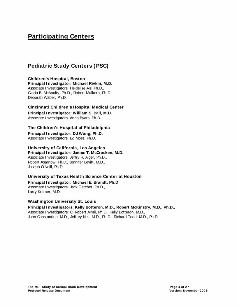
Participating Centers
Pediatric Study Centers (PSC) Children's Hospital, Boston Principal Investigator: Michael Rivkin, M.D. Associate Investigators: Heidelise Als, Ph.D., Gloria B. McAnulty, Ph.D., Robert Mulkern, Ph.D. Deborah Waber, Ph.D.
Cincinnati Children’s Hospital Medical Center Principal Investigator: William S. Ball, M.D. Associate Investigators: Anna Byars, Ph.D.
The Children's Hospital of Philadelphia Principal Investigator: DJ Wang, Ph.D. Associate Investigators: Ed Moss, Ph.D. University of California, Los Angeles Principal Investigator: James T. McCracken, M.D. Associate Investigators: Jeffry R. Alger, Ph.D., Robert Asarnow, Ph.D., Jennifer Levitt, M.D., Joseph O'Neill, Ph.D.
University of Texas Health Science Center at Houston Principal Investigator: Michael E. Brandt, Ph.D. Associate Investigators: Jack Fletcher, Ph.D., Larry Kramer, M.D.
Washington University St. Louis Principal Investigators: Kelly Botteron, M.D., Robert McKinstry, M.D., Ph.D., Associate Investigators: C. Robert Almli, Ph.D., Kelly Botteron, M.D., John Constantino, M.D., Jeffrey Neil, M.D., Ph.D., Richard Todd, M.D., Ph.D.
The MRI Study of normal Brain Development Page 5 of 27 Protocol Release Document Version: November 2006
Clinical Coordinating Center (CCC) Washington University St. Louis Principal Investigator: Kelly Botteron, M.D., Associate Investigator: C. Robert Almli, Ph.D.
Data Coordinating Center (DCC) Montreal Neurological Institute Principal Investigator: Alan Evans, Ph.D. Associate Investigators: Louis Collins, Ph.D., Gabriel Leonard, Ph.D., Tomas Paus, M.D., Ph.D., Bruce Pike, Ph.D., Alex Zijdenbos, Ph.D. Harvard University/McLean Hospital Associate Investigator: Nicholas Lange, Sc.D. Georgetown Associate Investigator: Thomas Zeffiro, M.D., Ph.D.
Diffusion Tensor Processing Center (DPC) National Institute of Child Health and Human Development, NIH Principal Investigator: Carlo Pierpaoli, M.D. Ph.D.
Spectroscopy Processing Center (SPC) University of California, Los Angeles Principal Investigator: James T. McCracken , M.D. Associate Investigators: Jeffry R. Alger, Ph.D., Jennifer Levitt, M.D., Joseph O'Neill, Ph.D.
The MRI Study of normal Brain Development Page 6 of 27 Protocol Release Document Version: November 2006
GENERAL OVERVIEW
Introduction
The purpose of the study is to collect a representative sample of normal, healthy infants and children for a magnetic resonance imaging study that will serve two purposes: 1) to provide the largest normative database to date of the developing human brain for comparison with brain scan studies of children with neurological, developmental, and psychiatric disorders; and 2) provide longitudinal data for investigating brain maturation in relationship to behavioral and cognitive development in a normal sample. Such data will allow a greater understanding of deviations in brain structural development associated with pediatric brain disorders.
The study involves six different sites in the United States, as well as a Data Coordinating Center (DCC) in Montreal, Canada, and a Clinical Coordinating Center (CCC) in St. Louis, Missouri. The U.S. sites include three Children’s Hospitals (Boston, Cincinnati and Philadelphia), and Washington University in St. Louis, the University of Texas Health Science Center in Houston, and the University of California in Los Angeles. A seventh center, the University of California in Irvine, was initially involved in recruiting subjects who were subsequently followed at UCLA.
These sites will collaborate in the recruitment of a representative sample of approximately 546
children, ages 10 days through 18 years 3 months (at first scan), who will be studied using anatomic magnetic resonance imaging (aMRI), diffusion tensor imaging (DTI), magnetic resonance spectroscopy (MRS) and behavioral testing at three (or more) different time points over a five to six year period. The children will be scanned with techniques that will provide state of the art information concerning the structural and metabolic development of the brain, including the development of specific structures. Developmental changes in the brain and in specific regions of the brain will be related to maturational changes in behavior and cognition. Image analysis tools sensitive to brain developmental changes will be developed as part of this effort. A goal is to develop growth curves for various aspects of brain development.
The information gained will be made widely available to researchers and clinicians. This will
permit physicians and researchers to determine possible differences in brain structure and metabolism in children with known or suspected brain-based problems. The information and tools produced may also allow physicians to track brain-related changes associated with disease and to evaluate the effects of various treatments on the brain.
The MRI Study of normal Brain Development Page 7 of 27 Protocol Release Document Version: November 2006
Objectives
The Pediatric MRI study of Normal Brain Development has two main objectives and two ancillary objectives. Objective 1: Anatomic MRI and cognitive/behavioral cross-sectional and longitudinal studies of children between the ages 4 years, 6 months and 18 years, 3 months (at time of first scan). Objective 2: Anatomic MRI and cognitive/behavioral cross-sectional and longitudinal studies of children between the age of newborn (10-14 days) and 4 years, 5 months (at time of first scan). Ancillary A: MR Spectroscopy (MRS) of children between the ages of newborn (10-14 days) and 18 years, 3 months (at time of first scan). Ancillary B: Diffusion Tensor Imaging (DTI) of children between the ages of newborn (10-14 days) and 4 years, 5 months (at time of first scan). However, this was broadened to include subjects of all ages. An expanded DTI (eDTI) protocol was developed and acquisitions began in early 2006.
The MRI Study of normal Brain Development Page 8 of 27 Protocol Release Document Version: November 2006
Inclusion Criteria
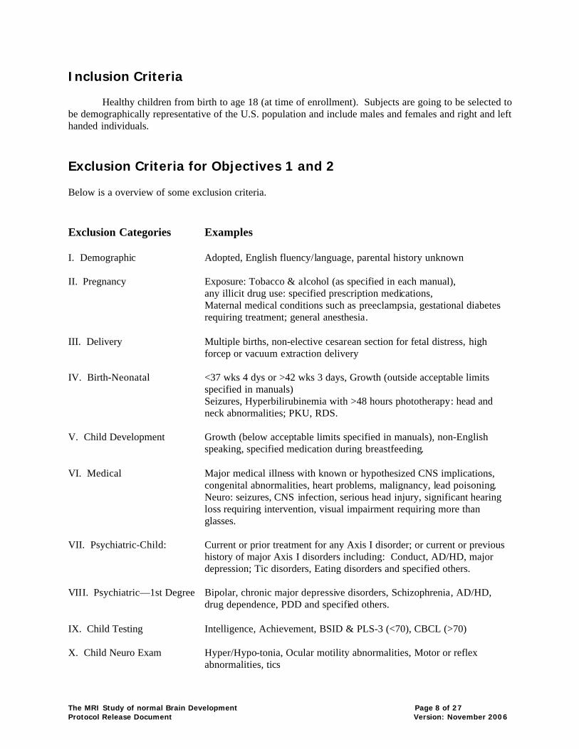
Healthy children from birth to age 18 (at time of enrollment). Subjects are going to be selected to be demographically representative of the U.S. population and include males and females and right and left handed individuals. Exclusion Criteria for Objectives 1 and 2 Below is a overview of some exclusion criteria.
Exclusion Categories Examples I. Demographic Adopted, English fluency/language, parental history unknown II. Pregnancy Exposure: Tobacco & alcohol (as specified in each manual),
any illicit drug use: specified prescription medications, Maternal medical conditions such as preeclampsia, gestational diabetes requiring treatment; general anesthesia.
III. Delivery Multiple births, non-elective cesarean section for fetal distress, high
forcep or vacuum extraction delivery IV. Birth-Neonatal <37 wks 4 dys or >42 wks 3 days, Growth (outside acceptable limits
specified in manuals) Seizures, Hyperbilirubinemia with >48 hours phototherapy: head and neck abnormalities; PKU, RDS.
V. Child Development Growth (below acceptable limits specified in manuals), non-English
speaking, specified medication during breastfeeding. VI. Medical Major medical illness with known or hypothesized CNS implications,
congenital abnormalities, heart problems, malignancy, lead poisoning. Neuro: seizures, CNS infection, serious head injury, significant hearing loss requiring intervention, visual impairment requiring more than glasses.
VII. Psychiatric-Child: Current or prior treatment for any Axis I disorder; or current or previous history of major Axis I disorders including: Conduct, AD/HD, major depression; Tic disorders, Eating disorders and specified others.
VIII. Psychiatric—1st Degree Bipolar, chronic major depressive disorders, Schizophrenia, AD/HD,
drug dependence, PDD and specified others. IX. Child Testing Intelligence, Achievement, BSID & PLS-3 (<70), CBCL (>70) X. Child Neuro Exam Hyper/Hypo-tonia, Ocular motility abnormalities, Motor or reflex
abnormalities, tics
The MRI Study of normal Brain Development Page 9 of 27 Protocol Release Document Version: November 2006
OBJECTIVE 1 Subjects
Children between the ages of 4 years, 6 months and 18 years, 3 months will be recruited across sites to reflect the current demographic composition of the U.S. population with respect to gender, socioeconomic status, and race/ethnicity. Approximately 440 children will participate in Objective 1 with equal distribution across ages and gender. Subjects will be recruited from the general metropolitan area associated with each study site through lists provided by a national marketing agency.
An agency will provide mailing lists of household addresses within various income levels, known to have children between specified ages. A subset of the subjects at selected sites will participate in an additional MRS study that may require a second scanning session. Recruitment and Screening
Briefly, the following recruitment steps are followed: 1) Introductory letters discussing the study are mailed to families obtained from the agency as outlined above. 2) Families are screened by telephone interviews, and then 3) completion of parent Childhood Behavior Checklist (CBCL) forms, ages 4:6 – 18:3). If they pass these initial screening procedures, they are followed by structured interviews as outlined below.
The Full Telephone Screening Interview will consist of the administration of a medical and
developmental history phone interview. If the subject remains eligible, a structured psychiatric interview (the Diagnostic Interview Schedule for Children (C-DISC4) for children >7:00 yrs), and the Family History Interview for Genetic Studies (FIGS-MRI) will be administered to the parent. Children and adolescents over 10:00 years of age will complete the related interview to the C-DISC – the Diagnostic Predictive Scales (DPS-4). If diagnoses are indicated on the DPS-4, the corresponding C-DISC4 modules will be administered. Following this and assuming all inclusion criteria have been reviewed and met, an appointment for an on-site evaluation and MRI will be made with eligible families.
On-Site Evaluations
The on-site evaluation and MR scans may require up to two days. Standardized Neurological and Physical Exam A physician will conduct a physical/neurological examination. A standardized format was
developed by a consensus process with neurologists associated with the study. Tanner Staging Questionnaire when appropriate Carskadon and Acebo, J Adol Health. 1993;14:190-195, Peterson AC et al. A Self-Report Measure of Pubertal Status: Reliability, Validity and Initial Norms. J Youth Adolesc. 1988;17:117-133. Two saliva samples for the measurement of gonadal steroids as a more sensitive indicator of stage
of maturation than chronological age will be collected. In addition, a urine sample will be collected for
The MRI Study of normal Brain Development Page 10 of 27 Protocol Release Document Version: November 2006
endocrine measurements, which will complement Tanner staging and growth data. Adrenal and gonadal steroids will be measured in saliva and urine (under direction of J.McCracken – UCLA).
The following testing will be completed as screening tests & subject characterization:
Wechsler Abbreviated Scale of Intelligence (WASI) (ages 6:00 – 24:00) 4 subtests: Vocabulary, Similarities, Block Design, Matrix Reasoning WISC-III (Wechsler Intelligence Scale for Children – 3rd edition)
Coding A (ages 6:00-7:11) Coding B (ages 8:00-16:11) Digit Span (ages 6:00-16:11)
or WAIS-R (Wechsler Adult Intelligence Scale – Revised ) (ages 17:00 +) Digit Symbol Digit Span
Woodcock-Johnson Psycho-Educational Battery-III – 3 Subtests (Ages 4:6 to adult) Passage Comprehension Letter-Word Identification Calculation
Differential Ability Scales (DAS) (ages 4:6-5:11) Subtests: Verbal ability –
Verbal comprehension, Picture similarities, Naming vocabulary
Nonverbal ability – Pattern construction, Early number concepts, Copying
The following battery of neuropsychological tests will be completed for purposes of brain-behavior correlations:
Rating Scales – Behavior Rating Inventory (BRIEF) – parent completed Ages 4:6 - adult Junior Temperament and Character Inventory (JTCI)
ages 4:6 –15:11 - parent completed (Parent JTCI) ages 10:00 –15:11 – child completed (JTCI) ages 16:00 to adult – parent & adolescent completed versions of TCI (Temperament and Character Inventory)
Administered Tests: California Verbal Learning Test (CVLT) CVLT-C (ages 4:6-15:11) CVLT-II (ages 16:00 to adult

The MRI Study of normal Brain Development Page 11 of 27 Protocol Release Document Version: November 2006
Handedness Ages 4:6-5:11 12 item performance scale (non published) Ages 6:00 – adult 8 item pantomime performance scale (non published) (contact Botteron for further details if you are interested in these scales) NEPSY – Verbal fluency Semantic (ages 4:6-adult) Phonemic – “F,A,S” (ages 7:00-adult)
Purdue Pegboard Half-board (ages 4:6-5:11) Full Board (ages 6:00-adult) Computerized Tests:
Cambridge Neuropsychological Test Automated Battery (CANTAB) (ages 4:6 – adult) Subtests:
Motor Screening Task Spatial Span Task Spatial Working Memory Task. Big Little Circle. Intradimensional/Extradimensional Set-Shifting Task
The MRI Study of normal Brain Development Page 12 of 27 Protocol Release Document Version: November 2006
OBJECTIVE 2 Subjects
Children ranging from newborns to 4 years, 6 months of age will be recruited at the two sites (Boston and St. Louis) participating in Objective 2. The sites will attempt to recruit a sample that is representative of the U.S. population with respect to gender, socioeconomic status and race/ethnicity.
A total of 106 children will participate in Objective 2. Newborns will be recruited on-site at hospital maternity wards and from associated well-baby clinics. Neonates recruited for study must be between 37 weeks 4 days and 41 weeks 3 days at birth. Children between 3 months and 3 years, 11 months will be recruited from satellite physician offices affiliated with the institution sites and/or the hospital maternity wards. Additional subjects in the 4-year-old age range will be recruited from the general metropolitan area associated with each study site through lists provided by the marketing agency (as in Objective 1). Recruitment and Screening
For the majority of potential participants, except newborns, recruitment will occur through well-baby and well-child clinics. Staff will introduce the study during medical visits and/or by telephone. The study will be explained to parents, and any questions answered. Additional screening will be conducted in person or by telephone. Informed consent will be obtained for this screening. Screening includes and extended version of the long screening interview used in Objective 1, and includes a FIGS interview for information on family history of psychiatric disorder. If the parents wish to participate, the appointments will be made for on-site evaluation and MR scanning.
For newborns, parents of potentially eligible infants will be approached as soon after delivery as appropriate, and the study will be explained to them. Children recruited as newborns will receive the first time-point evaluation (neurobehavioral, neurological, and MRI examination) between the post-conceptional ages of 41 weeks 4 days and 42 weeks 3 days. Consent will be obtained and the evaluation and MR study will be scheduled not sooner than 10 days after birth, a time when initial physiologic post-delivery adjustment has been accomplished for most infants.
On-Site Evaluations
All evaluations will be conducted on site. The evaluation and MR study may take more than one appointment day, depending upon the preference of the parents and the temperament and fatigue of the child.
Standardized Neurological and Physical Exam. A standardized format was developed by a consensus process with neurologists associated with the study.
(ages birth – 4:5 yrs) Parenting Stress Index (PSI) (ages 1 month – 4:5 yrs) Carey Temperament Scales
The MRI Study of normal Brain Development Page 13 of 27 Protocol Release Document Version: November 2006
Early Infant Temperament Scale (EITQ) (ages 1 – 4 months) Infant Temperament Scale (RITQ) (ages 4-11 months) Toddler Temperament Scale (TTS) (ages 1:00 - 2:11 yrs) Toddler Temperament Scale (BSQ) (ages 3:0 – 4:5 yrs)
Childhood Behavior Checklist 1.5-5 year old (ages 1:6 – 4:5 yrs) Bayley Scales of Infant Development – 2nd Edition (BSID-II) (ages 1 month – 3:6 yrs)
Mental Development Scale Psychomotor Development scale Behavior Rating Scale
Preschool Language Scale (PLS-3), (ages 1month – 4:5 yrs) Handedness (ages 1:00-4:5 yrs) non published performance evaluation (contact us for further details if interested). Purdue Pegboard task – half board (ages 3:00 – 4:5 yrs) NEPSY – Semantic Verbal Fluency (ages 3:00 – 4:5) Cambridge Neuropsychological Test Automated Battery (CANTAB) (ages 4:00-4:5 yrs)
same 5 subtests as above in Objective 1
MRI Protocols – Objective 2 caveats: The actual scanning time for anatomical MRI, DTI, relaxometry and MRS is approximately 40-
60 minutes. For children who do not remain comfortable and cooperative the study may be terminated early. All infants and toddlers will be scanned without sedation. The entire procedure from arriving to the MR suite to leaving the suite upon completion of the MR studies should take approximately 1 to 1-1/2 hours (2-2 ½ hours for newborns and young infants). For older children (toddlers and preschoolers) who do not fall asleep or who express anxiety about the MR procedures, one or more 15–20 minute sessions with a mock scanner may be scheduled. At one site, selected subjects may be asked to return for a more prolonged (70 minute) MRS study scheduled at a different time. Families will be asked to return for follow-up evaluations and MR scans at intervals that vary depending upon the age at which the child enters the study.
The following procedures will be used to monitor young infants while they are in the scanner: • Pulse oximetry • Electrocardiogram
The MRI Study of normal Brain Development Page 14 of 27 Protocol Release Document Version: November 2006
• Visual inspection by a skilled attendant. This person will be trained and knowledgeable in a) rapidly removing the infant from the bore should that be needed, and b) neonatal/child CPR and resuscitation.
The MRI Study of normal Brain Development Page 15 of 27 Protocol Release Document Version: November 2006
FOLLOW-UP VISITS AND TIME INTERVALS BETWEEN VISITS
Objective 1 Subjects: An Objective 1 subject is followed for maximum of three visits. After the initial visit (V1), subjects are invited back for the second (V2) and third (V3) visits 24 and 48 months respectively after the initial visit.
Objective 2 Subjects: An Objective 2 subject is followed for a minimum of three to a maximum of eleven visits. The time interval between visits for Objective 2 subjects varies since it depends on the post-EDC age of the cohort at the time of the visit.
The MRI Study of normal Brain Development Page 16 of 27 Protocol Release Document Version: November 2006
Quality Control (QC) Procedures for Screening and Behavioral Data
The issue of quality control and standardization of procedures across sites is an important element in a multi-site study. Therefore specific procedures were developed and implemented in this study to address general QC issues. The goals of the QC plan were the following: 1) to establish uniform application of recruitment procedures and assessment protocols, 2) to establish initial certification procedures prior to the testing of actual protocol subjects and 3) To monitor and evaluate the appropriateness and reliability of the administration of assessment measures.
The QC procedures were developed by process of a subcommittee and are briefly summarized here. These procedures address the maintenance of project-wide quality control and certification for all behavioral, psychological, neurological, psychiatric, biological specimen data collection and recording. This includes periodic assessment of statistical inter and intra-rater reliabilities for selected clinical/behavioral measures.
Certification for in house neurobehavioral testing was based on expert review of the rater’s videotaped testing sessions and a review of the associated test scoring. Certification, prior to testing actual subjects, was based on videotaped sessions for several age groups for each rater. If areas of concern were noted and the tape was reviewed as “not passing”, then the rater will complete another videotape with a mock/practice subject for the specified test or test battery depending upon the situation. Ongoing QC monitoring consists of videotaping and complete review of the first five completed subjects (per examiner) and random videotaping and complete review of and average of every 6th subject. If the testing procedure is rated as non-standard the specific rater/tester can be asked to repeat the certification process with mock subjects. If recertification was requested, the rater/tester will need to stop testing actual enrolled subjects. Quality control for screening interviews, including structured and semi-structured psychiatric interviews, is based on the review of audio taped interviews. The initial review consists of 5 audio taped interviews will be reviewed/edited per interviewer at each site. Specified criteria were established for definition of passing and non- passing interviews. If the interview were judged as non-passing then the rater would provide audiotapes for review of the next 5 interviews. During the ongoing study phase, 5 additional interviews would be reviewed/edited per interviewer at approximately 4-month intervals. Structured interviews such as the DISC, DPS and FIGS Interviews will be reviewed and recoded to establish inter-rater reliability. QC review of videotaped neurological exams is also completed at the beginning of each sampling period based on review of one passing videotaped exam for each examiner.
The MRI Study of normal Brain Development Page 17 of 27 Protocol Release Document Version: November 2006
MRI PROTOCOL Rationale
The goal of the imaging portion of the protocol is to use MRI for the in vivo characterization of developing brain structure in children from birth to age 4 years 5 months (Objective 2) and aged 4 years 6 months to 18 years 3 months (Objective 1). All MRI scans will be completed without sedation. The MRI data should be collected in a time period feasible for this age range and should be amenable to automated computer analysis to determine global and regional brain volumes, regional morphological measurements, and global and regional measures of tissue composition. In the view of the DCC, CCC, PSCs, and NIH advisors this translated into goals of 30-45 minute acquisition duration, 1-2 mm spatial resolution, whole brain coverage, and multiple contrast weightings (T1, T2, and PD). The minimum requirements for each subject scan are the T1W and PD/T2W sequences as per protocol. If an Objective 1 subject does not endure the 3D T1W or PD/T2W as specified for that age group, then the shorter “alternative” sequences used for the younger age group could be acquired. Only the Objective 1 or Objective 2 (alternative) protocol will be used. In addition to these aMRI sequences, the Objective 2 sites may acquire a second dual contrast and a T1 relaxometry. Several sites may also be acquiring MRS, MRSI and DTI data for both Objective 1 and 2.
The MRI Study of normal Brain Development Page 18 of 27 Protocol Release Document Version: November 2006
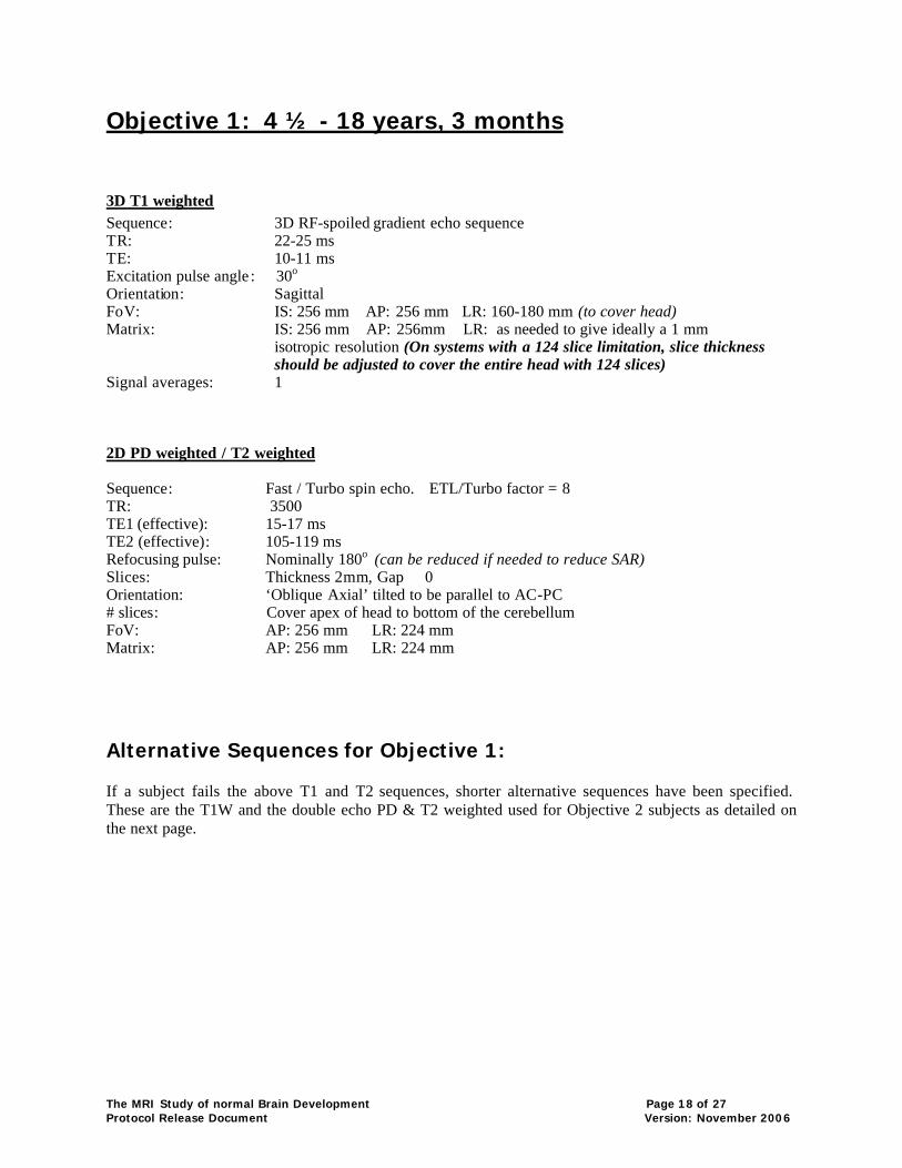
Objective 1: 4 ½ - 18 years, 3 months
3D T1 weighted Sequence: 3D RF-spoiled gradient echo sequence TR: 22-25 ms TE: 10-11 ms Excitation pulse angle : 30o Orientation: Sagittal FoV: IS: 256 mm AP: 256 mm LR: 160-180 mm (to cover head) Matrix: IS: 256 mm AP: 256mm LR: as needed to give ideally a 1 mm isotropic resolution (On systems with a 124 slice limitation, slice thickness
should be adjusted to cover the entire head with 124 slices) Signal averages: 1 2D PD weighted / T2 weighted Sequence: Fast / Turbo spin echo. ETL/Turbo factor = 8 TR: 3500 TE1 (effective): 15-17 ms TE2 (effective): 105-119 ms Refocusing pulse: Nominally 180o (can be reduced if needed to reduce SAR) Slices: Thickness 2mm, Gap 0 Orientation: ‘Oblique Axial’ tilted to be parallel to AC-PC # slices: Cover apex of head to bottom of the cerebellum FoV: AP: 256 mm LR: 224 mm Matrix: AP: 256 mm LR: 224 mm Alternative Sequences for Objective 1: If a subject fails the above T1 and T2 sequences, shorter alternative sequences have been specified. These are the T1W and the double echo PD & T2 weighted used for Objective 2 subjects as detailed on the next page.
The MRI Study of normal Brain Development Page 19 of 27 Protocol Release Document Version: November 2006
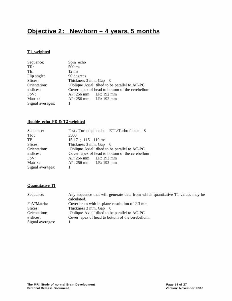
Objective 2: Newborn – 4 years, 5 months
T1_weighted Sequence: Spin echo TR: 500 ms TE: 12 ms Flip angle: 90 degrees Slices: Thickness 3 mm, Gap 0 Orientation: ‘Oblique Axial’ tilted to be parallel to AC-PC # slices: Cover apex of head to bottom of the cerebellum FoV: AP: 256 mm LR: 192 mm Matrix: AP: 256 mm LR: 192 mm Signal averages: 1 Double_echo_PD & T2 weighted Sequence: Fast / Turbo spin echo ETL/Turbo factor = 8 TR : 3500 TE 15-17 ; 115 - 119 ms Slices: Thickness 3 mm, Gap 0 Orientation: ‘Oblique Axial’ tilted to be parallel to AC-PC # slices: Cover apex of head to bottom of the cerebellum FoV: AP: 256 mm LR: 192 mm Matrix: AP: 256 mm LR: 192 mm Signal averages: 1 Quantitative T1 Sequence: Any sequence that will generate data from which quantitative T1 values may be
calculated. FoV/Matrix: Cover brain with in-plane resolution of 2-3 mm Slices: Thickness 3 mm, Gap 0 Orientation: ‘Oblique Axial’ tilted to be parallel to AC-PC # slices: Cover apex of head to bottom of the cerebellum. Signal averages: 1
The MRI Study of normal Brain Development Page 20 of 27 Protocol Release Document Version: November 2006
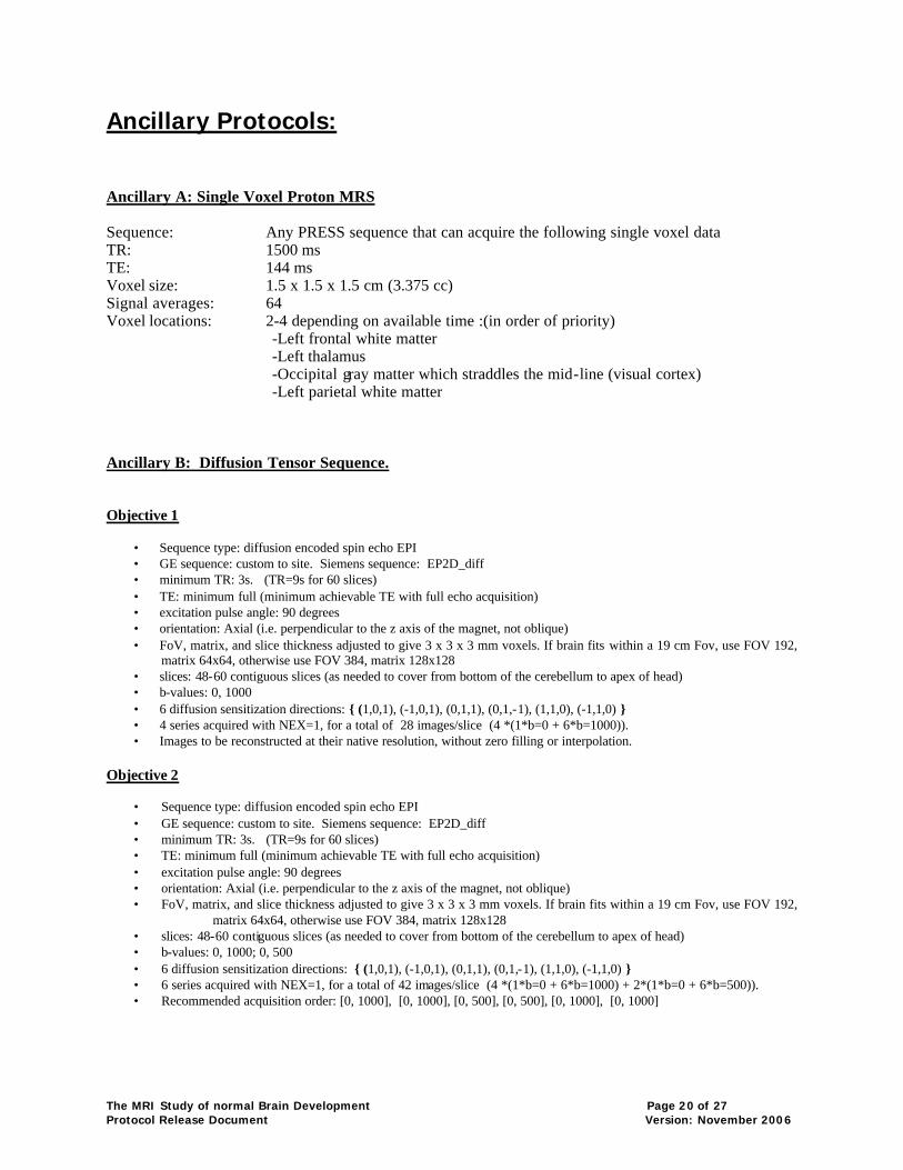
Ancillary Protocols: Ancillary A: Single Voxel Proton MRS Sequence: Any PRESS sequence that can acquire the following single voxel data TR: 1500 ms TE: 144 ms Voxel size: 1.5 x 1.5 x 1.5 cm (3.375 cc) Signal averages: 64 Voxel locations: 2-4 depending on available time :(in order of priority)
-Left frontal white matter -Left thalamus -Occipital gray matter which straddles the mid-line (visual cortex) -Left parietal white matter
Ancillary B: Diffusion Tensor Sequence. Objective 1
• Sequence type: diffusion encoded spin echo EPI • GE sequence: custom to site. Siemens sequence: EP2D_diff • minimum TR: 3s. (TR=9s for 60 slices) • TE: minimum full (minimum achievable TE with full echo acquisition) • excitation pulse angle: 90 degrees • orientation: Axial (i.e. perpendicular to the z axis of the magnet, not oblique) • FoV, matrix, and slice thickness adjusted to give 3 x 3 x 3 mm voxels. If brain fits within a 19 cm Fov, use FOV 192,
matrix 64x64, otherwise use FOV 384, matrix 128x128 • slices: 48-60 contiguous slices (as needed to cover from bottom of the cerebellum to apex of head) • b-values: 0, 1000 • 6 diffusion sensitization directions: { (1,0,1), (-1,0,1), (0,1,1), (0,1,-1), (1,1,0), (-1,1,0) } • 4 series acquired with NEX=1, for a total of 28 images/slice (4 *(1*b=0 + 6*b=1000)). • Images to be reconstructed at their native resolution, without zero filling or interpolation.
Objective 2
• Sequence type: diffusion encoded spin echo EPI • GE sequence: custom to site. Siemens sequence: EP2D_diff • minimum TR: 3s. (TR=9s for 60 slices) • TE: minimum full (minimum achievable TE with full echo acquisition) • excitation pulse angle: 90 degrees • orientation: Axial (i.e. perpendicular to the z axis of the magnet, not oblique) • FoV, matrix, and slice thickness adjusted to give 3 x 3 x 3 mm voxels. If brain fits within a 19 cm Fov, use FOV 192,
matrix 64x64, otherwise use FOV 384, matrix 128x128 • slices: 48-60 contiguous slices (as needed to cover from bottom of the cerebellum to apex of head) • b-values: 0, 1000; 0, 500 • 6 diffusion sensitization directions: { (1,0,1), (-1,0,1), (0,1,1), (0,1,-1), (1,1,0), (-1,1,0) } • 6 series acquired with NEX=1, for a total of 42 images/slice (4 *(1*b=0 + 6*b=1000) + 2*(1*b=0 + 6*b=500)). • Recommended acquisition order: [0, 1000], [0, 1000], [0, 500], [0, 500], [0, 1000], [0, 1000]
The MRI Study of normal Brain Development Page 21 of 27 Protocol Release Document Version: November 2006
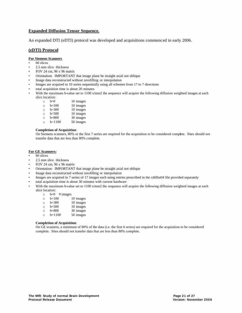
Expanded Diffusion Tensor Sequence. An expanded DTI (eDTI) protocol was developed and acquisitions commenced in early 2006. (eDTI) Protocol For Siemens Scanners • 60 slices • 2.5 mm slice thickness • FOV 24 cm, 96 x 96 matrix • Orientation: IMPORTANT that image plane be straight axial not oblique • Image data reconstructed without zerofilling or interpolation • Images are acquired in 10 series sequentially using all schemes from 17 to 7 directions • total acquisition time is about 20 minutes • With the maximum b-value set to 1100 s/mm2 the sequence will acquire the following diffusion weighted images at each
slice location: o b=0 10 images o b=100 10 images o b=300 10 images o b=500 10 images o b=800 30 images o b=1100 50 images
Completion of Acquisition: On Siemens scanners, 80% or the first 7 series are required for the acquisition to be considered complete. Sites should not transfer data that are less than 80% complete.
For GE Scanners: • 60 slices • 2.5 mm slice thickness • FOV 24 cm, 96 x 96 matrix • Orientation: IMPORTANT that image plane be straight axial not oblique • Image data reconstructed without zerofilling or interpolation • Images are acquired in 7 series of 17 images each using entries prescribed in the cdiflist04 file provided separately • total acquisition time is about 30 minutes with current hardware • With the maximum b-value set to 1100 s/mm2 the sequence will acquire the following diffusion weighted images at each
slice location: o b=0 9 images o b=100 10 images o b=300 10 images o b=500 10 images o b=800 30 images o b=1100 50 images
Completion of Acquisition: On GE scanners, a minimum of 80% of the data (i.e. the first 6 series) are required for the acquisition to be considered complete. Sites should not transfer data that are less than 80% complete.
The MRI Study of normal Brain Development Page 22 of 27 Protocol Release Document Version: November 2006
Quality Control (QC) Procedures for MRI Data
The magnetic resonance imaging (MRI) quality control (QC) procedure was developed to
evaluate the quality of the MRI data coming in from the different pediatric study centers (PSCs) and to ensure its applicability for automated processing. The procedure involves verification of parameters for protocol adherence, automated QC estimation of movement, noise and contrast and visual inspection of the MR image data. Different levels of QC of MRI data occurs at different locations from the time of acquisition at the PSC MRI console to the Data Coordinating Center (DCC) in Montreal. The first level of MRI QC is a visual inspection by the PSC while the subject is scanned. The image data is examined at the scanner console to determine if a rescan or a fallback scan is necessary. The reconstructed coronal and sagittal data should be examined for inter-packet motion of the multi-slice acquisitions. Once scanning is completed, all image volumes are transferred to the PSC SWS where they are reviewed by the PSC principal investigator (PI) or his/her designate. If the site is uncertain of the quality of the scans, the PI can request an expedited review.
Once the data arrives at the DCC in Montreal, it is automatically verified for proper identification and protocol compliance with respect to acquisition parameters. An automatic program runs a number of scripts to accomplish the following tasks:
• Movement artifacts are estimated. • Noise and contrast are estimated. • Image preprocessing (cropping, image intensity non-uniformity correction, intensity
normalization) is completed.
Visual inspection of all data sets is performed at the DCC. The goal of the visual inspection is to rate:
• the amount of movement artifacts – either within the slices or volume, or between packets for the multi-packet acquisitions
• the level of intensity homogeneity within slices, between slices and throughout the volume the amount of noise in the scan
• the level of contrast between grey matter, white matter and CSF • the adherence to scanning protocol in terms of coverage of the head/brain from left to right, top to
bottom and front to back • the amount of geometric distortion due to susceptibility artifacts • the appearance of any other artifacts in the images.
Individual volumes are given a ”PASS” or “FAIL” QC status received based on the categories above.
Passing MRI Datasets: The complete dataset for a subject visit is then given a “PASS” or “FAIL”
status determined by the successful acquisition of the structural MRIs (T1W, PD/T2W either initial or fallback). A dataset must have a successful T1W and a PD/T2W to receive a final “PASS” status. If a scan is failed, a rescan is requested (assuming the repeat is acquired within the allowable time window).

The MRI Study of normal Brain Development Page 23 of 27 Protocol Release Document Version: November 2006
ACR and Living Phantom Rationale:
Quality control if a major issue in a large multi-center MRI study such as this. A key component of the DCC’s MRI QC program is the use of both the ACR and living phantoms. The ACR (American College of Radiology) Phantom is a standardized MRI test object that is to be scanned on a regular basis at all the PSCs. The purpose of this procedure is to monitor the overall performance and stability of the scanners used to collect data at every site. The ACR phantom can be used to assess: geometric accuracy, high contrast spatial resolution, image intensity uniformity, signal ghosting, and slice thickness. Much of this will be automated at the DCC and should allow easy detection of scanner problems and/or changes at the PSCs. It will also allow correction of geometric distortions due to any magnetic field gradient non-linearities. While the ACR phantom can provide considerable information it still has a number of shortcomings (e.g. does not contain the geometric complexity of a human brain) that make the living phantom desirable. The living phantom is a single individual who is scanned at every site at regular intervals throughout the study. This data can then be analyzed using the full pipeline to assess reliability and reproducibility of derived morphometrics (e.g. white matter tissue volume) which are the primary imaging outcomes in this study.
The quality control procedure will consist of acquiring the protocol on an ACR phantom and on a
living phantom, a single human volunteer travelling to each site, at various time points. The data will be sent to the DCC for analysis. If there are any unusual events at a PSC (quench, power failure, hardware/software upgrade, etc) a new phantom scan should be performed. It is the responsibility of the site to closely monitor QC at their site.
Sites recruiting Objective 1 subjects are asked to acquire the FULL Objective 1 protocol for which
the site is contracted on the ACR phantom at the beginning of each scanning phase (years 1, 3, and 5). In addition, the site should run the anatomical acquisitions (3D T1W, PD/T2W) monthly on the ACR phantom during each scanning phase.
The FULL protocol should be acquired on the “living phantom” at the beginning of each scanning
phase (years 1, 3, and 5). This should be done on the same day as the ACR phantom. Sites recruiting Objective 2 subjects are asked to acquire the FULL Objective 2 protocol for which the site is contracted should be acquired on both the ACR phantom at the beginning of each scanning phase and repeated every six months.
The FULL protocol should be acquired on the “living” phantom yearly at the beginning of each scanning phase. This should be done on the same day as the ACR. The site should run the anatomical acquisitions monthly on the ACR phantom during each scanning phase. Sites recruiting both Objective 1 and 2 subjects should acquire both sets of anatomical scans.
Note: Due to the size of the ACR phantom, some of the parameters in the anatomical acquisitions required slight revisions in order to obtain complete coverage. These revisions apply to the ACR phantom ONLY! Subjects and the “living” phantom are scanned as per the protocol.
The MRI Study of normal Brain Development Page 24 of 27 Protocol Release Document Version: November 2006
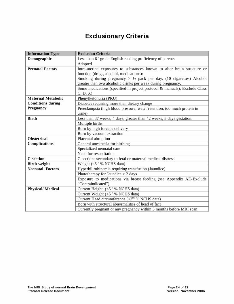
Exclusionary Criteria
Information Type Exclusion Criteria
Less than 6th grade English reading proficiency of parents Demographic Adopted Intra-uterine exposures to substances known to alter brain structure or function (drugs, alcohol, medications): Smoking during pregnancy > ½ pack per day. (10 cigarettes) Alcohol greater than two alcoholic drinks per week during pregnancy.
Prenatal Factors
Some medications (specified in project protocol & manuals); Exclude Class C, D, X) Phenylketonuria (PKU) Diabetes requiring more than dietary change
Maternal Metabolic Conditions during Pregnancy Preeclampsia (high blood pressure, water retention, too much protein in
urine) Less than 37 weeks, 4 days, greater than 42 weeks, 3 days gestation. Multiple births Born by high forceps delivery
Birth
Born by vacuum extraction Placental abruption General anesthesia for birthing Specialized neonatal care
Obstetrical Complications
Need for resuscitation C-section C-sections secondary to fetal or maternal medical distress Birth weight Weight (<5th % NCHS data)
Hyperbilirubinemia requiring transfusion (Jaundice) Phototherapy for Jaundice > 2 days
Neonatal Factors Exposure to medications via breast feeding (see Appendix AE-Exclude
“Contraindicated”) Current Height (<5th % NCHS data) Current Weight (<5th % NCHS data) Current Head circumference (<3rd % NCHS data) Born with structural abnormalities of head of face
Physical/ Medical
Currently pregnant or any pregnancy within 3 months before MRI scan
The MRI Study of normal Brain Development Page 25 of 27 Protocol Release Document Version: November 2006
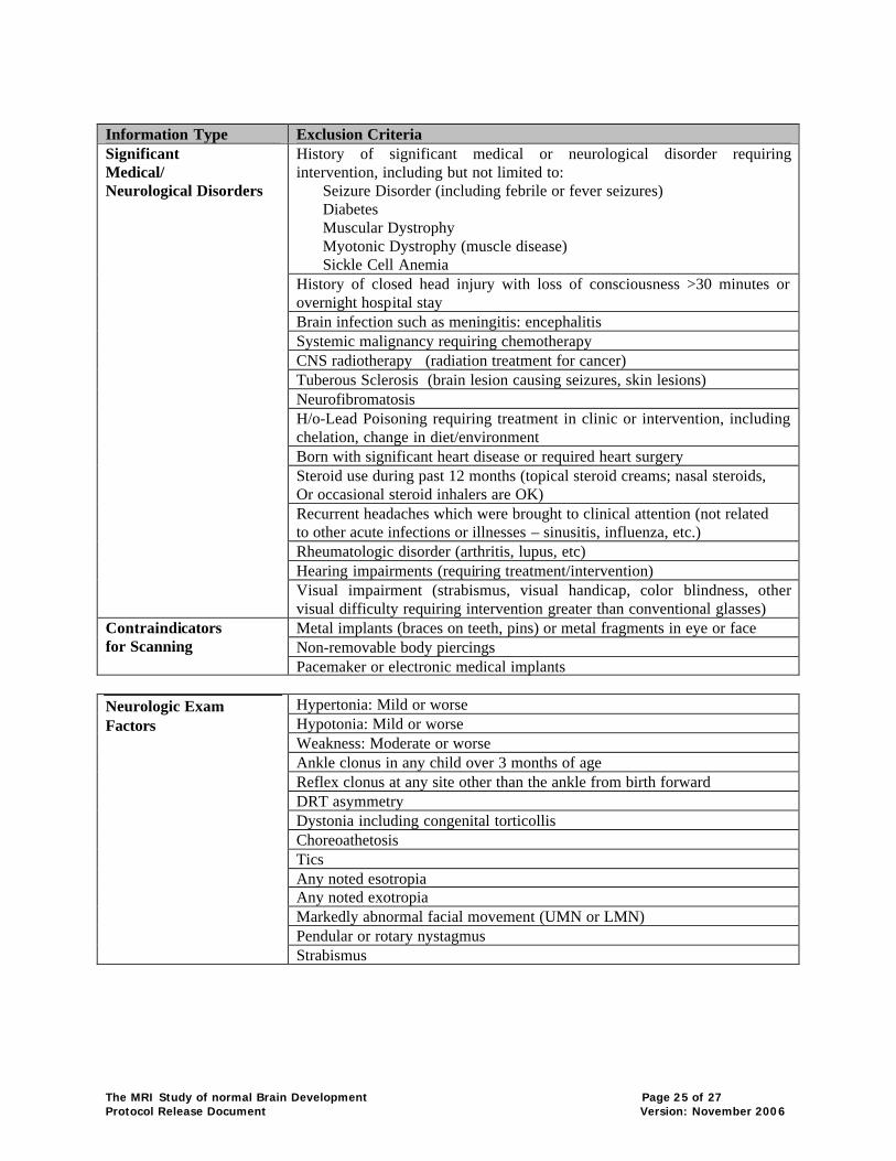
Information Type Exclusion Criteria
History of significant medical or neurological disorder requiring intervention, including but not limited to: Seizure Disorder (including febrile or fever seizures) Diabetes Muscular Dystrophy Myotonic Dystrophy (muscle disease) Sickle Cell Anemia History of closed head injury with loss of consciousness >30 minutes or overnight hospital stay Brain infection such as meningitis: encephalitis Systemic malignancy requiring chemotherapy CNS radiotherapy (radiation treatment for cancer) Tuberous Sclerosis (brain lesion causing seizures, skin lesions) Neurofibromatosis H/o-Lead Poisoning requiring treatment in clinic or intervention, including chelation, change in diet/environment Born with significant heart disease or required heart surgery Steroid use during past 12 months (topical steroid creams; nasal steroids, Or occasional steroid inhalers are OK) Recurrent headaches which were brought to clinical attention (not related to other acute infections or illnesses – sinusitis, influenza, etc.) Rheumatologic disorder (arthritis, lupus, etc) Hearing impairments (requiring treatment/intervention)
Significant Medical/ Neurological Disorders
Visual impairment (strabismus, visual handicap, color blindness, other visual difficulty requiring intervention greater than conventional glasses) Metal implants (braces on teeth, pins) or metal fragments in eye or face Non-removable body piercings
Contraindicators for Scanning
Pacemaker or electronic medical implants
Hypertonia: Mild or worse Hypotonia: Mild or worse Weakness: Moderate or worse Ankle clonus in any child over 3 months of age Reflex clonus at any site other than the ankle from birth forward DRT asymmetry Dystonia including congenital torticollis Choreoathetosis Tics Any noted esotropia Any noted exotropia Markedly abnormal facial movement (UMN or LMN) Pendular or rotary nystagmus
Neurologic Exam Factors
Strabismus
The MRI Study of normal Brain Development Page 26 of 27 Protocol Release Document Version: November 2006
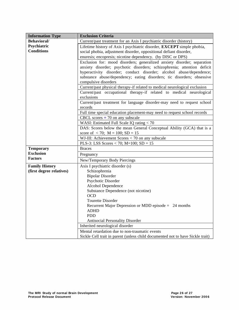
Information Type Exclusion Criteria
Current/past treatment for an Axis I psychiatric disorder (history) Lifetime history of Axis I psychiatric disorder, EXCEPT simple phobia, social phobia, adjustment disorder, oppositional defiant disorder, enuresis; encopresis; nicotine dependency. (by DISC or DPS) Exclusion for: mood disorders; generalized anxiety disorder; separation anxiety disorder; psychotic disorders; schizophrenia; attention deficit hyperactivity disorder; conduct disorder; alcohol abuse/dependence; substance abuse/dependency; eating disorders; tic disorders; obsessive compulsive disorders Current/past physical therapy-if related to medical neurological exclusion Current/past occupational therapy-if related to medical neurological exclusions Current/past treatment for language disorder-may need to request school records Full time special education placement-may need to request school records CBCL scores = 70 on any subscale WASI: Estimated Full Scale IQ rating < 70 DAS: Scores below the mean General Conceptual Ability (GCA) that is a score of < 70; M = 100; SD = 15 WJ-III: Achievement Scores < 70 on any subscale
Behavioral/ Psychiatric Conditions
PLS-3: LSS Scores < 70; M=100; SD = 15 Braces Pregnancy
Temporary Exclusion Factors New/Temporary Body Piercings
Axis I psychiatric disorder (s) Schizophrenia Bipolar Disorder Psychotic Disorder Alcohol Dependence Substance Dependence (not nicotine) OCD Tourette Disorder Recurrent Major Depression or MDD episode = 24 months ADHD PDD Antisocial Personality Disorder Inherited neurological disorder
Family History (first degree relatives)
Mental retardation due to non-traumatic events Sickle Cell trait in parent (unless child documented not to have Sickle trait)
The MRI Study of normal Brain Development Page 27 of 27 Protocol Release Document Version: November 2006
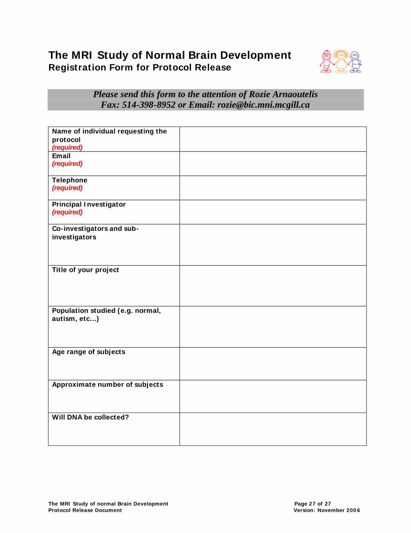
The MRI Study of Normal Brain Development Registration Form for Protocol Release
Please send this form to the attention of Rozie Arnaoutelis Fax: 514-398-8952 or Email: [email protected]
Name of individual requesting the protocol (required)
Email (required)
Telephone (required)
Principal Investigator (required)
Co-investigators and sub-investigators
Title of your project
Population studied (e.g. normal, autism, etc…)
Age range of subjects
Approximate number of subjects
Will DNA be collected?