the mysterious world of ob ultrasound coding -...
TRANSCRIPT

1
The Mysterious World of OB Ultrasound Coding
The Mysterious World of
OB Ultrasound Coding
Presented by:Lori-Lynne A. WebbCPC CCS-P CCP CHDA COBGCCPC, CCS-P, CCP, CHDA, COBGC,AHIMA Accredited ICD-10 TrainerAHIMA ACE mentor

2
The Mysterious World of OB Ultrasound Coding
Lori-Lynne’s Bio: She is a Specialty based E&M, and Procedure Coding, Compliance, Data Charge entry analyst and HIPAA Privacy specialist. Over the last 20 years she has successfully conducted pre-payment, post-payment and audit charge services for medical providers and insurance payers. She has worked closely with contracted 3rd party insurance payers for successful reimbursement outcomes. She has experience with both inpatient and outpatient coding for physician based, and hospital based providers and facilities, in addition to supervising coding and clinical staff.
Ms. Webb contributes educationally based coding articles and educational updates for national coding publications. She has her own ‘Lori-Lynne’s coding coach blog’ and is the coding resource for obgynhospitalist.com . She has presented at the National AHIMA and AAPC conferences, IdHIMA(State of Idaho) conferences, and local AAPC chapters. She is an AHIMA ( ) , pACE mentor; teaches CPT®, ICD-9 & 10, HCPCS; and is an AHIMA accredited ICD-10 Certified Trainer. Her major specialty is Women’s Services. This includes Maternal Fetal Medicine, OB/GYN office and facility, OB/GYN Hospitalist Labor/Trauma Services, OB/GYN Oncology, Urology, and General surgical coding.
Learning Objectives:
1. What is involved and visualized in an OB Ultrasound
2. Understand and use the approved abbreviations pertinent to OB Ultrasound and Maternal Fetal Medicine p
3. Understand the documentation criteria needed to code and bill CPT® and diagnosis codes for OB Ultrasound and Maternal Fetal Medicine
4. Understand the differences in clinical application of how and why a Trans-Vaginal and Trans Abdominal ultrasound is performed and the clinical utilization of ultrasound is performed and the clinical utilization of these scans

3
Let’s start at the beginning…g g
What is an Ultrasound?
Ultrasonic sound (Ultrasound) is:Th f lt i ( d) f di ti The use of ultrasonic (sound) waves for diagnostic or therapeutic purposes
to image an internal body structure
monitor a developing fetus
generate localized deep heat to the tissues.

4
Ultrasound Safety/Risks
Ultrasound is considered a very safe procedure for both the mother and the fetus.
Ultrasound does not produce ionizing radiation or pose radiation risk to mother or fetus.
What is an Ultrasound?
The currently used equipment for such a scan are called real-time scanners with the ability to provide a continuous picture of a moving fetus on a monitor screen.
Very high frequency sound waves of between 3.5 to 7.0 megahertz are generally used for this purpose.

5
What is an Ultrasound?
These waves are emitted from a transducer, which is placed in contact with the maternal abdomen
d i d t th ti l t f th t and is moved to the particular part of the uterus.
These frequencies when reflected back from the fetal surface produce a typical sonographic image, which can be read and categorized with various computer software.
The Ultrasound MachineA basic ultrasound machine has the following parts:
•Transducer probeprobe that sends and receives the sound waves
•Central processing unit (CPU)computer that does all of the calculations and contains the electrical
li f it lf d th t d bpower supplies for itself and the transducer probe
•Transducer pulse controlschanges the amplitude, frequency and duration of the pulses emitted from the transducer probe
•Displaydisplays the image from the ultrasound data processed by the CPU
•Keyboard/cursor inputs data and takes measurements from the display
•Disk storage device (CD/DVD Hard Drive)stores the acquired images
•Printerprints the image from the displayed data

6
The Ultrasound Machine
The Ultrasound Machine
Transducer probe probe that sends and receives the sound waves probe that sends and receives the sound waves

7
The Ultrasound Machine
Display Unit: Displays Display Unit: Displays the image from the ultrasound data processed by the CPU
The Ultrasound Machine
Keyboard/cursor: Input data and take measurements measurements from the display

8
The Ultrasound Machine
Disk storage device: (CD/DVD Hard Drive)
stores the acquired images
Printerprints the image from the gdisplayed data
What the images look like…

9
The Ultrasound Images
Profile of fetal face 2nd trimester
The Ultrasound Images
Transvaginal ultrasound uterus with 6 week gestational sac before appearance of embryo

10
The Ultrasound Images
Fetal Profile – 1st Trimester
The Ultrasound Images
Sextuplets – 1st Trimester

11
In the world of Obstetrics, Maternal Fetal Medicine (MFM)/Perinatology is a sub-specialty that is focused on the fetus, and its growth during the pregnancy.
Let’s start at the beginning…
Perinatology specialists work closely with obstetricians, and genetic counselors to provide care for high risk pregnancies, and to provide screening services for potential fetal anomalies prior to birth.
The perinatal period is generally defined as the time from 8-12 weeks gestation to approximately 30-45 days after g pp y ydelivery.
Background information
MFM/perinatal specialists provide extensive care for High risk pregnanciesg p g Multiple gestation (twins, triplets etc) In-vitro fertilization pregnancies (IVF) Advanced maternal age (AMA) Chronic maternal diagnoses (i.e. hypertension,
diabetes, seizure disorder)

12
Background information
Perinatologists perform and provide extensive ultrasound procedures with interpretation of:
Fetal growth and/or anomalies Fetal growth and/or anomalies
Placenta location and/or anomalies
Amniotic fluid
Umbilical cord complications during the Umbilical cord complications during the pregnancy
Background information
MFM Perinatologists provide highly complex surgical fetal procedures performed in-utero such as:
Chorionic Villus Sampling (CVS)
Amniocentesis (Amnio)
Percutaneous umbilical cord blood sampling procedure (PUBS) also known as a procedure (PUBS) also known as a cordocentesis
13
Background information
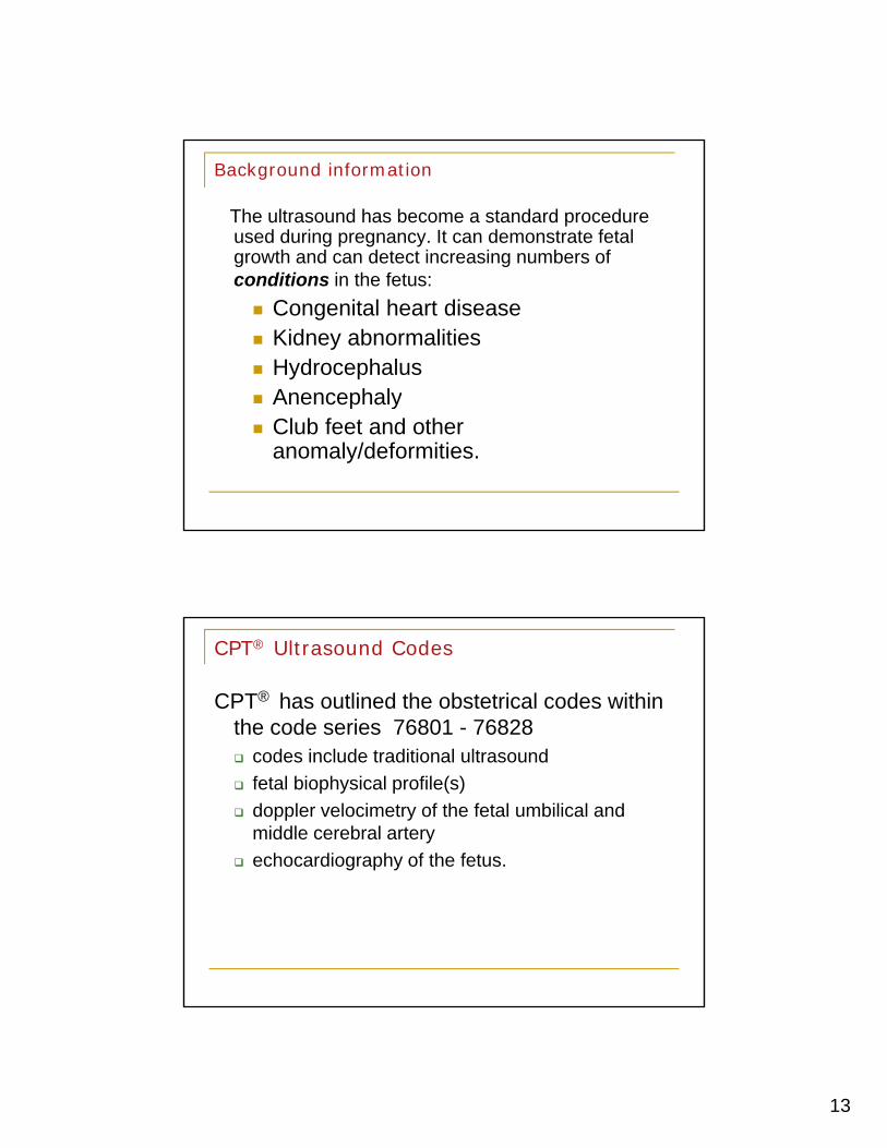
The ultrasound has become a standard procedure used during pregnancy. It can demonstrate fetal growth and can detect increasing numbers of conditions in the fetus:
Congenital heart disease Kidney abnormalities Hydrocephalus Anencephaly Anencephaly Club feet and other
anomaly/deformities.
CPT® Ultrasound Codes
CPT® has outlined the obstetrical codes within the code series 76801 - 76828 codes include traditional ultrasound
fetal biophysical profile(s)
doppler velocimetry of the fetal umbilical and middle cerebral artery
echocardiography of the fetus.

14
Conquering the Ultrasound Criteria
According to the guidelines in CPT® all diagnostic ultrasounds requirediagnostic ultrasounds require
a permanently recorded image
a final written report.
Conquering the CPT Ultrasound Criteria
Coders need to fully understand if they are billing and coding ultrasound scans as:
A) Global or complete scan
B) The recorded image or technical component only (TC Modifier)
Th i t t ti /d t ti l f th C) The interpretation/documentation only of the ultrasound scan (26 Modifier)

15
Conquering the CPT® Ultrasound Criteria
Carefully review the CPT code definitions to determine if the CPT code itself specifies for the first or singlep ggestation
– such as found in CPT code 7680176801 – Ultrasound, pregnant uterus, real time with image documentation, fetal and
maternal evaluation, first trimester (<14 weeks 0 days),transabdominal approach; single or first gestation.
Conquering the CPT® Ultrasound Criteria
Check if the add-on code “” symbol is denoted at the beginning of the CPT code
do not use a 51 modifier with the ”symbol code, as per the CPT definitions of an ‘add on code’

16
Conquering the CPT® Ultrasound Criteria
Review code 76802 to understand how the add-on code is used to denote ‘each additional gestation”
Code 76802 is an add-on code to CPT code 76801
Definition: +76802, each additional gestation (List separately in addition to code for primary procedure)
Conquering the CPT® Ultrasound Criteria
If the CPT ultrasound code criteria does not specify ‘units’ (such as in the code 76815) it should never be billed as a multiple unit, only as a single unit
CPT Code 76815 states 1 or more fetuses within the guidelines, so only 1 unit would be appropriate… even though
more than 1 fetus may be documented
17
Conquering the CPT® Ultrasound Criteria
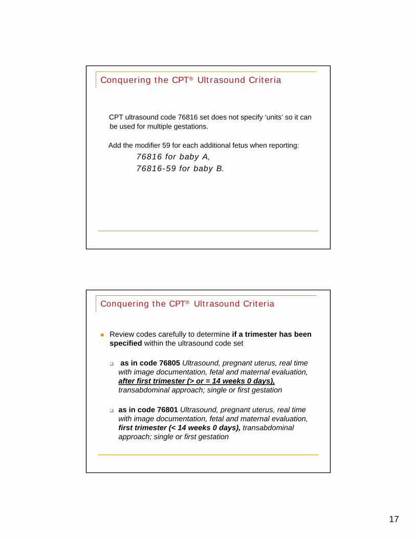
CPT ultrasound code 76816 set does not specify ‘units’ so it can be used for multiple gestationsbe used for multiple gestations.
Add the modifier 59 for each additional fetus when reporting:
76816 for baby A, 76816-59 for baby B.
Conquering the CPT® Ultrasound Criteria
Review codes carefully to determine if a trimester has been specified within the ultrasound code set
as in code 76805 Ultrasound, pregnant uterus, real time with image documentation, fetal and maternal evaluation, after first trimester (> or = 14 weeks 0 days),transabdominal approach; single or first gestation
as in code 76801 Ultrasound, pregnant uterus, real time as in code 76801 Ultrasound, pregnant uterus, real time with image documentation, fetal and maternal evaluation, first trimester (< 14 weeks 0 days), transabdominal approach; single or first gestation

18
Conquering the CPT® Ultrasound Criteria
Review to determine the approach of how the ultrasound was performed
CPT Code 76817 Ultrasound, pregnant uterus, real time with image documentation, transvaginal approach
CPT Code 76811 Ultrasound, pregnant uterus, real time with image documentation, fetal and maternal evaluation plus detailed fetal anatomic examination, transabdominal approach; single or first gestation
CPT code 76813 Ultrasound, pregnant uterus, real time with image documentation, first trimester fetal nuchal translucency measurement, transabdominal or transvaginal approach; single or first gestation
TRANSABDOMINALVIEW ILLUSTRATION
TRANSVAGINAL VIEW ILLUSTRATION

19
Deciphering the Terminology
In MFM/Perinatology medicine, there are many strange words and procedures
a coder needs a good understanding of ultrasound terminology & clinical documentation
Standard Medical Dictionary Reference
Standard Medical Abbreviations Reference Standard Medical Abbreviations Reference
Term Abbreviation Definition Amniocentesis Amnio A procedure to draw a sample of amniotic fluid which is then analyzed to
detect chromosome abnormalities, structural defects and metabolic disorders.
Amniotic Fluid
Amnio Fluid The fluid in which the embryo or fetus is suspended within the womb (the embryonic sac inside the uterus).
Beats per minute bpm the number of heartbeats per unit of time (beats per minute) Chorionic Villus Sampling
CVS An alternative to amniocentesis to detect chromosomal abnormalities. The CVS can be performed earlier in fetal development than amniocentesis, and thereby allows earlier diagnosis.
Congenital Defect A problem or condition existing at or dating from birth; acquired during development in the womb (uterus) and not through heredity
Crown Rump Length CRL the ultrasound measurement of a fetus Diagnostic Fetosocopy A minimally invasive examination of the fetus by a miniature video camera
Abbreviations/Terminology
Diagnostic Fetosocopy A minimally-invasive examination of the fetus by a miniature video camera inserted through a small tube
Estimated Date of Confinement
EDC a term for the estimated delivery date for a pregnant woman
Fetal Abnormality A condition detected in the unborn human that is not the normal or average.
Fetal Echocardiography A high resolution ultrasound test to detect heart abnormalities in the fetus. Fetal Pole a thickening on the margin of the yolk sac of a fetus during pregnancy Genetic Counseling Medical guidance concerning inherited (genetic) disorders. In Utero Relating to being in the womb Intra-Uterine Pregancy IUP the normal location for a pregnancy to occu In-vitro Fertilization IVF a process by which egg cells are fertilized by sperm outside the body Last Menstrual Period LMP the first day of the menstrual period prior to conceiving, used to calculate
Expected Date of Delivery Magnetic Resonance Imaging MRI A noninvasive diagnostic technique that produces computerized images of
internal body tissue induced by the application of radio waves Maternal Fetal Medicine MFM The testing and management of high-risk pregnancies; also called
perinatology Neonatal Intensive Care Unit NICU An area within a hospital dedicated to the care and treatment of pre-term
and critically ill babies N l T b D f NTD i i h i l d b i h l i h Neural Tube Defect NTD an opening in the spinal cord or brain that occurs very early in human
development, visualized by ultrasound Nuchal Translucency NT The area around the neck of the fetus, also known as the nuchal fold Postnatal Occurring, existing or performed after birth Prenatal Occurring, existing or performed before birth Trans-abdominal ultrasound TAUS Ultrasound procedure performed to visualize the pelvic cavity through
application of sound waves by a device placed upon the abdomen Trans-Vaginal ultrasound TVUS Ultrasound procedure performed to visualize the cervix and uterine
contents by application of sound waves through a device inserted into the vagina.
Trimester the division of pregnancy into three-month sections Ultrasound US or
U/sound A technique involving the formation of a 2D-or 3D dimensional image used for the examination and measurement of bodily abnormalities.

20
Code Set Criteria Detail of the Ultrasound Code
CPT Definition & Guidelines
Documentation Criteria Needed Clinical Indications (Diagnosis)
Pre-Service Work
Intra-Service Work
Post-Service Work
Static/Video Ultrasound Record Permanent Recorded Image
Pre-Service Work :
Chart Review of prior clinical informationinformation
Review of pertinent prior imaging studies
Proper Draping & Positioning of the Patient

21
Intra Service WorkWork involved in performing the actual scan
Always includes the physical performance of the scan by the provider or sonographer
If performed by a sonographer – Supervision of the sonography
Standard Clinical work detail criteria noted (Key elements) and of the actual scan findings
Inte p et nd p ep e epo t fo the Pe m nent Medi l Interpret and prepare report for the Permanent Medical Record (electronic or paper)
Post-Service Work :
Discuss the findings with the patient and referring physician patient and referring physician (when appropriate)
Review and sign the permanent record/prepared report

22
Code-Set SpecificsMost Common Fetal Ultrasounds
76801 +76802 76815
76805 +76810
76811 +76812
76816
76817
CPT® Code-Set Specifics
76801: Ultrasound, pregnant uterus, real time with image documentation, fetal and maternal evaluation, first trimester (< 14 weeks 0 days), transabdominal approach; single or first gestation
+ 76802: Ultrasound, pregnant uterus, real time with image documentation, fetal and maternal evaluation, first trimester (< 14 weeks 0 days), transabdominal approach; Each additional gestation (List separately in addition to code for primary procedure)

23
76801-76802 Ultrasound First Trimester Key Elements
Determination of the number of gestational sacs and fetuses
G t ti l /f t l t i t f Gestational sac/fetal measurement appropriate for gestation
Survey of visible fetal and placental anatomic structure
Qualitative assessment of the amniotic fluid volumevolume
Evaluation of maternal uterus and adnexa
76801-76802 Key Elements
These ultrasound images show a normal early fetus of 9 weeks gestational age (1st trimester) and are taken via the transabdominal route.
Among the structures seen are the fetus with the bulkier head (cephalic part) and the fetal heart on Color Doppler and Power Doppler imaging.
Spectral Doppler waveform (Topmost row- right) shows the cardiac pulsations with heart rate.
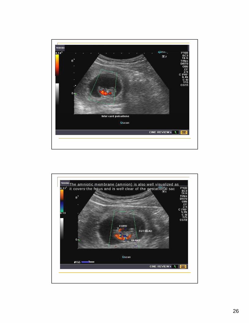
The fetal cardiac pulsations are also well visualized in the Power Doppler image of the fetus (2nd row from bottom- Right).
The amniotic membrane (amnion) is also well visualized as it covers the fetus and is well clear of the gestational sac (Topmost row -left). g ( p )
At a later date, the amnion merges with the gestational sac and would not be visualized. The early umbilical cord is also visualized as it extends from the fetus to the uterine wall (ultrasound/ Doppler image on bottom row).

24
Ultrasound images show a normal early fetus of 9 weeks gestational age (1st trimester) and are taken via the transabdominal route
Fetal heart on Color Doppler and Power Doppler imaging. Spectral Doppler waveform shows the cardiac pulsations with heart rate

25
The fetal cardiac pulsations are also well visualized in the Power Doppler image of the fetus
26
The amniotic membrane (amnion) is also well visualized as it covers the fetus and is well clear of the gestational sac

27
The early umbilical cord is also visualized as it extends from the fetus to the uterine wall
CPT® Code-Set Specifics
76805: Ultrasound, pregnant uterus, real time with image documentation, fetal and maternal evaluation, first trimester (> or = 14 weeks 0 days), transabdominal approach; single or first gestation
+ 76810: Ultrasound, pregnant uterus, real time with image documentation, fetal and maternal evaluation, first trimester (> or = 14 weeks 0 days), transabdominal approach; Each additional gestation (List separately in addition to code for primary procedure)

28
76805, +76810 After First TrimesterKey Elements
Determination of the number Fetuses amniotic/chorionic sacsamniotic/chorionic sacs
Survey of intracranial, spinal and abdominal anatomy
Evaluate the 4-chamber heart view
Assessment of the umbilical cord insertion site
A t f th i ti fl id l Assessment of the amniotic fluid volume
Evaluation of maternal adnexa when visible
Second Trimester View

29
CPT® Code-Set Specifics76811: Ultrasound, pregnant uterus, real time with image
documentation, fetal and maternal evaluation, plus detailed fetal anatomic examination, transabdominal
h i l fi t t tiapproach, single or first gestation
+ 76812:Ultrasound, pregnant uterus, real time with image documentation, fetal and maternal evaluation, plus detailed fetal anatomic examination, transabdominal approach, each additional gestation (list separately in pp , g ( p yaddition to code for primary procedure)
Fetal Anatomic FeaturesSpine &
Abdominal Wall 2nd
Trimester
Fetal long
bones
Fetal Foot
Fetal Hand
Fetal Heart in 2nd
Trimester

30
CPT® Code-Set Specifics
76815: Ultrasound, pregnant uterus, real time with image documentation limitedtime with image documentation, limited (e.g., fetal heart beat, placental location, fetal position and/or qualitative amniotic fluid volume), 1 or more fetuses
76815 Limited Ultrasound Key Elements
Examination is limited for a focused “quick-look/quick-q /qpeek” assessment of one or more of these key elements
Fetal Position Fetal Heartbeat Placental location Qualitative amniotic fluid volume
Note: If an AFI only is noted/performed in the record – this is the CPT code/scan that should be billed
31
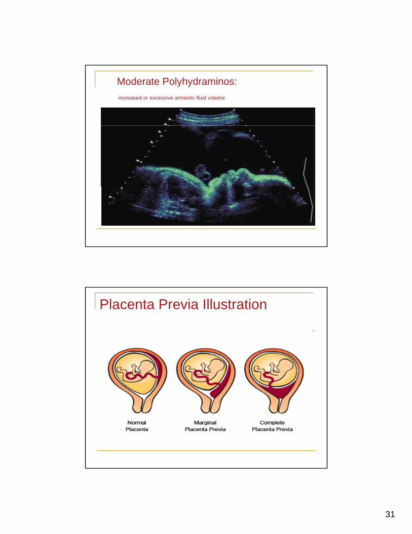
Moderate Polyhydraminos:
increased or excessive amniotic fluid volume
Placenta Previa Illustration

32
CPT® Code-Set Specifics
76816: Ultrasound, pregnant uterus, real time with image documentation follow uptime with image documentation, follow-up (e.g., re-evaluation of fetal size by measuring standard growth parameters and amniotic fluid volume, re-evaluation of organ system(s) suspected or confirmed to be abnormal on a previous scan), p ),transabdominal approach, per fetus
Transabdominal View

33
76816 Follow-Up Ultrasound Key Elements
Examination is limited to the following elementsExamination is limited to the following elements Focused Assessment of fetal size by measuring BPD
(Bi-Parietal Diameter), abdominal circumference, femur length or other appropriate measurement
OR Detailed re-examination of a specific organ or
system known or suspected to be abnormalC i f thi i ti t i t d t Comparison of this examination to a prior study to evaluate the interval change (i.e. fetal growth)

34
CPT® Code-Set Specifics
76817: Ultrasound, pregnant t l ti ith iuterus, real time with image
documentation, transvaginal
76817 Transvaginal Ultrasound Key Elements
Interpretation of the examination including ANY of the following elementsfollowing elements Evaluation of the fetus and placenta Evaluation of the maternal uterus and adnexa Evaluation of characteristics of cervix including
length and structure

35
Transvaginal view: embryo at 10 weeks
Code-Set SpecificsTargeted & Procedural Fetal Ultrasounds
76818 BPP w/NST
76819 BPP w/o NST
59025 Fetal NST
76820 Umbilical Artery Doppler
76821 Mid-Cerebral Artery Doppler
36
Code-Set SpecificsTargeted Fetal Ultrasound Testing
76818 Bio-Physical Profile (BPP) with
76819 Bio-Physical Profile (BPP) withoutProfile (BPP) with
Fetal Non-Stress Test (NST)
Profile (BPP) without Fetal Non-Stress Test (NST)
CPT® Code-Set Specifics
76818: Fetal bio-physical profile; with non-stress testingtesting
76819: Fetal bio-physical profile; without non-stress testing

37
76818/19 Bio-Physical Profile Key Elements
Interpretation of the examination76818P f d t th h i l i t t f th Performed to assess the physiologic status of the fetus (and scored)
Fetal Breathing movements (score: 0-2)
Fetal Movement (score: 0-2)
Fetal Tone (score: 0-2)
Amniotic Fluid Volume (score: 0-2)
Final Score documented on interpretation Fetal Non-Stress Test (score: 0-2)
Note: for code 76819 – no NST is performed
76819 Follow-Up UltrasoundKey Elements
The LMP of this 31 year old, gravida 1, para 0 patient was MAY 3 2011, giving her an EDD of FEB 7 2012 and a current gestational age of 33 weeks 1 day by dates. A sonographic examination was performed on DEC 21 2011
IMPRESSIONTwin IUP (Fetus A)33 weeks and 1 day by dates. (EDD=FEB 7 2012)33 weeks and 1 day by this ultrasound. (EDD=FEB 7 2012)Fetal position = Maternal Left Vertex presentationsonographic examination was performed on DEC 21 2011
using real time equipment.The amniotic fluid volume for fetus A appears to be normal. The
amniotic fluid volume for fetus B appears to be normal.INDICATIONS: TwinsRESULTSFetus # 1 of 2Vertex presentationFetal growth appeared normalFetal position = Maternal LeftPlacenta Location = Right lateralNo placenta previaPlacenta Grade = IIChorionicity = Monochorionic, DiamnioticBIOPHYSICAL PROFILE
Fetal position = Maternal , Left Vertex presentationFetal growth appeared normalEstimated Fetal Weight = 2032 gramsEstimated Fetal Weight = 4 lbs 8 ozNo placenta previaMonochorionic, diamnioticTwin IUP (Fetus B)33 weeks and 1 day by dates. (EDD=FEB 7 2012)32 weeks and 6 days by this ultrasound. (EDD=FEB 9 2012)Fetal position = Superior, Right Transverse presentationFetal growth appeared normalEstimated Fetal Weight = 2015 gramsEstimated Fetal Weight = 4 lbs 7 ozNo placenta previaMonochorionic, diamniotic
BIOPHYSICAL PROFILEFetus AThe Biophysical Profile score was 8/8.Breathing: 2 Movement: 2 Tone: 2 AFV: 2Fetus BThe Biophysical Profile score was 8/8.Breathing: 2 Movement: 2 Tone: 2 AFV: 2
RECOMMENDATION: BPP: 1 WeekGENERAL COMMENT: Patient is seen in follow up of MC twins. She is without problems. Fetal growth remains appropriate and concordant, with concordant MVPs. MCA dopplers for Twin B are stable and at median for gestational age. Twin A’s MCA could not be accurately insonated. Fetal testing is normal for both fetuses. I reviewed considerations for delivery with patient and her husband given the ongoing risks for morbidity and mortality with MC twins. Will continue weekly surveillance and MCA dopplers until delivery.

38
CPT® Code-Set Specifics
59025 Fetal Non-Stress Test
59025 Fetal Non-Stress Test Key Elements
The NST can be performed in conjunction with or without the Bio-Physical Profile
Global Procedure 59025 Interpretation only Procedure 59025-26 Technical Component Only Procedure 59025-TCp y

39
59025 Fetal Non-Stress Test Key Elements
The NST is performed by auscultation of the fetal heart rate using an external electronic monitor attached to the pregnant patient.
Documentation needs to include
Baseline Fetal Heart Rate (FHR)
Baseline FHR Variability
Presence of Accelerations Presence of Accelerations
Presence of Periodic or episodic Decelerations
Changes or Trends of FHR pattern over time (30 minutes minimum)
Frequency and intensity of uterine contractions
59025 Fetal Non-Stress Test Key Elements “Stand Alone NST”

40
Code-Set SpecificsTargeted Fetal Ultrasound Testing
76820 Doppler velocimetry fetal;
76821 Doppler velocimetry fetal;velocimetry fetal;
umbilical artery velocimetry fetal; middle cerebral artery
CPT® Code-Set Specifics
76820 Doppler Velocimetry, Fetal; Umbilical Artery
41
76820 Doppler Key Elements
Interpretation of the examination76820
Study is performed to assess the velocity of the blood flow through the umbilical artery.
Velocity waveforms through the umbilical artery of a normal fetus are different from a growth-retarded fetus.
Can be performed either transabdominally or transvaginally.
CPT® Code-Set Specifics
76821 Doppler velocimetry, Fetal; Middle Cerebral Artery

42
76821 Doppler Key ElementsInterpretation of the examination 76821
Study is performed to assess the velocity of the blood fl th h th Middl C b l tflow through the Middle Cerebral artery.
The peak systolic velocity is inversely related to the amount of hematocrit (HCT) in fetal blood
Helps to determine the timing of labor induction or when fetal anemia is severe enough to require a transfusion transfusion
Can be performed either transabdominally or transvaginally
76820 Umbilical Artery
76821 Middle Cerebral ArteryCerebral Artery

43
Doppler of umbilical artery
Doppler of umbilical artery

44
Doppler of Mid-cerebral artery
Doppler of Mid-cerebral artery

45
Cordocentesis: PUBS ProcedurePercutaneous Umbilical Blood Sampling
An advanced imaging ultrasound determines the location where the umbilical cord inserts into thelocation where the umbilical cord inserts into the placenta.
The ultrasound guides a thin needle through the abdomen and uterine walls to the umbilical cord. The needle is inserted into the umbilical cord to retrieve a small sample of fetal blood.
The sample is sent to the laboratory for analysis, and results are usually available within 72 hours.
When is cordocentesis performed?
Cordocentesis is usually done when diagnostic information can not be obtained through amniocentesis, CVS, ultrasound or results of above tests were/are inconclusive.above tests were/are inconclusive.
Cordocentesis is performed after 17 weeks into pregnancy.
Entire procedure takes approx. 45-60 minutes to perform due to the fragility of the umbilical veinvein.

46
What does the cordocentesis test look for?
Cordocentesis detects chromosome abnormalities and/or blood disorders
Cordocentesis may be performed to help diagnose any of the following : Malformations of the fetus
Fetal infection (i.e. toxoplasmosis or rubella)
Fetal platelet count in the mother
Fetal anemia Fetal anemia
Isoimmunization
http://www.youtube.com/watch?v=Hg10v0Pa5Sc

47
The Amniocentesis Procedure Amniocentesis Background
During the pregnancy, the uterus houses and protects a developing f f f ffetus for about 9 months. The fetus is surrounded by fluid, which in the 2nd and 3rd trimester is produced primarily by fetal urination also known as amniotic fluid. .
Amniotic fluid, allows the fetus to float and move within the uterus. It also cushions and protects the fetus from injury and helps to maintain a constant temperature in the uterus.
Pregnant women who are over 35 or have a family history of genetic disorders may undergo a procedure called an amniocentesis.
The Amniocentesis procedure is used to test for infections, genetic disorders, metabolic problems, and fetal lung maturity, sex of the infant.
The Amniocentesis Procedure itself…
During the procedure, the position of the fetus is first located using ultrasound guidance. g
The ultrasound locates a safe place for aspiration that is away from the placenta.
Aspiration of fluid from the amniotic sac is performed, via a needle inserted through the skin of the abdomen. Approximately 1 ounce of amniotic fluid is withdrawnamniotic fluid is withdrawn.
Following the procedure, the fetus will be monitored by ultrasound for a brief period. And, the amniotic fluid will be naturally replaced in about 3 to 4 hours.
48
CPT® Code-Set SpecificsAmniocentesis: 59000/59001 Coding for the Amniocentesis involves codes:
59000 A i t i Di ti (M di i d ) 59000 Amniocentesis Diagnostic. (Medicine code)
76946 The Ultrasound guidance of the test
59001 Amniocentesis for Therapeutic Fluid Reduction (includes ultrasound guidance)
The Amniocentesis Procedure
d fl
Amniocentesis video 01082012 flv
Amniocentesis animation video.flv
Amniocentesis video 01082012.flv

49
THANK YOU!!!
I appreciate you spending this time with me, I hope you enjoyed it as much as I enjoyedI hope you enjoyed it as much as I enjoyed bringing this to you.
References
•http://www.ultrasound-images.com
•http://www.sonosite.com
•http://prosono.ieasysite.com/index.html
•http://www.bing.com/images/search?q=second+trimester+ultrasound+image&qpvt=second+trimester+ultrasound+image&FORM=IGRE
•http://www.medicalvideos.us/
•http://www.babyzone.com/pregnancy/fetal_development/photos_second_trimester_ultrasounds/2