the n-terminal domain of human dna ligase i contains the nuclear
TRANSCRIPT
The EMBO Journal vol.14 no.21 pp.5379-5386, 1995
The N-terminal domain of human DNA ligase Icontains the nuclear localization signal and directsthe enzyme to sites of DNA replication
A.Montecuccol, E.Savini, F.Weighardt,R.Rossi, G.Ciarrocchi, A.Villa2 andG.BiamontiIstituto di Genetica Biochimica ed Evoluzionistica CNR,via Abbiategrasso 207, 1-27100 Pavia and 2Department ofPharmacology, CNR and B.Ceccarelli Centers and DIBIT ScientificInstitute S.Raffaele, via Olgettina 60, University of Milan,20132 Milano, Italy
'Corresponding author
DNA replication in mammalian cells occurs in discretenuclear foci called 'replication factories'. Here we showthat DNA ligase I, the main DNA ligase activity inproliferating cells, associates with the factories duringS phase but displays a diffuse nucleoplasmic distribu-tion in non-S phase nuclei. Immunolocalization analysisof both chloramphenicol acetyltransferase (CAT)-DNAligase I fusion proteins and epitope tagged DNA ligaseI mutants allowed the identification of a 13 amino acidfunctional nuclear localization signal (NLS) locatedin the N-terminal regulatory domain of the protein.Furthermore, the NLS is immediately preceded by a115 amino acid region required for the association ofthe enzyme with the replication factories. We proposethat in vivo the activity of DNA ligase I could bemodulated through the control of its sub-nuclear com-partmentalization.Keywords: DNA ligase I/DNA replication/factories/nuclearlocalization signal
IntroductionDNA ligases play essential roles in both DNA replicationand DNA repair by joining single- and double-strandedbreaks in an ATP-requiring reaction (Kornberg and Baker,1991). Three distinct DNA ligases, whose function isnot yet completely understood, have been biochemicallyidentified in mammalian cells (Lindahl and Barnes, 1992).New perspectives to elucidate the role of DNA ligase Icame from the isolation of both the human (Barnes et al.,1990) and the murine (Savini et al., 1994) cDNAs. DNAligase I (-102 kDa) is divided into two clearly distinctregions: a highly conserved C-terminal region containingthe active site (Tomkinson et al., 1990), and a lessconserved N-terminal portion dispensable for catalyticactivity and probably corresponding to a regulatory domain(Kodama et al., 1991; Savini et al., 1994). The analysisof the human and murine protein sequences (Prigent et al.,1992; Savini et al., 1994) suggests the presence of a(nuclear localization signal) NLS and of putative phos-phorylation sites in this domain.
Several observations indicate an involvement of DNAligase I in DNA replication: (i) DNA ligase I level is
higher in proliferating cells (Elder and Rossignol, 1990;Montecucco et al., 1992); (ii) the human DNA ligase IcDNA complements the replicative defect of the Saccharo-myces cerevisiae cdc 9 DNA ligase mutant (Barnes et al.,1990); (iii) the enzyme co-purifies with a protein complexable to replicate SV40 DNA in vitro (Congjun et al.,1994); (iv) it cannot be substituted for by DNA ligase IIIin an in vitro SV40 DNA replication assay (Waga et al.,1994). The strongest evidence of a role in DNA replicationderives from the human 46 BR cell line in which amutation in the DNA ligase I gene (Barnes et al., 1992)correlates with a delay in the joining of the Okazakifragments (Prigent et al., 1994). Interestingly, the samecell line is hypersensitive to a wide range of DNA-damaging agents (Lehmann et al., 1988), providing geneticevidence for a role of DNA ligase I in DNA repair.Consistent with this, both the catalytic activity and themRNA levels ofDNA ligase I increase after UV treatmentof human fibroblasts (Montecucco et al., 1995). Takentogether, these data suggest that DNA ligase I is involved indifferent aspects ofDNA metabolism, probably dependingupon associations with different enzymatic complexes.The long half-lives of both DNA ligase I mRNA
(Montecucco et al., 1992) and its protein (Lasko et al.,1990) indicate that transcriptional regulation takes placein the long-range period and suggests the existence of ashort-term regulation at the post-translational level. Short-term regulation could control the recruitment of theenzyme in different complexes involved in chromosomereplication and DNA repair as already suggested forPCNA (Bravo and Macdonald Bravo, 1987), DNA methyltransferase (Leonhardt et al., 1992), cyclin-A and cdk2(Cardoso et al., 1993). All these proteins localize in vivoat specific dense structures, called replication factories,that undergo characteristic changes during S phase andwhere DNA synthesis occurs (Hozak et al., 1993, 1994).
In this paper we analyse the nuclear and sub-nuclearlocalization of DNA ligase I during the cell cycle bymeans of indirect immunofluorescence experiments andreport the identification of the functional NLS and of areplication factory targeting sequence.
ResultsSpecific localization of DNA ligase I at sites ofcellular DNA replication throughout S phaseThe subcellular distribution of DNA ligase I was assessedby indirect immunofluorescence experiments. Methanol-fixed HeLa cells were incubated with a mouse mono-specific antiserum directed to human DNA ligase I.Binding to the antigen was visualized using a RHODOS-conjugated sheep anti-mouse Ig antibody. In agreementwith results obtained by Lasko et al. (1990), the antigenwas exclusively localized to the nucleoplasm (Figure 1)
5379

A.Montecucco et aL
Lig I
Fig. 1. Immunolocalization patterns of endogenous DNA ligase I(Lig I) and PCNA in exponentially growing HeLa cells stained witheither mouse polyclonal antiserum against human DNA ligase I orPCNA(PCIO) mAb. Lig I was revealed with a RHODOS-conjugatedsheep anti-mouse Ig secondary antibody; PCNA with a FITC-conjugated sheep anti-mouse Ig antibody.
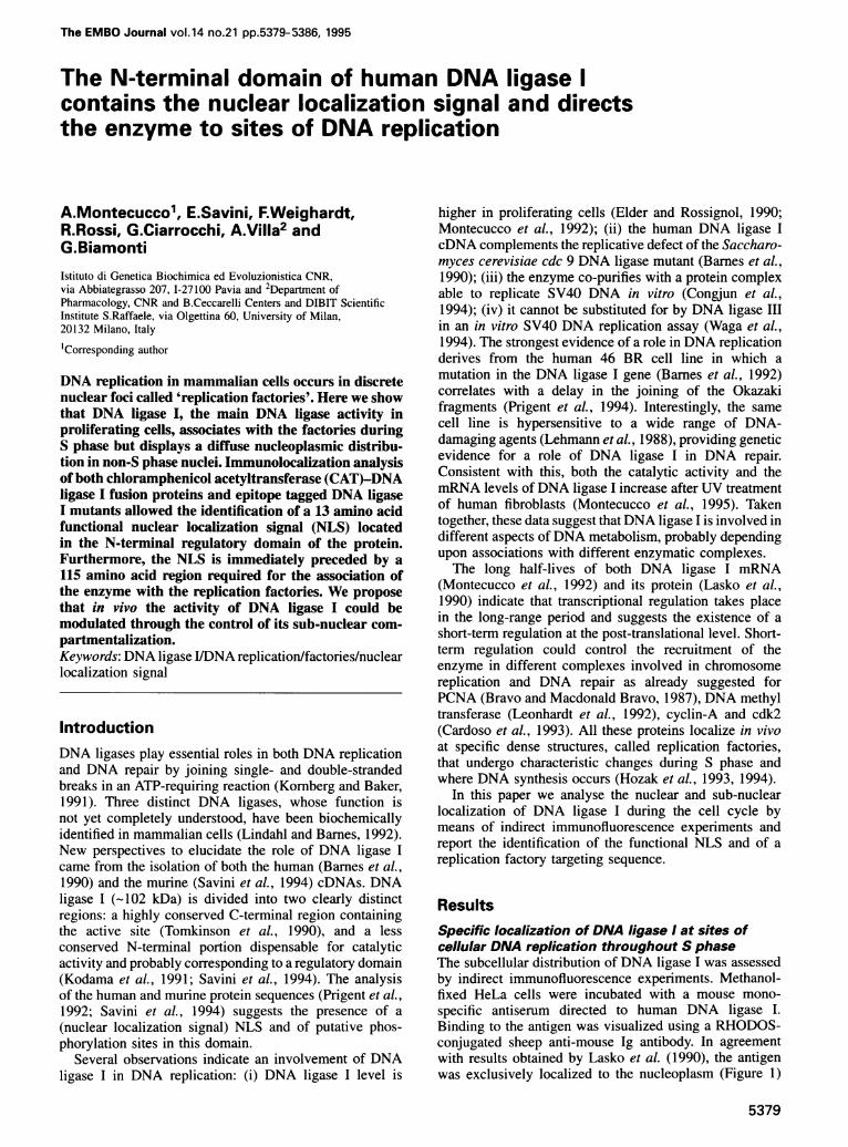
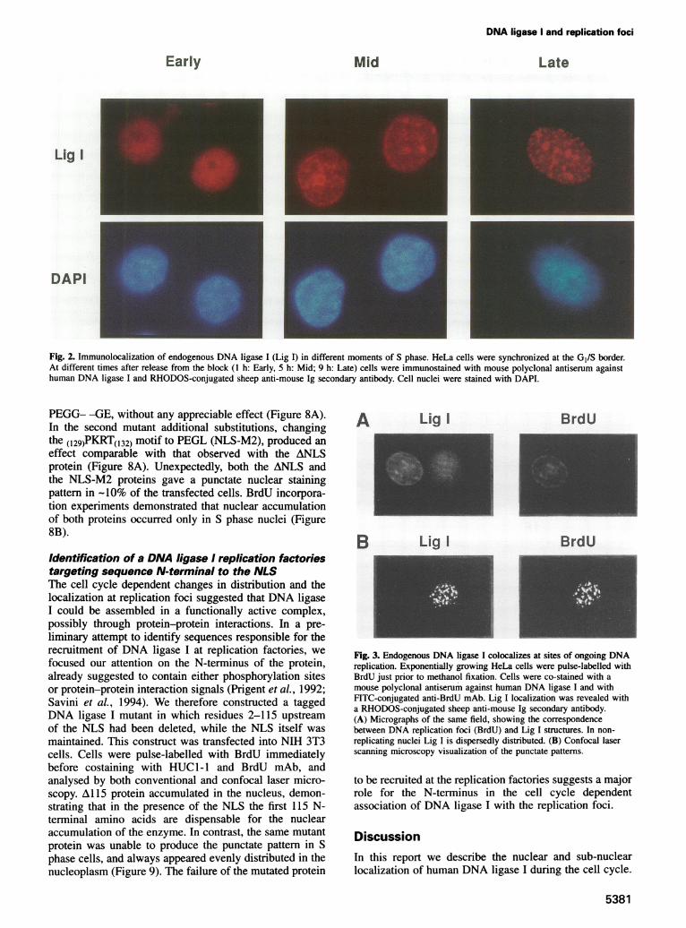
and nucleoli were not stained. DNA ligase I distributionvaried from homogeneous to granular-like encompassingthe wide range of intermediate possibilities. A similarpattern could be observed when a monoclonal antibody(mAb) to PCNA was used (Figure 1). To test whetherthe different staining patterns were related to cell cycleprogression, the enzyme localization was analysed inHeLa cells synchronized at the GI/S border by a doubleblock with thymidine and aphidicolin, and processed atdifferent times after releasing the block. Cell nuclei wererevealed by DAPI DNA staining. As exemplified in Figure2, at each time-point a definite fluorescent patten couldbe observed in most of the cells. In early S phase,i hafter the release from the block, a fine, punctate pattemrcould be observed, evenly distributed throughout thenucleoplasm. In the mid-S, 5 h after release, dots appearedmore concentrated around nucleoli and in the nuclearperiphery. In the late S phase, 9 h after release, only afew, large dots could be observed, on a homogeneousstaining of the nucleoplasm. The change in staining patterwas reminiscent of the time-course of DNA replication,suggesting an association of DNA ligase I with sites ofDNA replication. To prove this, HeLa cells were pulse-labelled for 1 h with 50en M BrdU and immediatelycostained with anti-DNA ligase I antibodies and FITC-conjugated anti-BrdU mAb. In non-replicating cells,evidenced by the lack of BrdU incorporation, DNAligase I appeared homogeneously dispersed throughoutthe nucleoplasm. Vice versa, in replicating cells the
staining patterns of BrdU and DNA ligase I were identical(Figure 3A). The colocalization was confirmed by opticaldissection with confocal laser scanning microscopy for abetter visualization of the punctate structure (Figure 3B).This analysis demonstrates that DNA ligase I indeedlocalizes at sites of ongoing DNA replication.
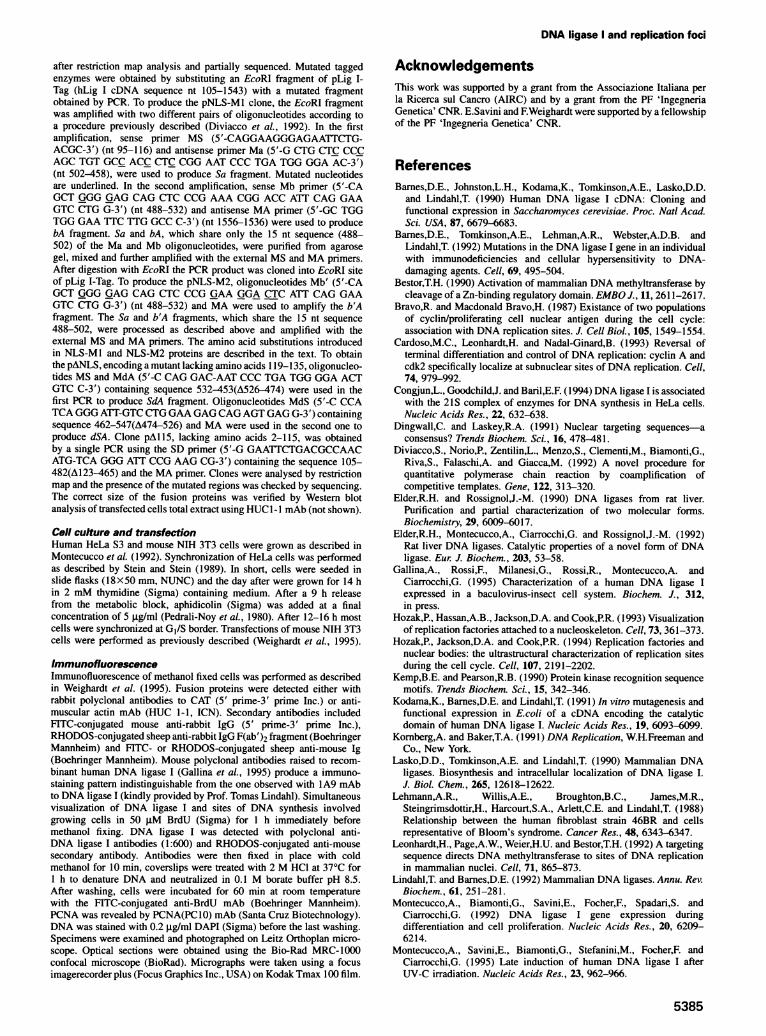
The DNA ligase I NLS resides in the N-terminaldomain of the proteinIn order to identify the DNA ligase I NLS, we producedfusions of chloramphenicol acetyltransferase (CAT) withvarious portions of the enzyme (Figure 4). CAT is a smallprotein that can passively diffuse through the nuclearmembrane and evenly distributes in the cell volume. Wehave previously described that the specific subcellularaccumulation of CAT depends on either canonical NLSor retention motifs. Moreover when the fusion protein islarger than 50-60 kDa nuclear accumulation can beobserved only in the presence of a functional NLS(Weighardt et al., 1995).
Specific human DNA ligase I cDNA fragments werecloned into the pAl-CAT vector (Weighardt et al., 1995)and transfected into NIH 3T3 cells with the calciumphosphate coprecipitation technique. After 48 h, cells wereimmunostained with anti-CAT antibodies. Three chimericgenes were initially tested: one coding for the catalyticdomain (C-ter) and the others for two regions spanningthe entire N-terminal domain (N-ter and EF). As shownin Figure 5, the CAT/N-ter(-119) fusion was evenly dis-tributed in the whole cell volume whilst the CAT/C-ter(202_919) protein, because of its large size, remainedconfined in the cytoplasmic compartment. In contrast,nuclear staining was observed in cells transfected with theCAT/EF(52 203) construct. The same pattern was observedwhen the corresponding region of the murine DNA ligaseI was tested (not shown). Further constructs (Figure 4)allowed us to identify a 13 amino acid peptide (II9PKRRT-ARKQLPKRI31), 100% identical between mouse and man,showing nuclear targeting activity [CAT/DF(119-131), Figure5]. This sequence shows some classical features of theNLS so far described, such as lysine/arginine clustersimmediately preceded by a proline (Dingwall andLaskey, 1991).The function of this putative NLS was directly assessed
by means of specific DNA ligase I mutants. To distinguishthe transfected from the endogenous enzyme we developeda vector (pAl -HUC, Figure 6) that directs the expressionin mammalian cells of proteins tagged at their C-terminuswith a 15 amino acid muscular actin epitope recognizedby the HUC 1-1 mAb. The tagged enzyme was nuclear andduring S phase colocalized at sites ofBrdU incorporation asdemonstrated by confocal laser scanning analysis (Figure7). Therefore, the presence of the actin epitope did notaffect the sub-nuclear localization of the protein, makingthis system suitable for the subsequent mutation analysis.When the putative NLS was removed (ANLS, Al 19-
135) nuclear localization was no longer observed in mostof the transfected cells (Figure 8A), indicating that thisregion is indeed necessary for nuclear targeting. In anattempt to dissect this sequence, we produced two addi-tional mutants carrying substitutions in the basic aminoacids (K/R). In one mutant (NLS-M1) the cluster(119)PKRR- -KR(126) was substituted with the sequence
5380
DNA ligase I and replication foci
Early Mid Late
Lig I
DAPI
Fig. 2. Immunolocalization of endogenous DNA ligase I (Lig I) in different moments of S phase. HeLa cells were synchronized at the GI/S border.At different times after release from the block (1 h: Early, 5 h: Mid; 9 h: Late) cells were immunostained with mouse polyclonal antiserum againsthuman DNA ligase I and RHODOS-conjugated sheep anti-mouse Ig secondary antibody. Cell nuclei were stained with DAPI.
PEGG- -GE, without any appreciable effect (Figure 8A).In the second mutant additional substitutions, changingthe (129)PKRT(l32) motif to PEGL (NLS-M2), produced aneffect comparable with that observed with the ANLSprotein (Figure 8A). Unexpectedly, both the ANLS andthe NLS-M2 proteins gave a punctate nuclear stainingpattern in -10% of the transfected cells. BrdU incorpora-tion experiments demonstrated that nuclear accumulationof both proteins occurred only in S phase nuclei (Figure8B).
Identification of a DNA ligase I replication factoriestargeting sequence N-terminal to the NLSThe cell cycle dependent changes in distribution and thelocalization at replication foci suggested that DNA ligaseI could be assembled in a functionally active complex,possibly through protein-protein interactions. In a pre-liminary attempt to identify sequences responsible for therecruitment of DNA ligase I at replication factories, wefocused our attention on the N-terminus of the protein,already suggested to contain either phosphorylation sitesor protein-protein interaction signals (Prigent et al., 1992;Savini et al., 1994). We therefore constructed a taggedDNA ligase I mutant in which residues 2-115 upstreamof the NLS had been deleted, while the NLS itself wasmaintained. This construct was transfected into NIH 3T3cells. Cells were pulse-labelled with BrdU immediatelybefore costaining with HUCI-1 and BrdU mAb, andanalysed by both conventional and confocal laser micro-scopy. A115 protein accumulated in the nucleus, demon-strating that in the presence of the NLS the first 115 N-terminal amino acids are dispensable for the nuclearaccumulation of the enzyme. In contrast, the same mutantprotein was unable to produce the punctate pattern in Sphase cells, and always appeared evenly distributed in thenucleoplasm (Figure 9). The failure of the mutated protein
A
B
Lig I BrdU
Lig I BrdU
Fig. 3. Endogenous DNA ligase I colocalizes at sites of ongoing DNAreplication. Exponentially growing HeLa cells were pulse-labelled withBrdU just prior to methanol fixation. Cells were co-stained with amouse polyclonal antiserum against human DNA ligase I and withFITC-conjugated anti-BrdU mAb. Lig I localization was revealed witha RHODOS-conjugated sheep anti-mouse Ig secondary antibody.(A) Micrographs of the same field, showing the correspondencebetween DNA replication foci (BrdU) and Lig I structures. In non-replicating nuclei Lig I is dispersedly distributed. (B) Confocal laserscanning microscopy visualization of the punctate patterns.
to be recruited at the replication factories suggests a majorrole for the N-terminus in the cell cycle dependentassociation of DNA ligase I with the replication foci.
DiscussionIn this report we describe the nuclear and sub-nuclearlocalization of human DNA ligase I during the cell cycle.
5381

A.Montecucco et al.
h~~~~~~~~~~~~~~~~~~~~~~~~~~DAAI.g_MS.MMEMMl 7., 1jr-
I
I( lo
N-ter /.19 _- Lig - IS1 --L i.4- I
C-ter 292 9W9 N LU S Lit 2
EF N: - ig --
GF -1¼ - 4 Li --g3
IF 3?1.Zi§. S 1
.
Vt'
.4 I -) /07 N - 1 i ¢ S 1- ig5 4
S 1L i
Cff /I f y 43) 'i%7 - J ig - S J,igl 0
)F I/ ; 7/ .ig
AI
.N'
Fig. 4. Strategy for the identification of human DNA ligase I NLS. Black bars indicate the fragments of human DNA ligase I fused to CAT. Thename of the clones and the set of primers used for PCR amplifications of the corresponding cDNA fragments are shown. The cellular localization ofeach fusion protein, as revealed by immunofluorescent staining of transfected cells, is also indicated. W, whole cell staining; C, cytoplasmic;N, nuclear.
N-Ter EF
IF CF
C-Ter
DF
5. SI llulCIIIa ):l Ii / h ( )II ) A IiIllIccTll(p In Ir:I n.lCICLd iStrcII1. O cd tli Ii l [I CCWhll
N;,Xt- .iiiAcd-m Epritpc
'.-7 p ri t pri't'1j
A.........l-H|
p16;
E1011o i
Fig. 6. Schematic rapresentation of pAl-HUC vector. The T7 and T3 promoters in the pBluescript SK(+) plasmid flank a cassette formed by: thehnRNP Al gene promoter (Al promoter), the EcoRI-SalI cloning sites, the muscular actin epitope encoding region, followed by the TAG stop
codon and by the SV40-derived RNA processing site.
5382
:.-A A4i Pr i nrlt I S Lncalisutron
VI'
(.'
DNA ligase I and replication foci
We show that the enzyme has a diffuse nucleoplasmicdistribution in non-S phase nuclei, while being specificallylocalized at sub-nuclear sites of ongoing DNA replicationthroughout S phase. Colocalization of DNA ligase I atsites of BrdU incorporation suggests that the enzyme isassembled in the DNA replisome at replication foci in vivo.A similar pattern has been reported for other replicationfactors such as DNA polymerase a, PCNA (Hozak et al.,1993) and DNA methyl transferase (Leonhardt et al.,1992). All these proteins appear to be relatively stable
Lig I-Tag BrdU
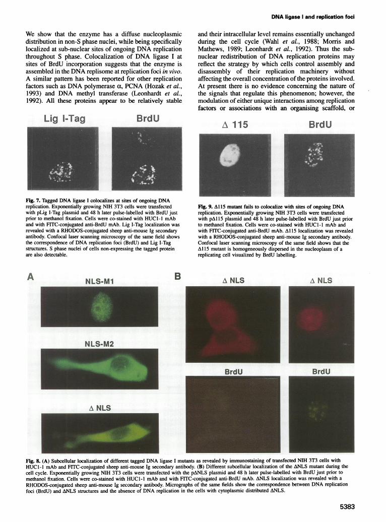
Fig. 7. Tagged DNA ligase I colocalizes at sites of ongoing DNAreplication. Exponentially growing NIH 3T3 cells were transfectedwith pLig I-Tag plasmid and 48 h later pulse-labelled with BrdU justprior to methanol fixation. Cells were co-stained with HUCG-l mAband with FITC-conjugated anti-BrdU mAb. Lig I-Tag localization wasrevealed with a RHODOS-conjugated sheep anti-mouse Ig secondaryantibody. Confocal laser scanning microscopy of the same field showsthe correspondence of DNA replication foci (BrdU) and Lig I-Tagstructures. S phase nuclei of cells non-expressing the tagged proteinare also detectable.
A BNLS-M1
and their intracellular level remains essentially unchangedduring the cell cycle (Wahl et al., 1988; Morris andMathews, 1989; Leonhardt et al., 1992). Thus the sub-nuclear redistribution of DNA replication proteins mayreflect the strategy by which cells control assembly anddisassembly of their replication machinery withoutaffecting the overall concentration of the proteins involved.At present there is no evidence concerning the nature ofthe signals that regulate this phenomenon; however, themodulation of either unique interactions among replicationfactors or associations with an organising scaffold, or
A 115 BrdU
Fig. 9. Al 15 mutant fails to colocalize with sites of ongoing DNAreplication. Exponentially growing NIH 3T3 cells were transfectedwith pAl 15 plasmid and 48 h later pulse-labelled with BrdU just priorto methanol fixation. Cells were co-stained with HUCl-l mAb andwith FITC-conjugated anti-BrdU mAb. Al 15 localization was revealedwith a RHODOS-conjugated sheep anti-mouse Ig secondary antibody.Confocal laser scanning microscopy of the same field shows that theA115 mutant is homogeneously dispersed in the nucleoplasm of areplicating cell visualized by BrdU labelling.
A NLS A NLS
NLS-M2
RrdU
A NLS
Fig. 8. (A) Subcellular localization of different tagged DNA ligase I mutants as revealed by immunostaining of transfected NIH 3T3 cells withHUCl-I mAb and FITC-conjugated sheep anti-mouse Ig secondary antibody. (B) Different subcellular localization of the ANLS mutant during thecell cycle. Exponentially growing NIH 3T3 cells were transfected with the pANLS plasmid and 48 h later pulse-labelled with BrdU just prior tomethanol fixation. Cells were co-stained with HUCI-l mAb and with FITC-conjugated anti-BrdU mAb. ANLS localization was revealed with a
RHODOS-conjugated sheep anti-mouse Ig secondary antibody. Micrographs of the same fields show the correspondence between DNA replicationfoci (BrdU) and ANLS structures and the absence of DNA replication in the cells with cytoplasmic distributed ANLS.
5383

A.Montecucco et aL
both, could have a major role. In this regard, it is importantto identify the protein determinants involved. Thus far,only DNA methyl transferase has been analysed in detailand its association with replication foci was found todepend on a sequence of -200 residues downstream ofthe NLS (Leonhardt et al., 1992) in the N-terminus of theprotein that corresponds to a large regulatory regiondispensable for the enzymatic activity (Bestor, 1990).The results reported here demonstrate that the DNA
ligase I determinants for nuclear and sub-nuclear accumu-lation are also located in the N-terminal regulatory domain,which is both a target of post-translational modifications(Prigent et al., 1992) and dispensable for the catalyticactivity (Kodama et al., 1991).A DNA ligase I mutant, in which the NLS has been
deleted (ANLS), failed to enter the cell nucleus in mostof the expressing cells. However, an unforeseen nuclearlocalization of the ANLS protein was detected in -10%of transfected cells and proved to be S phase-specific.Interestingly, the ANLS protein localized at the replicationfoci, ruling out a role for the NLS in the sub-nuclearrecruitment. This S phase-specific nuclear accumulationcould be due to the presence of a cell cycle dependentNLS that we failed to detect, although its physiologicalsignificance would be difficult to understand. We favouran alternative explanation which takes into account theability of DNA ligase I, and of its ANLS mutant, toassociate with the nuclear factories. This behaviour sug-gests the occurrence of specific protein interaction. Wepropose that a protein able to bind DNA ligase I onlyenters the cell nucleus immediately before the S phase,either because it is sequestered in the cytoplasm duringmost of the cell cycle or because it is synthesized at theend of the GI period. If this were the case, then thecytoplasmic ANLS protein would be 'piggybacked' intothe cell nucleus as described for other proteins with theirNLS deleted (Zacksenhaus et al., 1993). According to thishypothesis, ANLS DNA ligase I would interact withan as yet unidentified replication factor whose nuclearlocalization is strictly controlled during the cell cycle.This interaction could be relevant for the recruitment ofthe wild-type protein at the replication foci and couldexplain why DNA ligase III is unable to replace DNAligase I in a SV40 DNA replication assay (Waga et al.,1994), in spite of the overlapping substrate specificitybetween the two enzymes in vitro (Tomkinson et al., 1991;Elder et al., 1992). A validation of this hypothesis couldderive from the identification of the region of DNA ligaseI involved, a possible candidate being the 115 N-terminalresidues that seem to be a replication factories targetingsequence. This sequence has no homology with the func-tionally analogous region of the DNA methyl transferase.The 115 amino acid region is rich in charged residues, inprolines and in (SP) and (PS) motifs, all features conservedin evolution from mouse to man, in spite of the fact thatthe N-terminal domain is quite different in these twospecies (Savini et al., 1994). In particular the conservationof (SP) and (PS) residues could have a functional signific-ance since these motifs are present in the recognition siteof many protein kinases (Kemp and Pearson, 1990).Phosphorylation of the DNA ligase I N-terminal domainhas been shown to modulate the catalytic activity (Prigent
et al., 1992). Moreover it could have a role in modulatingthe interaction at replication foci.
Materials and methodsConstruction of CAT-DNA ligase I fusionsRecombinant plasmids were produced by cloning either oligonucleotidesor PCR products into the NsiI-SalI sites of the pA1-CAT vector(Weighardt et al., 1995). Different portions of human DNA ligase I wereobtained by PCR amplification of the cDNA [ATCC No. 65857 (Barneset al., 1990)] with suitable primers (Figure 4 and Table I). N (NsiI)designates sense sequence while S (SalI) designates antisense. N primerswere designed to obtain C-terminal fusions in-frame with the CATprotein. The amplification reactions were carried out with the Taqpolymerase (GeneAmp; Perkin Elmer Cetus); the correct sequence ofthe products was verified by the dideoxy method (Sequenasem DNASequencing Kit, USB) after cloning into the TA vector (Invitrogen Co.).Inserts were then excised by double digestion with NsiI-SaIl and clonedinto pAl-CAT. To produce the CAT-DF fusion two complementaryoligonucleotides, N-Lig-7 (+) and S-Lig-7 (-) (Table I), flanked by NsiIand SalI sites were annealed (5 min at 95°C and slow cooling at roomtemperature) and cloned into pAl-CAT. The correct size of the fusionproteins was verified by Western blot analysis of transfected cells totalextract using anti-CAT antibodies (not shown).
Construction ofpA 1-HUC expression vector and of taggedDNA ligase I mutantspAl-HUC expression vector was produced by cloning into EcoRI-PstIsites of pAl-SV40 plasmid (Weighardt et al., 1995), the fragmentobtained by the annealing of the following oligonucleotides: (+) 5'-AATTFC ATGCAT TATATATATA GTCGAC GGG ATG TGG ATA TCCAAA CAA GAA TAT GAT GAA GCA GGG CCA AGTA7TTAG CTGCA-3'; (-) 5'-G CTA AAT ACT TGG CCC TGC TTC ATC ATA TTC TTGTTT GGA TAT CCA CAT CCC GTCGAC TATATATATA ATGCAT G-3'. This fragment contains, from the 5' end: EcoRI and NsiI sites(underlined) followed, after a 10 nucleotide (nt) spacer, by a SalI site(underlined); a Gly codon preceding the muscular actin epitope (italics)recognised by HUC1-I mAb (ICN); a stop codon and a PstI site(underlined). To obtain the tagged DNA ligase I construct we first PCRamplified the cDNA region from nt 2503-2877 with primers: Lig-5':5'-GGCAGGCATTCA (2503)AAG CTT GGA ACT GGC TTC AGTGAT GA(2528)-3' and Lig-3': 5'-GGCAGGCATTCA GTCGAC(2877)GTA GGT ATC TTC AGG GTC AG(2858)-3'. In this way wereplaced the original stop codon with a SalI (underlined) site. The PCRproduct was digested with HindlIl and Sall and ligated in the presenceof the KpnI-HindIII hLig I cDNA fragment (nt 1-2503) into KpnI-SalJsites of pUC19 vector (Boehringer Mannheim). The resulting construct(pUC-Lig) contains 120 nt of the 5'-UTR followed by the entire codingsequence. The pUC-Lig plasmid was linearized with KpnI, blunted withKlenow fragment (Boehringer Mannheim) and further digested withSalI. The excised cDNA was then cloned into pAl-HUC linearized withEcoRI, blunted and digested with Sail. A clone (pLig I-Tag) was selected
Table I. Oligonucleotide primers used in PCR amplification to obtainthe DNA ligase I cDNA fragments encoding the fusion proteins shownin Figure 4
Pnmer
N-U -1N - Lig -2N-U-3N- ig-4N-ig-5N-Ug-6N - Lig -7
Poon (nt)
S GGCAGGCATTCATGAT
5.
5 T
- ( 121- 148 )- ( 724- 748 )- ( 274- 300)- ( 415. 439 )- ( ms $o )- ( 475 4 )
( 475 513 ) -G
3,3'3'3'3,3,3,
S- Li-1 GGCATTCAGTGAC - ( 477- 43 )S-L -2 - (7-M4)S-L-3 S-(57,-7M)S-L-44, - ( 2- m )S-L;S 5' -( 57. ss )S-Li-6 1 -(5C1o sos)S-Lg-7 F5 TCGAC - 513.- 7 ) ATC3A
3'3'
3,3,3,3,3,
5384
DNA ligase I and replication foci
after restriction map analysis and partially sequenced. Mutated taggedenzymes were obtained by substituting an EcoRI fragment of pLig I-Tag (hLig I cDNA sequence nt 105-1543) with a mutated fragmentobtained by PCR. To produce the pNLS-Ml clone, the EcoRI fragmentwas amplified with two different pairs of oligonucleotides according toa procedure previously described (Diviacco et al., 1992). In the firstamplification, sense primer MS (5'-CAGGAAGGGAGAATTCTG-ACGC-3') (nt 95-116) and antisense primer Ma (5'-G CTG CTC CCCAGC TGT GCC ACC CTC CGG AAT CCC TGA TGG GGA AC-3')(nt 502-458), were used to produce Sa fragment. Mutated nucleotidesare underlined. In the second amplification, sense Mb primer (5'-CAGCT GGG GAG CAG CTC CCG AAA CGG ACC ATT CAG GAAGTC CTG G-3') (nt 488-532) and antisense MA primer (5'-GC TGGTGG GAA TTC TTG GCC C-3') (nt 1556-1536) were used to producebA fragment. Sa and bA, which share only the 15 nt sequence (488-502) of the Ma and Mb oligonucleotides, were purified from agarosegel, mixed and further amplified with the external MS and MA primers.After digestion with EcoRI the PCR product was cloned into EcoRI siteof pLig I-Tag. To produce the pNLS-M2, oligonucleotides Mb' (5'-CAGCT GGG GAG CAG CTC CCG GAA GGA CTC ATT CAG GAAGTC CTG G-3') (nt 488-532) and MA were used to amplify the b'Afragment. The Sa and b'A fragments, which share the 15 nt sequence488-502, were processed as described above and amplified with theexternal MS and MA primers. The amino acid substitutions introducedin NLS-Ml and NLS-M2 proteins are described in the text. To obtainthe pANLS, encoding a mutant lacking amino acids 119-135, oligonucleo-tides MS and MdA (5'-C CAG GAC-AAT CCC TGA TGG GGA ACTGTC C-3') containing sequence 532-453(A526-474) were used in thefirst PCR to produce SdA fragment. Oligonucleotides MdS (5'-C CCATCA GGG ATT-GTC CTGGAA GAGCAG AGT GAG G-3') containingsequence 462-547(A474-526) and MA were used in the second one toproduce dSA. Clone pAl 15, lacking amino acids 2-115, was obtainedby a single PCR using the SD primer (5'-G GAATTCTGACGCCAACATG-TCA GGG ATT CCG AAG CG-3') containing the sequence 105-482(A123-465) and the MA primer. Clones were analysed by restrictionmap and the presence of the mutated regions was checked by sequencing.The correct size of the fusion proteins was verified by Western blotanalysis of transfected cells total extract using HUC 1-1 mAb (not shown).
Cell culture and transfectionHuman HeLa S3 and mouse NIH 3T3 cells were grown as described inMontecucco et al. (1992). Synchronization of HeLa cells was performedas described by Stein and Stein (1989). In short, cells were seeded inslide flasks (18X50 mm, NUNC) and the day after were grown for 14 hin 2 mM thymidine (Sigma) containing medium. After a 9 h releasefrom the metabolic block, aphidicolin (Sigma) was added at a finalconcentration of 5 tg/ml (Pedrali-Noy et al., 1980). After 12-16 h mostcells were synchronized at G1/S border. Transfections of mouse NIH 3T3cells were performed as previously described (Weighardt et al., 1995).
ImmunofluorescenceImmunofluorescence of methanol fixed cells was performed as describedin Weighardt et al. (1995). Fusion proteins were detected either withrabbit polyclonal antibodies to CAT (5' prime-3' prime Inc.) or anti-muscular actin mAb (HUC 1-1, ICN). Secondary antibodies includedFITC-conjugated mouse anti-rabbit IgG (5' prime-3' prime Inc.),RHODOS-conjugated sheep anti-rabbit IgG F(ab')2 fragment (BoehringerMannheim) and FITC- or RHODOS-conjugated sheep anti-mouse Ig(Boehringer Mannheim). Mouse polyclonal antibodies raised to recom-binant human DNA ligase I (Gallina et al., 1995) produce a immuno-staining pattern indistinguishable from the one observed with lA9 mAbto DNA ligase I (kindly provided by Prof. Tomas Lindahl). Simultaneousvisualization of DNA ligase I and sites of DNA synthesis involvedgrowing cells in 50 tM BrdU (Sigma) for 1 h immediately beforemethanol fixing. DNA ligase I was detected with polyclonal anti-DNA ligase I antibodies (1:600) and RHODOS-conjugated anti-mousesecondary antibody. Antibodies were then fixed in place with coldmethanol for 10 min, coverslips were treated with 2 M HCI at 370C for1 h to denature DNA and neutralized in 0.1 M borate buffer pH 8.5.After washing, cells were incubated for 60 min at room temperaturewith the FITC-conjugated anti-BrdU mAb (Boehringer Mannheim).PCNA was revealed by PCNA(PC1O) mAb (Santa Cruz Biotechnology).DNA was stained with 0.2 tg/ml DAPI (Sigma) before the last washing.Specimens were examined and photographed on Leitz Orthoplan micro-scope. Optical sections were obtained using the Bio-Rad MRC-1000confocal microscope (BioRad). Micrographs were taken using a focusimagerecorder plus (Focus Graphics Inc., USA) on Kodak Tmax 100 film.
AcknowledgementsThis work was supported by a grant from the Associazione Italiana perla Ricerca sul Cancro (AIRC) and by a grant from the PF 'IngegneriaGenetica' CNR. E.Savini and F.Weighardt were supported by a fellowshipof the PF 'Ingegneria Genetica' CNR.
ReferencesBarnes,D.E., Johnston,L.H., Kodama,K., Tomkinson,A.E., Lasko,D.D.
and Lindahl,T. (1990) Human DNA ligase I cDNA: Cloning andfunctional expression in Saccharomyces cerevisiae. Proc. Natl Acad.Sci. USA, 87, 6679-6683.
Barnes,D.E., Tomkinson,A.E., Lehman,A.R., Webster,A.D.B. andLindahl,T. (1992) Mutations in the DNA ligase I gene in an individualwith immunodeficiencies and cellular hypersensitivity to DNA-damaging agents. Cell, 69, 495-504.
Bestor,T.H. (1990) Activation of mammalian DNA methyltransferase bycleavage of a Zn-binding regulatory domain. EMBO J., 11, 2611-2617.
Bravo,R. and Macdonald Bravo,H. (1987) Existance of two populationsof cyclin/proliferating cell nuclear antigen during the cell cycle:association with DNA replication sites. J. Cell Biol., 105, 1549-1554.
Cardoso,M.C., Leonhardt,H. and Nadal-Ginard,B. (1993) Reversal ofterminal differentiation and control of DNA replication: cyclin A andcdk2 specifically localize at subnuclear sites of DNA replication. Cell,74, 979-992.
Congjun,L., Goodchild,J. and Baril,E.F. (1994) DNA ligase I is associatedwith the 21S complex of enzymes for DNA synthesis in HeLa cells.Nucleic Acids Res., 22, 632-638.
Dingwall,C. and Laskey,R.A. (1991) Nuclear targeting sequences-aconsensus? Trends Biochem. Sci., 16, 478-481.
Diviacco,S., Norio,P., Zentilin,L., Menzo,S., Clementi,M., Biamonti,G.,Riva,S., Falaschi,A. and Giacca,M. (1992) A novel procedure forquantitative polymerase chain reaction by coamplification ofcompetitive templates. Gene, 122, 313-320.
Elder,R.H. and Rossignol,J.-M. (1990) DNA ligases from rat liver.Purification and partial characterization of two molecular forms.Biochemistry, 29, 6009-6017.
Elder,R.H., Montecucco,A., Ciarrocchi,G. and Rossignol,J.-M. (1992)Rat liver DNA ligases. Catalytic properties of a novel form of DNAligase. Eur J. Biochem., 203, 53-58.
Gallina,A., Rossi,F., Milanesi,G., Rossi,R., Montecucco,A. andCiarrocchi,G. (1995) Characterization of a human DNA ligase Iexpressed in a baculovirus-insect cell system. Biochem. J., 312,in press.
Hozak,P., Hassan,A.B., Jackson,D.A. and Cook,P.R. (1993) Visualizationof replication factories attached to a nucleoskeleton. Cell, 73, 361-373.
Hozak,P., Jackson,D.A. and Cook,P.R. (1994) Replication factories andnuclear bodies: the ultrastructural characterization of replication sitesduring the cell cycle. Cell, 107, 2191-2202.
Kemp,B.E. and Pearson,R.B. (1990) Protein kinase recognition sequencemotifs. Trends Biochem. Sci., 15, 342-346.
Kodama,K., Barnes,D.E. and Lindahl,T. (1991) In vitro mutagenesis andfunctional expression in Ecoli of a cDNA encoding the catalyticdomain of human DNA ligase I. Nucleic Acids Res., 19, 6093-6099.
Kornberg,A. and Baker,T.A. (1991) DNA Replication, W.H.Freeman andCo., New York.
Lasko,D.D., Tomkinson,A.E. and Lindahl,T. (1990) Mammalian DNAligases. Biosynthesis and intracellular localization of DNA ligase I.J. Biol. Chem., 265, 12618-12622.
Lehmann,A.R., Willis,A.E., Broughton,B.C., James,M.R.,Steingrimsdottir,H., Harcourt,S.A., Arlett,C.E. and Lindahl,T. (1988)Relationship between the human fibroblast strain 46BR and cellsrepresentative of Bloom's syndrome. Cancer Res., 48, 6343-6347.
Leonhardt,H., Page,A.W., Weier,H.U. and Bestor,T.H. (1992) A targetingsequence directs DNA methyltransferase to sites of DNA replicationin mammalian nuclei. Cell, 71, 865-873.
Lindahl,T. and Barnes,D.E. (1992) Mammalian DNA ligases. Annu. Rev.Biochem., 61, 251-281.
Montecucco,A., Biamonti,G., Savini,E., Focher,F., Spadari,S. andCiarrocchi,G. (1992) DNA ligase I gene expression duringdifferentiation and cell proliferation. Nucleic Acids Res., 20, 6209-6214.
Montecucco,A., Savini,E., Biamonti,G., Stefanini,M., Focher,F. andCiarrocchi,G. (1995) Late induction of human DNA ligase I afterUV-C irradiation. Nucleic Acids Res., 23, 962-966.
5385
A.Montecucco et al.
Morris,G.F. and Mathews,M.B. (1989) Regulation of proliferating cellnuclear antigen during the cell cycle. J. Bio. Chem., 264, 13856-13864.
Pedrali-Noy,G., Spadari,S., Miller-Faures,A.,. Miller,A.O.A., Kruppa,J.and Koch,G. (1980) Synchronization ofHeLa cell cultures by inhibitionof DNA polymerase a with aphidicolin. Nucleic Acids Res., 8,377-387.
Prigent,C., Lasko,D.D., Kodama,K., Woodgett,J.R. and Lindahl,T. (1992)Activation of mammalian DNA ligase I through phosphorylation bycasein kinase II. EMBO J., 11, 2925-2933.
Prigent,C., Satoh,M.S., Daly,G., Barnes,D.E. and Lindahl,T. (1994)Aberrant DNA repair and DNA replication due to an inheritedenzymatic defect in human DNA ligase I. Mol. Cell. Biol., 14,310-317.
Savini,E., Biamonti,G., Ciarrocchi,G. and Montecucco,A. (1994) Cloningand sequence analysis of a cDNA coding for the murine DNA ligaseI enzyme. Gene, 144, 253-257.
Stein,G.S. and Stein,J.L. (1989) In Baserga,R. (ed.), Cell Synchronization.IRL Press, Oxford, pp. 133-137.
Tomkinson,A.E., Lasko,D.D., Daly,G. and Lindahl,T. (1990) MammalianDNA ligases. Catalytic domain and size of DNA ligase I. J. Bio.Chem., 265, 12611-12617.
Tomkinson,A.E., Roberts,E., Daly,G., Totty,N.F. and Lindahl,T. (1991)Three distinct DNA ligases in mammalian cells. J. Biol. Chem., 266,21728-21735.
Waga,S., Bauer,G. and Stillman,B. (1994) Reconstitution of completeSV40 DNA replication with purified replication factor. J. Biol. Chem.,269, 10923-10934.
Wahl,A.F., Geis,A.M., Spain,B.H., Wong,S.W., Kom,D. and Wang,T.S.-F.(1988) Gene expression of human DNA polymerase a during cellproliferation and the cell cycle. Mol. Cell. Biol., 8, 5016-5025.
Weighardt,F., Biamonti,G. and Riva,S. (1995) Nucleo-cytoplasmicdistribution of human hnRNP proteins: a search for the targetingdomains in hnRNP Al. J. Cell Sci., 108, 545-555.
Zacksenhaus,E., Bremner,R., Phillips,R.A. and Gallie,B.L. (1993) Abipartite nuclear localization signal in the retinoblastoma gene productand its importance for biological activity. Mol. Cell. Biol., 13,4588-4599.
Received on May 31, 1995; revised on July 25, 1995
5386