the nervous system - ms. rickard's anatomy...
TRANSCRIPT

Essentials of Human Anatomy & Physiology
Copyright © 2003 Pearson Education, Inc. publishing as Benjamin Cummings
Slides 7.1 – 7.22
Seventh Edition
Elaine N. Marieb
Chapter 7
The Nervous System
Lecture Slides in PowerPoint by Jerry L. Cook

Functions of the Nervous System
Slide 7.1a Copyright © 2003 Pearson Education, Inc. publishing as Benjamin Cummings
Sensory input – gathering information
To monitor changes occurring inside and outside the body
Changes = stimuli
Integration
To process and interpret sensory input and decide if action is needed

Functions of the Nervous System
Slide 7.1b Copyright © 2003 Pearson Education, Inc. publishing as Benjamin Cummings
Motor output
A response to integrated stimuli
The response activates muscles or glands

Structural Classification of the Nervous System
Slide 7.2 Copyright © 2003 Pearson Education, Inc. publishing as Benjamin Cummings
Central nervous system (CNS)
Brain
Spinal cord
Peripheral nervous system (PNS)
Nerve outside the brain and spinal cord

Functional Classification of the Peripheral Nervous System
Slide 7.3a Copyright © 2003 Pearson Education, Inc. publishing as Benjamin Cummings
Sensory (afferent) division
Nerve fibers that carry information to the
central nervous system
Figure 7.1

Functional Classification of the Peripheral Nervous System
Slide 7.3b Copyright © 2003 Pearson Education, Inc. publishing as Benjamin Cummings
Motor (efferent) division
Nerve fibers that carry impulses away from
the central nervous system
Figure 7.1

Functional Classification of the Peripheral Nervous System
Slide 7.3c Copyright © 2003 Pearson Education, Inc. publishing as Benjamin Cummings
Motor (efferent) division
Two subdivisions
Somatic nervous system = voluntary
Autonomic nervous system = involuntary
Figure 7.1

Organization of the Nervous System
Slide 7.4 Copyright © 2003 Pearson Education, Inc. publishing as Benjamin Cummings
Figure 7.2

Nervous Tissue: Support Cells (Neuroglia)
Slide 7.5 Copyright © 2003 Pearson Education, Inc. publishing as Benjamin Cummings
Astrocytes
Abundant, star-shaped cells
Brace neurons
Form barrier between capillaries and neurons
Control the chemical environment of the brain
Figure 7.3a

Nervous Tissue: Support Cells
Slide 7.6 Copyright © 2003 Pearson Education, Inc. publishing as Benjamin Cummings
Microglia
Spider-like phagocytes
Dispose of debris
Ependymal cells
Line cavities of the brain and spinal cord
Circulate cerebrospinal fluid
Figure 7.3b, c

Nervous Tissue: Support Cells
Slide 7.7a Copyright © 2003 Pearson Education, Inc. publishing as Benjamin Cummings
Oligodendrocytes
Produce myelin
sheath around
nerve fibers in the
central nervous
system
Figure 7.3d

Nervous Tissue: Support Cells
Slide 7.7b Copyright © 2003 Pearson Education, Inc. publishing as Benjamin Cummings
Satellite cells
Protect neuron cell bodies
Schwann cells
Form myelin sheath in the peripheral nervous system
Figure 7.3e

Nervous Tissue: Neurons
Slide 7.8 Copyright © 2003 Pearson Education, Inc. publishing as Benjamin Cummings
Neurons = nerve cells
Cells specialized to transmit messages
Major regions of neurons
Cell body – nucleus and metabolic center of the cell
Processes – fibers that extend from the cell body
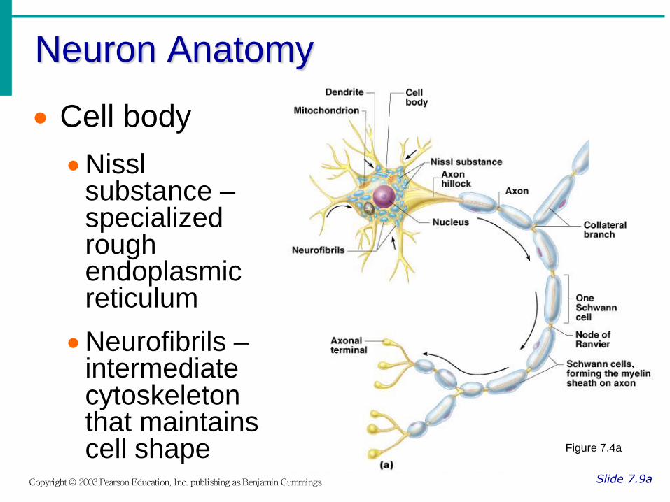
Neuron Anatomy
Slide 7.9a Copyright © 2003 Pearson Education, Inc. publishing as Benjamin Cummings
Cell body
Nissl substance – specialized rough endoplasmic reticulum
Neurofibrils – intermediate cytoskeleton that maintains cell shape Figure 7.4a

Neuron Anatomy
Slide 7.9b Copyright © 2003 Pearson Education, Inc. publishing as Benjamin Cummings
Cell body
Nucleus
Large nucleolus
Figure 7.4a

Neuron Anatomy
Slide 7.10 Copyright © 2003 Pearson Education, Inc. publishing as Benjamin Cummings
Extensions outside the cell body
Dendrites – conduct impulses toward the cell body
Axons – conduct impulses away from the cell body
Figure 7.4a

Axons and Nerve Impulses
Slide 7.11 Copyright © 2003 Pearson Education, Inc. publishing as Benjamin Cummings
Axons end in axonal terminals
Axonal terminals contain vesicles with neurotransmitters
Axonal terminals are separated from the next neuron by a gap
Synaptic cleft – gap between adjacent neurons
Synapse – junction between nerves
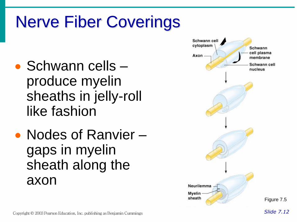
Nerve Fiber Coverings
Slide 7.12 Copyright © 2003 Pearson Education, Inc. publishing as Benjamin Cummings
Schwann cells – produce myelin sheaths in jelly-roll like fashion
Nodes of Ranvier – gaps in myelin sheath along the axon
Figure 7.5

Neuron Cell Body Location
Slide 7.13 Copyright © 2003 Pearson Education, Inc. publishing as Benjamin Cummings
Most are found in the central nervous system
Gray matter – cell bodies and unmylenated fibers
Nuclei – clusters of cell bodies within the white matter of the central nervous system
Ganglia – collections of cell bodies outside the central nervous system

Functional Classification of Neurons
Slide 7.14a Copyright © 2003 Pearson Education, Inc. publishing as Benjamin Cummings
Sensory (afferent) neurons
Carry impulses from the sensory receptors
Cutaneous sense organs
Proprioceptors – detect stretch or tension
Motor (efferent) neurons
Carry impulses from the central nervous system

Functional Classification of Neurons
Slide 7.14b Copyright © 2003 Pearson Education, Inc. publishing as Benjamin Cummings
Interneurons (association neurons)
Found in neural pathways in the central nervous system
Connect sensory and motor neurons

Neuron Classification
Slide 7.15 Copyright © 2003 Pearson Education, Inc. publishing as Benjamin Cummings
Figure 7.6

Structural Classification of Neurons
Slide 7.16a Copyright © 2003 Pearson Education, Inc. publishing as Benjamin Cummings
Multipolar neurons – many extensions from the cell body
Figure 7.8a

Structural Classification of Neurons
Slide 7.16b Copyright © 2003 Pearson Education, Inc. publishing as Benjamin Cummings
Bipolar neurons – one axon and one dendrite
Figure 7.8b
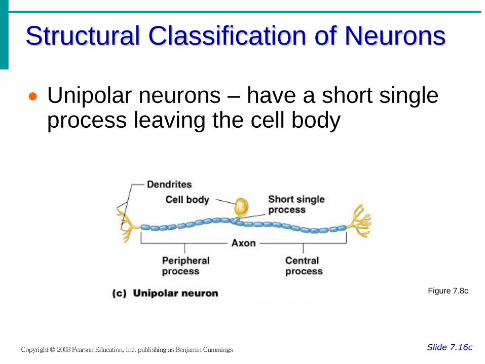
Structural Classification of Neurons
Slide 7.16c Copyright © 2003 Pearson Education, Inc. publishing as Benjamin Cummings
Unipolar neurons – have a short single process leaving the cell body
Figure 7.8c

Functional Properties of Neurons
Slide 7.17 Copyright © 2003 Pearson Education, Inc. publishing as Benjamin Cummings
Irritability – ability to respond to stimuli
Conductivity – ability to transmit an impulse
The plasma membrane at rest is polarized
Fewer positive ions are inside the cell than outside the cell

Starting a Nerve Impulse
Slide 7.18 Copyright © 2003 Pearson Education, Inc. publishing as Benjamin Cummings
Depolarization – a stimulus depolarizes the neuron’s membrane
A deploarized membrane allows sodium (Na+) to flow inside the membrane
The exchange of ions initiates an action potential in the neuron
Figure 7.9a–c

The Action Potential
Slide 7.19 Copyright © 2003 Pearson Education, Inc. publishing as Benjamin Cummings
If the action potential (nerve impulse) starts, it is propagated over the entire axon
Potassium ions rush out of the neuron after sodium ions rush in, which repolarizes the membrane
The sodium-potassium pump restores the original configuration
This action requires ATP

Nerve Impulse Propagation
Slide 7.20 Copyright © 2003 Pearson Education, Inc. publishing as Benjamin Cummings
The impulse continues to move toward the cell body
Impulses travel faster when fibers have a myelin sheath
Figure 7.9c–e

Continuation of the Nerve Impulse between Neurons
Slide 7.21 Copyright © 2003 Pearson Education, Inc. publishing as Benjamin Cummings
Impulses are able to cross the synapse to another nerve
Neurotransmitter is released from a nerve’s axon terminal
The dendrite of the next neuron has receptors that are stimulated by the neurotransmitter
An action potential is started in the dendrite

How Neurons Communicate at Synapses
Slide 7.22 Copyright © 2003 Pearson Education, Inc. publishing as Benjamin Cummings
Figure 7.10
Essentials of Human Anatomy & Physiology
Copyright © 2003 Pearson Education, Inc. publishing as Benjamin Cummings
Slides 7.23 – 7.42
Seventh Edition
Elaine N. Marieb
Chapter 7
The Nervous System
Lecture Slides in PowerPoint by Jerry L. Cook

The Reflex Arc
Slide 7.23 Copyright © 2003 Pearson Education, Inc. publishing as Benjamin Cummings
Reflex – rapid, predictable, and involuntary responses to stimuli
Reflex arc – direct route from a sensory neuron, to an interneuron, to an effector
Figure 7.11a

Simple Reflex Arc
Slide 7.24 Copyright © 2003 Pearson Education, Inc. publishing as Benjamin Cummings
Figure 7.11b, c
Types of Reflexes and Regulation
Slide 7.25 Copyright © 2003 Pearson Education, Inc. publishing as Benjamin Cummings
Autonomic reflexes
Smooth muscle regulation
Heart and blood pressure regulation
Regulation of glands
Digestive system regulation
Somatic reflexes
Activation of skeletal muscles

Central Nervous System (CNS)
Slide 7.26 Copyright © 2003 Pearson Education, Inc. publishing as Benjamin Cummings
CNS develops from the embryonic neural tube
The neural tube becomes the brain and spinal cord
The opening of the neural tube becomes the ventricles
Four chambers within the brain
Filled with cerebrospinal fluid

Cerebral Hemispheres (Cerebrum)
Slide 7.28b Copyright © 2003 Pearson Education, Inc. publishing as Benjamin Cummings
The surface is made of ridges (gyri) and grooves (sulci)
Figure 7.13a

Regions of the Brain
Slide 7.27 Copyright © 2003 Pearson Education, Inc. publishing as Benjamin Cummings
Cerebral hemispheres
Diencephalon
Brain stem
Cerebellum Figure 7.12

Cerebral Hemispheres (Cerebrum)
Slide 7.28a Copyright © 2003 Pearson Education, Inc. publishing as Benjamin Cummings
Paired (left and right) superior parts of the brain
Include more than half of the brain mass
Figure 7.13a
Lobes of the Cerebrum
Slide 7.29a Copyright © 2003 Pearson Education, Inc. publishing as Benjamin Cummings
Fissures (deep grooves) divide the cerebrum into lobes
Surface lobes of the cerebrum
Frontal lobe
Parietal lobe
Occipital lobe
Temporal lobe

Lobes of the Cerebrum
Slide 7.29b Copyright © 2003 Pearson Education, Inc. publishing as Benjamin Cummings
Figure 7.15a

Specialized Areas of the Cerebrum
Slide 7.30 Copyright © 2003 Pearson Education, Inc. publishing as Benjamin Cummings
Somatic sensory area – receives impulses from the body’s sensory receptors
Primary motor area – sends impulses to skeletal muscles
Broca’s area – involved in our ability to speak

Sensory and Motor Areas of the
Cerebral Cortex
Slide 7.31 Copyright © 2003 Pearson Education, Inc. publishing as Benjamin Cummings
Figure 7.14

Specialized Area of the Cerebrum
Slide 7.32a Copyright © 2003 Pearson Education, Inc. publishing as Benjamin Cummings
Cerebral areas involved in special senses
Gustatory area (taste)
Visual area
Auditory area
Olfactory area

Specialized Area of the Cerebrum
Slide 7.32b Copyright © 2003 Pearson Education, Inc. publishing as Benjamin Cummings
Interpretation areas of the cerebrum
Speech/language region
Language comprehension region
General interpretation area

Specialized Area of the Cerebrum
Slide 7.32c Copyright © 2003 Pearson Education, Inc. publishing as Benjamin Cummings
Figure 7.13c

Layers of the Cerebrum
Slide 7.33a Copyright © 2003 Pearson Education, Inc. publishing as Benjamin Cummings
Gray matter
Outer layer
Composed mostly of neuron cell bodies
Figure 7.13a

Layers of the Cerebrum
Slide 7.33b Copyright © 2003 Pearson Education, Inc. publishing as Benjamin Cummings
White matter
Fiber tracts inside the gray matter
Example: corpus callosum connects hemispheres
Figure 7.13a
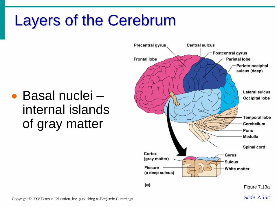
Layers of the Cerebrum
Slide 7.33c Copyright © 2003 Pearson Education, Inc. publishing as Benjamin Cummings
Basal nuclei – internal islands of gray matter
Figure 7.13a

Diencephalon
Slide 7.34a Copyright © 2003 Pearson Education, Inc. publishing as Benjamin Cummings
Sits on top of the brain stem
Enclosed by the cerebral heispheres
Made of three parts
Thalamus
Hypothalamus
Epithalamus

Diencephalon
Slide 7.34b Copyright © 2003 Pearson Education, Inc. publishing as Benjamin Cummings
Figure 7.15

Thalamus
Slide 7.35 Copyright © 2003 Pearson Education, Inc. publishing as Benjamin Cummings
Surrounds the third ventricle
The relay station for sensory impulses
Transfers impulses to the correct part of the cortex for localization and interpretation
Hypothalamus
Slide 7.36a Copyright © 2003 Pearson Education, Inc. publishing as Benjamin Cummings
Under the thalamus
Important autonomic nervous system center
Helps regulate body temperature
Controls water balance
Regulates metabolism
Hypothalamus
Slide 7.36b Copyright © 2003 Pearson Education, Inc. publishing as Benjamin Cummings
An important part of the limbic system (emotions)
The pituitary gland is attached to the hypothalamus
Epithalamus
Slide 7.37 Copyright © 2003 Pearson Education, Inc. publishing as Benjamin Cummings
Forms the roof of the third ventricle
Houses the pineal body (an endocrine gland)
Includes the choroid plexus – forms cerebrospinal fluid

Brain Stem
Slide 7.38a Copyright © 2003 Pearson Education, Inc. publishing as Benjamin Cummings
Attaches to the spinal cord
Parts of the brain stem
Midbrain
Pons
Medulla oblongata

Brain Stem
Slide 7.38b Copyright © 2003 Pearson Education, Inc. publishing as Benjamin Cummings
Figure 7.15a
Midbrain
Slide 7.39 Copyright © 2003 Pearson Education, Inc. publishing as Benjamin Cummings
Mostly composed of tracts of nerve fibers
Has two bulging fiber tracts – cerebral peduncles
Has four rounded protrusions – corpora quadrigemina
Reflex centers for vision and hearing

Pons
Slide 7.40 Copyright © 2003 Pearson Education, Inc. publishing as Benjamin Cummings
The bulging center part of the brain stem
Mostly composed of fiber tracts
Includes nuclei involved in the control of breathing

Medulla Oblongata
Slide 7.41 Copyright © 2003 Pearson Education, Inc. publishing as Benjamin Cummings
The lowest part of the brain stem
Merges into the spinal cord
Includes important fiber tracts
Contains important control centers
Heart rate control
Blood pressure regulation
Breathing
Swallowing
Vomiting

Reticular Formation
Slide 7.42a Copyright © 2003 Pearson Education, Inc. publishing as Benjamin Cummings
Diffuse mass of gray matter along the brain stem
Involved in motor control of visceral organs
Reticular activating system plays a role in awake/sleep cycles and consciousness

Reticular Formation
Slide 7.42b Copyright © 2003 Pearson Education, Inc. publishing as Benjamin Cummings
Figure 7.15b

Essentials of Human Anatomy & Physiology
Copyright © 2003 Pearson Education, Inc. publishing as Benjamin Cummings
Slides 7.43 – 7.62
Seventh Edition
Elaine N. Marieb
Chapter 7
The Nervous System
Lecture Slides in PowerPoint by Jerry L. Cook
Cerebellum
Slide 7.43a Copyright © 2003 Pearson Education, Inc. publishing as Benjamin Cummings
Two hemispheres with convoluted surfaces
Provides involuntary coordination of body movements
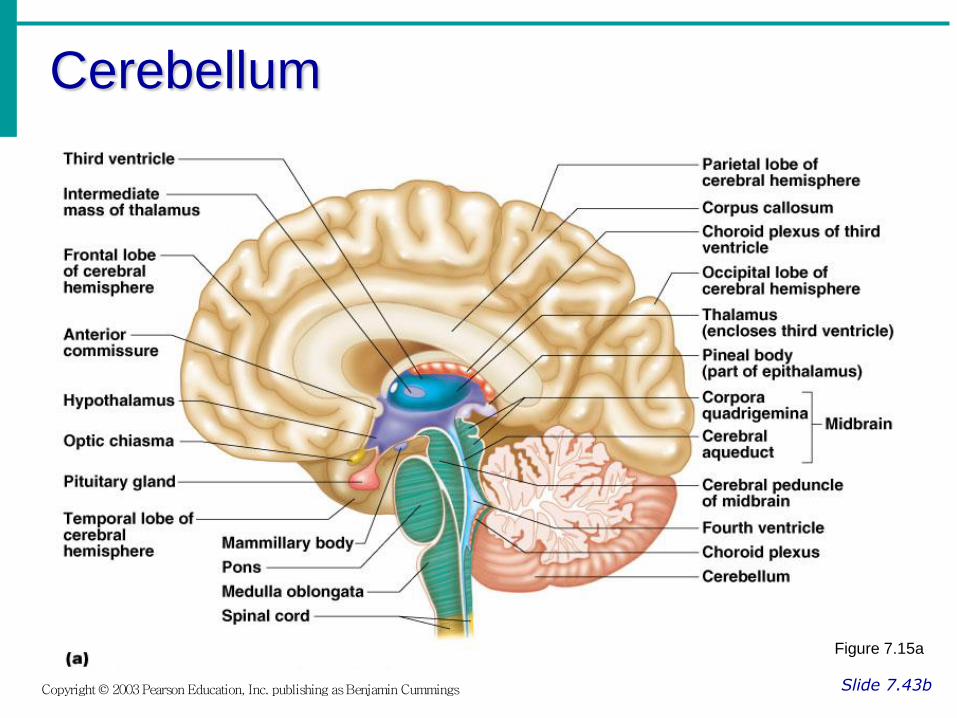
Cerebellum
Slide 7.43b Copyright © 2003 Pearson Education, Inc. publishing as Benjamin Cummings
Figure 7.15a

Protection of the Central Nervous System
Slide 7.44a Copyright © 2003 Pearson Education, Inc. publishing as Benjamin Cummings
Scalp and skin
Skull and vertebral column
Meninges
Figure 7.16a

Protection of the Central Nervous System
Slide 7.44b Copyright © 2003 Pearson Education, Inc. publishing as Benjamin Cummings
Cerebrospinal fluid
Blood brain barrier
Figure 7.16a

Meninges
Slide 7.45a Copyright © 2003 Pearson Education, Inc. publishing as Benjamin Cummings
Dura mater
Double-layered external covering
Periosteum – attached to surface of the skull
Meningeal layer – outer covering of the brain
Folds inward in several areas
Meninges
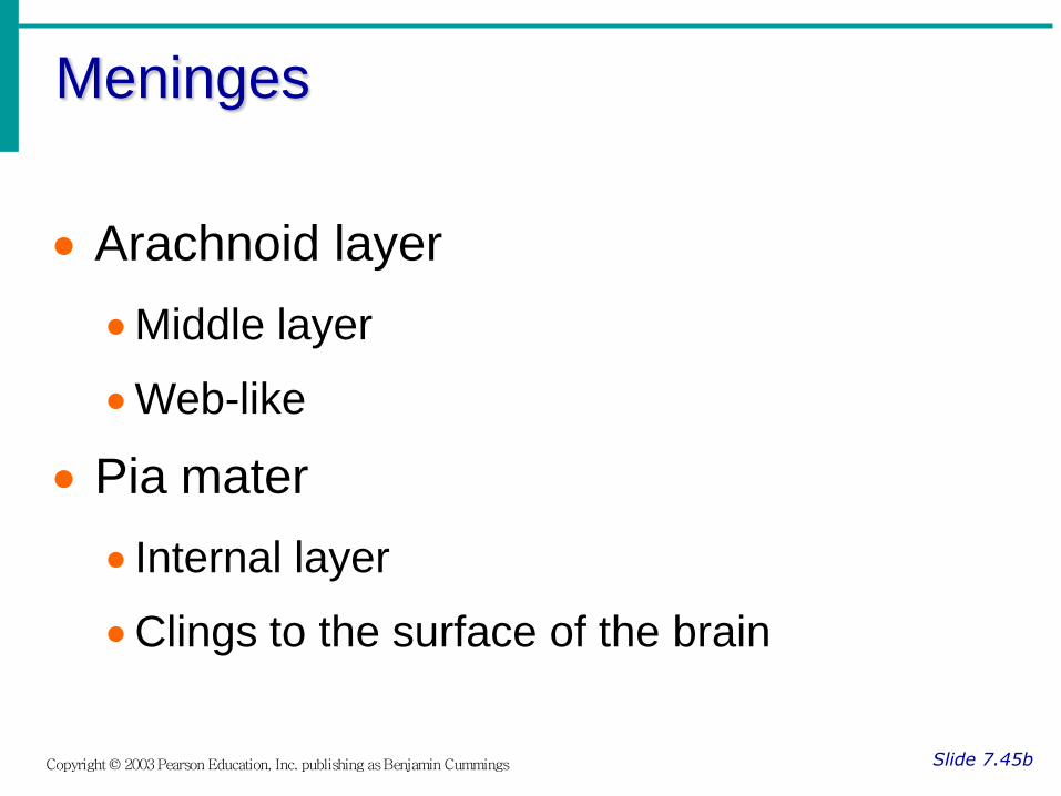
Slide 7.45b Copyright © 2003 Pearson Education, Inc. publishing as Benjamin Cummings
Arachnoid layer
Middle layer
Web-like
Pia mater
Internal layer
Clings to the surface of the brain

Cerebrospinal Fluid
Slide 7.46 Copyright © 2003 Pearson Education, Inc. publishing as Benjamin Cummings
Similar to blood plasma composition
Formed by the choroid plexus
Forms a watery cushion to protect the brain
Circulated in arachnoid space, ventricles, and central canal of the spinal cord

Blood Brain Barrier
Slide 7.48 Copyright © 2003 Pearson Education, Inc. publishing as Benjamin Cummings
Includes the least permeable capillaries of the body
Excludes many potentially harmful substances
Useless against some substances
Fats and fat soluble molecules
Respiratory gases
Alcohol
Nicotine
Anesthesia
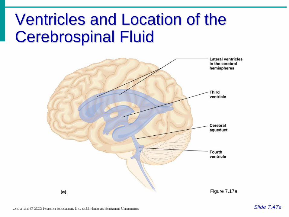
Ventricles and Location of the Cerebrospinal Fluid
Slide 7.47a Copyright © 2003 Pearson Education, Inc. publishing as Benjamin Cummings
Figure 7.17a

Ventricles and Location of the Cerebrospinal Fluid
Slide 7.47b Copyright © 2003 Pearson Education, Inc. publishing as Benjamin Cummings
Figure 7.17b

Traumatic Brain Injuries
Slide 7.49 Copyright © 2003 Pearson Education, Inc. publishing as Benjamin Cummings
Concussion
Slight brain injury
No permanent brain damage
Contusion
Nervous tissue destruction occurs
Nervous tissue does not regenerate
Cerebral edema
Swelling from the inflammatory response
May compress and kill brain tissue
Cerebrovascular Accident (CVA)
Slide 7.50 Copyright © 2003 Pearson Education, Inc. publishing as Benjamin Cummings
Commonly called a stroke
The result of a ruptured blood vessel supplying a region of the brain
Brain tissue supplied with oxygen from that blood source dies
Loss of some functions or death may result
Alzheimer’s Disease
Slide 7.51 Copyright © 2003 Pearson Education, Inc. publishing as Benjamin Cummings
Progressive degenerative brain disease
Mostly seen in the elderly, but may begin in middle age
Structural changes in the brain include abnormal protein deposits and twisted fibers within neurons
Victims experience memory loss, irritability, confusion and ultimately, hallucinations and death

Spinal Cord
Slide 7.52 Copyright © 2003 Pearson Education, Inc. publishing as Benjamin Cummings
Extends from the medulla oblongata to the region of T12
Below T12 is the cauda equina (a collection of spinal nerves)
Enlargements occur in the cervical and lumbar regions
Figure 7.18

Spinal Cord Anatomy
Slide 7.53a Copyright © 2003 Pearson Education, Inc. publishing as Benjamin Cummings
Exterior white mater – conduction tracts
Figure 7.19

Spinal Cord Anatomy
Slide 7.53b Copyright © 2003 Pearson Education, Inc. publishing as Benjamin Cummings
Internal gray matter - mostly cell bodies
Dorsal (posterior) horns
Anterior (ventral) horns
Figure 7.19

Spinal Cord Anatomy
Slide 7.53c Copyright © 2003 Pearson Education, Inc. publishing as Benjamin Cummings
Central canal filled with cerebrospinal fluid
Figure 7.19
Spinal Cord Anatomy
Slide 7.54 Copyright © 2003 Pearson Education, Inc. publishing as Benjamin Cummings
Meninges cover the spinal cord
Nerves leave at the level of each vertebrae
Dorsal root
Associated with the dorsal root ganglia – collections of cell bodies outside the central nervous system
Ventral root

Peripheral Nervous System
Slide 7.55 Copyright © 2003 Pearson Education, Inc. publishing as Benjamin Cummings
Nerves and ganglia outside the central nervous system
Nerve = bundle of neuron fibers
Neuron fibers are bundled by connective tissue
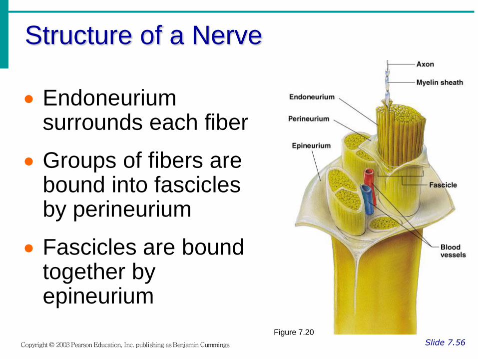
Structure of a Nerve
Slide 7.56 Copyright © 2003 Pearson Education, Inc. publishing as Benjamin Cummings
Endoneurium surrounds each fiber
Groups of fibers are bound into fascicles by perineurium
Fascicles are bound together by epineurium
Figure 7.20
Classification of Nerves
Slide 7.57 Copyright © 2003 Pearson Education, Inc. publishing as Benjamin Cummings
Mixed nerves – both sensory and motor fibers
Afferent (sensory) nerves – carry impulses toward the CNS
Efferent (motor) nerves – carry impulses away from the CNS
Cranial Nerves
Slide 7.58 Copyright © 2003 Pearson Education, Inc. publishing as Benjamin Cummings
12 pairs of nerves that mostly serve the head and neck
Numbered in order, front to back
Most are mixed nerves, but three are sensory only

Distribution of Cranial Nerves
Slide 7.59 Copyright © 2003 Pearson Education, Inc. publishing as Benjamin Cummings
Figure 7.21

Cranial Nerves
Slide 7.60 Copyright © 2003 Pearson Education, Inc. publishing as Benjamin Cummings
I Olfactory nerve – sensory for smell
II Optic nerve – sensory for vision
III Oculomotor nerve – motor fibers to eye muscles
IV Trochlear – motor fiber to eye muscles

Cranial Nerves
Slide 7.61 Copyright © 2003 Pearson Education, Inc. publishing as Benjamin Cummings
V Trigeminal nerve – sensory for the face; motor fibers to chewing muscles
VI Abducens nerve – motor fibers to eye muscles
VII Facial nerve – sensory for taste; motor fibers to the face
VIII Vestibulocochlear nerve – sensory for balance and hearing

Cranial Nerves
Slide 7.62 Copyright © 2003 Pearson Education, Inc. publishing as Benjamin Cummings
IX Glossopharyngeal nerve – sensory for taste; motor fibers to the pharynx
X Vagus nerves – sensory and motor fibers for pharynx, larynx, and viscera
XI Accessory nerve – motor fibers to neck and upper back
XII Hypoglossal nerve – motor fibers to tongue

Essentials of Human Anatomy & Physiology
Copyright © 2003 Pearson Education, Inc. publishing as Benjamin Cummings
Slides 7.63 – 7.75
Seventh Edition
Elaine N. Marieb
Chapter 7
The Nervous System
Lecture Slides in PowerPoint by Jerry L. Cook

Spinal Nerves
Slide 7.63 Copyright © 2003 Pearson Education, Inc. publishing as Benjamin Cummings
There is a pair of spinal nerves at the level of each vertebrae for a total of 31 pairs
Spinal nerves are formed by the combination of the ventral and dorsal roots of the spinal cord
Spinal nerves are named for the region from which they arise

Spinal Nerves
Slide 7.64 Copyright © 2003 Pearson Education, Inc. publishing as Benjamin Cummings Figure 7.22a

Anatomy of Spinal Nerves
Slide 7.65 Copyright © 2003 Pearson Education, Inc. publishing as Benjamin Cummings
Spinal nerves divide soon after leaving the spinal cord
Dorsal rami – serve the skin and muscles of the posterior trunk
Ventral rami – forms a complex of networks (plexus) for the anterior
Figure 7.22b
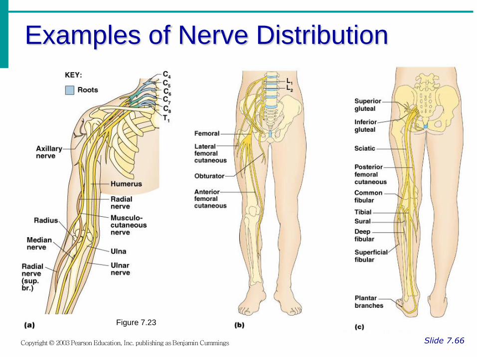
Examples of Nerve Distribution
Slide 7.66 Copyright © 2003 Pearson Education, Inc. publishing as Benjamin Cummings
Figure 7.23

Autonomic Nervous System
Slide 7.67 Copyright © 2003 Pearson Education, Inc. publishing as Benjamin Cummings
The involuntary branch of the nervous system
Consists of only motor nerves
Divided into two divisions
Sympathetic division
Parasympathetic division

Differences Between Somatic and Autonomic Nervous Systems
Slide 7.68a Copyright © 2003 Pearson Education, Inc. publishing as Benjamin Cummings
Nerves
Somatic – one motor neuron
Autonomic – preganglionic and postganglionic nerves
Effector organs
Somatic – skeletal muscle
Autonomic – smooth muscle, cardiac muscle,and glands

Differences Between Somatic and Autonomic Nervous Systems
Slide 7.68b Copyright © 2003 Pearson Education, Inc. publishing as Benjamin Cummings
Nerurotransmitters
Somatic – always use acetylcholine
Autominic – use acetylcholine, epinephrine, or norepinephrine
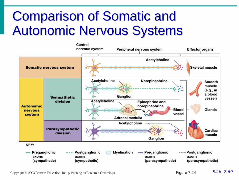
Comparison of Somatic and Autonomic Nervous Systems
Slide 7.69 Copyright © 2003 Pearson Education, Inc. publishing as Benjamin Cummings Figure 7.24

Anatomy of the Sympathetic
Division
Slide 7.70 Copyright © 2003 Pearson Education, Inc. publishing as Benjamin Cummings
Originates from T1 through L2
Ganglia are at the sympathetic trunk (near the spinal cord)
Short pre-ganglionic neuron and long postganglionic neuron transmit impulse from CNS to the effector
Norepinephrine and epinephrine are neurotransmitters to the effector organs

Sympathetic Pathways
Slide 7.71 Copyright © 2003 Pearson Education, Inc. publishing as Benjamin Cummings
Figure 7.26
Anatomy of the Parasympathetic Division
Slide 7.72 Copyright © 2003 Pearson Education, Inc. publishing as Benjamin Cummings
Originates from the brain stem and S1 through S4
Terminal ganglia are at the effector organs
Always uses acetylcholine as a neurotransmitter

Anatomy of the Autonomic Nervous System
Slide 7.73 Copyright © 2003 Pearson Education, Inc. publishing as Benjamin Cummings
Figure 7.25

Autonomic Functioning
Slide 7.74a Copyright © 2003 Pearson Education, Inc. publishing as Benjamin Cummings
Sympathetic – “fight-or-flight”
Response to unusual stimulus
Takes over to increase activities
Remember as the “E” division = exercise, excitement, emergency, and embarrassment

Autonomic Functioning
Slide 7.74b Copyright © 2003 Pearson Education, Inc. publishing as Benjamin Cummings
Parasympathetic – housekeeping activites
Conserves energy
Maintains daily necessary body functions
Remember as the “D” division - digestion, defecation, and diuresis

Development Aspects of the Nervous System
Slide 7.75a Copyright © 2003 Pearson Education, Inc. publishing as Benjamin Cummings
The nervous system is formed during the first month of embryonic development
Any maternal infection can have extremely harmful effects
The hypothalamus is one of the last areas of the brain to develop

Development Aspects of the Nervous System
Slide 7.75b Copyright © 2003 Pearson Education, Inc. publishing as Benjamin Cummings
No more neurons are formed after birth, but growth and maturation continues for several years
The brain reaches maximum weight as a young adult