the nervous system -...
TRANSCRIPT

The Nervous SystemChapter 9
Written Response #1
1. What are the organs of the central nervous system?
2. What nerves arise from the spinal cord?
3. What body structures receive their nerve supply from the autonomic system?

The Nervous System
“The right half of the brain controls the left half of the body. This means that only left
handed people are in their right mind.”

Organization

Central Nervous System (CNS)
• Made up of brain and spinal cord
• Acts as body’s control center, coordinates body’s activities• Impulses travel through
the neurons in your body to reach the brain

Peripheral Nervous System (PNS)
• Made up of all the nerves that carry messages to and from the central nervous system.• Similar to telephone wires
that connect all of our houses in the community
• Central Nervous System and Peripheral Nervous System work together to make rapid changes in your body in response to stimuli.

Peripheral Nervous System (PNS)
• Spinal nerves: arise from the spinal cord to supply the body below the head.
• Cranial nerves: originate from the brain and extend to the head and neck
• Cranial and spinal nerves give rise to smaller nerves that branch extensively throughout the body.

Peripheral Nervous System: Two Parts• Somatic Nervous System (SNS): “The Voluntary Nervous
System”• Relay information between skin, skeletal muscles and central
nervous system• You consciously control this pathway by deciding whether or
not to move muscles (except reflexes)• Reflexes: Automatic response to stimulus
• Autonomic Nervous System (ANS): “The Automatic Nervous System”• Relay information from central nervous system to organs• Involuntary: You do not consciously control these
• Sympathetic Nervous System: controls in times of stress, such as the flight or fight response
• Parasympathetic Nervous System: controls body in times of rest
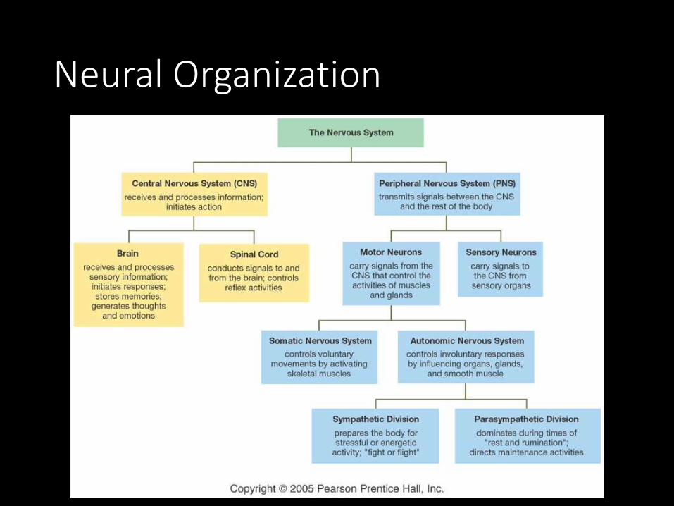
Neural Organization
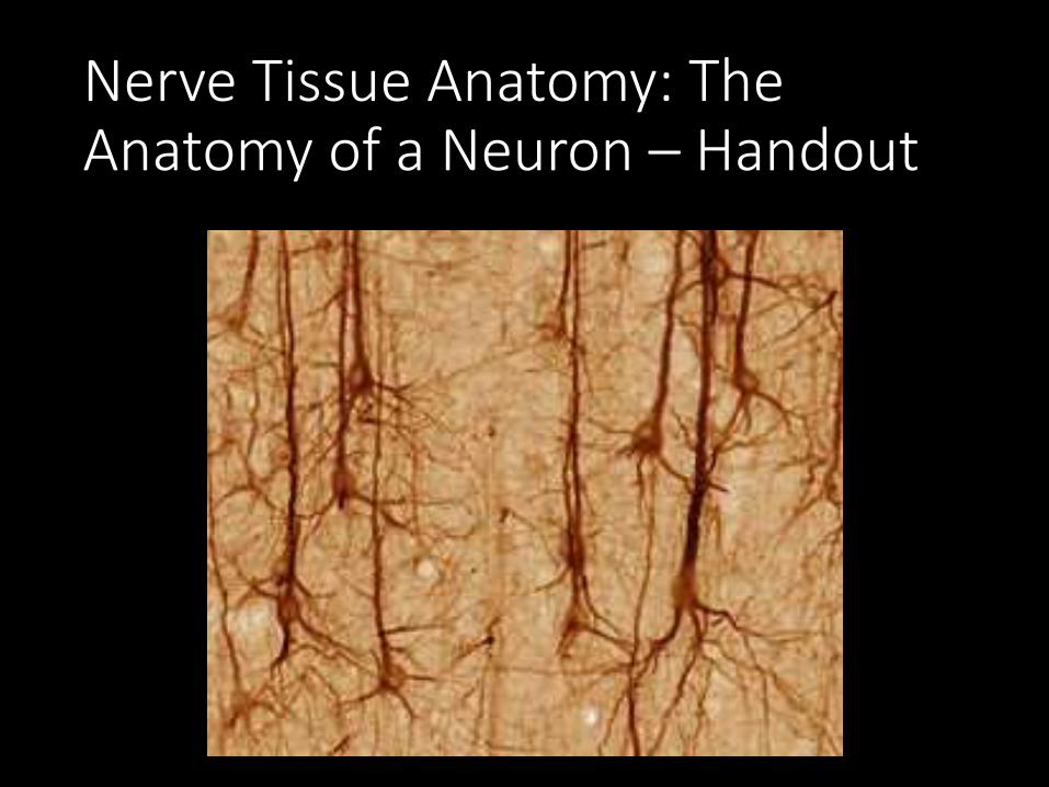
Nerve Tissue Anatomy: The Anatomy of a Neuron – Handout
Nerve Tissue Anatomy: The Anatomy of a Neuroglia – Handout
Activity: Mindboggling –Handout
Visual Imagery (Visual Cortex)
• Visualize a place you’d like to be. Maybe it’s riding a bike, sitting in the park or just hanging out in your bedroom. Create an image of that place and hold it in your mind for a minute.

Auditory Cortex
• Listen to the sounds in the room – really listen. What do you hear? Is it the cracking of someone’s gum, a whisper, the hum of the air conditioner? How many sounds can you differentiate?
Cognitive
• Starting at 100, count backwards by 7.

Hippocampus
• Recall an event from your past, try to remember as many details as you can from it. (Example: the first time you rode a bike, a family holiday). What emotions were you feeling?
Memory
• Memorize this list:• Candy• Head• Bear• Farm• Ring• Cat• Jennifer• Necklace• Nine• Pen

• What were the items on the list?
• How can you make it easier to remember a list?

P.E.T (Positron Emission Tomography) Scans:


14 or 16? Depends on how you look at it.

List the colors.
Train Your Brain.
• Don’t look at the clock, but have you partner watch it. Try to guess how long it takes for 30 seconds to pass. After a few tries you should become more accurate.

Processing Words and Letters
• Read the Sentence and count how many letter F's are in it. • FINISHED FILES ARE THE RESULT OF YEARS OF SCIENTIFIC
STUDY COMBINED WITH THE EXPERIENCE OF YEARS...

Brain Teaser – Word Rhymes
• Headache = Brain ________
• Chief Genius = ________ Brain
• Tune you can’t get out of your head = Brain ________
• Vehicle that transports cerebrums = Brain ___________
• What a neurology professor does = ________ Brains
• Not abnormal = _____ Brain

• Aoccdrnig to rscheearch at Cmabrigde Uinervtisy, it deosn't mttaer in waht oredr the ltteers in a wrod are, the olny iprmoetnt tihng is taht the frist and lsat ltteer be at the rghit pclae. The rset can be a toatl mses and you can sitll raed it wouthit a porbelm. Tihs is bcuseae the huamn mnid deos not raed ervey lteter by istlef, but the wrod as a wlohe.
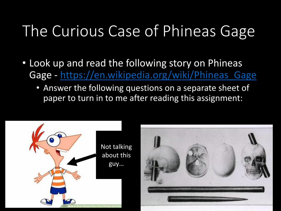
The Curious Case of Phineas Gage
• Look up and read the following story on Phineas Gage - https://en.wikipedia.org/wiki/Phineas_Gage• Answer the following questions on a separate sheet of
paper to turn in to me after reading this assignment:
Not talking about this
guy…
Phineas Gage Questions:
1. How was Phineas Gage's brain damaged?
2. Why is Phineas Gage's story important to neuroscientists today?
3. Compared to Gage's personality before the accident, after the accident Gage was:
4. How do neuroscientists today determine the exact damage done to Gage's brain?
5. After the initial accident, Gage almost died due to:
6. Which part of the brain is associated with personality?
7. Which statement is an inference?A. Gage's body was exhumed and his skull displayedB. Gage's personality was changed dramatically after the accident.C. The part of the brain damaged by the iron was the frontal lobe.D. Gage would have recovered fully had it not been for the infection.
8. What is an abscess?
9. What does exhume mean?
10. After the accident, Gage is said to have vacillated. What does vacillate mean?
11. What was the most likely cause of Gage’s death?

Written Response #2
1. What is a myelinated nerve fiber, and why does it conduct impulses faster than unmyelinated fibers?
2. What are the structural and functional types of neurons?
3. How is an action potential generated in a resting neuron?
4. What is the difference between an excitatory transmission and an inhibitory transmission?

Brain: Structure
• Hindbrain carries out the most basic functions.
• Midbrain coordinates signals.
• Forebrain processes signals, stores memories, creates thought.

Neuroglia vs. Neurons
• Neuroglia divide.
• Neurons do not.
• Most brain tumors are “gliomas.”
• Most brain tumors involve the neuroglia cells, not the neurons.
• Consider the role of cell division in cancer!
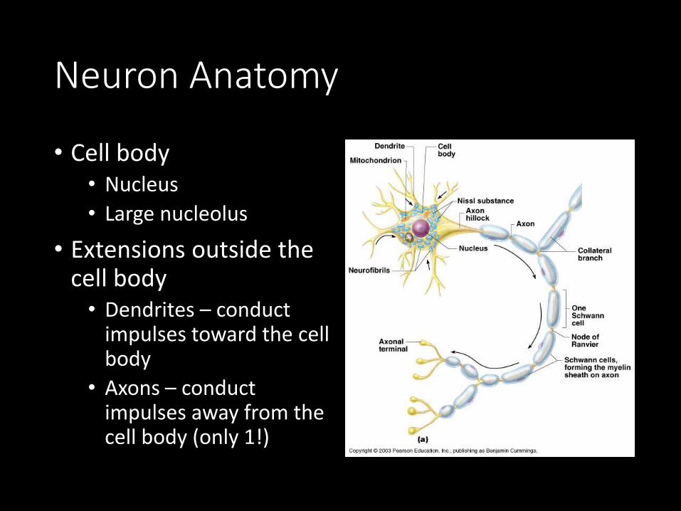
Neuron Anatomy
• Cell body• Nucleus
• Large nucleolus
• Extensions outside the cell body• Dendrites – conduct
impulses toward the cell body
• Axons – conduct impulses away from the cell body (only 1!)
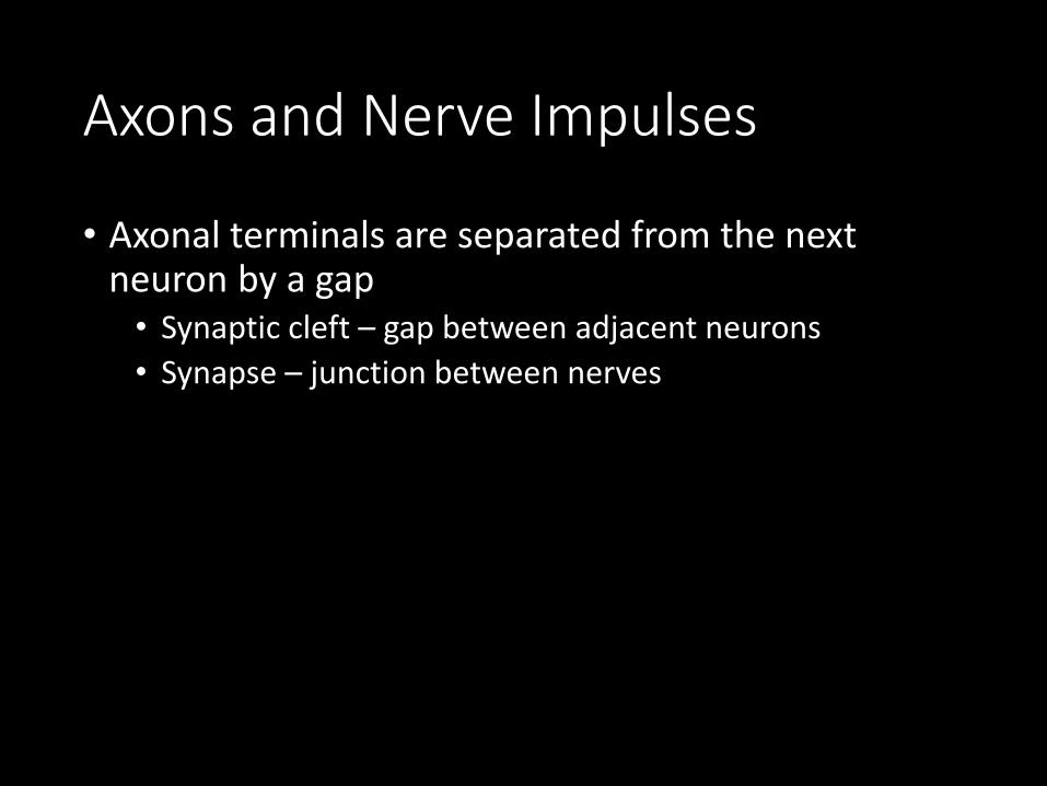
Axons and Nerve Impulses
• Axonal terminals are separated from the next neuron by a gap• Synaptic cleft – gap between adjacent neurons
• Synapse – junction between nerves
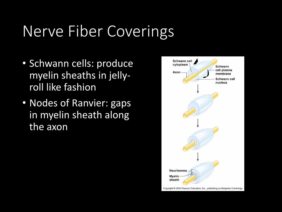
Nerve Fiber Coverings
• Schwann cells: produce myelin sheaths in jelly-roll like fashion
• Nodes of Ranvier: gaps in myelin sheath along the axon

Nervous Tissue: Support Cells (Neuroglia or Glia)• Astrocytes
• Abundant, star-shaped cells
• Brace neurons
• Form barrier between capillaries and neurons
• Control the chemical environment of the brain (CNS)
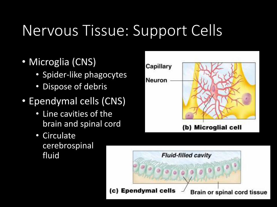
Nervous Tissue: Support Cells
• Microglia (CNS)• Spider-like phagocytes
• Dispose of debris
• Ependymal cells (CNS)• Line cavities of the
brain and spinal cord
• Circulate cerebrospinal fluid

Nervous Tissue: Support Cells
• Oligodendrocytes (CNS)• Produce myelin sheath
around nerve fibers in the central nervous system

Support Cells of the PNS
• Satellite cells• Protect neuron cell bodies
• Schwann cells• Form myelin sheath in the peripheral nervous system

Written Response #3: Application
• In Multiple Scleroses the myelin sheath is destroyed. The myelin sheath hardens to a tissue called the scleroses. This is considered an autoimmune disease.
• Why does MS appear to affect the muscles?
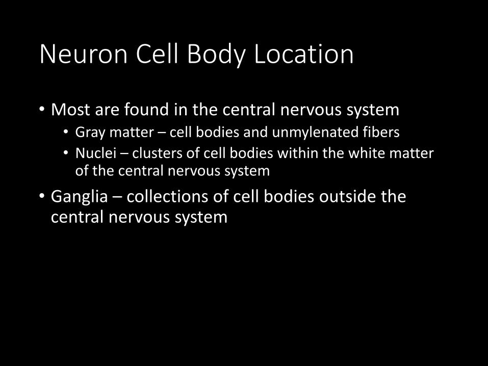
Neuron Cell Body Location
• Most are found in the central nervous system• Gray matter – cell bodies and unmylenated fibers
• Nuclei – clusters of cell bodies within the white matter of the central nervous system
• Ganglia – collections of cell bodies outside the central nervous system

Three types of neurons
• Sensory Neurons (Afferent): carry impulses from inside and outside the body to brain and spinal cord.
• Interneurons : found within brain and spinal cord, process incoming impulses and pass them on to motor neurons.
• Motor Neurons (Efferent): carry impulses away from the brain and spinal cord.
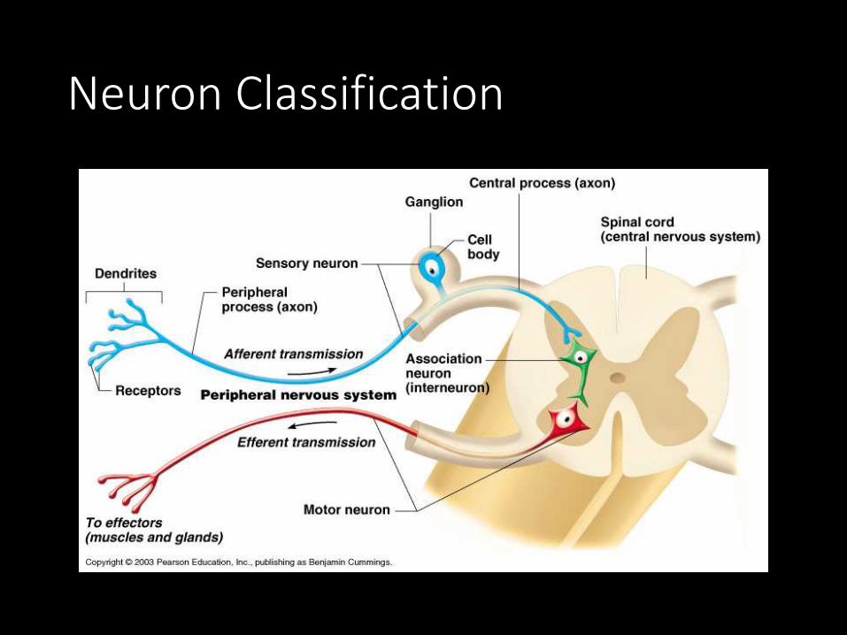
Neuron Classification

Types of Neurons

How Neurons Function (Physiology)• Irritability – ability to respond to stimuli
• Conductivity – ability to transmit an impulse
• The plasma membrane at rest is polarized• Fewer positive ions are inside the cell than outside the
cell
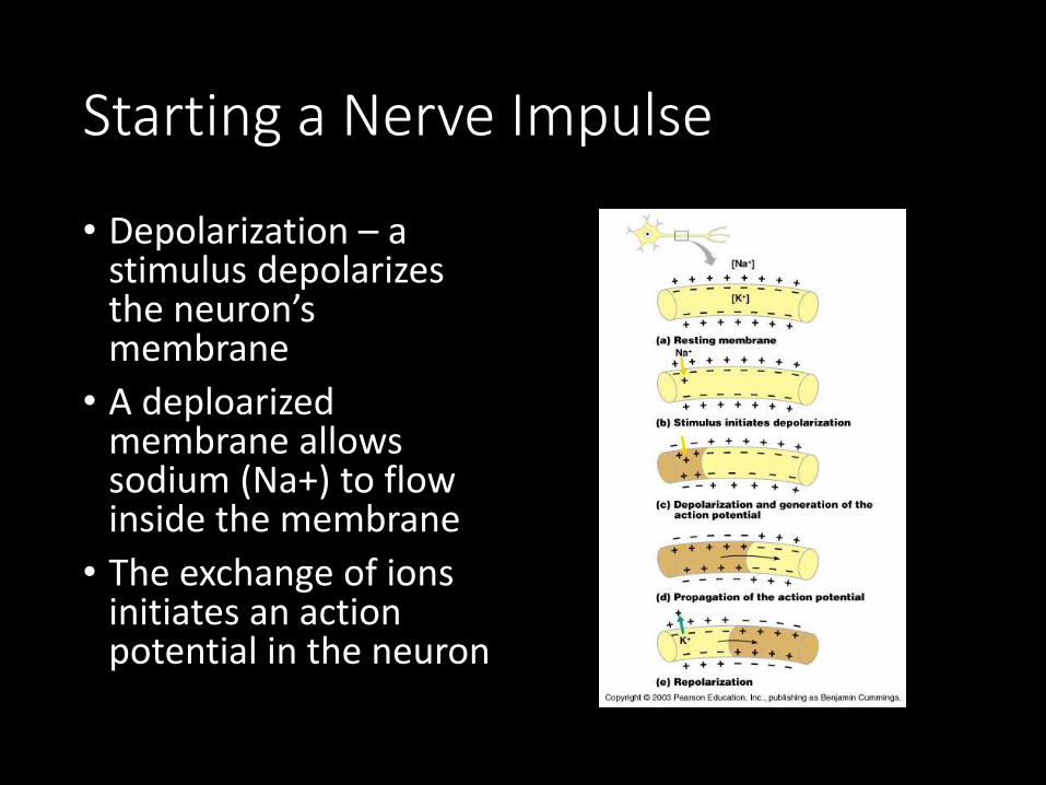
Starting a Nerve Impulse
• Depolarization – a stimulus depolarizes the neuron’s membrane
• A deploarized membrane allows sodium (Na+) to flow inside the membrane
• The exchange of ions initiates an action potential in the neuron

The Action Potential
• If the action potential (nerve impulse) starts, it is propagated over the entire axon. “All or Nothing”• Potassium ions rush out of the neuron after sodium ions
rush in, which repolarizes the membrane
• The sodium-potassium pump restores the original configuration
• This action requires ATP (active transport…remember from Biology?)

Nerve Impulse Propagation
• The impulse continues to move toward the cell body
• Impulses travel faster when fibers have a myelin sheath

Continuation of the Nerve Impulse between Neurons• Impulses are able to cross the synapse to another
nerve• Neurotransmitter is released from a nerve’s axon
terminal
• The dendrite of the next neuron has receptors that are stimulated by the neurotransmitter
• An action potential is started in the dendrite
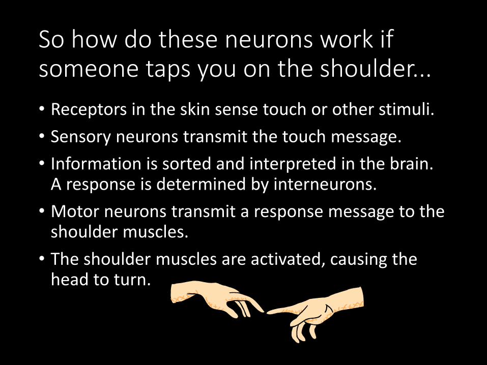
So how do these neurons work if someone taps you on the shoulder...
• Receptors in the skin sense touch or other stimuli.
• Sensory neurons transmit the touch message.
• Information is sorted and interpreted in the brain. A response is determined by interneurons.
• Motor neurons transmit a response message to the shoulder muscles.
• The shoulder muscles are activated, causing the head to turn.

How is an impulse transmitted?
• Stimulus excites sensory neuron.
• Depolarization (a change in charge due to sodium ions) creates a wave of changing charges down the axon.
• Impulse moves across synapse (tiny space between one neuron’s axon and another’s dendrites) with the help of neurotransmitters
This is an image of neurons
located in the cerebral cortex of a
hamster.
OPTICAL ILLUSION
• How does your brain perceive objects?


Brain Videos
• Brain Games: Video Episode
• Brain Heiroglyphics
• Brain Game at NatGeo
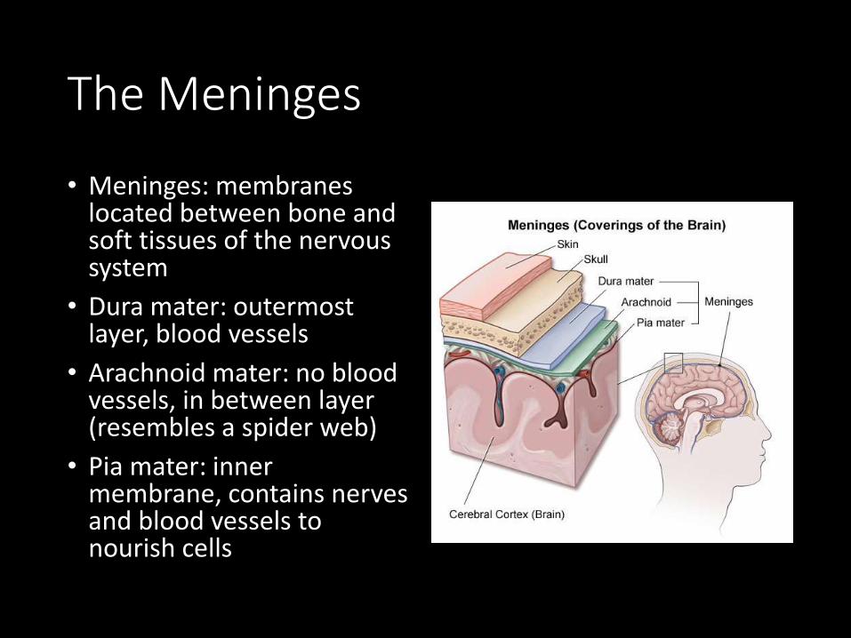
The Meninges
• Meninges: membranes located between bone and soft tissues of the nervous system
• Dura mater: outermost layer, blood vessels
• Arachnoid mater: no blood vessels, in between layer (resembles a spider web)
• Pia mater: inner membrane, contains nerves and blood vessels to nourish cells

Brain
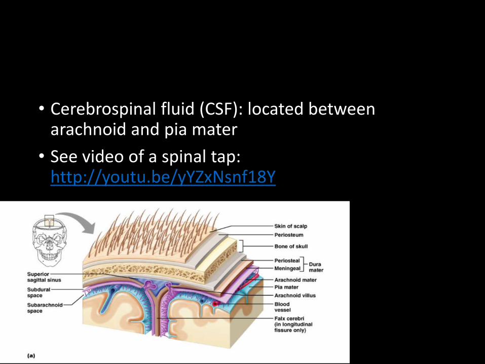
• Cerebrospinal fluid (CSF): located between arachnoid and pia mater
• See video of a spinal tap: http://youtu.be/yYZxNsnf18Y

Dura mater is being peeled away in the photo to the right

Subdural Hematoma
• A type of hematoma, usually associated with traumatic brain injury. Blood gathers between the dura mater, and the brain, putting pressure on brain.

Repair of a subdural hematoma in an 83 year old patient.
Natgeo Brain Surgery Video -removal of tumor while patient is awake

Spinal Cord
• Passes down the vertebral canal; has 31 segments (each with a pair of spinal nerves)
• Cervical enlargement = supplies nerves to upper limbs (neck)
• Lumbar enlargement = supplies nerves to the lower limbs (lower back)
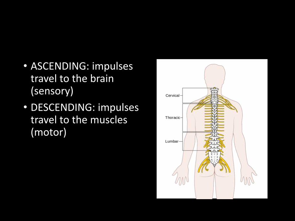
• ASCENDING: impulses travel to the brain (sensory)
• DESCENDING: impulses travel to the muscles (motor)

• The nerves are numbered based on their location:• C1 - C8
• T1 - T12
• L1 - L5

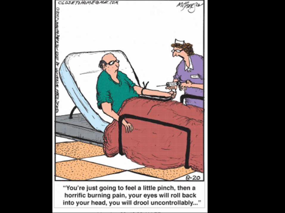
Mental Hospital Phone Menu
1. Please select from the following options menu:
2. If you are obsessive-compulsive, press 1 repeatedly.
3. If you are co-dependent, please ask someone to press 2 for you.
4. If you have multiple personalities, press 3, 4, 5 and 6.
5. If you are paranoid, we know who you are and what you want, stay on the line so we can trace your call.
6. If you are schizophrenic, listen carefully and a little voice will tell You which number to press.
7. If you are manic-depressive, hang up. It doesn't matter which number you press, nothing will make you happy anyway.
8. If you are dyslexic, press 9-6-9-6.
9. If you are bipolar, please leave a message after the beep or before the beep or after the beep. But Please wait for the beep.
10. If you have low self-esteem, please hang up. Our operators are too busy to talk with you.
The Brain
• 3 Major Parts:• Cerebrum
• Cerebellum
• Brain Stem

Cerebrum
• CEREBRUM - wrinkly large part of the brain
• higher mental function, solving problems
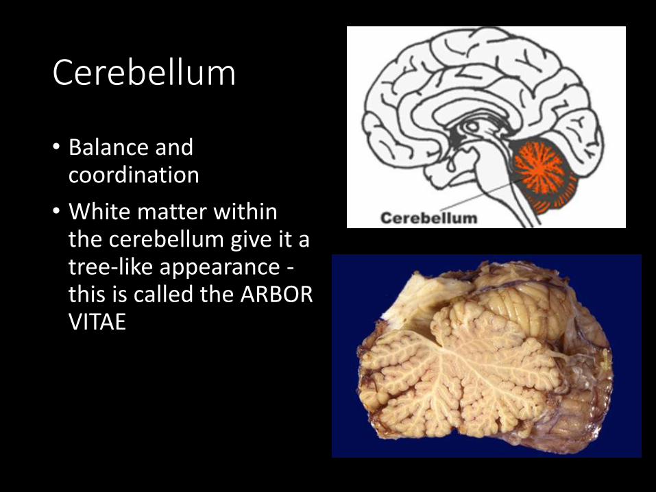
Cerebellum
• Balance and coordination
• White matter within the cerebellum give it a tree-like appearance -this is called the ARBOR VITAE

Brain Stem
• Brain Stem - regulates visceral functions (autonomic system)
Activity: Brain Cap – Handout


• 1. Cerebral Hemispheres - left and right side separated by the....
• 2. Corpus Callosum -connects the two hemispheres
The Cerebral Hemispheres

Corpus callosum
3. Convolutions of the Brain
• Convolutions: the wrinkles and grooves of the cerebrum• Fissures = deep groove
• Sulcus = shallow groove
• Gyrus = bump

4. Fissures: separate lobes
• Longitudinal fissure -separate right and left sides

• Transverse Fissure -separates cerebrum from cerebellum
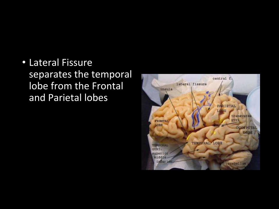
• Lateral Fissure separates the temporal lobe from the Frontal and Parietal lobes

Hemisphere Dominance - Activity


Lobes of the Brain
5.6.
7.
8.
9.10.
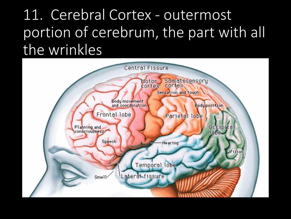
11. Cerebral Cortex - outermost portion of cerebrum, the part with all the wrinkles

Functional and Structural Areas of the Cerebral Cortex

Fluid filled cavities, contain CSF
11. VENTRICLES OF THE BRAIN

12. Cerebrospinal Fluid (CSF) - fluid that protects and supports brain
˙pınןɟ ןɐuıdsoɹqǝɹǝɔ ɟo ǝןdɯɐsɐ ǝʌoɯǝɹ oʇ (ǝɐɹqǝʇɹǝʌ) sǝuoq ɹɐqɯn ןoʍʇ uǝǝʍʇǝqpǝʇɹǝsuı sı ǝןpǝǝu ɐ 'ǝɹnʇɔundɹɐqɯn ןƃuıɹnp ˙uoıƃǝɹɹɐqɯn ןǝɥʇ uı 'ʞɔɐq ɹǝʍo ןɹnoʎ uı pǝɯɹoɟɹǝd sı (dɐʇɐuıds) ǝɹnʇɔundן ɹɐqɯnן .pɹoɔɐuıdsן ɹo uıɐɹq ǝɥʇ ɟo sɹǝɔuɐɔɹo ɔsןsısoɹǝ؛ ǝןdıʇןnɯ sɐ ɥɔns'ɯǝʇsʎs snoʌɹǝu ɐɹʇuǝɔן ǝɥʇɟo sɹǝpɹosıp sıʇıƃuıuǝɯ؛ sɐɥɔns 'suoıʇɔǝɟuı ǝsouƃɐıp uɐɔsıɥʇ
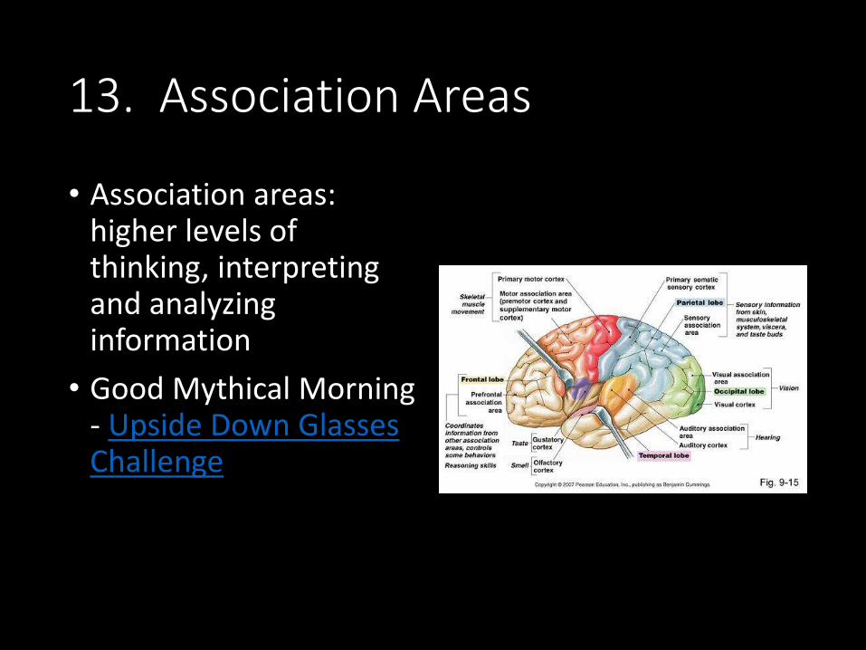
13. Association Areas
• Association areas: higher levels of thinking, interpreting and analyzing information
• Good Mythical Morning - Upside Down Glasses Challenge
Brain Stem
Pituitary
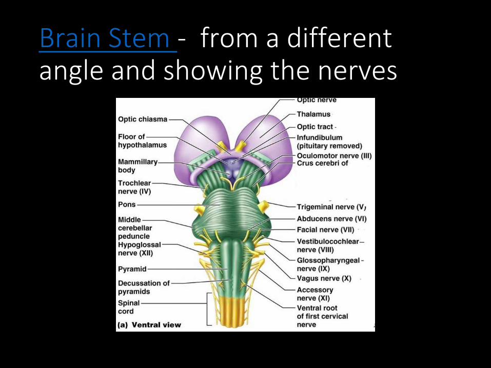

1. Diencephalon has two parts...
• Hypothalamus -hormones, heart rate, blood pressure, body temp, hunger, attaches to pituitary
• Thalamus - relay station

4. Optic Tract / Chiasma - optic nerves cross over each other
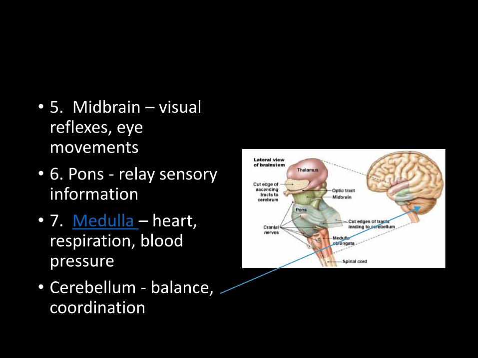
• 5. Midbrain – visual reflexes, eye movements
• 6. Pons - relay sensory information
• 7. Medulla – heart, respiration, blood pressure
• Cerebellum - balance, coordination

Pituitary Gland
• The "master gland" of the endocrine system. It controls hormones.

Corpus callosumThalamus
Hypothalamus
Pineal gland

MidbrainMedulla
OblongataPons

Hippocampus and Amygdala
• Hippocampus: memory is controlled (“sea horse”; that’s its shape). The hippocampus plays a major role in memories.
• Amygdala: storage of memories associated with emotional events.• Also associated with fear
response and aggression

The Limbic System
• Composed of hypothalamus, hippocampus, amygdala (and several other nearby areas ) • main function is in
emotions
• Why are teenagers impulsive?
Memory
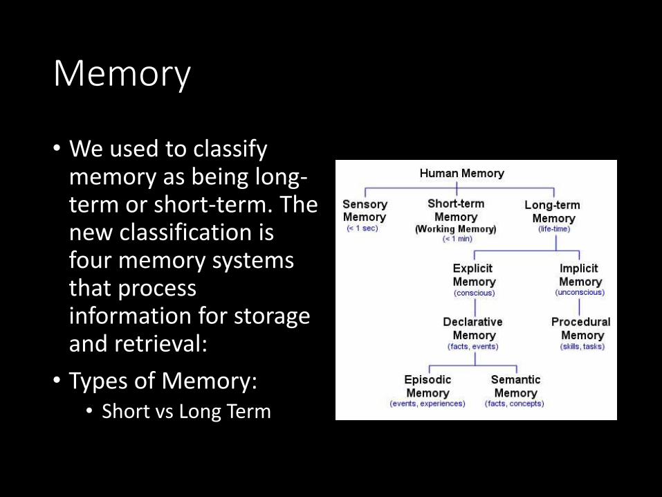
• We used to classify memory as being long-term or short-term. The new classification is four memory systems that process information for storage and retrieval:
• Types of Memory:• Short vs Long Term

How important are your memories?• If you were involved in
a traumatic event, such as a rape or a terrorist attack, would you take a pill that would make it so that you did not remember the event?

Episodic Memory
• Involved in remembering personal experiences, such as a phone conversation you had yesterday or the movie you watched last week.
• Challenge:• Do you remember the first Harry Potter movie? What
was the story? How many details do you remember?
Semantic Memory
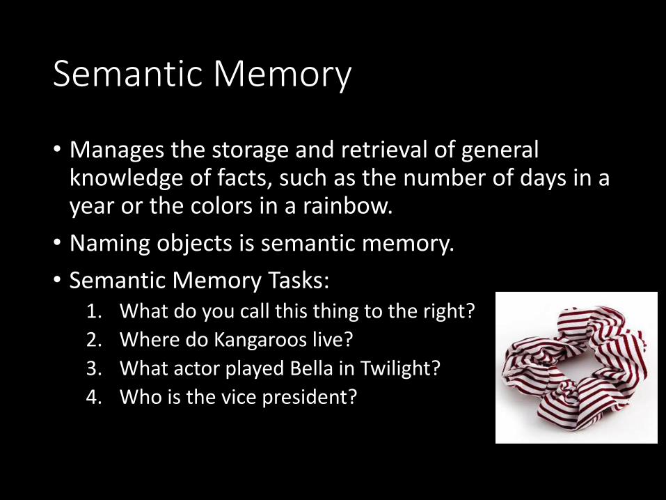
• Manages the storage and retrieval of general knowledge of facts, such as the number of days in a year or the colors in a rainbow.
• Naming objects is semantic memory.
• Semantic Memory Tasks:1. What do you call this thing to the right?
2. Where do Kangaroos live?
3. What actor played Bella in Twilight?
4. Who is the vice president?
Procedural Memory
• Allows us to learn activities and skills that will then be performed automatically with little or no conscious thought.• Examples are riding a bicycle or driving a car.
• Challenge:1. Can you make a teacup with a length of string?
2. Do you know how to use a can opener?

Working Memory
• Provides temporary storage of information necessary for such complex cognitive tasks as language comprehension, learning, and reasoning.
• Working Memory Test

Memory - Sometimes it's good to forget things…why?• Video on memory and where memories are stored
and retrieved.
Check out these brain images!

Brain Crossword – Handout
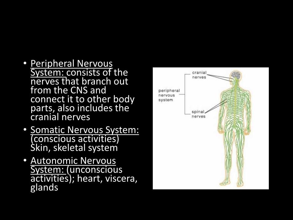
• Peripheral Nervous System: consists of the nerves that branch out from the CNS and connect it to other body parts, also includes the cranial nerves
• Somatic Nervous System: (conscious activities) Skin, skeletal system
• Autonomic Nervous System: (unconscious activities); heart, viscera, glands
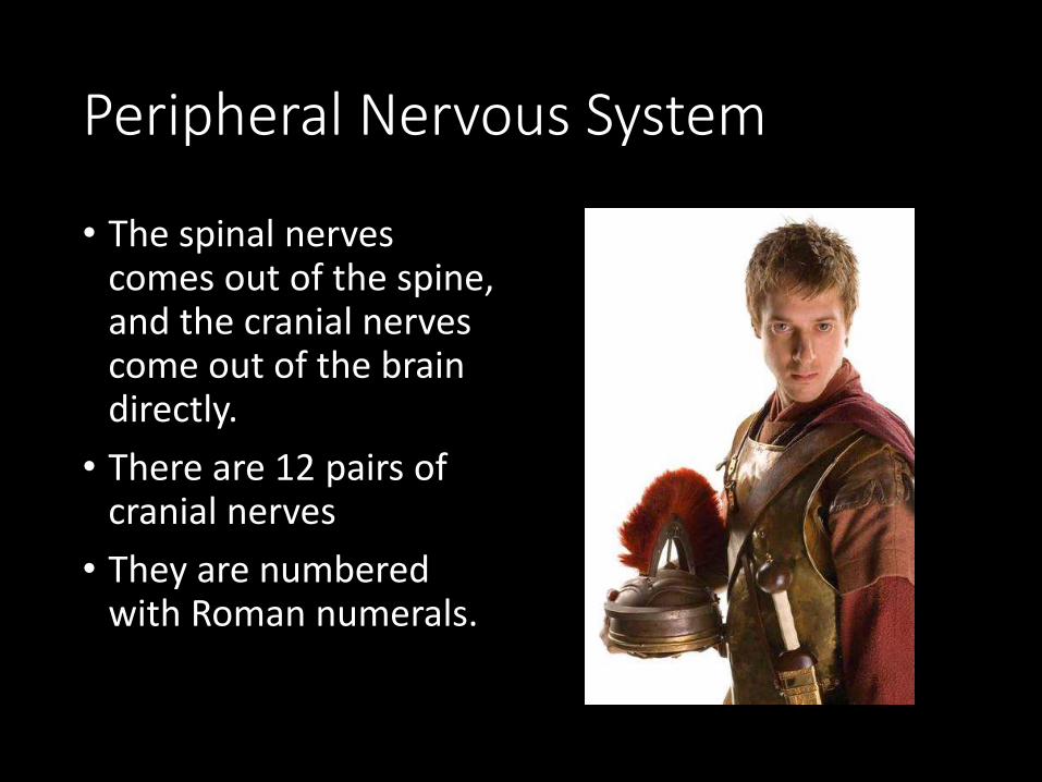
Peripheral Nervous System
• The spinal nerves comes out of the spine, and the cranial nerves come out of the brain directly.
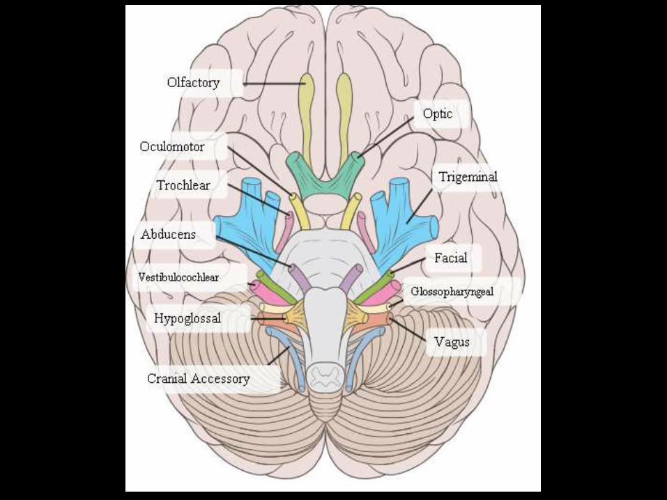
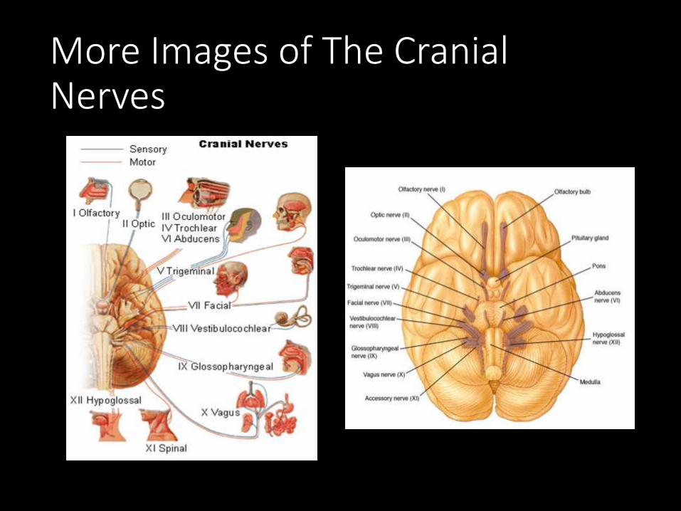
• There are 12 pairs of cranial nerves
• They are numbered with Roman numerals.
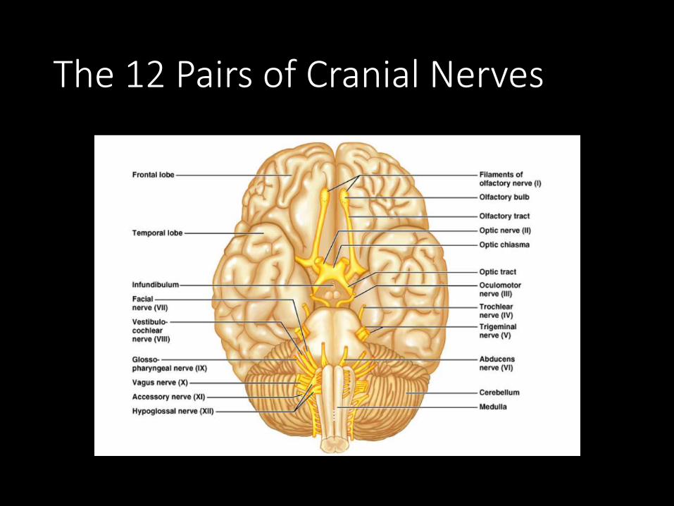
The 12 Pairs of Cranial Nerves
Figure 14.8
I. Olfactory Nerve
• Sense of smell. Outside of the CNS they are called olfactory nerves, and inside of the CNS they are called the olfactory tract.

II. Optic Nerve
• Transmits information from the eye’s retina. VISION
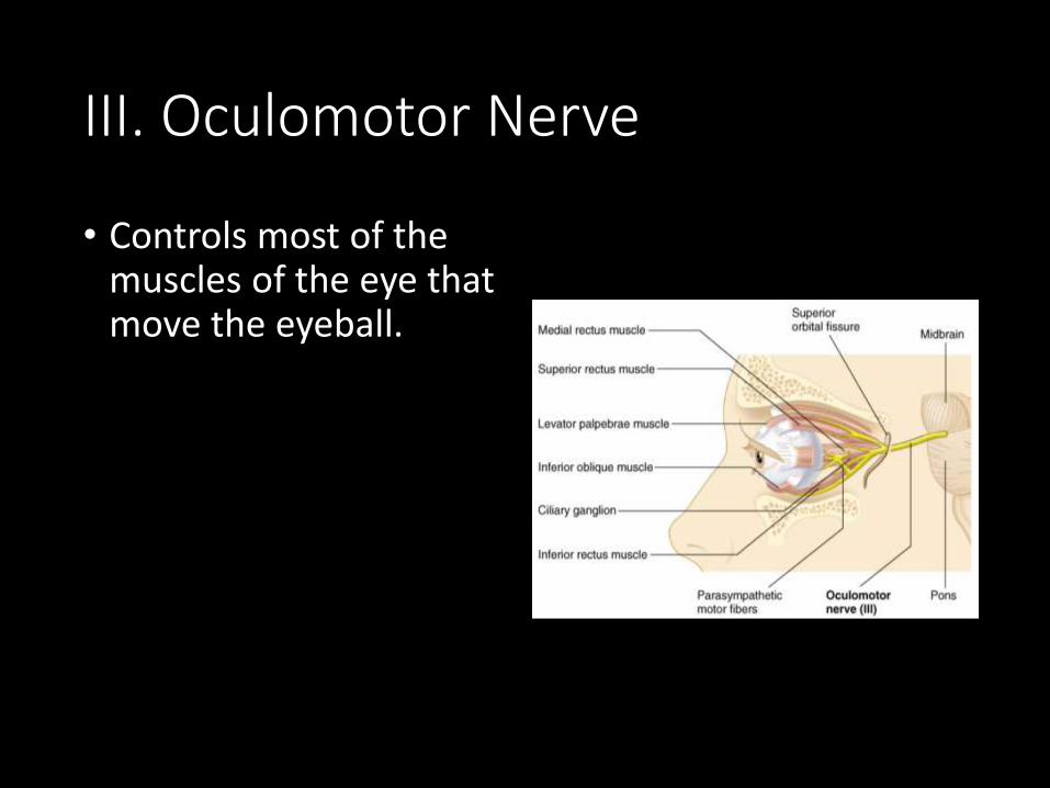
III. Oculomotor Nerve
• Controls most of the muscles of the eye that move the eyeball.

IV. Trochlear Nerve
• Innervates an extrinsic eye muscle, eyelid
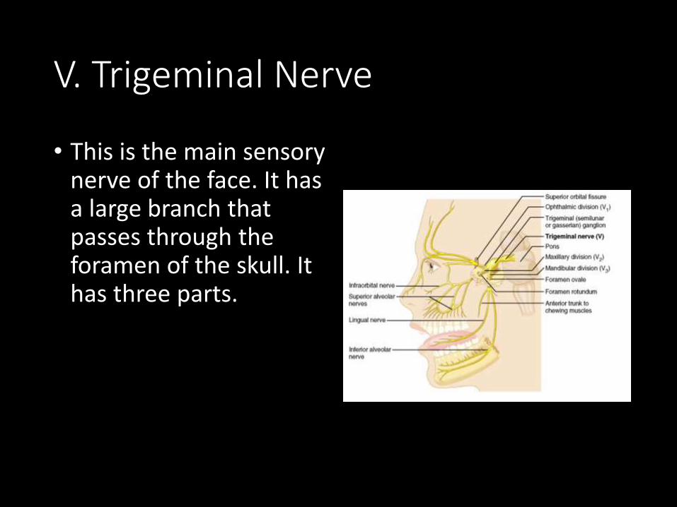
V. Trigeminal Nerve
• This is the main sensory nerve of the face. It has a large branch that passes through the foramen of the skull. It has three parts.
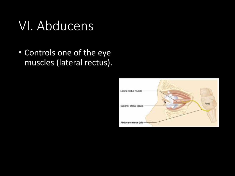
VI. Abducens
• Controls one of the eye muscles (lateral rectus).

VII. Facial Nerve
• Innervates the muscles of facial expression.
• A person who cannot blink or smile may have damage to this nerve.
• BELL’S PALSY is damage of the facial nerve causing paralysis on one side.
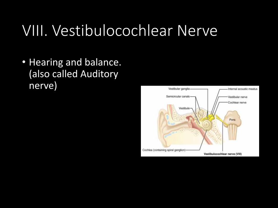
VIII. Vestibulocochlear Nerve
• Hearing and balance. (also called Auditory nerve)

IX: Glossopharyngeal Nerve
• pharynx, tongue
• swallowing, speech, saliva
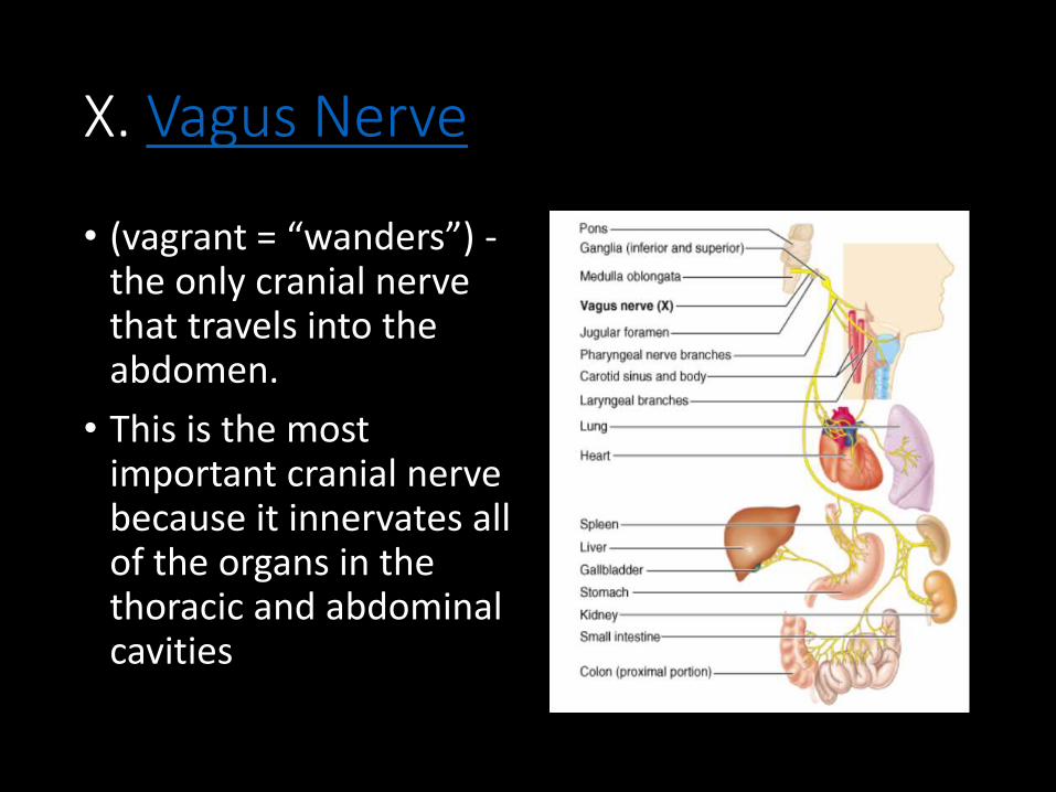
X. Vagus Nerve
• (vagrant = “wanders”) -the only cranial nerve that travels into the abdomen.
• This is the most important cranial nerve because it innervates all of the organs in the thoracic and abdominal cavities
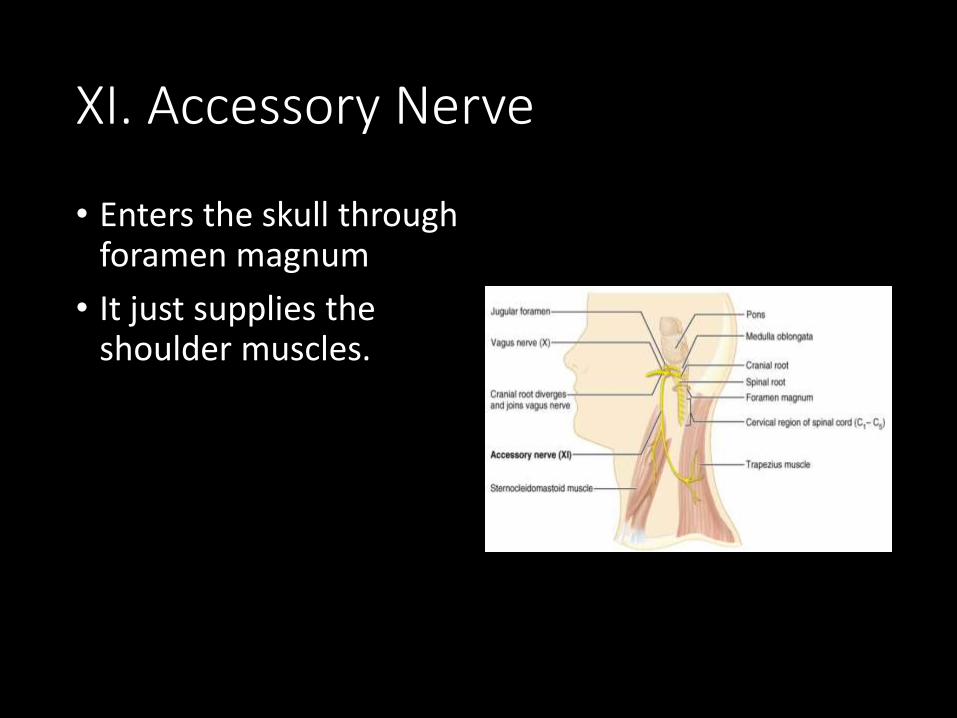
XI. Accessory Nerve
• Enters the skull through foramen magnum
• It just supplies the shoulder muscles.
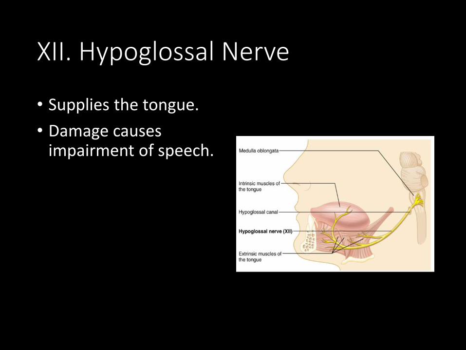
XII. Hypoglossal Nerve
• Supplies the tongue.
• Damage causes impairment of speech.

Overview of Cranial Nerves
I. Olfactory Sense of smell
II. Optic Sight
III. Occulomotor Move eyelids
IV. Trochlear Move eyes
V. Trigeminal Face, jaw, chewing
VI. Abducens Eyes
VII. Facial Facial expressions
VIII. Vestibulocochlear(auditory)
Sense of equilibrium,hearing
IX. Glossopharyngeal Pharynx, tongue
X. Vagus Major organs
XI. Accessory Shoulders
XII. Hypoglossal Tongue

Need to know all of the cranial nerves?• On Old Olympus
Towering Top A Fin And German Viewed A Hop


Spinal Nerves
• 8 pairs of cervical nerves (C1 - C8)
• 12 pairs of thoracic nerves (T1-T12)
• 5 pairs of lumbar nerves (L1-L5)
• 5 pairs of sacral nerves (S1-S5)
• 1 pair of coccygeal nerves (Co)
• 31 Total
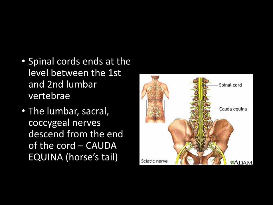
• Spinal cords ends at the level between the 1st and 2nd lumbar vertebrae
• The lumbar, sacral, coccygeal nerves descend from the end of the cord – CAUDA EQUINA (horse’s tail)
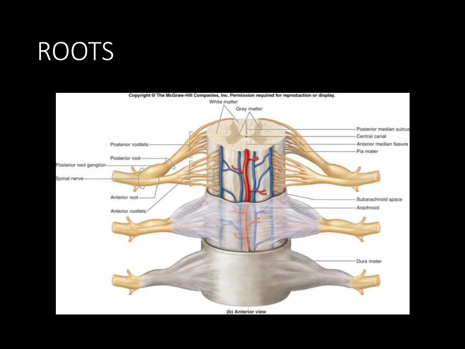
ROOTS
• Each nerve emerges from the spinal cord at points called ROOTS• Dorsal Root Ganglion
• Ventral root ganglion

PLEXUSES
• Main portions of the spinal nerves combine to form complex networks called PLEXUSES

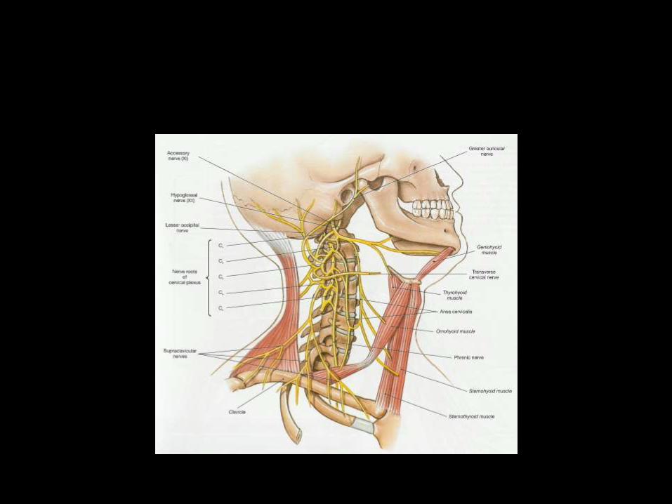
1. Cervical Plexus - neck2. Brachial Plexus - shoulders, arms, hands
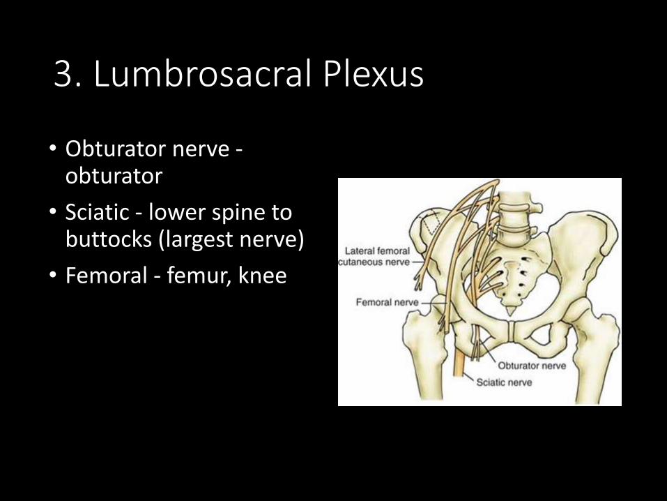
3. Lumbrosacral Plexus
• Obturator nerve -obturator
• Sciatic - lower spine to buttocks (largest nerve)
• Femoral - femur, knee

Autonomic Nervous System
• Sympathetic - energy, high stress, emergency• Fight or Flight
• Parasympathetic -resting, normal
• Divisions act antagonistically - one is exhitatory, other inhibits

More Images of The Cranial Nerves

The sheep brain also has the 12 cranial nerves,
but they can be difficult to find


SensesChapter 10
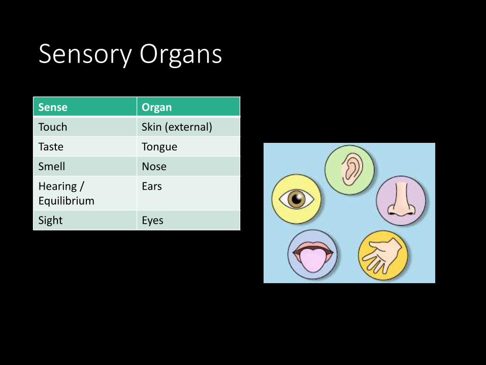
Sensory Organs
Sense Organ
Touch Skin (external)
Taste Tongue
Smell Nose
Hearing / Equilibrium
Ears
Sight Eyes
Types of Sensory Receptors
1. Mechanoreceptors – stimulated by changes in pressure or movement• Found in skin and muscles
2. Thermoreceptors – stimulated by changes in temperature• Found in skin
3. Pain receptors – stimulated by tissue damage• Found in skin and viscera
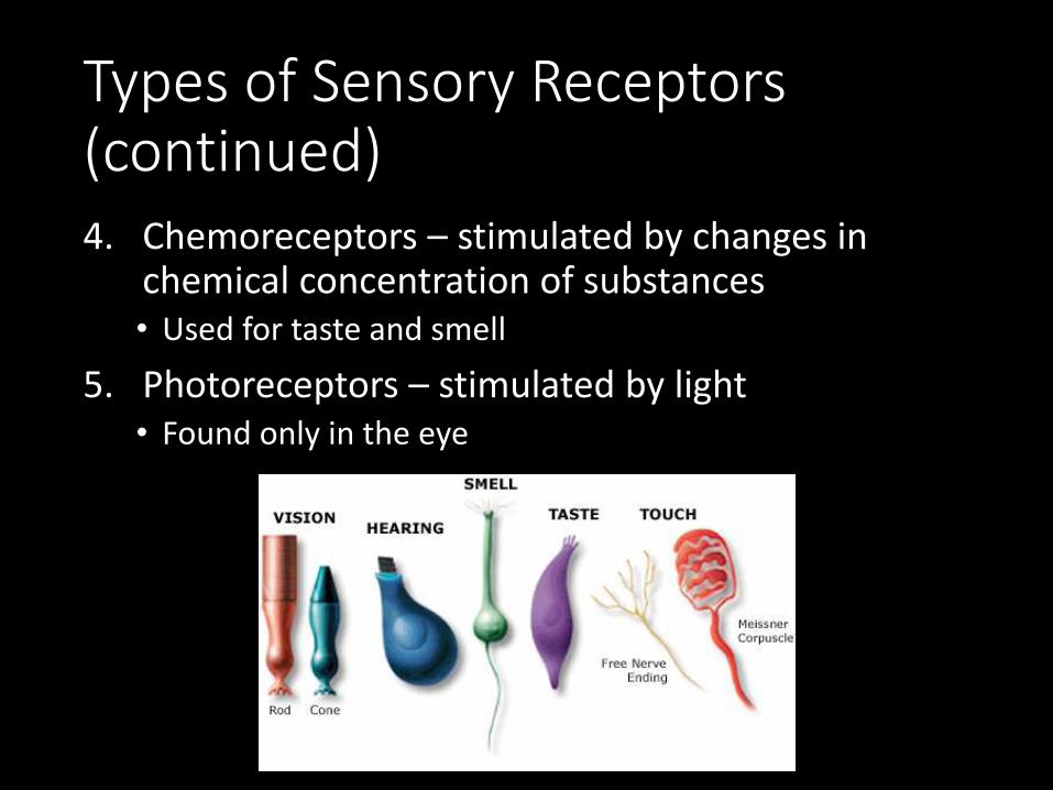
Types of Sensory Receptors (continued)4. Chemoreceptors – stimulated by changes in
chemical concentration of substances• Used for taste and smell
5. Photoreceptors – stimulated by light• Found only in the eye

Sense of Touch
• Mechanoreceptors in the skin and viscera detect varying degrees of pressure.
• Free nerve endings have pain receptors and thermoreceptors.

Sense of Touch – Pain
• Pain is caused by chemicals released by inflamed tissues.• Aspirin and ibuprofen
reduce pain by blocking synthesis of these chemicals
• Referred pain: inside the body’s organs, pain is often felt in another area.• Ex: Pain from the heart is
felt in the left shoulder and arm

Senses of Taste & Smell
• Taste and smell are “chemical senses”
• Taste – tastebuds containing chemoreceptors are found in the epithelium of the tongue
• Papillae (bumps) on the tongue contain many receptors
• Receptors can distinguish between sweet, sour, salty, and bitter tastes.

Senses of Taste & Smell
• Smell – within the nasal cavity, chemoreceptors in the olfactory bulb are stimulated by odor molecules

Senses of Taste & Smell
• Smells have been shown to be linked to memories because the olfactory bulb is linked to the limbic system of the brain.

Sense of Hearing
• Anatomy of the Ear• 1. Outer Ear – includes:
• pinna (external ear)
• auditory canal
Sense of Hearing
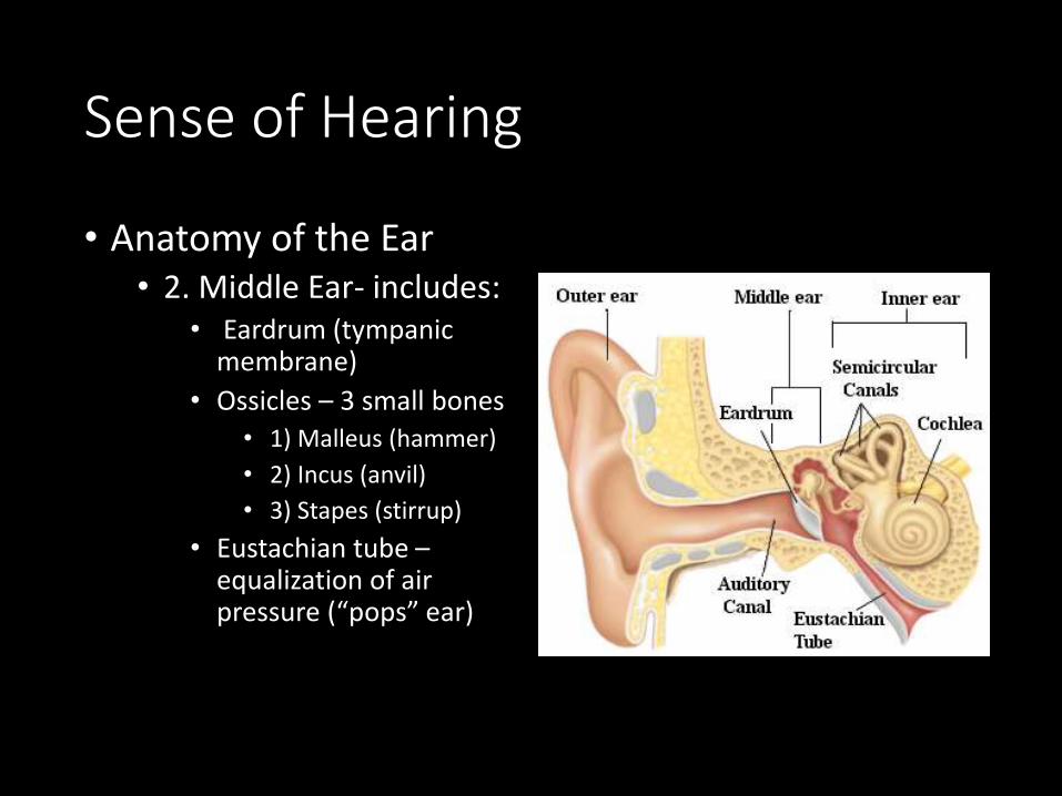
• Anatomy of the Ear• 2. Middle Ear- includes:
• Eardrum (tympanic membrane)
• Ossicles – 3 small bones• 1) Malleus (hammer)
• 2) Incus (anvil)
• 3) Stapes (stirrup)
• Eustachian tube –equalization of air pressure (“pops” ear)

Sense of Hearing
• Anatomy of the Ear• 3. Inner Ear – includes:
• Semicircular canals –involved with equilibrium
• Cochlea – snail-shaped structure involved with hearing

Sense of Hearing
• How we Hear1. Sound waves travel through the auditory canal to the
eardrum.
2. The sound waves cause the eardrum to vibrate.
3. The vibration causes the malleus (hammer) to hit the incus (anvil) and then the stapes (stirrup).
4. The vibration passes to the fluid in the cochlea of the inner ear.
5. Each part of the spiral cochlea is sensitive to different frequencies of sound.
6. The auditory nerve takes impulses to the brain.
Sense of Hearing
• Equilibrium• Mechanoreceptors in
the semicircular canals detect rotation and movement of the head
• Little hair cells send information to the brain to cause appropriate motor output so as to correct position when it is unbalanced.
• Vertigo (dizziness)

Sense of Sight
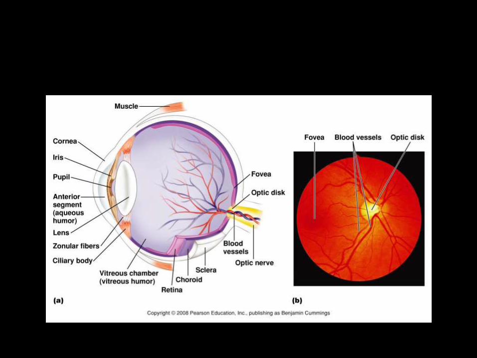
• Anatomy of the Eye• Sclera – protection
(white of eye)• Cornea – refracts light• Vitreous humor –
maintains eyeball shape• Retina
• Rods – black & white vision
• Cones – color vision
• Optic nerve – sends impulses to brain

Sense of Sight
• Anatomy of the Eye• Lens – focuses light
• Cilliary body – holds lens in place, accommodation
• Iris – regulates light entrance (muscle)
• Pupil – admits light

Sense of Sight
• How we see• 1. Light enters through the pupil.
• The iris can contract or dilate to allow different amounts of light into the eye.
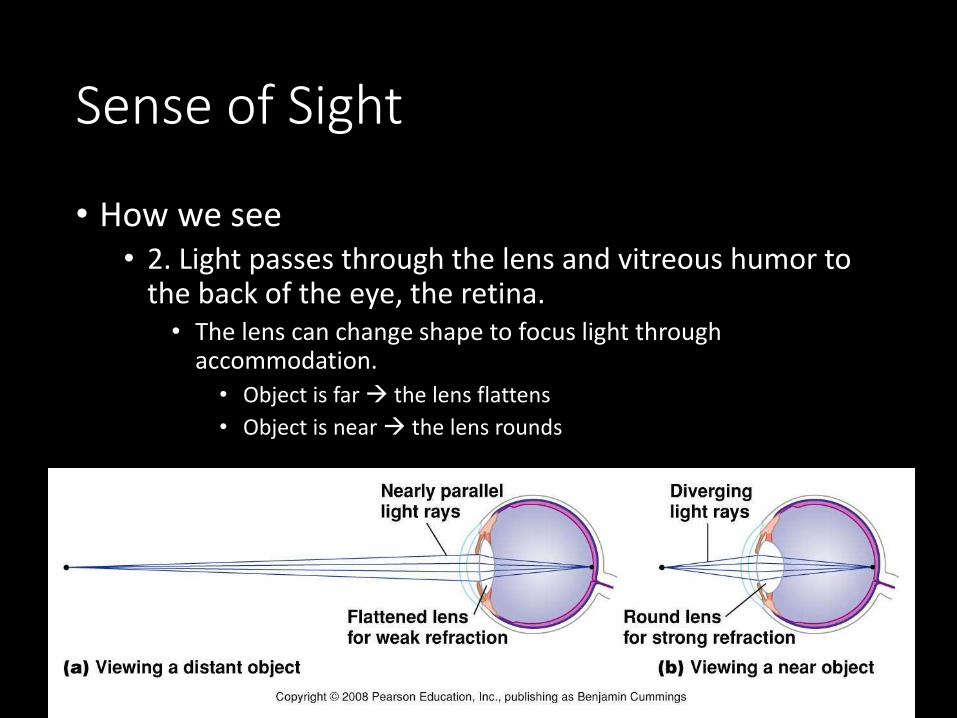
Sense of Sight
• How we see• 2. Light passes through the lens and vitreous humor to
the back of the eye, the retina.• The lens can change shape to focus light through
accommodation.
• Object is far the lens flattens
• Object is near the lens rounds
Sense of Sight
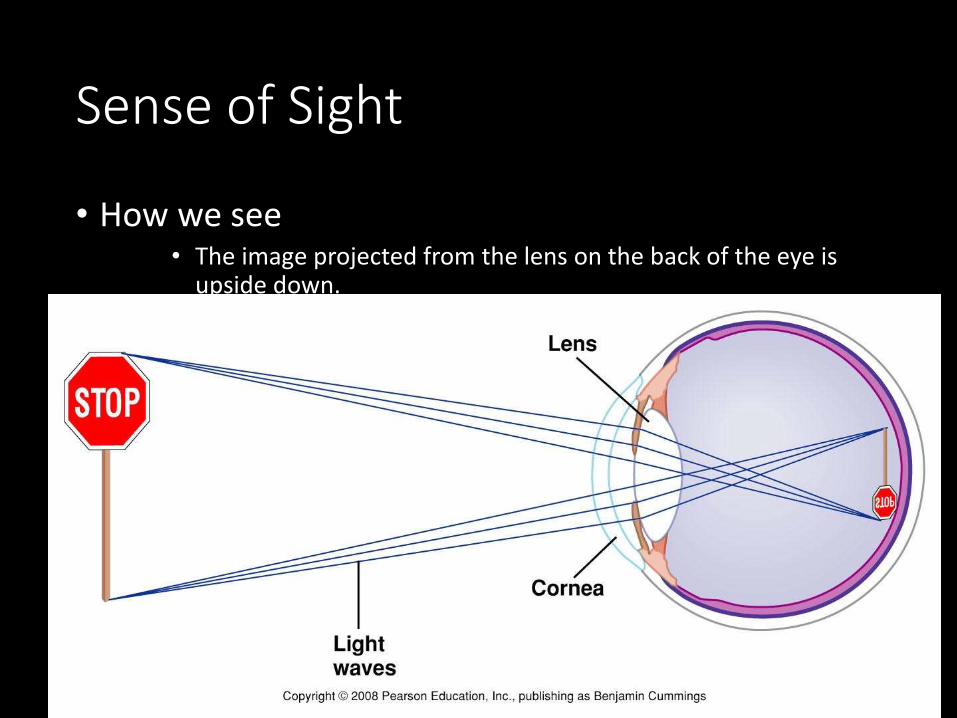
• How we see• The image projected from the lens on the back of the eye is
upside down.

Sense of Sight
• How we see• 3. The retina has photoreceptor cells that detect light
and send impulses to the brain.• Rods – black and white vision
• sensitive to light; night vision
• Cones – color vision & detail• Sensitive to bright light
• Blue, green, and red pigment cones detect different wavelengths of light
Sense of Sight
• How we see• 4. Impulses from the rods and cones in the retina are
sent to the optic nerve• This spot on the retina has not rods or cones and creates a
blind spot.

Sense of Sight
• How we see• 5. The optic nerves from each eye cross at the optic
chiasm.• Input from the right eye goes to the left occipital lobe
• Input from the left eye goes to the right occipital lobe
• 6. Visual integration centers in the occipital lobe process visual input.
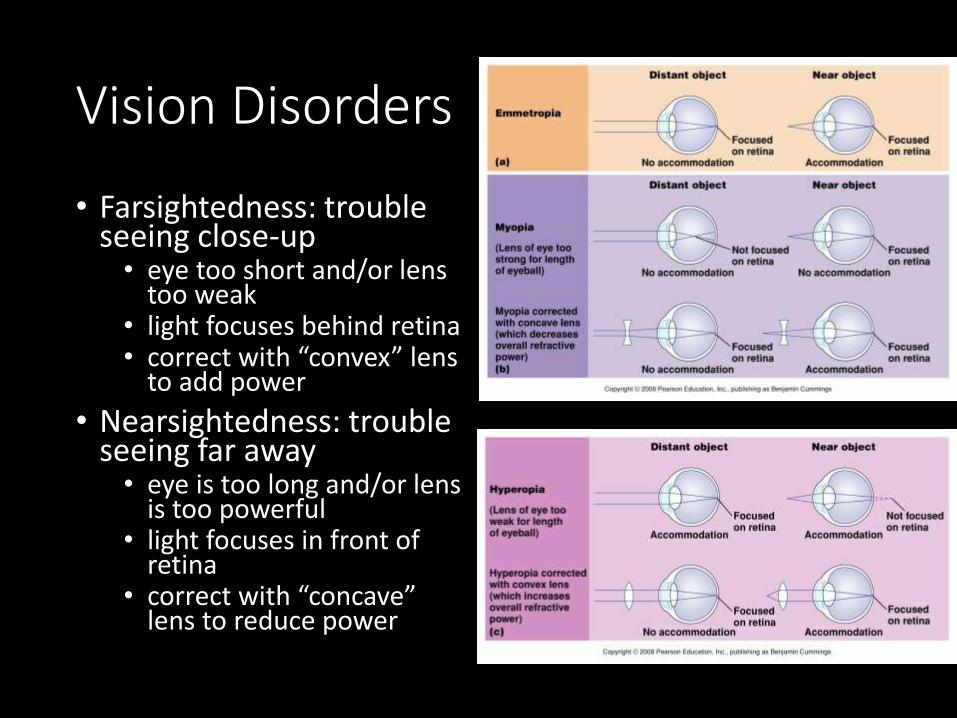
Vision Disorders
• Farsightedness: trouble seeing close-up• eye too short and/or lens
too weak• light focuses behind retina• correct with “convex” lens
to add power
• Nearsightedness: trouble seeing far away• eye is too long and/or lens
is too powerful• light focuses in front of
retina• correct with “concave”
lens to reduce power

• Presbyopia: Oldsightedness• The crystalline lens tends to harden with age
• The near point of distinct vision moves further and further away from the eye with age.
Astigmatism
• Abnormal curvature of the cornea
• Light from vertical and horizontal direction do not focuses in the same point
• Correct with “cylindrical” lens to compensate
Color Blindness
• Red-green color-blindness – occurs when red or green cones or pigments are missing• Due to sex-linked gene (on X chromosomes) so more
common in men.
• Non-sex-linked condition• Blue-color blindness- missing blue cones or pigments
• Monochromats: people who are totally colorblind, more severe

Disorders of the Eye
• Glaucoma• Damage to the optic
nerve occurs due to increased eye pressure
• Can lead to blindness
• Cataracts • Clouding of the lens
that affects vision
• Very common in older people
Figure 10.27b