the neurological examination - …westernbiomed.weebly.com/uploads/1/5/4/7/15477822/... · the...
TRANSCRIPT

The Neurological Examination
Dr. Gary Mumaugh – Western Physical Assessment

Common or Concerning Symptoms
• Headache
• Dizziness
• Generalized, proximal or distal weakness
• Numbness
• Abnormal or loss of sensations
• Loss of consciousness, syncope or near-syncope
• Seizures
• Tremors or involuntary movements

Health Promotion and Counseling
• Preventing stroke or TIA
• Reducing risk of peripheral neuropathy
• Detecting the “three D’s”
– Delirium
– Dementia
– depression

The Neurological Examination
Six Parts of the Neurological Exam
• Mental Status
• Cranial Nerves
• Motor
• Coordination
• Sensory
• Gait

Concept of a Screening Exam
• Screening each of the parts allows one to check on
the entire neuroaxis (Cortex, Subcortical White
Matter, Basal Ganglia/Thalamus, Brainstem,
Cerebellum, Spinal Cord, Peripheral Nerves, NMJ,
and Muscles)
• Expand evaluation of a given part to either
– Answer questions generated from the History
– Confirm or refute expected or unexpected findings on Exam

Mental Status
• Level of Alertness
– Subjective view of Examiner
– Definition of Consciousness
– Terminology for Depressed Level of Consciousness
– Concept of Coma
– Delerium • Degree of Orientation
– To what?
– “A and O x 4”
Mental Status
• Concentration
– Serial 7’s or 3’s
– “WORLD” backwards
– Months of the Year Backwards
– Try to quantify degree of impairment
* A and O and Concentration need to be
intact for other aspects of the Mental Status
Exam to have localizing value!

Mental Status Memory
• Immediate Recall
– A task of concentration
• Short-Term Memory
– “3/3 objects after 5 minutes”
• Long-Term Memory
– Last thing to go

Mental Status Language
• Aphasia vs. Dysarthria
• Receptive Language
– Command Following
• Expressive Language
– Fluency
– Word Finding
• Repetition
– Screens for Receptive, Expressive, and
Conductive Aphasias

Language
What is Altered Mental Status?
• What are some symptoms that would make
you think your patient has:
“Altered Mental Status”
• Altered level of conscious
• Disorientation
• Inappropriate behavior
• Altered cognition

What is Level of Consciousness?
What Is Altered LOC?
• Decreased wakefulness
• Unable to follow commands
• Decreased awareness
• Decreased responsiveness
• Unresponsive

Altered Level of
Consciousness versus Cognition?
• Alertness
• Wakefulness
• Awareness
• Responsive
• Sedation
• Coma
• Lethargic
• Orientation
• Confusion
• Concentration
• Comprehension
• Logic
• Able to follow instructions
• Memory –long and short term
• Appropriate behavior relative to the situation/environment
• Age specific
• Acute or chronic
• Continuum

What is Altered Neurologic Status?
• Altered Level of Consciousness
• Altered Mental Status
• Abnormal cranial nerves
• Abnormal speech
• Abnormal motor function
• Abnormal sensory function
• Alteration in balance/gait

What’s Normal?
• Alert and oriented X 3 (Person, Place,
Time/Date) consistent with developmental age
• Follows commands
• Responds to questions appropriately
• Speech Clear (?)
My name
is
Barbara
But there’s more to it….

Specialized Neuro Assessments
• AVPU (Alert, Verbal, Pain, Unresponsive)
• NIHSS (National Institute of Health Stroke Scale)
• Glasgow Coma Scale
• CAM – Confusion Assessment Method
• Cranial Nerve Assessment
• CMS –Circulation Movement Sensation
• Withdrawal Alcohol Screening (WAS)
• Mini mental health
• Electrolyte (Chvostec, Trousseau)
• Spinal

What can cause alteration in
mental status?
• Neurologic System vs. Other Systems
• Age Related
• Genetic/Hereditary
• Emotional/Psychiatric
• Acute vs. Chronic
• Sudden Onset vs. Slow Onset

AEIOU TIPS
A Alcohol
E Electrolytes, Endocrine
I Insulin
O Oxygenation
U Uremia
T Trauma, Toxicity, Temperature
I Infection
P Psych, Pharmacy, Perfusion
S Space Occupying lesion, Stroke, Seizure

A is for Alcohol

E is for Electrolytes

E is for Endocrine

I is for Insulin
• Hypoglycemia
• Hyperglycemia
• DKA
• Hyper Osmolar (HHNK)
• Renal failure
• Chronic Changes to brain

O is for Oxygenation
• Hypoxia
• Hyperventilation (Anxiety vs. Hypoxia)
• Respiratory Distress
• Airway
• Upper
• Lower
• Systemic

U is for Uremia
• Kidney failure-toxins
build up
• Encephalopathy
• Muscle weakness
• Mental status
changes
• Seizures
• coma
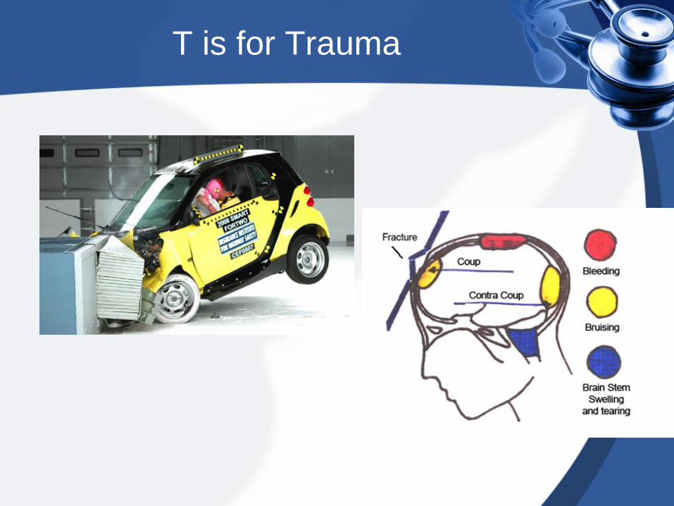
T is for Trauma

T is for Toxicity (Poisoning)

Intracranial Pressure
• Caused by
– Tumor
– Bleeding
– Infection
– Hydrocephalus
– Swelling
– Hyperosmolar
– Seizures


Temperature
• Fever
• Hyperthermia
• Hypothermia

I is for Infection
• Meningitis
• Encephalitis
• Brain Abscess

P is for Psychiatric
• Acute psychosis
• Schizophrenia
• Bipolar
• Delirium
– Hallucinations
– Auditory
– Delusional

P is for Pharmaceuticals
Toxicity Focused Physical Exam Findings
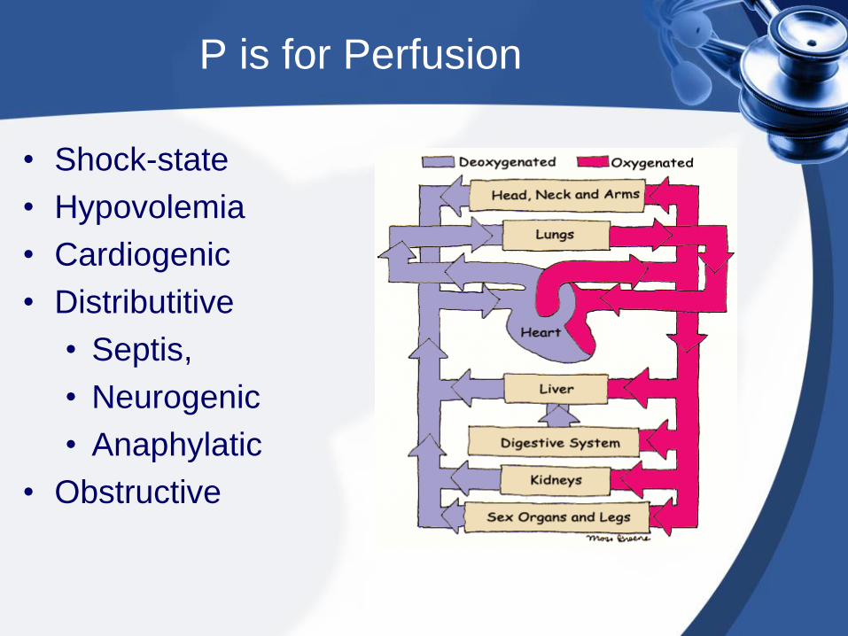
P is for Perfusion
• Shock-state
• Hypovolemia
• Cardiogenic
• Distributitive
• Septis,
• Neurogenic
• Anaphylatic
• Obstructive

S is for Space Occupying Lesion
• Tumor
• Hemorrhagic
• Hydrocephalus
• Brain Swelling – brain injury

S is for Stroke

S is for Seizures
• Generalized
• Tonic-Clonic
• Complex partial
• Absence
• Febrile
The Neurological Examination
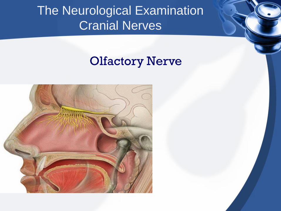
Cranial Nerves
Olfactory Nerve

The Cranial Nerves
Olfactory Nerve
• Distinguish Coffee from Cinnamon
• Smelling Salts irritate nasal mucosa and test
V2 Trigemminal Sense
• Disorders of Smell result from closed head
injuries

Cranial Nerves
Optic Nerve Cranial Nerve II

Cranial Nerves
Optic Nerve
• Visual Acuity
• Visual Fields
• Afferent input to Pupillary Light Reflex
– APD
• Look at the Nerve (Fundoscopic Exam)

Cranial Nerves
Trochlear Nerve
c.n. IV
Oculomotor Nerve
Cn III
Abducens Nerve
Cn VI

Cranial Nerves
Cranial Nerves III, IV, VI
• Extra-Ocular Muscles • Efferent limb of pupillary light reflex (III) • Ptosis
– Oculomotor Nerve Palsy
– Part of Horner’s Syndrome • Cardinal Directions of Gaze • Efferent output for Oculocephalic Reflex • Look for Nystagmus
“EOMI without nystagmus”

Cranial Nerves
Trigeminal Nerve
C.N. V

Cranial Nerves
Trigeminal Nerve
• Motor Component
• Opthalmic (V1), Maxillary (V2), and
Mandibular (V3) Distributions
• All modes of Primary Sensation Modalities
can be tested
• Afferent input for the Corneal Blink Reflex
“Facial sensation intact in all distributions”

Cranial Nerves
Facial Nerve
C.N. VII

Cranial Nerves
Facial Nerve
• Motor innervation to facial muscles • UMN versus LMN Facial Weakness • Efferent output to Corneal Blink Reflex • Other Functions
– Parasympathetic input to lacrimal, sublingual, and submandibular glands, taste to anterior 2/3 of tongue, general sensation to concha of earlobe and small part of scalp, motor input to stapedius muscle
“Facial motor intact”

Cranial Nerves
Vestibulocochlear Nerve
C.N. VIII

Cranial Nerves
Vestibulocochlear Nerve
• Hearing and Balance
– Patients will complain of tinnitis, hearing
loss, and/or vertigo
• Weber and Renee Test
– Differentiates Conductive vs Sensorineural
hearing loss
• Afferent input to the Oculocephalic Reflex
– Doll’s Eye Maneuver
– Cold Calorics
Cranial Nerves
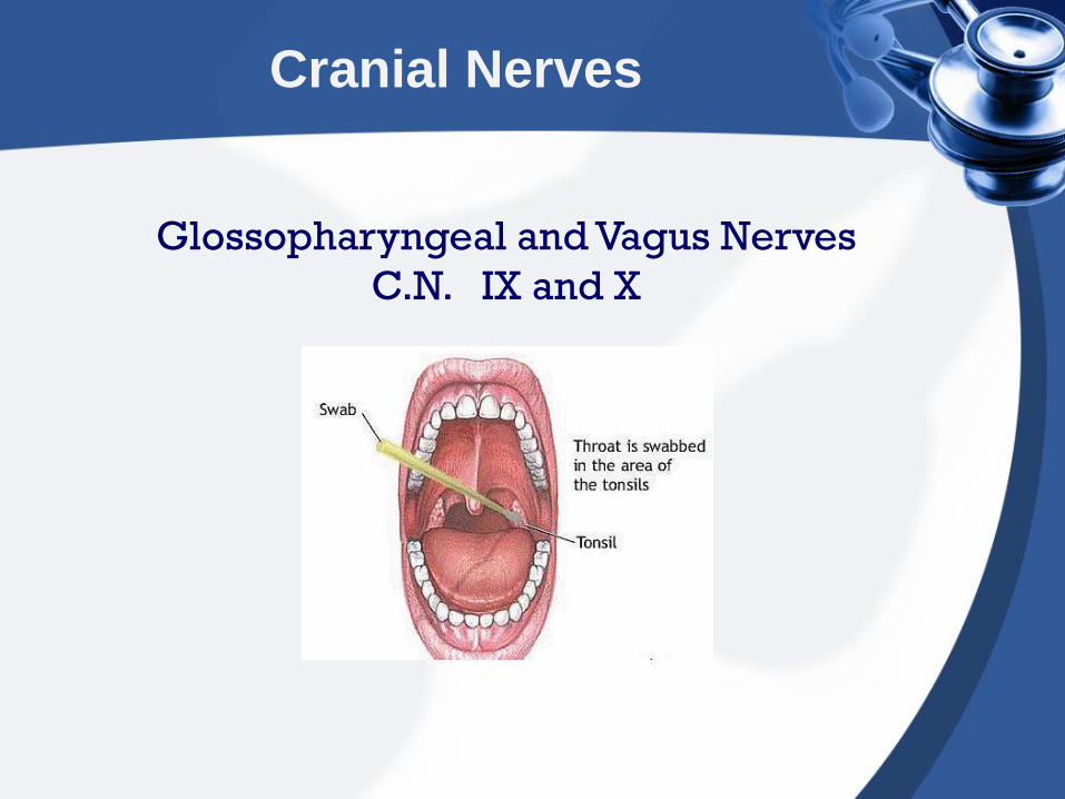
Glossopharyngeal and Vagus Nerves
C.N. IX and X

Cranial Nerves
Glossopharyngeal and Vagus Nerves
• Afferent (IX) and Efferent (X) components for
the Gag Reflex
• Vagus Nerve also does all parasympathetics
from the neck down until the mid-transverse
colon

Cranial Nerves
Spinal Accessory Nerve
C.N. XI
Trapezius
strength
Sternocleido-
Mastoid
strength

Cranial Nerves
Hypoglossal Nerve
C.N. XII

Cranial Nerves
Hypoglossal Nerve
• Protrudes the tongue to the opposite side
• Tongue in cheek (strength)
• Hemi-atrophy and fasiculations (LMN)

The Motor Examination
• Strength
• Tone
• DTR’s
• Plantar Responses
• Involuntary Movements

Exam of the Motor System
• Position, movement, muscle bulk and tone
– Observe body position and involuntary
movements such as tremors, tics, fasiculations
– Inspect muscle bulk, note any atrophy
– Assess muscle tone – flex and extend the arm
and lower the leg for residual tension
– Tics (sudden, rapid, non-rhytmic)

The Motor Examination
Strength
The Motor Examination
Strength
Medical Research Council Scale
• 5/5 = Full Strength
• 4/5 = Weakness with Resistance
• 3/5 = Can Overcome Gravity Only
• 2/5 = Can Move Limb without Gravity
• 1/5 = Can Activate Muscle without
Moving Limb
• 0/5 = Cannot Activate Muscle

The Motor Examination
Weakness
• Describe the Distribution of Weakness
– Upper Motor Neuron Pattern
– Peripheral Neuropathy Pattern
– Myopathic Pattern

The Motor Examination
• Strength
– Decreased
• Tone
– increased spasticty
• DTR
– increased or brisk
• Plantar reflexes
– upgoing toes
• Atrophy/Fasiculations
– None
• Strength
– Decreased
• Tone
– decreased hypotonia
• DTR
– diminished or absent
• Plantar reflexes
– downgoing toes
• Atrophy/Fasiculations
– positive or negative
Upper Motor Neuron Lower Motor Neuron

Muscle Strength
• Test the following muscle groups and movements
– Biceps and triceps – wrist flexion & extension
– Handgrip, finger – abduction, adduction, thumb
opposition
– Trunk – flexion, extension, lateral bending
– Thorax – expansion, diaphragmatic excursion
during respiration
– Hip – flexion, extension, abduction, adduction
– Knee and ankle – flexion and extension

The Motor Examination
Tone
• Tone is the resistance appreciated when
moving a limb passively
• “Normal Tone”
• Hypotonia
– “Central Hypotonia”
– “Peripheral Hypotonia”
• Increased Tone
– Spasticity (Corticospinal Tract)
– Rigidity (Basal Ganglia, Parkinson’s Disease)
– Dystonia (Basal Ganglia)

The Motor Examination
DTR’s
• 0/4 = Absent
• 1-2/4 = Normal Range
• 3/4 = Pathologically Brisk
• 4/4 = Clonus

The Motor Examination
Involuntary Movements
• Hyperkinetic Movements
– Chorea
– Athetosis
– Tics
– Myoclonus
• Bradykinetic Movements
– Parkinsonism (Bradykinesia, Rigidity,
Postural Instability, Resting Tremor)
– Dystonia

The Sensory Examination
Primary Sensory Modalities
• Light Touch (Multiple Pathways)
• Pain/Temperature Sensation (Spinothalamic
Tract)
• Vibration/Position Sensation (Posterior
Columns)
Cortical Sensory Modalities
• Stereognosis
• Graphesthesia
• Two-Point Discrimination
• Double Simultaneous Extinction

The Sensory Examination
Primary Sensory Modalities
• Reflect Input from sensory receptors,
sensory nerves, spinal cord, brainstem,
through to the level of the Thalamus.
Cortical Sensory Modalities
• Reflect Processing by the Somatosensory
Cortex (post-central gyrus)

The Sensory Examination
Pain and Temperature
• Small-unmyelinated fibers provide pain and
temperature input which travels through the
dorsal roots
• Second-order neurons cross midline at the
anterior commissure and travel up the lateral
spinothalamic tract

The Sensory Examination
Joint Position and Vibration
• Larger myelinated fibers bring sensory
information concerning vibration and joint
position
• Second-order neurons cross at the Thalamus

The Sensory Examination

The Sensory Examination
• Pain and Temperature
– Pinprick (One pin per patient!)
– Sensation of Cold
– Look for Sensory Nerve or
Dermatomal Distribution
• Vibration Sensation
– C-128 Hz Tuning Fork (check great toe)
• Joint Position Sensation
– Check great toe
– Romberg Sign

The Sensory Examination
Higher Cortical Sensory Function
• Graphesthesia
• Stereognosis
• Two-Point Discrimination
• Double Simultaneous Extinction
• Gerstmann’s Syndrome (acalculia, right-left
confusion, finger agnosia, agraphia)
– Usually seen in Dominant Parietal Lobe
lesions

Coordination
Cerebellum
Inputs Outputs

Coordination Examination
• Test coordination including
– Rapid alternating movements
• Repeating pronation supination palms
• Tapping thumbs and fingers
• Tapping ball of foot with fingers
– Point-to-point movements
• Touching face and nose
• Moving heel down opposite leg

• Test coordination including
– Gait – assess gait as patient
• Walks across room
• Walks heel-to-toe
• Walks on toes and heels
• Hops in place

• Test coordination including
– Stance, namely
• Rhomberg test
– Patient stands with feet together and arms
forward, eyes closed, for 30-60 seconds
without support
– Loss of balance with eyed closed is +
• Pronator drift
– Patient stands for 20-30 seconds with both
arms forward, palms up, eyes closed, tap
arms briskly downward
– Pronation and downward drift is +

Sensory Exam
• General Principles of Exam
– Compare symmetric areas on both sides
of the body
– When testing pain, temperature, and
touch, compare distal with proximal areas
of extremities
– Map out the boundaries of any area of
sensory loss or hypersensitivity

Sensory Exam
• Test pain using a disposable broken Q-tip or
pin and discard after usage
– Ask if the prick is sharp or dull or ask the patient
to compare two sensations
• Test light tough using cotton wisp
• Test vibration with tuning fork
• Test proprioception – hold big toe by its sides
between your thumb and index finger, pull it away
from other toes, and move it up and down. Ask
patient to identify the direction of movement.

Sensory Exam
• Assess discriminating sensation to test the
ability of the sensory cortex to analyze and
interpret sensations
– Stereogenesis – place a key or familiar object in
patient’s hand and ask patient to identify
– Number identification (graphesthesia) – outline a
large number in patient’s palm and ask them to
identify
– Two-point discrimination – using two ends of an
open paper clip, touch the finger pad in two
places simultaneously and ask patient to identify
1 touch or 2


Sensory Exam
• Assess discriminating sensation
– Point location – lightly touch a point on the
patient’s skin and ask the patient to point
to the spot
– Extinction – touch and area on both sides
of the body at the same time and ask if
patient feels 1 spot or 2

Deep Tendon Reflexes
• Select a properly weighted hammer
• Encourage the patient to relax, position the
limbs properly and symmetrically
• Strike the tendon with a brisk direct
movement using the minimum force needed
to obtain a response
• Use reinforcement when needed
• Grade the response

Reflex Grading
• + 4 Very brisk, hyperactive, with clonus
(rhythmic oscillations between flexion and
extension)
• + 3 Brisker than average; possibly, but not
necessarily indicative of disease
• + 2 Average; normal
• + 1 Somewhat diminished; low normal
• 0 No response


Deep Tendon Reflexes
• Biceps reflex C5-6
• Triceps reflex C6-7
• Brachioradialis or supinator C5-6
• Knee reflex L2-4
• Ankle reflex Mainly S1
• Clonus
– A hyperactive response required for
assigning a reflex grade of 4, usually
elicited at the ankle
Cutaneous Sensation Reflexes
• Abdominal reflex
– Upper T8-10
– Lower T10-12
• Plantar response – L5-S1
• Anal reflex – S2-4

The Gait
• A normal Gait requires multiple levels of the
neuroaxis to be intact
– Vision
– Strength
– Balance/Coordination
– Joint Position

The Gait
Observe Different Aspects of Gait
• Arm Swing
• Base of Gait
• Heel Strike
• Time Spent on Each Leg
• Posture of Trunk
• Toe Walking
• Heel Walking
• Tandem Walking

The Gait
Classical Patterns of Abnormal Gait
• Parkinsonism Gait
• Hemiparetic Gait
• Spastic Diplegia Gait
• Acute Ataxia Gait
• Chronic Ataxia Gait
• Waddling Gait (Hip Girdle Weakness)
• High Stepping Gait