the new genetics: paradigm shifts in … new genetics: paradigm shifts in prenatal diagnosis...
TRANSCRIPT
Jennifer Hoskovec, MS, CGC
Assistant Professor
Director, Prenatal Genetic Counseling Services
Department of Ob/Gyn and Reproductive Sciences
UT Health
THE NEW GENETICS:
PARADIGM SHIFTS IN
PRENATAL DIAGNOSIS

Overview of standard screening and testing options for
aneuploidy
Non-Invasive Prenatal Testing for aneuploidy (NIPT)
Chromosomal Microarray Analysis in the prenatal setting
OUTLINE

Screening
Screening Options: First Trimester Screening
Quadruple Marker Screen
Integrated, Sequential, or Contingency Screens
Anatomy Scan
Benefit(s)
Non-invasive = no risk
Identifies women from low risk pool who are at increased risk
Disadvantage(s)
Risk calculation only
False positive/negative
Limited to trisomy 18, 13, 21
Timing, insurance coverage
Patient anxiety
Invasive Prenatal Diagnosis
Testing Options:
Chorionic Villus
Sampling (CVS)
Amniocentesis
Benefit(s)
Diagnostic information
on all aneuploidies
Additional testing
available such as
microarray, PCR
Disadvantage(s)
Invasive, risk of
pregnancy loss (1/300-
1/500)
Standard Screening and Testing Options for
Fetal Aneuploidy

NIPT NON-INVASIVE
PRENATAL
TESTING
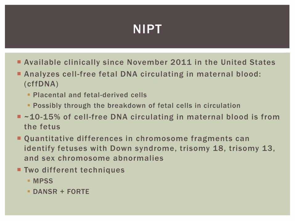
Available clinically since November 2011 in the United States
Analyzes cell -free fetal DNA circulating in maternal blood:
(cf fDNA)
Placental and fetal-derived cells
Possibly through the breakdown of fetal cells in circulation
~10-15% of cell -free DNA circulating in maternal blood is from
the fetus
Quantitative dif ferences in chromosome fragments can
identify fetuses with Down syndrome, trisomy 18, trisomy 13,
and sex chromosome abnormalies
Two dif ferent techniques
MPSS
DANSR + FORTE
NIPT

Simultaneously sequence millions of short segments from
amplified DNA
Hundreds of sequences generated in a single run
Amplifies maternal and fetal DNA together
Increases number of samples run
Decreases cost
Different platforms used
Technique currently used by Sequenom and Verinata
MASSIVELY PARALLEL SHOTGUN
SEQUENCING
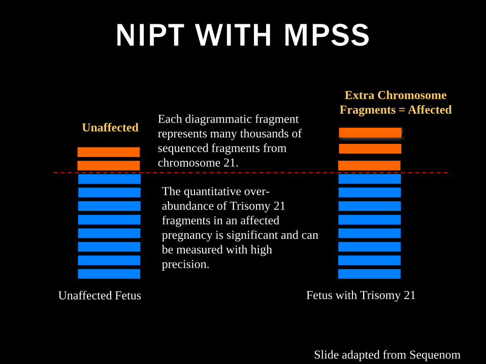
NIPT WITH MPSS
Unaffected
Unaffected Fetus Fetus with Trisomy 21
Each diagrammatic fragment
represents many thousands of
sequenced fragments from
chromosome 21.
The quantitative over-
abundance of Trisomy 21
fragments in an affected
pregnancy is significant and can
be measured with high
precision.
Extra Chromosome
Fragments = Affected
Slide adapted from Sequenom
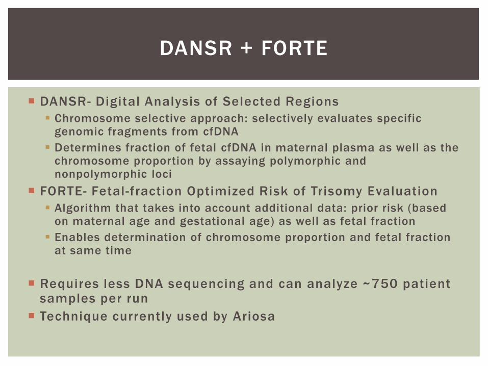
DANSR + FORTE
DANSR- Digital Analysis of Selected Regions
Chromosome selective approach: selectively evaluates specific genomic fragments from cfDNA
Determines fraction of fetal cfDNA in maternal plasma as well as the chromosome proportion by assaying polymorphic and nonpolymorphic loci
FORTE- Fetal-fraction Optimized Risk of Trisomy Evaluation
Algorithm that takes into account additional data: prior risk (based on maternal age and gestational age) as well as fetal fraction
Enables determination of chromosome proportion and fetal fraction at same time
Requires less DNA sequencing and can analyze ~750 patient samples per run
Technique currently used by Ariosa
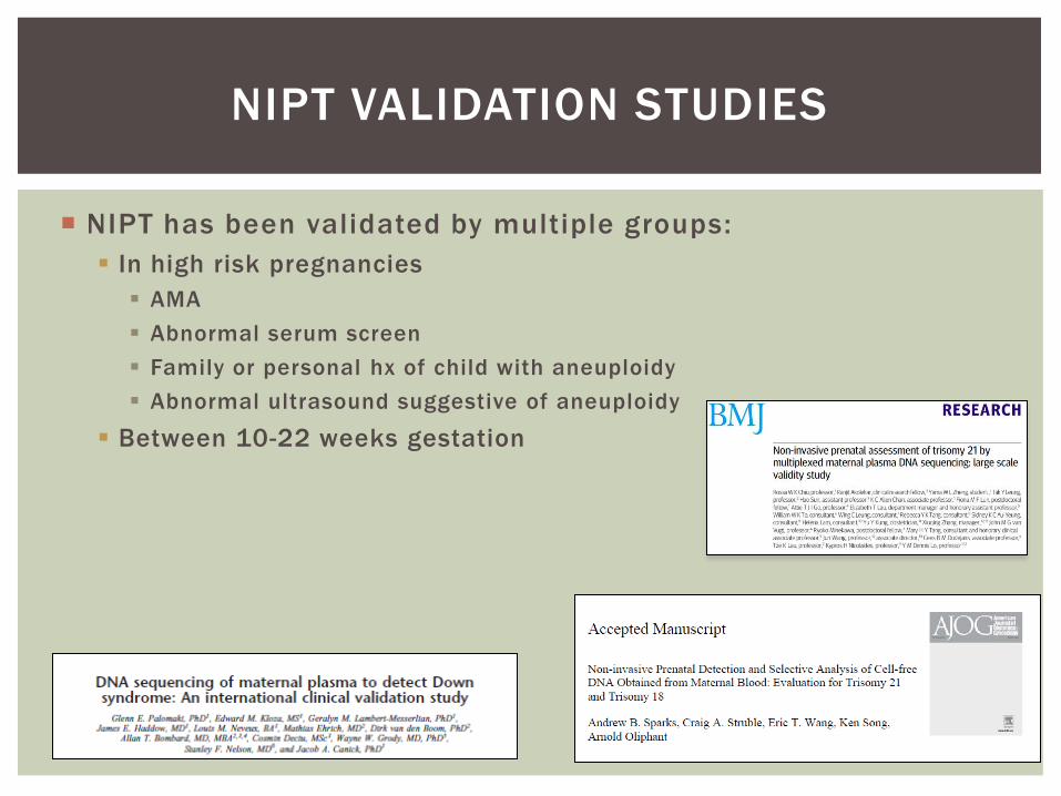
NIPT has been validated by multiple groups:
In high risk pregnancies
AMA
Abnormal serum screen
Family or personal hx of child with aneuploidy
Abnormal ultrasound suggestive of aneuploidy
Between 10-22 weeks gestation
NIPT VALIDATION STUDIES
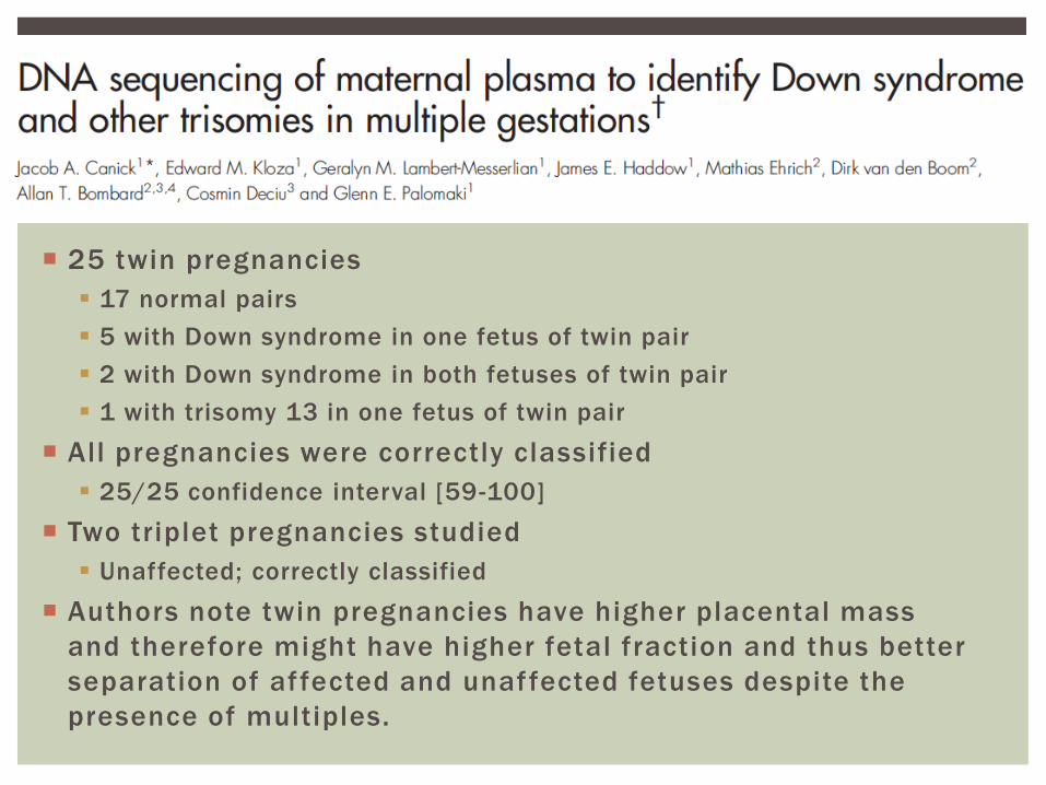
25 twin pregnancies
17 normal pairs
5 with Down syndrome in one fetus of twin pair
2 with Down syndrome in both fetuses of twin pair
1 with trisomy 13 in one fetus of twin pair
All pregnancies were correctly classified
25/25 confidence interval [59-100]
Two triplet pregnancies studied
Unaffected; correctly classified
Authors note twin pregnancies have higher placental mass
and therefore might have higher fetal fraction and thus better
separation of affected and unaffected fetuses despite the
presence of multiples.
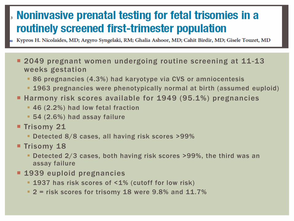
2049 pregnant women undergoing routine screening at 11 -13 weeks gestation
86 pregnancies (4.3%) had karyotype via CVS or amniocentesis
1963 pregnancies were phenotypically normal at birth (assumed euploid)
Harmony risk scores available for 1949 (95.1%) pregnancies
46 (2.2%) had low fetal fraction
54 (2.6%) had assay failure
Trisomy 21
Detected 8/8 cases, all having risk scores >99%
Trisomy 18
Detected 2/3 cases, both having risk scores >99%, the third was an assay failure
1939 euploid pregnancies
1937 has risk scores of <1% (cutoff for low risk)
2 = risk scores for trisomy 18 were 9.8% and 11.7%
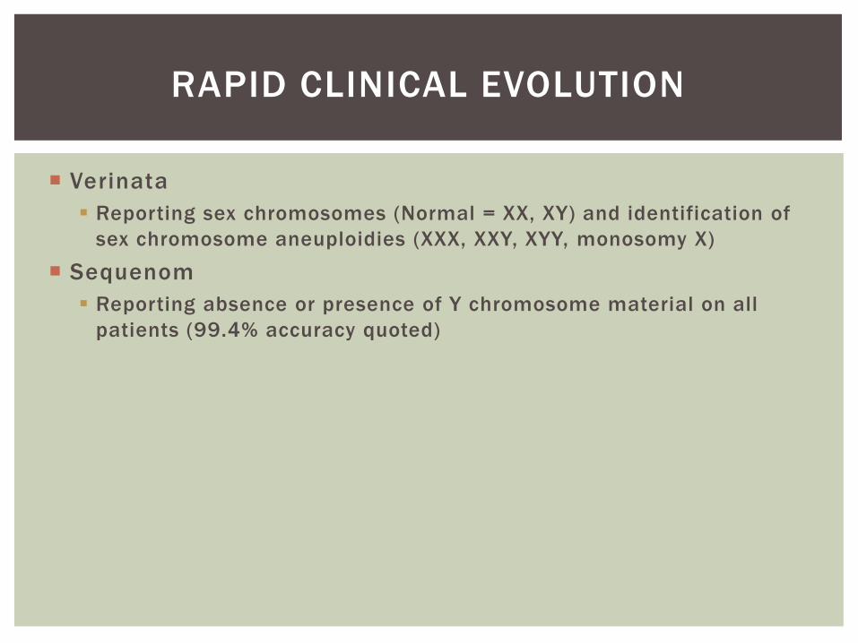
RAPID CLINICAL EVOLUTION
Verinata
Reporting sex chromosomes (Normal = XX, XY) and identification of
sex chromosome aneuploidies (XXX, XXY, XYY, monosomy X)
Sequenom
Reporting absence or presence of Y chromosome material on all
patients (99.4% accuracy quoted)
Three
separate
groups
have now
shown
high
sensitivity
and
specificity
with low
false
positive
rate
NIPT IN CLINICAL CARE
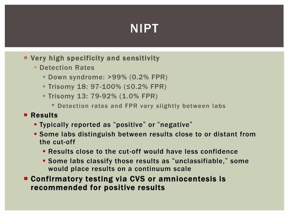
Very high specificity and sensitivity
Detection Rates
Down syndrome: >99% (0.2% FPR)
Trisomy 18: 97-100% (≤0.2% FPR)
Trisomy 13: 79-92% (1.0% FPR)
* Detection rates and FPR vary sl ightly between labs
Results
Typically reported as “positive” or “negative”
Some labs distinguish between results close to or distant from the cut-off
Results close to the cut-off would have less confidence
Some labs classify those results as “unclassifiable,” some would place results on a continuum scale
Confirmatory testing via CVS or amniocentesis is recommended for positive results
NIPT
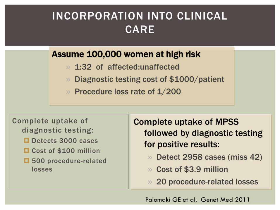
Complete uptake of
diagnostic testing:
Detects 3000 cases
Cost of $100 million
500 procedure-related
losses
INCORPORATION INTO CLINICAL
CARE
Assume 100,000 women at high risk
» 1:32 of affected:unaffected
» Diagnostic testing cost of $1000/patient
» Procedure loss rate of 1/200
Complete uptake of MPSS
followed by diagnostic testing
for positive results:
» Detect 2958 cases (miss 42)
» Cost of $3.9 million
» 20 procedure-related losses
Palomaki GE et al. Genet Med 2011

Current limitations
Validation
Limited validation studies in low risk women
Validation study in twins had only 25 sets
Not validated in triplet or higher order multiples
Not validated in pregnancies conceived with egg donors
Not validated past 22 weeks gestation
Cost and insurance coverage
Does not include screening for ONTD
NIPT: LIMITATIONS

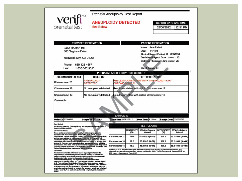
NIPT Specifics Laboratory (Test name)
Technology Conditions Tested For
Sensitivity Specificity Reporting
Sequenom (MaterniT21Plus)
MPSS Trisomy 21 Trisomy 18 Trisomy 13
T21 = 99.1% T18 = >99.9% T13 = 91.7%
T21 = 99.9% T18 = 99.6% T13 = 99.7%
Positive Negative Failure
Verinata (Verify)
MPSS Trisomy 21 Trisomy 18 Trisomy 13 Sex Chromosomes
T21 = 100% T18 = 97.2% T13 = 78.6% 45X = 95% XXX, XXY, XYY = Limited data
T21 = 100% T18 = 100% T13 = 100% 45X = 100%
Positive Negative Aneuploidy suspected Failure
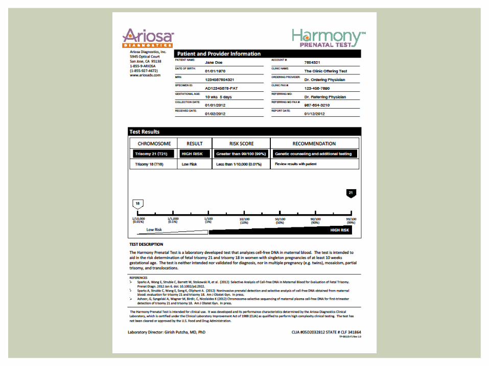
Ariosa (Harmony) Partnered with Integrated Genetics (LabCorp)
DANSR (assay) + FORTE (algorithm)
Trisomy 21 Trisomy 18 Trisomy 13
T21 = 100% T18 = 97.4%
T21 = 99.9% T18 = 99.9%
Risk Ratio via algorithm 1/10,000 – 99/100 (0.5% results fell between the two extreme values)


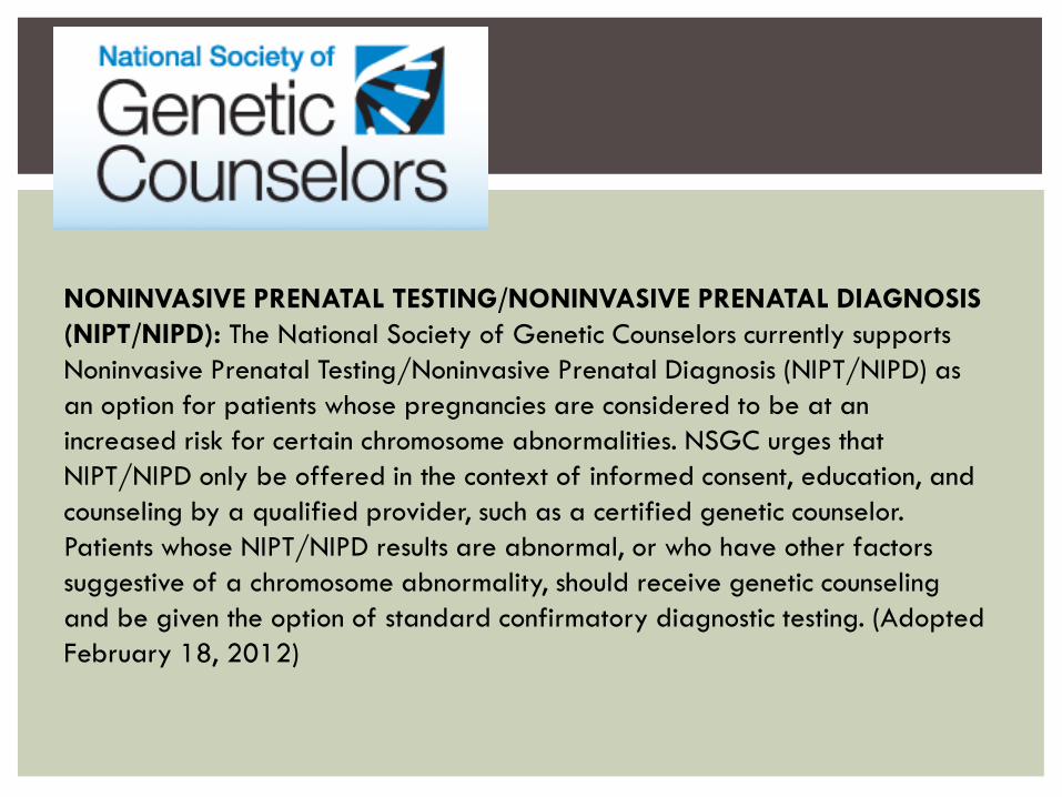
NONINVASIVE PRENATAL TESTING/NONINVASIVE PRENATAL DIAGNOSIS
(NIPT/NIPD): The National Society of Genetic Counselors currently supports
Noninvasive Prenatal Testing/Noninvasive Prenatal Diagnosis (NIPT/NIPD) as
an option for patients whose pregnancies are considered to be at an
increased risk for certain chromosome abnormalities. NSGC urges that
NIPT/NIPD only be offered in the context of informed consent, education, and
counseling by a qualified provider, such as a certified genetic counselor.
Patients whose NIPT/NIPD results are abnormal, or who have other factors
suggestive of a chromosome abnormality, should receive genetic counseling
and be given the option of standard confirmatory diagnostic testing. (Adopted
February 18, 2012)
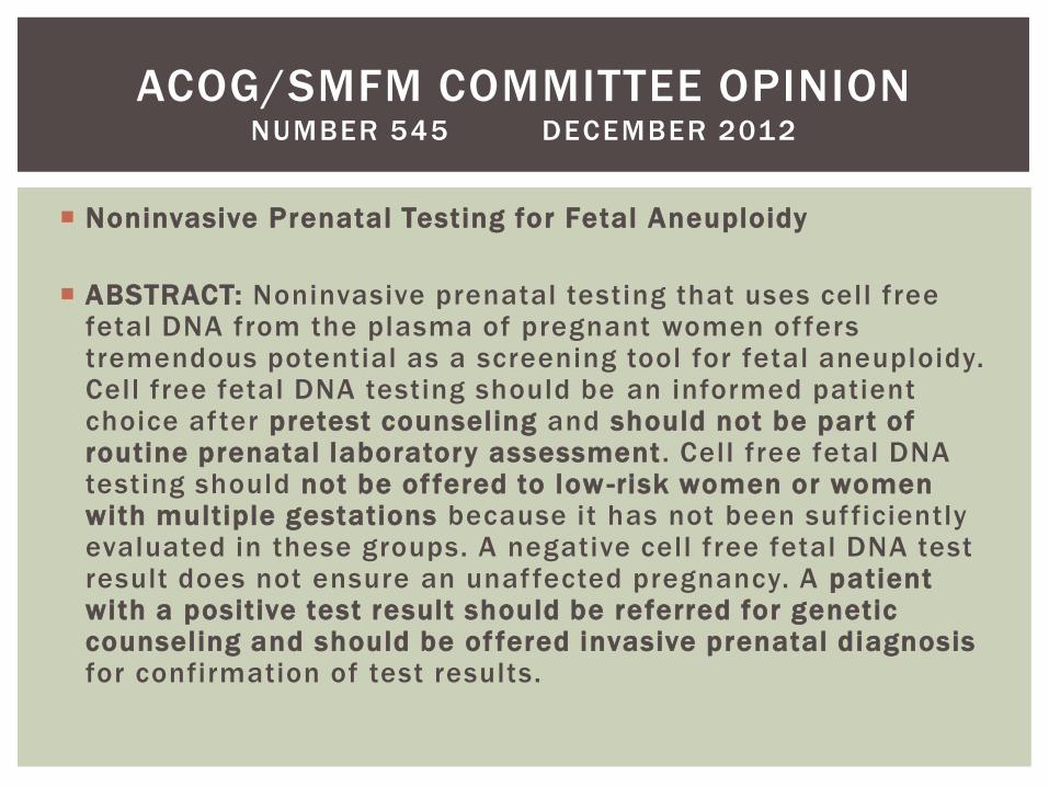
Noninvasive Prenatal Testing for Fetal Aneuploidy
ABSTRACT: Noninvasive prenatal testing that uses cell free fetal DNA from the plasma of pregnant women offers tremendous potential as a screening tool for fetal aneuploidy. Cell free fetal DNA testing should be an informed patient choice after pretest counseling and should not be part of routine prenatal laboratory assessment. Cell free fetal DNA testing should not be of fered to low -risk women or women with multiple gestations because it has not been sufficiently evaluated in these groups. A negative cell free fetal DNA test result does not ensure an unaffected pregnancy. A patient with a positive test result should be referred for genetic counseling and should be of fered invasive prenatal diagnosis for confirmation of test results.
ACOG/SMFM COMMITTEE OPINION NUMBER 545 DECEMBER 2012
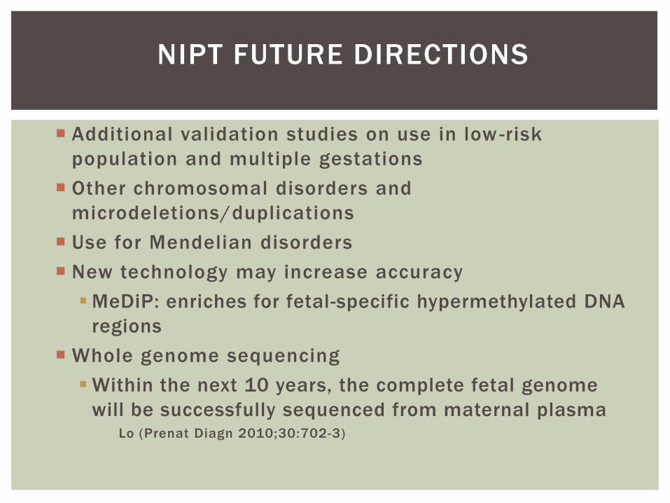
NIPT FUTURE DIRECTIONS
Additional validation studies on use in low -risk
population and multiple gestations
Other chromosomal disorders and
microdeletions/duplications
Use for Mendelian disorders
New technology may increase accuracy
MeDiP: enriches for fetal-specific hypermethylated DNA
regions
Whole genome sequencing
Within the next 10 years, the complete fetal genome
will be successfully sequenced from maternal plasma Lo (Prenat Diagn 2010;30:702-3)

So many options!
Accurate and balanced discussion of options with patient is very
important
Benefits
Limitations
Risks
Assist the patient in making informed, autonomous decision
Be sensitive to personal nature of decision
Family values
Religious beliefs
Family and life situations
Concerns about having a child with an abnormality
Concerns about risk of miscarriage
SUMMARY
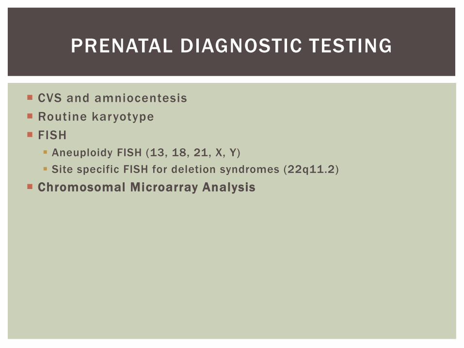
CVS and amniocentesis
Routine karyotype
FISH
Aneuploidy FISH (13, 18, 21, X, Y)
Site specific FISH for deletion syndromes (22q11.2)
Chromosomal Microarray Analysis
PRENATAL DIAGNOSTIC TESTING

CHROMOSOMAL MICROARRAY ANALYSIS
CMA platforms use thousands of DNA probes spread across the genome to detect gains and losses of genetic material.
Extracted DNA from the patient (fetus) is compared with a reference (normal) genome.
Allows identification of abnormal copy number changes (gains and losses).
Aneuploidy
Duplications and deletions – too small to be seen by conventional cytogenetics
Limitations:
Cannot detect balanced chromosome rearrangements (identifies dosage differences, not positional differences)
Cannot identify triploidy
Possible Pitfalls:
Identification of a copy variant of unknown significance (~1.5%)
Requires parental bloods for comparison
Possible out of pocket expense to the patient
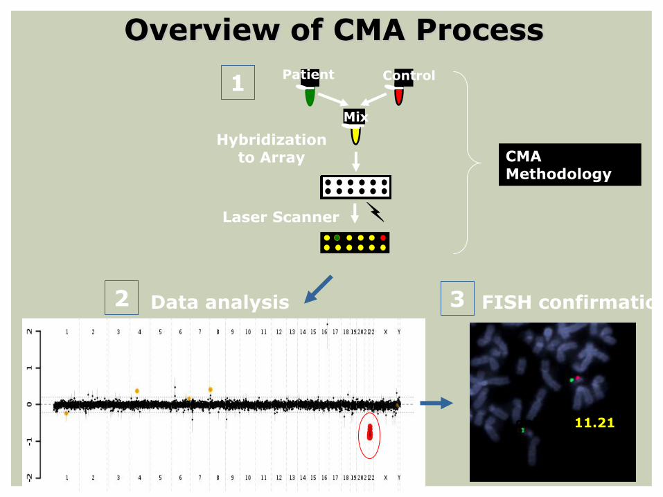
Mix
Patient
Hybridization to Array
Control
Laser Scanner
Data analysis
del 22:q11.21
FISH confirmation
CMA Methodology
1
2 3
Overview of CMA Process
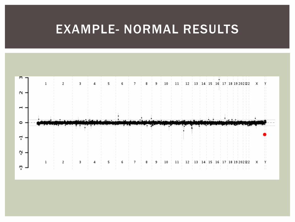
EXAMPLE- NORMAL RESULTS
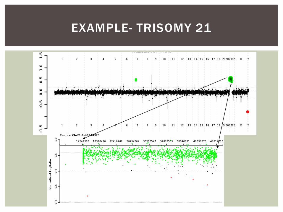
EXAMPLE- TRISOMY 21
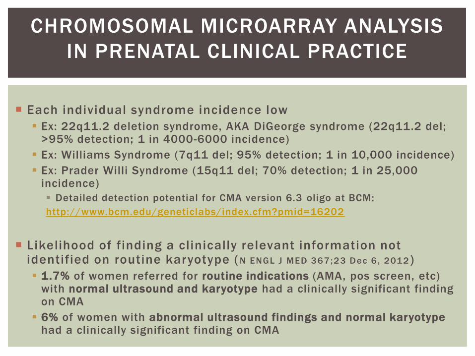
CHROMOSOMAL MICROARRAY ANALYSIS
IN PRENATAL CLINICAL PRACTICE
Each individual syndrome incidence low
Ex: 22q11.2 deletion syndrome, AKA DiGeorge syndrome (22q11.2 del; >95% detection; 1 in 4000-6000 incidence)
Ex: Williams Syndrome (7q11 del; 95% detection; 1 in 10,000 incidence)
Ex: Prader Willi Syndrome (15q11 del; 70% detection; 1 in 25,000 incidence)
Detailed detection potential for CMA version 6.3 oligo at BCM:
http://www.bcm.edu/geneticlabs/index.cfm?pmid=16202
Likelihood of finding a clinically relevant information not identified on routine karyotype ( N ENGL J MED 367;23 Dec 6 , 2012 )
1.7% of women referred for routine indications (AMA, pos screen, etc) with normal ultrasound and karyotype had a clinically significant finding on CMA
6% of women with abnormal ultrasound findings and normal karyotype had a clinically significant finding on CMA

QUESTIONS
REFERENCES
Lo et al (1997) Presence of fetal DNA in maternal plasma and serum.
Lancet
Finning et al (2002) Prediction of fetal D status from maternal plasma:
introduction of a new noninvasive fetal RHD genotyping service.
Transfusion
Bianchi DW (2004) Circulating fetal DNA: its origin and diagnostic
potential - a review.
Ding et al (2004) MS analysis of single -nucleotide dif ferences in
circulating nucleic acids: application to non -invasive prenatal
diagnosis. PNAS
Gautier et al (2005) Fetal RhD genotyping by maternal serum analysis:
a two-year experience. AJOG
Scheffer et al (2011) Noninvasive fetal blood group genotyping if rhesus D,
c, E, and of K in alloimmunised pregnant women: evaluation of a 7-year
clinical experience. BJOG
Chiu et al (2011), Non-invasive prenatal assessment of trisomy 21 by
multiplexed maternal plasma DNA sequencing: large scale validity study.
BMJ
REFERENCES
Palomaki et al (2011), DNA sequencing of maternal plasma to detect Down
syndrome: an international clinical validation study. Genetics in Medicine
Palomaki et al (2012), DNA sequencing of maternal plasma reliably identifies
trisomy 18 and trisomy 13 as well as Down syndrome: an international collaborative
study. Genetics in Medicine
Bianchi et al (2012) Genome-wide fetal aneuploidy detection by maternal
plasma DNA sequencing. Obstetr ics and Gynecology
Sparks et al (2012) Non-invasive prenatal detection and selective analysis of
cel l - free DNA obtained from maternal blood: evaluation for tr isomy 21 and
trisomy 18. AJOG
Ashoor et al (2012) Chromosome-selective sequencing of maternal plasma cel l -
free DNA for first -tr imester detection of tr isomy 21 and trisomy 18. AJOG
Colah et al (2011) Invasive and non -invasive approaches for prenatal diagnosis
of haemoglobinopathies : experiences from India. Indian J Med Res
Norton et al (2012) Non Invasive Chromosomal Evaluation (NICE) Study: results
of a multicenter prospective cohort study for detection of fetal tr isomy 21 and
trisomy 18. AJOG .
Canick et al (2012) DNA sequencing of maternal plasma to identify Down
syndrome and other tr isomies in multiple gestations . Prenat Diagnosis
REFERENCES
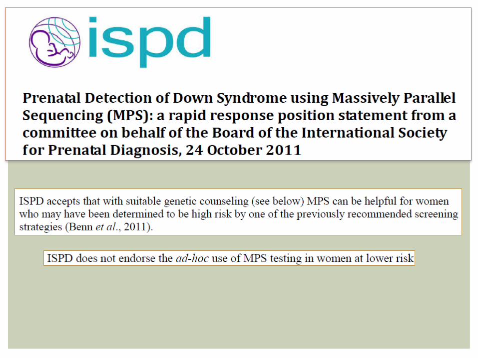
Benn et al (2011) Prenatal detection of Down syndrome using massively paral lel
shotgun sequencing : a rapid response posit ion statement from a committee on
behalf of the board of the international society for prenatal diagnosis.
NSGC (2012) Posit ion statement on noninvasive prenatal testing/noninvasive
prenatal diagnosis.
Sehnert et al (2011) Optimal detection of fetal chromosomal abnormalit ies by
massively paral lel DNA sequencing of cel l - free fetal DNA from maternal blood.
Clinical Chemistry
Ladha, S (2012) A new era of non -invasive prenatal genetic diagnosis: exploit ing
fetal epigenetic dif ferences. Clin Genet
Devaney et al (2011) Noninvasive fetal sex determination using cel l - free fetal
DNA: a systematic review and meta -analysis. JAMA
Hil l et al (2011) Non-invasive prenatal determination of fetal sex: translating
research into cl inical practice. Clin Genet
Geifman-Holtzman et al (2006) Diagnostic accuracy of noninvasive fetal Rh
genotyping from maternal blood - a meta-analysis. AJOG
Lo, Y (1994) Non-invasive prenatal diagnosis using fetal cel ls in maternal blood. J
Clin Pathol
Benn et al (2012) Non-invasive prenatal diagnosis for Down syndrome: the
paradigm wil l shif t , but slowly. Ultrasound Obstet Gynecol
REFERENCES
Nicolaides et al (2012) Noninvasive prenatal testing for fetal tr isomies in a
routinely screened first -tr imester population. AJOG
Wapner et al (212) Chromosomal Microarray versus Karyotyping for Prenatal
Diagnosis. NEJM