the novel synthetic serine protease inhibitor cu-2010 dose-dependently reduces postoperative blood...
TRANSCRIPT

ET
/BS
Evolving Technology/Basic Science Szabo et al
The novel synthetic serine protease inhibitor CU-2010dose-dependently reduces postoperative blood loss and improvespostischemic recovery after cardiac surgery in a canine model
Gabor Szabo, MD, PhD,a, Gabor Veres, MD,b Tamas Radovits, MD,a Humaira Haider,a Nelli Krieger,a
Susanne Bahrle, MD,c Silke Niklisch,d Christiane Miesel-Groschel,a Andreas van de Locht, PhD,d and
Matthias Karck, MDa
From th
Germ
Medi
and P
Medi
Disclosu
report
Read at
Surge
Receive
public
Address
Neue
uni-h
0022-52
Copyrig
doi:10.1
732
Background: Serine protease inhibitors such as aprotinin reduce perioperative blood loss and may improve post-
pump cardiac performance owing to their anti-inflammatory properties. After the ‘‘aprotinin era,’’ we investi-
gated the efficacy of the novel synthetic serine protease inhibitors CU-2010 with improved coagulatory and
anti-inflammatory profile on blood loss and reperfusion injury in a canine model.
Methods: Thirty-six dogs were divided into 6 groups: control, aprotinin (n ¼ 8; Hammersmith scheme), and
CU-2010 (0.5, 0.83, 1.25, and 1.66 mg/kg). All animals underwent 90 minutes of cardiopulmonary bypass
with 60 minutes of hypothermic cardioplegic arrest. End points were blood loss during the first 2 hours after
application of protamine, as well as recovery of myocardial contractility (slope of the end-systolic
pressure–volume relationship, coronary blood flow, and vascular reactivity.
Results: CU-2010 dose-dependently reduced blood loss to a degree comparable with that of aprotinin at lower
doses and even further improved at higher doses (control/aprotinin/CU-2010 in increasing doses: 142� 13, 66�17, 95� 16, 57� 17, 46� 3, and 13� 4 mL; P<.05). Whereas aprotinin did not influence myocardial function,
CU-2010 improved the recovery of end-systolic pressure–volume relationship (control 60� 6 mg kg vs aprotinin
73� 7 mg/kg vs CU-2010 1.66 mg/kg; 102%� 8%; P<.05). Coronary blood flow (52� 4 vs 88� 7 vs 96� 7;
P< .05) and response to acetylcholine (44% � 6% vs 77% � 7% vs 81% � 6%; P< .05) were improved by
both aprotinin and CU-2010.
Conclusions: The novel serine protease inhibitor CU-2010 significantly reduced blood loss after cardiac surgery
comparable with aprotinin. Furthermore, an additionally improved anti-inflammatory profile led to a significantly
improved postischemic recovery of myocardial and endothelial function. (J Thorac Cardiovasc Surg
2010;139:732-40)
In cardiac surgery, the use of cardiopulmonary bypass
(CPB) triggers activation of the coagulation system and
evokes systemic inflammatory responses, which may result
in excessive bleeding and other complications in the perio-
perative and postoperative periods.1 Despite administration
of large amounts of heparin, contact with the artificial sur-
faces of the heart–lung machine results in generation of
thrombin2 and activation of plasmin.
e Department of Cardiac Surgery,a University of Heidelberg, Heidelberg,
any; the Department of Cardiovascular Surgery,b Semmelweis University
cal School, Budapest, Hungary; the Department of Cardiology,c Angiology
ulmonology, University of Heidelberg, Heidelberg, Germany; and The
cine Company,d Munchen, Germany.
res: Andreas van de Locht is an employce of Curacyte, GmbH; Gabor Szabo
s fees and grant support from Curacyte.
the Eighty-ninth Annual Meeting of The American Association for Thoracic
ry, Boston, Massachusetts, May 9–13, 2009.
d for publication May 4, 2009; revisions received Oct 4, 2009; accepted for
ation Oct 31, 2009.
for reprints: Gabor Szabo, MD, PhD, Department of Cardiac Surgery, Im
nheimer Feld 110, 69120 Heidelberg, Germany (E-mail: Gabor.Szabo@urz.
eidelberg.de).
23/$36.00
ht � 2010 by The American Association for Thoracic Surgery
016/j.jtcvs.2009.10.059
The Journal of Thoracic and Cardiovascular Surg
Prophylactic antifibrinolytic therapies during cardiac sur-
gery are now widely accepted as a strategy to attenuate
bleeding complications.3,4 The most thoroughly evaluated
antifibrinolytic agents are aprotinin, an active-site serine
protease inhibitor isolated from bovine lung, and tranexamic
acid, a synthetic lysine analog that specifically blocks the ly-
sine binding site in plasminogen and prevents its conversion
to plasmin as well as access to fibrin.3 Many studies and
meta-analyses have proven that both drugs reduce bleeding
tendency and allogeneic blood requirement in cardiac4-6 and
noncardiac7 surgery.
The kallikrein–kinin system is an endogenous metabolic
cascade, the triggering of which results in the release of va-
soactive kinins (bradykinin-related peptides). The pharma-
cologically active kinins, which are often considered as
either proinflammatory or cardioprotective, are implicated
in many physiologic and pathologic processes. It was shown
that plasma kallikrein inhbitors, in particular aprotinin,
reduce inflammatory response and ischemia/reperfusion
injury in various models.8
The nonspecific serine protease inhibitor aprotinin had
been used to reduce the perioperative blood loss and the
need for blood transfusion after CPB. Aprotinin is produced
ery c March 2010

Abbreviations and AcronymsACT ¼ activated clotting time
aPTT ¼ partial thromboplastin time
CPB ¼ cardiopulmonary bypass
dP/dt ¼ left ventricular pressure development
ESPVR ¼ end-systolic pressure–volume relationship
MCF ¼ maximum clot strength
PRSW ¼ preload recruitable stroke work
PT ¼ prothrombin time
Szabo et al Evolving Technology/Basic Science
ET
/BS
from bovine tissue; therefore, the possibility of an allergic
reaction must be considered whenever this drug is used.
The safety of aprotinin has recently been questioned because
of suspected excess mortality9,10 and impaired renal func-
tion,11,12 which, despite some controversy,13–15 finally led
to suspension of worldwide marketing.16
With aprotinin’s multiple interactions indicating a possi-
ble positive effect of broad-spectrum serine protease inhibi-
tion during cardiac or major surgery, a targeted modulation
of protease activity by synthetic peptidomimetic inhibitors
was considered a plausible approach to more effective ther-
apeutics. Beyond aprotinin’s primary targets, plasmin and
plasma kallikrein, the inhibition of procoagulant enzymes
is of interest because the activation of thrombin plays a cen-
tral role in CPB-related coagulopathy, and attenuation of en-
zyme activity upstream in the coagulation pathways might
mitigate such activation. As a result, a small synthetic mol-
ecule with an adapted selectivity profile and equal antifibri-
nolytic efficacy compared with aprotinin or tranexamic acid
and an improved safety profile is desirable.
CU-2010 (Curacyte Discovery, Leipzig, Germany) is
a novel, small synthetic compound (molecular weight, 700
Da) with peptide-like characteristics that allow substrate-like
binding to the active site of serine proteases (see Katz and as-
sociates17 for a review of this class of compounds). CU-2010
shows rapid clearance after intravenous infusion, with an elim-
ination half-life of approximately 20 minutes in rats and dogs.
We18 previously showed that CU-2010 effectively reduced
postoperative blood loss either applied according to the Ham-
mersmith scheme or as a simple continuous infusion. This
effect was comparable with that of aprotinin.
The aim of the present study was to establish a dose–re-
sponse relation in a clinically relevant model (ie, CPB
with cardioplegic arrest and reperfusion). A further aim of
the present study was to investigate whether CU-2010 is
able to improve the recovery of myocardial and coronary
vascular function after cardioplegic arrest and reperfusion.
MATERIAL AND METHODSAnimals
Thirty-six dogs (foxhounds) weighing 22 to 35 kg were used in this ex-
periment. All animals received humane care in compliance with the ’’Prin-
The Journal of Thoracic and Ca
ciples of Laboratory Animal Care’’ formulated by the National Society for
Medical Research and the ’’Guide for the Care and Use of Laboratory An-
imals’’ prepared by the Institute of Laboratory Animal Resources and pub-
lished by the National Institutes of Health (NIH Publication No. 86-23,
revised 1996). The experiments were approved by the Ethical Committee
of the Land Baden-Wurttemberg for Animal Experimentation.
Surgical Preparation and CPBThe dogs were premedicated with propionylpromazine and anesthetized
with a bolus of pentobarbital (15 mg/kg initial bolus and then 0.5 mg $ kg�1 $
h�1 intravenously), paralyzed with pancuronium bromide (0.1 mg/kg as a bo-
lus and then 0.2 mg $ kg�1 $ h�1 intravenously), and endotracheally intu-
bated. The dogs were ventilated with a mixture of room air and oxygen
(inspired oxygen fraction ¼ 60%) at a frequency of 12 to 15 breaths/min
and a tidal volume starting at 15 mL $ kg�1 $ min�1. The settings were ad-
justed by maintaining arterial partial carbon dioxide pressure levels between
35 and 40 mm Hg. The femoral artery and vein were cannulated for record-
ing mean arterial pressure and taking blood samples for the analysis of blood
gases, electrolytes and pH, and parameters of blood coagulation. Intrave-
nous volume substitution was carried out with Ringer solution to maintain
hemodynamic stability. According to the values of potassium, bicarbonate,
and base excess, substitution included administration of potassium chloride
and sodium bicarbonate (8.4%). Neither catecholamines nor other hor-
monal or pressor substances were administered. Rectal temperature and
standard peripheral electrocardiogram were monitored continuously.
After left anterolateral thoracotomy in the fourth intercostal space, peri-
cardiotomy and isolation of the great vessels were performed. After systemic
anticoagulation with sodium heparin (Liquemin, 300 U/kg; Hoffmann-La
Roche, Basel, Switzerland), the left subclavian artery was cannulated for ar-
terial perfusion. The venous cannula was placed in the right atrium. The ex-
tracorporeal circuit consisted of a heat exchanger, a venous reservoir, a roller
pump, and a membrane oxygenator primed with Ringer lactate solution
(1000 mL) supplemented with heparin (150 U/kg) and 20 mL sodium bicar-
bonate (8.4%). Hypothermic CPB was performed for 90 minutes at a lowest
temperature of 28 �C. Pump flow rate was adjusted to keep cardiac output
constant at preoperative levels. Alpha-stat management was applied. Hemat-
ocrit values were in the physiologic range in all animals (28%–32%) at the
beginning of the experiments, decreased to a level of 24% owing to hemo-
dilution during CPB, and recovered spontaneously on reperfusion in all an-
imals. There were no differences among the groups at any time point (data
not shown). After initiation of CPB, the hearts were arrested with 1000 mL
crystalloid cardioplegia (Custodiol HTK solution; Odyssey Pharmaceuti-
cals, Inc, Florham Park, NJ). After 60 minutes, the aortic crossclamp was re-
leased and the reperfusion phase was initiated. After weaning from CPB,
heparin was antagonized by intravenous protamine (300 U/kg) over 10 min-
utes and the animals were monitored for 120 minutes.
Experimental GroupsThirty-six dogs were divided into 6 experimental groups. Control animals
received placebo. Dogs of the aprotinin treatment group received bovine
aprotinin: the treatment scheme was applied according to Hammersmith
(ie, intravenous bolus/pump prime/continuous infusion). The applied dose
of aprotinin was adjusted to the actual body weight of the animals. Accord-
ingly, the animals received 28,333 KIU/kg as an initial bolus and 28333 KIU/
kg into the pump prime. During CPB 10,000 KIU/kg aprotinin was infused
over 90 minutes. The remaining dogs were divided into 4 treatment groups
and received CU-2010 at 4 different doses in a slow infusion for 90 minutes
starting 5 minutes after administration of heparin. The applied dose of CU-
2010 was adjusted to the actual body weight of the animals in all groups
as follows: 0.5 mg/kg, 0.83 mg/kg, 1.25 mg/kg, and 1.66 mg/kg.
Measurements of Blood Loss and CoagulationParameters
The primary end point of the study was the total blood loss after weaning
from CPB during the first 2 hours after application of protamine. Blood loss
rdiovascular Surgery c Volume 139, Number 3 733

Evolving Technology/Basic Science Szabo et al
ET
/BS
was measured by gauze bandages after weaning from CPB and 15 minutes
after finishing of protamine at different time points (in 120, 160, and 220
minutes after initiation of CPB). Bandages were placed into the operating
area (pericardial sack and surrounding tissues). Weight of gauze bandages
was measured before and after cleaning the operating area. We calculated
blood loss from the difference in the weight of gauze bandages. The same
number of bandages (3 pieces at once) were removed every 10 minutes dur-
ing the first 30 minutes of the observation period and thereafter every 20
minutes to standardize the blood loss measurement protocol and to avoid
overfilling of the operation area with blood.
Secondary end points were parameters of coagulation: (activated clotting
time (ACT), prothrombin time (PT), and partial thromboplastin time (aPTT).
These parameters were monitored before CPB and before application of heparin
and study medication, at 45 and 90 minutes on CPB, and after weaning from
CPB at 105 and 220 minutes from the time point of the initiation of CPB.
ACT (Celite method), PT, and aPTT were assessed by routine clinical assays.
The influence on whole blood clotting was assayed with rotational
thrombelastometry using ellagic acid (INTEM reagent; Pentapharm
GmbH, Munich, Germany) as activator of the intrinsic system or tissue fac-
tor (EXTEM reagent; Pentapharm) as extrinsic coagulation activator. After
a 5-minute preincubation with test compounds or saline, citrated human
blood was subjected to rotation thromboelastography analysis (ROTEM;
Pentapharm) according to the manufacturer’s instructions. ROTEM clotting
time (equal to reaction time) and maximum clot strength (MCF; equal to
maximum amplitude) were obtained as coagulation parameters. In addition,
fibrinolysis in whole blood was also studied with ROTEM. Activation of
test samples accelerated the measurement process and enhanced reproduc-
ibility compared with conventional thromboelastography. To allow obser-
vation of fibrinolysis, we modified ROTEM analysis with tissue factor
activation (EXTEM) through addition of tissue plasminogen activator.19 Ci-
trated blood was preincubated at 37�C for 5 minutes with test compound or
saline before tissue factor, CaCl2, and tissue plasminogen activator (100 U/
mL final concentration) were added to start the reaction. Fibrinolysis was
observed over a period of 60 minutes and determined by measuring loss
of clot strength with time (lysis time) and was recorded at the decline of
the amplitude to 10% of MCF.
Hemodynamic MeasurementsAortic pressure was monitored with 5F Millar catheter-tip manometer
(Millar Instruments Inc, Houston, Tex) inserted via the femoral artery. A
6F combined Millar conductance–pressure catheter was inserted into the
left ventricle via the apex to register left venricular pressures and volumes
as well as intracardiac electrograms. A perivascular ultrasonic flow probe
was attached to the ascending aorta.
Heart rate, aortic pressure, left ventricular end-systolic pressure, maxi-
mum and minimum pressure development (dP/dt max, dP/dt min), end-di-
astolic pressure, and the time constant of the isovolumetric pressure decay
(t), as well as cardiac output as the equivalent of aortic flow, were monitored
continuously. Stroke volume was calculated from the integrated flow signal
and was used to calibrate the volume signal from the conductance catheter.
Parallel conductance was estimated by rapid injection of 1 mL of hypertonic
saline into the left atrium.
The volume signal provided by the conductance catheter was registered
continuously (Sigma F5, Leycom; Leiden, The Netherlands) and computed
by the Conduct PC software (Leycom). Left ventricular pressure–volume
loops were constructed online. Vena caval occlusions were performed to ob-
tain a series of loops for calculation of the slope and intercept of the left and
right ventricular end-systolic pressure–volume relationships. In addition,
the slope of the left ventricular end-systolic pressure–volume relationship
(ESPVR) and preload recruitable stroke work (PRSW) were calculated as
load-independent indices of myocardial contractility.
Coronary blood flow was measured by an ultrasonic flow meter placed
on the left anterior descending coronary artery. Coronary vascular resistance
was calculated as the difference between mean aortic pressure and central
734 The Journal of Thoracic and Cardiovascular Surg
venous pressure divided by coronary blood flow. Coronary endothelium–
dependent vasodilatation was assessed after intracoronary administration
of a single bolus of acetylcholine (10�7 mol/L) and endothelium-indepen-
dent vasodilatation after nitroglycerin (10�4 mol/L). The vasoresponse
was expressed as percent change of baseline coronary vascular resistance.
Biochemical MeasurementsPlasma myeloperoxidase concentrations an index of neutrophil activa-
tion and tissue malonaldahyde concentrations as an index of free radical
generation and lipid peroxidation were determined by standard enzyme-
linked immunosorbent assay kits from IBL, Hamburg, Germany. Tissue
probes for malonaldehyde analysis were taken by biopsy before CPB and
at the end of the experiments.
StatisticsAll values were expressed as mean� SEM. Paired t test was used to com-
pare 2 means within groups. Individual means between the groups were com-
pared by 1-way analysis of variance followed by an unpaired t test with
Bonferroni correction for multiple comparisons and the post hoc Scheffe
test. In the figures only the significances between the groups were indicated.
Significant changes over the time within each group were indicated in the text.
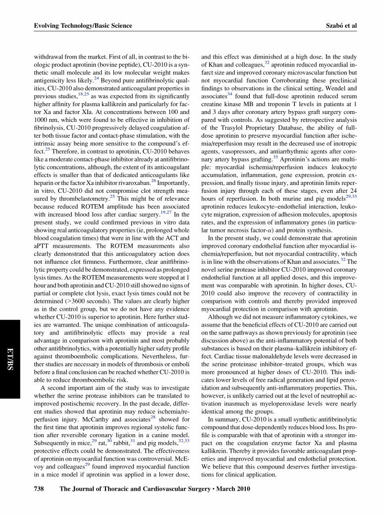
RESULTSBlood Loss
Postoperative blood loss during the first 2 hours after in-
jection of protamine was significantly reduced in the aproti-
nin-treated group in comparison with control (P < .05).
Application of CU-2010 resulted in a significant, dose-de-
pendent reduction of postoperative blood loss (Figure 1).
Blood Coagulation ParametersAs expected, aPTT and ACT increased significantly in all
groups after heparin and remained elevated during CPB. Af-
ter protamine, ACT and aPTT returned to baseline levels in
the control and aprotinin groups, but remained elevated
(ACT only slightly, aPTT remarkably so) in the CU-2010
treatment groups (Figure 1).
The PT values increased slightly during CPB and re-
mained at this level until the end of the observation period
in all groups (Figure 1).
ROTEM parameters are shown in Table 1 at baseline and
at the end of the observation period. Baseline values were
similar in all groups. At the end of the observation period,
clotting time (both EXTEM and INTEM) was prolonged
in the CU-2010 groups in comparison with the vehicle and
the aprotinin groups. EXTEM MCF showed a significant de-
crease after CPB but did not differ among the groups at base-
line and at the end of the observation period. If INTEM
reagents were used, similar data could be obtained, except
that no significant decrease could be observed in the aproti-
nin group in comparison with baseline, and the MCF was
significantly lower in comparison with aprotinin in the
CU-2010 1.25 mg/kg group. However, in all groups except
the aprotinin group, INTEM MCF still did not reached its
maximum in one or more individuals within the measure-
ment time and therefore the values are somewhat
ery c March 2010

FIGURE 1. Blood loss after weaning from cardiopulmonary bypass (CPB) and protamine (left top). Activated clotting time (ACT, right top), partial throm-
boplastin time (aPTT, left bottom), and prothrombin time (PT, right bottom).Time points indicate the time from the initiation of CPB (t0); t90 corresponds the
end of CPB, t105, directly as well as 20 (T120), 50 (T150), and 120 (T220) minutes after protamine, respectively. All values are mean� SEM. *P<.05 versus
control; #P< .05 versus aprotinin.
Szabo et al Evolving Technology/Basic Science
ET
/BS
underestimated. Lysis time (EXTEM) reached the desig-
nated end point (reduction of the amplitude to 10% MCF)
neither in the CU-2010 nor in the aprotinin groups, but did
so in the control group.
Hemodynamic ParametersHeart rate, cardiac output, left ventricular end-systolic
pressure, end-diastolic pressure, time constant of relaxation
(t), maximum and minimum pressure development (dP/dt
max, dP/dt min), mean arterial pressure, and coronary blood
flow are shown in Table 2. Baseline values did not differ
among the groups. Although most hemodynamic parameters
did not change significantly, mean arterial pressure and dP/
dt min decreased in all groups. dP/dt max and coronary
blood flow showed a significant decrease in the control
group after CPB (P < .05), whereas they remained un-
changed in the treatment groups.
After CPB, both left ventricular ESPVR and PRSW de-
creased significantly in the control and the aprotinin group;
The Journal of Thoracic and Ca
these decreases could be partly up to completely preserved
by application of CU-2010 (Table 2). Owing to the phys-
iologic variations of the absolute values of ESPVR and
PRSW, a comparison between the groups was somewhat
difficult; therefore, percent recovery values were calculated
(Figure 2). CU-2010 in higher doses significantly im-
proved percent recovery in comparison with controls
(and in case of PRSW in comparison with the aprotinin
group as well).
Coronary Vascular FunctionVasoresponse to acethylcholine was similar in all
groups before CPB. After CPB, endothelial function
(vasodilatative response to acetylcholine) was signifi-
cantly reduced in the control group and was abolished
by aprotinin and all doses of CU-2010 (Figure 2). Endo-
thelium-independent vasodilatation after nitroglycerin
did not differ between the groups and over time
(Figure 2).
rdiovascular Surgery c Volume 139, Number 3 735

TABLE 1. ROTEM parameters
Control Aprotinin
CU-2010
0.5 mg/kg
CU-2010
0.83 mg/kg
CU-2010
1.25 mg/kg
CU-2010
1.66 mg/kg
Baseline
EXTEM CT (s) 34 � 4 33 � 1 35 � 1 31 � 1 33 � 4 38 � 4
EXTEM MCF (mm) 65 � 2 67 � 2 66 � 1 67 � 2 65 � 2 62 � 2
EXTEM LT (s) 2466 � 252 2498 � 248 2331 � 256 3051 � 281 2505 � 302 2729 � 220
INTEM CT (s) 169 � 8 201 � 11 187 � 12 178 � 9 182 � 14 172 � 9
INTEM MCF (mm) 65 � 3 65 � 3 65 � 1 67 � 2 64 � 3 66 � 3
Two hours of reperfusion
EXTEM CT (s) 43 � 5 39 � 3 58 � 4*yz 86 � 8*yz 82 � 6*yz 51 � 5*yzEXTEM MCF (mm) 50 � 7* 53 � 5* 48 � 2* 45 � 4* 43 � 3* 54 � 3*
EXTEM LT (s) 2736 � 360 >3600 >3600 >3600 >3600 3290 � 222
INTEM CT (s) 312 � 28* 268 � 31* 845 � 112*yz 1587 � 180*yz 1520 � 120*yz 823 � 67*yzINTEM MCF (mm) 49 � 4* 57 � 7 41 � 3* 45 � 5* 30 � 8*z 47 � 4*
The influence on whole blood clotting was assayed with rotational thrombelastometry using ellagic acid (INTEM reagent) as activator of the intrinsic system or tissue factor (EX-
TEM reagent) as extrinsic coagulation activator. Clotting time (CT), maximum clot strength (MCF), and lysis time (LT) were measured at baseline (before heparin and test sub-
stance) and after 2 hours of reperfusion. All values are given as mean � SEM. *P< .05 versus baseline; yP< .05 versus control; zP< .05 versus aprotinin.
Evolving Technology/Basic Science Szabo et al
ET
/BS
Biochemical MeasurementsPlasma myeloperoxidase levels increased significantly in
all groups in comparison with baseline, but there were no
differences among the groups (Table 3). Cardiac tissue ma-
lonaldehyde levels increased after CPB in the control group,
but this increase was partly reversed by the use of the serine
protease inhibitors. The most pronounced decrease was
observed at the highest doses of CU-2010.
TABLE 2. Hemodynamic variables
Control Aprotinin
C
0.
Baseline
HR (beats/min) 114 � 8 140 � 9 1
CO (mL/min) 2599 � 353 2344 � 290 22
LVESP (mm Hg) 104 � 9 106 � 6 1
LVEDP (mm Hg) 9.92 � 1.53 8 � 0.91 6
t (ms) 32 � 1 34 � 1
dPt/dt max (mm Hg/s) 1811 � 78 1784 � 236 20
dP/dt min (mm Hg/s) �1566 � 160 �1866 � 108 �20
MAP (mm Hg) 104 � 3.67 95 � 11
CBF (mL/min) 40 � 5 46 � 4
ESPVR (mm Hg/mL) 3.04 � 0.61 2.99 � 0.33 2.
PRSW (kerg) 68 � 7 66 � 5
Two hours of reperfusion
HR (beats/min) 128 � 4 136 � 8 1
CO (mL/min) 2275 � 344 2320 � 264 22
LVESP (mm Hg) 87 � 4 89 � 3
LVEDP (mm Hg) 11 � 1 8 � 0.
t (ms) 34 � 2 35 � 1
dP/dt max (mm Hg/s) 1244 � 93* 1394 � 165 16
dP/dt min (mm Hg/s) �1111 � 107* �1444 � 78* �14
MAP (mm Hg) 68 � 2* 62 � 4
CBF (mL/min) 24.00 � 3* 45 � 5yESPVR (mm Hg/mL) 1.45 � 0.5* 1.82 � 0.32* 2.
PRSW (kerg) 47 � 6* 37 � 5*
Heart rate (HR), cardiac output (CO), left ventricular end-systolic pressure (LVESP), end-
pressure development (dP/dt max, dP/dt min), mean arterial pressure (MAP), coronary blood
load recruitable stroke work (PRSW). All values are given as mean � SEM. *P< .05 vers
736 The Journal of Thoracic and Cardiovascular Surg
DISCUSSIONIn the present study, we demonstrated that the novel syn-
thetic small molecule serine protease inhibitor CU-2010
dose-dependently reduced postoperative blood loss after
CPB to a degree comparable with that produced by aproti-
nin. This decrease of blood loss in the CU-2010 groups oc-
curred despite prolonged ACT and aPTT values. Both
aprotinin and CU-2010 attenuated endothelial dysfunction
U-2010
5 mg/kg
CU-2010
0.83 mg/kg
CU-2010
1.25 mg/kg
CU-2010
1.66 mg/kg
51 � 7 144 � 6 135 � 5 118 � 5
61 � 288 2534 � 259 1449 � 141 1596 � 180
07 � 2 91 � 6 104 � 5 97 � 4
.8 � 1 7 � 1 9.83 � 1 12.5 � 1.76
32 � 1 27 � 3 42 � 3 46 � 6
76 � 230 1503 � 47 1575 � 80 1570 � 137
12 � 63 �1609 � 99 �1727 � 100 �1542 � 130
88 � 4 79 � 8 95 � 6 87 � 6
46 � 2 40 � 3 44 � 5 39 � 4
77 � 0.33 3.34 � 0.51 4.20 � 0.33 3.05 � 0.80
62 � 6 54 � 4 51 � 8 56 � 6
62 � 8y 136 � 6 145 � 4y 133 � 8
36 � 286 1952 � 224 1917 � 209 1648 � 198
85 � 6* 78 � 5 93 � 5 84 � 3*
7 � 1 8 � 2 10 � 1 11 � 2
28 � 1 32 � 5 42 � 5 42 � 5
20 � 132y 1202 � 149 1377 � 128 1388 � 147
73 � 117* �1215 � 108* �1377 � 64* �1220 � 49*
60 � 3* 58 � 5 70 � 5.9 59 � 3
51 � 7y 42 � 6y 47 � 5y 37 � 3y32 � 0.34 2.81 � 0.23 3.57 � 0.58 2.96 � 0.63y48 � 3* 40 � 3* 55 � 6z 57 � 7zdiastolic pressure (LVEDP), time constant of relaxation (t), maximum and minimum
flow (CBF), slope of the endsystolic pressure–volume relationship (ESPVR), and pre-
us baseline; yP< .05 versus control; zP< .05 veersus aprotinin.
ery c March 2010

FIGURE 2. Percent recovery of the load-independent slope of the end-systolic pressure–volume relationship (ESPVR, left top) and preload recruitable stroke
work (PRSW, right top) 2 hours after weaning from cardiopulmonary bypass. Endothelium-dependent relaxation after application of acethylcholine (Ach, left
bottom). Endothelium-independent relaxation after application of nitroglycerin (NTG, right bottom). CVR, Coronary vascular resistance. All values are mean
� SEM.*P< .05 versus control;�P< .05 versus baseline; #P< .05 versus aprotinin; $P< .05 versus all other groups.
Szabo et al Evolving Technology/Basic Science
ET
/BS
after cardioplegic arrest and reperfusion. Furthermore, high-
dose CU-2010 but not aprotinin improved postischemic re-
covery of left ventricular function.
Apart from the concerns raised about possible side effects
of aprotinin, there is evidence that aprotinin reduces postop-
erative blood loss and transfusion requirements up to 40% to
50% in different population of patients (eg, first time, redo,
patients with aspirin) undergoing cardiac surgery with20-22
TABLE 3. Biochemical parameters
Control Aprotinin
Baseline
MPO (ng/L) 1.2 � 0.1 1.2 � 0.2
MDA (mmol/L/g dry weight) 0.9 � 0.1 1.2 � 0.1
Two hours of reperfusion
MPO (ng/L) 2.5 � 0.2* 2.7 � 0.3*
MDA (mmol/L/g dry weight) 5.3 � 0.2*,z 3.4 � 0.2*yMyeloperoxidase (MPO) and Malonaldehyde (MDA). All values are given as mean � SEM
The Journal of Thoracic and Ca
or without CPB.23 This study demonstrates for the first
time that a member of a new class of synthetic small molecule
serine protease inhibitors, CU-2010, significantly decreases
postoperative blood loss after CPB in a canine model. The ef-
fects of CU-2010 were comparable with those of aprotinin.
The novel serine protease inhibitors may have some poten-
tial advantages that may also overcome some concerns raised
currently about the use of aprotinin and leading to its
CU-2010
0.5 mg/kg
CU-2010
0.83 mg/kg
CU-2010
1.25 mg/kg
CU-2010
1.66 mg/kg
1.6 � 0.2 1.1 � 0.1 0.9 � 0.1 1.3 � 0.2
1.1 � 0.1 1.0 � 0.2 1.4 � 0.1 1.1 � 0.2
3.1 � 0.3* 2.4 � 0.2* 2.5 � 0.4* 2.6 � 0.2
2.9 � 0.2�y 3.1 � 0.3*y 2.4 � 0.3*y 1.9 � 0.2*yz, *P< .05 versus baseline; yP< .05 versus control; zP< .05 versus aprotinin.
rdiovascular Surgery c Volume 139, Number 3 737
Evolving Technology/Basic Science Szabo et al
ET
/BS
withdrawal from the market. First of all, in contrast to the bi-
ologic product aprotinin (bovine peptide), CU-2010 is a syn-
thetic small molecule and its low molecular weight makes
antigenicity less likely.24 Beyond pure antifibrinolytic qual-
ities, CU-2010 also demonstrated anticoagulant properties in
previous studies,18,25 as was expected from its significantly
higher affinity for plasma kallikrein and particularly for fac-
tor Xa and factor XIa. At concentrations between 100 and
1000 nm, which were found to be effective in inhibition of
fibrinolysis, CU-2010 progressively delayed coagulation af-
ter both tissue factor and contact-phase stimulation, with the
intrinsic assay being more sensitive to the compound’s ef-
fect.25 Therefore, in contrast to aprotinin, CU-2010 behaves
like a moderate contact-phase inhibitor already at antifibrino-
lytic concentrations, although, the extent of its anticoagulant
effects is smaller than that of dedicated anticoagulants like
heparin or the factor Xa inhibitor rivaroxaban.26 Importantly,
in vitro, CU-2010 did not compromise clot strength mea-
sured by thrombelastometry.25 This might be of relevance
because reduced ROTEM amplitude has been associated
with increased blood loss after cardiac surgery.19,27 In the
present study, we could confirmed previous in vitro data
showing real anticoagulatory properties (ie, prolonged whole
blood coagulation times) that were in line with the ACT and
aPTT measurements. The ROTEM measurements also
clearly demonstrated that this anticoagulatory action does
not influence clot firmness. Furthermore, clear anitfibrino-
lytic property could be demonstrated, expressed as prolonged
lysis times. As the ROTEM measurements were stopped at 1
hour and both aprotinin and CU-2010 still showed no signs of
partial or complete clot lysis, exact lysis times could not be
determined (>3600 seconds). The values are clearly higher
as in the control group, but we do not have any evidence
whether CU-2010 is superior to aprotinin. Here further stud-
ies are warranted. The unique combination of anticoagula-
tory and antifibrinolytic effects may provide a real
advantage in comparison with aprotinin and most probably
other antifibrinolytics, with a potentially higher safety profile
against thromboembolic complications. Nevertheless, fur-
ther studies are necessary in models of thrombosis or emboli
before a final conclusion can be reached whether CU-2010 is
able to reduce thromboembolic risk.
A second important aim of the study was to investigate
whether the serine protease inhibitors can be translated to
improved postischemic recovery. In the past decade, differ-
ent studies showed that aprotinin may reduce ischemia/re-
perfusion injury. McCarthy and associates28 showed for
the first time that aprotinin improves regional systolic func-
tion after reversible coronary ligation in a canine model.
Subsequently in mice,29 rat,30 rabbit,31 and pig models,32,33
protective effects could be demonstrated. The effectiveness
of aprotinin on myocardial function was controversial. McE-
voy and colleagues29 found improved myocardial function
in a mice model if aprotinin was applied in a lower dose,
738 The Journal of Thoracic and Cardiovascular Surg
and this effect was diminished at a high dose. In the study
of Khan and colleagues,32 aprotinin reduced myocardial in-
farct size and improved coronary microvascular function but
not myocardial function Corroborating these preclinical
findings to observations in the clinical setting, Wendel and
associates34 found that full-dose aprotinin reduced serum
creatine kinase MB and troponin T levels in patients at 1
and 3 days after coronary artery bypass graft surgery com-
pared with controls. As suggested by retrospective analysis
of the Trasylol Proprietary Database, the ability of full-
dose aprotinin to preserve myocardial function after ische-
mia/reperfusion may result in the decreased use of inotropic
agents, vasopressors, and antiarrhythmic agents after coro-
nary artery bypass grafting.35 Aprotinin’s actions are multi-
ple: myocardial ischemia/reperfusion induces leukocyte
accumulation, inflammation, gene expression, protein ex-
pression, and finally tissue injury, and aprotinin limits reper-
fusion injury through each of these stages, even after 24
hours of reperfusion. In both murine and pig models29,33
aprotinin reduces leukocyte–endothelial interaction, leuko-
cyte migration, expression of adhesion molecules, apoptosis
rates, and the expression of inflammatory genes (in particu-
lar tumor necrosis factor-a) and protein synthesis.
In the present study, we could demonstrate that aprotinin
improved coronary endothelial function after myocardial is-
chemia/reperfusion, but not myocardial contractility, which
is in line with the observations of Khan and associates.32 The
novel serine protease inhibitor CU-2010 improved coronary
endothelial function at all applied doses, and this improve-
ment was comparable with aprotinin. In higher doses, CU-
2010 could also improve the recovery of contractility in
comparison with controls and thereby provided improved
myocardial protection in comparison with aprotinin.
Although we did not measure inflammatory cytokines, we
assume that the beneficial effects of CU-2010 are carried out
on the same pathways as shown previously for aprotinin (see
discussion above) as the anti-inflammatory potential of both
substances is based on their plasma–kallikrein inhibitory ef-
fect. Cardiac tissue malonaldehyde levels were decreased in
the serine proteinase inhibitor–treated groups, which was
more pronounced at higher doses of CU-2010. This indi-
cates lower levels of free radical generation and lipid perox-
idation and subsequently anti-inflammatory properties. This,
however, is unlikely carried out at the level of neutrophil ac-
tivation inasmuch as myeloperoxidase levels were nearly
identical among the groups.
In summary, CU-2010 is a small synthetic antifibrinolytic
compound that dose-dependently reduces blood loss. Its pro-
file is comparable with that of aprotinin with a stronger im-
pact on the coagulation enzyme factor Xa and plasma
kallikrein. Thereby it provides favorable anticoagulant prop-
erties and improved myocardial and endothelial protection.
We believe that this compound deserves further investiga-
tions for clinical application.
ery c March 2010

Szabo et al Evolving Technology/Basic Science
ET
/BS
References1. Ferraris VA, Ferraris SP, Saha SP, Hessel EA 2nd, Haan CK, Royston BD, et al.
Perioperative blood transfusion and blood conservation in cardiac surgery: The
Society of Thoracic Surgeons and The Society of Cardiovascular Anesthesiolo-
gists clinical practice guideline. Ann Thorac Surg. 2007;83:S27-86.
2. Boisclair MD, Lane DA, Philippou H, Esnouf MP, Sheikh S, Hunt B, et al. Mech-
anisms of thrombin generation during surgery and cardiopulmonary bypass.
Blood. 1993;82:3350-7.
3. Mannucci PM, Levi M. Prevention and treatment of major blood loss. N Engl J
Med. 2007;356:2301-11.
4. Brown JR, Birkmeyer NJ, O’Connor GT. Meta-analysis comparing the effective-
ness and adverse outcomes of antifibrinolytic agents in cardiac surgery. Circula-
tion. 2007;115:2801-13.
5. Henry DA, Carless P, Moxey A, O’Connell D, Stokes B, McClelland B, et al.
Anti-fibrinolytic use for minimising perioperative allogeneic blood transfusion.
Cochrane Database Syst Rev. 2007. CD001886.
6. Sedrakyan A, Treasure T, Elefteriades JA. Effect of aprotinin on clinical outcomes
in coronary artery bypass graft surgery: a systematic review and meta-analysis of
randomized clinical trials. J Thorac Cardiovasc Surg. 2004;128:442-8.
7. Zufferey P, Merquiol F, Laporte S, Decousus H, Mismetti P, Auboyer C, et al. Do
antifibrinolytics reduce allogeneic blood transfusion in orthopedic surgery? Anes-
thesiology. 2006;105:1034-46.
8. Harmon D, Lan W, Shorten G. The effect of aprotinin on hypoxia-reoxygenation–
induced changes in neutrophil and endothelial function. Eur J Anaesthesiol. 2004;
21:973-9.
9. Fergusson DA, Hebert PC, Mazer CD, Fremes S, MacAdams C, Murkin JM, et al.
The BI: a comparison of aprotinin and lysine analogues in high-risk cardiac sur-
gery. N Engl J Med. 2008;358:2319-31.
10. Shaw AD, Stafford-Smith M, White WD, Phillips-Bute B, Swaminathan M,
Milano C, et al. The effect of aprotinin on outcome after coronary-artery bypass
grafting. N Engl J Med. 2008;358:784-93.
11. Mangano DT, Tudor IC, Dietzel C. The risk associated with aprotinin in cardiac
surgery. N Engl J Med. 2006;354:353-65.
12. Karkouti K, Beattie WS, Dattilo KM, McCluskey SA, Ghannam M, Hamdy A,
et al. A propensity score case–control comparison of aprotinin and tranexamic
acid in high-transfusion–risk cardiac surgery. Transfusion. 2006;46:327-38.
13. Mouton R, Finch D, Davies I, Binks A, Zacharowski K. Effect of aprotinin on re-
nal dysfunction in patients undergoing on-pump and off-pump cardiac surgery:
a retrospective observational study. Lancet. 2008;371:475-82.
14. Westaby S. Aprotinin: twenty-five years of claim and counterclaim. J Thorac Car-
diovasc Surg. 2008;135:487-91.
15. Dietrich W, Busley R, Boulesteix AL. Effects of aprotinin dosage on renal func-
tion: an analysis of 8,548 cardiac surgical patients treated with different dosages of
aprotinin. Anesthesiology. 2008;108:189-98.
16. Edmunds LH Jr, Colman RW. Thrombin during cardiopulmonary bypass. Ann
Thorac Surg. 2006;82:2315-22.
17. Katz BA, Sprengeler PA, Luong C, Verner E, Elrod K, Kirtley M, et al. Engineer-
ing inhibitors highly selective for the S1 sites of Ser190 trypsin-like serine prote-
ase drug targets. Chem Biol. 2001;8:1107-21.
18. Szabo G, Veres G, Radovits T, Haider H, Krieger.N, Bahrle S, et-al. Effects of
novel synthetic serine protease inhibitors on postoperative blood loss, coagulation
parameters, and vascular function after cardiac surgery. J Thorac Cardiovasc
Surg. In press
19. Cammerer U, Dietrich W, Rampf T, Braun SL, Richter JA. The predictive
value of modified computerized thromboelastography and platelet function
analysis for postoperative blood loss in routine cardiac surgery. Anesth Analg.
2003;96:51-7.
20. Blauhut B, Gross C, Necek S, Doran JE, Spath P, Lundsgaard-Hansen P. Effects
of high-dose aprotinin on blood loss, platelet function, fibrinolysis, complement,
and renal function after cardiopulmonary bypass. J Thorac Cardiovasc Surg.
1991;101:958-67.
21. Royston D, Bidstrup BP, Taylor KM, Sapsford RN. Effect of aprotinin on
need for blood transfusion after repeat open-heart surgery. Lancet. 1987;2:
1289-91.
22. Murkin JM, Lux J, Shannon NA, Guiraudon GM, Menkis AH, McKenzie FN,
et al. Aprotinin significantly decreases bleeding and transfusion requirements in
patients receiving aspirin and undergoing cardiac operations. J Thorac Cardio-
vasc Surg. 1994;107:554-61.
23. Bittner HB, Lamke J, Lange M, Rastan A, Mohr FW. The impact of aprotinin on
blood loss and blood transfusion in off pump coronary artery bypass grafting. Ann
Thorac Surg. 2008;85:1662-8.
The Journal of Thoracic and Ca
24. Byun Y, Chang LC, Lee LM, Han IS, Sinh VK, Yang VC. Low molecular weight
protamine: a potent but nontoxic antagonist to heparin/low weight heparin. ASAIO
J. 2000;46:435-9.
25. Dietrich W, Nicklisch S, Koster A, Spannagl M, Giersiefen H, van de Locht A.
CU-2010—a novel small molecule protease inhibitor with antifibrinolytic and an-
ticoagulant properties. Anesthesiology. 2009;110:123-30.
26. Gerotziafas GT, Elalamy I, Depasse F, Perzborn E, Samama MM. In vitro
inhibition of thrombin generation, after tissue factor pathway activation, by
the oral, direct factor Xa inhibitor rivaroxaban. J Thromb Haemost. 2007;5:
886-8.
27. Avidan MS, Alcock EL, Da Fonseca J, Ponte J, Desai JB, Despotis GJ, et al. Com-
parison of structured use of routine laboratory tests or near-patient assessment
with clinical judgement in the management of bleeding after cardiac surgery. Br
J Anaesth. 2004;92:178-86.
28. McCarthy RJ, Tuman KJ, O’Connor C, Ivankovich AD. Aprotinin pretreatment
diminishes postischemic myocardial contractile dysfunction in dogs. Anesth An-
alg. 1999;89:1096-100.
29. McEvoy MD, Sabbagh MJ, Taylor AG, Zavadzkas JA, Koval CN, Stroud RE,
et al. Aprotinin modifies left ventricular contractility and cytokine release after is-
chemia–reperfusion in a dose-dependent manner in a murine model. Anesth An-
alg. 2009;108:399-406.
30. Buerke M, Pruefer D, Sankat D, Carter JM, Buerke U, Russ M, et al. Effects of
aprotinin on gene expression and protein synthesis after ischemia and reperfusion
in rats. Circulation. 2007;116(11 Suppl):I121-6.
31. Carter JM, Buerke U, Rossner E, Russ M, Schubert S, Schmidt H, et al. Anti-in-
flammatory actions of aprotinin provide dose-dependent cardioprotection from re-
perfusion injury. Br J Pharmacol. 2008;155:93-102.
32. Khan TA, Bianchi C, Voisine O, Feng J, Baker J, Hart M, et al. Reduction of myo-
cardial reperfusion injury by aprotinin after regional ischemia and cardioplegic ar-
rest. J Thorac Cardiovasc Surg. 2004;128:602-8.
33. Khan TA, Bianchi C, Araujo E, Voisine P, Xu SH, Feng J, et al. Aprotinin pre-
serves cellular junctions and reduces myocardial edema after regional ischemia
and cardioplegic arrest. Circulation. 2005;112(9 Suppl):I196-201.
34. Wendel HP, Heller W, Michel J, Mayer G, Ochsenfahrt C, Graeter U, et al. Lower
cardiac troponin T levels in patients undergoing cardiopulmonary bypass and re-
ceiving high-dose aprotinin therapy indicate reduction of perioperative myocar-
dial damage. J Thorac Cardiovasc Surg. 1995;109:1164-72.
35. Royston D, Levy JH, Fitch J, Dietrich W, Body SC, Murkin JM, et al. Full-
dose aprotinin use in coronary artery bypass graft surgery: an analysis of
perioperative pharmacotherapy and patient outcomes. Anesth Analg. 2006;
103:1082-8.
DiscussionDr John A. Elefteriades (New Haven, Conn). I congratulate the
authors on an excellent experimental investigation. This is an im-
portant topic because patients with complex cardiac surgical dis-
ease currently have excess bleeding since the withdrawal of
aprotinin. The members of our group are strong proponents of apro-
tinin. In 2006 in this Journal we published a study of patients with
aneurysms operated on with and without aprotinin. Everything was
better with aprotinin, not only blood product transfusion but also
respiratory function and other parameters.
I have a few simple questions for you. The first one has to do
with your measurement of blood loss. Why did you use sponges
rather than chest tube drainage? Also, were the operators blinded
as to study drug or no study drug?
Dr Szabo. First of all, the operator was blinded to control and the
de novo compound but not aprotinin, because aprotinin is a different
dosing scheme. The control group received continuous infusion as
placebo.
Regarding the measurement of blood loss, in our experience,
gauze bandages were more exact than using a chest tube drain.
We measured blood loss volume in the first 2 hours at the open
rdiovascular Surgery c Volume 139, Number 3 739

Evolving Technology/Basic Science Szabo et al
ET
/BS
chest situation. With drying the operative area with the gauze ban-
dages, we could remove all blood from the operative area.
Dr Elefteriades. Since you were doing this experiment, why
didn’t you allow the animals to survive and look for renal function
or evidence of thrombosis?
Dr Szabo. These experiments sought to establish a dose–
response relationship and to explore whether the compound has
some cardioprotective effects. As such it was designed as an acute
experiment without keeping the animals alive. However, safety is-
sues are very important, before moving to the clinical arena. These
experiments are required from the authorities in Germany and they
are running now.
740 The Journal of Thoracic and Cardiovascular Surg
Dr Elefteriades. Some of your cardiac functional parameters
exceeded the preoperative baseline. Do you mean to imply that
your drug has a positive inotropic effect?
Dr Szabo. Actually, it was within the physiologic ranges of
a few percent, and I think it is just the physiologic variation. How-
ever, we also did pharmacokinetic experiments as dose finding pre-
vious to these experiments and in that case we measured
hemodynamics, even at the larger range of doses. We did not see
any direct inotropic effect of this drug.
Dr Elefteriades. Congratulations and I hope your drug is borne
out in further studies.
Dr Szabo. We hope so as well. Thank you very much.
ery c March 2010