the odontological identification of adolf hitler - dental age · the odontological identification...
TRANSCRIPT

The odontological identification of Adolf Hitler Definitive documentation by X-rays, interrogations and autopsy findings
REIDAR F. SOGNNAES & FERDINAND S T R o M
Schools of Medicine and Dentistry, Center for the Health Sciences, University of California, Los Angeles, California, and Faculty of Odontology, University of Oslo, Norway
Sognnaes, R. F. & F. Strom. The odontological identification of Adolf Hitler. Definitive documentation by X-rays, interrogations and autopsy findings. Acta Odont. Scand. 31, 43-69, 1973.
A definitive odontological identification of Hitler has been based on several sources of documentary evidence: (1) complete testimonies recorded by American intelligence officers during the 1945 interroga- tions of Hitler’s dentist and physicians; (2) attached to one of the above mentioned reports five head X-ray plates taken in 1944 follow- ing an assassination attempt and revealing several very characteristic dental conditions, including (a) a maxillary left central incisor with a radiopaque (metallic) restoration and with a radiolucent zone typical of a so-called ))Window Crown)), (b) a special dental bridge construction in the right mandibular area, in which a radiopaque (metallic) restoration on the right canine is connected by means of a metallic lingual bar to the second premolar with a cantilever extension to replace the first molar, and (c) periodontal bone break- down around the mandibular incisor roots; (3) the above observa- tions were compared with the various dental features depicted jn the Russian autopsy report and discussed in terms of other published reports; (4) the authors’ comparisons and reappraisal of these and other documentary data. The accumulated evidence now provides definite odontologic proof that Hitler did in fact die, and that the Russians did indeed recover and autopsy the right body.
Ke-v-words: Forensic dentistry; forensic medicine; autopsy; history
Ferdinand Strom, Faculty of Odontotogy, University of Oslo, Geit- myrsvn. 69, Oslo, Norway
The first exhaustive investigation of the last days of Adolf Hitler was undertaken by the Oxford professor Dr. H. R . Trevor- Roper in 1945. The British Intelligense authorities in Germany in September of that year had given him the task to collect all available evidence on the last days of Hitler and to determine, if possible, the truth about his disappearance or demise.
The resulting report of November 1 st, 1945, followed by the book, ))The Last Days of Hitler)) by Trevor-Roper (1947), appeared to leave little doubt in the Western world that Hitler had indeed met his end. Furthermore, in the intro- duction to the third edition of his book, published in 1956, Trevor-Roper concluded that there was considerable circumstantial
Received for publication, December 5, 1972.
Act
a O
dont
ol S
cand
Dow
nloa
ded
from
info
rmah
ealth
care
.com
by
Kin
g's
Col
lege
Lon
don
For
pers
onal
use
onl
y.

44 R E I D A R F. SOGNNAES AND FERDINAND STROM
evidence that Hitler’s remains must have come into the hands of the Russians who first occupied Berlin in 1945.
However as late as i n 1965. The Soviet General Boltin. one of the co-editors of the Russian War Histor). stated to the )>Spiegeln correspondent Eric Kubv, that as far as he (Boltin) was concerned. Hitler’s body had not been found (k‘ubv, 1965). Even today no official Soviet statement has been released regarding the death of Adolf Hitler.
In 1968 the Russian writer Lev Bez.1.- iiienski published in West Germany a book entitled >>Der Toti des Adolf Hitler)) (The Death of Adolf Hitler. J 968 b). Ber)*mrwski’s book carried the subtitle: ~ ) C ‘ n k n o ~ ~ n Docirtnents from Soviet Ar- diives)). Thus. twenty-three years after the events. Soviet sources finally per- mitted disclosure of details of the autopsy with various dental and medical data on the death of Adolf Hitler as well as Eva Braun; Goebbels and his family, General Krebs and two dogs. These documents provided us at long last the possibility to evaluate what might justify the claim that the Russians had recovered and identified with >)fair certainty)> the corpse of Adolf Hitler.
Bezymenski’s report included for the first time photographs of Hitler’s partially burned remains. including his dental bridgework and some of his natural teeth still in the lower jaw bone. However. no x-rays were reproduced to indicate any clear-cut conformity between subjective clinical and objective roentgenological observations.
In between these two major reports of British and Russian origins. which were in book format, there appeared in 1965 a brief summary chart on the dental status of Adolf Hitler prepared by a former captain in the U.S. Army. Dr. Ellsu~orth
Kell\, (1965), who had occasion to prepare his sketch from the U.S. Army report by his fellow officers who in 1945-46 had interviewed Hitler’s captured dentist, Dr. Hugo Blaschke.
With reference to our comparative observations on data reported in the past. i t became apparent that there were certain descriptive discrepancies, subject to sub- jective interpretation. Most regrettably there were not to be found the kind of objective documentation illustrating defini- tive x-ray observations. If available, much more conclusive evidence could be provided to supplement the information based on memory through personal inter- rogation.
Under these circumstances it seemed important to us (a) to re-explore all available reports by American, British and Russian investigators placed on record in the published literature, and (b) to seek out original archives and documents through which further research might contribute to a conclusive settlement of controversial questions regarding Hitler’s fate and identity.
MATERIAL
The following types of observations served as documentary evidence for the analysis, correlation and conclusion reached in th i s study:
I . Documents from Soviet archives, originally recorded during Hitler’s alleged autopsy in May 1945. and published for the first time by Bezynienski in 1968.
2 . Currently declassified documents from American archives, originally recor- ded during the 1945-46 interrogation of Hitler’s dentist and filed in the head- quarters of the USA National Archives and Records Service, Washington, D.C.
3. Recently located roentgenological
Act
a O
dont
ol S
cand
Dow
nloa
ded
from
info
rmah
ealth
care
.com
by
Kin
g's
Col
lege
Lon
don
For
pers
onal
use
onl
y.

THE ODONTOLOGICAL IDENTIFICATION OF ADOLF HITLER 45
evidence in the form of five authenticated head x-ray plates, originally taken of Hitler during late 1944 and uncovered in early 1972 while one of the authors (RFS) was searching wartime medical files at a satellite U.S. federal archive facility in the town of Suitland, Mary- land.
The evaluation of these several sources of information will be introduced with further details on the collection and nature of the material. Particular reference will be made to the validity and objectivity of the documentation vis a vis the specula- tions, hearsay and even legends and myths that still surround this enigma, as exenipli- fied bq recent writings in Germany (Maser, 1971). We shall make little reference to much of the often repeated and already widely published and publicized state- ments from those who claim inside in- formation from various unconfirmed re- ports and individual sources. Instead, we shall emphasize some of the most reliable objective kinds of data available to forensic science, notably the odontological findings and especially the newly un- covered x-ray evidence.
SOVIET ARCHIVES
The wartime autopsy documents from Soviet Archives, first recorded in May, 1945, were not reported until 1968 by the Soviet journalist, Lev Bezymenski, first in a German edition (1968a) and then immediately in an English edition (1968b). We will primarily be citing the English edition with a few cross-references to the German version when indicated. The discussion to follow will concentrate on three sub-topics: namely, descriptive, photographic and diagrammatic informa- tion.
1 . Descriptive information *
The autopsy report ))concerning the forensic examination of a male corpse disfigured by fire (presumably the corpse of Hitler))) is given verbatim as ~ D o c u - ment 1 2 ~ in Bezymenski’s book. The report dated May 8, 1945, originates from Mortuary CAFS (Surgical Army Field Hospital) at Berlin-Buch. It commences with the composition of the commission with their ranks and titles, followed by a detailed description of the examination of the charred remains.
The first part of the external examina- tion, dealing mainly with the head and the teeth is here reprinted as cited from the autopsy report in the English version of Bezymenski’s book (1968b, pp. 44-46):
))The remains of a male corpse dis- figured by fire were delivered in a wooden box (Length 163 cm., Width 55 cm., Height 53 cm.) On the body was found a piece of yellow jersey, 25 x 8 cm., charred around the edges, resembling a knitted undervest.
In view of the fact that the corpse is greatly damaged, it is difficult to gauge the age of the deceased. Pre- sumably it lies between 50 and 60 years. The dead man’s height is 165 cm. (the measurements are approximate since the tissue is charred), the right shinbone measures 39 cm. The corpse is severely charred and smells of burned flesh.
Part of the cranium is missing. Parts of the occipital bone, the left
temporal bone, the lower cheekbones, the nasal bones, and the upper and lower jaws are preserved. The burns
* The information quoted below from the Soviet autopsy report is reprinted by permission of Harcourt, Brace & World, Inc. from ))The Death of Adolf Hiilem by Bezymenski, C 1968 by Christian Wegner Verlag, Hamburg.
Act
a O
dont
ol S
cand
Dow
nloa
ded
from
info
rmah
ealth
care
.com
by
Kin
g's
Col
lege
Lon
don
For
pers
onal
use
onl
y.

46 K E I D A R F. SOGNNAES A N D F E R D I N A N D STROM
are more pronounced on the right side of the cranium than on the left. In the brain cavity parts of the fire- damaged brain and of the dura mater are visible. On face and body the skin is completely missing: only remnants of charred muscles are preserved. There are many small cracks in the nasal bone and the upper jawbones. The tongue is charred. its tip is firmly locked between the teeth of the upper and lower jaws.
In the upper jaw there are nine teeth connected by a bridge of yellow metal (gold). The bridge is anchored by pins on the second left and the second right incisor. This bridge consists of 4 upper incisors (21 k)*. 2 canine teeth (31 _ - 13). the first !eft bicuspid (14). and the first and second right bGuspids (41 51). as indicated in the sketch. The-first left incisor (b) consists of a white platelet. whith cracks and a black spot in the porcelain (enamel) at the bottom. This platelet is inset into the visible side of the metal (gold) tooth. The second incisor. the canine tooth. and the left bicuspid, as well as the first and second incisors and the first bicuspid on the right. are the usual porcelain (enamel) dental plates. their posterior parts fastened to the bridge. The right canine tooth is fully capped by yellow metal (gold). The maxillary bridge is vertically sawed off behind the second left bicuspid (I 5 ). The lower jawbone lies loose in t h e singed oral
* Ediroriol f u o r w f e : The journal normally uses t he F.D.I. tuo-digit system of designating teeth (Tor reference, see ))Instructions to Constribu- t orsn ) .
This system has not been adhered to in the present paper, particularly because the author's comparisons have required citations of the accumulated documentary evidence. In order to avoid confusion, two differing systems of designating teeth have not been simultaneously Liwd.
cavity. The alveolar processes are broken in the back and have ragged edges. The front surface and the lower edge of the mandibula are scorched. On the front surface the charred prongs of dental roots are recognizable. The lower jaw consists of fifteen teeth, ten of which are artificial. The incisors ( 2 17 p) and the first right bicuspid ( 4 ) are natural, exhibiting considerable wear on the masticating surface and considerably exposed necks. The dental enamel has a bluish shimmer and a dirty yellow coloration around the necks. The teeth to the left (p, 13, p, and 1%) are artificial. of yellow metal (gold), and consist of a bridge of gold crowns. The bridge is fastened to the third. the fifth (in the bridge, the sixth tooth), and the eight tooth (in the bridge, the ninth tooth). The second bicuspid to the right (71) is topped by a crown of yellow metal (gold) which is linked to the right canine tooth by an arching plate. Part of the masticating surface and the posterior surface of the right canine tooth is capped by a yellow metal (gold) plate as part of the bridge. The first right molar is artificial, white, and secured by a gold clip connected with the bridge of the second bicuspid and the right incisor. ))
With translations and retyping through Russian. German, and English it is to be expected that certain technical terms are not readdy explained while specific designations may even cause more or less obvious misprints. We have been on the alert to both possibilities and deemed it appropriate to point out our findings in detail in the attached footnote.*
1
* Some corrections in the translation of the details described above are necessary for the sake of completion o f this historical record. Firstly, it concerns the following statement:
- >
Act
a O
dont
ol S
cand
Dow
nloa
ded
from
info
rmah
ealth
care
.com
by
Kin
g's
Col
lege
Lon
don
For
pers
onal
use
onl
y.

THE ODONTOLOGICAL IDENTIFICATION OF A DOLF HITLER 47
In addition to the dental details de- scribed and reproduced above from the autopsy report published by Bezymenski (1968b, p. 44--46), it is stated (p. 47) that certain objects were taken from the corpse and handed over to the so-called SMERSH Section of the 3rd Soviet Shock Army on May 8, 1945. These objects were: ))a maxillary bridge of yellow metal, consisting of 9 teeth; and a singed lower jaw, consisting of 15 teeth.)) Heusermann, the chair-side dental assistant of Hitler’s dentist, Dr. Hugo Blaschke (see later), was interrogated in a talk with the Soviet Chief Expert of Forensic Medicine, Lieute- nant Colonel Shkaravski on May 11, 1945, in the offices of CAFS No. 496. Frau Heusermann described the state of Hitler’s teeth in every detail. Her de- scription tallies with the anatomical data pertaining to the oral cavity of the un- known man whose burned corpse we dissected,)) concludes the report.
))Part of the cranium is missing)). In the original German edition, on the other hand, it is stated: ))Ein Teil der Schadeldaches fehlt)). The latter is more precise, so the correct translation should have stated that a part of the occipital vault is missing. The second correction concerns the following sentence: ))The first left incisor (IL) consists of a white platelet, with cracks and a black spot in the porcelain (enamel) a t the bottom)). In the original German edition it is stated: ))Der linke erste Schneidezahn (11) stellt eine weisse Zahnplatte dar mit Sprungen und einem schwarzen Defekt im Email unten)). Thus the word )>porcelain)) in the English transla- tion is inappropriately used as a descriptive term for what simply should have been dental enamel. Then there are in the German as well as in the English edition some obvious typewriting errors. First of all, in the following statement: ))The first right molar is artificial, white, and secured by a gold clip connected with the bridge of the second bicuspid and the right incisor)), the last word should read the right cuspid. Further it is stated in the dental description that ))The maxillary bridge is vertically sawed off behind the second left bicuspid (IL))), instead of the first left bicuspid (15). This important correc- tion is supported by Rzhevsknya (1967) when quoting from the same autopsy report and also by the treatment description of Hitler’s dentist Dr. Hugo Blaschke (1946).
Later on, Bezymenski adds this footnote (1968b, p. 47): ))I asked N. Krayevski (the autopsy pathologist) how it was possible for the date of May 1 I th, 1945, to appear in an autopsy report that had been written on May 8. He explained that the report had originally been written by hand; only later was it decided to add the statements of Heusermann. As was mentioned above, the delay between evidence and conclusion is absolutely normal.))
In any event, the Soviet autopsy report placed great weight on the significance of the odontological findings. Thus the conclusion of the autopsy report culmi- nated with the following statement as cited by Bezymenski (1968b p. 49): ))The most important anatomical finding for identi- fication of the person are the teeth, with much bridgework, artificial teeth, crowns, and fillings (see documents).)) The report is signed by the five members of the Autopsy Commission, headed by Lieute- nant Colonel F. I. Shkaravski, Chief Expert, Forensic Medicine, (1st Byelo- russian Front, Medical Service), and by Lieutenant Colonel N. A. Krayevski. Chief Anatomical Pathologist Service, Red Army).
2. Photographic Illustrations
One of the most important
(Medical
exhibits recovered from the Soviet Archives con- cerns the objective photographic evidence illustrating the remains of the dentition of the corpse described in autopsy docu- ment No. 12. Included were photographs of a fixed dental prosthesis, namely, a 9-unit bridge, and the charred fragment of a mandible, the latter with several intact anterior teeth and two bridges, one on the right and one on the left side. The photographs which appear in two of the several inserts of illustrations
Act
a O
dont
ol S
cand
Dow
nloa
ded
from
info
rmah
ealth
care
.com
by
Kin
g's
Col
lege
Lon
don
For
pers
onal
use
onl
y.

48 REIDAR F. SOGNNAES AND FERDINAND S T R h 4
Fig. I A . Lingual view of the nine-unit maxillary Fig. IB. Another vieM of the maxillary bridge bridge reproduced from Soviet autopsy report reproduced from Bezymenski. N o t e the metal by Bezymenski (1968b). For proper orientation posts emerging from the Dowel crowns of the a n d esaluation, see text. upper right central incisor and upper left lateral
incisor. Lingually a re seen the ends of tiny (platinum) pins, evidently retaining anter ior porcelain facings, not seen directly in this view.
presented by Bezymenski (1968b between Moving towards the left jaw quadrant, the printed Fages 54 & 5 5 ) are reprodu- there then follows a metallic crown for the ced with his permission as our Figs. left central incisor. The seventh tooth seen 1 A. B (upper teeth) and 1 C. D (lower in this view is again a crown with a barely teeth ) . :$ visible central post emerging from the
middle (typical of a so-called Richmond from the lingual view and indicates a crown). The eight and ninth portion of distinct curvature of this large bridge. this bridge (the left canine and the first Consequently. one photograph shows premolar described above) cannot be to the best advantage those seven elements seen in full view in this photograph due of the nine-unit bridge which happens to to the curvature of the dental arch. culminate with the right second premolar However. the other view, Fig. l B , (Fig. I A ) . namely. a solid metal pontic accomodates a completion of the photo- replacement not prepared for any direct graphic record. In this illustration, it is the tooth or root attachment. This pontic upper right two premolars which are is next followed by a similarly independent blocked from direct view, and one can replacement. a metal back with a sug- barely see the lingual portion of the crown pestion of a different front facing. a slit of the right canine. On the other hand, barely separating the anterior from the the seven remaining portions of this posterior portion. Continuing in a mesial nine-unit bridge can be readily identified. direction. there follows then what is To begin with, we note that the pontic for obviously a metallic crown for the canine. the upper right lateral incisor clearly This. in turn. connects with another politic shows tcvo minute lingual marks, as two for the lateral incisor. and then the large light neyewlike dots indicative of the right central incisor. i n the center of typical platinum posts emerging through vvhich there is barely visible what appears the gold from an anterior porcelain facing. to be a metal post. This politic is next attached to a large
central incisor. This tooth also shows two
flirior. i R e : . i ~ t w ? i ~ / ~ i , 1968 b). porcelain facing. More importantly, this
The maxillary bridge is photographed
-~
* Figs. I and 3 were reproduced by courtesy of ; l L l f h ~ r publisher of T/ /e ([f’ Arlolf )’eyes)), evidence indicatitl& ‘ISe Of a
Act
a O
dont
ol S
cand
Dow
nloa
ded
from
info
rmah
ealth
care
.com
by
Kin
g's
Col
lege
Lon
don
For
pers
onal
use
onl
y.

THE ODONTOLOGICAL IDENTIFICATION OF ADOLF HITLER 49
Fig. 1C. Labial-buccal view of the left side of Fig. ID. Lingual view of the incisors and right the charred remains of the lower jaw. There is quadrant of the lower jaw showing the special evidence of bone resorption and tooth erosion bridge construction with the characteristic in the mandibular incisor region. Also note the lingual bar, bypassing the intact first premolar. distal drift of the left canine and second pre- molar, creating wide replacement with what appear to be twin porcelain facings for the first premolar.
clearly shows a metal post, albeit rather striation in keeping with the previous short, typical of the Richmond Crown or description that it was ))vertically sawed Dowel Crown, as it is now commonly off)). called. Turning to the left quadrant, this In the lower jaw, several of the natural restoration is attached to a smooth- teeth were remarkably well preserved, backed left central incisor restoration with being attached to the charred remnants no markings to suggest either a platinum of the mandible, together with a lower left post facing or any post attachments. On bridge, seen from the buccal view, as the other hand, such evidence is clearly reproduced in Fig. 1C and a right bridge. present on the next tooth, the left lateral seen from the lingual view in Fig. ID. incisor, which again shows the lingual With regard to the natural teeth, ))eyes)) caused by the platinum posts typical of a porcelain facing, as well as a relatively short central post or dowel, typical of the Richmond Crown or ))Sift- Zahm. This crown is then attached to a relatively thin reconstruction for the left canine position, obviously a metal-backed porcelain facing with no evidence of any
the lower incisors evidently exhibit a good deal of dental erosion, mainly below the enamel-cementum junction. In the case of the lateral incisors, the right one shows considerable incisor wear, if not even a partially fractured incisal edge, whereas the left lateral incisor shows some dark areas possibility suggestive of
provision for root attachments; in other dental decay. The latter may in part be words, a pontic rather than crown. Finally, super-imposed upon the erosion, since this cantilevered pontic is in turn attached we now know that such seemingly in- to an additional cantilevered replacement compatible conditions are not necessarily for the left first premolar. One cannot mutually exclusive (Sognnaes et af., 1972). readily see whether or not this latter Both the anterior and posterior views of restoration has the markings of a porcelain these incisor teeth (Fig. 1 C & 1D) suggest facing. However, one can see that the distal that, aside from any postmortem destruc- left termination of this bridge is not tion, there appears to have been con- amoothly polished, but has some rough siderable alveolar bone resorption during
Act
a O
dont
ol S
cand
Dow
nloa
ded
from
info
rmah
ealth
care
.com
by
Kin
g's
Col
lege
Lon
don
For
pers
onal
use
onl
y.

50 REIDAR F. SOGNKAES AND FERDINAND STROM
the life of the patient. indicative of periodontal disease.
The left lower bridge spans a space from the canine to the last molar. In addition to the full crown attachments on these terminal ends, there is a middle pillar where a full crown appears to be attached to what must be a distally drifted second premolar. The missing teeth, which have been replaced by the pontics between these three bridge attachments. are the first premolar and the first and second molar. However. due to distal drift of the second premolar and mesial drift of the third molar, the ))twin)) pontic replacement ot' the first and second molars has been reduced in width to little more than that of a single molar. By the same token. the distal drift of the second premolar has increased the first premolar space. Con- sequently. the pontic replacement. attached to the distal aspect of the canine crown. appears to be as wide as a molar. In the photographs of the charred remains. this latter replacement does not have as polished a buccal surface as the rest of this bridge. Instead. there seems to be a certain surface porousity which may suggest the use of porcelain facing. partly concealed by a secondary darkening from the charred buccal flesh.
The lower right bridge involves three teeth directly. but bypasses one seemingly uninvolved tooth, the right first premolar. due to a very special type of bridge constiuction (Fig. 1 D). As will be seen from this lingual view. there is a distinct fixed connecting bar between the restora- tion5 on the canine and second premolar. B) cross checking with the anterior view i n Fig. 2C, it will be noted that the canine restoration does not involve the labial portion of this tooth except for a consider- able slice out of the mesial-incisal corner (thi.; niay have served to restore a serious
defect in this area caused either by a fracture or caries). Principally covering the lingual and interproximal surfaces this canine restoration must be classified as a so-called 3/4 crown used for attachment of the lingual bar, and thereby connected with the crown of the second premolar. Lastly. the photograph clearly shows a freehanging, cantilevered extension for replacement of the lower right first molar tooth. Being this far back in the mouth, one would not expect to need a buccal tooth-colored facing for aesthetic reasons. Yet. one can clearly detect the two little shiny ))eyes)) piercing through the lingual gold backing, indicating the platinum posts emerging from a porcelain facing.
3. Diugramnratic Representation
To a dentist, it is essential to keep records by charting a patient's dentition with its characteristically individualized anatomi- cal. pathological and restorative features. For this purpose, various preprinted dia- grammatic forms are used to depict the dental arches and individual tooth shapes. Apparently there was no dentist or ordinary dental chart available to the pathologists who performed the autopsy of Hitler's alleged body as recorded above. But fortunately an effort was made to sketch some of the dental configurations in diagrammatic form as a supplement to the descriptive and photographic data. Specifically. this sketch, which indicates the number of natural and artificial teeth present at the time of the autopsy. IS illustrated in the English edition of Beziwwnski ( 1 968b) between text pages 54-55 and is reproduced in our Fig. 2.*
The Soviet autopsy report, dated May 8, 1945. actually made specific cross reference
* See footnote, p. 48.
Act
a O
dont
ol S
cand
Dow
nloa
ded
from
info
rmah
ealth
care
.com
by
Kin
g's
Col
lege
Lon
don
For
pers
onal
use
onl
y.

THE ODONTOLOGICAL IDENTIFICATION OF ADOLF HITLER 51
Fig. 2. Soviet pathologist’s sketch alleged to be the teeth and tooth replacements of Hit- ler’s upper and lower jaws. An- notations on the right are attri- buted to Mrs. Kaethe Heuser- mann, chairside assistant for Hitler’s dentist. This illus- tration is reproduced from Bezymenski’s English edition (1968b), but did not appear in the German version. For translation* of the Russian wri- ting and interpretation of the diagram, see text.
between the descriptive findings and the dental diagram when stating: ))as in- dicated in the sketch.))
The Soviet dental sketch reproduced in Fig. 2 shows in the upper jaw, a nine-unit bridge attached to four teeth with roots, namely, the upper right canine, the right central incisor and the upper left central and lateral incisors. Among the pontics of the bridge are the upper right lateral incisor and the upper right first and second pre- molars. The latter two teeth are freehan- ging cantilevered extensions on the right terminal (distal) extension of the bridge.
Similarly, the upper left side of the bridge terminates with the two cantilevered distal extensions, namely, for the replace- ment of the left canine and first premolar. Distally to the profile of the first premolar, a sharp black line has been drawn as the termination point. This, according to the autopsy description, is where the maxillary bridge was ))vertically sawed off)).
No other teeth are shown in the upper ja,, indicating that several teeth were missing without being replaced; namely, the upper right first, second and third molars. and the upper left second pre- molar and first, second, and third mo- lars.
With regard to the lower jaw, the sketch
* The translation of the Russian notes was done at The Institute of Slavic and Baltic Studies, University of Oslo, Head Professor Arne Gallis.
indicates that the lower right second and third molars were missing without being replaced. The remaining fourteen teeth are included in the diagram as being either present or replaced by bridgework.
The lower right first permanent molar is sketched in an uninterrupted profile together with the second premolar in the shape of a figure ) ) 8 ~ , as if the two were made in one piece. The first premolar, however, is drawn in a completely in- dependent isolated fashion, without any bridge connection on either side, in- dicating that this particular tooth was not involved in the adjacent bridge construc- tion. Similarly, the four lower incisor teeth are each drawn independently. Un- like the maxillary incisors, these teeth are thus completely separated to indicate that they were not involved in any kind of bridge construction. On the other hand, on the lower left side, there are six teeth diagrammed in complete contact with each other, much like the maxillary bridge, suggesting that a fixed bridge extended from the lower left canine through the premolars and molars finally culminating distally with the third molar or wisdom tooth.
There are several annotations, hand- written in Russian, on this same sketch. To the left of the chart (see Fig. 21, next to the profile of the upper bridge, the translation reads ))upper jaw)) and next
Act
a O
dont
ol S
cand
Dow
nloa
ded
from
info
rmah
ealth
care
.com
by
Kin
g's
Col
lege
Lon
don
For
pers
onal
use
onl
y.
52 R E I D A K F. SOGNNAES A N D F E R D I N A N D STROM
to the mandibular teeth. dower jaw)). and near the bottom, ))corpse of a man)). Flirthermore. the notes identify the chart as belonging to the ))document of the dissection of the corpse of Hitler)). Lastly. it is written, rather importantly. that ))the drawing on the right side is carried out by citizen Hojzerman)). with an illegibie first name ())Kate?))). After this. the family name of ))Hojzermann is repeated with improved writing and dated May 1 1 . 1945; in other words, suggesting her commentary was made three days after the actual autopsy date.
The signature oil this sketch is not easily legible but would appear to be that of Shkaravski. who. as indicated else- where. was the Chief Expert, Forensic Medicine. Kathe Heusermann‘s annota- tions on the right side of the chart are >omec\.hat scattered, but include drawings of what probably depicts the profile of a so-called ))windown crown with a distal- iilcisal dark defect. Attached to this tooth, a smaller onc is sketched in. with a clear facing and post (probably to mean Dowel crown). The sketch shows at least one other isolated tooth profile. suggesting repiacement with a post a 18 the Dowel crown type. One additional tooth profile suggests what may have been made to emphasize the otheruise stated presence of an open-face crown or so-called window)) cro\\ ti.
All in all. while apparently there was no dentisr and no standard dental form in- volved i n the autopsy or in the post- mortem review of the dental status. there can be no question that such a chart. as reproduced i n Fig. 2. does prove to be a very important kind of exhibit in forensic odoiitology. Indeed, what has been in- cluded in the autopsy sketch we have found to be in harmony with the essential in- t‘ormation obtained from the descriptive
and photographic documentation re- produced from the Soviet autopsy report by Bezymenski (1968b), and not in any important conflict with the data recently recovered from the American Archives to be presented below.
AMERICAN ARCHIVES
Presentation of the information obtained through the search of the American Archives will first be concerned with the very important interrogation of Hitler’s dentist, including (a) description of the dental status quo, (b) appropriate dental charts and diagrams, and (c) history of Hitler’s dental treatment record. These dental data will be supplemented with information obtained by interrogation of Hitler’s physicians. Finally. a detailed analysis will be made of the most im- portant new forensic exhibit, namely, the five head x-ray plates discovered in one of the files located in the Modern Record Branch of the U.S. National Archives, the National Document Center, Suitland. Maryland.
I . Interrogation of Hitler ’s Personal Dentist. Dr. Blaschke
Hitler’s principal and only full-fledged dentist, hitherto known to us, was Dr. Hugo Johannes Blaschke, D.D.S., a dental graduate of the University of Pennsylva- nia. class of 191 1. Returning to practice in Berlin. lie became well known to the Nazi leaders. He was Hitler’s dentist from 1934 to 1945, and was rewarded by being awarded the title of Professor and Briga- dier General. Waffen S.S.
Following his capture in 1945, an eight page document was prepared at the headquarters of the United States Forces, European Theatre. Military Intelligence
Act
a O
dont
ol S
cand
Dow
nloa
ded
from
info
rmah
ealth
care
.com
by
Kin
g's
Col
lege
Lon
don
For
pers
onal
use
onl
y.

THE ODONTOLOGICAL IDENTIFICATION OF ADOLF HITLER 53
Fig. 3. Front page of the American interrogation re- port recording the testi- mony of Hitler's dentist, Dr. Hugo Blaschke (U.S. Nat. Archives No . 0 1 FIR 31). For detailed discus- sion of the report, see text.
01 F I M . INTm.OCI.TICIN Ki'CRT (01 - FIR) No. 31
This i s the third mprt of a ser i e s dealing r i t h Hitler'a phyeical and mental condition. rbo treated E i t l e r fmm the beginning of 1934 tc 20 Apr 45.
I t is based on information obtained from the dentiat
p b l t of Contents
1. PzF&iicis 2
2. &rSOI< FCFl XnrORT 2
3. FLFOFtTz ' t I T i S a ' S TSTE' 2
a. Intmduction: Source 2 b. Cherscterist ics of Hlt ler'a Tebth 2
(1) Verbal Bscr ip t ion 2 (a) €aturn1 Teeth 2 (b) Replacerants 3 (0) Cavities untreated 4 (d) color of Teeth k
(2) brarings 5 4. c o ~ . c w s I c ~ 7
5. CoAwxiTs 1st E&O!.?&NiLTIGiS 7
Service Center, APO-757, known as a ))Final Interrogation Report)) (01 FIR 31), signed by Malcolm S. Hilty, Chief of 01 section. This document also includes several diagrams and has an appendix (Annex I) of three pages in which Dr. Blaschke gives the dental treatment history of Hitler; and three additional appendices contain information regarding Eva Baun's and Martin Bormann's teeth, and about Dr. Blaschke's professional and personal background.
The frontice piece of this document is duplicated here (Fig. 3) together with two of the several sketches prepared during the interrogation to represent Hitler's dental status in the upper and lower jaws
(Figs. 4 & 5).* In addition, reference will be made to some critical comments regard- ing one of Dr. Blaschke's last professional contacts with Hitler as a patient, and with the last contact with Hitler's dental chart and x-rays before Dr. Blaschke left Berlin in 1945.
As a preamble to the interrogation report regarding Hitler's dentistry, there is said to be three reasons for the report, namely, to provide (a) data useful in the ))identification of Hitler or his remains,)) (b) information to expose what might be
*Figs. 3 to 7 were reproduced from records re- leased to the first author through the courtesy of U.S. National Archives and Records Service, Washington, D.C. and from the U.S. National Document Center, Suitland, Maryland.
Act
a O
dont
ol S
cand
Dow
nloa
ded
from
info
rmah
ealth
care
.com
by
Kin
g's
Col
lege
Lon
don
For
pers
onal
use
onl
y.
54 R E I D A R F. SOGNNAES AND FERDINAND STROM
future ))frauds.)) and (c) research material for ))the historian. the doctor, and the scientist . ))
Dr. Blaschke was interned on May 28. 1945, and interrogated during November and December of that year. During the interrogation. while recording the status of Hitler’s teeth. Dr. Blaschke was given the privilege of consulting head x-rays taken of Hitler on September 19. 1944. The interrogators stated in connection with the ensuing report. that ))information on Hitler’s teeth is considered reliable.))
2. Descriptive Denrul Informution
In describing the characteristics of Hitler’s teeth as existing in April 1945, the Blaschke testimony first enumerates each individual natural tooth that was present either en- tirely or partially during Hitler’s last dental examination in early 1945. From the listing of each jaw quadrant. i t is indicated that the only teeth remaining without complete or partial prosthetic replacement were the four incisor teeth and the first right premolar in the lower jaw. Next, the report describes the complete replacement of missing teeth irr toto, by means of so- called pontics. and partial replacement of rooted teeth by means of crowns and so- called Richmond crowns (or Dowel crowns) with a ))pivot)) or post in the root canal to make what i n German terms be- comes a ))Stift-Zahn.,,
Musil lu. In the upper jaw is described a bridge which extends from a Richmond crown on the upper right central incisor to a full gold crown on the right canine tooth. terminating with a cantilevered. freely suspended, gold crown replacing the second bicuspid. Between these units of the bridge, the right lateral incisor is recorded as being a pontic made from a gold-backed porcelain facing. Lastly. on
the right side, the report states that there is a ))full gold crown over the first bicuspid)) (to this we shall return for further comment later).
In the other upper jaw quadrant, the bridge continues beginning with the left central incisor which carries an incomplete crown covering three-fourths of the tooth enamel, identified as a ))windown crown. This is followed by another Richmond crown (Dowel crown) on the lateral incisor and culminates again with a freely suspended porcelain-faced, gold-backed replacement of the first left premolar. Between the two last mentioned teeth, the report lists a )>full gold crown on cuspid.,) (In our later discussion we shall return to this replacement).
In describing this whole maxillary nine- unit bridge, Blaschke reports that the bridge originally extended farther ))until the end of October, 1944)). At that time, Dr. Blaschke had to remove the second left premolar by cutting the bridge ))between the first and second bicuspids)). The report adds that ))the straight edge produced by the cutting is strikingly characteristic)) (cf. pp, 47,Sl & 54 above).
itfunriible. In the lower jaw, two pros- thetic replacements are described. On the right side, a bridge extends from a three- quarter gold crown on the canine, ))leaving jn view most of frontal part of natural tooth.)) To this gold backing, there was attached a )>golden arch behind the first bicuspid,)) which in turn was attached to a full gold crown on the second premolar. Terminating the distal portion of the bridge was a cantilevered ())freely suspend- ed))) replacement for the missing first molar.
On the left side of the lower jaw, another bridge extended over most of the left quadrant, namely, from the canine to the third molar, both covered with full gold
Act
a O
dont
ol S
cand
Dow
nloa
ded
from
info
rmah
ealth
care
.com
by
Kin
g's
Col
lege
Lon
don
For
pers
onal
use
onl
y.

THE ODONTOLOGICAL IDENTIFICATION OF ADOLF HITLER 55
F! L
Fig. 4. Diagram showing anterior view o f nine-unit bridge from Hitler's upper jaw reproduced from Dr. Blaschke's interrogation report.
crowns. As a pillar in between, the report also lists a gold crown on the second pre- molar. Between these three bridge attach- ments, the missing first premolar was replaced by a pontic consisting of a gold- backed porcelain facing and the missing first and second molars replaced in gold.
In addition, the descriptive aspect of the dental interrogation report calls attention to a few problems of restorative den- tistry afflicting Hitler as follows: (a) ))ex- tensive caries existed for several years at the distal-lateral corner of the upper left central incisor,)) and (b) in the lower left jaw ))a porcelain-cement filling of the lateral incisor.))
3. Diagrammatic hfornxztion
Following the descriptive portion of the above report on Hitler's dentistry, Dr. Blaschke and his interrogators proceeded to prepare a total of eight diagrams or charts of Hitler's teeth as viewed from various projections. Two of these diagrams have been reproduced; Fig. 4, represent- ing upper jaw, and Fig. 5, representing the lower jaw.
Upper jaw. The upper jaw diagram clearly reflects the profile of a nine-unit bridge, the distal ends of which are cantilevered, freely-suspended pontics.
Besides the shaded areas of crowns and pontics, the rooted tooth attachments have been indicated by dotted lines. Thereby, we found clear evidence that the so-calied Richmond crowns ())Stift-Zahne))), which were attached by metal posts in the root canals, were limited to two of the upper teeth; namely, the upper right central incisor and upper left lateral incisor.
From the frontal view of the maxillary bridge, one notes that the upper left central incisor is the only tooth which includes the sketch of a labial band at the junction between the crown and root, a documentary evidence in keeping with the ))window)) crown type of restoration. Also, this same tooth has a short oblique line drawn across the distal incisor cor- ner, evidently confirming the problem of dental decay to which Dr. Blaschke made reference in the descriptive portion of his report.
In general, the use of the so-called cantilevered, ))freely suspended)) bridge ex- tensions are only resorted to if absolutely necessary or demanded to avoid remov- able replacements, as Hitler demanded, according to Dr. Blaschke. We note that, in order to fill a few more empty spaces, the sketch as well as the descriptive text indicate that such was the case, not only on one side, but indeed on both termi-
Act
a O
dont
ol S
cand
Dow
nloa
ded
from
info
rmah
ealth
care
.com
by
Kin
g's
Col
lege
Lon
don
For
pers
onal
use
onl
y.

56 REIDAR F. SOGNNAES AND FERDINAND STROP4
. , '. *.:
Fig. 5 . Sketch from Dr. Blaschke's interrogation report showing lingual view of the lower jaw indi- cating clearly the premolar bypass a n d cantilevered bridge extension on the lower right, a s well as the six-unit hridge o n the left. A small defect has been suggested o n the distal corner of the left
lateral incisor; for details see text.
nal ends of Hitler's nine-unit maxillary bridge.
Lo,~.er juw. The diagram depicting the teeth and their replacement in the lower ja\\ (Fig. 5) indicates that ten of the mandibular teeth were rooted and that tive of these. the four incisors and the first right premolar. were completely uninvolved in the porsthetic bridge repla- cements. The replaced missing teeth. as noted in the diagram i n Fig. 5. were the lovver right first molar. a cantilevered extension. and the lower left first premolar and first and second molars. The lingual version shows a profile of a bypass between the louer right canine and sccond pre- molar. circumventing or bypassing the intact first premolar by a curved ))barn. Distally. this unusual bridge construction i s then shown to connect and culniinate with a cantilevered suspension, as a pontic substitute to replace the first right molar.
Several of the sketches indicate consi- derable periodontal bone destruction around the roots of the lower incisors. particularly towards the left side. The diagram (Fig. 5 ) shows an oblique line along the distal-incisal corner of the lower left lateral incisor, suggesting the presence of some defect, presumably caused by either tooth decay or traumatic fracture.
4 . Past History of Hitler's Dental Treatnietit The interrogation of Hitler's dentist, Dr. Hugo Blaschke, included a separate section. Annex I, regarding Blaschke's earlier care of Hitler, a supplement to the overall report (01 FIR 31). Made avail- able through the U.S. National Archives, this Annex represents an English transla- tion of >)notes written for this report by BLASCHKE. giving Hitler's abridged case history as a dental patient in the years 1934 to 1945.))
From his notes, there is no evidence that Dr. Blaschke ever constructed or was called upon to repair or adjust the curious bridge construction on the right side of Hitler's lower jaw. This is a point of some importance. For example, i t means that Dr. Blaschke would have no record of the treatment of the lower right canine tooth. In other words, he could not know (and did not pretend to know) whether this corner tooth, which anchored the bridge anteriorly, had been injured by trauma or decay to require replacement of a large part of the mesial corner; or if the defect had been deep enough to require a root filling; or whether or not the inlay or 314 crown was in fact fortified with a metal post in the root canal. From the outside appearance of the completed
Act
a O
dont
ol S
cand
Dow
nloa
ded
from
info
rmah
ealth
care
.com
by
Kin
g's
Col
lege
Lon
don
For
pers
onal
use
onl
y.

THE ODONTOLOGICAL IDENTIFICATION OF ADOLF HITLER 57
bridge, there would be no way to tell if this tooth had been root-filled at all. In brief, the lower right bridge was clearly not made by Blaschke, but by someone else treating Hitler, presumably before 1934, when Dr. Blaschke entered the scene. The same may well have been the case with the bridge on the left side of Hitler’s lower jaw. Dr. Blaschke makes no specific refer- ence to the history of this construction in the treatment record.
With regard to the positive treatment situations which Dr. Blaschke himself had handled, special reference is made to three problem areas, namely in connection with trouble arising from the lower left lateral incisor, the upper left central incisor and the upper left second premolar.
It was in connection with his very first professional contact with Hitler (at the instigation of Goering) that Blaschke was called upon to remedy, in 1934, an infec- tion, swelling and pain arising from the area of the lower left lateral incisor. In treating this condition Blaschke states: ))The tooth was filled with iodoform paste (Walkhoff) and temporarily closed.))
In the upper jaw, )>much more extensive work was necessary,)) requiring the remov- al of a right and left defective bridge, and the construction of a new single fixed bridge, because ))Hitler rejected a remov- able prosthesis.))
Dr. Blaschke states that fairly regular check-ups of Hitler’s teeth were made until the outbreak of the war, after which all subsequent dental care had to wait until requested, which was not before pain arose. This did occur at least once as a result of ))extensive caries)) in the distal corner of the upper left incisor. This tooth, partly covered by the ))window)) crown, already had a pulp involvement. However, this could only be treated in a temporary fashion because of Hitler’s impatience, as 5
a result of which Blaschke states: ))a conclusion of the treatment, i.e. filling of the root and final filling of the cavity, was never achieved.))
The next and apparently last major episode in Hitler’s dental care followed shortly after the assassination attempt i n 1944. Dr. Blaschke explained it this way: ))Towards the end of September (1944) I was called to the headquarters. Hitler complained about slight tenderness of the gingiva of the upper left jaw. He was bed- ridden. He was, as Professor Morel1 told me, suffering from an inflammation of the naso-pharyngeal area. )) Blaschke’s clinical and roentgenological dental examina- tions revealed a deep pocket around the root of the upper left second premolar. Apparently, this had not caused much direct pain, or as Blaschke put it, ))The pain - wrongly diagnosed as neuralgic - was killed through obtundents.)) He adds that he was unable to get a clear picture of the history of these pains from Hitler who ))disliked intensely talking about his health.)) Not until the end of October 1944 did Dr. Blaschke get per- mission to perform the necessary treatment as follows: ))The old bridge on the left upper jaw was then cut in front of the gold crown on the 2nd bicuspid. The tooth, together with the cut-off part of the bridge, came out very easily. . (cf. p. 54).
From the middle of January 1945, Hitler was constantly in the Berlin Reich Chan- cellery where Dr. Blaschke had installed an office, but Hitler only ))came once to the dental station for a short while in mid- February for a superficial examination.)) From that time on, Dr. Blaschke gives no record of any additional treatment, but he concludes with a very important statement regarding the whereabouts of the dental charts and intra-oral x-ray pictures of the teeth, two of the potentially most impor-
Act
a O
dont
ol S
cand
Dow
nloa
ded
from
info
rmah
ealth
care
.com
by
Kin
g's
Col
lege
Lon
don
For
pers
onal
use
onl
y.

58 R E I D A R F. SOGNNAES A N D FERDINAND STROM
tant elements for future forensic documen- tation. This concluding statement, translat- ed from Dr. Blaschke’s own written notes. as found in the United States Archives (01 FIR 31. Annex I , page 3) reads as follows:
))The patients’ file cards as well as the X-ray pictures of their teeth were, since the middle of January 1945, constantly kept at the dental station in the Voss- Strasse shelter of the Reich Chancellery.
On the night of 20/21 January 1945. 1 was ordered to be ready for movement, with a minimum of baggage, within an hour. I was helped in packing the little portable dental station which I wanted to take with me by my dental helper, Mrs. Kaethe Heitserttiann, and my assistant. Dr. Rohkamnr. It is possible, and even likely that the files were put in the same box as the dental station. My baggage was then supposed to be sent from the Tempelhof airfield to SALZBURG in a transport plane car- rying baggage exclusively. This plane ne- ver arrived at SALZBURG. and from the Obersalzberg it coiild never be as- certained what had become of it.))
I n the above statement. Blaschke is said to have been leaving 20121 January. The days are right, but the month is misprinted, for he was leaving the night between Apri l 20j21 (Kempner 1969). Anyhow, from the above statement, it is regrettably clear that Dr. Blaschke’s detailed diagnostic and therapeutic dental record charts for Hitler and the small individual dental x-ray iilms used in these connections were last seen by Dr. Blaschke in the spring of 1945. For on the night of April 20-21, he was ordered to be ready for his move south after his files were sent with a transport baggage plane from Berlin’s Tempelhof Airfield to Salzburg. However. this plane,
according to Blaschke, evidently did not arrive, and as far as he knew, it ))could never be ascertained what had become of it.)) Whatever efforts may have been made since then in any part of Europe or else- where to recover Blaschke’s files, no one has to date recovered or reproduced a single one of the ordinary kinds of dental x-ray films, to which Hitler’s dentist referred.
5. Interrogation of Hitler’s Ph~~sicians
During the search for pertinent informa- tion in the Unitcd States National Archives and Records Service, three additional documents were located regarding Hitler’s health problems, namely, those originating from Hitler’s several physicians.
Two of these reports deal specifically with the head and neck examinations of Hitler following the assassination attempt during the summer of 1944. In these two reports are recorded observations by two ear, nose and throat specialists, Dr. Erwin Giesing, who in 1945 was subjected to extensive interrogation by the American Army officers, and Dr. Karl von Eicken, who contributed a day by day record of the times he was consulted in connection with Hitler’s sinusitis and vocal cord tLimor (which Dr. Eicken earlier had removed). These two documents were located i n the central Washington office of the U.S. National Archives and Records Service.
But the most elaborate medical report, compiled after interrogation of Hitler’s principal private physician, Professor Doc- tor Theo Morell, was located together with several consultant supplements in a satel- lite division of the U.S. National Archives in the town of Suitland, Maryland. Evi- dently. Dr. Morell and his fellow physicians did not have any detailed insight into Hitler’s oral and dental health. Aside from
Act
a O
dont
ol S
cand
Dow
nloa
ded
from
info
rmah
ealth
care
.com
by
Kin
g's
Col
lege
Lon
don
For
pers
onal
use
onl
y.

THE ODONTOLOGICAL IDENTIFICATION OF A DOLF HITLER 59
the remark that the teeth were ))defective>) and that Hitler at times had suffered from gingivitis and halitosis, there was not a single reference to any useful forensic dental identification features, such as specific missing teeth, intact teeth, restored teeth or replaced teeth.
This deficiency turned out to be far outweighed by a very significant appendix to the document, namely, ))Annex 11)). This Annex to Dr. Morell’s interrogation report included five head-x-rays of Hitler, taken following the assassination attempt in 1944. As a result, it now became possible for the first time to make direct compara- tive documentations, and evaluate con- clusions based on objective vis-a-vis subjective data.
6. Head X-ray Plates of Adolf Hitler
Before analysing each one of the five x-ray plates located in 1972 in the U.S. National Archives, it seems appropriate to cite verbatim the explanatory note at- tached to these five films when placed on file in 1945:
))Annex II. Five X-rays of Hider’s Head. Sources: Giesing and Morell. The five x-ray plates attached are copies of originals found among Dr. Morell‘s records. They have been positively identified by him as well as by Dr. Giesing as X-rays taken of Hitler. The three plates marked with the day 19 Sep 44 were made by Dr. Giesing at the Army Hospital at Rastenburg, East Prussia, using German army portable equipment (plate-target distance app 1 m.). This was during the period when Dr. Giesing was treating Hitler for ear injuries suffered at the time of the assassination attempt on 20 Jul 44. (See also Annex IV, e.g., ))Results of Ear Examination))). The plates were
made to assist in diagnosing pain com- plained of in the sinus regions. Four views were taken including a left lateral of the sinus cavities. This however could not be found. The 3 plates available include 1 each of the frontal sinuses (nose-forehead position), the sphenoidal sinuses (mouth-chin position), and the maxillary, ethnoidal and frontal sinuses (chin-nose position). The two plates marked 21 Oct 44 were also found among Dr. Morell’s records, but he can no longer remember the circum- stances under which they were made. These views are both of the maxillary, ethnoidal, and frontal sinuses (chin-nose position). >)
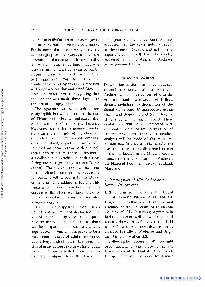
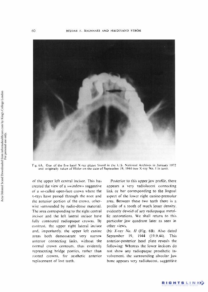
In the following paragraphs each of these important head x-rays of Adolf Hitler will be described in detail as items (a) to (e). (a): X-ray No. I (Fig. 6A). Dated in Eu- ropean style: 19. 9. 44 (Sept. 19, 1944), this x-ray photograph was taken in a manner referred to as a ))niouth-chin position.)) With a submental-vertical beam direction, one notes that an occlusal view of the teeth is seen to best advantage. Although there is some overlapping be- tween the profile of the upper and lower teeth, it is nevertheless possible to make several diagnostic observations of im- portance as a contribution to identifica- tion.
First of all, it is evident that most of the large posterior teeth on the right side are missing. On the left side of the jaw, on the other hand, there are evidently very radiopaque profiles all the way back towards the ramus of the mandible, sug- gesting the presence of teeth or their artificial replacements all the way up to the third molar region. In the anterior portion, one of the most striking features is a radiolucent zone in the front portion
Act
a O
dont
ol S
cand
Dow
nloa
ded
from
info
rmah
ealth
care
.com
by
Kin
g's
Col
lege
Lon
don
For
pers
onal
use
onl
y.
c 0 KEIDAR F. SOGNNAES AND FERDINAND STROM
Fig. 6A. O n e of the f ibe head X-ray plates found 111 the U.S. National Archives in January 1972 a n d originally taken of Hltler on the date of September 19, 1944 (see X-ray No. 1 i n text) .
of the upper left central incisor. This has created the view of a windown suggestive of a so-called open-face crown where the x-rays have passed through the root and the anterior portion of the crown. other- wise surrounded by radio-dense material. The area corresponding to the right central incisor and the left lateral incisor have fully contoured radiopaque crowns. By contrast. the upper right lateral incisor and. importantly, the upper left canine areas both demonstrate very narrow anterior connecting links. without the normal crown contours. thus evidently representing bridge pontics. rather than rooted crowns, for aesthetic anterior replacement of lost teeth.
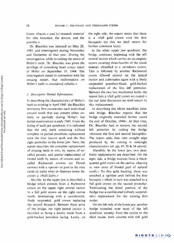
Posterior to this upper jaw profile, there appears a very radiolucent connecting link or bar corresponding to the lingual aspect of the lower right canine-premolar area. Between these two teeth there is a profile of a tooth of much lesser density, evidently devoid of any radiopaque metal- lic restorations. We shall return to this particular jaw quadrant later as seen in other views. (b): X-rar No. I / (Fig. 6B). Also dated September 19, 1944 (19.9.44). This anterior-posterior head plate reveals the following: Whereas the lower incisors do not show any radiopaque prosthetic in- volvement, the surrounding alveolar jaw bone appears very radiolucent, suggestive
Act
a O
dont
ol S
cand
Dow
nloa
ded
from
info
rmah
ealth
care
.com
by
Kin
g's
Col
lege
Lon
don
For
pers
onal
use
onl
y.
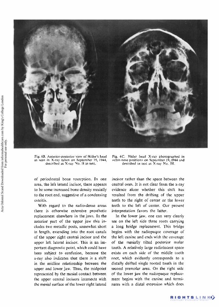
Fig. 6B. Anterior-posterior view of Hitter’s head Fig. 6C. Hitler head X-ray photographed in as seen in X-ray taken on September 19, 1944, >>chin-nose position)) on September 19, 1944 and
described as X-ray No. I1 in text. described in text as X-ray No. 111.
of periodontal bone resorption. In one area, the left lateral incisor, there appears to be some increased bone density mesially to the root end, suggestive of a condensing osteitis.
With regard to the radio-dense areas there is otherwise extensive prosthetic replacement elsewhere in the jaws. In the anterior part of the upper jaw this in- cludes two metallic posts, somewhat short in length, extending into the root canals of the upper right central incisor and the upper left lateral incisor. This is an im- portant diagnostic point, which could have been subject to confusion, because the x-ray also indicates that there is a shift in the midline relationship between the upper and lower jaw. Thus, the midpoint represented by the mesial contact between the upper central incisors intersects with the mesial surface of the lower right lateral
incisor rather than the space between the central ones. It is not clear from the x-ray evidence alone whether this shift has resulted from the drifting of the upper teeth to the right of center or the lower teeth to the left of center. Our present interpretation favors the latter.
In the lower jaw, one can very clearly see on the left side three roots carrying a long bridge replacement. This bridge begins with the radiopaque coverage of the left canine and ends with the coverage of the mesially tilted posterior molar tooth. A relatively large radiolucent space exists on each side of the middle tooth root, which evidently corresponds to a distally drifted single rooted tooth in the second premolar area. On the right side of the lower jaw the radiopaque replace- ment begins with the canine and termi- nates with a distal extension which does
Act
a O
dont
ol S
cand
Dow
nloa
ded
from
info
rmah
ealth
care
.com
by
Kin
g's
Col
lege
Lon
don
For
pers
onal
use
onl
y.

62 REIDAR F . SOGNNAES A N D F E R D I N A N D STROM
not appear to rest on a root and, hence, suggests a cantilevered pontic, replacing a tooth in the anterior molar area. In the root canal of the right canine tooth, there is a barely visible thin radiopaque central zone or line extending about half way down the root length. This could either be a radiopaque root canal filling material necessitated by pulp exposure due to decay or trauma. or a very thin post to help support the bridge carrying the radiopaque restoration on the canine tooth.
Lastly, this x-ray has an area of radio- dense bone structure lingual to the mental foramen area, possibly indicative of a minor bone exostosis or a small mandi- bular torus. ( c ) : X-rai. N o . I I I (Fig. 6C). The third x-ray dated September 19, 1944 (19.9.44), is the least satisfactory from a dental standpoint. I t has a relatively low contrast in connection with the prosthetic replace- ments. It is also the least elegant from a photographic standpoint. For there are a number of vertical lines and bands extending all through the x-ray, pre- sumably due to some technical difficulties i n the preparation or processing of this f i l l l l .
Nevertheless, even if not of crucial importance, this x-ray does serve to con- firm two singular dental features already noted in the first x-ray (Fig. 6A). Taken in the chin-nose position, we note again, first of all, the radiolucent window in the upper left central incisor and, secondly. the radiopaque bar or by-pass from the canine to the second premolar area in the lower right jaw.
Aside from the above. we attach a very special importance to this x-ray, for an initially different and non-dental reason. For we are fortunate to know for sure that this x-ray plate was taken on September
19, 1944 by the instigation of one of Hitler’s surviving physicians. Dr. Erwin Giesing, who was consulted regarding Hitler’s head and neck problems fol- lowing the assassination attempt during the summer of 1944, as already noted in Dr. Morell’s interrogation report. With the confirmed authenticity of this particu- lar x-ray, attributed to Ciesing and here reproduced in Figure 6C, we are now able to turn with special interest to a comparison with the next x-ray headplate taken one month later. Was that one also of Hitler? We believe it is, according to our evidence below. (d): X - r q No. IV (Fig. 7A). Taken October 21, 1944 (21.10.44) this head x-ray. also a chin-nose position, shows excellent contrasting reproduction of the dental restorations. A direct comparison with x-ray No. I11 (19.9.44) indicates the identical nature of the individual of whom these two x-rays were taken. Thus, we note a comparable configuration of the very exhuberant frontal sinuses which, for identification purposes, can be shown to precisely overlap each other when the transparencies of these x-rays are placed on top of each other.
It is well known that there is a finger- pint quality with regard to the special anatomical patterns of these sinuses i n each individual human being. To further signify the similarity of these x-rays, there is also a certain related pathological aspect to be noted; namely, the sclerotic reaction exhibited by the margins of the sinus cavities. This phenomenon, which may be assumed to have been due to chronic sinusitis, is noted in both of the latter x-rays (Nos. 111 & TV), as evidenced by a characteristic radiopaque blurring along the edges of the sinus cavities.
Observing other details of x-ray No. IV, the identity of the odontological forensic
Act
a O
dont
ol S
cand
Dow
nloa
ded
from
info
rmah
ealth
care
.com
by
Kin
g's
Col
lege
Lon
don
For
pers
onal
use
onl
y.
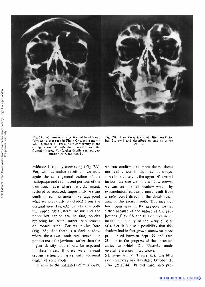
Fig. 7A. ))Chin-nose)) projection of head X-ray Fig. 7B. Head X-ray taken of Hitelr on Octo- (similar to that seen in Fig. 6 C) taken a month ber 21, 1944 and described in text as X-ray later, October 21, 1944. Note conformity in the No. V. configuration of both the dentition and the frontal sinuses. For further details, see text des-
cription of X-ray No. IV.
evidence is equally convincing (Fig. 7A). For, without undue repetition, we note again the same general outline of the radiopaque and radiolucent portions of the dentition, that is, where it is either intact, restored or replaced. Importantly, we can confirm, from an anterior vantage point what we previously concluded from the occlusal view (Fig. 6A), namely, that both the upper right lateral incisor and the upper left canine are, in fact, pontics replacing lost teeth, rather than crowns on rooted teeth. For we notice here (Fig. 7A) that there is a dark shadow where these two tooth replacements or pontics meat the jawbone, rather than the higher density that should be expected in these areas, if these were instead crowns resting on the cementum-covered dentin of solid roots.
Thanks to the sharpness of this x-ray,
we can confirm one more dental detail not readily seen in the previous x-rays. If we look closely at the upper left central incisor, the one with the window crown, we can see a small shadow which, by extrapolation, evidently must result from a radiolucent defect in the distal-incisal area of this incisor tooth. This may not have been seen in the previous x-rays, either because of the nature of the pro- jections (Figs. 6A and 6B) or because of inadequate quality of the x-ray (Figure 6C). Yet, it is also a possibility that this shadow had in fact grown somewhat more pronounced between Sept. 19 and Oct. 21, due to the progress of the untreated caries to which Dr. Blaschke made several references noted above. (e): X-ray No. V. (Figure 7B). The fifth available x-ray was also dated October 21, 1944 (21.10.44). In this case, also pro-
Act
a O
dont
ol S
cand
Dow
nloa
ded
from
info
rmah
ealth
care
.com
by
Kin
g's
Col
lege
Lon
don
For
pers
onal
use
onl
y.

64 REIDAR F. SOGNNAES A N D FERDINAND STROM
jected in the so-called chin-nose position, the mouth was evidently slightly open when the x-ray was taken. It will be noted that the profiles of the three bridges fall precisely into place as indicated in the x-ray taken on September 19, 1944 (Figure 6C). Thus, if the transparencies of these two x-rays are placed so as to compare the patient’s dentition, one can demonstrate perfect overlapping of the two dentition profiles of the upper and lower jaws. While this last x-ray is not as sharp as the other one taken on the same date (Figure 7A). one can, at least on the transparency, once again detect a shadow on the upper left central incisor, but mainly in the distal aspect, whereas the incisal portion, because of the slightly difrerent projection, is obscured by the radio-dense restoration.
Principal Roentgenological Interpretations
In comparing the five x-ray plates located in the IJ.S. National Archives, several principal observations emerge, which are considered of major significance in cor- relating subjective and objective evidence regarding the odontological identification of Hitler.
Firstly, it became clear that Hitler had only four remaining teeth which were not involved i n either bridging a gap or supporting a bridge between adjacent teeth. These four teeth were the right and left mandibular incisors. However, while they were free from dental decay, as far as could be seen in the x-ray, there was considerable bone resorption around the roots of these anterior teeth. These ))naked)) portions of the root cementum would have tended to expose the necks of the teeth to the injuries of erosion and abrasion. (It should be noted that this is precisely what is evident from the visual
appearance of the mandibular anterior teeth reproduced in the Russian autopsy report depicting the remains of Hitler’s burned mandible).
Secondly, the x-rays indicate that the maxillary right central incisor and left lateral incisor, while completely opaque to the x-ray beam, do show short metallic posts for insertion into the root canals. This suggests the type of dental crown restoration known as a ))Richmond Crown)), frequently used jn the past and precisely the type of restoration de- scribed by the reports on Hitler’s dental condition.
Thirdly, the x-rays show a very peculiar and very unusual dental bridge construc- tion on the right lingual aspect of the mandible. This involves a lingual high- density (metallic) bar extending distally from the right canine region, bypassing the first premolar, evidently connecting with a radiopaque (metallic) crown on the second premolar. From that point, an additional metallic extension evidently had been constructed as a cantilevered distal extension of the bridge. ( In this connection, it is noteworthy that Hitler’s dentist made very special verbal reference to this unique prosthetic construction while being interrogated by the American officers.)
Fourthly, the x-rays show that the left maxillary incisor has the very charac- teristic feature of a partially opaque or metallic crown, typical of the now out- moded, but in times past, not infrequently used, >bwindow))-crown.
HITLER’S DENTAL CHART BASED ON
COMBINED RECORDS
After careful study of the various types of descriptive, diagrammatic and photo-
Act
a O
dont
ol S
cand
Dow
nloa
ded
from
info
rmah
ealth
care
.com
by
Kin
g's
Col
lege
Lon
don
For
pers
onal
use
onl
y.

'THE ODONTOLOGICAL IDENTIFICATION OF ADOLF HITLER 6 5
HITLER'S DENTAL STATUS - 1945
METALLIC PORTION 0 PORCELAIN PORTION 0 NATURAL PORTION
Fig. 8. Dental chart prepared by authors to summarize their conclusions regarding the dental condi- tion of Hitler as it existed in 1945.
graphic evidence presented above, a dental chart has been prepared (Fig. 8) with a view to summarizing what, in our judgment, represented the dental status of Adolf Hitler in 1945."
Where metallic material was the only substance covering or replacing the teeth, the diagram is shaded black. Where the frontal portion of the replaced teeth was represented by a porcelain facing, the diagram presents a shaded area. Where
natural teeth were still present, the areas are unshaded. Where the teeth were neither present, nor replaced, the areas are left blank.
In the upper jaw, we have indicated that the nine-unit bridge which Hitler had i n place in 1945 was resting on only four remaining natural supporting teeth; namely, the upper right canine, the two central incisors and the left lateral incisor. Of the five totally replaced teeth, only one was supported as a typical pontic, that is, anchored on both sides, namely, the upper right lateral incisor. The terminal
l.Fig. 8. was drawn by Mr. George Robbins, School of Dentistry, University o f California, 1 0 s Angeles.
Act
a O
dont
ol S
cand
Dow
nloa
ded
from
info
rmah
ealth
care
.com
by
Kin
g's
Col
lege
Lon
don
For
pers
onal
use
onl
y.

66 REIDAR F. SOCNNAES A N D FERDINAND STROM
wings of the bridge consisted of two pontics at each end, completely canti- levered without any visible means of support by dental root attachments. These freely suspended teeth, on the right side, were the first and second premolars and, on the left side, the canine and first premolar.
In the lower jaw. the diagram indicates the presence of five natural teeth without any metallic restoration; namely the four incisors and the right first premolar. In addition, the natural tooth substance, kept blank, is retained by the labial surface of the lower right canine, except for a large portion of the mesial-incisal corner, which is indicated in black to represent part of a metallic restoration. The latter is shown to be connected by a metallic bar soldered onto a metallic crown on the lower right second premolar, from which there extends a cantilevered, goldbacked, porcelain-faced, freely suspended replace- ment of the first molar.
On the left side of the lower jaw, the diagram indicates absenses and replace- ment of the Srst premolar by a porcelain- faced gold pontic and of the first and second molars by solid metal. These pontics are supported by fixed attach- ments to full crowns on the canine, second premolar, and third molar.
The diagram finally indicates metal posts extending into the root canals of the upper right central and left lateral incisors, the crowns of which have restorations typical of the Richmond crowns or Dowel crowns (sStift-Zahns). For the remaining apical portions of the root canals in these teeth, a dotted line has been in- dicated, since there is no clear-cut opacity visible in any of the x-rays to suggest the presence of a radiodense root canal filling material.
Similarly, the lower right canine, in
keeping with the x-ray evidence, has been shown to include some radio-dense material near the coronal part of the root canal. It is not known from any inspection or objective evidence whether or not this is a root canal filling or a metal post or both. Obviously, the canine restoration is not a Richmond crown or Dowel crown, but the root canal opacity could be due to a thin supporting pin to strengthen the retension of the metallic restoration which covers the posterior three-quarters of the crown.
We have indicated no radiopaque filling material in the lower left lateral incisor even though this tooth, according to Blaschke, was treated with what he referred to as Walkhoff’s iodoform paste. Whatever was used, it may not have been sufficiently radiopaque to stand out in contrast to the primary root dentin or secondary dentin, which, in part, may have obliterated the vaguely visible root canal in this tooth.
For this diagrammatic representation of Hitler’s dental chart, a standard format has been used, and no effort has been made to modify the size and shape of any individual teeth or their replacements.
CONCLUDING COMPARISON
Once we had reached agreement re- garding the interpretation of the roent- genological, diagrammatic and descriptive documents from Hitler’s files, uncovered in 1972 through the United States National Archives and Records Service, it became possible to make a concluding comparison, tooth by tooth, between this information and that deduced from the photographic, diagrammatic and descriptive autopsy data uncovered through the ))Unknown Documents from Soviet Archives)), re- ported by Bezymenski in 1968.
Act
a O
dont
ol S
cand
Dow
nloa
ded
from
info
rmah
ealth
care
.com
by
Kin
g's
Col
lege
Lon
don
For
pers
onal
use
onl
y.
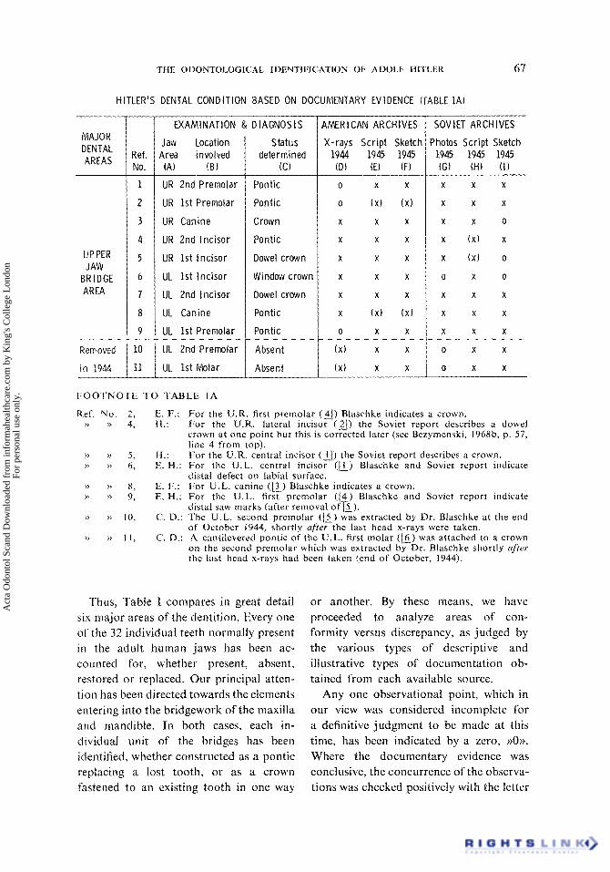
THE ODONTOLOGICAL IDENTiFiCATION OF ADOLF HITLER
HITLER'S DENTAL CONDITION BASED ON DOCUMENTARY EVIDENCE (TABLE 1A)
67
MAJOR DENTAL AREAS
UPPER JAW
BRIDGE AREA
Ref. No.
EXAMINATION L? DIAGNOSIS
Jaw Location 4rea involved (A) (B) UR 2nd Premolar
UR 1st Premolar
UR Canine
UR 2nd Inc i so r
UR 1st I nc i so r
UL 1st I nc i so r
UL 2nd Inc i so r
UL Canine
UL 1st Premolar
Ref. >>
No. >>
>>
?)
Status determined
(C)
Pontic
Pontic
Crown
Pontic
Dowel crown
Window crown
Dowel crown
Pontic
Pontic - - _ - - - -
4MERICAN ARCHIVES
X-rays Scr ip t Sketch 1944 1945 1945 (D) (El (F)
0 x x
0 ( x ) ( x )
x x X
X x x
x x X
X x x
X x x
x ( x ) ( x )
0 x x _ _ _ _ - - _ - - - -
SOVIET ARCHIVES
'hotos Scr ip t Sketch 1945 1945 1945 (G) (H) ( 1 )
x x x
x x x
x x o
x ( x ) x
x ( x ) 0
O X 0
x x x
x x x
x x x _ - _ - - _ - _ _ _ o x x
x x ( x ) x x
Absent
Absent
2 , For the U.R. first premolar (51) Blaschke indicates a crown. 4, H.: For the U.R. lateral incisor (21) the Soviet report describes a dowel
crown at one point but this is corrected later (see Bezymenski, 1968b, p. 57, line 4 from topj.
5 , For the U.R. central incisor (I]) the Soviet report describes a crown. 6, E. H.: For the U.L. central incisor (]I) Blaschke and Soviet report indicate
distal defect on labial surface. 8 , For U . L . canine (13) Blaschke indicates a crown. 9, E. H.: For the U.L. first premolar (14) Blaschke and Soviet report indicate
distal saw marks (after removal of 12). C. D.: The U.L. second premolar (12) was extracted by Dr. Blaschke at the end
of October 1944, shortly after the last head x-rays were taken. C. D.: A cantilevered pontic of the U.L. first molar (15) was attached to a crown
on the second premolar which was extracted by Dr. Blaschke shortly after the last head x-rays had been taken (end of October, 1944).
E. F.:
H.:
E. F.:
10,
I I ,
Thus, Table I compares in great detail six major areas of the dentition. Every one of the 32 individual teeth normally present in the adult human jaws has been ac- counted for, whether present, absent, restored or replaced. Our principal atten- tion has been directed towards the elements entering into the bridgework of the maxilla and mandible. In both cases, each in- dividual unit of the bridges has been identified, whether constructed as a pontic replacing a lost tooth, or as a crown fastened to an existing tooth in one way
or another. By these means, we have proceeded to analyze areas of con- formity versus discrepancy, as judged by the various types of descriptive and illustrative types of documentation ob- tained from each available source.
Any one observational point, which in our view was considered incomplete for a definitive judgment to be made at this time, has been indicated by a zero, DO)). Where the documentary evidence was conclusive, the concurrence of the observa- tions was checked positively with the letter
Act
a O
dont
ol S
cand
Dow
nloa
ded
from
info
rmah
ealth
care
.com
by
Kin
g's
Col
lege
Lon
don
For
pers
onal
use
onl
y.

68 REIDAR F. SOGNNAES A N D FERDINAND STROM
Ref. No.
Jaw Location Area involved (A) (€3)
MAJOR DENTAL AREAS
LL Canine LL 1st Premolar LL 2nd Premola
, LL 1st Mo lar LL 2nd Molar LL 3rd Molar
LR Inc isors LL Inc isors
LOWER JAW
BRIDGE ( r i gh t )
UR MI M2M3 UL M z & M3
, LR M z & M3
LOWER JAW
BRIDGE ( lef t )
INTACT AREAS
V O I D AREAS
12 13 14 15
LR 1st Mo lar LR 2nd Premola LR 1st Premolar LR Canine
16 17 18 19 20 21
22/23 24125
26128 29130 31/32
Bridge bypass f rom LR C-P,
S PEC. I 33 i Alveolar bone mand. inc isors
AREAS 1 34 i
Status determined
(C)
Pont ic Crown (Bypassed) 314 Crown
X-rays Scr ip t Sketch 1944 1945 1945 (DI (E) (F)
X o x X x x
x x X
X x x
Photos Scr ip t Sketch 1945 1945 1945 (GI (H) ( 1 )
x x o x x o x x o x x o
Crown Pont ic Crown Pont ic Pont ic Crown
Present Present
I x j X X
X x x I x x X
x x o x x o x x o x x o x x o x x o
x x x x x x
Absent Absent
L ingua l bar x x X x x x
Periodontal resorpt ion I x x x j x x 0
FOOTNOTE TO TABLE IB Ref. No. 17, G . :
>) n 19/20. G.:
T h e pontic for the L.L. first premolar (17) is wider than normal due to distal drift of the L.L. second premolar (13). The combined pontic for the L.L. first and second molars (167) is narrow- er than normal due to distal drift of the L.L. second premolar (IT) and mesial tipping of the L.L. third molar (IT).
DXD. Finally, we have used a bracket D(X)D when such a positive record was apparent only from indirect, or less than adequate, observations or from data pertinent to Hitler’s life, rather than post- mortem dental status, as, for example, items No. 10 and I I , Table IA , referring to Hitler’s 1944 x-rays.
Where the material at hand permitted definitive conclusions, it will be noticed that there exists a remarkable conformity between the individual tooth identifica- tions established through the analysis of the American and Soviet data. In addition to the individual teeth which were present, absent, restored or replaced, as
the case may be, we have also noted a few other special areas. namely, the unique lingual bar serving as a fixed bridge bypass between the lower right canine and second premolar, and also the alveolar bone resorption around the roots of the incisor teeth (Table 16).
From this overall comparison of the odontological evidence (Tables TA, B), we conclude that the individual identified by means of the 1945 Hitler files located i n the U.S. National Archives in 1972, is the same person as that whose 1945 autopsy report was published in 1968 on the basis of the previously unknown documents from Soviet Archives of 1945.
Act
a O
dont
ol S
cand
Dow
nloa
ded
from
info
rmah
ealth
care
.com
by
Kin
g's
Col
lege
Lon
don
For
pers
onal
use
onl
y.

THE ODONTOLOGICAL IDENTIFICATION OF ADOLF HITLER 69
REFERENCES
Amrrictttr Archives trrid Records Service. 1945. Interrogation of Hitler’s dentist and physi- cians (Nos . OI-FIR/3 I ; Or-C1R/2; and OI-C1R/4)
Bez.yi?ieii.ski, L . 1968a. Der Tod des Adorf Hitler: Uiihekonii/~ Dokumente nus Moskauer Arclri- reti. Christian Wegner Verlag, Hamburg, W. Germany
Bez,vinerrski, L . 1968b. The Deuth of Adolf Hitler: Unkttown documents from Soviet Archives. Harcourt, Brace and World, New York, N.Y.
Bltrschke, H . 1946. Hitler’s Teeth. U.S. Forces European Theater, Military Intelligence Ser- vice Center ( A P O 757), Final Interrogation Report, N o . OI-FIR/31 Feb. 1946
Giesirrg, E. 1945. Hitler a s Seen by his Doctors. U.S. Forces European Theater Military Intelligence Service Center ( A P O 757), Consolidated Interrogation Report, No. OI-CIR/2
Kr/l . t , E. 1965. Hitler’s dental chart. J. Calif. Dent. Assoc. 41, 424-425
tiempiwr, R. 1969. Das Dritte Rrich im Kreuz- wrhiir. Bechtle Verlag. Munich and Ess- lingen, W. Germany
t i i t b y , E . 1945. The Russinm in Berlin. Translated by Arnold J . Pomerans. William Heine-
mann, Ltd., London, England. Copyright by Verlag Miinchen-Bern-Wien, 1965
Moser , W. 197 I . Adolf Hiiler: Legende, Mytos, Wirklichkeit. Bechtle Verlag, Munich and Esslingen, W. Germany
Morell, T. 1945. Hitler as Seen by his Doctors. U.S . Forces European Theater, Military Intelligence Service Center ( A P O 7571, Consolidated Interrogation Report, No. 01-C 1 R-4
Rzhrvslta.vu, E. 1967. Hiilrr Elide o h m M j i h o s . Deutsche Militfr Verlag (Original in Russian, 1965)
Sognnues, R. F., Wolcoti, R . B. & Xliongu, R. 1972. Dental Erosion: I. Erosion-like pat- terns occurring in association with other dental conditions. J . Amer. Dent. Assoc.
Trevor-Roper, H . R. 1970. The Last Dujs of Hiiler. Colier-Macmillan Company, Cana- da, Ltd., Toronto, Ontario, Canada. Sixth printing (Original copyright, 1947, 1962)
V ( J ~ Z Eiclten, K. 1945. Notes o n medical treatment or patient HM.F.D ())Mein Fuhrer))) from May 15, 1935 to December 30, 1944. U.S. National Archives and Records Divisions M L 125, 125a & 131 (In German only)
WrilkhoJj; 0. 1928. Mein System der mediku- mentosen Behnndlung sch werer Erkrurrkungen der Zahnpulpn itnd des Periodontiitms. Meusser Verlag, Berlin
84, 571-576
Act
a O
dont
ol S
cand
Dow
nloa
ded
from
info
rmah
ealth
care
.com
by
Kin
g's
Col
lege
Lon
don
For
pers
onal
use
onl
y.