the open orthopaedics journal · understanding the deformity and the deforming force is ... the...
TRANSCRIPT

Send Orders for Reprints to [email protected]
The Open Orthopaedics Journal, 2017, 11, (Suppl-4, M5) 651-659 651
1874-3250/17 2017 Bentham Open
The Open Orthopaedics Journal
Content list available at: www.benthamopen.com/TOORTHJ/
DOI: 10.2174/1874325001711010651
REVIEW ARTICLE
Reconstruction of Cavus Foot: A Review
Bom Soo Kim*
Department of Orthopaedic Surgery, Inha University College of Medicine 7-241, Sinheung-dong 3-ga, Jung-gu,Incheon 400-712, Republic of Korea
Received: February 10, 2016 Revised: July 16, 2016 Accepted: July 23, 2016
Abstract: Cavus foot ranges from flexible subtle to rigid severe deformities, and is related to many pathological conditions of thefoot and ankle. Understanding the deformity and the deforming force is essential in treating the cavus foot as well as the associatedcomorbidities. Since every deformity is different, surgical plans should be customized to each patient.
Keywords: Cavovarus, Cavus, Deformity, Osteotomy, Reconstruction, Subtle.
1. INTRODUCTION
Cavus foot encompasses a wide range of deformities, from a subtle flexible deformity to severe rigid cavus foot.The prevalence of cavus foot is reported to be 10 to 25% of the population or as common as flat foot [1 - 3]. Withincreasing awareness of the milder or subtle cavus, the whole disease entity may be more prevalent.
Cavus foot is related to many pathological conditions of the foot and ankle. Even a subtle structural deformity ormuscle imbalance can create repeated problems. However, cavus foot seldom draws attention unless the deformity isvery severe. Understanding the underlying deformity and eliminating the deforming force is essential to successfultreatment. In this article, the clinical and radiological diagnosis of the cavus foot and the state-of-the-art surgicalprocedures have been reviewed.
2. ETIOLOGY
The term “pes cavus” or “cavus foot” is used to describe a wide spectrum of foot shapes that have an abnormalelevation of the medial longitudinal arch [4]. High arch of the foot is frequently associated with hindfoot varus, forefootadduction and plantar flexion, and ankle equinus.
The etiology is most frequently attributed to the neuromuscular disorders involving brain, spinal cord, or theperipheral nerves. Two thirds of adults with symptomatic cavus foot have an underlying neurological condition [5].Among them, Charcot-Marie-Tooth (CMT) disease, a hereditary sensory motor neuropathy, is most frequently reported.The probability of a patient who has bilateral cavovarus feet being diagnosed with CMT is 78% [6].
The progressive muscle involvement from distal to proximal, most frequently affects the intrinsic muscles, thetibialis anterior, and the peroneus brevis. Extensor hallucis longus is relatively spared. Relative weakness in one of thetwo opposing muscles causes muscle imbalance and structural deformity. Structural deformation is more substantialwhen the motor imbalance begins before maturation of the skeleton [4, 7].
Muscle imbalance can occur between the extrinsic and intrinsic muscles, between the posterior tibial and theperoneus brevis muscles, and between the anterior tibial and the peroneus longus muscles. Weak anterior tibial relativeto the peroneus longus results in plantar flexion of the first metatarsal. The flexion power of the peroneus longusbecomes much stronger as the foot is positioned in equinus [2, 8]. Recruitment of extensor hallucis longus produces
* address correspondance to this author at the Department of Orthopaedic Surgery, Inha University College of Medicine, 7-241, Sinheung-dong 3-ga,Jung-gu, Incheon 400-712, Republic of Korea; Tel: +82-10-5332-5875; Fax: +82-32-890-3560; E-mail: [email protected]

652 The Open Orthopaedics Journal, 2017, Volume 11 Bom Soo Kim
cock up deformity of the great toe, which further depresses the metatarsal head. With weak intrinsic muscles, theunopposed extensor digitorum longus hyperextends the unstable lessor toes at the metatarsophalangeal joint while theflexor digitorum longus and brevis flex the phalanges. The resultant claw toe deformity and plantarflexed metatarsalheads amplify forefoot equinus [4, 7].
The plantar flexed forefoot forces the hindfoot into varus. Hindfoot varus is initially flexible, but can graduallybecome rigid over time. With the rigid hindfoot varus, the Achilles tendon becomes a secondary invertor and becomescontracted [4, 7].
A mild variation of the cavovarus deformity without an identifiable underlying neurological deficit has beenincreasingly reported in recent literature [2, 9, 10]. This foot shape has been referred to as the subtle, nonneurologic, oridiopathic cavus. Although the exact etiology for this entity has been subject to debate, both intrinsic and extrinsicmuscle imbalances may play a role [11, 12]. Chronic ankle instability with a varus-tilted mortise can also result in acavovarus foot.
3. CLINICAL MANIFESTATIONS
Cavus foot, even subtle deformity, can cause various problems through out the foot and ankle (Table 1).Metatarsalgia due to forefoot overload is related to the combined effect of cavus foot and tight heel cord. Whenexamining a patient with metatarsalgia, cavus foot should be in the list of differential diagnoses along with Morton’sneuroma and long metatarsals. Overload on the 1st metatarsal head can lead to sesamoiditis or sesamoid fractures.Overload on the lateral border can result in stress fracture of the 5th metatarsal. Stress fracture of the 5th metatarsal isdifficult to treat without addressing the underlying cavus deformity.
Table 1. Clinical manifestations associated with cavus foot.
Forefoot and Midfoot Metatarsalgia Callus under 1st, 5th metatarsal heads Morton’s neuroma Sesamoid problems (sesamoiditis, chondromalacia, avascular necrosis) Stress fracture of metatarsal bones Metatarsus adductus Midfoot arthritisAnkle and hindfoot Plantar fasciitis Achilles tendinitis Chronic lateral ankle instability Subtalar instability Peroneal tendon problems (tear or split, rupture, tendinopathy) Enlarged or posteriorly placed distal fibular Recurrent dislocation of the peroneal tendons Painful os peroneum syndrome Painful Haglund deformity Varus ankle arthritis
Reduced shock absorption due to rigid hindfoot and tight heel cord can lead to plantar fasciitis or Achilles tendinitis.Haglund deformity can become symptomatic more easily if the heel is in varus because the posterior superior calcanealtuberosity will become more prominent. Rigid joints can progress to joint destruction and develop arthritis over time.
Chronic lateral ankle instability and recurrent sprain is inevitable in a patient with cavus foot. Prolonged lateraloverload and recurrent sprain can lead to peroneal tendon problems. Any attempt to repair the lateral ligamentousproblems will not be successful if the bony structure is remained in varus. If left untreated, prolonged cavus foot willeventually lead to varus ankle osteoarthritis.
4. PHYSICAL EXAMINATION
An elevated medial longitudinal arch in a weight bearing foot is indicative of a cavus foot (Fig. 1A). Along with thehigh arch, prominent dorsolateral foot and the extensor digitorum brevis, claw toes, depression of 1st metatarsal (Fig.

Cavus Foot Reconstruction The Open Orthopaedics Journal, 2017, Volume 11 653
1B), callosity under first or fifth metatarsal heads, thickening or callosities over the lateral border of the foot may bepresent in a cavus foot.
Fig. (1). A 19 year-old male patient presented with recurrent ankle sprains on the right side. Elevated medial longitudinal arch isindicative of a cavus foot (A)M. Depressed 1st metatarsal head can accompany plantar callus (B). Observation of the medial border ofthe heel from the front, a positive peek-a-boo heel sign, is suggestive of heel varus (C).
While severe cavus foot can be easily determined, a subtle cavus deformity requires careful inspection of the footwith high threshold of suspicion. A “peek-a-boo heel sign [13]” is gaining popularity as a sign of heel varus determinedby mere observation from the front. With the patient standing with the feet at shoulder width apart and the medialborders of the hallux of both feet in parallel alignment, any appearance of the medial border of the heel is considered asign of heel varus (Fig. 1C). This sign is not present when there is valgus or neutral hindfoot position. Heel varus isthen confirmed from the rear (Fig. 2A) to rule out any possible false positive peek-a-boo sign due to a very large heelpad or severe metatarsus adductus with externally rotated lower extremities.
Fig. (2). The same patient as in Fig. (1), seen from behind. Varus alignment of the right heel (A) is corrected by Coleman block test(B).

654 The Open Orthopaedics Journal, 2017, Volume 11 Bom Soo Kim
Once the heel varus is confirmed, the flexibility of the hindfoot varus should be checked using the Coleman blocktest [14]. A block is placed under the lateral side of the foot, allowing the first metatarsal bone to drop. If the hindfoot isflexible and the hindfoot varus is completely driven by the pronated forefoot, the heel varus will be corrected into slightvalgus (Fig. 2B). If the heel varus persists, further evaluation is required to investigate accompanying tarsal coalition,subtalar arthrosis, previous fracture, or muscular spasm.
A thorough neurological examination should be performed, paying special attention to the peroneus longus andbrevis, as well as tibialis posterior and anterior muscles.
5. RADIOGRAPHIC SIGNS
The following radiographic features can help in considering the diagnosis of a cavus foot [4, 9, 15, 16] (Fig. 3):
Fig. (3). Radiographic findings of a patient with severe cavovarus foot. A standing lateral view (A) shows a plantar flexed 1st ray,elevated navicular height, depressed 5th metatarsal base, and claw toe deformities. Exact Meary angle can not be measured due tosevere tilting of the talus. The posterior facet of the subtalar joint as well as the Chopart joints are clearly viewed in this lateralradiograph due to rotation of the hindfoot. The distal fibular is enlarged and posteriorly located. Secondary osteoarthritis of the anklejoint is noted. A heel-alignment view (B) confirms the degree of heel varus. From the dorsoplantar foot X-ray, the metatarsals areadducted and the 1st metatarsal appears short due to plantar flexion.
Increased calcaneal pitch (angle between a line along the undersurface of the calcaneus and the floor; normal is<30°).
Increased Meary angle (due to the plantar flexed first metatarsal, the angle between a line drawn along the axis ofthe first metatarsal and that of the talus is increased. Normal is 0 ± 5°)
Increased Hibbs angle (angle between a line through the axis of the calcaneus and the first metatarsal; normal is

Cavus Foot Reconstruction The Open Orthopaedics Journal, 2017, Volume 11 655
<45°; cavus is near 90°).
Increased navicular height
Posterior position of fibula (the fibula appears more posterior to the tibia than normal due to the varus hindfootposition and external rotation of the lower limb.).
Subtalar view (Due to the inversion of the hindfoot, the posterior facet of the subtalar joint is clearly visible in alateral foot radiograph).
In order to correctly measure some of the angles mentioned above, true dorsoplantar and lateral weight-bearing footradiographs are required. However, when the deformity is severe, the talus and calcaneus tilt into varus, making itimpossible to draw a correct axis of the bone. Therefore, the reference values mentioned above are to be used asguidelines rather than definitive diagnostic criteria.
6. MANAGEMENT
6.1. Nonsurgical Treatment
Patients with milder symptoms associated with a cavus deformity can benefit from conservative treatmentconsisting of gastrocnemius muscle stretching exercise and specialized foot orthotics. The aim of applying an orthotic isto realign the hindfoot correctly to offload the lateral border of the foot. Therefore, an ideal orthotic for a subtle cavusfoot should support the lateral hindfoot and midfoot with a wedge [17]. Medial arch support should be minimized sinceit can further tilt the foot in supination [9].
6.2. Surgical Reconstruction
When considering an operative treatment for a cavus foot, the goal is to obtain a stable plantigrade foot withpreservation of joints if possible. In order to do that, one should recognize the muscle imbalance and understand thestructural alterations in the foot. The foot will not be balanced with any uncorrected structural deformity and thedeformity will recur if the foot is not balanced. So, for any cavus foot, one has to correct the muscle imbalance andcorrect any structural deformity.
Since every deformity is unique, there is no such thing as a standard protocol that can be applied universally.Instead, there is a list of many procedures and surgical options that we can choose from to optimally reconstruct eachcavus foot (Table 2).
Table 2. List of surgical procedures for cavus foot.
Correction of the structural deformity Soft tissue procedures Achilles tendon lengthening Plantar fascia release Abductor hallucis fascia release Deltoid ligament release Lateral ankle ligament reconstruction Osteotomies First metatarsal dorsiflexion osteotomy Midtarsal closing wedge osteotomy Calcaneal valgizational osteotomy Arthrodesis Double or Triple fusion First tarsometatarsal fusion Naviculocuneiform arthrodesisCorrection of dynamic muscle imbalance Tendon transfers Peroneus longus tendon transfer to peroneus brevis Posterior tibial tendon transfer to dorsum of foot Anterior tibial tendon transfer to the middle of the foot Extensor hallucis longus transfer to 1st metatarsal (Jones procedure)
656 The Open Orthopaedics Journal, 2017, Volume 11 Bom Soo Kim
Correction of the structural deformityCorrection of claw toes Soft tissue release Resection arthroplasty Proximal interphalangeal joint fusion Girdlestone Taylor transfer
6.3. Soft Tissue Releases
Prolonged cavovarus deformities are almost always accompanied by tight heel cord and contracted medial andplantar soft tissues. Since it is impossible to correct structural deformity in the presence of contracted soft tissues,release of tight soft tissues must preceded to any other procedures.
Tight Achilles tendon can be lengthened by percutaneous triple hemisection, open Z-plasty, or by gastrocnemiusrecession. Silfverskiold test is useful to determine the components of Achilles tendon that require lengthening. Once theAchilles tendon is lengthened, more accurate assessment of the residual varus deformity becomes possible.
Plantar fascia should be completely released. It can be performed through a 3 cm long incision over its calcanealinsertion. In severe cavus feet, the abductor hallucis fascia may also require a release, which can be performed throughthe same incision. Care should be exerted not to injure the medial calcaneal branch of the tibial nerve as well as thenerve branch that inserts to the abductor hallucis muscle.
In severe cavovarus cases, additional release of posteromedial structures including flexor hallucis longus, flexordigitorum longus, posterior tibialis tendon can also be necessary. Release of the deltoid ligament can be performed ifthere is talar tilt in the ankle joint due to deltoid contracture.
6.4. Bony Reconstruction
Correction of structural deformity requires either osteotomy or arthrodesis. If the hindfoot is flexible, determined bythe positive Coleman block test, osteotomy can realign the cavovarus without scarifying the joint. Whenever possible,osteotomies are preferred over fusions. However, if the hindfoot varus is rigid, arthrodesis may be inevitable.
A positive Coleman block sign implies that the hindfoot varus is due to the plantarflexed 1st ray and the hindfoot isflexible. Therefore, removing the deforming force by elevating the first ray must be performed. It can be achieved by adorsiflexion osteotomy at the base of the first metatarsal. A dorsal wedge is removed at a point 10mm distally from thefirst tarsometatarsal joint. If the apex of the deformity is more proximal, arthrodesis of the 1st tarsometatarsal joint orclosing wedge osteotomy at the medial cuneiform can be considered.
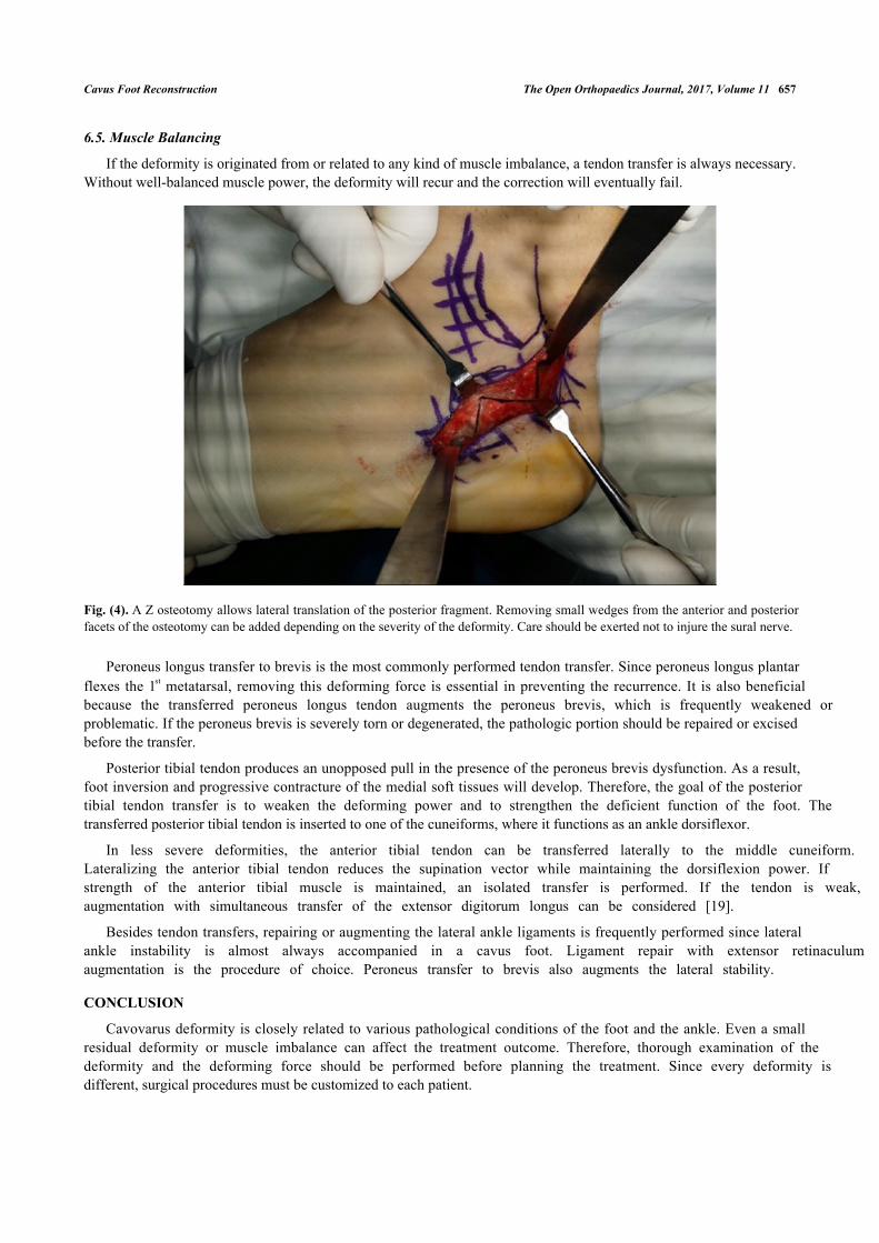
If hindfoot varus is fully corrected with 1st metatarsal osteotomy, then calcaneal osteotomy is not necessary.However, if there is residual varus after the dorsiflexion osteotomy, or if the Coleman block did not completely correctthe hindfoot varus, a calcaneal osteotomy must be done. For a mild varus, a Dwyer closing wedge osteotomy [18] maybe sufficient. For a greater amount of correction, lateralization osteotomy is necessary. An oblique osteotomy has theadvantage of three-dimensional correction as the posterior fragment can be rotated, translated, and elevated. Rotationcan be achieved with additional resection of a lateral based wedge. Elevation of the posterior fragment is helpful todecrease the calcaneal pitch. A Z osteotomy [7] is another powerful tool to correct the heel varus (Fig. 4). Theosteotomy primarily allows translation, but a little bit of rotation can be added by removing small wedges. Since thecenter of rotation is more anterior, the Z osteotomy allows a greater degree of correction compared to Dwyerosteotomy.
Salvaging joints wherever possible is beneficial because it allows more flexibility and shock absorption. However,rigid or severe cavus foot can only be reconstructed using arthrodesis. For a triple arthrodesis, the subtalar,talonavicular, and calcaneocuboid joints are denuded and fixed in a mild heel valgus position. The forefoot should besupinated through the Chopart joint [8]. When performing a triple arthrodesis, the cuboid can slide slightly beneath thecalcaneus due to the natural shape of the calcaneocuboid joint. This causes a painful bony bump on weight bearing. Toavoid this, it is useful to flat cut the calcaneocuboid joint with a saw. Excluding the calcaneocuboid joint in a triplefusion is also feasible since the calcaneocuboid joint is rarely arthritic. This is also beneficial because it reduces thepotential of problematic nonunion of the calcaneocuboid joint.
(Table 2) contd.....
Cavus Foot Reconstruction The Open Orthopaedics Journal, 2017, Volume 11 657
6.5. Muscle Balancing
If the deformity is originated from or related to any kind of muscle imbalance, a tendon transfer is always necessary.Without well-balanced muscle power, the deformity will recur and the correction will eventually fail.
Fig. (4). A Z osteotomy allows lateral translation of the posterior fragment. Removing small wedges from the anterior and posteriorfacets of the osteotomy can be added depending on the severity of the deformity. Care should be exerted not to injure the sural nerve.
Peroneus longus transfer to brevis is the most commonly performed tendon transfer. Since peroneus longus plantarflexes the 1st metatarsal, removing this deforming force is essential in preventing the recurrence. It is also beneficialbecause the transferred peroneus longus tendon augments the peroneus brevis, which is frequently weakened orproblematic. If the peroneus brevis is severely torn or degenerated, the pathologic portion should be repaired or excisedbefore the transfer.
Posterior tibial tendon produces an unopposed pull in the presence of the peroneus brevis dysfunction. As a result,foot inversion and progressive contracture of the medial soft tissues will develop. Therefore, the goal of the posteriortibial tendon transfer is to weaken the deforming power and to strengthen the deficient function of the foot. Thetransferred posterior tibial tendon is inserted to one of the cuneiforms, where it functions as an ankle dorsiflexor.
In less severe deformities, the anterior tibial tendon can be transferred laterally to the middle cuneiform.Lateralizing the anterior tibial tendon reduces the supination vector while maintaining the dorsiflexion power. Ifstrength of the anterior tibial muscle is maintained, an isolated transfer is performed. If the tendon is weak,augmentation with simultaneous transfer of the extensor digitorum longus can be considered [19].
Besides tendon transfers, repairing or augmenting the lateral ankle ligaments is frequently performed since lateralankle instability is almost always accompanied in a cavus foot. Ligament repair with extensor retinaculumaugmentation is the procedure of choice. Peroneus transfer to brevis also augments the lateral stability.
CONCLUSION
Cavovarus deformity is closely related to various pathological conditions of the foot and the ankle. Even a smallresidual deformity or muscle imbalance can affect the treatment outcome. Therefore, thorough examination of thedeformity and the deforming force should be performed before planning the treatment. Since every deformity isdifferent, surgical procedures must be customized to each patient.

658 The Open Orthopaedics Journal, 2017, Volume 11 Bom Soo Kim
CONSENT FOR PUBLICATION
Not applicable.
CONFLICT ON INTEREST
The authors declare no conflict of interest, financial or otherwise.
ACKNOWLEDGEMENT
This article is supported by Inha University Research Grant (55949-01).
REFERENCES
[1] Irwin TA, Anderson RB, Davis WH, Cohen BE. Effect of ankle arthritis on clinical outcome of lateral ankle ligament reconstruction incavovarus feet. Foot Ankle Int 2010; 31(11): 941-8.[http://dx.doi.org/10.3113/FAI.2010.0941] [PMID: 21189185]
[2] Manoli A II, Graham B. The subtle cavus foot, “the underpronator”. Foot Ankle Int 2005; 26(3): 256-63.[http://dx.doi.org/10.1177/107110070502600313] [PMID: 15766431]
[3] Sachithanandam V, Joseph B. The influence of footwear on the prevalence of flat foot. A survey of 1846 skeletally mature persons. J BoneJoint Surg Br 1995; 77(2): 254-7.[PMID: 7706341]
[4] Aminian A, Sangeorzan BJ. The anatomy of cavus foot deformity. Foot Ankle Clin 2008; 13(2): 191-8.[http://dx.doi.org/10.1016/j.fcl.2008.01.004] [PMID: 18457768]
[5] Alexander IJ, Johnson KA. Assessment and management of pes cavus in Charcot-Marie-tooth disease. Clin Orthop Relat Res 1989; (246):273-81.[PMID: 2766615]
[6] Nagai MK, Chan G, Guille JT, Kumar SJ, Scavina M, Mackenzie WG. Prevalence of Charcot-Marie-Tooth disease in patients who havebilateral cavovarus feet. J Pediatr Orthop 2006; 26(4): 438-43.[http://dx.doi.org/10.1097/01.bpo.0000226278.16449.c4] [PMID: 16791058]
[7] Ortiz C, Wagner E, Keller A. Cavovarus foot reconstruction. Foot Ankle Clin 2009; 14(3): 471-87.[http://dx.doi.org/10.1016/j.fcl.2009.03.006] [PMID: 19712886]
[8] Manoli A II, Beals TC, Hansen ST Jr. Technical factors in hindfoot arthrodesis. Instr Course Lect 1997; 46: 347-56.[PMID: 9143979]
[9] Abbasian A, Pomeroy G. The idiopathic cavus foot-not so subtle after all. Foot Ankle Clin 2013; 18(4): 629-42.[http://dx.doi.org/10.1016/j.fcl.2013.08.004] [PMID: 24215829]
[10] Chilvers M, Manoli A II. The subtle cavus foot and association with ankle instability and lateral foot overload. Foot Ankle Clin 2008; 13(2):315-24.[http://dx.doi.org/10.1016/j.fcl.2008.01.003] [PMID: 18457776]
[11] Tynan MC, Klenerman L, Helliwell TR, Edwards RH, Hayward M. Investigation of muscle imbalance in the leg in symptomatic forefoot pescavus: a multidisciplinary study. Foot Ankle Jan 13(9): 489-501.[http://dx.doi.org/10.1177/107110079201300901]
[12] Helliwell TR, Tynan M, Hayward M, Klenerman L, Whitehouse G, Edwards RH. The pathology of the lower leg muscles in pure forefoot pescavus. Acta Neuropathol 1995; 89(6): 552-9.[http://dx.doi.org/10.1007/BF00571511] [PMID: 7676811]
[13] Manoli A II, Smith DG, Hansen ST Jr. Scarred muscle excision for the treatment of established ischemic contracture of the lower extremity.Clin Orthop Relat Res 1993; (292):[PMID: 8519125]
[14] Coleman SS, Chesnut WJ. A simple test for hindfoot flexibility in the cavovarus foot Clin Orthop Relat Res Jan123: 60-2.[http://dx.doi.org/10.1097/00003086-197703000-00022]
[15] Solis G, Hennessy MS, Saxby TS. Pes cavus: A review. Foot Ankle Surg 2000; 6(3): 145-53.[http://dx.doi.org/10.1046/j.1460-9584.2000.00217.x]
[16] Maskill MP, Maskill JD, Pomeroy GC. Surgical management and treatment algorithm for the subtle cavovarus foot. Foot Ankle Int 2010;31(12): 1057-63.[http://dx.doi.org/10.3113/FAI.2010.1057] [PMID: 21189205]
[17] LoPiccolo M, Chilvers M, Graham B, Manoli A II. Effectiveness of the cavus foot orthosis. J Surg Orthop Adv 2010; 19(3): 166-9.[PMID: 21086930]
[18] Dwyer FC. Osteotomy of the calcaneum for pes cavus. J Bone Joint Surg Br 1959; 41-B(1): 80-6.[PMID: 13620710]

Cavus Foot Reconstruction The Open Orthopaedics Journal, 2017, Volume 11 659
[19] Huber M. What is the role of tendon transfer in the cavus foot? Foot Ankle Clin 2013; 18(4): 689-95.[http://dx.doi.org/10.1016/j.fcl.2013.08.002] [PMID: 24215833]
© 2017 Bom Soo Kim.
This is an open access article distributed under the terms of the Creative Commons Attribution 4.0 International Public License (CC-BY 4.0), acopy of which is available at: https://creativecommons.org/licenses/by/4.0/legalcode. This license permits unrestricted use, distribution, andreproduction in any medium, provided the original author and source are credited.