the oral pathology top seven list - north state perio
TRANSCRIPT

The Oral Pathology Top Seven List
Brad W. Neville, DDS Distinguished University Professor
Division of Oral Pathology Medical University of South Carolina

The Oral Pathology Top Seven List
Brad W. Neville, DDS
Case #7
Denture-Related Hyperplasia
CHAPTER 12 Soft Tissue Tumors 475
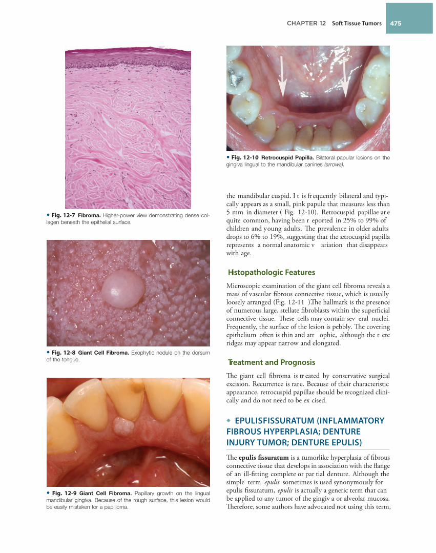
the mandibular cuspid. I t is frequently bilateral and typi-cally appears as a small, pink papule that measures less than 5 mm in diameter ( Fig. 12-10 ). Retrocuspid papillae ar e quite common, having been r eported in 25% to 99% of children and young adults. Th e prevalence in older adults drops to 6% to 19%, suggesting that the retrocuspid papilla represents a normal anatomic v ariation that disappears with age.
Histopathologic Features
Microscopic examination of the giant cell fi broma reveals a mass of vascular fi brous connective tissue, which is usually loosely arranged ( Fig. 12-11 ). Th e hallmark is the presence of numerous large, stellate fi broblasts within the superfi cial connective tissue. Th ese cells may contain sev eral nuclei. Frequently, the surface of the lesion is pebbly. Th e covering epithelium often is thin and atr ophic, although the r ete ridges may appear narrow and elongated.
Treatment and Prognosis
Th e giant cell fi broma is tr eated by conservative surgical excision. Recurrence is rare. Because of their characteristic appearance, retrocuspid papillae should be recognized clini-cally and do not need to be ex cised.
◆ EPULIS FISSURATUM (INFLAMMATORY FIBROUS HYPERPLASIA; DENTURE INJURY TUMOR; DENTURE EPULIS)
Th e epulis fi ssuratum is a tumorlike hyperplasia of fi brous connective tissue that develops in association with the fl ange of an ill-fi tting complete or par tial denture. Although the simple term epulis sometimes is used synonymously for epulis fi ssuratum, epulis is actually a generic term that can be applied to any tumor of the gingiv a or alveolar mucosa. Th erefore, some authors have advocated not using this term,
• Fig. 12-7 Fibroma. Higher-power view demonstrating dense col-
lagen beneath the epithelial surface.
• Fig. 12-8 Giant Cell Fibroma. Exophytic nodule on the dorsum
of the tongue.
• Fig. 12-9 Giant Cell Fibroma. Papillary growth on the lingual
mandibular gingiva. Because of the rough surface, this lesion would
be easily mistaken for a papilloma.
• Fig. 12-10 Retrocuspid Papilla. Bilateral papular lesions on the
gingiva lingual to the mandibular canines (arrows).

CHAPTER 12 Soft Tissue Tumors476
vestibule. Th e epulis fi ssuratum usually dev elops on the facial aspect of the alv eolar ridge, although occasional lesions are seen lingual to the mandibular alv eolar ridge ( Fig. 12-14 ).
Th e epulis fi ssuratum most often occurs in middle-aged and older adults, as would be expected with a dentur e-related lesion. It may occur on either the maxilla or man-dible. Th e anterior por tion of the jaws is aff ected much
• Fig. 12-12 Epulis Fissuratum. Hyperplastic folds of tissue in the
anterior maxillary vestibule.
• Fig. 12-13 Epulis Fissuratum. A, Several folds of hyperplastic
tissue in the maxillary vestibule. B, An ill-fi tting denture fi ts into the
fi ssure between two of the folds. (Courtesy of Dr. William Bruce.)
A
B
• Fig. 12-11 Giant Cell Fibroma. A, Low-power view showing a
nodular mass of fi brous connective tissue covered by stratifi ed squa-
mous epithelium. Note the elongation of the rete ridges. B, High-power
view showing multiple large stellate-shaped and multinucleated
fi broblasts.
A
B
preferring to call these lesions infl ammatory fi brous hyperpla-sia or other descriptiv e names. H owever, the term epulis fi ssuratum is still widely used today and is w ell understood by virtually all clinicians. O ther examples of epulides include the giant cell epulis (peripheral giant cell granu-loma) (see page 485 ), ossifying fi broid epulis (peripheral ossifying fi broma) (see page 487 ), and congenital epulis (see page 503 ).
Clinical Features
Th e epulis fi ssuratum typically appears as a single or mul-tiple fold or folds of hyperplastic tissue in the alv eolar ves-tibule ( Figs. 12-12 and 12-13 ). Most often, there are two folds of tissue, and the fl ange of the associated dentur e fi ts conveniently into the fi ssure between the folds. Th e redun-dant tissue is usually fi rm and fi brous, although some lesions appear erythematous and ulcerated, similar to the appear-ance of a py ogenic granuloma. O ccasional examples of epulis fi ssuratum demonstrate surface areas of infl ammatory papillary hyperplasia (see page 478 ). Th e size of the lesion can vary from localized hyperplasias less than 1 cm in size to massive lesions that inv olve most of the length of the

CHAPTER 12 Soft Tissue Tumors 477
squamous epithelium. Like the epulis fi ssuratum, the over-lying epithelium may be hyperplastic.
Treatment and Prognosis
Th e treatment of the epulis fi ssuratum or fi broepithelial polyp consists of surgical r emoval, with micr oscopic
more often than the posterior ar eas. Th ere is a pronounced female predilection; most studies sho w that two-thirds to three-fourths of all cases submitted for biopsy occur in women.
Another similar but less common fi brous hyperplasia, often called a fi broepithelial polyp or leafl ike denture fi broma, occurs on the har d palate beneath a maxillar y denture. Th is characteristic lesion is a fl attened pink mass that is attached to the palate by a narrow stalk ( Fig. 12-15 ). Usually, the fl attened mass is closely applied to the palate and sits in a slightly cupped-out depr ession. However, it is easily lifted up with a probe, which demonstrates its pedun-culated nature. Th e edge of the lesion often is serrated and resembles a leaf.
Histopathologic Features
Microscopic examination of the epulis fi ssuratum reveals hyperplasia of the fi brous connective tissue. Often multiple folds and grooves occur where the denture impinges on the tissue ( Fig. 12-16 ). Th e overlying epithelium is fr equently hyperparakeratotic and demonstrates irr egular hyperplasia of the rete ridges. In some instances, the epithelium sho ws infl ammatory papillary hyperplasia (see page 478 ) or pseu-doepitheliomatous (pseudocarcinomatous) hyperplasia. Focal areas of ulceration ar e not unusual, especially at the base of the gr ooves between the folds. A v ariable chronic infl ammatory infi ltrate is present; sometimes, it may include eosinophils or sho w lymphoid follicles. I f minor saliv ary glands are included in the specimen, then they usually show chronic sialadenitis.
In rare instances, the formation of osteoid or chondr oid is observed. Th is unusual-appearing pr oduct, known as osseous and chondromatous metaplasia, is a reactive phe-nomenon caused b y chronic irritation b y the ill-fi tting denture (see page 292 ). Th e irregular nature of this bone or cartilage can be microscopically disturbing, and the pathol-ogist should not mistake it for a sar coma.
Th e denture-related fi broepithelial polyp has a narr ow core of dense fi brous connective tissue covered by stratifi ed
• Fig. 12-14 Epulis Fissuratum. Redundant folds of tissue arising
in the fl oor of the mouth in association with a mandibular denture.
• Fig. 12-15 Fibroepithelial Polyp. Flattened mass of tissue arising
on the hard palate beneath a maxillary denture; note its pedunculated
nature. Because of its serrated edge, this lesion also is known as a
leafl ike denture fi broma. Associated infl ammatory papillary hyperplasia
is visible in the palatal midline.
A
B
• Fig. 12-16 Epulis Fissuratum. Low-power photomicrograph
demonstrating folds of hyperplastic fi brovascular connective tissue
covered by stratifi ed squamous epithelium.

CHAPTER 12 Soft Tissue Tumors478
papillary surface. Many cases ar e associated with dentur e stomatitis.
Histopathologic Features
Th e mucosa in infl ammatory papillary hyperplasia exhibits numerous papillary growths on the surface that are covered by hyperplastic, stratifi ed squamous epithelium ( Fig. 12-19 ). In advanced cases, this hyperplasia is pseudoepithelioma-tous in appearance, and the pathologist should not mistake it for carcinoma ( Fig. 12-20 ). Th e connective tissue can vary from loose and edematous to densely collageniz ed. A chronic infl ammatory cell infi ltrate is usually seen, which consists of lymphocytes and plasma cells. Less fr equently, polymorphonuclear leukocytes are also present. If underly-ing salivary glands are present, then they often show scleros-ing sialadenitis.
Treatment and Prognosis
For very early lesions of infl ammatory papillary hyperplasia, removal of the denture may allow the erythema and edema
• Fig. 12-17 Infl ammatory Papillary Hyperplasia. Erythematous,
pebbly appearance of the palatal vault.
• Fig. 12-18 Infl ammatory Papillary Hyperplasia. An advanced
case exhibiting more pronounced papular lesions of the hard palate.
• Fig. 12-19 Infl ammatory Papillary Hyperplasia. Medium-power
view showing fi brous and epithelial hyperplasia resulting in papillary
surface projections. Heavy chronic infl ammation is present.
examination of the ex cised tissue. Th e ill-fi tting denture should be remade or relined to prevent a recurrence of the lesion.
◆ INFLAMMATORY PAPILLARY HYPERPLASIA (DENTURE PAPILLOMATOSIS)
Infl ammatory papillary hyperplasia is a r eactive tissue growth that usually, although not always, develops beneath a denture. Some investigators classify this lesion as par t of the spectrum of denture stomatitis (see page 194 ). Although the exact pathogenesis is unkno wn, the condition most often appears to be r elated to the following: • An ill-fi tting denture • Poor denture hygiene • Wearing the denture 24 hours a day
Approximately 20% of patients who wear their dentures 24 hours a day hav e infl ammatory papillary hyperplasia. Candida organisms also have been suggested as a cause, but any possible role appears uncertain.
Clinical Features
Infl ammatory papillary hyperplasia usually occurs on the hard palate beneath a denture base ( Figs. 12-17 and 12-18 ). Early lesions may inv olve only the palatal v ault, although advanced cases cover most of the palate. Less frequently, this hyperplasia develops on the edentulous mandibular alveolar ridge or on the sur face of an epulis fi ssuratum. On rare occasions, the condition occurs on the palate of a patient without a dentur e, especially in people who habitually breathe through their mouth or hav e a high palatal v ault. Candida -associated palatal papillar y hyperplasia also has been reported in dentate patients with human immunode-fi ciency virus (HIV) infection.
Infl ammatory papillary hyperplasia is usually asymptom-atic. Th e mucosa is er ythematous and has a pebbly or

CHAPTER 12 Soft Tissue Tumors 479
• Fig. 12-20 Infl ammatory Papillary Hyperplasia. Higher-power
view showing pseudoepitheliomatous hyperplasia of the epithelium.
This epithelium has a bland appearance that should not be mistaken
for carcinoma.
to subside, and the tissues may r esume a mor e normal appearance. Th e condition also may sho w improvement after topical or systemic antifungal therapy . For more advanced and collagenized lesions, many clinicians prefer to excise the hyperplastic tissue befor e fabricating a ne w denture. Various surgical methods have been used, includ-ing the following: • Partial-thickness or full-thickness surgical blade
excision • Curettage • Electrosurgery • Cryosurgery • Laser surgery
After surgery, the existing dentur e can be lined with a temporary tissue conditioner that acts as a palatal dr essing and promotes greater comfort. After healing, the patient should be encouraged to leave the new denture out at night and to keep it clean.
◆ FIBROUS HISTIOCYTOMA
Fibrous histiocytomas are a diverse group of tumors that exhibit fi broblastic and histiocytic diff erentiation, although the cell of origin is uncertain. Because of the variable nature of these lesions, an array of terms has been used for them, including dermatofi broma, sclerosing hemangioma, fi broxanthoma, and nodular subepidermal fi brosis. Unlike other fi brous growths discussed pr eviously in this chapter, the fi brous histiocytoma is generally considered to represent a true neoplasm.
Clinical Features
Th e fi brous histiocytoma can dev elop almost anywhere in the body. Th e most common site is the skin of the extremi-ties, where the lesion is called a dermatofi broma. Tumors of the oral and perioral r egion are rare, and it is likely that many previously reported examples would be r eclassifi ed
today as solitary fi brous tumors (see next topic). Rar e intra-bony lesions of the jaws also hav e been r eported. Oral fi brous histiocytomas tend to occur in middle-aged and older adults; cutaneous examples ar e most fr equent in young adults. Th e tumor is usually a painless nodular mass and can vary in size from a few millimeters to several cen-timeters in diameter ( Fig. 12-21 ). Deeper tumors tend to be larger.
Histopathologic Features
Microscopically, the fi brous histiocytoma is characterized by a cellular pr oliferation of spindle-shaped fi broblastic cells with vesicular nuclei ( Figs. 12-22 and 12-23 ). Th e margins of the tumor often are not sharply defi ned. Th e tumor cells are arranged in shor t, intersecting fascicles, kno wn as a storiform pattern because of its resemblance to the irregular, whorled appearance of a straw mat. Rounded histiocyte-like cells, lipid-containing xanthoma cells, or multinucleated giant cells can be seen occasionally , as may scatter ed lym-phocytes. Th e stroma may demonstrate ar eas of myx oid change or focal hyalinization.
• Fig. 12-21 Fibrous Histiocytoma. Nodular mass on the dorsum
of the tongue.
• Fig. 12-22 Fibrous Histiocytoma. Medium-power view of a skin
tumor (dermatofi broma) showing spindle-shaped cells arranged in a
storiform pattern.

The Oral Pathology Top Seven List
Brad W. Neville, DDS
Case #6
Sialolithiasis

CHAPTER 11 Salivary Gland Pathology 427
• Fig. 11-13 Sialolithiasis. Hard mass at the orifi ce of Wharton
duct.
• Fig. 11-14 Sialolithiasis. Radiopaque mass located at the left
angle of the mandible. (Courtesy of Dr. Roger Bryant.)
lesions of patients with multiple “ mucus retention cysts” also show prominent oncocytic metaplasia of the epithelial lining.
Treatment and Prognosis
Isolated salivary duct cysts are treated by conservative surgi-cal excision. For cysts in the major glands, par tial or total removal of the gland may be necessar y. Th e lesion should not recur.
For rare patients who develop multifocal salivary ductal ectasia (“mucus retention cysts”), local excision may be per-formed for the more problematic swellings; however, surgi-cal management does not appear feasible or advisable for all of the lesions, which may number as many as 100. I n one reported case, systemic er ythromycin and chlorhexidine mouth rinses w ere helpful in r elieving pain and r educing drainage of pus. Sialagogue medications also may be helpful in stimulating salivary fl ow, thereby preventing the accumu-lation of inspissated mucus within the dilated ex cretory ducts.
◆ SIALOLITHIASIS (SALIVARY CALCULI; SALIVARY STONES)
Sialoliths are calcifi ed structures that dev elop within the salivary ductal system. R esearchers believe that they arise from deposition of calcium salts ar ound a nidus of debris within the duct lumen. Th is debris may include inspis-sated mucus, bacteria, ductal epithelial cells, or for eign bodies. Th e cause of sialoliths is unclear , but their forma-tion can be pr omoted by chronic sialadenitis and par tial obstruction. Th eir development typically is not r elated to any systemic derangement in calcium and phosphor us metabolism.
Clinical and Radiographic Features
Sialoliths most often dev elop within the ductal system of the submandibular gland, which accounts for about 80% of cases; the formation of stones within the par otid gland system is distinctly less frequent. Th e long, tortuous, upward path of the submandibular (Wharton) duct and the thicker, mucoid secretions of this gland may be r esponsible for its greater tendency to form salivary calculi. Sialoliths also can form within the minor saliv ary glands, most often within the glands of the upper lip or buccal mucosa. Salivary stones can occur at almost any age, but they ar e most common in young and middle-aged adults.
Major gland sialoliths most fr equently cause episodic pain or swelling of the aff ected gland, especially at meal-time. Th e severity of the symptoms v aries, depending on the degree of obstruction and the amount of resultant back-pressure produced within the gland. I f the stone is located toward the terminal por tion of the duct, then a har d mass may be palpated beneath the mucosa ( Fig. 11-13 ).
Sialoliths typically appear as radiopaque masses on radio-graphic examination. H owever, not all stones ar e visible on standard radiographs (perhaps because of the degr ee of calcifi cation of some lesions). Th ey may be disco vered anywhere along the length of the duct or within the gland itself ( Fig. 11-14 ). Stones in the terminal por tion of the submandibular duct are best demonstrated with an occlusal radiograph. On panoramic or periapical radiographs, the calcifi cation may appear superimposed on the mandible and care must be exercised not to confuse it with an intrabony lesion ( Fig. 11-15 ). Multiple parotid stones radiographically can mimic calcifi ed parotid lymph nodes, such as might occur in tuber culosis. Sialography, ultrasound, and CT scanning may be helpful additional imaging studies for sialoliths. Diagnostic sialendoscopy also can be a v aluable tool in the evaluation and diagnosis of ductal obstructions. In this technique, a miniaturized endoscope is inserted into the duct orifi ce, allowing visualization of the ductal system for any stones, strictures, or adhesions.
Minor gland sialoliths often ar e asymptomatic but may produce local swelling or tenderness of the aff ected gland ( Fig. 11-16 ). A small radiopacity often can be demonstrated with a soft tissue radiograph.
Histopathologic Features
On gross examination, sialoliths appear as hard masses that are round, oval, or cylindrical. Th ey are typically y ellow,

CHAPTER 11 Salivary Gland Pathology428
often demonstrates squamous, oncocytic, or mucous cell metaplasia. Periductal infl ammation is also evident. Th e ductal obstruction frequently is associated with an acute or chronic sialadenitis of the feeding gland.
Treatment and Prognosis
Small sialoliths of the major glands sometimes can be treated conservatively by gentle massage of the gland in an
although they may be a white or y ellow-brown color. Sub-mandibular stones tend to be larger than those of the parotid or minor glands. S ialoliths are usually solitar y, although occasionally two or more stones may be discovered at surgery.
Microscopically, the calcifi ed mass exhibits concentric laminations that may surround a nidus of amorphous debris ( Fig. 11-17 ). If the associated duct also is r emoved, then it
• Fig. 11-15 Sialolithiasis. A, Periapical fi lm showing discrete radi-
opacity (arrow) superimposed on the body of the mandible. Care must
be taken not to confuse such lesions with intrabony pathosis.
B, Occlusal radiograph of same patient demonstrating radiopaque
stone in Wharton duct.
A
B
• Fig. 11-16 Sialolithiasis. A, Minor salivary gland sialolith present-
ing as a hard nodule in the upper lip. B, A soft tissue radiograph of
the same lesion revealed a laminated calcifi ed mass.
A
B
• Fig. 11-17 Sialolithiasis. Intraductal calcifi ed mass showing con-
centric laminations. The duct exhibits squamous metaplasia.
CHAPTER 11 Salivary Gland Pathology 429
Recurrent or persistent ductal obstr uction (most com-monly caused by sialoliths) can lead to a chr onic sialadeni-tis. Periodic swelling and pain occur within the aff ected gland, usually dev eloping at mealtime when saliv ary fl ow is stimulated. S ialography often demonstrates sialectasia (ductal dilatation) proximal to the area of obstruction ( Fig. 11-20 ). In chronic parotitis, Stensen duct may show a char-acteristic sialographic pattern known as “sausaging,” which refl ects a combination of dilatation plus ductal strictur es from scar formation. Chronic sialadenitis also can occur in the minor glands, possibly as a r esult of blockage of ductal fl ow or local trauma.
Juvenile recurrent parotitis is the most common infl ammatory salivary disorder of childr en in the U nited States and the second most common such disor der world-wide (following mumps). I t is characteriz ed by recurring episodes of unilateral or bilateral, non-suppurativ e parotid swelling, usually beginning betw een the ages of 3 and 6 years. Th e exact cause is unkno wn, although congenital duct malformations, genetic factors, immunologic disor-ders, and dental malocclusion hav e been suggested as pos-sible contributing factors. Although multiple r ecurrences often develop, the condition usually r esolves around the time of puberty.
eff ort to milk the stone toward the duct orifi ce. Sialagogues (drugs that stimulate salivary fl ow), moist heat, and increased fl uid intake also may pr omote passage of the stone. Larger sialoliths usually need to be removed surgically. If signifi cant infl ammatory damage has occurr ed within the feeding gland, then the gland may need to be removed. Minor gland sialoliths are best treated by surgical removal, including the associated gland.
Extracorporeal shock wave lithotripsy and interventional sialendoscopy with basket r etrieval are newer techniques that can be eff ective in the r emoval of sialoliths fr om the major glands. Th ese minimally invasive techniques have low morbidity and may preclude the necessity of gland removal.
◆ SIALADENITIS
Infl ammation of the salivary glands (sialadenitis) can arise from various infectious and noninfectious causes. Th e most common viral infection is mumps (see page 238 ), although a number of other vir uses also can inv olve the saliv ary glands, including Coxsackie A, ECHO, choriomeningitis, parainfl uenza, human immunodefi ciency virus (HIV), and cytomegalovirus (CMV) (in neonates). Most bacterial infec-tions arise as a result of ductal obstruction or decreased sali-vary fl ow, allowing retrograde spread of bacteria throughout the ductal system. B lockage of the duct can be caused b y sialolithiasis (see page 427 ), congenital strictures, or com-pression by an adjacent tumor . Decreased fl ow can r esult from dehydration, debilitation, or medications that inhibit secretions.
One of the more common causes of sialadenitis is recent surgery (especially abdominal surgery), after which an acute parotitis (surgical mumps) may arise because the patient has been kept without food or fl uids (NPO) and has r eceived atropine during the surgical pr ocedure. Other medications that produce xerostomia as a side eff ect also can predispose patients to such an infection. M ost community-acquired cases of acute bacterial sialadenitis are caused by Staphylococ-cus aureus or streptococcal species. Hospital-acquired infec-tions are also most frequently associated with S. aureus, but they also may be caused by a variety of other species, includ-ing Eikenella corrodens, Escherichia coli, Fusobacterium, Hae-mophilus infl uenzae, Klebsiella, Prevotella, Proteus, and Pseudomonas . Noninfectious causes of saliv ary infl amma-tion include Sjögren syndrome (see page 434 ), sarcoidosis (see page 310 ), radiation therapy (see page 266 ), and various allergens.
Clinical and Radiographic Features
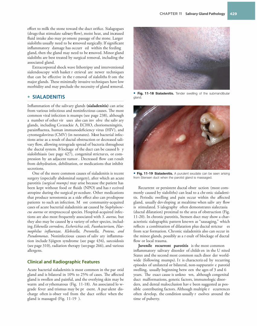
Acute bacterial sialadenitis is most common in the par otid gland and is bilateral in 10% to 25% of cases. Th e aff ected gland is swollen and painful, and the overlying skin may be warm and er ythematous ( Fig. 11-18 ). An associated lo w-grade fever and trismus may be pr esent. A pur ulent dis-charge often is obser ved from the duct orifi ce when the gland is massaged ( Fig. 11-19 ).
• Fig. 11-18 Sialadenitis. Tender swelling of the submandibular
gland.
• Fig. 11-19 Sialadenitis. A purulent exudate can be seen arising
from Stensen duct when the parotid gland is massaged.
The Oral Pathology Top Seven List
Brad W. Neville, DDS
Case #5
Pernicious Anemia

CHAPTER 17 Oral Manifestations of Systemic Diseases 773
intrinsic factor are also found in the serum of these patients. Vitamin B 12 defi ciency may occur for other r easons, and although the r esulting signs and symptoms may be identical to those of pernicious anemia, these should be considered as distinctly diff erent defi ciency disorders. For example, a decreased ability to absorb cobalamin may also occur after gastrointestinal bypass operations. In addition, because cobalamin is primarily derived from animal sources, some strict v egetarians (vegans) may dev elop vitamin B 12 defi ciency.
Because cobalamin is necessar y for normal nucleic acid synthesis, anything that disr upts the absorption of the vitamin causes problems, especially for cells that ar e multi-plying rapidly and, ther efore, synthesizing large amounts of nucleic acids. Th e cells that ar e the most mitotically active are aff ected to the gr eatest degree, especially the hematopoietic cells and the gastr ointestinal lining epithe-lial cells.
Clinical Features
With respect to systemic complaints, patients with perni-cious anemia often r eport fatigue, w eakness, shortness of breath, headache, and feeling faint. S uch symptoms ar e associated with most anemias and pr obably refl ect the reduced oxygen-carrying capacity of the blood. Vitamin B 12 also functions to maintain my elin throughout the nervous system; therefore, with reduced levels of the vitamin, many patients report paresthesia, tingling, or numbness of the extremities. Diffi culty in walking and diminished vibratory and positional sense may be pr esent. Psychiatric symptoms of memory loss, irritability, depression, and dementia have also been described.
Oral symptoms often consist of a burning sensation of the tongue, lips, buccal mucosa, or other mucosal sites. Clinical examination may sho w focal patchy ar eas of oral mucosal erythema and atrophy ( Fig. 17-17 ), or the process may be more diff use, depending on the sev erity and dura-tion of the condition. Th e tongue may be aff ected in as many as 50% to 60% of patients with pernicious anemia, but it may not show as much involvement as other areas of the oral mucosa in some instances. Th e atrophy and er y-thema may be easier to appreciate on the dorsal tongue than at other sites, however.
Histopathologic Features
Histopathologic examination of an er ythematous portion of the oral mucosa sho ws marked epithelial atrophy with loss of r ete ridges, an incr eased nuclear-to-cytoplasmic ratio of the sur face epithelial cells, and pr ominent nucleoli ( Fig. 17-18 ). Th is pattern can be misinterpreted as epithelial dysplasia at times, although the nuclei in pernicious anemia typically are pale staining and show peripheral chro-matin clumping. A patchy diff use chronic infl ammatory cell infi ltrate is usually noted in the underlying connectiv e tissue.
with endoscopy or esophageal barium contrast radiographic studies usually sho ws the pr esence of abnormal bands of tissue in the esophagus, called esophageal webs. Another sign is an alteration of the growth pattern of the nails, which results in a spoon-shaped confi guration (koilonychia). Th e nails may also be brittle.
Symptoms of anemia may prompt patients with Plummer-Vinson syndrome to seek medical care. Fatigue, shortness of breath, and weakness are characteristic symptoms.
Laboratory Findings
Hematologic studies sho w a hypochr omic microcytic anemia that is consistent with an ir on-defi ciency anemia.
Histopathologic Features
A biopsy specimen of inv olved mucosa fr om a patient with Plummer-Vinson syndrome typically shows epithelial atrophy with varying degrees of submucosal chronic infl am-mation. In advanced cases, evidence of epithelial atypia or dysplasia may be seen.
Treatment and Prognosis
Treatment of P lummer-Vinson syndrome is primarily directed at correcting the iron-defi ciency anemia by means of dietary iron supplementation. Th is therapy usually resolves the anemia, r elieves the glossodynia, and may reduce the severity of the esophageal symptoms. O ccasion-ally, esophageal dilation is necessar y to help impr ove the symptoms of dysphagia. P atients with P lummer-Vinson syndrome should be ev aluated periodically for oral, hypo-pharyngeal, and esophageal cancer because a 5% to 50% prevalence of upper aerodigestive tract malignancy has been reported in aff ected persons.
◆ PERNICIOUS ANEMIA
Pernicious anemia is an uncommon condition that occurs with greatest frequency among older patients of N orthern European heritage, although r ecent studies have identifi ed the disease in black and Hispanic populations as well. Asian populations seem to be aff ected much less fr equently. Th e disease is a megaloblastic anemia caused by poor absorption of cobalamin (vitamin B 12 , extrinsic factor). Intrinsic factor, which is pr oduced by the parietal cells of the stomach lining, is needed for vitamin-B 12 absorption. N ormally, when cobalamin is ingested, it binds to intrinsic factor in the duodenum. B ecause the lining cells of the intestine preferentially take up the cobalamin-intrinsic factor complex, signifi cant amounts of the vitamin cannot be absorbed unless both components ar e present.
In the case of pernicious anemia, most patients lack intrinsic factor because of an autoimmune destr uction of the parietal cells of the stomach; this r esults in decr eased absorption of cobalamin. Antibodies dir ected against

CHAPTER 17 Oral Manifestations of Systemic Diseases774
the elimination of painful injections. One recent systematic literature review has identifi ed what appears to be an increased risk of gastric carcinoma, with pernicious anemia patients being seven times more likely to develop this tumor compared to the general population. Both vitamin B 12 defi -ciency and folate defi ciency will cause megaloblastic anemia, and it is impor tant to distinguish betw een the two pr ob-lems. Treatment of vitamin B 12 defi ciency with folate will resolve the anemia and the oral mucosal atr ophy, but reduced myelin production will continue, r esulting in further CNS damage.
◆ PITUITARY DWARFISM
Pituitary dwarfi sm is a relatively rare condition that results from either the diminished production of growth hormone by the anterior pituitary gland, abnormalities of the growth hormone molecule, or a r educed capacity of the tissues to respond to growth hormone. Aff ected patients are typically much shorter than normal, although their body proportions are generally appropriate.
Several conditions may cause short stature, and a careful evaluation of the patient must be per formed to r ule out other possible causes, such as the follo wing: 1. Intrinsic defects in the patient ’ s tissues (e.g., certain skel-
etal dysplasias, chr omosomal abnormalities, and idio-pathic short stature)
2. Alterations in the envir onment of the gr owing tissues (e.g., malnutrition, hypothyr oidism, and diabetes mellitus) If a lack of growth hormone is detected, the cause should
be determined. Sometimes the fault lies with the pituitar y gland itself (e.g., aplasia or hypoplasia). I n other instances, the problem may be r elated to destruction of the pituitar y or hypothalamus b y tumors, therapeutic radiation, or infection.
If the hypothalamus is aff ected, a defi ciency in growth hormone–releasing hormone, which is pr oduced by the hypothalamus, results in a defi ciency of growth hormone. Often defi ciencies in other hormones, such as thyr oid
Laboratory Findings
Hematologic evaluation of vitamin B 12 defi ciency shows a macrocytic anemia and r educed serum cobalamin lev els. Th e Schilling test for pernicious anemia has been used to determine the pathogenesis of the cobalamin defi ciency by comparing absorption and ex cretion rates of radiolabeled cobalamin. However, this study is rather complicated to perform, and is now considered to be obsolete. Th e presence of serum antibodies directed against intrinsic factor is quite specifi c for pernicious anemia.
Treatment and Prognosis
Once the diagnosis of pernicious anemia is established, treatment traditionally has consisted of monthly intramus-cular injections of cyanocobalamin. Th e condition responds rapidly once therapy is initiated, with reports of clearing of oral lesions within 5 days. High-dose oral cobalamin therapy has also been sho wn to be an equally eff ective treatment, however, with adv antages being its cost-eff ectiveness and
• Fig. 17-17 Pernicious Anemia. A, The dorsal tongue shows erythema and atrophy. B, After therapy
with vitamin B 12 , the mucosal alteration resolved.
A B
• Fig. 17-18 Pernicious Anemia. This medium-power photomicro-
graph shows epithelial atrophy and atypia with chronic infl ammation
of the underlying connective tissue. These features are characteristic
of a megaloblastic anemia, such as pernicious anemia.
The Oral Pathology Top Seven List
Brad W. Neville, DDS
Case #4
Cinnamon Stomatitis

CHAPTER 9 Allergies and Immunologic Diseases322
Histopathologic Features
Biopsy of perioral dermatitis demonstrates a v ariable pattern. In many cases there is a chronic lymphohistiocytic dermatitis that often exhibits spongiosis of the hair follicles. In other patients a r osacea-like pattern is noted in which there is perifollicular granulomatous infl ammation. On occasion, this histopathologic pattern has been misdiag-nosed as sarcoidosis.
Treatment and Prognosis
Most cases resolve with “zero therapy,” which includes dis-continuation of corticosteroids, cosmetics, and facial creams. Discontinuation of potent topical cor ticosteroid use often is followed by a period of exacerbation, which can be mini-mized by substitution of a less potent cor ticosteroid before total cessation. Oral tetracycline is considered the therapeu-tic gold standard for perioral dermatitis but must be avoided during childhood and pregnancy. In addition, a shortage of tetracycline in the United States has made its use problem-atic. No strong evidence has been presented to demonstrate that doxycycline or minocycline is equivalent or superior to tetracycline. Luckily, perioral dermatitis also r esponds well to topical pimecr olimus or topical er ythromycin. Weaker therapeutic recommendations include topical metr onida-zole, clindamycin, tacrolimus, tetracycline, adapalene, or azelaic acid, plus systemic erythromycin or isotretinoin. Th e pathosis typically demonstrates signifi cant improvement within several weeks and total r esolution in a few months. Recurrence is uncommon.
◆ CONTACT STOMATITIS FROM ARTIFICIAL CINNAMON FLAVORING
Mucosal abnormalities secondar y to the use of ar tifi cially fl avored cinnamon pr oducts are fairly common, but the
Th e antigen can be placed on the mucosa in a mixture with Orabase or in a r ubber cup that is fi xed to the mucosa.
Treatment and Prognosis
In mild cases of acute contact stomatitis, r emoval of the suspected allergen is all that is r equired. In more severe cases, antihistamine therapy, which is combined with topical anesthetics, usually is benefi cial. Chronic reactions respond to removal of the antigenic sour ce and application of a topical corticosteroid gel or oral suspension.
When attempting to disco ver the sour ce of a diff use allergic mucositis, use of plain baking soda or toothpaste that is fr ee of fl avoring or pr eservatives is r ecommended. Th e patient also should be instr ucted to avoid mouthwash, gum, mints, chocolate, cinnamon-containing products, car-bonated drinks, and excessively salty, spicy, or acidic foods. If an association cannot be found, then cutaneous patch testing may provide helpful information.
◆ PERIORAL DERMATITIS (PERIORIFICIAL DERMATITIS)
Perioral dermatitis does not refer to every rash that occurs around the mouth but is specifi c for a unique infl ammatory skin disease that involves the cutaneous sur faces surround-ing the facial orifi ces. Because the disor der also often involves the paranasal and periorbital skin, periorifi cial dermatitis is the most appr opriate designation. Although the process is idiopathic, the dermatitis is associated strongly with uncritical use of potent topical cor ticosteroids on the facial skin. F luorinated toothpaste and o veruse of heavy facial cosmetics, cr eams, and moisturiz ers also ar e impli-cated in many patients. Weaker correlations have been seen with systemic cor ticosteroids, corticosteroid inhalers, and nasal corticosteroids. Heavy exposure to ultraviolet light, heat, and wind appears to worsen the dermatitis. S ome of these substances may initially induce an irritant or allergic contact dermatitis, whereas others are thought to pr oduce inappropriate occlusion of the skin surface with subsequent proliferation of skin fl ora.
Clinical Features
Perioral dermatitis appears with persistent er ythematous papules, papulovesicles, and papulopustules that involve the skin surrounding the v ermilion border of the upper and lower lips. In addition, involvement of the perinasal skin is seen in approximately 40% of aff ected patients, and 25% have periorbital dermatitis ( Fig. 9-36 ). Classically, there is a zone of spared skin immediately adjacent to the vermilion border. Pruritus and burning are variable. Th e vast majority of the cases are diagnosed in women between the ages of 20 to 45 years, lending further support to the association with cosmetic use. In spite of this, the process does occur in men and in children of either sex.
• Fig. 9-36 Perioral Dermatitis. Multiple erythematous papules of
the skin surrounding the vermilion border of the lips. Note similar
involvement around the nasal orifi ces (periorifi cial dermatitis). (Courtesy
of Dr. Billy Millay.)
CHAPTER 9 Allergies and Immunologic Diseases 323
range of changes was not r ecognized widely until the late 1980s. Cinnamon oil is used as a fl avoring agent in confec-tionery, ice cr eam, soft drinks, alcoholic bev erages, pro-cessed meats, gum, candy , toothpaste, br eath fresheners, mouthwashes, and even dental fl oss. Concentrations of the fl avoring are up to 100 times that in the natural spice. Th e reactions are documented most commonly in those pr od-ucts associated with prolonged or frequent contact, such as candy, chewing gum, and toothpaste. Th e anticalculus com-ponents of tar tar-control toothpastes hav e a str ong bitter fl avor and r equire a signifi cant concentration of fl avoring agents including cinnamon to hide the taste, r esulting in a greater chance these formulations will cause oral mucosal lesions. Although much less common, r eactions to cinna-mon in its natural spice form hav e been documented.
Clinical Features
Th e clinical presentations of contact stomatitis v ary some-what, according to the medium of deliv ery. Toothpaste results in a mor e diff use pattern; the signs associated with chewing gum and candy ar e more localized. Pain and burning are common symptoms in all cases.
Th e gingiva is the most fr equent site aff ected by tooth-paste, often r esembling plasma cell gingivitis (see page 145 ); enlargement, edema, and er ythema are common. Sloughing of the superfi cial oral epithelium without cr e-ation of an erosion is seen commonly. Erythematous muco-sitis, occasionally combined with erosion, has been reported on the buccal mucosa and tongue. E xfoliative cheilitis and circumoral dermatitis also may occur.
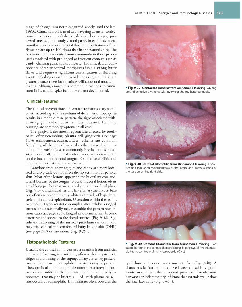
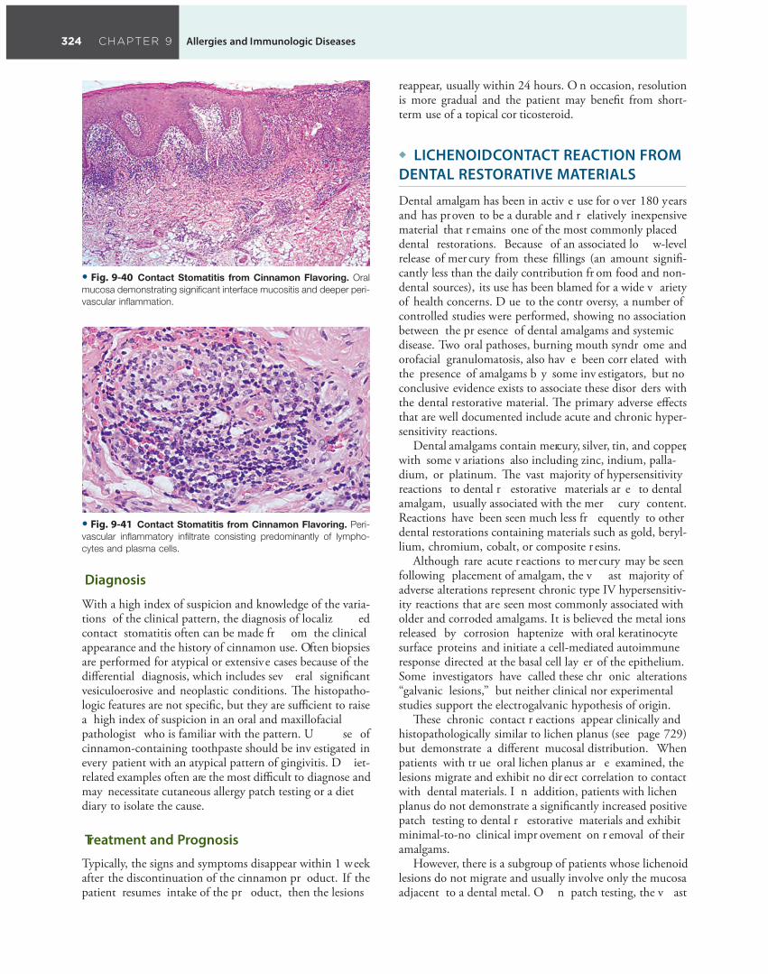
Reactions from chewing gum and candy are more local-ized and typically do not aff ect the lip vermilion or perioral skin. Most of the lesions appear on the buccal mucosa and lateral borders of the tongue. B uccal mucosal lesions often are oblong patches that are aligned along the occlusal plane ( Fig. 9-37 ). Individual lesions have an er ythematous base but often are predominantly white as a result of hyperkera-tosis of the surface epithelium. Ulceration within the lesions may occur. Hyperkeratotic examples often exhibit a ragged surface and occasionally may r esemble the pattern seen in morsicatio (see page 259 ). Lingual involvement may become extensive and spread to the dorsal sur face ( Fig. 9-38 ). Sig-nifi cant thickening of the surface epithelium can occur and may raise clinical concern for oral hairy leukoplakia (OHL) (see page 242 ) or carcinoma ( Fig. 9-39 ).
Histopathologic Features
Usually, the epithelium in contact stomatitis fr om artifi cial cinnamon fl avoring is acanthotic, often with elongated rete ridges and thinning of the suprapapillary plates. Hyperkera-tosis and extensive neutrophilic exocytosis may be present. Th e superfi cial lamina propria demonstrates a heavy infl am-matory cell infi ltrate that consists pr edominantly of lym-phocytes that may be intermix ed with plasma cells, histiocytes, or eosinophils. Th is infi ltrate often obscures the
• Fig. 9-37 Contact Stomatitis from Cinnamon Flavoring. Oblong
area of sensitive erythema with overlying shaggy hyperkeratosis.
• Fig. 9-38 Contact Stomatitis from Cinnamon Flavoring. Sensi-
tive and thickened hyperkeratosis of the lateral and dorsal surface of
the tongue on the right side.
• Fig. 9-39 Contact Stomatitis from Cinnamon Flavoring. Left
lateral border of the tongue demonstrating linear rows of hyperkerato-
sis that resemble oral hairy leukoplakia (OHL).
epithelium and connectiv e tissue inter face ( Fig. 9-40 ). A characteristic feature in localiz ed cases caused b y gum, mints, or candies is the fr equent presence of an ob vious perivascular infl ammatory infi ltrate that extends well below the interface zone ( Fig. 9-41 ).
CHAPTER 9 Allergies and Immunologic Diseases324
reappear, usually within 24 hours. O n occasion, resolution is more gradual and the patient may benefi t from short-term use of a topical cor ticosteroid.
◆ LICHENOID CONTACT REACTION FROM DENTAL RESTORATIVE MATERIALS
Dental amalgam has been in activ e use for o ver 180 years and has proven to be a durable and r elatively inexpensive material that r emains one of the most commonly placed dental restorations. Because of an associated lo w-level release of mer cury from these fi llings (an amount signifi -cantly less than the daily contribution fr om food and non-dental sources), its use has been blamed for a wide v ariety of health concerns. D ue to the contr oversy, a number of controlled studies were performed, showing no association between the pr esence of dental amalgams and systemic disease. Two oral pathoses, burning mouth syndr ome and orofacial granulomatosis, also hav e been corr elated with the presence of amalgams b y some inv estigators, but no conclusive evidence exists to associate these disor ders with the dental restorative material. Th e primary adverse eff ects that are well documented include acute and chronic hyper-sensitivity reactions.
Dental amalgams contain mercury, silver, tin, and copper, with some v ariations also including zinc, indium, palla-dium, or platinum. Th e vast majority of hypersensitivity reactions to dental r estorative materials ar e to dental amalgam, usually associated with the mer cury content. Reactions have been seen much less fr equently to other dental restorations containing materials such as gold, beryl-lium, chromium, cobalt, or composite r esins.
Although rare acute r eactions to mercury may be seen following placement of amalgam, the v ast majority of adverse alterations represent chronic type IV hypersensitiv-ity reactions that are seen most commonly associated with older and corroded amalgams. It is believed the metal ions released by corrosion haptenize with oral keratinocyte surface proteins and initiate a cell-mediated autoimmune response directed at the basal cell lay er of the epithelium. Some investigators have called these chr onic alterations “galvanic lesions,” but neither clinical nor experimental studies support the electrogalvanic hypothesis of origin.
Th ese chronic contact r eactions appear clinically and histopathologically similar to lichen planus (see page 729 ) but demonstrate a diff erent mucosal distribution. When patients with tr ue oral lichen planus ar e examined, the lesions migrate and exhibit no dir ect correlation to contact with dental materials. I n addition, patients with lichen planus do not demonstrate a signifi cantly increased positive patch testing to dental r estorative materials and exhibit minimal-to-no clinical impr ovement on r emoval of their amalgams.
However, there is a subgroup of patients whose lichenoid lesions do not migrate and usually involve only the mucosa adjacent to a dental metal. O n patch testing, the v ast
• Fig. 9-40 Contact Stomatitis from Cinnamon Flavoring. Oral
mucosa demonstrating signifi cant interface mucositis and deeper peri-
vascular infl ammation.
• Fig. 9-41 Contact Stomatitis from Cinnamon Flavoring. Peri-
vascular infl ammatory infi ltrate consisting predominantly of lympho-
cytes and plasma cells.
Diagnosis
With a high index of suspicion and knowledge of the varia-tions of the clinical pattern, the diagnosis of localiz ed contact stomatitis often can be made fr om the clinical appearance and the history of cinnamon use. Often biopsies are performed for atypical or extensive cases because of the diff erential diagnosis, which includes sev eral signifi cant vesiculoerosive and neoplastic conditions. Th e histopatho-logic features are not specifi c, but they are suffi cient to raise a high index of suspicion in an oral and maxillofacial pathologist who is familiar with the pattern. U se of cinnamon-containing toothpaste should be inv estigated in every patient with an atypical pattern of gingivitis. D iet-related examples often are the most diffi cult to diagnose and may necessitate cutaneous allergy patch testing or a diet diary to isolate the cause.
Treatment and Prognosis
Typically, the signs and symptoms disappear within 1 week after the discontinuation of the cinnamon pr oduct. If the patient resumes intake of the pr oduct, then the lesions

The Oral Pathology Top Seven List
Brad W. Neville, DDS
Case #3
Neurofibromatosis Type I

CHAPTER 12 Soft Tissue Tumors 495
Clinical and Radiographic Features
Th e diagnostic criteria for NF1 ar e summarized in Box 12-1 . Patients have multiple neurofi bromas that can occur anywhere in the body but ar e most common on the skin. Th e clinical appearance can v ary from small papules to larger soft nodules to massiv e baggy, pendulous masses (elephantiasis neuromatosa) on the skin ( Figs. 12-64 and 12-65 ). Th e plexiform variant of neurofi broma, which feels like a “bag of worms, ” is consider ed pathognomonic for NF1. Th e tumors may be pr esent at bir th, but they often begin to appear during puber ty and may continue to develop slowly throughout adulthood. Accelerated growth may be seen during pr egnancy. Th ere is a wide v ariability in the expression of the disease. S ome patients have only a few neurofi bromas; others have literally hundreds or thou-sands of tumors. However, two-thirds of patients have rela-tively mild disease.
Another highly characteristic featur e is the pr esence of café au lait (coff ee with milk) pigmentation on the skin ( Fig. 12-66 ). Th ese spots occur as yellow-tan to dark-brown
clinically for the possibility of neurofi bromatosis (see next topic). Malignant transformation of solitary neurofi bromas can occur, although the risk appears to be remote, especially compared with that in patients with neur ofi bromatosis.
◆ NEUROFIBROMATOSIS TYPE I (VON RECKLINGHAUSEN DISEASE OF THE SKIN)
Neurofi bromatosis type I is a r elatively common heredi-tary condition that is estimated to occur in one of ev ery 2,500 to 3,000 bir ths (see Table 12-1 , page 493 ). At least eight forms of neurofi bromatosis have been recognized, but the most common form is neurofi bromatosis type I (NF1), which is discussed her e. Th is form of the disease, also known as von Recklinghausen disease of the skin, accounts for 85% to 97% of neurofi bromatosis cases and is inherited as an autosomal dominant trait (although 50% of all patients have no family history and apparently represent new mutations). I t is caused b y a variety of mutations of the NF1 gene, which is located on chr omosome region 17q11.2 and is r esponsible for a tumor suppr essor protein product known as neurofi bromin.
• Fig. 12-62 Neurofi broma. Low-power view showing a cellular
tumor mass below the epithelial surface.
• Fig. 12-63 Neurofi broma. High-power view showing spindle-
shaped cells with wavy nuclei.
Diagnostic Criteria for Neurofi bromatosis Type I (NF1)
• BOX 12-1 �
The diagnostic criteria are met if a patient has two or more of
the following features:
1. Six or more café au lait macules more than 5 mm in greatest
diameter in prepubertal persons and more than 15 mm in
greatest diameter in postpubertal persons
2. Two or more neurofi bromas of any type or one plexiform
neurofi broma
3. Freckling in the axillary or inguinal regions
4. Optic glioma
5. Two or more Lisch nodules (iris hamartomas)
6. A distinctive osseous lesion such as sphenoid dysplasia or
thinning of long bone cortex with or without pseudoarthrosis
7. A fi rst-degree relative (parent, sibling, or offspring) with NF1,
based on the previously mentioned criteria
• Fig. 12-64 Neurofi bromatosis Type I. Multiple tumors of the
trunk and arms.

CHAPTER 12 Soft Tissue Tumors496
develop actual intraoral neurofi bromas ( Fig. 12-67 ). Radio-graphic fi ndings may include enlargement of the mandibu-lar foramen, enlargement or branching of the mandibular canal, increased bone density , concavity of the medial surface of the ramus, and increase in dimension of the coro-noid notch. Cephalometric analysis often sho ws a shor t length of the mandible, maxilla, and cranial base.
Several unusual clinical v ariants of NF1 hav e been described. On occasion, the condition can include unilat-eral enlargement that mimics hemifacial hyperplasia (see page 35 ). In addition, several patients with NF1 have been described with associated Noonan syndrome or with central giant cell granulomas of the jaw .
Treatment and Prognosis
Th ere is no specifi c therapy for NF1, and tr eatment often is directed toward prevention or management of complica-tions. Facial neurofi bromas can be r emoved for cosmetic purposes. Carbon dio xide (CO 2 ) laser and dermabrasion have been used successfully for extensiv e lesions. NF1 patients with pr ominent hemifacial enlargement may require more signifi cant cosmetic remodeling surgery.
One of the most feared complications is the development of cancer, most often a malignant peripheral nerve sheath tumor (neurofi brosarcoma; malignant schwannoma), which has been r eported to occur in about 5% of cases. Th ese tumors are most common on the trunk and extremi-ties, although head and neck inv olvement is occasionally seen ( Figs. 12-68 to 12-70 ). Th e 5-year survival rate for malignant peripheral ner ve sheath tumors associated with NF1 is 35% to 54%. O ther malignancies also hav e been associated with NF1, including CNS tumors, pheochromo-cytoma, leukemia, rhabdomyosarcoma, and Wilms tumor. Th e average lifespan of individuals with NF1 is 8 to 15 years
macules that vary in diameter fr om 1 to 2 mm to several centimeters. In NF1, this pigmentation typically has a smooth edge (“coast of California”), in contrast to the irreg-ular border (“coast of Maine”) of the café au lait spots that may occur with polyostotic fi brous dysplasia (see page 593 ). Th e pigmentation usually is pr esent at bir th or it may develop during the fi rst year of life. F reckling of the axilla (Crowe sign) or of other inter triginous zones is also a highly suggestive sign.
Lisch nodules, translucent brown-pigmented spots on the iris, ar e found in nearly all aff ected individuals. Th e most common general medical pr oblem is hyper tension, which may develop secondary to coarctation of the aor ta, pheochromocytoma, or renal artery stenosis. Other possible abnormalities include CNS tumors, macr ocephaly, mental defi ciency, seizures, short stature, and scoliosis.
Studies indicate that oral manifestations may occur in as many as 72% to 92% of cases, especially if a detailed clinical and radiographic examination is performed. Th e most com-monly described fi nding is enlargement of the fungiform papillae, which has been reported in up to 50% of patients; however, the specifi city of this fi nding for neurofi bromatosis is unknown. Only about 25% to 37% of patients will
• Fig. 12-66 Neurofi bromatosis Type I. Same patient as depicted
in Fig. 12-64 . Note the café au lait pigmentation on the arm.
• Fig. 12-67 Neurofi bromatosis Type I. Diffuse neurofi broma
resulting in unilateral enlargement of the tongue.
• Fig. 12-65 Neurofi bromatosis Type I. Baggy, pendulous neuro-
fi broma of the lower neck.

CHAPTER 12 Soft Tissue Tumors 497
that he most likely had a rar e condition known as Proteus syndrome. Because patients with NF1 may fear acquiring a similar clinical appearance, they should be r eassured that they have a diff erent condition. Th e phrase “Elephant Man disease” is incorr ect and misleading, and it should be avoided. Genetic counseling is extr emely important for all patients with neurofi bromatosis.
◆ MULTIPLE ENDOCRINE NEOPLASIA TYPE 2B
Th e multiple endocrine neoplasia (MEN) syndromes are a group of rare autosomal dominant conditions character-ized by tumors or hyperplasias of the neuroendocrine tissues (see Table 12-1 , page 493 ). MEN type 1 is caused by muta-tions of the MEN1 gene located on chr omosome 11. Aff ected individuals can develop a variety of tumors of the parathyroid glands, pancr eatic islets, anterior pituitar y gland, and adr enal cortex. MEN type 2 encompasses a family of disorders (familial medullar y thyroid carcinoma [MTC] syndrome, MEN type 2A, and MEN type 2B) that are characterized by the development of MTC. Th ese three conditions are caused by mutations at v arious sites of the RET proto-oncogene on chr omosome 10. P atients with familial MTC syndrome develop MTC but ar e not at increased risk for other neur oendocrine tumors. P atients with MEN type 2A ar e at incr eased risk for MT C (over 95% of patients), adr enal pheochromocytomas (50% of patients), and primary hyperparathyroidism (20% to 30% of patients).
Over 95% of cases of MEN type 2B ar e caused b y a germline mutation at codon 918 (M918T ) of the RET proto-oncogene, although a fe w examples hav e been described with a mutation at codon 883 (A883F). I n addi-tion to MT C and pheochr omocytomas, patients dev elop mucosal neuromas that especially inv olve the oral mucous membranes. Because oral manifestations ar e prominent
• Fig. 12-68 Neurofi bromatosis Type I. Malignant peripheral nerve
sheath tumor of the left cheek in a patient with type I neurofi bromato-
sis. (From Neville BW, Hann J, Narang R, et al: Oral neurofi brosarcoma
associated with neurofi bromatosis type I, Oral Surg Oral Med Oral
Pathol 72:456-461, 1991.)
• Fig. 12-69 Neurofi bromatosis Type I. Same patient as depicted
in Fig. 12-68 . Note the intraoral appearance of malignant peripheral
nerve sheath tumor of the mandibular buccal vestibule. The patient
eventually died of this tumor. (From Neville BW, Hann J, Narang R,
et al: Oral neurofi brosarcoma associated with neurofi bromatosis type
I, Oral Surg Oral Med Oral Pathol 72:456-461, 1991.)
less than the general population, mostly r elated to vascular disease and malignant neoplasms.
In recent years, there has been considerable inter est in Joseph (not J ohn) Merrick, the so-called E lephant Man. Although Merrick once was mistakenly consider ed to have NF1, it is no w generally accepted that his horribly disfi g-ured appearance was not because of neurofi bromatosis, but
• Fig. 12-70 Malignant Peripheral Nerve Sheath Tumor. High-
power view of an intraoral tumor that developed in a patient with
neurofi bromatosis type I. There is a cellular spindle cell proliferation
with numerous mitotic fi gures.
The Oral Pathology Top Seven List
Brad W. Neville, DDS
Case #2
Multiple Endocrine Neoplasia Type 2B

CHAPTER 12 Soft Tissue Tumors 497
that he most likely had a rar e condition known as Proteus syndrome. Because patients with NF1 may fear acquiring a similar clinical appearance, they should be r eassured that they have a diff erent condition. Th e phrase “Elephant Man disease” is incorr ect and misleading, and it should be avoided. Genetic counseling is extr emely important for all patients with neurofi bromatosis.
◆ MULTIPLE ENDOCRINE NEOPLASIA TYPE 2B
Th e multiple endocrine neoplasia (MEN) syndromes are a group of rare autosomal dominant conditions character-ized by tumors or hyperplasias of the neuroendocrine tissues (see Table 12-1 , page 493 ). MEN type 1 is caused by muta-tions of the MEN1 gene located on chr omosome 11. Aff ected individuals can develop a variety of tumors of the parathyroid glands, pancr eatic islets, anterior pituitar y gland, and adr enal cortex. MEN type 2 encompasses a family of disorders (familial medullar y thyroid carcinoma [MTC] syndrome, MEN type 2A, and MEN type 2B) that are characterized by the development of MTC. Th ese three conditions are caused by mutations at v arious sites of the RET proto-oncogene on chr omosome 10. P atients with familial MTC syndrome develop MTC but ar e not at increased risk for other neur oendocrine tumors. P atients with MEN type 2A ar e at incr eased risk for MT C (over 95% of patients), adr enal pheochromocytomas (50% of patients), and primary hyperparathyroidism (20% to 30% of patients).
Over 95% of cases of MEN type 2B ar e caused b y a germline mutation at codon 918 (M918T ) of the RET proto-oncogene, although a fe w examples hav e been described with a mutation at codon 883 (A883F). I n addi-tion to MT C and pheochr omocytomas, patients dev elop mucosal neuromas that especially inv olve the oral mucous membranes. Because oral manifestations ar e prominent
• Fig. 12-68 Neurofi bromatosis Type I. Malignant peripheral nerve
sheath tumor of the left cheek in a patient with type I neurofi bromato-
sis. (From Neville BW, Hann J, Narang R, et al: Oral neurofi brosarcoma
associated with neurofi bromatosis type I, Oral Surg Oral Med Oral
Pathol 72:456-461, 1991.)
• Fig. 12-69 Neurofi bromatosis Type I. Same patient as depicted
in Fig. 12-68 . Note the intraoral appearance of malignant peripheral
nerve sheath tumor of the mandibular buccal vestibule. The patient
eventually died of this tumor. (From Neville BW, Hann J, Narang R,
et al: Oral neurofi brosarcoma associated with neurofi bromatosis type
I, Oral Surg Oral Med Oral Pathol 72:456-461, 1991.)
less than the general population, mostly r elated to vascular disease and malignant neoplasms.
In recent years, there has been considerable inter est in Joseph (not J ohn) Merrick, the so-called E lephant Man. Although Merrick once was mistakenly consider ed to have NF1, it is no w generally accepted that his horribly disfi g-ured appearance was not because of neurofi bromatosis, but
• Fig. 12-70 Malignant Peripheral Nerve Sheath Tumor. High-
power view of an intraoral tumor that developed in a patient with
neurofi bromatosis type I. There is a cellular spindle cell proliferation
with numerous mitotic fi gures.

CHAPTER 12 Soft Tissue Tumors498
abdominal distention, megacolon, constipation, and diarrhea.
Th e most signifi cant aspect of this condition is the devel-opment of MTC, which occurs in vir tually all cases. Th is aggressive tumor arises from the parafollicular cells (C cells) of the thyroid gland, which ar e responsible for calcitonin production. MTC silently develops early in life and, without prophylactic thyroidectomy before 1 y ear of age, most patients will develop metastatic tumor during childhood or adolescence.
Laboratory Values
If MTC is present, then serum or urinary levels of calcitonin are elevated. An incr ease in calcitonin lev els may herald the onset of the tumor , and calcitonin also can be moni-tored to detect local r ecurrences or metastases after tr eat-ment. Pheochromocytomas may r esult in incr eased levels of urinary vanillylmandelic acid ( VMA) and incr eased epinephrine-to-norepinephrine ratios.
Histopathologic Features
Th e mucosal neuromas are characterized by marked hyper-plasia of ner ve bundles in an other wise normal or loose connective tissue background ( Figs. 12-73 and 12-74 ). Prominent thickening of the perineurium is typically seen.
Treatment and Prognosis
Th e prognosis for patients with MEN type 2B centers on early recognition of the oral featur es. Because of the extremely poor pr ognosis for MT C, the thyr oid gland should be removed as soon as possible—pr eferably within the fi rst year of life. Th e average age of death fr om this neoplasm is 21 y ears. It has been suggested that patients with the A883F mutation of the RET proto-oncogene may develop a less aggr essive form of MT C than patients with the M918T mutation. Patients also should be observed for the development of pheochromocytomas because they may
only in MEN type 2B, the r emainder of the discussion is limited to this condition.
Clinical Features
Patients with MEN type 2B usually have a marfanoid body build characterized by thin, elongated limbs with muscle wasting. Th e face is narrow, but the lips are characteristically thick and protuberant because of the diff use proliferation of nerve bundles. Th e upper ey elid sometimes is ev erted because of thickening of the tarsal plate ( Fig. 12-71 ). Small, pedunculated neuromas may be observable on the conjunc-tiva, eyelid margin, or cornea.
Oral mucosal neuromas are usually the fi rst sign of the condition and may be detectable during infancy. Th ese neu-romas appear as soft, painless papules or nodules that prin-cipally aff ect the lips and anterior tongue but also may be seen on the buccal mucosa, gingiva, and palate ( Fig. 12-72 ). Bilateral neuromas of the commissural mucosa ar e highly characteristic.
Pheochromocytomas of the adrenal glands develop in at least 50% of all patients and become mor e prevalent with increasing age. Th ese neuroendocrine tumors are frequently bilateral or multifocal. Th e tumor cells secr ete catechol-amines, which result in symptoms such as profuse sweating, intractable diarrhea, headaches, fl ushing, heart palpitations, and severe hypertension. Also, appr oximately 40% of patients with MEN type 2B will dev elop ganglioneuroma-tosis of the gastr ointestinal tract, which can r esult in
• Fig. 12-71 Multiple Endocrine Neoplasia (MEN) Type 2B. Note
the narrow face and eversion of the upper eyelids.
• Fig. 12-72 Multiple Endocrine Neoplasia (MEN) Type 2B. Mul-
tiple neuromas along the anterior margin of the tongue and bilaterally
at the commissures. (Courtesy of Dr. Emmitt Costich.)

CHAPTER 12 Soft Tissue Tumors 499
9% of cases are diagnosed after the age of 12 months. Th ere is a striking predilection for the maxilla, which accounts for 69% of reported cases. Less frequently reported sites include the skull (11%), epididymis and testis (9%), mandible (6%), and brain (4%). A slight male pr edilection has been noted.
Th e lesion is most common in the anterior region of the maxilla, where it classically appears as a rapidly expanding mass that is fr equently blue or black ( Fig. 12-75 ). Th e tumor often destroys the underlying bone and may be asso-ciated with displacement of the dev eloping teeth ( Fig. 12-76 ). In some instances, there may be an associated osteo-genic reaction, which exhibits a “ sun ray” radiographic pattern that can be mistaken for osteosar coma.
Laboratory Values
High urinary levels of vanillylmandelic acid (VMA) often are found in patients with melanotic neur oectodermal tumor of infancy. Th ese levels may return to normal once
• Fig. 12-73 Multiple Endocrine Neoplasia (MEN) Type 2B. Low-
power view of an oral mucosal neuroma showing marked hyperplasia
of nerve bundles.
• Fig. 12-74 Multiple Endocrine Neoplasia (MEN) Type 2B. High-
power view of the same neuroma as depicted in Fig. 12-73 . Note the
prominent thickening of the perineurium.
result in a life-thr eatening hypertensive crisis, especially if surgery with general anesthesia is per formed.
◆ MELANOTIC NEUROECTODERMAL TUMOR OF INFANCY
Th e melanotic neuroectodermal tumor of infancy is a rare pigmented neoplasm that usually occurs during the fi rst year of life. I t is generally accepted that this lesion is of neural crest origin. In the past, however, a number of tissues were suggested as possible sour ces of this tumor . Th ese included odontogenic epithelium and retina, which resulted in various older terms for this entity , such as pigmented ameloblastoma, retinal anlage tumor, and melanotic progonoma. Because these names are inaccurate, however, they should no longer be used.
Clinical and Radiographic Features
Melanotic neuroectodermal tumor of infancy almost always develops in young children during the fi rst year of life; only
• Fig. 12-75 Melanotic Neuroectodermal Tumor of Infancy. Infant with an expansile mass of the anterior maxilla. (From Steinberg
B, Shuler C, Wilson S: Melanotic neuroectodermal tumor of infancy:
evidence for multicentricity, Oral Surg Oral Med Oral Pathol 66:666-
669, 1988.)
• Fig. 12-76 Melanotic Neuroectodermal Tumor of Infancy. Radiolucent destruction of the anterior maxilla associated with dis-
placement of the developing teeth. (Courtesy of Dr. Len Morrow.)

The Oral Pathology Top Seven List
Brad W. Neville, DDS
Case #1
Pyostomatitis Vegetans

CHAPTER 17 Oral Manifestations of Systemic Diseases792
used successfully to manage refractory oral ulcers of Crohn disease.
◆ PYOSTOMATITIS VEGETANS
Pyostomatitis vegetans is a r elatively rare condition that has a controversial history. It has been associated in the past with diseases such as pemphigus or pyodermatitis vegetans. Most investigators today, however, believe that pyostomati-tis vegetans is an unusual oral expr ession of infl ammatory bowel disease, par ticularly ulcerative colitis or Crohn disease. Th e pathogenesis of the condition, like that of infl ammatory bowel disease, is poorly understood. A fe w patients with pyostomatitis vegetans have also been noted to have one of several concurrent liver abnormalities.
Clinical Features
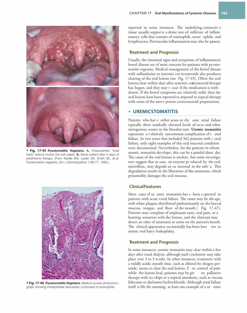
Patients with py ostomatitis vegetans exhibit characteristic yellowish, slightly elevated, linear, serpentine pustules set on an erythematous oral mucosa. Th e lesions primarily aff ect the buccal and labial mucosa, soft palate, and ventral tongue ( Figs. 17-44 and 17-45 ). Th ese lesions hav e been called “snail track” ulcerations, although in most instances the lesions are probably not truly ulcerated. Oral discomfort is variable but can be surprisingly minimal in some patients. Th is variation in symptoms may be r elated to the number of pustules that have ruptured to form ulcerations. Th e oral lesions may appear concurrently with the bowel symptoms, or they may precede the intestinal involvement.
Histopathologic Features
A biopsy specimen of an oral lesion of py ostomatitis vege-tans usually shows marked edema, causing an acantholytic appearance of the inv olved epithelium. Th is may be the result of the accumulation of numer ous eosinophils within the spinous lay er, often forming intraepithelial abscesses ( Fig. 17-46 ). Subepithelial eosinophilic abscesses have been
may not necessarily r ule out a diagnosis of C rohn disease. As with the clinical lesions, the histopathologic pattern is relatively nonspecifi c, resembling orofacial granulomatosis. Special stains should be performed to rule out the possibil-ity of deep fungal infection, syphilis, or my cobacterial infection.
Treatment and Prognosis
Most patients with mild C rohn disease are initially treated medically with mesalamine (5-aminosalicylic acid) or sul-fasalazine, a dr ug that is enzymatically br oken down by bacteria in the colon to form sulfapyridine and 5-aminosalicylic acid. Some patients respond well to this medication, typically when it is combined with an antibiotic such as metr onida-zole. With moderate to severe involvement, systemic pred-nisone may be used and is often eff ective, particularly when combined with an immunosuppressive drug, such as azathio-prine, methotrexate, or 6-mercaptopurine. For more severe or refractory cases of Crohn disease, one of the tumor necro-sis factor- α inhibitors (such as, infl iximab, adalimumab, or certolizumab pegol) may be used. S ometimes the disease cannot be maintained in remission by medical therapy, and complications develop that r equire surgical inter vention. Complications may include bowel obstruction or fi stula or abscess formation. If a signifi cant segment of the terminal ileum has been r emoved surgically or is inv olved with the disease, then periodic injections of vitamin B 12 may be neces-sary to prevent megaloblastic anemia secondary to the lack of ability to absorb the vitamin. Similar supplementation of magnesium, iron, the fat-soluble vitamins, and folate may also be required because of malabsorption. Cigarette smoking is known to exacerbate Crohn disease, and patients should be advised to stop this habit.
Oral lesions have been reported to clear with tr eatment of the gastrointestinal process in many cases. O ccasionally persistent oral ulcerations will dev elop, and these may have to be tr eated with topical or intralesional cor ticoste-roids. Systemic thalidomide and infl iximab have been
• Fig. 17-44 Pyostomatitis Vegetans. The characteristic lesions
are seen on the buccal mucosa, appearing as yellow-white pustules.
• Fig. 17-43 Crohn Disease. This medium-power photomicrograph
of an oral lesion shows a nonnecrotizing granuloma in the submucosal
connective tissue.
CHAPTER 17 Oral Manifestations of Systemic Diseases 793
reported in some instances. Th e underlying connectiv e tissue usually suppor ts a dense mix ed infi ltrate of infl am-matory cells that consists of eosinophils, neutr ophils, and lymphocytes. Perivascular infl ammation may also be present.
Treatment and Prognosis
Usually, the intestinal signs and symptoms of infl ammatory bowel disease are of most concern for patients with pyosto-matitis vegetans. Medical management of the bowel disease with sulfasalazine or systemic cor ticosteroids also produces clearing of the oral lesions (see Fig. 17-45 ). Often the oral lesions clear within days after systemic corticosteroid therapy has begun, and they may r ecur if the medication is with-drawn. If the bowel symptoms are relatively mild, then the oral lesions have been reported to respond to topical therapy with some of the mor e potent corticosteroid preparations.
◆ UREMIC STOMATITIS
Patients who hav e either acute or chr onic renal failure typically show markedly elevated levels of ur ea and other nitrogenous wastes in the bloodstr eam. Uremic stomatitis represents a r elatively uncommon complication of r enal failure. In two series that included 562 patients with r enal failure, only eight examples of this oral mucosal condition were documented. Nevertheless, for the patients in whom uremic stomatitis develops, this can be a painful disor der. Th e cause of the oral lesions is unclear , but some investiga-tors suggest that ur ease, an enzyme pr oduced by the oral microfl ora, may degrade ur ea secreted in the saliv a. Th is degradation results in the liberation of free ammonia, which presumably damages the oral mucosa.
Clinical Features
Most cases of ur emic stomatitis hav e been r eported in patients with acute renal failure. Th e onset may be abr upt, with white plaques distributed predominantly on the buccal mucosa, tongue, and fl oor of the mouth ( Fig. 17-47 ). Patients may complain of unpleasant taste, oral pain, or a burning sensation with the lesions, and the clinician may detect an odor of ammonia or urine on the patient ’ s breath. Th e clinical appearance occasionally has been kno wn to mimic oral hairy leukoplakia.
Treatment and Prognosis
In some instances, uremic stomatitis may clear within a few days after renal dialysis, although such resolution may take place over 2 to 3 weeks. In other instances, treatment with a mildly acidic mouth rinse, such as diluted hy drogen per-oxide, seems to clear the oral lesions. F or control of pain while the lesions heal, patients may be giv en palliative therapy with ice chips or a topical anesthetic, such as viscous lidocaine or dyclonine hydrochloride. Although renal failure itself is life thr eatening, at least one example of a ur emic
• Fig. 17-45 Pyostomatitis Vegetans. A, Characteristic “snail
track” lesions involve the soft palate. B, Same patient after 5 days of
prednisone therapy. (From Neville BW, Laden SA, Smith SE, et al:
Pyostomatitis vegetans, Am J Dermatopathol 7:69-77, 1985.)
A
B
• Fig. 17-46 Pyostomatitis Vegetans. Medium-power photomicro-
graph showing intraepithelial abscesses composed of eosinophils.