the orbit and visual pathways - wong -...
TRANSCRIPT
The Orbit and Visual Pathways
Wade Wong DO FACR FAOCR
Emeritus Professor of Radiology
University of California, San Diego
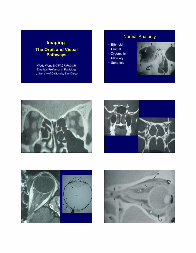
ImagingNormal Anatomy
• Ethmoid
• Frontal
• Zygomatic
• Maxillary
• Sphenoid
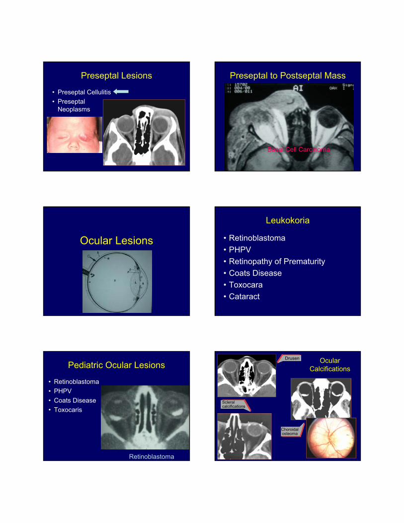
Preseptal Lesions
• Preseptal Cellulitis
• PreseptalNeoplasms
Preseptal to Postseptal Mass
Basal Cell Carcinoma
Ocular Lesions
Leukokoria
• Retinoblastoma
• PHPV
• Retinopathy of Prematurity
• Coats Disease
• Toxocara
• Cataract
Pediatric Ocular Lesions
• Retinoblastoma
• PHPV
• Coats Disease
• Toxocaris
Retinoblastoma
Ocular Calcifications
Choroidal osteoma
Drusen
Scleral calcifications
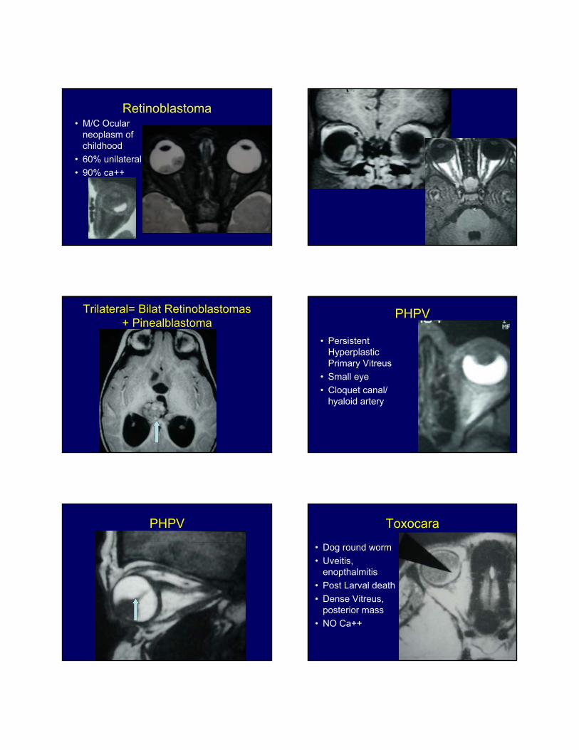
Retinoblastoma• M/C Ocular
neoplasm ofchildhood
• 60% unilateral
• 90% ca++
Trilateral= Bilat Retinoblastomas+ Pinealblastoma
PHPV
• PersistentHyperplasticPrimary Vitreus
• Small eye
• Cloquet canal/hyaloid artery
PHPV Toxocara
• Dog round worm
• Uveitis,enopthalmitis
• Post Larval death
• Dense Vitreus,posterior mass
• NO Ca++
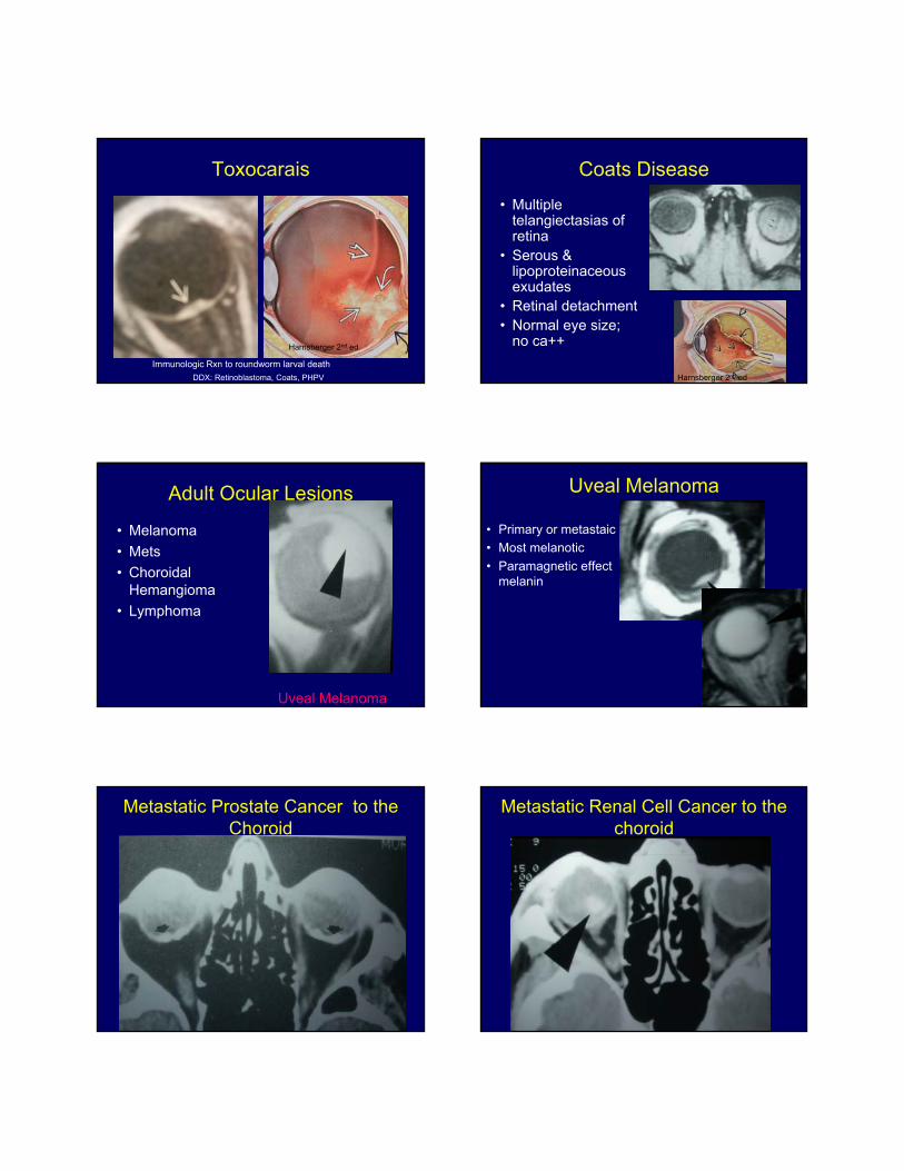
Toxocarais
Harnsberger 2nd ed
Immunologic Rxn to roundworm larval death
DDX: Retinoblastoma, Coats, PHPV
Coats Disease
• Multipletelangiectasias ofretina
• Serous &lipoproteinaceousexudates
• Retinal detachment• Normal eye size;
no ca++
Harnsberger 2nd ed
Adult Ocular Lesions
• Melanoma
• Mets
• ChoroidalHemangioma
• Lymphoma
Uveal Melanoma
Uveal Melanoma
• Primary or metastaic
• Most melanotic
• Paramagnetic effect ofmelanin
Metastatic Prostate Cancer to the Choroid
Metastatic Renal Cell Cancer to the choroid
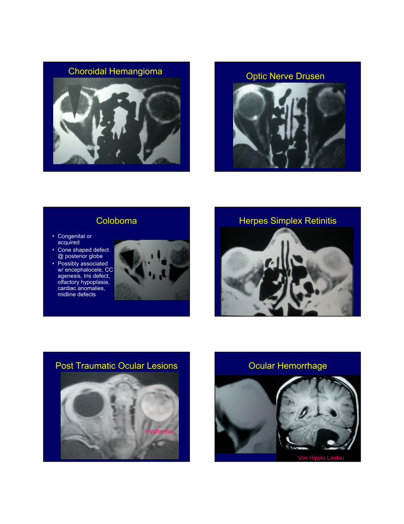
Choroidal Hemangioma Optic Nerve Drusen
Coloboma
• Congenital oracquired
• Cone shaped defect@ posterior globe
• Possibly associatedw/ encephalocele, CCagenesis, Iris defect,olfactory hypoplasia,cardiac anomalies,midline defects
Herpes Simplex Retinitis
Post Traumatic Ocular Lesions
Hyphema
Ocular Hemorrhage
Von Hipple Lindau
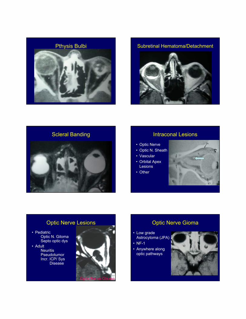
Pthysis Bulbi Subretinal Hematoma/Detachment
Scleral Banding Intraconal Lesions
• Optic Nerve
• Optic N. Sheath
• Vascular
• Orbital ApexLesions
• Other
Optic Nerve Lesions
• PediatricOptic N. GilomaSepto optic dys
• AdultNeuritisPseudotumorIncr. ICP/ Sys
Disease
Optic Nerve Glioma
Optic Nerve Gioma
• Low gradeAstrocytoma (JPA)
• NF-1
• Anywhere alongoptic pathways
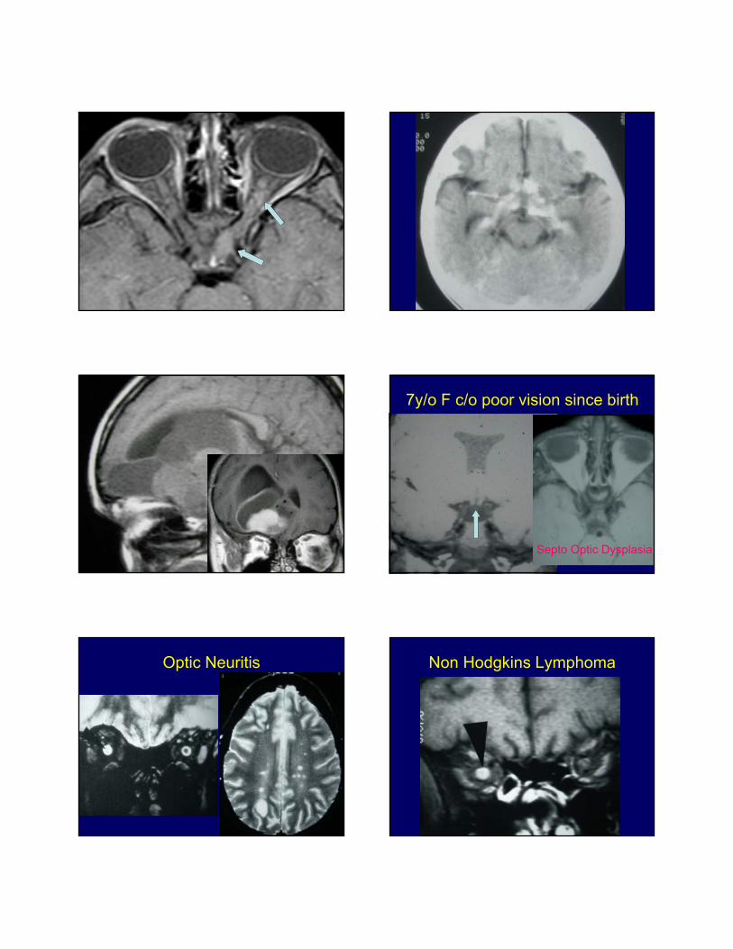
7y/o F c/o poor vision since birth
Septo Optic Dysplasia
Optic Neuritis Non Hodgkins Lymphoma
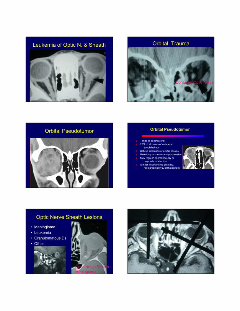
Leukemia of Optic N. & Sheath Orbital Trauma
Contused Optic Nerve
Orbital Pseudotumor Orbital Pseudotumor
) Tends to be unilateral) 25% of all cases of unilateral
exophthalmos) Diffuse infiltration of orbital tissues) Remitting or chronic and progressive) May regress spontaneously or
responds to steroids) Similar to lymphoma clinically,
radiographically & pathologically
Optic Nerve Sheath Lesions
• Meningioma
• Leukemia
• Granulomatous Ds.
• Other
Optic Nerve Sheath Meningioma
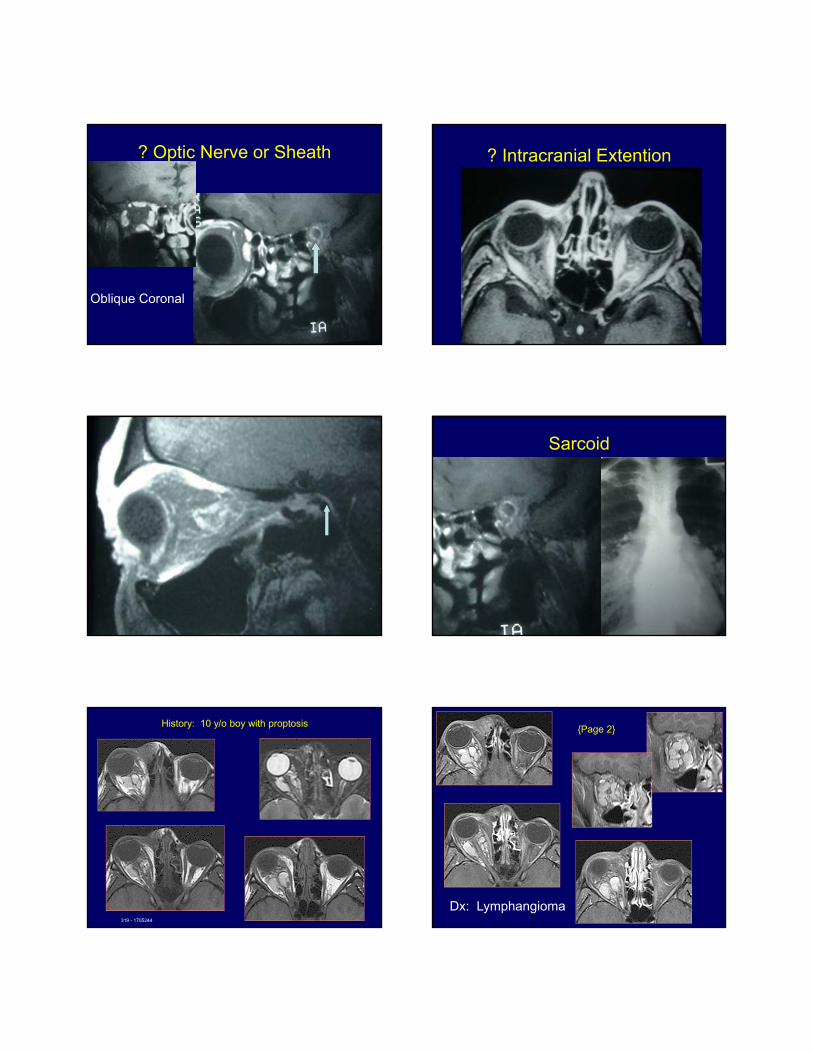
? Optic Nerve or Sheath
Oblique Coronal
? Intracranial Extention
Sarcoid
History: 10 y/o boy with proptosis
319 - 1705244
{Page 2}
Dx: Lymphangioma
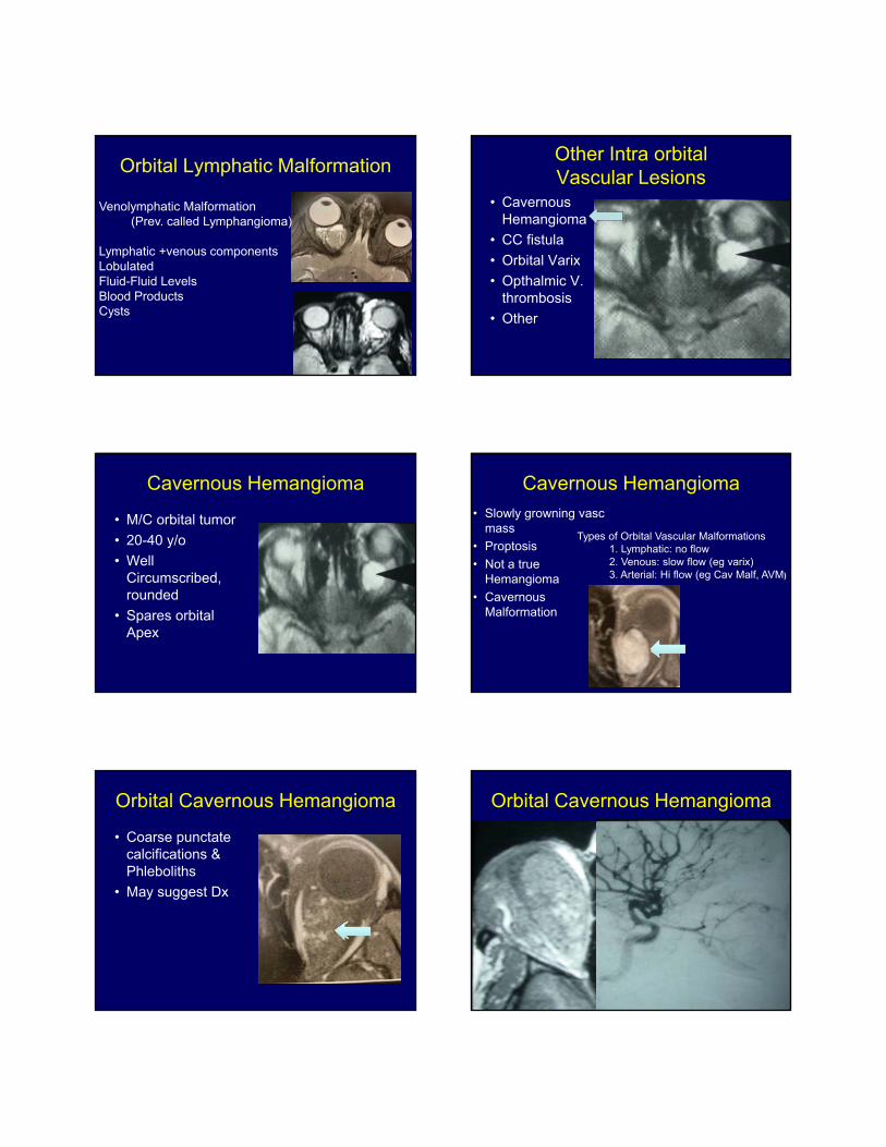
Orbital Lymphatic Malformation
Venolymphatic Malformation(Prev. called Lymphangioma)
Lymphatic +venous componentsLobulatedFluid-Fluid LevelsBlood ProductsCysts
Other Intra orbital Vascular Lesions
• CavernousHemangioma
• CC fistula
• Orbital Varix
• Opthalmic V.thrombosis
• Other
Cavernous Hemangioma
• M/C orbital tumor
• 20-40 y/o
• WellCircumscribed,rounded
• Spares orbitalApex
Cavernous Hemangioma
• Slowly growning vascmass
• Proptosis
• Not a trueHemangioma
• CavernousMalformation
Types of Orbital Vascular Malformations1. Lymphatic: no flow2. Venous: slow flow (eg varix)3. Arterial: Hi flow (eg Cav Malf, AVM)
Orbital Cavernous Hemangioma
• Coarse punctatecalcifications &Phleboliths
• May suggest Dx
Orbital Cavernous Hemangioma
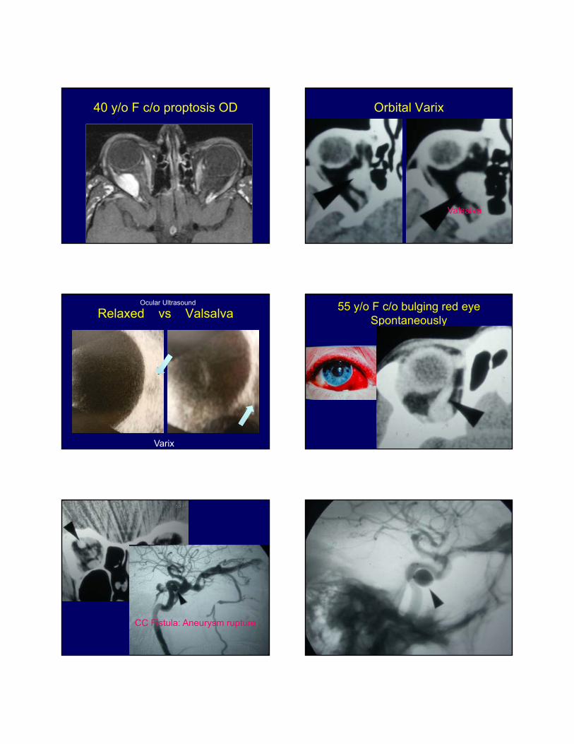
40 y/o F c/o proptosis OD Orbital Varix
Valsalva
Relaxed vs ValsalvaOcular Ultrasound
Varix
55 y/o F c/o bulging red eye Spontaneously
CC Fistula: Aneurysm rupture
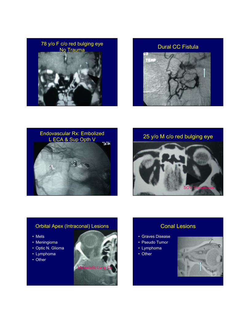
78 y/o F c/o red bulging eyeNo Trauma
Dural CC Fistula
Endovascular Rx: Embolized L ECA & Sup Opth V
25 y/o M c/o red bulging eye
SOV Thrombosis
Orbital Apex (Intraconal) Lesions
• Mets
• Meningioma
• Optic N. Glioma
• Lymphoma
• Other
Metastatic Lung Ca
Conal Lesions
• Graves Disease
• Pseudo Tumor
• Lymphoma
• Other
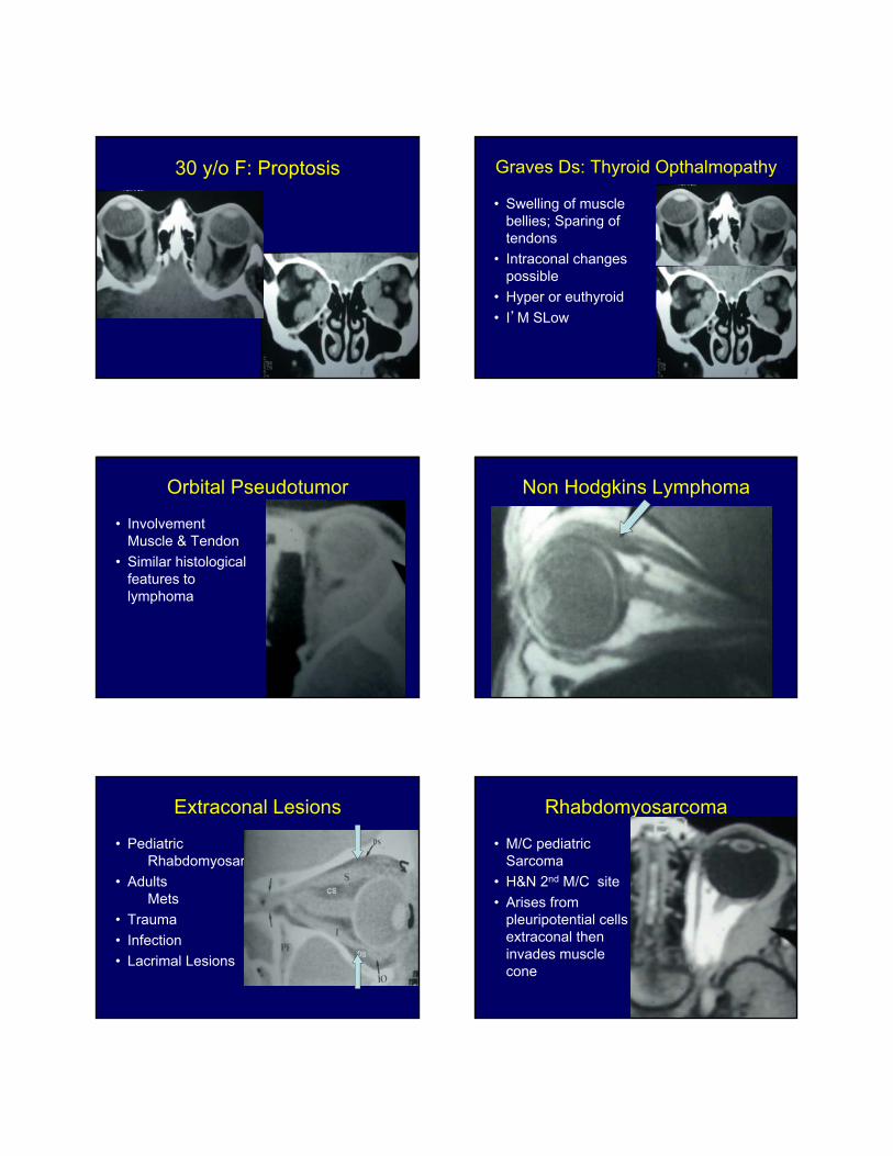
30 y/o F: Proptosis Graves Ds: Thyroid Opthalmopathy
• Swelling of musclebellies; Sparing oftendons
• Intraconal changespossible
• Hyper or euthyroid
• I’M SLow
Orbital Pseudotumor
• InvolvementMuscle & Tendon
• Similar histologicalfeatures tolymphoma
Non Hodgkins Lymphoma
Extraconal Lesions
• PediatricRhabdomyosar
• AdultsMets
• Trauma
• Infection
• Lacrimal Lesions
Rhabdomyosarcoma
• M/C pediatricSarcoma
• H&N 2nd M/C site
• Arises frompleuripotential cellsextraconal theninvades musclecone
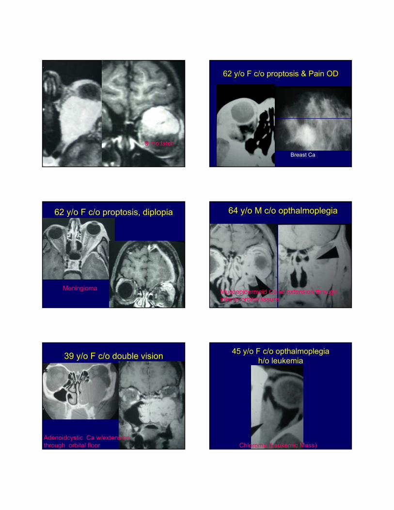
9 mo later
62 y/o F c/o proptosis & Pain OD
Breast Ca
62 y/o F c/o proptosis, diplopia
Meningioma
64 y/o M c/o opthalmoplegia
Mucoepidermoid Ca w/ extension through inferior orbital fissure
39 y/o F c/o double vision
Adenoidcystic Ca w/extension through orbital floor
45 y/o F c/o opthalmoplegiah/o leukemia
Chloroma (Leukemic Mass)
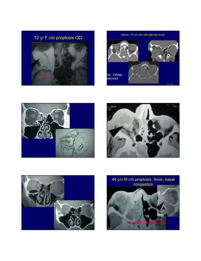
12 y/ F c/o proptosis OD
Dermoid Dx: Orbital dermoid
History: 57 y/o man with right eye droop
527 - r0819
44 y/o M c/o proptosis, fever, nasal congestion
Sinusits, Orbital Abscess
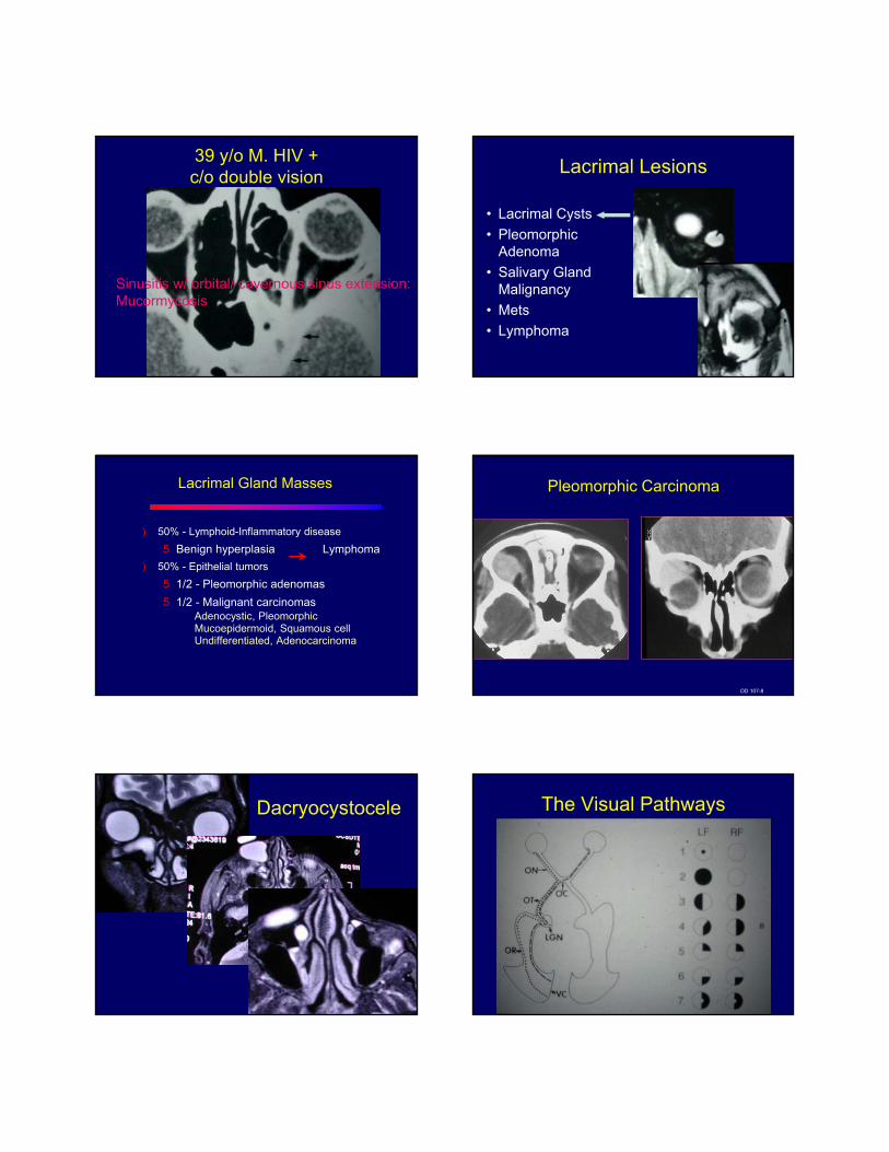
39 y/o M. HIV + c/o double vision
Sinusitis w/ orbital/ cavernous sinus extension: Mucormycosis
Lacrimal Lesions
• Lacrimal Cysts
• PleomorphicAdenoma
• Salivary GlandMalignancy
• Mets
• Lymphoma
Lacrimal Gland Masses
) 50% - Lymphoid-Inflammatory disease
5 Benign hyperplasia Lymphoma
) 50% - Epithelial tumors
5 1/2 - Pleomorphic adenomas
5 1/2 - Malignant carcinomasAdenocystic, PleomorphicMucoepidermoid, Squamous cellUndifferentiated, Adenocarcinoma
Pleomorphic Carcinoma
OD 107-8
Dacryocystocele
• Dacryocystocele
The Visual Pathways
Chiasmatic Lesions
• Sellar/SuprasellarMasses
• Vascular
Chiasmatic: Bitemporal Hemianopsia
Pituatary Adenoma
77 y/o F: progressive visual loss
Meningioma
18 y/o M: Bitemporal Hemianopsia
Germinoma
47 y/o M: Bitemporal Hemianopsia
ACOM Aneurysm
Optic Tract Lesions
• Optic NerveGlioma
• Other adjacentNeoplasms
• Other
Optic Nerve Glioma along Optic Tracts
Astrocytoma: Mass effect on optic tracts
Optic Radiation Lesions
• Neoplasms
• Vascular
• Inflammatory
47 y/o F c/o defects in vision
AVM
Tumefactive M.S.
Sudden Onset quadrantic visual loss
PCA Infarct
18 y/o M c/o visual loss
Sturge Weber
Affectations of eye movement: Cranial Nerve 3, 4, 6 deficits
• Brainstem Mass
• Vascular
• Inflammatory
• Other
Brainstem Glioma
57 y/o F c/o double vision
Cavernous Sinus Met
E.B.
• 44 y/o M
• Progressive doublevision
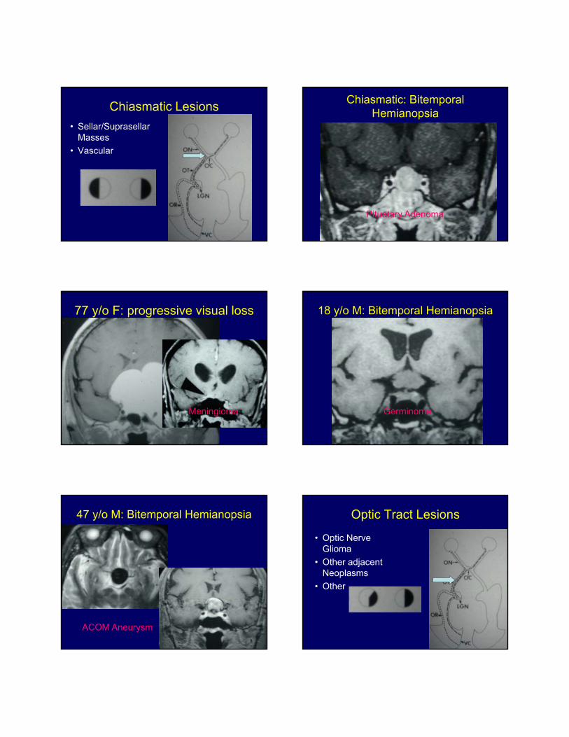
Rapidly Worsening 3rd N plasy
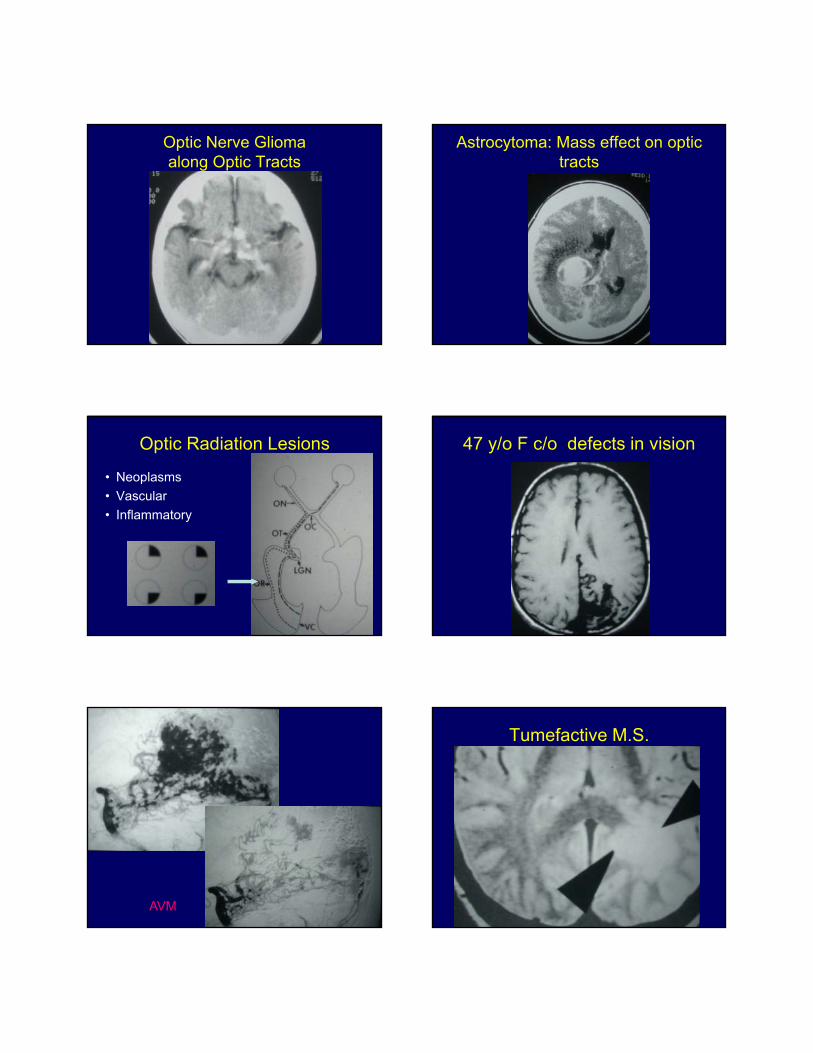
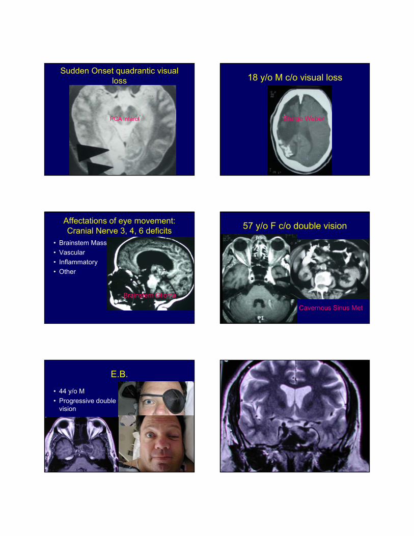
Passed BTO & HMPAONext Day
New involvement: L 6 th N• NOW What?
• ?Surgery
• Coiling?
• Stent
• Coils/Stent
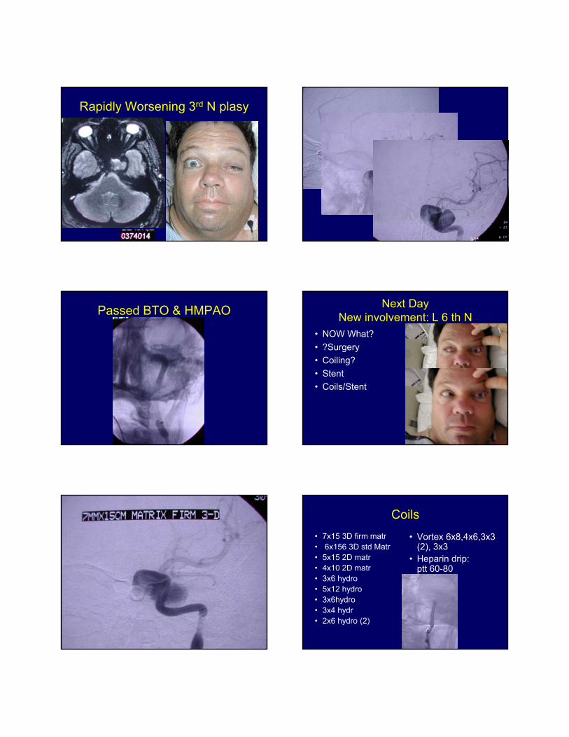
Coils
• 7x15 3D firm matr• 6x156 3D std Matr• 5x15 2D matr• 4x10 2D matr• 3x6 hydro• 5x12 hydro• 3x6hydro• 3x4 hydr• 2x6 hydro (2)
• Vortex 6x8,4x6,3x3(2), 3x3
• Heparin drip:ptt 60-80
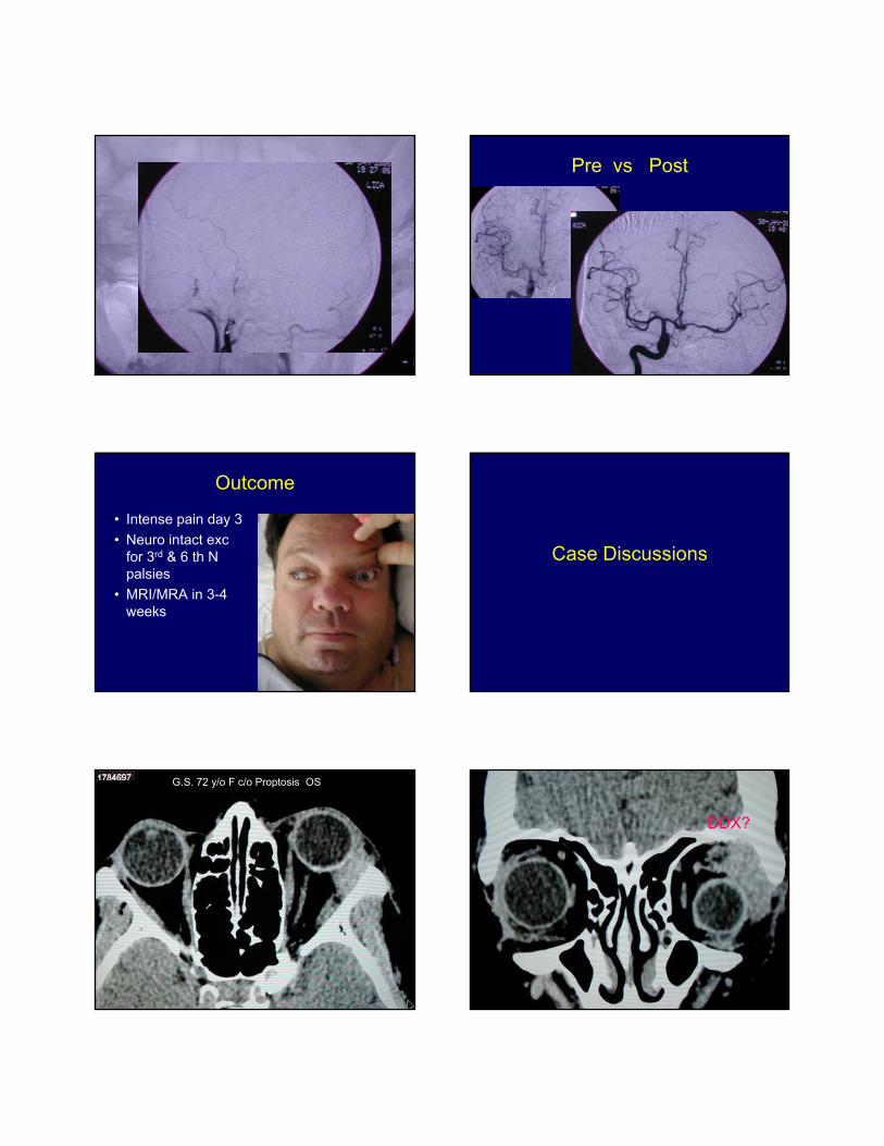
Pre vs Post
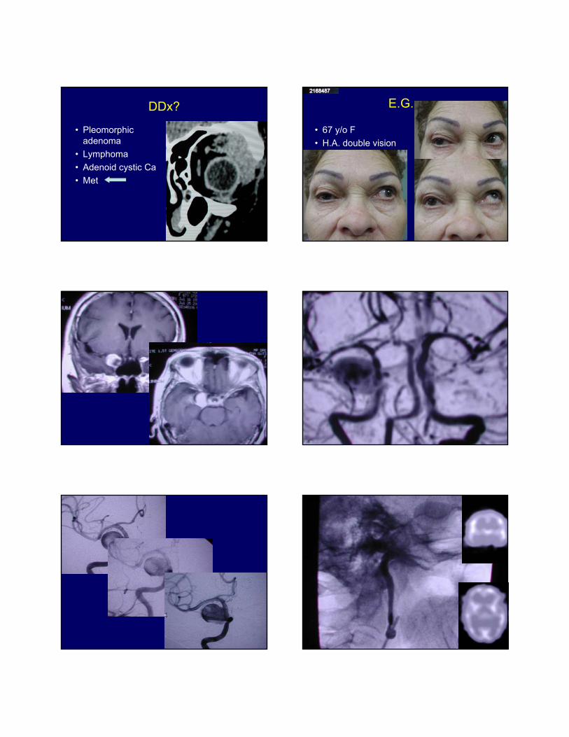
Outcome
• Intense pain day 3
• Neuro intact excfor 3rd & 6 th Npalsies
• MRI/MRA in 3-4weeks
Case Discussions
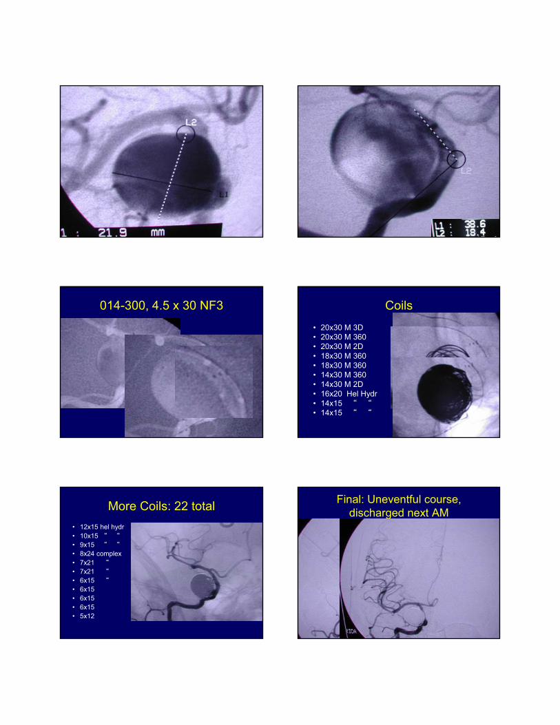
G.S. 72 y/o F c/o Proptosis OS
DDX?
DDx?
• Pleomorphicadenoma
• Lymphoma
• Adenoid cystic Ca
• Met
E.G.
• 67 y/o F
• H.A. double vision
014-300, 4.5 x 30 NF3 Coils
• 20x30 M 3D• 20x30 M 360• 20x30 M 2D• 18x30 M 360• 18x30 M 360• 14x30 M 360• 14x30 M 2D• 16x20 Hel Hydr• 14x15 “ “• 14x15 “ “
More Coils: 22 total
• 12x15 hel hydr• 10x15 “ “
• 9x15 “ “
• 8x24 complex• 7x21 “• 7x21 “• 6x15 “• 6x15• 6x15• 6x15• 5x12
Final: Uneventful course, discharged next AM
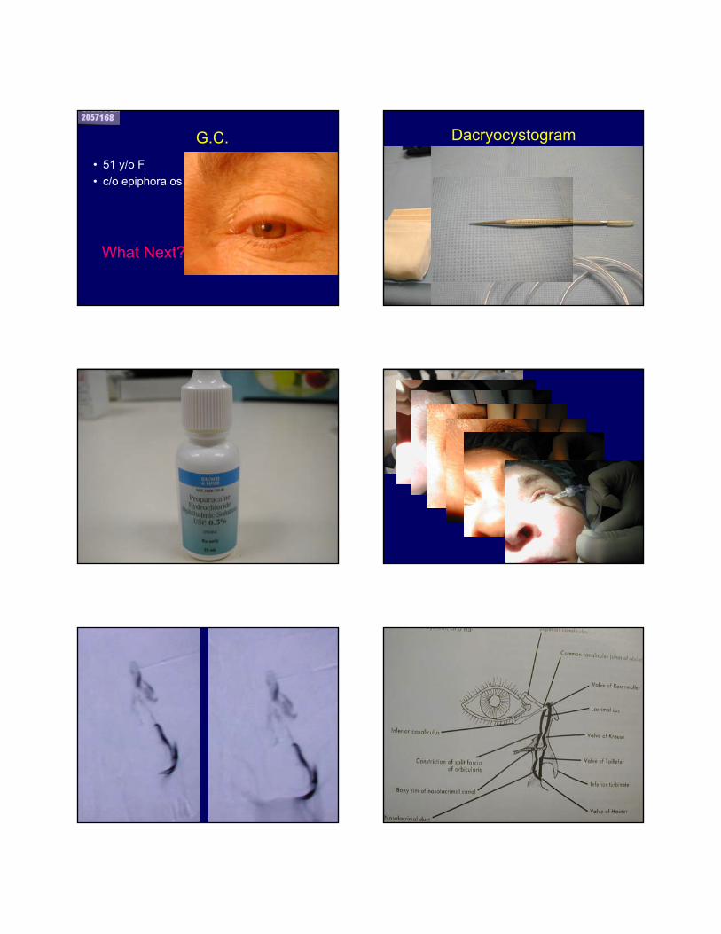
G.C.
• 51 y/o F
• c/o epiphora os
What Next?
Dacryocystogram
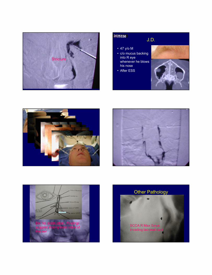
Stricture
J.D.
• 47 y/o M
• c/o mucus backinginto R eyewhenever he blowshis nose
• After ESS
Dx: No obstruction.. Findings suggest incompetent valve of Hauser
Other Pathology
SCCA R Max Sinus invading lacrimal duct