the origin of the gran canarian xenoliths - diva...
TRANSCRIPT

Independent Project at the Department of Earth Sciences Självständigt arbete vid Institutionen för geovetenskaper
2016: 38
The Origin of the Gran Canarian Xenoliths
Ursprunget till Gran Canarias xenoliter
Beatrice Jägerup
DEPARTMENT OF EARTH SCIENCES
I N S T I T U T I O N E N F Ö R
G E O V E T E N S K A P E R


Independent Project at the Department of Earth Sciences Självständigt arbete vid Institutionen för geovetenskaper
2016: 38
The Origin of the Gran Canarian Xenoliths
Ursprunget till Gran Canarias xenoliter
Beatrice Jägerup

Copyright © Beatrice Jägerup Published at Department of Earth Sciences, Uppsala University (www.geo.uu.se), Uppsala, 2016

Sammanfattning Ursprunget till Gran Canarias xenoliter Beatrice Jägerup Xenoliter är bitar av sten från omkringliggande berg som förts till jordytan av magma. På Gran Canaria, Kanarieöarnas största ö, har man hittat märkliga xenoliter. De är ljusa, porösa och väldigt annorlunda från den basaltiska magman som burit med sig dem. Genom att undersöka xenoliternas petrologiska egenskaper kommer den här rapporten fokusera på att bestämma deras ursprung. 14 provers mineralogi och textur studerades i tunnslip, och ett δ18O-värde bestämdes för 17 prover. Den mineralogiska sammansättningen av det vulkaniska glaset i en av xenoliterna testades i EPMa. Resultaten visar att xenoliterna till störst del består av kiselrikt glas, kvarts och fältspatsmineral, men även höga värden av kalcium. 9 av 14 xenoliter har en textur och höga δ18O-värden (från 8,1 ‰ to 16,77 ‰) som stämmer överens med sedimentära stenar. De kvarstående xenoliterna uppvisar tydligt påverkade texturer och altererade phenocryster, vilket indikerar att de har smälts och omkristalliserats. Den sist nämnda gruppen har även extremt låga δ18O–värden, vilket skulle kunna förklaras av effekter från hydrotermiska processer. Mest sannolikt är att samtliga xenoliter härstammar från de äldre sedimentära avlagringarna under Gran Canaria. Nyckelord: Gran Canaria, xenoliter, syreisotoper, tunnslipsanalys, EPM Självständigt arbete i geovetenskap, 1GV029, 15 hp, 2016. Handledare: Abigail Barker, Valentin R. Troll och Kirsten Zaczek. Institutionen för geovetenskaper, Uppsala universitet, Villavägen 16, 752 36 Uppsala (www.geo.uu.se) Hela publikationen finns tillgänglig på www.diva-portal.org

Abstract The Origin of the Gran Canarian Xenoliths Beatrice Jägerup Xenoliths are pieces from the surrounding bedrock, brought to the surface of the earth by host magma. On Gran Canaria, the largest island in the Canary Islands archipelago, strange xenoliths have been found. They are light in color, porous and very different from the basaltic magma carrying them. By studying petrological features and oxygen isotope content of the xenoliths, the focus of this report will be to investigate their origin. The minerals and texture of 14 samples were studied in thin section, and the δ18O–value was determined for 17 samples. The mineralogical composition of xenolith glass was examined by EPMa. The results show that the xenoliths are rich in silica rich glass, quartz and feldspars, but also have high calcium content. 9 of 14 xenoliths have textures and δ18O–values from 8.1 ‰ to 16.77 ‰, similar to sedimentary rocks. The remaining xenoliths are metamorphosed and exhibit altered phenocrysts, indicating they have been melted and recrystallized. The latter group also has extremely low δ18O–value, which could be explained by the effects of hydrothermal processes. Most likely all the xenoliths originate from the pre-volcanic sedimentary deposits beneath Gran Canaria. Key words: Gran Canaria, Xenoliths, Oxygen Isotope Value, Thin Section Analysis, EPM Independent Project in Earth Science, IGV029, 15 credits, 2016. Supervisors: Abigail Barker, Valentin R. Troll and Kirsten Zaczek. Department of Earth Sciences, Uppsala University, Villavägen 16, SE-752 36 Uppsala (www.geo.uu.se) The whole document is available at www.diva-portal.org

Table of Contents 1. Introduction…………………………………………….............. 1 2. Geological settings……………………………………………... 1
2.1 The Canary Islands……………………………….... 1 2.2 Gran Canaria...……………………………………... 2
3. Methodology…………………………………………………….. 4 3.1 Microscopy analysis………………………………... 4 3.2 Electronprobe Micro analysis (EPMa)……………... 4 3.3 Oxygen isotope analysis…...………………………. 4
4. Results…………………………………………………………... 5 4.1 Petrographic features………………………………. 5 4.2 Mineralogical composition………………………….. 10 4.3 Oxygen isotopes……………………………………. 12
5. Discussion……………………………………………………..… 12 5.1 Microclimate in xenolith pumice…………………. 12 5.2 The origin of the xenoliths……………………….. 13 6. Conclusion……………………………………………..………… 14 Acknowledgments………………………………………………….. 15 References………………………………………………………….. 16


1. Introduction A volcanic eruption is among natures most powerful and destructive events, yet many communities are situated close to or in volcanically active areas (The Geography Site, 2010). It is of great importance to understand the inner mechanics of volcanism, because of the immediate threat a volcano can pose for people in its surroundings.
One way to gather vital information about a volcano is to study erupted material. Xenoliths are pieces of foreign rock brought to the surface of the Earth by rising magma (Encyclopedia Britannica, 2015). They can be especially important since the processes of magma-crust interaction can affect the outcome of the eruption.
On the volcanic hills of Gran Canaria, the largest island of the Canary Islands, you can find strange, pumice-like xenoliths scattered on the ground (Figure 1). With just a quick look it is clear that they are different from the basaltic magma that carried them. During an eruption at El Hierro in 2011 similar xenoliths were found, as they bubbled up from the bottom of the ocean and rose to the sea surface where they stayed afloat (Troll et.al. 2012). “The Floating stones of El Hierro”, also called “restingolites”, consist of an outer crust of dense basalt with a core of white and porous pumice-like xenoliths (Troll et.al. 2012). The existence of the rocks was at first a mystery. Why was there pumice within basaltic magma? Was it juvenile rhyolite or formed by crustal assimilation? After further examination the samples were found to contain remnant quartz crystals, jasper fragments and wollastonite. This composition together with relatively high oxygen isotope values suggested that the xenoliths were of sedimentary origin (Troll et.al. 2012). When calcareous nanofossils were found within the xenoliths this theory was confirmed; at least 50 % of the pumice was indeed partly melted sediments from thick sedimentary deposits beneath El Hierro (Zaczek et.al 2015). The fossils and the sediment were later dated. Fascinatingly enough the sedimentary ages correspond with what is thought to be the time for initial shield building volcanism at El Hierro, promoting the mantle-plume theory (Zaczek et.al. 2015).
The interesting result from the investigations of restingolites once again brought attention to the xenoliths found on Gran Canaria. Is it possible that they also are of sedimentary origin? The purpose of this project is to examine the Gran Canarian xenoliths for petrological features, including mineralogical assemblages, texture and chemical characteristics, with the aim of presenting new insights on the origin of the xenoliths and to enhance the understandings of the volcanism on Gran Canaria. 2. Geological settings 2.1 The Canary Islands The Canary Islands are a densely populated archipelago of seven larger and several smaller islands, some still volcanically active. The Canary Islands stretch from about 500 to 100 km off the northwestern coast of Africa and are situated on top of a slow moving oceanic plate (McDougall & Schmincke, 1976). The north western margin between the African continent and the oceanic plate is passive, meaning it manifests no well-defined tectonic processes, such as large faults, earthquakes or rift zone volcanoes (Schmincke 1982). The Canary Islands are a result of 'intraplate volcanism' and during several decades the volcanism on the islands has been investigated and discussed vigorously (Schmincke 1982). But despite being one of the most researched island chains on the planet the geological processes are yet not completely understood. A handful of plausible theories have been proposed to explain the origin
1

of the magmas, and so far the evidence seems to promote two models; the model based on a mantle plume or 'hot spot', where melt originates from an active source deep down in the mantle (Schminke 1973; Hoernle & Schmincke, 1993) or the model of fracture propagation, where fractures in the crust lower the pressure and allow melt to rise (Fúster, 1972). The reason for the debate is because the complicated geology of the Canary Islands does not fit completely with either of these two models. The classical example of mantle plume generated volcanism is the island chain of Hawaii. According to the Wilson-Morgan model (1963; 1971) the islands follow a linear pattern with a younging direction consistent with tectonic movements, which also is somewhat true for the Canary Islands. They are aligned, and the oldest island is Fuertaventura in the East, while the youngest volcanism is located at the western island El Hierro, which is still in an early shield building phase (Schmincke & Sumita, 1998). But evidence for a prominent E-W age-progression in the island chain are lacking, indicating the geological situation is more complex than the one in the Hawaiian Islands (Anguita & Hernan, 1975; McDougall & Schmincke, 1976). Adding to the dispute, the propagating fracture model is also questioned since it does not comply with geophysical measurements beneath the islands (Troll, 2012). 2.2 Gran Canaria Gran Canaria is a shield volcano that reaches 1900 m above sea level, with a total height of 2500 m from the bottom of the sea and a base of almost 45 km in diameter it is the largest seamount of the Canary Islands (Schmincke, 1982). The geological structure can be described as four layers, most of it beneath the sea surface. At the bottom is Jurassic tholeiitic oceanic crust, overlain by a thick layer of mesozoic to tertiary sediments. The seamount and shield basalts are situated on top of the sediments, covered with more recent volcanic sedimentary deposits, the so called 'volcanic apron' (Schmincke & Sumita, 1998).
Gran Canaria volcanic evolution is usually divided in to three volcanic phases, each succeeded by erosional hiatuses. The magmatic composition and other details of the first phase, the 'seamount phase', are not well known since its eruptives are covered with newer material (McDougall & Schmincke, 1976). The short lived, main shield building phase began during the Miocene, between 14.5 to 16 Ma. It was a fundamental phase of alkali basaltic volcanism that produced enough material for an island to emerge from the ocean. The major part of the Miocene phase consisted of extensive felsic volcanism, which lasted from 14 to 8.3 Ma (Cousens et.al. 1990). The volcanism stayed dormant from 8 Ma to 5 Ma, before the Pliocene phase was initiated. The third and less intensive phase started in quaternary times with regular eruptions, the youngest date back to just a few thousand years (Schmincke & Sumita, 1998). The majority of the xenoliths examined are retrieved from the grounds around such a quaternary volcano; Montañón Negro (Figure 1). The map of Gran Canaria in figure 2 shows the volcanic material exposed at surface today. The youngest rocks are found in the north-eastern part of the island, where the majority derives from quaternary times. The basaltic shield is exposed in the west, covered by felsic eruptives.
2

Source: Valentin Troll, 2015
Figure 1. Montañón Negro, a quaternary volcano and one of the sample sites on Gran Canaria. The xenoliths can be seen as small, light dots on the darker ground.
Source: Lisa Samrock, 2015.
Figure 2. Geological Map of Gran Canaria.
3

3. Methodology 3.1 Microscopy analysis Fourteen samples of xenoliths retrieved at various sites on Gran Canaria were selected for a thin section analysis, so that the texture, mineralogy and other important features could be examined. The samples were prepared and later examined in an optical microscope, model Nikon Eclipse e600 pol, with a magnification from 2,5X to 50X, in plane polarized and crossed polarized light. Minerals and features were identified according to standard procedures (Jones & Bloss, 1980; MacKenzie, 1980.).
If higher magnification or more detailed pictures were necessary for the investigation, selected samples were examined with Scanning Electron Microscope (SEM). SEM is a technique especially used for imaging and with a wide area of application within geological research. SEM can provide images with higher resolution than ordinary optical microscopes, since the best spatial resolution of SEM spans from 50 to 100 nm and magnification from 20X up to 30 000X (Reed, 2005).
The SEM-analysis was conducted at the Evolutionary Biology Centre (EBC), Uppsala University, using a Gemini Tardis, Zeiss Supra 35 VP. To make the sample conductive it is firstly covered with a gold-palladium coating.
3.2 Electron probe Micro Analysis (EPMa) The mineralogical composition of the glass was tested in one sample with an electron probe, using the field emission source JEOL-JXA-8530F Hyperprobe at CEMPEG (Centre for Experimental Mineralogy, Petrology and Geochemistry). The method is suitable for identifying the composition of small selected areas in the sample (Reed, 2005). 3.3 Oxygen Isotope Analysis Oxygen has three stable (non-radioactive) isotopes; O16, O17 and O18, where the two most abundant are O16 (99.76 %) and O18 (0.2%). The amount of O18 can be normalized by using equation 1, where the ratio of O18 and O16 is compared with the standard value Standard Mean Ocean Water (SMOW). The result is given as the difference in O18 between the sample and SMOW, consequently the value is notated as δ18O per-mil (‰).
Isotope fractionation is a term used to describe the distribution of oxygen isotopes in a substance. Oxygen is the most common element in the crust and the study of oxygen isotope fractionation can provide important information about conditions present during the formation of a rock (Carmichael et.al. 1974).
Equation 1: describes the delta notation for reporting oxygen isotope values. (Carmichael et.al. 1974)
Fractionation can occur kinetically or derive from the systems energy equilibrium. The latter has the largest effect on the ratio; this is because equilibrium fractionation mainly
4
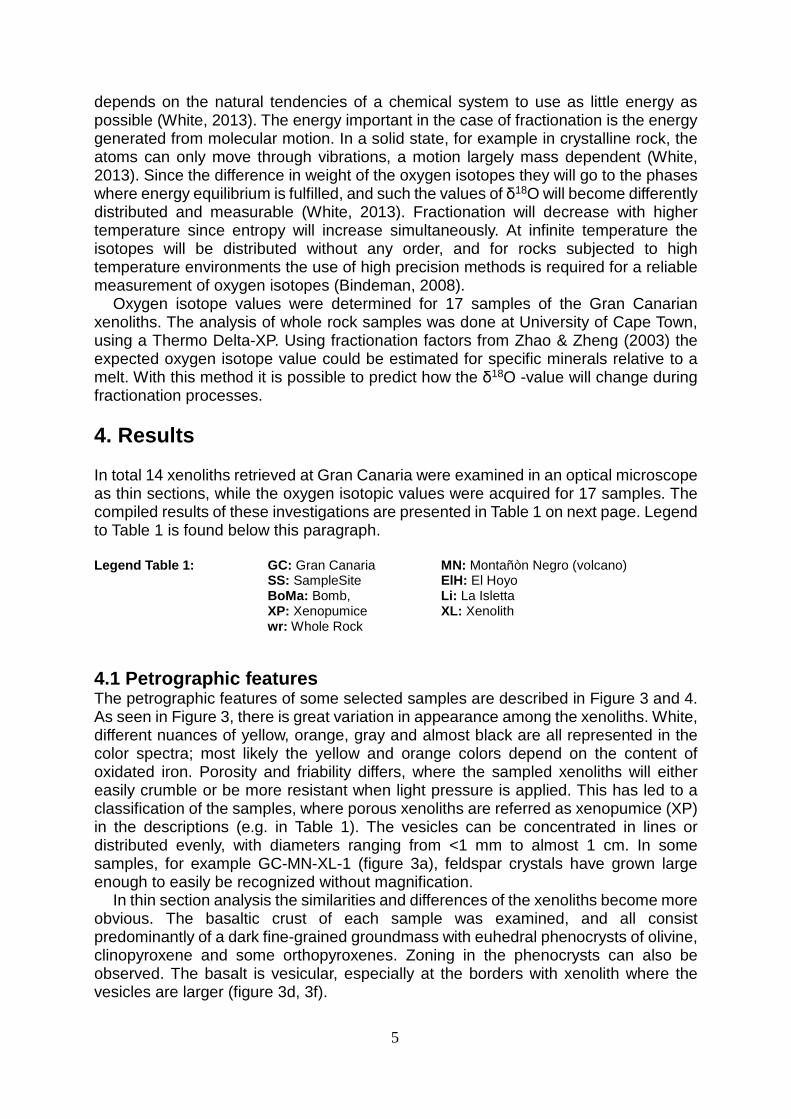
depends on the natural tendencies of a chemical system to use as little energy as possible (White, 2013). The energy important in the case of fractionation is the energy generated from molecular motion. In a solid state, for example in crystalline rock, the atoms can only move through vibrations, a motion largely mass dependent (White, 2013). Since the difference in weight of the oxygen isotopes they will go to the phases where energy equilibrium is fulfilled, and such the values of δ18O will become differently distributed and measurable (White, 2013). Fractionation will decrease with higher temperature since entropy will increase simultaneously. At infinite temperature the isotopes will be distributed without any order, and for rocks subjected to high temperature environments the use of high precision methods is required for a reliable measurement of oxygen isotopes (Bindeman, 2008).
Oxygen isotope values were determined for 17 samples of the Gran Canarian xenoliths. The analysis of whole rock samples was done at University of Cape Town, using a Thermo Delta-XP. Using fractionation factors from Zhao & Zheng (2003) the expected oxygen isotope value could be estimated for specific minerals relative to a melt. With this method it is possible to predict how the δ18O -value will change during fractionation processes. 4. Results In total 14 xenoliths retrieved at Gran Canaria were examined in an optical microscope as thin sections, while the oxygen isotopic values were acquired for 17 samples. The compiled results of these investigations are presented in Table 1 on next page. Legend to Table 1 is found below this paragraph. Legend Table 1: GC: Gran Canaria MN: Montañòn Negro (volcano) SS: SampleSite ElH: El Hoyo BoMa: Bomb, Li: La Isletta XP: Xenopumice XL: Xenolith wr: Whole Rock 4.1 Petrographic features The petrographic features of some selected samples are described in Figure 3 and 4. As seen in Figure 3, there is great variation in appearance among the xenoliths. White, different nuances of yellow, orange, gray and almost black are all represented in the color spectra; most likely the yellow and orange colors depend on the content of oxidated iron. Porosity and friability differs, where the sampled xenoliths will either easily crumble or be more resistant when light pressure is applied. This has led to a classification of the samples, where porous xenoliths are referred as xenopumice (XP) in the descriptions (e.g. in Table 1). The vesicles can be concentrated in lines or distributed evenly, with diameters ranging from <1 mm to almost 1 cm. In some samples, for example GC-MN-XL-1 (figure 3a), feldspar crystals have grown large enough to easily be recognized without magnification.
In thin section analysis the similarities and differences of the xenoliths become more obvious. The basaltic crust of each sample was examined, and all consist predominantly of a dark fine-grained groundmass with euhedral phenocrysts of olivine, clinopyroxene and some orthopyroxenes. Zoning in the phenocrysts can also be observed. The basalt is vesicular, especially at the borders with xenolith where the vesicles are larger (figure 3d, 3f).
5

Table 1 Results compiled. δ18O notated in ‰. Sample name Type Features in thinsection δ18O wr
GC-MN-XP-1 Xenopumice Vesicular. Mainly consists of feldspar grains with rough edges, rounded quartz grains and aggregations of clay minerals.
13.73
GC-MN-XL-1 Xenolith Feldspar phenocrysts, flow alignment (featherlike).
3.6
GC-MN-XP-2 Xenopumice Zoned feldspar phenocrysts, flow alignment (parallel), groups of quartz crystals.
4.09
GC-MN-XP-3 Xenopumice Vesicular. Mainly feldspar and plagioclase grains with rough edges, euhedral sphene crystal.
6.63
GC-MN-XP-6 Xenopumice Irregular distributed vesicles of larger size. Flow alignment (featherlike and parallel).
3.77
GC-MN-XP-8 Xenopumice Very vesicular, with both elongated and round vesicles. Feldspar phenocrysts in groundmass of small quartz grains.
4.91
GC-MN-XL-9 Xenolith Feldspar phenocrysts with carlsbad twinning, some fractured, flow alignment. Elongated vesicle trails.
4.92
GC-SS1-XP-1 Xenopumice Vesicular. Irregular grain shapes and sizes of feldspars and quartz. The smaller grains are rounded. Apatite needles. Some iron staining. Contains shells of diatoms.
15.07
GC-SS1-XP-4 Xenopumice Vesicular. Irregular grain shapes and sizes of feldspars and quartz. Apatite needles. Larger patches of iron staining. Frequently occurring shells of diatoms.
8.14
GC-SS2-XP-1 Xenopumice Elongated vesicle trails. Mainly consists of evenly sized feldspar grains with rough edges. Groundmass of quartz and clay minerals. Some iron staining.
16.77
GC-SS2-XP-1.2 Xenopumice Vesicular. Mainly consists of feldspar grains with rough edges and aggregates of small, evenly sized quartz/clay mineral grains. Brown glass containing opaque, euhedral cubic crystals and apatite needles.
No data
GC-ElH-XP-1 Xenopumice No data. 8.1
GC-ElH-XP-2 Xenopumice Fewer small vesicles. Mainly consists of similar small sized feldspar and quartz grains. Some iron precipitation, orange in color.
15.99
GC-ElH-XP-4 Xenopumice Mainly consists of larger feldspar grains with rough edges and aggregates of rounded quartz grains and clay minerals.
16.32
GC-BoMa-XP-1.1 Xenopumice No data 6.71
GC-BoMa-XP-1.2 Xenopumice No data 7.39
GC-BoMa-XP-1.3 Xenopumice No data 6.48
GC-LI-XP-1 Xenopumice Large vesicles of irregular shape. 14.48
6
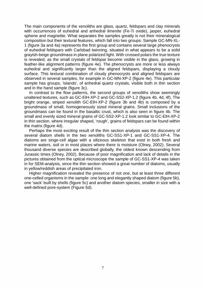
The main components of the xenoliths are glass, quartz, feldspars and clay minerals with occurrences of euhedral and anhedral ilmenite (Fe-Ti oxide), jasper, euhedral sphene and magnetite. What separates the samples greatly is not their mineralogical composition but their textural features, which fall into two groups. Sample GC-MN-XL-1 (figure 3a and 4a) represents the first group and contains several large phenocrysts of euhedral feldspars with Carlsbad twinning, situated in what appears to be a solid grayish-beige groundmass in plane polarized light. With crossed polars the true texture is revealed, as the small crystals of feldspar become visible in the glass, growing in feather-like alignment patterns (figure 4e). The phenocrysts are more or less always euhedral and significantly larger than the aligned feldspars, displaying a cloudy surface. This textural combination of cloudy phenocrysts and aligned feldspars are observed in several samples, for example in GC-MN-XP-2 (figure 4e). This particular sample has groups, 'islands', of anhedral quartz crystals, visible both in thin section and in the hand sample (figure 3c).
In contrast to the flow patterns, the second groups of xenoliths show seemingly unaltered textures, such as GC-ElH-XP-2 and GC-SS2-XP-1.2 (figure 4b, 4d, 4f). The bright orange, striped xenolith GC-ElH-XP-2 (figure 3b and 4b) is composed by a groundmass of small, homogeneously sized mineral grains. Small inclusions of the groundmass can be found in the basaltic crust, which is also seen in figure 4b. The small and evenly sized mineral grains of GC-SS2-XP-1.2 look similar to GC-ElH-XP-2 in thin section, where irregular shaped, ‘rough’, grains of feldspars can be found within the matrix (figure 4d).
Perhaps the most exciting result of the thin section analysis was the discovery of several diatom shells in the two xenoliths GC-SS1-XP-1 and GC-SS1-XP-4. The diatoms are singe-cell algae with a siliceous skeleton that exist in both fresh and marine waters, soil or in most places where there is moisture (Olney, 2002). Several thousand diverse species are described globally, the oldest known descending from Jurassic times (Olney, 2002). Because of poor magnification and lack of details in the pictures obtained from the optical microscope the sample of GC-SS1-XP-4 was taken in for SEM-analysis, since the thin section showed a great number of diatoms, usually in yellow/reddish areas of precipitated iron.
Higher magnification revealed the presence of not one, but at least three different one-celled organisms in the sample: one long and elegantly shaped diatom (figure 5b), one 'sack' built by shells (figure 5c) and another diatom species, smaller in size with a well-defined pore-system (Figure 5d).
7

a. b.
c. d.
e. f. Figure 3. Photographs of handsamples. a. Sample GC-MN-XL-1. Note the light colored feldspar phenocryst in the middle, homogenous sizes of vesicles and no distinct flow pattern. δ18O of 3.6 ‰. b. Sample GC-ElH-XP-2 has visible layering and a bright orange color. The vesicles follow a linear pattern, crossing the layers. δ18O of 15.99‰. c. Sample GC-MN-XP-2 has some large vesicles and visible blobs of quartz (gray areas in the picture). δ18O of 4.09 ‰. d. Sample GC-SS2-XP-12 with its with its basaltic crust. Observe the vascularity of the basalt at the contact of the xenolith. The vesicles of the xenolith are homogenously sized, no visible larger crystals. e. Sample GC-MN-XP-8 is considerably darker in color and shows some folding at the right edge. ). δ18O of 4.91 ‰. f. Sample GC-SS1-XP-4 has elongated vesicles that indicate a flow direction. Also observe the vescularity of the basalt at the contact with the xenolith. ). δ18O of 8.14 ‰.
8

a.
b.
c.
d.
e. f.
Figure 4. Photographs of thin sections. a. Phenocryst of feldspar showing a cloudy, altered surface with distinct carlsbad twinning in sample of GC-MN-XL-1, δ18O of 3.6 ‰. Plane polarized light. b. Inclusion of xenolith in the contact with the basalt in sample GC-ElH-XP-2. δ18O of 15.99 ‰. Plane polarized light. c. Aligned feldspar crystals in sample GC-MN-XP-2. An “Island” of recrystallized quartz is visible in the left corner. δ18O of 4.09 ‰. Cross polarized light. d. Detrital feldspars and fine grained groundmass of different minerals (mainly quartz, feldspars and clay minerals) in sample GC-SS2-XP-1.2. No oxygen isotope data. e. Feldspar phenocryst with cloudy and altered surfaces surrounded by aligned feldspars and opaque glass in xenolith GC-MN-XL-1. δ18O 3.6‰. f. Rounded and sorted detrital grains in sample GC-SS2-XP-1.2, magnification x50.
9

a. b.
c. d. Figure 5. Photographs from the SEM. a. Shells of several diatoms inside a vesicle. b. The well preserved shell of a diatom, possibly of the genus Pinnularia, a fresh-water algae found in soil. Length ~25 µm c. Two shells similar to Euglyphid Thecamoebian. d. The shell of a diatom, similar to the genus Luticola. Length ~7 µm. The surrounding material seems to have merged with the shell, but it is not clarified whether the material is made of glass or other organics. 4.2 Mineralogical composition An EPMa analysis was made on the glass in sample SS2-XP-1, illustrated in figure 6. The result shows the glass to large extent consists of silica (70.43 wt %), but also aluminum (III) oxide (10.69 wt %), calcium oxide (9.16 wt %) and iron oxide (3.9 wt %).
The xenoliths that have previously been suggested to have a sedimentary origin (Hansteen & Troll, 2003) all share a similar silica content of approximately 86 wt%, but the values of Al2O3, CaO and MgO are distributed in a wide range (Figure 6). The rhyolites all have a silica content of about 69 wt %, but in contrast to the sedimentary xenoliths they contain the same amount of Al2O3, MgO and CaO. With a value of 9.16 wt % the xenolith GC-SS2-XP-1 stands out as being rich in CaO compared to the other rocks, that have values from below 1 wt % (phonolitic dyke and rhyolites) to 3 wt % (sedimentary xenoliths).
10

Figure 6. Composition of selected major elements plotted against SiO2 in GC-SS2-XP-1 and compared with same major elements of reference rocks. Data sources: Phonolitic dyke (Donoghue et.al, 2010), Sedimentary xenoliths (Hansteen & Troll, 2003), Rhyolite (GeoRoc, 2015).
11

Figure 7. Isotope oxygen values for Gran Canaria xenoliths and reference rocks (‰, wr). Data sources: Altered basalts (Hansteen & Troll, 2003), Sedimentary xenoliths (Troll & Schmincke, 2002), Altered tuffs and unaltered tuffs (Donoghue et.al. 2008), Alkali basalt (Troll & Schmincke, 2002) 4.3 Oxygen Isotopes The data from the oxygen isotope measurements are given in Table 1 and Figure 7, where they are plotted with reference values of altered and unaltered tuffs (solidified ash), altered and unaltered basalt, sedimentary xenoliths and the mean mantle value of δ18O=5.7 ‰ (Bindeman, 2008).
The xenolith samples can be allocated into two groups according to their oxygen isotope values. One group has high values, ranging from δ18O 13.73 to 16.77 ‰, and the other group plots at values from δ18O 3.6 to 8.14 ‰. The high-value group has a range of δ18O similar to the confirmed sedimentary xenoliths and altered tuffs, while the lower group could be comparable with the unaltered tuffs. The low-value group could possibly be divided once more to a third group of intermediate values above mean mantle values, from δ18O 6.48 to 8.14 ‰. 5. Discussion 5.1 Microclimate in xenolith pumice At first the diatoms were assumed to be fossilized and part of pre-volcanism sediment, an assumption made because the shells appear to be intergrown with the silica glass, as if the surrounding material covers or is attached with the shells (visible in figure 5a and 5d). If part of the sedimentary xenolith, the diatoms could have helped age determine the sediment it originated from. The sediments that would have been interesting to date are those directly beneath the seamount of Gran Canaria, since that also may give an age for the initial volcanism.
But, when comparing the shells with known species of diatoms it became clear that they share features with some modern day fresh-water diatoms, and could be classified as the genera Pinnularia (figure 5b) and Luticola (figure 5d). The third organism (figure
12

5c) is likely to be a euglyphid thecamoeba, a freshwater creature also found in 15 Ma old Miocene limnic sediment from volcanic crater lakes in southern Germany (Foissner & Schiller, 2001).
The genera of Luticola and Pinnularia are adapted to fresh water environments, which contradicts samplings from a depositional environment of the marine pre-volcanic sedimentary basin. The small sizes of the diatoms (10-25 µm) indicate that they are suitable for living in soil (Lund, 1946). The shells of the diatoms are in general concentrated to reddish areas of iron precipitation, corresponding with previous research about iron functioning as a fertilizer for diatoms (Boyd et.al, 2000). The fact that fossilized specimens of euglyphid thecamoeba have been found in limnic crater lakes, suggests that these are also not a part of marine sedimentary relicts (Foissner & Schiller, 2001).
In summary, the shells of the three organisms are most likely to be a remains of a microscopic ecosystem, where the porous texture of the two xenoliths from sample site 1 in combination with high air humidity at the site seem to have provided a favorable microclimate for soil living, fresh-water microorganisms. Figure 1 shows one of the sites where xenolith samples have been retrieved, illustrating how the xenoliths are lying on the ground as a part of the upper soil layer. The 'intergrowth' with silica glass and the diatom shells (figure 5d) is the only evidence for the diatoms to be marine, associated with the pre-volcanic sedimentary rocks. Although this intergrowth could be a cover of organic material, deriving from biological processes and which later glued the shells to the glass. 5.2 Origin of the xenoliths The origin of the high δ18O xenoliths from Gran Canaria seems to be sedimentary and the most prominent evidence for them not being of magmatic origin is the mineralogical composition and the textural features. Jasper and clay minerals are commonly found in sedimentary deposits and a high value of CaO is not common in magmatic rock, and more likely to be a result of deposition of calcareous organisms in a sedimentary rock.
Nine of the fourteen thin sections show features comparable with sedimentary rocks (table 1), and show few signs of melting and alteration. For example sample GC-SS2-XP-1.2. Its appearance in an optical microscope is seemingly similar to a sedimentary rock, where the feldspars grains have irregular edges and an almost ‘rough’ shape. The quartz grains are of a smaller size but show the same rough shape, an indication of them being detrital. Another interesting feature identified is the layering of GC-ElH-XP-2 (figure 3.b). The layers could be a result of fluid interaction, causing substances to precipitate and color the rock. Though, this explanation would be unlikely, since the only flow patterns visible are the lines of small vesicles transverse to the layers; the coloring should have followed the flow, which is not the case. Also in consideration that the quartz grains within are all rounded and sorted, the layers are more likely to be lamination within a sandstone.
The big contrast between the low and high δ18O-values is puzzling, but the values are also important evidence to support the sedimentary origin. As described in the section 4.3, the xenoliths can be divided in two distinct and possibly three groups with different δ18O-values. Sedimentary rocks usually exhibit higher δ18O, as seen in figure 8, where the high value group shows almost identical values to the confirmed sedimentary xenoliths (Hansteen & Troll 2003).
The lower values are more difficult to explain. The lowest was measured to be 3.6 ‰ is several units below mean mantle value of 5.7 ‰ (Figure 7). Even when melted completely, the values should not be that low. The calculation of expected oxygen
13
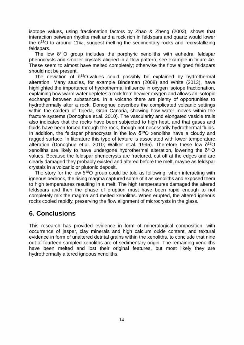
isotope values, using fractionation factors by Zhao & Zheng (2003), shows that interaction between rhyolite melt and a rock rich in feldspars and quartz would lower the δ18O to around 11‰, suggest melting the sedimentary rocks and recrystallizing feldspars.
The low δ18O group includes the porphyric xenoliths with euhedral feldspar phenocrysts and smaller crystals aligned in a flow pattern, see example in figure 4e. These seem to almost have melted completely; otherwise the flow aligned feldspars should not be present.
The deviation of δ18O-values could possibly be explained by hydrothermal alteration. Many studies, for example Bindeman (2008) and White (2013), have highlighted the importance of hydrothermal influence in oxygen isotope fractionation, explaining how warm water depletes a rock from heavier oxygen and allows an isotopic exchange between substances. In a volcano there are plenty of opportunities to hydrothermally alter a rock. Donoghue describes the complicated volcanic settings within the caldera of Tejeda, Gran Canaria, showing how water moves within the fracture systems (Donoghue et.al. 2010). The vascularity and elongated vesicle trails also indicates that the rocks have been subjected to high heat, and that gases and fluids have been forced through the rock, though not necessarily hydrothermal fluids. In addition, the feldspar phenocrysts in the low δ18O xenoliths have a cloudy and ragged surface. In literature this type of texture is associated with lower temperature alteration (Donoghue et.al. 2010; Walker et.al. 1995). Therefore these low δ18O xenoliths are likely to have undergone hydrothermal alteration, lowering the δ18O values. Because the feldspar phenocrysts are fractured, cut off at the edges and are clearly damaged they probably existed and altered before the melt, maybe as feldspar crystals in a volcanic or plutonic deposit.
The story for the low δ18O group could be told as following; when interacting with igneous bedrock, the rising magma captured some of it as xenoliths and exposed them to high temperatures resulting in a melt. The high temperatures damaged the altered feldspars and then the phase of eruption must have been rapid enough to not completely mix the magma and melted xenoliths. When erupted, the altered igneous rocks cooled rapidly, preserving the flow alignment of microcrysts in the glass. 6. Conclusions This research has provided evidence in form of mineralogical composition, with occurrence of jasper, clay minerals and high calcium oxide content, and textural evidence in form of unaltered detrital grains within the xenoliths, to conclude that nine out of fourteen sampled xenoliths are of sedimentary origin. The remaining xenoliths have been melted and lost their original features, but most likely they are hydrothermally altered igneous xenoliths.
14

Acknowledgments I would like to thank my supervisors Abigail Barker, Valentin Troll and Kirsten Zazcek for sharing their knowledge, providing excellent guidance and bringing beautiful xenoliths to Uppsala, without them this project would not exist. I would also like to thank everyone else at Uppsala University involved in the research, especially paleontologist Michael Streng. Michael spent many hours looking at diatom shells in SEM with me, and identified the organisms found. Lisa Samrock is thanked for the contribution of the geological map of Gran Canaria. Lastly, I would like to thank my mother Ulrika Jägerup, Matilda Svensson and Erika Wikmark for taking your time proof reading my report. You have all been a part of this work!
15

References Bindeman, I. (2008), Oxygen Isotopes in Mantle and Crustal Magmas as Revealed by
Single Crystal Analysis. Reviews in Mineralogy & Geochemistry, vol 69, pp 445-478. DOI:10.2138/rmg.2008.69.12
Boyd, P. W., Watson, A. J., Law, C. S., Abraham, E. R., Trull, T., Murdoch., Bakker, C.E., Bowie, A. R., Buesseler, K. O, Chang, H., Charette, M., Croot, P., Downing, K., Frew, R., Gall, M., Hadfield, M., Hall, J., Harvey, M., Jameson, G., LaRoche, J., Liddicoat, M., Ling, R., Maldonado, M. T., McKay, R. M., Nodder, S., Pickmere, S., Pridmore, R., Rintoul, S., Safi, K., Sutton, P., Strzepek, R., Tanneberger, K., Turner, S., Waite, A. & Zeldis, J. (2000), A mesoscale phytoplankton bloom in the polar Southern Ocean stimulated by iron fertilization. Nature, 407, pp 695-702. DOI: 10.1038/35037500
Carmichael, I. S. E., Turner, F. J. & Verhoogen, J. (1974), Igneous Petrology. 1. ed. USA: McGraw-Hill.
Cousens, B.L., Spera, F. J. & Tilton, G. R. (1990), Isotopic Patterns in silicic ignimbrites and lawa flows of the Mogan and the lower Fataga Formations, Gran Canaria, Canary Islands: temporal changes in mantle source composition. Earth Planet, Scientific Letter, vol 96, pp 319-335.
Donoghue, E., Troll, V. R., Harris, C., O’Halloran, A., Walter, T. R & Pèrez Torrado, F. J. (2008), Low-temperature hydrothermal alteration of intra-caldera tuffs, Miocene Tejeda caldera, Gran Canaria, Canary Islands. Journal of Volcanology and Geothermal Research, vol 176, pp 551-564.
Donoghue, E., Troll, V. R. & Harris, C. (2010), Fluid-Rock Interaction in the Miocene, Post-Caldera,Tejeda Intrusive Complex,Gran Canaria (Canary Islands): Insights from Mineralogy, and O- and H-Isotope Geochemistry. Journal of Petrology, vol 51, pp 2149-2176.
Foissner, W. & Schiller, W. (2001), Stable for 15 million years: scanning electron microscope investigation of Miocene euglyphid thecamoebians from Germany, with description of the new genus Scutiglypha. European Journal of Protistology, vol 37, pp 167-180.
Hansteen, T. H. & Troll, V. R. (2003), Oxygen isotope composition of xenoliths from the oceanic crust and volcanic edifice beneath Gran Canaria (Canary Islands): consequences for crustal contamination of ascending magmas. Chemical Geology, vol 193, pp 181-193.
Jones, N. W. & Bloss, F. D. (1980), Laboratory Manual for Optical Microscopy. USA: Burgess Publishing Company.
Lund, J. W. G. (1946), Observations of soil algae. The ecology, size and taxonomy of British soil diatoms. New Pythologist, vol 44, pp 196-219.
McKenzie, W. S. (1980), Atlas of rock-forming minerals in Thin Section. USA: Prentice Hall.
McDougall, I. & Schmincke, H.U. (1976), Geochronology of Gran Canaria, Canary Islands: Age of Shield Building Volcanism and Other Magmatic Phases. Bulletin of Vulcanology, vol. 40-1, pp 57-77.
Morgan, W. J. (1971), Convection Plumes in the Lower Mantle. Nature, vol. 230, pp 42-43. DOI:10.1038/230042a0
Reed, S. J. B. (2005), Electron Microprobe Analysis and Scanning Electron Microscopy in Geology. 2nd ed. Cambridge: Cambridge University Press
Schmincke, H. U. (1982), Volcanic and Chemical Evolution of the Canary Islands. In:
16

Geology of the Northwest African Continental Margin. Berlin & Heidelberg: Springer-Verlag. pp 273-306.
Schmincke, H. U & Sumita, M. (1998), Volcanic Evolution of Gran Canaria reconstructed from apron sediments: synthesis of VICAP project drilling. In: Weaver, P.P.E., Schmincke, H.-U., Firth, J.V., and Duffield,W. (eds.). Proceedings of the Ocean Drilling Program, Scientific Results. Vol. 157. College Station, TX: Ocean Drilling Program, pp 219–266. DOI:10.2973/odp.proc.sr.157.135.1998.
Troll, V. R. (2012), Volcanological Field Methods – Time Travel on an Ocean Island (Report Series A, No. 75).Uppsala University, Department of Earth Science.
Troll, V. R. & Schmincke, H. U. (2002), Magma mixing and crustal recycling in ternary feldspar from compositionally zoned peralkaline ignimbrite ‘A’, Gran Canaria, Canary Islands. Journal of Petrology, vol. 43, pp 243-270.
Troll, V. R., Klügel, A., Deegan, F. M., Carracedo, J. C., Wiesmaier, S., Kueppers, U., Dahren, B., Blythe, L. S., Hansteen, T. H., Freda, C., Budd, D. A., Jolis, E. M., Jonsson, E., Meade,F. C., Harris, C., Berg, S. E., Mancini, L., Polacci, M. & Pedroza, K. (2012), Floating stones off El Hierro, Canary Islands: xenoliths of pre-island sedimentary origin in the early products of the October 2011 eruption. Solid Earth, 3, pp 97-110. DOI: 10.5194/se-3-97-2012
Walker, F. D. L., Lee, M. R. & Parsons, I. (1995). Micropores and micropermeable texture in alkali feldspars: geochemical and geophysical implications. Mineralogical Magazine, Vol 59, pp 505-534
White, W. M. (2013), Geochemistry. Hoboken: Wiley – Blackwell Wilson, J. T. (1963), A possible origin of the Hawaiian Islands. Canadian Journal of
Physics, vol 41, pp 863-870. Zaczek, K., Troll, V. R., Cachao, M., Ferreira, J., Deegan, F. M., Juan, C., Soler, V.,
Meade, F. C. & Burchardt, S. (2015), Nannofossils in 2011 El Hierro eruptive products reinstate plume model for Canary Islands. Scientific Reports, DOI: 10.1038/srep07945
Zhao, Z. F. & Zheng, Y. F. 2003. Calculation of oxygen isotope fractionation in magmatic rocks. Chemical Geology, vol 193, pp 59-80.
Internet sources GEOROC. (2015). Precompiled Files, Rocks, Rhyolite 1. Available at:
http://georoc.mpch-mainz.gwdg.de/georoc/CompFiles.aspx#rockfiles [2015-05-29] Encyclopedia Britannica (2015). Xenoliths. Available at:
http://global.britannica.com/EBchecked/topic/650624/xenolith [2015-05-20] Olney, M. (2002). Diatoms – Microfossil image recovery and circulation for learning and education. Available at:
http://www.ucl.ac.uk/GeolSci/micropal/diatom.html [2015-05-06 The Geography Site (2010). Why do people live near volcanoes? Available at:
http://www.geography-site.co.uk/pages/physical/earth/volcanoes/volcanoliving.html [2016-02-10]
17


