the origin of the mullerian duct in chick and mouse - … · the origin of the mullerian duct in...
TRANSCRIPT

02 (2007) 389–398www.elsevier.com/locate/ydbio
Developmental Biology 3
The origin of the Mullerian duct in chick and mouse
Silvana Guioli ⁎, Ryohei Sekido, Robin Lovell-Badge
Division of Developmental Genetics, MRC National Institute for Medical Research, The Ridgeway, Mill Hill, London NW7 1AA, UK
Received for publication 24 July 2006; revised 25 September 2006; accepted 27 September 2006Available online 3 October 2006
Abstract
In vertebrates the female reproductive tracts derive from a pair of tubular structures called Mullerian ducts, which are composed of threeelements: a canalised epithelial tube, mesenchymal cells surrounding the tube and, most externally, coelomic epithelial cells. Since the firstdescription by Johannes Peter Muller in 1830, the origin of the cells making up the Mullerian duct has remained controversial. We report theresults from lineage-tracing experiments in chicken and mouse embryos aimed to provide information of the dynamics of Mullerian ductformation. We show that all Mullerian duct components derive from the coelomic epithelium in both species. Our data support a model of aMullerian epithelial tube derived from an epithelial anlage at the mesonephros anterior end, which then segregates from the epithelium and extendscaudal of its own accord, via a process involving rapid cell proliferation. This tube is surrounded by mesenchymal cells derived from localdelamination of coelomic epithelium. We exclude any significant influx of cells from the Wolffian duct and also the view of a tube forming bycoelomic epithelium invagination along the mesonephros. Our data provide clues of the underlying mechanism of tubulogenesis relevant to bothnormal and abnormal development of the female reproductive tract.© 2006 Elsevier Inc. All rights reserved.
Keywords: Mullerian duct; Wolffian duct; Coelomic epithelium; Tubulogenesis; Endometriosis
Introduction
Tubulogenesis is a process vital to the development andfunction of many tissues and organs. Despite the apparentsimplicity of a tube, there are many different ways that tubes canform during embryogenesis including folding, branching,mesenchymal to epithelial transitions and formation of alumen in an initial solid cord (Myat, 2005). Moreover, tubu-logenesis is a process requiring precise control over cellularevents, including changes in cell shape and adhesion, celldivision and apoptosis, as well as active alteration of the cellularenvironment. How these different processes are coordinated inspace and time to form a tube is not sufficiently understood forany system.
The female reproductive tract represents a tubular structureof obvious importance to the continuation of a species. Itsanatomy varies markedly among vertebrates, depending on thetype of fertilisation, mode of reproduction, type of placentationand other factors. Anatomical differences can even be observed
⁎ Corresponding author. Fax: +44 2088162009.E-mail address: [email protected] (S. Guioli).
0012-1606/$ - see front matter © 2006 Elsevier Inc. All rights reserved.doi:10.1016/j.ydbio.2006.09.046
within a species. Despite the extremely high degree ofspecialisation of the adult structures, the internal genital tractsderive, with few exceptions such as the Teleost fish (Suzuki andShibata, 2004), from a pair of tubular structures calledMullerian ducts or paramesonephric ducts.
The Mullerian ducts arise during embryogenesis as part ofthe urogenital system in both sexes. They develop along theanterior–posterior (A–P) axis of the embryos in close proximityand lateral to the mesonephric (or Wolffian) ducts. It is possibleto distinguish three cellular components in the Mullerian duct:the epithelial cells forming the inner tube (Mullerian ductepithelium: MDE), the mesenchymal cells surrounding the tube(Mullerian duct mesenchyme: MDM) and the coelomicepithelial cells defining the external borders of the duct(Mullerian coelomic epithelium: MCE).
Molecular genetic studies in mouse have contributed to theidentification of a number of proteins essential for the formationof the Mullerian duct. These include the homeodomaintranscription factors Pax2, Pax8, Lim1, Emx2, Hoxa13 andthe signalling molecule Wnt4, defining a genetic cascade forearly Mullerian development (Kobayashi and Behringer, 2003;Kobayashi et al., 2004).

390 S. Guioli et al. / Developmental Biology 302 (2007) 389–398
Classical embryological studies of extirpation or blockage ofthe Wolffian duct in chick embryos have shown that theWolffian duct is required for the formation and caudal extensionof the Mullerian duct (Bishop-Calame, 1966; Didier, 1971,1973; Grunwald, 1941). Further evidence for this requirementcomes from mouse and human studies. Mice with a conditionaldeletion of Lim1 in the Wolffian duct have a truncatedMullerian duct due to the inability of maintaining themesonephric duct (Kobayashi et al., 2005; Pedersen et al.,2005), while in humans, several syndromes have been identifiedwhere Mullerian duct growth is affected by the absence of theWolffian duct. One such a condition is a form of kidney aplasiaassociated with the absence of a fallopian tube on one side anduterus unicornus on the other side (Grunwald, 1941). How thedependence of the Mullerian duct on the Wolffian duct isachieved is not yet known.
To begin understanding the mechanisms of early develop-ment of the Mullerian tube, it will be necessary to integrate themolecular data with a cellular understanding of the process,which is still poor. One of the key issues is the origin of the cellscontributing to the duct during its formation and during itsgrowth. Most of our current knowledge derives from light andelectron microscopy observations of Mullerian duct sections atdifferent development time-points in different organisms(Abdel-Malek, 1950; Del Vecchio, 1982; Dohr and Tarmann,1984; Frutiger, 1969; Furbringer, 1878; Grunwald, 1941; Hall,1904; Hashimoto, 2003; Inomata et al., 1989; Jacob et al., 1999;Wrobel, 2003; Wrobel and Sub, 2000).
In the chick, the first sign of Mullerian development has beendescribed as the appearance of a Mullerian ridge consisting of athickening of the coelomic epithelium adjacent to themesonephric duct. According to Jacob et al. (1999), theprimordium of the MDE becomes apparent later, at stageHamburger and Hamilton (HH) 25, as an aggregation of cellsextending caudally from a funnel (ostial funnel) formed in thecranial area of the Mullerian ridge. By HH30, the MDE hasgrown a considerable length. The rostral end is a luminalepithelial tube surrounded by layers of mesenchymal cells,while the caudal tip is mesenchymal and stays in close contactwith the Wolffian duct. Other studies performed in Amniota(such as human, rat, bovine,) do agree that the Mullerian duct isformed by the caudally directed growth of the ostial funnel, butthere are at least two important unresolved issues. The first,which is controversial, concerns the origin of the cellscontributing to the duct epithelium. One model predicts thatthe duct anlage derives from a placode-like thickening anddeepening of the coelomic epithelium, which then extendscaudally, of its own accord, forming the epithelial Mullerianduct itself (Jacob et al., 1999). A second model predicts a majoror sole contribution of Wolffian duct cells to the growing MDEvia a budding or splitting off from the Wolffian duct itself (DelVecchio, 1982; Frutiger, 1969; Inomata et al., 1989). Thesecond question that has not been clearly answered in any studyis the origin of the MDM along the A–P axis of the embryo.
All these issues need to be tackled with approaches thatdepend on following cell fate in a dynamic way. We decidedfirst to investigate the contribution of the coelomic epithelial
cells to MDE and MDM by performing lineage-tracingexperiments in chick embryos. We electroporated GFP-expres-sing plasmids into coelomic epithelial cells in ovo just beforethe appearance of a Mullerian ridge and followed the fate of theGFP cells after 3–4 days of development. We then performed asecond set of lineage tracing experiments in mouse urogenitalridges as a comparative study, to address how well the processof Mullerian duct formation is conserved.
Materials and methods
Animals
Fertilised chicken eggs were obtained from Winter Egg Farm. Mouseembryos for electroporation and MitoTracker injections were from the outbredParkes strain maintained at the NIMR. The transgenic line Sox9(1.9)LacZ wasmaintained as heterozygote (abbreviated in the text as 1.9LacZ/+) on a CBA/B10background. These mice express LacZ under the control of a 1.9 kb regulatoryregion from mouse Sox9 (Sekido and Lovell-Badge, unpublished data).
Electroporation of chick and mouse embryos
Fertilised chicken eggs were incubated at 37.5°C for 2.5 days up to stageHamburger and Hamilton (HH) 14–17 (Hamburger and Hamilton, 1992).Following the removal of 2.5 ml of albumen with a syringe, a window was cut inthe egg shell. The electroporation was performed as described in Sekido andLovell-Badge (in press),with somemodifications. In brief,DNAwas injected intoone side of the coelomic cavity using a glass capillary needle and an inject+maticpico-pump. Two small electrodes (4 mm length, 0.4–0.5 mm diameter) wereapplied in parallel, one on each side of the embryo and a difference in voltagewasgenerated to allow directional entry of DNA into cells towards the midline. Thelow fixed voltage was applied with a BTX ECM-830 electroporator. The eggswere then sealed with standard tape and allowed to develop at 37.5°C. Thesurvival rate up to stage HH30–32 was variable, ranging from one-third to two-thirds of the total number of injected embryos.
11.5 dpc wild type mouse embryos were dissected in Dulbecco's MinimalEagle's medium (DMEM). The exact age of the embryos was then establishedby counting tail somites (ts) number as described in Hacker et al. (1995).Embryos were placed in a dish in PBS and the injection/electroporation wasperformed as for the chicken embryos. The urogenital ridges were subsequentlyplaced into grooves of a 1% agarose support in 35 mm dishes containing DMEMwith 100 units/ml penicillin, 0.05 mg/ml streptomycin, 2 mM Glutamine and10% fetal calf serum. The samples were incubated at 37°C, in 5% CO2 for acouple of days.
Conditions of electroporation: chicken embryos: five 50 ms pulses at 24 V;mouse embryos: five 50 ms pulses at 52 V. Electroporated DNA: plasmid pCS2+
expressing EGFP under the ubiquitous promoter CMV IE94.
MitoTracker injections
MitoTracker red (CMXRos from Molecular Probes) was dissolved inDMSO at 2 mg/ml and diluted 1:4 in 10% Sucrose just before labelling theurogenital ridges of ts 13–19 wild type embryos. Using a microcapillary glassneedle, the dye was delivered onto the surface of the urogenital ridge epitheliumat the anterior tip or along the mesonephros. The injected samples were thencultured as described above.
Light microscopy, immunohistochemistry and β−galactosidasestaining
Electroporated and mitoTracker labelled samples were screened using aLeica stereomicroscope (MZFLIII) equipped with GFP Plus Fluorescence filter(GFP2, excitation 480/40 nm) and Green Fluorescence filter (G, excitation 546/10 nm). Chick positive samples were fixed for 1 h in 4% Paraformaldehyde(PFA), rinsed in PBS, transferred to 30% Sucrose at 4°C overnight and
391S. Guioli et al. / Developmental Biology 302 (2007) 389–398
embedded in OCT. Transverse sections were cut with a Leica cryostat. Mousesamples were treated for whole-mount immunohistochemistry. After a 30′fixation in 4% PFA at 4°C, they were rinsed in PBS and incubated in blockingsolution (PBS/2% BSA, 0.5% Triton) for at least 4 h at 4°C. The incubation withthe antibody was carried out overnight at 4°C in blocking solution. After three1 h rinses in PBS/0.1% Tween, samples were incubated with secondaryantibodies in PBS/0.1% Tween overnight at 4°C. Samples were then rinsed fewhours in PBS/0.1% Tween before visualisation under the fluorescent stereo-microscope. Selected samples were transferred to 30% sucrose overnight at 4°Cand then embedded in OCT for sectioning.
Primary antibodies: rabbit polyclonal anti-PAX2 (1:1200 dilution) (PRB-276P from Covance). Secondary antibodies: goat anti-rabbit Alexa-555 orAlexa-488 (1:400 dilution) from Molecular Probes. Fluorescence images werecaptured on an Olympus IX70 inverted microscope using a Deltavision cooledCCD imaging system (Photometrics CH350L liquid cooled CCD camera;Softworx image acquisition software) (Applied Precision), with the exception ofimages in Fig. 3B: confocal Z-stack of images captured with a Leica TCS-SP1system and processed using the volume rendering software Volocity(Improvision).
12.5 dpc mouse embryos were dissected from pregnant females hetero-zygous for the 1.9LacZ transgene, fixed 30′ in 4% PFA, rinsed in PBS andstained in X-Gal solution overnight at 37°C. Positive samples were embedded inparaffin, sectioned with a microtome and eosin counterstained.
5-bromo-2′-deoxyuridine (BrdU) labelling
Pregnant mice received a single intra-peritoneal injection of BrdU (SigmaB-5002) at 50 mg/kg of body weight 2 h before dissection.
Following the removal of 2.5 ml of albumen from chicken eggs incubated at37.5°C for 7 days, 100 μg of BrdU dissolved in 100 μl of PBS was dropped onthe vitelline membrane. The eggs were sealed with tape and incubated foranother 2 h.
Chicken and mouse embryo urogenital ridges were fixed in 4% PFA at 4°Cfor 2 h, rinsed in PBS and put in 30% Sucrose overnight at 4°C beforeembedding in OCT. 12 μm sections cut with a Leica cryostat were washed threetimes 5′ in PBS, fixed again for 5′ in 4% PFA at RT, rinsed in PBS and incubatedin 2 M HCl at 37°C for 30′. Following three 5′ washes in 0.1 M Borate bufferpH8.5 and one wash in PBS, the sections were incubated in blocking buffer(PBS/0.5% BSA, 2% sheep serum, 0.1% triton) for 1 h. Primary antibodies wereadded to the blocking solution overnight at 4°C (mouse monoclonal anti-BrdU1:300 from Chemicon and rabbit polyclonal anti-PAX2 1:1200). After threewashes in PBS/0.1% Tween, the sections were incubated with secondaryantibodies (goat anti-rabbit Alexa 555 and goat anti-mouse Alexa 488 1:400,from Molecular Probes) for 2 h and finally washed in PBS/0.1% Tween.
Results and discussion
MDE and MDM of chick embryos derive from differentpopulations of coelomic epithelial cells
Lineage tracing experiments were performed in ovo, be-ginning with chick embryos at HH14–17. At these stages theWolffian duct has already reached the cloaca, while theMullerian duct is not yet apparent. In order to label the rightor left dorsal coelomic epithelium, including the cells over-laying the mesonephros, a plasmid expressing GFP undercontrol of an ubiquitous promoter was injected into the right orleft coelomic cavity and electroporated towards the dorsal/medial side. The embryos were screened for GFP expression 3–4 days after manipulation, at HH30–32.
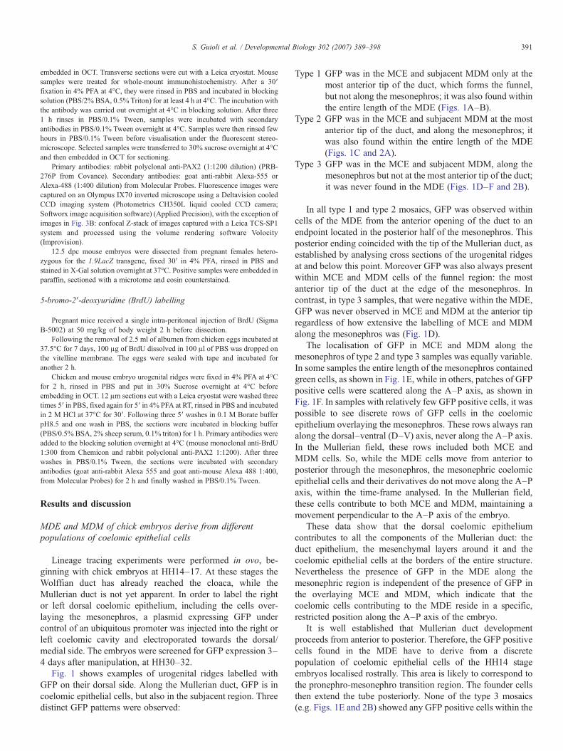
Fig. 1 shows examples of urogenital ridges labelled withGFP on their dorsal side. Along the Mullerian duct, GFP is incoelomic epithelial cells, but also in the subjacent region. Threedistinct GFP patterns were observed:
Type 1 GFP was in the MCE and subjacent MDM only at themost anterior tip of the duct, which forms the funnel,but not along the mesonephros; it was also found withinthe entire length of the MDE (Figs. 1A–B).
Type 2 GFP was in the MCE and subjacent MDM at the mostanterior tip of the duct, and along the mesonephros; itwas also found within the entire length of the MDE(Figs. 1C and 2A).
Type 3 GFP was in the MCE and subjacent MDM, along themesonephros but not at the most anterior tip of the duct;it was never found in the MDE (Figs. 1D–F and 2B).
In all type 1 and type 2 mosaics, GFP was observed withincells of the MDE from the anterior opening of the duct to anendpoint located in the posterior half of the mesonephros. Thisposterior ending coincided with the tip of the Mullerian duct, asestablished by analysing cross sections of the urogenital ridgesat and below this point. Moreover GFP was also always presentwithin MCE and MDM cells of the funnel region: the mostanterior tip of the duct at the edge of the mesonephros. Incontrast, in type 3 samples, that were negative within the MDE,GFP was never observed in MCE and MDM at the anterior tipregardless of how extensive the labelling of MCE and MDMalong the mesonephros was (Fig. 1D).
The localisation of GFP in MCE and MDM along themesonephros of type 2 and type 3 samples was equally variable.In some samples the entire length of the mesonephros containedgreen cells, as shown in Fig. 1E, while in others, patches of GFPpositive cells were scattered along the A–P axis, as shown inFig. 1F. In samples with relatively few GFP positive cells, it waspossible to see discrete rows of GFP cells in the coelomicepithelium overlaying the mesonephros. These rows always ranalong the dorsal–ventral (D–V) axis, never along the A–P axis.In the Mullerian field, these rows included both MCE andMDM cells. So, while the MDE cells move from anterior toposterior through the mesonephros, the mesonephric coelomicepithelial cells and their derivatives do not move along the A–Paxis, within the time-frame analysed. In the Mullerian field,these cells contribute to both MCE and MDM, maintaining amovement perpendicular to the A–P axis of the embryo.
These data show that the dorsal coelomic epitheliumcontributes to all the components of the Mullerian duct: theduct epithelium, the mesenchymal layers around it and thecoelomic epithelial cells at the borders of the entire structure.Nevertheless the presence of GFP in the MDE along themesonephric region is independent of the presence of GFP inthe overlaying MCE and MDM, which indicate that thecoelomic cells contributing to the MDE reside in a specific,restricted position along the A–P axis of the embryo.
It is well established that Mullerian duct developmentproceeds from anterior to posterior. Therefore, the GFP positivecells found in the MDE have to derive from a discretepopulation of coelomic epithelial cells of the HH14 stageembryos localised rostrally. This area is likely to correspond tothe pronephro-mesonephro transition region. The founder cellsthen extend the tube posteriorly. None of the type 3 mosaics(e.g. Figs. 1E and 2B) showed any GFP positive cells within the

Fig. 1. Urogenital ridges from stage HH30–32 chick embryos electroporated with pCS2-EGFP (in green). (A, B) Examples of type 1 GFP pattern; (C) example of type2 pattern; (D, E, F) examples of type 3 pattern. A=anterior end, P=posterior end. White arrows indicate areas where GFP is only within the MDE; white arrowheadsindicate areas where GFP is only in MCE and MDM; red arrows indicate areas where GFP is in all components of the Mullerian duct. The most common pattern foundamong the injected sample is the type 3: out of a total of 46 samples from a few electroporation experiments, 6 were type 1, 7 were type 2 and 33 were type 3.
392 S. Guioli et al. / Developmental Biology 302 (2007) 389–398
MDE, even in areas along the mesonephros where the MCE washeavily targeted with GFP. This indicates that the MDE does notintermingle with coelomic epithelial cells localised moreposteriorly along the mesonephros, during its caudal extension.This confirms that the coelomic epithelial cells of the MDEanlage segregate from the epithelium before extending throughthe mesonephros. Cell recruitment from the MCE does notoccur at all below the point of segregation.
In some type 1 and 2 samples, most MDE cells were found tobe GFP positive, indicating that the coelomic epithelium mustbe at least the major contributor to the Mullerian duct. Sodespite not being possible to test directly any potentialcontribution from the Wolffian duct, it is clear that, if there isany contribution at all during MDE migration, it has to beminor. Typically, in samples where the duct was heavilylabelled, such as the example in Fig. 2A, less than 20% of theduct cells were GFP negative and, while these cells could be ofWolffian duct origin, they could also be from untargetedcoelomic epithelial cells or from targeted cells that haveproliferated and diluted out or lost the marker.
In our hands, the stage of the chick embryos used for theelectroporation was critical for successful targeting of GFP tothe MDE. Jacob et al. (1999) report the appearance of a MDEanlage at HH24 in chick, but we were only successful intargeting GFP to the MDE when we used HH14–15 embryos.Embryos slightly older (e.g. HH17) never showed GFP withinthe MDE, suggesting two possibilities: the MDE anlage isalready segregating from the surface epithelium immediatelyafter these stages, or the target area is difficult to reach at laterstages (HH17), using our technique.
The origin of MDE and MDM cells is conserved between chickand mouse
At 11.5 dpc in the mouse, the Mullerian duct has not yetformed, but a group of cells positive for Lim1 and Pax2 arepresent at the rostral end of the urogenital ridge in the proximityof the Wolffian duct. This patch of cells is thought to be theanlage of the Mullerian duct. By 12.5 dpc, the Mullerian ducthas grown considerably along the A–P axis, running along

Fig. 2. Transverse sections of urogenital ridges from stage HH32 chick embryos electroporated with pCS2-EGFP (in green). Nuclei are counterstained with DAPI (inblue). Three sections per sample are shown; from left to right: anterior to posterior. (A) Example of type 2 GFP pattern (corresponding to sample c in Fig. 1); at theanterior end GFP is in MCE, MDM and MDE, at the posterior end GFP is only in the MDE. (B) Example of type 3 GFP pattern (corresponding to sample e in Fig. 1);GFP is only in MCE and MDM.
393S. Guioli et al. / Developmental Biology 302 (2007) 389–398
about one half to two thirds of the mesonephros; by 13.5 dpcthe duct has run the entire length of the mesonephros(Kobayashi et al., 2004; Torres et al., 1995).
We wanted to ask if the contribution of the coelomicepithelial cells to the Mullerian duct is similar or differentbetween chick and mouse. In order to do this comparative study,we designed strategies to trace the fate of the coelomicepithelium overlaying the urogenital ridge in culture.
It is well established that the mouse genital ridge canundergo normal differentiation into testis or ovary, whengrown together with the mesonephros in culture containingserum-enriched medium. We first tested if the Mullerian duct
develops normally in this organ culture, by analysing theexpression of PAX2. This marker, known to be essential forthe formation and maintenance of the duct, is normallyexpressed in Wolffian duct, Mullerian duct epithelium andmesonephric tubules (Torres et al., 1995). A proportion of11.5 dpc urogenital ridges grown in culture for 2 days didshow the expected MDE Pax2 staining, confirming thenormal growth of the Mullerian duct. When the cultureswere initiated from embryos at ts 14–19 (11.1–11.6 dpc), thepercentage of normal growth was above 60%, while wheninitiated at ts11–12 (10.7–10.8 dpc), it was much lower,about 35%. The difference could reflect the presence or
394 S. Guioli et al. / Developmental Biology 302 (2007) 389–398

Fig. 4. Two examples of mouse urogenital ridges injected with MitoTracker dye and cultured in vitro from 11.5 dpc for 2.5 days. The samples were stained with anantibody against PAX2. (A) Whole-mount fluorescence images showing normal growth of Mullerian duct and Wolffian duct in both samples (as confirmed by PAX2staining, in green). The sample on the left contains MitoTracker within the MDE (in red). (B) Transverse sections across the two samples in (A) confirming the resultsdisplayed in (A). Pax2 in green; MitoTracker in red. MD: Mullerian duct; WD: Wolffian duct; A=anterior end, P=posterior end.
395S. Guioli et al. / Developmental Biology 302 (2007) 389–398
absence of either the responding or inducing tissue after thedissection, or to stage-dependent sensitivity to the cultureconditions. To follow the fate of coelomic epithelial cellsoverlaying the urogenital ridge, we used two procedures:injection/electroporation of a GFP expressing plasmid, orlabelling with MitoTracker dye (see Materials and methods).After culture, only samples with a Mullerian duct, as assayedby PAX2 staining, were analysed.
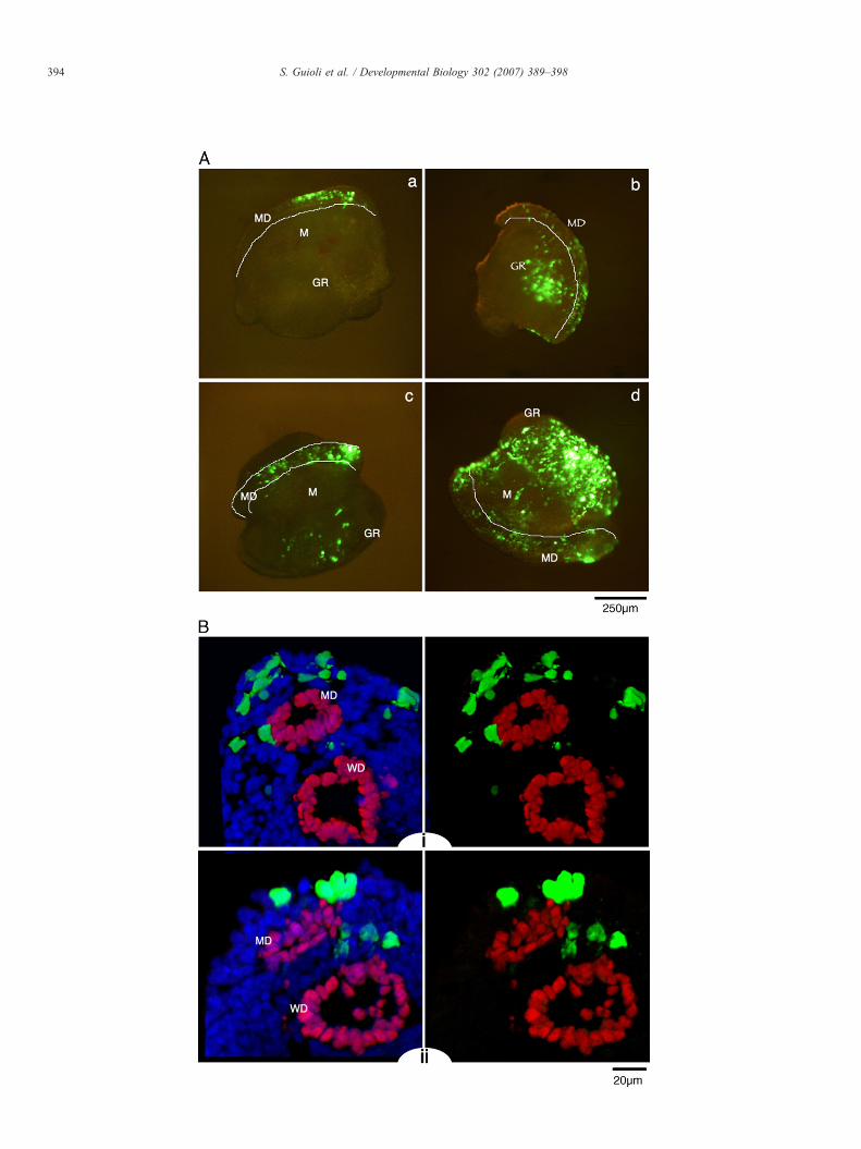
The electroporation of GFP was never as extensive as in thechick, but it allowed us to clearly mark patches of cells along theurogenital ridge, targeting the Mullerian field, as well as otherparts of the mesonephros itself and the genital ridge. Fig. 3A
Fig. 3. Mouse urogenital ridges from 11.5 dpc embryos cultured in vitro for 2 days aimages from four different samples showing GFP localisation to the urogenital ridges.selected transverse sections from sample b and d, respectively, stained with an antibodGreen: GFP; Red: Pax2; Blue: DAPI counterstaining. MD: Mullerian duct; WD: W
shows a few examples from the collection of samples labelled atts17–19 (11.4–11.6 dpc). As with the chick, in the Mullerianfield GFP positive cells were found not only in the epitheliallayer but also in the inner portion (Fig. 3B). This suggests thatcoelomic epithelial cells along the mesonephric portion of theurogenital ridge either stay in the coelomic epithelium, orcontribute to the mesenchymal cells surrounding the Mullerianduct, similar to the type 3 chick samples.
The MitoTracker labelling of discrete areas of the epitheliumoverlaying the mesonephros in proximity to the Wolffian ductconfirmed that the coelomic epithelial cells along the mesone-phros contribute to the MDM (data not shown). In several
fter electroporation with pCS2-EGFP. (A) (a, b, c, d) Whole-mount fluorescenceThe Mullerian duct field borders are marked white. (B) (i, ii) Confocal images ofy against Pax2 which marks the epithelium of Mullerian duct and Wolffian duct.olffian duct, GR: genital ridge; M: mesonephros.
396 S. Guioli et al. / Developmental Biology 302 (2007) 389–398
samples labelled at their rostral tip, MitoTracker was foundwithin the entire length of the MDE. This was independent fromthe presence of dye within the MDM along the mesonephros(Fig. 4). These results show that the MDE anlage originatesfrom coelomic epithelial cells at the rostral tip of themesonephros or in the residual pronephric region and segregatefrom the surface before extending caudal along the mesonephricregion. Moreover the MDE does not intermingle with thecoelomic epithelial cells overlaying the mesonephros followingits segregation from the surface. This pattern is similar to theone found in chick. The samples labelled with MitoTracker atthe anterior end also showed what appeared to be stainingwithin the Wolffian duct (Fig. 4). At the time of injection, theWolffian duct was already developed and some diluted dye mayhave become trapped by capillary action in the canal, where thestaining was later observed. This was unexpected, but found inthe Wolffian duct of all injected samples, whether MitoTrackerpositive or negative within MDE and/or MDM/MCE, indicatingthat the labelling of the Mullerian duct was independent of thisapparent staining.
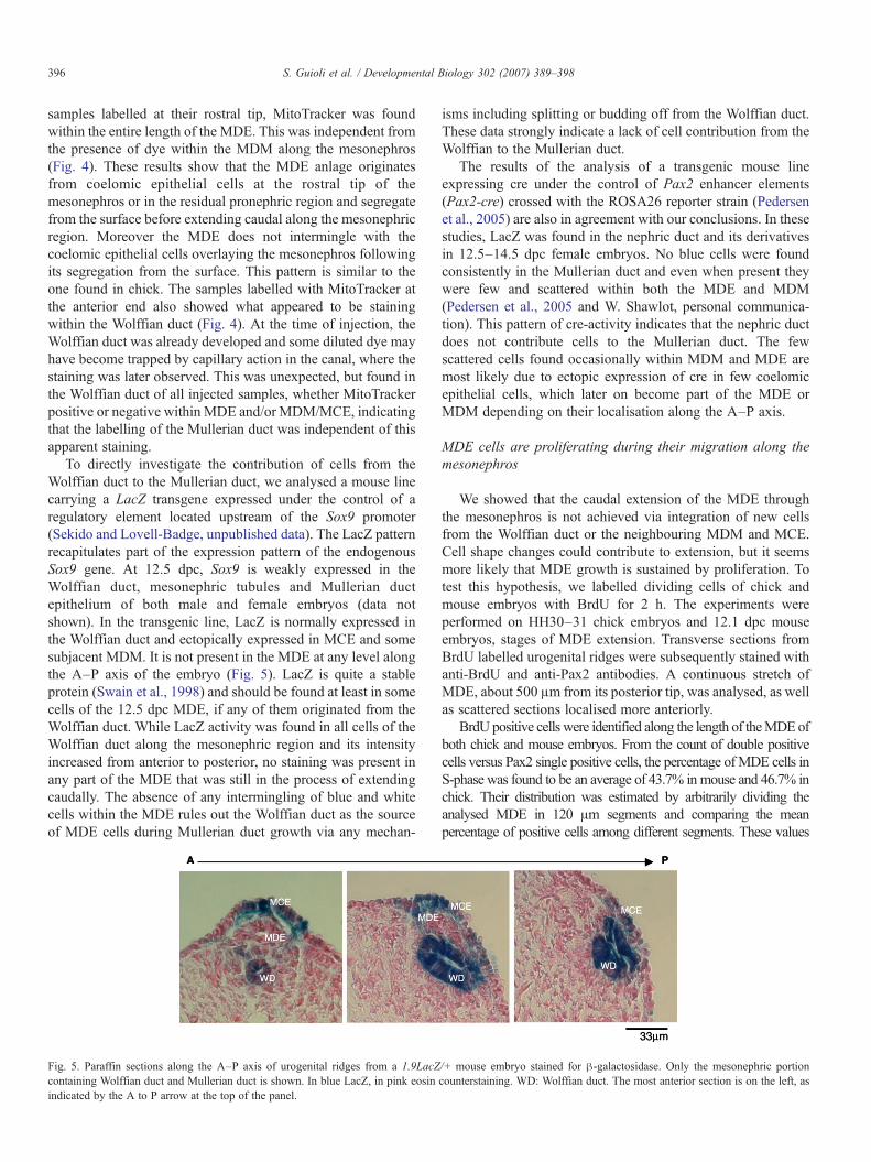
To directly investigate the contribution of cells from theWolffian duct to the Mullerian duct, we analysed a mouse linecarrying a LacZ transgene expressed under the control of aregulatory element located upstream of the Sox9 promoter(Sekido and Lovell-Badge, unpublished data). The LacZ patternrecapitulates part of the expression pattern of the endogenousSox9 gene. At 12.5 dpc, Sox9 is weakly expressed in theWolffian duct, mesonephric tubules and Mullerian ductepithelium of both male and female embryos (data notshown). In the transgenic line, LacZ is normally expressed inthe Wolffian duct and ectopically expressed in MCE and somesubjacent MDM. It is not present in the MDE at any level alongthe A–P axis of the embryo (Fig. 5). LacZ is quite a stableprotein (Swain et al., 1998) and should be found at least in somecells of the 12.5 dpc MDE, if any of them originated from theWolffian duct. While LacZ activity was found in all cells of theWolffian duct along the mesonephric region and its intensityincreased from anterior to posterior, no staining was present inany part of the MDE that was still in the process of extendingcaudally. The absence of any intermingling of blue and whitecells within the MDE rules out the Wolffian duct as the sourceof MDE cells during Mullerian duct growth via any mechan-
Fig. 5. Paraffin sections along the A–P axis of urogenital ridges from a 1.9LacZcontaining Wolffian duct and Mullerian duct is shown. In blue LacZ, in pink eosinindicated by the A to P arrow at the top of the panel.
isms including splitting or budding off from the Wolffian duct.These data strongly indicate a lack of cell contribution from theWolffian to the Mullerian duct.
The results of the analysis of a transgenic mouse lineexpressing cre under the control of Pax2 enhancer elements(Pax2-cre) crossed with the ROSA26 reporter strain (Pedersenet al., 2005) are also in agreement with our conclusions. In thesestudies, LacZ was found in the nephric duct and its derivativesin 12.5–14.5 dpc female embryos. No blue cells were foundconsistently in the Mullerian duct and even when present theywere few and scattered within both the MDE and MDM(Pedersen et al., 2005 and W. Shawlot, personal communica-tion). This pattern of cre-activity indicates that the nephric ductdoes not contribute cells to the Mullerian duct. The fewscattered cells found occasionally within MDM and MDE aremost likely due to ectopic expression of cre in few coelomicepithelial cells, which later on become part of the MDE orMDM depending on their localisation along the A–P axis.
MDE cells are proliferating during their migration along themesonephros
We showed that the caudal extension of the MDE throughthe mesonephros is not achieved via integration of new cellsfrom the Wolffian duct or the neighbouring MDM and MCE.Cell shape changes could contribute to extension, but it seemsmore likely that MDE growth is sustained by proliferation. Totest this hypothesis, we labelled dividing cells of chick andmouse embryos with BrdU for 2 h. The experiments wereperformed on HH30–31 chick embryos and 12.1 dpc mouseembryos, stages of MDE extension. Transverse sections fromBrdU labelled urogenital ridges were subsequently stained withanti-BrdU and anti-Pax2 antibodies. A continuous stretch ofMDE, about 500 μm from its posterior tip, was analysed, as wellas scattered sections localised more anteriorly.
BrdU positive cells were identified along the length of theMDEofboth chick and mouse embryos. From the count of double positivecells versus Pax2 single positive cells, the percentage ofMDE cells inS-phase was found to be an average of 43.7% in mouse and 46.7% inchick. Their distribution was estimated by arbitrarily dividing theanalysed MDE in 120 μm segments and comparing the meanpercentage of positive cells among different segments. These values
/+ mouse embryo stained for β-galactosidase. Only the mesonephric portioncounterstaining. WD: Wolffian duct. The most anterior section is on the left, as
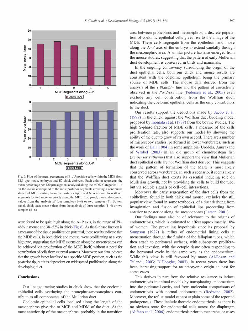
Fig. 6. Plots of the mean percentage of BrdU positive cells within the MDE from12.1 dpc mouse embryos and E7 chick embryos. Each column represents themean percentage per 120 μm segment analysed along the MDE. Categories 1–4on the X-axis correspond to the most posterior segments covering a continuousstretch of MDE starting from the posterior tip; 5 and 6 correspond to scatteredsegments located more anteriorly along the MDE. Top panel, mouse data; meanvalues from the analysis of four samples (1–4) or two samples (5). Bottompanel, chick data; mean values from the analysis of three samples (1–4) or twosamples (5–6).
397S. Guioli et al. / Developmental Biology 302 (2007) 389–398
were found to be quite high along the A–P axis, in the range of 39–48%inmouse and38–52%inchick (Fig. 6).As theS-phase fraction isameasure of the tissue proliferation potential, these results indicate thatthe MDE cells, in both chick and mouse, were proliferating at a veryhigh rate, suggesting that MDE extension along the mesonephros canbe achieved via proliferation of the MDE itself, without a need forcontributionof cells fromexternal sources.Moreover, our data indicatethat the growth is not localised to a specificMDE position, such as theposterior tip, but it is dependent on widespread proliferation along thedeveloping duct.
Conclusions
Our lineage tracing studies in chick show that the coelomicepithelial cells overlaying the pronephros/mesonephros con-tribute to all components of the Mullerian duct.
Coelomic epithelial cells localised along the length of themesonephros give rise to MCE and MDM of the duct. At themost anterior tip of the mesonephros, probably in the transition
area between pronephros and mesonephros, a discrete popula-tion of coelomic epithelial cells gives rise to the anlage of theMDE. These cells segregate from the epithelium and movealong the A–P axis of the embryo to extend caudally throughthe mesonephric area. A similar picture has also emerged fromthe mouse studies, suggesting that the pattern of early Mullerianduct development is conserved in birds and mammals.
In the ongoing controversy surrounding the origin of theduct epithelial cells, both our chick and mouse results areconsistent with the coelomic epithelium being the primarysource of MDE cells. The mouse data derived from theanalysis of the 1.9LacZ/+ line and the pattern of cre-activityobserved in the Pax2-cre line (Pedersen et al., 2005) evenexclude any cell contribution from the Wolffian duct,indicating the coelomic epithelial cells as the only contributorsto the duct.
Our results support the deductions made by Jacob et al.(1999) in the chick, against the Wolffian duct budding modelproposed by Inomata et al. (1989) from the bovine studies. Thehigh S-phase fraction of MDE cells, a measure of the cellsproliferation rate, also supports our model by showing theability of the duct to grow of its own accord. There are a numberof microscopy studies, performed in lower vertebrates, such asthe work of Hall (1904) in some amphibia (Urodela, Anura) andof Wrobel (2003) in an old group of chondreostean fish(Acipenser ruthenus) that also support the view that Mullerianduct epithelial cells are not Wolffian duct derived. This suggeststhat the pattern of formation of the MDE is most likelyconserved across vertebrates. In such a scenario, it seems likelythat the Wolffian duct exerts its essential inducing role onMullerian growth, not by providing the cells to build the tube,but via soluble signals or cell–cell interactions.
Moreover the early segregation of the duct cells from theepithelium, found in both chick and mouse, excludes the stillpopular view, found in some textbooks, of a duct deriving frominvagination and fusion of epithelial lips proceeding fromanterior to posterior along the mesonephros (Larsen, 2001).
Our findings may also be of relevance to the origins ofendometriosis, which is estimated to affect approximately 10%of women. The prevailing hypothesis since its proposal bySampson (1927) is reflux of endometrial lining cells atmenstruation through the fimbria of the fallopian tubes, whichthen attach to peritoneal surfaces, with subsequent prolifera-tion and invasion, with the ectopic tissue often responding tothe hormonal cycle in the same way as the endometrium.While this view is still favoured by many (Al-Fozan andTulandi, 2003; D'Hooghe, 2003), in recent years there hasbeen increasing support for an embryonic origin at least forsome cases.
This derives in part from the relative resistance to induceendometriosis in animal models by transplanting endometriuminto the peritoneal cavity and from molecular comparisons ofendometriosis with normal endometrium (Redwine, 2002).Moreover, the reflux model cannot explain some of the reportedpathogenesis. These include thoracic endometriosis, as there isno obvious route for endometrial cells across the diaphragm(Alifano et al., 2006); endometriosis prior to menarche, or cases

398 S. Guioli et al. / Developmental Biology 302 (2007) 389–398
of very young girls with MDE-like cells found in peritonealpockets (known as “Mullerian cell rests”) (Batt and Mitwally,2003; Goldstein et al., 1979). We have shown that the ductnormally develops from a discrete population of coelomicepithelial cells at the border between the mesonephros andpronephros. Presumably this is in response to a local source ofan inducing molecule that initiates the formation of a pocket thatis then dependent on the Wolffian duct for its caudal extension.However, such pockets could be initiated at other locations inclose contact with an appropriate signalling centre, in thepronephros or mesonephros. Remnants of the former couldremain within the thoracic cavity and of the latter within theperitoneal cavity. It will clearly be of interest to determine thefactors that initiate the MDE and the nature of their source.
Altogether our conclusions will inform future experiments todefine the molecular and cellular events responsible forMullerian duct tubulogenesis. The electroporation techniquein the chick allows targeting of all the Mullerian ductcomponents, and due to the stage-dependent targeting of theMDE even allows some control on which component to target.It is therefore a useful tool to deliver DNA and RNA moleculesfor the functional analysis of early Mullerian duct development,which will provide a better understanding of mechanisms oftube formation.
Acknowledgments
We thank W. Shawlot for showing us details of theexpression data of the Pax2-cre transgenic line generated inhis laboratory and for allowing us to refer to these; W. Hattonfor assistance with histology; P. Burgoyne for help with thestatistics; Kate Sullivan and Peter Laskey for help with confocaland Deltavision microscopy systems; J.M. Turner, C. Scott, C.Wise and F.W. Buaas for critical reading of the manuscript. R.S.was a recipient of EMBO and HFSP long-term fellowships.This work was supported by the MRC and the Louis-JeantetFoundation.
References
Abdel-Malek, E.T., 1950. Early development of the urogenital system in thechick. J. Morphol. 86, 599–626.
Al-Fozan, H., Tulandi, T., 2003. Left lateral predisposition of endometriosis andendometrioma. Obstet. Gynecol. 101, 164–166.
Alifano, M., Trisolini, R., Cancellieri, A., Regnard, J.F., 2006. Thoracicendometriosis: current knowledge. Ann. Thorac. Surg. 81, 761–769.
Batt, R.E., Mitwally, M.F., 2003. Endometriosis from thelarche to midteens:pathogenesis and prognosis, prevention and pedagogy. J. Pediatr. Adolesc.Gynecol. 16, 337–347.
Bishop-Calame, S., 1966. Experimental study of the organogenesis of theurogenital system of the chicken embryo. Arch. Anat. Microsc. Morphol.Exp. 55, 215–309.
D'Hooghe, T.M., 2003. Invisible microscopic endometriosis: how wrong is theSampson hypothesis of retrograde menstruation to explain the pathogenesisof endometriosis? Gynecol. Obstet. Invest. 55, 61–62.
Del Vecchio, F.R., 1982. Development of the caudal portions of the Mullerianducts in the rat (Rattus norvegicus). Acta Anat. (Basel) 113, 235–245.
Didier, E., 1971. The Wolffian duct induces the formation of the ostium of theMullerian duct: demonstration in the chick embryo. J. Embryol. Exp.Morphol. 25, 115–129.
Didier, E., 1973. Recherches sur la morphogenese du canal de Muller chez lesoiseaux, Etude experimental. Wilhelm Roux' Arch. 172, 287–302.
Dohr, G., Tarmann, T., 1984. Contacts between Wolffian and Mullerian cells atthe tip of the outgrowing Mullerian duct in rat embryos. Acta Anat. (Basel)120, 123–128.
Frutiger, P., 1969. On the early development of the ductus paramesonephricusand the Mullerian capsule in man. Acta Anat. (Basel) 72, 233–245.
Furbringer, M., 1878. Morph. Jb. 4, 1–111.Goldstein, D.P., deCholnoky, C., Leventhal, J.M., Emans, S.J., 1979. New
insights into the old problem of chronic pelvic pain. J. Pediatr. Surg. 14,675–680.
Grunwald, P., 1941. The relation of the growing Mullerian duct to the Wolffianduct and its importance for the genesis of malformations. Anat. Rec. 81,1–19.
Hacker, A., Capel, B., Goodfellow, P., Lovell-Badge, R., 1995. Expression ofSry, the mouse sex determining gene. Development 121, 1603–1614.
Hall, R., 1904. The development of the mesonephros and the Mullerian duct inAmphibia. Bull. Mus. Comp. Zool. Harvard Coll. 45, 32–125.
Hamburger, V., Hamilton, H.L., 1992. A series of normal stages in thedevelopment of the chick embryo. 1951. Dev. Dyn. 195, 231–272.
Hashimoto, R., 2003. Development of the human Mullerian duct in the sexuallyundifferentiated stage. Anat. Rec. A. Discov. Mol. Cell Evol. Biol. 272,514–519.
Inomata, T., Eguchi, Y., Nakamura, T., 1989. Origin of Mullerian duct and itslater developmental changes in relation to Wolffian duct in bovine fetuses.J. Vet. Med., A 36, 166–174.
Jacob, M., Konrad, K., Jacob, H.J., 1999. Early development of the mullerianduct in avian embryos with reference to the human. An ultrastructural andimmunohistochemical study. Cells Tissues Organs 164, 63–81.
Kobayashi, A., Behringer, R.R., 2003. Developmental genetics of the femalereproductive tract in mammals. Nat. Rev., Genet. 4, 969–980.
Kobayashi, A., Shawlot, W., Kania, A., Behringer, R.R., 2004. Requirement ofLim1 for female reproductive tract development. Development 131,539–549.
Kobayashi, A., Kwan, K.M., Carroll, T.J., McMahon, A.P., Mendelsohn, C.L.,Behringer, R.R., 2005. Distinct and sequential tissue-specific activities ofthe LIM-class homeobox gene Lim1 for tubular morphogenesis duringkidney development. Development 132, 2809–2823.
Larsen, W.J., 2001. Human Embryology. Churchill Livingstone, p. 277.Myat, M.M., 2005. Making tubes in the Drosophila embryo. Dev. Dyn. 232,
617–632.Pedersen, A., Skjong, C., Shawlot, W., 2005. Lim 1 is required for nephric duct
extension and ureteric bud morphogenesis. Dev. Biol. 288, 571–581.Redwine, D.B., 2002. Was Sampson wrong? Fertil. Steril. 78, 686–693.Sampson, J.A., 1927. Peritoneal endometriosis due to menstrual dissemination
of endometrial tissue into the peritoneal cavity. Am. J. Obstet. Gynecol. 14,422–469.
Sekido, R., Lovell-Badge, R., in press. Mechanisms of gonadal morphogen-esis are not conserved between chick and mouse. Dev. Biol. doi:10.1016/j.ydbio.2006.09.007.
Suzuki, A., Shibata, N., 2004. Developmental process of genital ducts in themedaka, Oryzias latipes. Zool. Sci. 21, 397–406.
Swain, A., Narvaez, V., Burgoyne, P., Camerino, G., Lovell-Badge, R., 1998.Dax1 antagonizes Sry action in mammalian sex determination. Nature 391,761–767.
Torres, M., Gomez-Pardo, E., Dressler, G.R., Gruss, P., 1995. Pax-2 controlsmultiple steps of urogenital development. Development 121, 4057–4065.
Wrobel, K.H., 2003. The genus Acipenser as a model for vertebrate urogenitaldevelopment: the mullerian duct. Anat. Embryol. (Berl.) 206, 255–271.
Wrobel, K.H., Sub, F., 2000. the significance of rudimentary nephrostomialtubules for the origin of the vertebrate gonad. Anat. Embryol. (Berl.) 201,273–290.